
Antioxidant and Anticancer Activities and Protein Interaction of the Oxidovanadium(IV) Naringin Complex

 , ,
, ,
Abstract
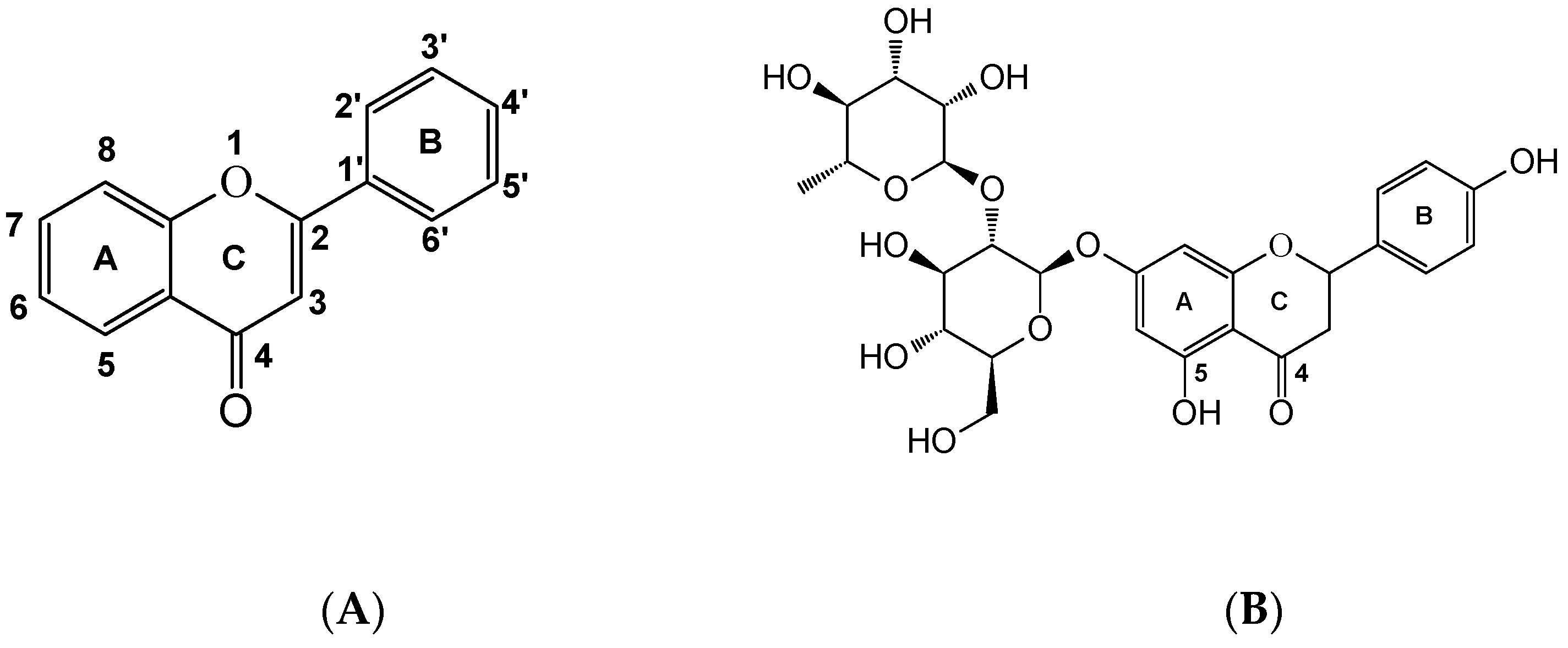
:1. Introduction
2. Results
2.1. Characterization of V(IV)O–Naringin Complex
2.1.1. Fourier Transformed Infrared Spectroscopy
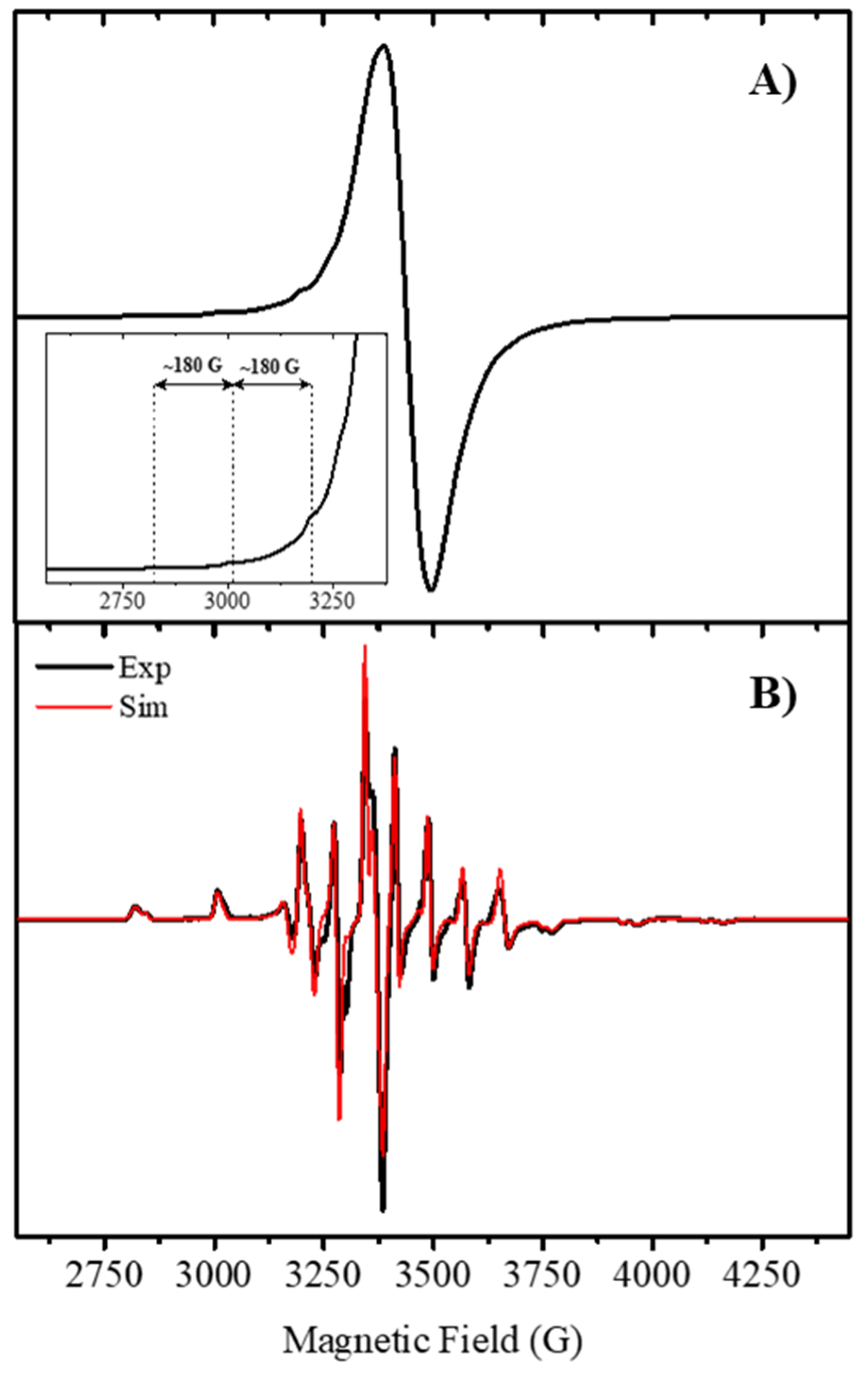
2.1.2. Powder and Frozen Solution EPR Spectra
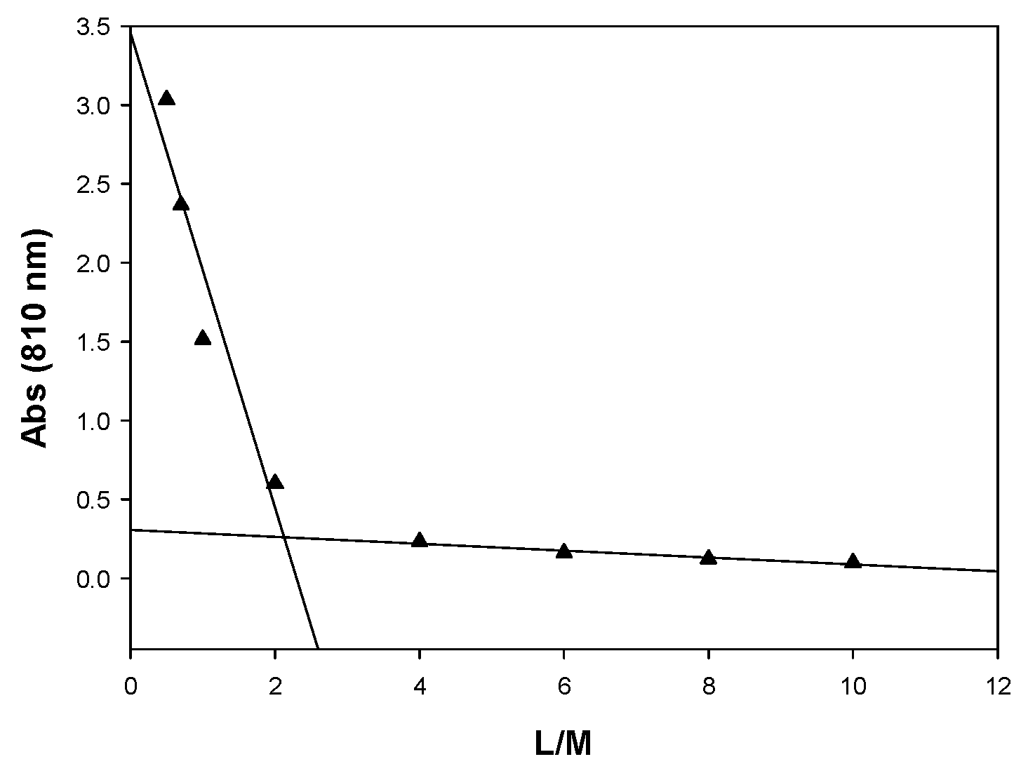
2.1.3. Spectrophotometric Titrations
2.2. Antiradical Behavior
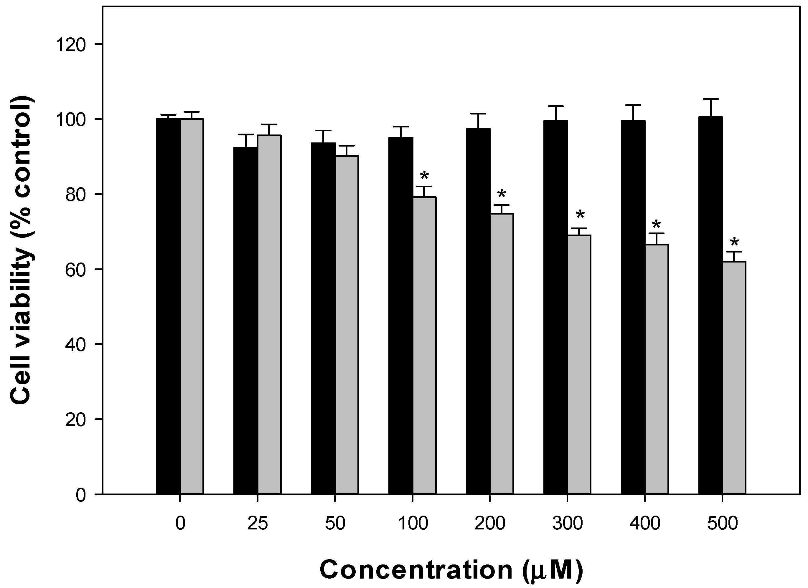
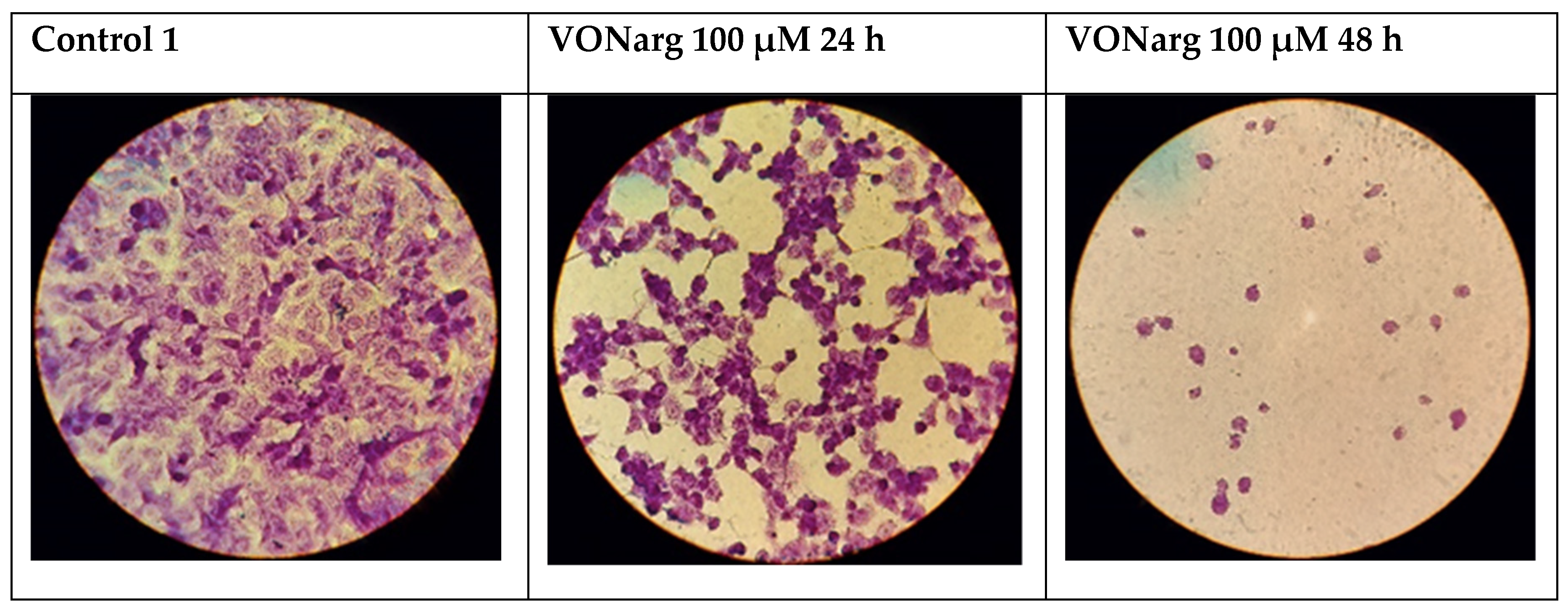
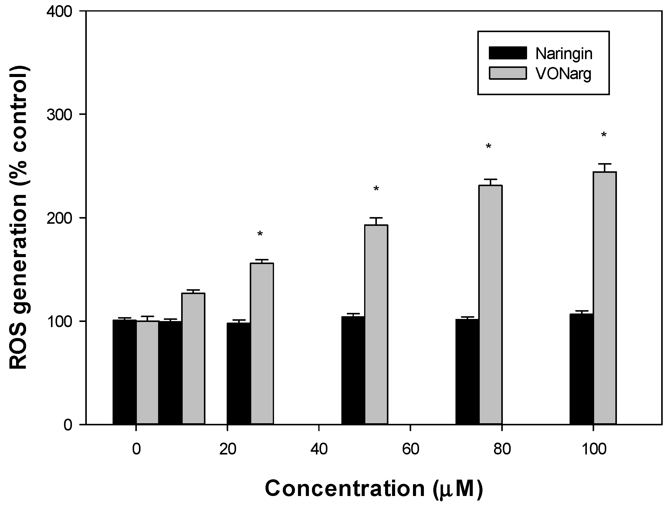
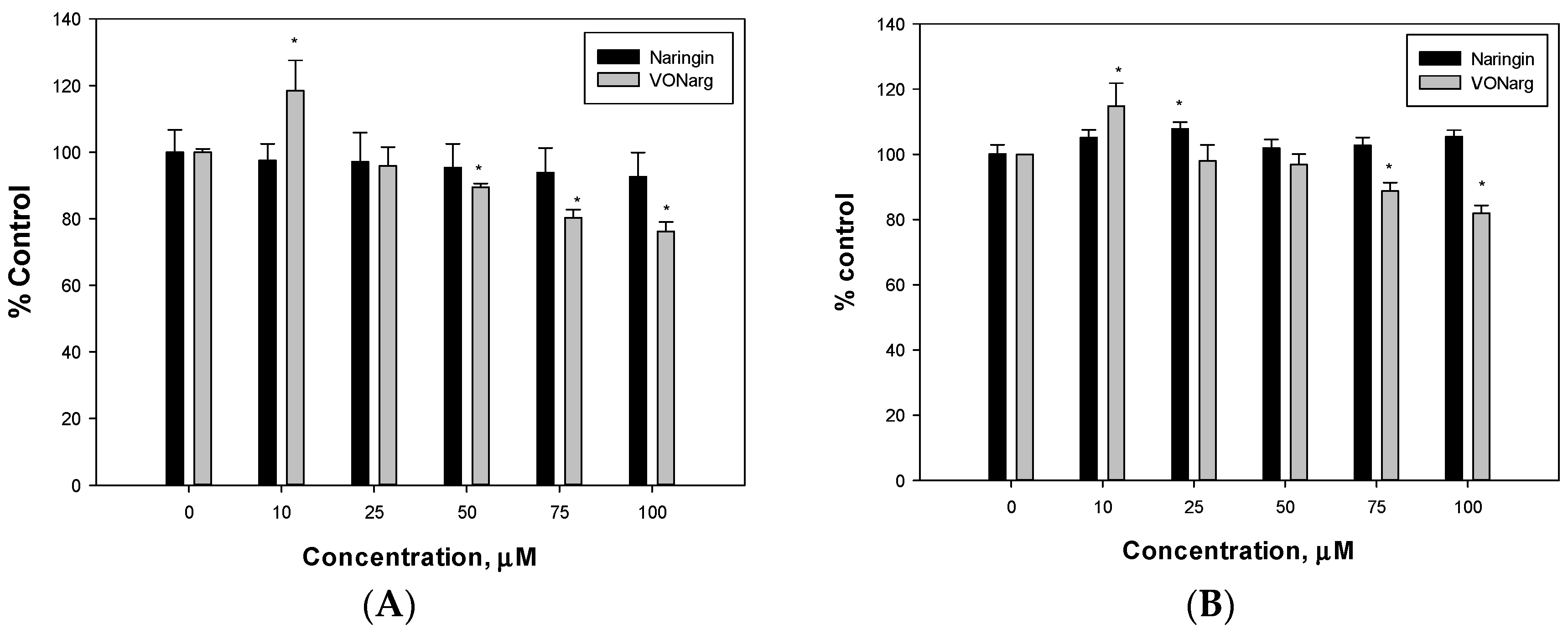
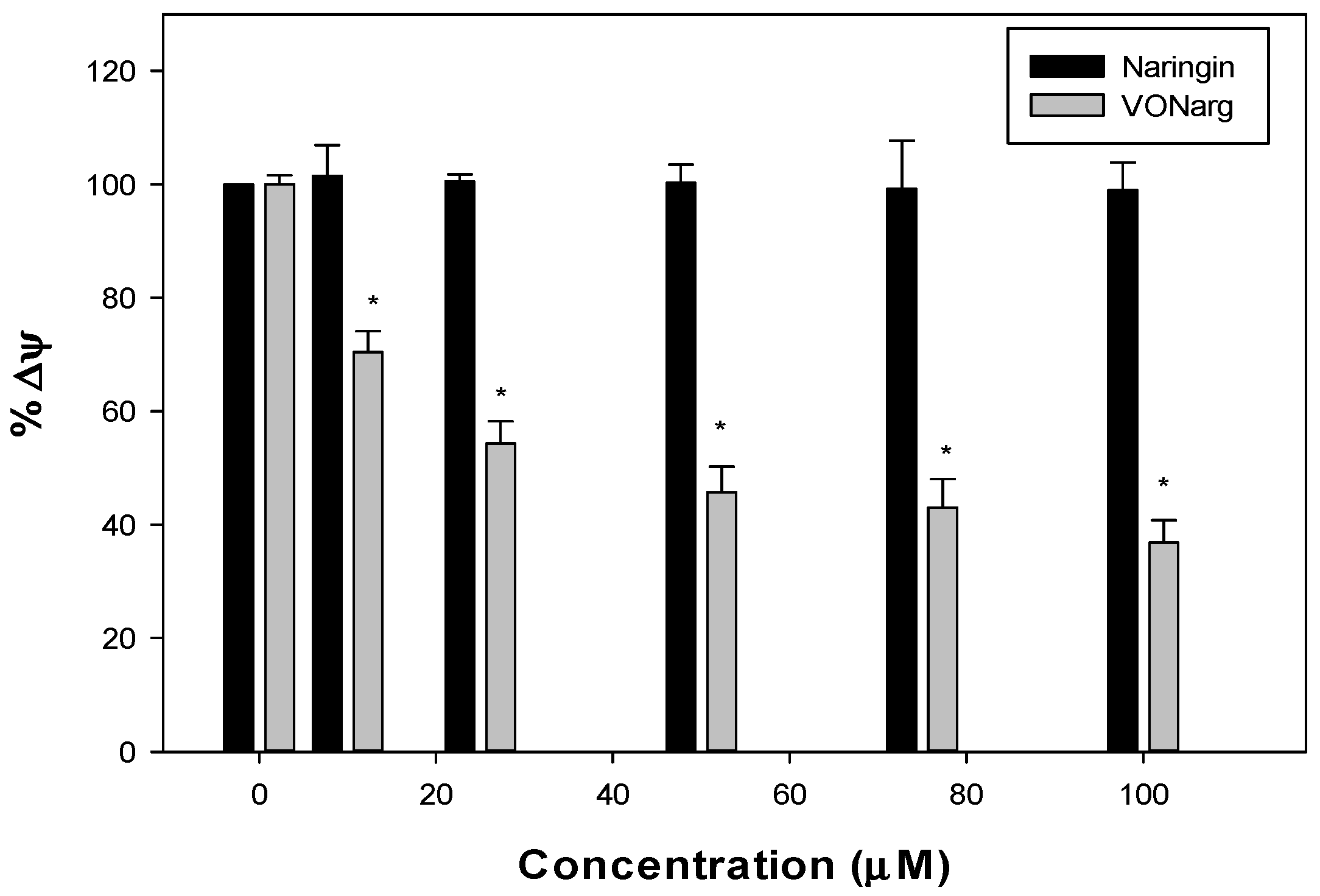
2.3. Anticancer Effects
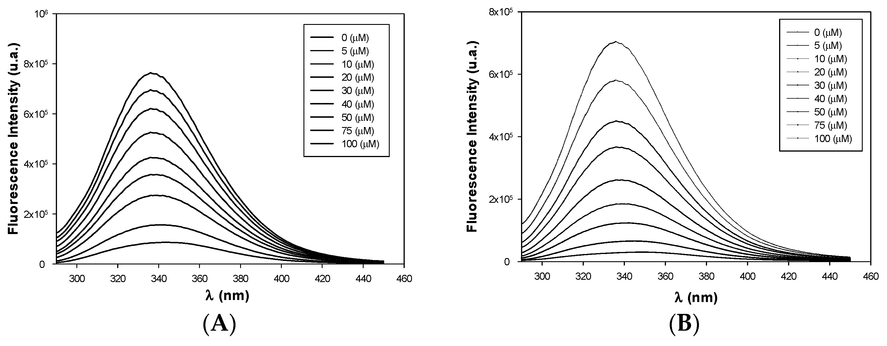
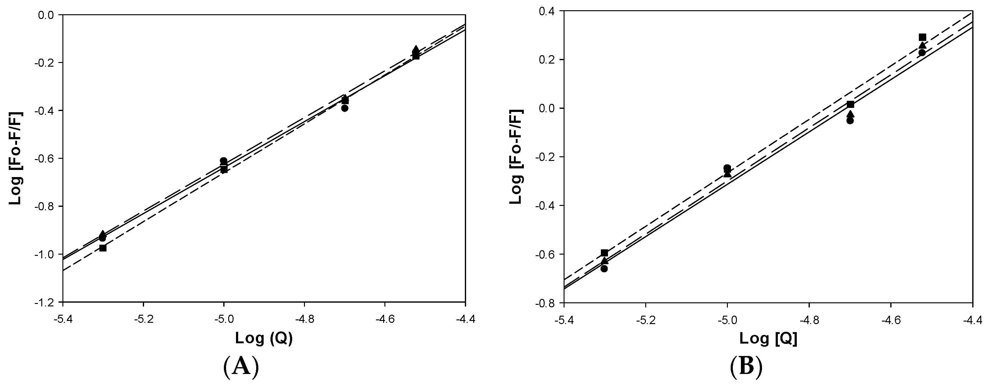
2.4. Bovine Serum Albumin (BSA) Interactions
3. Discussion
4. Material and Methods
4.1. Materials and Instrumentation
4.2. Synthesis of [VO(Narg)2]·8H2O (VONarg)
4.3. Spectrophotometric Titration
4.4. Antioxidant Properties
4.5. Biological Assays
4.5.1. Cell Culture
4.5.2. MTT Assay
4.5.3. Stress Oxidative Determinations
4.6. Data Analysis
4.7. Bovine Serum Albumin Interactions
5. Conclusions
Supplementary Materials
Author Contributions
Funding
Institutional Review Board Statement
Informed Consent Statement
Data Availability Statement
Acknowledgments
Conflicts of Interest
References
- Panche, A.N.; Diwan, A.D.; Chandra, S.R. Flavonoids: An overview. J. Nutr. Sci. 2016, 5, E47. [Google Scholar] [CrossRef] [PubMed] [Green Version]
- Wang, T.Y.; Li, Q.; Bi, K.S. Bioactive flavonoids in medicinal plants: Structure, activity and biological fate. Asian J. Pharm. Sci. 2018, 13, 12–23. [Google Scholar] [CrossRef]
- Dias, M.C.; Pinto, D.C.G.A.; Silva, A.M.S. Plant Flavonoids: Chemical Characteristics and Biological Activity. Molecules 2021, 26, 5377. [Google Scholar] [CrossRef]
- Zou, M.; Liu, H.; Li, J.; Yao, X.; Chen, Y.; Ke, C.; Liu, S. Structure-activity relationship of flavonoid bifunctional inhibitors against zika virus infection. Biochem. Pharmacol. 2020, 177, 113962. [Google Scholar] [CrossRef]
- Zhu, X.; Ouyang, W.; Lan, X.; Xiao, H.; Tang, L.; Liu, G.; Feng, K.; Zhang, L.; Song, M.; Cao, Y. Anti-hyperglycemic and liver protective effects of flavonoids from Psidium guajava L. (guava) leaf in diabetic mice. Food Biosci. 2020, 135, 100574. [Google Scholar] [CrossRef]
- Sun, Y.W.; Bao, Y.; Yu, H.; Chen, Q.J.; Lu, F.; Zhai, S.; Zhang, C.F.; Li, F.; Wang, C.Z.; Yuan, C.S. Anti-rheumatoid arthritis effects of flavonoids from Daphne genkwa. Int. Immunopharmacol. 2020, 83, 106384. [Google Scholar] [CrossRef]
- Tuentera, E.; Creylman, J.; Verheyen, G.; Pieters, L.; Van Miert, S. Development of a classification model for the antigenotoxic activity of flavonoids. Bioorg. Chem. 2020, 98, 103705. [Google Scholar] [CrossRef] [PubMed]
- Fusia, F.; Trezza, A.; Tramaglino, M.; Sgaragli, G.; Saponara, S.; Spiga, O. The beneficial health effects of flavonoids on the cardiovascular system: Focus on K+ channels. Pharmacol. Res. 2020, 152, 104625. [Google Scholar] [CrossRef]
- Khater, M.; Ravishankar, D.; Greco, F.; Osborn, H.M.I. Metal complexes of flavonoids: Their synthesis, characterization and enhanced antioxidant and anticancer activities. Future Med. Chem. 2019, 11, 2845–2867. [Google Scholar] [CrossRef]
- Allscher, T.; Klüfers, P.; Mayer, P. Carbohydrate-metal complexes: Structural Chemistry of Stable Solution Species. In Glycoscience; Springer: Berlin/Heidelberg, Germany, 2008; pp. 1077–1139. [Google Scholar] [CrossRef]
- Sharma, P.; Kumar, V.; Guleria, P. Naringin: Biosynthesis and pharmaceutical applications. Indian J. Pharm. Sci. 2019, 89, 988–999. [Google Scholar] [CrossRef]
- Kumar, S.; Pandey, A.K. Chemistry and biological activities of flavonoids: An overview. Sci. World J. 2013, 2013, 162750. [Google Scholar] [CrossRef] [Green Version]
- Sharma, M.; Dwivedi, P.; Singh Rawat, A.K.; Dwivedi, A.K. Nutrition nutraceuticals: A proactive approach for healthcare. In Nutraceuticals; Elsevier Inc.: Amsterdam, The Netherlands, 2016; pp. 79–116. [Google Scholar] [CrossRef]
- Chen, R.; Qi, Q.L.; Wang, M.T.; Li, Q.Y. Therapeutic potential of naringin: An overview. Pharm. Biol. 2016, 54, 3203–3210. [Google Scholar] [CrossRef]
- Zhao, Y.; Liu, S. Bioactivity of naringin and related mechanisms. Pharmazie 2021, 76, 359–363. [Google Scholar] [CrossRef] [PubMed]
- Zaragoz, C.; Villaescusa, L.; Monserrat, J.; Zaragoz, F.; Melchor, A. Potential therapeutic anti-inflammatory and immunomodulatory effects of dihydroflavones, flavones, and flavonols. Molecules 2020, 25, 1017. [Google Scholar] [CrossRef] [Green Version]
- Qi, Z.; Xu, Y.; Liang, Z.; Li, S.; Wang, J.; Wei, Y.; Dong, B. Naringin ameliorates cognitive deficits via oxidative stress, proinflammatory factors and the PPARγ signaling pathway in a type 2 diabetic rat model. Mol. Med. Rep. 2015, 12, 7093–7101. [Google Scholar] [CrossRef] [Green Version]
- Viswanatha, G.L.; Shylaja, H.; Moolemath, Y. The beneficial role of Naringin- a citrus bioflavonoid, against oxidative stress-induced neurobehavioral disorders and cognitive dysfunction in rodents: A systematic review and meta-analysis. Biomed. Pharmacother. 2017, 94, 909–929. [Google Scholar] [CrossRef] [PubMed]
- Memariani, Z.; Qamar Abbas, S.; Shams ul Hassan, S.; Ahmadi, A.; Chabra, A. Naringin and naringenin as anticancer agents and adjuvants in cancer combination therapy: Efficacy and molecular mechanisms of action, a comprehensive narrative review. Pharmacol. Res. 2021, 171, 105264. [Google Scholar] [CrossRef]
- Amin Hussen, N.H. Docking Study of Naringin Binding with COVID-19 Main Protease Enzyme. Iraqi J. Pharm. Sci. 2020, 29, 231–238. [Google Scholar] [CrossRef]
- Etcheverry, S.B.; Ferrer, E.G.; Naso, L.; Rivadeneira, J.; Salinas, V.; Williams, P.A.M. Antioxidant effects of the VO(IV) hesperidin complex and its role in cancer chemoprevention. J. Biol. Inorg. Chem. 2008, 13, 435–447. [Google Scholar] [CrossRef]
- Naso, L.; Martínez, V.R.; Lezama, L.; Salado, C.; Valcarcel, M.; Ferrer, E.G.; Williams, P.A.M. Antioxidant, anticancer activities and mechanistic studies of the flavone glycoside diosmin and its oxidovanadium(IV) complex. Interactions with bovine serum albumin. Bioorg. Med. Chem. 2016, 24, 4108–4119. [Google Scholar] [CrossRef] [PubMed]
- Goitia, H.; Quispe, P.; Naso, L.G.; Martínez, V.R.; Rey, M.; Rizzi, A.C.; Ferrer, E.G.; Williams, P.A.M. Interactions of rutin with the oxidovanadium(IV) cation. Anticancer improvement effects of glycosylated flavonoids. New J. Chem. 2019, 43, 17636–17646. [Google Scholar] [CrossRef]
- Pessoa, J.C.; Etcheverry, S.; Gambino, D. Vanadium compounds in medicine. Chem. Rev. 2015, 301–302, 24–48. [Google Scholar] [CrossRef]
- Islas, M.S.; Naso, L.G.; Lezama, L.; Valcarcel, M.; Salado, C.; Roura-Ferrer, M.; Ferrer, E.G.; Williams, P.A.M. Insights into the mechanisms underlying the antitumor activity of the new coordination complex with oxidovanadium(IV) and naringenin. Albumin binding studies. J. Inorg. Biochem. 2015, 149, 12–24. [Google Scholar] [CrossRef]
- Chasteen, N.D. Biological Magnetic Resonance; Berliner, L.J., Reuben, J., Eds.; Plenum: New York, NY, USA, 1981; p. 3. [Google Scholar]
- Gorelsky, S.; Micera, G.; Garribba, E. The Equilibrium Between the Octahedral and Square Pyramidal Form and the Influence of an Axial Ligand on the Molecular Properties of VIVO Complexes: A Spectroscopic and DFT Study. Chem. Eur. J. 2010, 16, 8167–8180. [Google Scholar] [CrossRef] [PubMed]
- Sanna, D.; Ugone, V.; Lubinu, G.; Micera, G.; Garribba, E. Behavior of the potential antitumor VIVO complexes formed by flavonoid ligands. 1. Coordination modes and geometry in solution and at the physiological pH. J. Inorg. Biochem. 2014, 140, 173–184. [Google Scholar] [CrossRef] [PubMed]
- Kivelson, D.; Lee, S. ESR Studies and the Electronic Structure of Vanadyl Ion Complexes. J. Chem. Phys. 1964, 41, 1896–1903. [Google Scholar] [CrossRef]
- Chand, P.; Murali Krishna, R.; Lakshamana Rao, J.; Lakshaman, S.V.J. EPR and optical studies of vanadyl complexes in two host-crystals of tutton salts of thallium. Rad. Eff. Def. Solids 1993, 127, 245–254. [Google Scholar] [CrossRef]
- Liu, K.T.; Yu, J.T.; Lou, S.H.; Lee, C.H.; Huang, Y.; Lii, K.H.J. Electron paramagnetic resonance study of V4+-doped KTiOPO4 single crystals. Phys. Chem. Solids 1994, 55, 1221–1226. [Google Scholar]
- Ferrer, E.G.; Salinas, M.V.; Correa, M.J.; Naso, L.; Barrio, D.A.; Etcheverry, S.B.; Lezama, L.; Rojo, T.; Williams, P.A.M. Synthesis, characterization, antitumoral and osteogenic activities of quercetin vanadyl(IV) complexes. J. Biol. Inorg. Chem. 2006, 11, 791–801. [Google Scholar] [CrossRef]
- Pereira, R.M.S.; Andrades, N.E.D.; Paulino, N.; Sawaya, A.C.H.F.; Eberlin, M.N.; Marcucci, M.C.; Marino Favero, G.; Novak, E.M.; Bydlowski, S.P. Synthesis and Characterization of a Metal Complex Containing Naringin and Cu, and its Antioxidant, Antimicrobial, Antiinflammatory and Tumor Cell Cytotoxicity. Molecules 2007, 12, 1352–1366. [Google Scholar] [CrossRef] [Green Version]
- Naso, L.G.; Lezama, L.; Valcarcel, M.; Salado, C.; Villacé, P.; Kortazar, D.; Ferrer, E.G.; Williams, P.A.M. Bovine serum albumin binding, antioxidant and anticancer properties of an oxidovanadium(IV) complex with luteolin. J. Inorg. Biochem. 2016, 157, 80–93. [Google Scholar] [CrossRef] [PubMed]
- Fanali, G.; di Masi, A.; Trezza, V.; Marino, M.; Fasano, M.; Ascenzi, P. Human serum albumin: From bench to bedside. Mol. Asp. Med. 2012, 33, 209–290. [Google Scholar] [CrossRef]
- Weinryb, I.; Steiner, R.F. The Luminescence of the Aromatic Amino Acids. In Excited States of Proteins and Nucleic Acids; Steiner, R.F., Weinryb, I., Eds.; Springer: Boston, MA, USA, 1971. [Google Scholar] [CrossRef]
- Lakowicz, J.R. Principles of Fluorescence Spectroscopy; Springer Science & Business Media: New York, NY, USA, 2013. [Google Scholar]
- Sun, Y.; Zhang, H.; Sun, Y.; Zhang, Y.; Liu, H.; Cheng, J.; Bi, S.; Zhang, H. Study of interaction between protein and main active components in Citrus aurantium L. by optical spectroscopy. J. Luminescence 2010, 130, 270–279. [Google Scholar] [CrossRef]
- Kragh-Hansen, U.; Chuang, V.T.G.; Otagiri, M. Practical aspects of the ligand-binding and enzymatic properties of human serum albumin. Biol. Pharm. Bull. 2002, 25, 695–704. [Google Scholar] [CrossRef] [Green Version]
- Zhang, X.; Li, L.; Xu, Z.; Liang, Z.; Su, J. Investigation of the Interaction of Naringin Palmitate with Bovine Serum Albumin: Spectroscopic Analysis and Molecular Docking. PLoS ONE. 2013, 8, e59106. [Google Scholar] [CrossRef]
- Roy, A.S.; Tripathy, D.R.; Chatterjee, A.; Dasgupta, S. A spectroscopic study of the interaction of the antioxidant naringin with bovine serum albumin. J. Biophys. Chem. 2010, 1, 141–152. [Google Scholar] [CrossRef] [Green Version]
- Shi, J.; Cao, H. Molecular structure-affinity relationship of dietary flavonoids for bovine serum albumin. Braz. J. Pharmacogn. 2011, 21, 594–600. [Google Scholar] [CrossRef] [Green Version]
- Karami, K.; Mehri Lighvan, Z.; Farrokhpour, H.; Dehdashti Jahromi, M.; Momtazi-borojeni, A.A. Synthesis and spectroscopic characterization study of new palladium complexes containing bioactive O,O-chelated ligands: Evaluation of the DNA/protein BSA interaction, in vitro antitumoural activity and molecular docking. J. Biomol. Struct. Dyn. 2018, 36, 3324–3340. [Google Scholar] [CrossRef] [PubMed]
- Badea, M.; Olar, R.; Uivarosi, V.; Marinescu, V.; Aldea, V. Synthesis and characterization of some vanadyl complexes with flavonoid derivatives as potential insulin-mimetic agents. J. Therm. Anal. Calorim. 2012, 107, 279–285. [Google Scholar] [CrossRef]
- Perillo, B.; Di Donato, M.; Pezone, A.; Di Zazzo, E.; Giovannelli, P.; Galasso, G.; Castoria, G.; Migliaccio, A. ROS in cancer therapy: The bright side of the moon. Exp. Mol. Med. 2020, 52, 192–203. [Google Scholar] [CrossRef]
- Yoshinaga, A.; Kajiya, N.; Oishi, K.; Kamada, Y.; Ikeda, A.; Chigwechokha, P.K.; Kibe, T.; Kishida, M.; Kishida, S.; Komatsu, M.; et al. NEU3 inhibitory effect of naringin suppresses cancer cell growth by attenuation of EGFR signaling through GM3 ganglioside accumulation. Eur. J. Pharmacol. 2016, 782, 21–29. [Google Scholar] [CrossRef] [PubMed]
- Nie, Y.; Wu, H.; Li, P.; Xie, L.; Luo, Y.; Shen, J.; Su, W. Naringin attenuates EGF-induced MUC5AC secretion in A549 cells by suppressing the cooperative activities of MAPKs-AP-1 and IKKs-IkB-NF-kB signaling pathways. Eur. J. Pharmacol. 2012, 690, 207–213. [Google Scholar] [CrossRef]
- Atta, E.M.; Hegab, K.H.; Abdelgawad, A.A.M.; Youssef, A.A. Synthesis, characterization and cytotoxic activity of naturally isolated naringin-metal complexes. Saudi Pharm. J. 2019, 27, 584–592. [Google Scholar] [CrossRef] [PubMed]
- Qian, J.; Li, J.; Ding, J.; Wang, Z.; Zhang, W.; Hu, G. Erlotinib activates mitochondrial death pathways related to the production of reactive oxygen species in the human non-small cell lung cancer cell line A549. Clin. Exp. Pharm. Physiol. 2009, 36, 487–494. [Google Scholar] [CrossRef]
- Chidambara Murthy, K.; Kim, J.; Vikram, A.; Patil, B.S. Differential inhibition of human colon cancer cells by structurally similar flavonoids of citrus. Food Chem. 2012, 132, 27–34. [Google Scholar] [CrossRef] [PubMed]
- Onishi, H. Photometric Determination of Traces of Metals, 4th ed.; John Wiley and Sons, Inc.: New York, NY, USA, 1986. [Google Scholar]
- Ali, I.; Wani, W.A.; Saleem, K. Empirical formulae to molecular structures of metal complexes by molar conductance. Synth. React. Inorg. Met. Nano-Metal Chem. 2013, 43, 1162–1170. [Google Scholar] [CrossRef]
- Martínez Medina, J.J.; Naso, L.G.; Pérez, A.L.; Rizzi, A.; Okulik, N.B.; Ferrer, E.G.; Williams, P.A.M. Apigenin oxidovanadium(IV) cation interactions. Synthesis, spectral, bovine serum albumin binding, antioxidant and anticancer studies. J. Photochem. Photobiol. A Chem. 2017, 344, 84–100. [Google Scholar] [CrossRef]
- Hissin, P.J.; Hilf, R. A fluorometric method for determination of oxidized and reduced glutathione in tissues. Anal. Biochem. 1976, 74, 214–226. [Google Scholar] [CrossRef]
- Bradford, M.M. A rapid and sensitive method for the quantitation of microgram quantities of protein utilizing the principle of protein-dye binding. Anal. Biochem. 1976, 72, 248–254. [Google Scholar] [CrossRef]
- Zamzami, N.; Métivier, D.; Kroemer, G. Quantitation of mitochondrial transmembrane potential in cells and in isolated mitochondria. Methods Enzymol. 2000, 322, 208–213. [Google Scholar] [CrossRef]


| Naringin | VONarg | Vibrational Modes-Functional Groups |
|---|---|---|
| 3422 br 3231 sh | 3395 br 3205 sh | ν O-H |
| 1645 vs | 1637 sh | ν C=O ring C |
| 1629 sh 1615 sh | 1614 vs | ν C=C |
| 1582 m | 1574 vs 1537 m | ν C=C |
| 1520 m 1504 m | 1520 m | ν C=C |
| 1355 sh 1341 m | 1357 sh | δ COH |
| 1295 m | 1292 w | δ HOC |
| 1281 w 1265 w | 1256 w | δ HOC, ν (C-O-C) |
| 1134 s | 1134 m | ν C-O secondary alcohol |
| 1074 vs | 1076 vs | ν O-C sugar |
| 1062 vs | 1060 sh | ν C-O primary alcohol |
| 1041 vs | 1040 sh | ν O-C |
| 987 m | 987 m | ν O-C |
| 980 m | ν V=O | |
| 822 m | 814 w | ν C-C, ν O-C |
| g‖ | g⊥ | (1) A‖ | (1) A⊥ | Δg‖/Δg⊥ | (1) P | k | (1) P × k | |
|---|---|---|---|---|---|---|---|---|
| S1 | 1.9332 | 1.9717 | 167.0 | 62.8 | 2.26 | 121.8 | 0.72 | 87.3 |
| S2 | 1.9398 | 1.9751 | 159.4 | 51.6 | 2.30 | 125.8 | 0.62 | 78.2 |
| Radical | % Scavenging | ||
|---|---|---|---|
| Naringin | VONarg | V(IV)O2+ | |
| SOD (IC50, µM) | >1000 | 870 ± 5.2 | 15 ± 0.2 |
| ROO•, lag (min), 100 µM | 0 | 3.8 ± 0.8 | 6.4 ± 1.1 |
| OH•, 100 µM | 28 ± 0.4 | 0 | 38 ± 2 |
| DPPH•, 100 µM | 2.0 ± 0.7 | 45.0 ± 5.0 | 37.0 ± 2 |
| T (K) | Ksv (104) (L·mol−1) | Kq (1012) (L·mol−1s−1) | Kb (104) (L·mol−1) | n | |
|---|---|---|---|---|---|
| Naringin * | 298 | 2.18 ± 0.15 | 2.18 ± 0.15 | 1.45 ± 0.23 | 0.96 ± 0.03 |
| 303 | 2.28 ± 0.09 | 2.28 ± 0.09 | 1.81 ± 0.44 | 0.98 ± 0.10 | |
| 310 | 2.32 ± 0.13 | 2.32 ± 0.13 | 2.81 ± 0.38 | 1.02 ± 0.09 | |
| VONarg | 298 | 5.67 ± 0.33 | 5.67 ± 0.33 | 11.6 ± 0.19 | 1.08 ± 0.04 |
| 303 | 5.88 ± 0.31 | 5.88 ± 0.31 | 14.1 ± 0.13 | 1.09 ± 0.03 | |
| 310 | 5.99 ± 0.36 | 5.99 ± 0.36 | 16.9 ± 0.19 | 1.10 ± 0.04 |
| Compounds | Kb × 104 M−1 |
|---|---|
| Naringenin * | 10.20 |
| naringin | 1.45 |
| VO(naringenin)2·H2O * | 0.31 |
| VONarg | 11.6 |
| [Pd{(C,N)-C6H4CH2NH(Et)(Narg)] * | 18 |
| Compounds | ΔH (KJ/mol) | ΔS (J/mol) | ΔG (KJ/mol) |
|---|---|---|---|
| Naringin | 42.7 | 222.7 | −23.7 (298 K) −24.8 (303 K) −26.3 (310 K) |
| VONarg | 23.9 | 177.0 | −28.8 (298 K) −29.7 (303 K) −31.0 (310 K) |
Publisher’s Note: MDPI stays neutral with regard to jurisdictional claims in published maps and institutional affiliations. |
© 2022 by the authors. Licensee MDPI, Basel, Switzerland. This article is an open access article distributed under the terms and conditions of the Creative Commons Attribution (CC BY) license (https://creativecommons.org/licenses/by/4.0/).
Share and Cite
Restrepo-Guerrero, A.G.; Goitia-Semenco, H.; Naso, L.G.; Rey, M.; Gonzalez, P.J.; Ferrer, E.G.; Williams, P.A.M. Antioxidant and Anticancer Activities and Protein Interaction of the Oxidovanadium(IV) Naringin Complex. Inorganics 2022, 10, 13. https://0-doi-org.brum.beds.ac.uk/10.3390/inorganics10010013
Restrepo-Guerrero AG, Goitia-Semenco H, Naso LG, Rey M, Gonzalez PJ, Ferrer EG, Williams PAM. Antioxidant and Anticancer Activities and Protein Interaction of the Oxidovanadium(IV) Naringin Complex. Inorganics. 2022; 10(1):13. https://0-doi-org.brum.beds.ac.uk/10.3390/inorganics10010013
Chicago/Turabian StyleRestrepo-Guerrero, Andrés Gonzalo, Helen Goitia-Semenco, Luciana G. Naso, Marilin Rey, Pablo J. Gonzalez, Evelina G. Ferrer, and Patricia A. M. Williams. 2022. "Antioxidant and Anticancer Activities and Protein Interaction of the Oxidovanadium(IV) Naringin Complex" Inorganics 10, no. 1: 13. https://0-doi-org.brum.beds.ac.uk/10.3390/inorganics10010013