Article

Identification and Virulence Characterization of Two Akanthomyces attenuatus Isolates Against Megalurothrips usitatus (Thysanoptera: Thripidae)

Key Laboratory of Bio-Pesticide Innovation and Application, Engineering Research Centre of Biological Control, South China Agricultural University, Guangzhou 510642, China
*
Author to whom correspondence should be addressed.
Submission received: 30 April 2019 / Revised: 31 May 2019 / Accepted: 3 June 2019 / Published: 13 June 2019

Abstract

:
Megalurothrips usitatus (Bagnall) is one of the most harmful pests of leguminous plants. In order to expand our knowledge on the infection of M. usitatus by entomopathogenic fungi, two newly identified isolates of Akanthomyces attenuatus (Zare & Gams) were tested for their pathogenicity against M. usitatus. Both isolates of A. attenuatus (SCAUDCL-38 and SCAUDCL-56) were isolated from soil and were identified by morphological and molecular analyses. The adult females of M. usitatus were treated with five different concentrations (1 × 10⁴, 1 × 10⁵, 1 × 10⁶, 1 × 10⁷, and 1 × 10⁸ conidia/mL) of the isolates. Our results revealed 76.25% and 57.5% mortality of M. usitatus after five days of treatment with 1 × 10⁸ conidia/mL of SCAUDCL-38 and SCAUDCL-56, respectively. The median lethal concentrations (LC50) of SCAUDCL-38 and SCAUDCL-56 calculated through linear regression analysis after five days of fungal treatment of M. usitatus were 1.9 × 10⁶ and 1.5 × 10⁷ conidia/mL, respectively, whereas the median lethal time (LT50) observed for 1 × 10⁸ conidia/mL of SCAUDCL-38 and SCAUDCL-56 were 3.52 days and 4.9 days, respectively. A. attenuatus isolates SCAUDCL-38 and SCAUDCL-56 are highly pathogenic strains of M. usitatus. These findings offer valuable information on the development and commercialization of alternative control measures against M. usitatus.

1. Introduction

Megalurothrips usitatus (Bagnall) (Thysanoptera: Thripidae), also known as bean flower thrips, is a major threat to snap bean and cowpea in southern regions of China [1]. Direct damage by thrips reduces the photosynthetic ability of the host plants [2]. Indirect losses due to fruit malformation and scarring caused by thrips are of greater economic significance compared to the direct losses [3,4]. The frequent use of conventional broad-spectrum chemical pesticides has dominated the management of M. usitatus [5]. The long-term use of synthetic chemicals to manage the M. usitatus is causing environmental pollution and adverse effects to live organisms [6]. This heavy application of pesticides has also resulted in the interruption of the biological balance between natural enemies and insect pests [5,7]. The above-mentioned circumstances have increased the awareness of the necessity as well as the desire to develop pest control strategies that are environmentally safe and biodegradable [8].
Many recent studies have shown that entomopathogenic fungi such as Metarhizium anisopliae Sorokin, Metarhizium brunneum Petch, Beauveria bassiana (Balsamo) Vuillemin, and Isaria fumosorosea Wize are effective against different thrips species [9,10,11,12,13,14,15]. Akanthomyces attenuatus Zare & Gams (previously known as Lecanicillium attenuatus, now designated as belonging to Akanthomyces clade, Pong et al. [16]) is a well-known pathogen of whitefly, aphid, and Thrips [17]. Some strains of this species have been developed as commercial biopesticides [17,18]. A. attenuatus is pathogenic to a variety of insect orders and mite groups [19,20]. Therefore, A. attenuatus may prove to be an effective microbial control option which can suppress M. usitatus populations. As the pathogenicity of A. attenuatus against M. usitatus may vary among different isolates [12], improvement in the basic knowledge as well as increasing the existing pool of A. attenuatus isolates can help in the selection of the most suitable isolate for commercial use.
This study presents the isolation, identification, and description of two A. attenuatus strains from China. The isolated strains were also tested for their pathogenicity against bean flower thrips, which can provide valuable information for the potential development of A. attenuatus as an effective bio-pesticide against M. usitatus.

2. Materials and Methods

2.1. Collection of Soil and Isolation of Fungi

Soil samples were collected from cultivated fields at the South China Agricultural University (SCAU), Guangzhou, China, beneath surface litter (to a depth of 10 cm), were individually placed in polyethylene bags, and were held at −4 °C until they were processed. Fungal isolation was performed following Inglis et al. [21] and Imoulan et al. [22]. Briefly, 3 g of soil was added to 30 mL sterile ddH2O containing 0.05% Tween-80. The mixture was stirred for 15 min on a time-controlled magnetic stirrer. After stirring, 1 mL suspension was inoculated to Petri dishes containing potato dextrose agar (PDA), and the plates were incubated at 25 ± 1 °C and 80 ± 5% R.H., with a 16:8 h (Light/ Dark) photoperiod. The Petri dishes were monitored for fungal sporulation after 7 days, which was followed by inoculation of individual germlings on new PDA plates. In this way, several rounds of inoculation were performed until a purified culture, based on phenotypic characteristics and fungal morphology, was obtained [12].

2.2. Insect Rearing

The population of M. usitatus originated in 2017 from a cowpea field in Guangzhou, China. This population was subsequently reared by the bean pod method. The colony was kept in a growth chamber at 26 ± 6 °C, (70 ± 5)% RH, and 16:8 h (Light/Dark)photoperiod.

2.3. Morphological Characterization

The morphological characteristics of two isolates (SCAUDCL-38 and SCAUDCL-53) were observed by culturing a small piece of fungal mycelia on a block of PDA overlaid by a coverslip for 10 days [23]. The slides were stained with lactophenol cotton blue and observed at 40X under a phase-contrast microscope. Conidial images were captured digitally with an Axio Cam HRc camera (Carl Zeiss) using the Axion Vision SE64 Release 4.9.1 software.

2.4. Radial Growth and Conidial Yield

The average daily growth rate and conidial yields of both strains were determined using the method of Ali et al. [24]. Fungal mycelial plugs (1 cm diameter) obtained from basic culture (as mentioned in Section 2.1) were cultured on fresh PDA plates for 10 days. The colony diameter was measured on a daily basis. After 10 days of growth, the conidia were scraped from the Petri dishes and suspended in 100 mL of 0.05% Tween-80. The conidial suspension was filtered through muslin cloth to remove the mycelia. The conidial concentration in suspension was quantified using a hemocytometer a phase-contrast microscope at 40X under.

2.5. DNA Extraction, PCR Amplification, and Sequence Analysis

The genomic DNA of the purified fungal strains (SCAUDCL-38 and SCAUDCL-56) was extracted with a fungal DNA isolation kit (Ezup, Sangon Biotech, Shanghai, China). The genomic DNA was used as a template for PCR amplification of the internal transcribed spacer (ITS) and elongation factor 1 alpha (TEF or EF1-α) regions [23,25]. All PCR reactions were performed in a 50 μL reaction system, which contained 25 μL 2 × Tap PCR Master (1 μL of each primer (10 μM), 1 μL genomic DNA, and 22 μL ddH2O). The ITS regions were amplified using the universal primers reported in Table 1 using the following cycling conditions: 5 min at 94 °C, 35 cycles at 94 °C for 30 s, 53 °C for 30 s, 72 °C for 1 min, and a final extension at 72 °C for 10 min. The EF1-α regions were amplified using the primers in Table 1. Touch-down PCR amplifications were performed under the following conditions: denaturation at 94 °C for 2 min, annealing temperature for the first amplification cycle 66 °C with subsequent reductions of 1 °C per cycle over the next nine cycles. Additional amplification cycles (36 cycles) were performed through denaturation at 94 °C for 30 s, annealing at 56 °C for 30 s, and final incubation at 72 °C for 10 min. The purity of the PCR products was confirmed through agarose gel electrophoresis followed by staining with GenGreen (TianGen Biotech, Beijing, China). The purified PCR products were dispatched to Shanghai Majorbio Bio-pharm Technology (Shanghai, China) for complete bidirectional sequencing with PCR primers.
The sequences were spliced with Genious version 7.1.4 and were blasted using GenBank, followed by connecting two gene fragments in series with TextPad 8.0.2. The sequences were compared using MEGA Version 7.0 [29] and the Kimura-2-parameter (K2P) to calculate a Maximum Likelihood (ML) tree [30].

2.6. Virulence of the Fungi in the Laboratory

Akanthomyces attenuatus isolates (SCAUDCL-38 and SCAUDCL-56) were cultured for 10 days on PDA plates and suspended in 0.01% Tween-80 as described in Section 2.3 to prepare a suspension containing 1 × 10⁸ conidia/mL. Lower concentrations (1 × 10⁷, 1 × 10⁶, 1 × 10⁵, and 1 × 10⁴ conidia/mL) were prepared through serial dilutions. The pathogenicity of A. attenuatus strains (SCAUDCL-38 and SCAUDCL-56) against adult M. usitatus under laboratory conditions was studied through a centrifuge tube residual bioassay. The centrifuge tubes along with a soya bean pod (1 cm length) were immersed individually in each conidial concentration for 2 hours. The centrifuge tubes and soya bean pods immersed in 0.01% Tween-80 only served as the control. Healthy females of M. usitatus (100 individuals) were transferred to each tube using a camel-hair brush. The tubes were sealed with a cotton plug to prevent thrips from escaping and were incubated in a growth chamber at 26 ± 1 °C, 75% R.H., and 16:8 h (Light/Dark) photoperiod. The insects were observed on a daily basis to record the number of dead M. usitatus females. The M. usitatus females infected with A. attenuatus were identified by the method of Ali et al. [31].
The complete experiment was performed three times using freshly prepared fungal suspensions.

2.7. Transmission Electron Microscopy

M. usitatus individuals were inoculated with 1.0 × 10⁸ conidia/mL of each strain and incubated at 26 °C and 75% relatively humidity. Gross changes in the appearance of the infected M. usitatus were directly monitored at different times after inoculation under a JEM1011 Transmission electron microscope (Nikon Co. Ltd., Japan). The infected M. usitatus were sampled at 1, 2, 3, 4, and 5 days after inoculation. They were fixed in 2.5% glutaraldehyde solution and then treated according to the method previously described [32].

2.8. Statistical Analysis

Radial growth and conidial yield data were subjected to a one-way ANOVA, and the means were compared using Tukey’s HSD test at a 5% level of significance. Mortality data were percent-transformed and subjected to probit analysis to calculate the medial lethal concentration (LC50) and the median lethal time (LT50) [33]. All the analyses were performed through SAS 9.1 [34].

3. Results

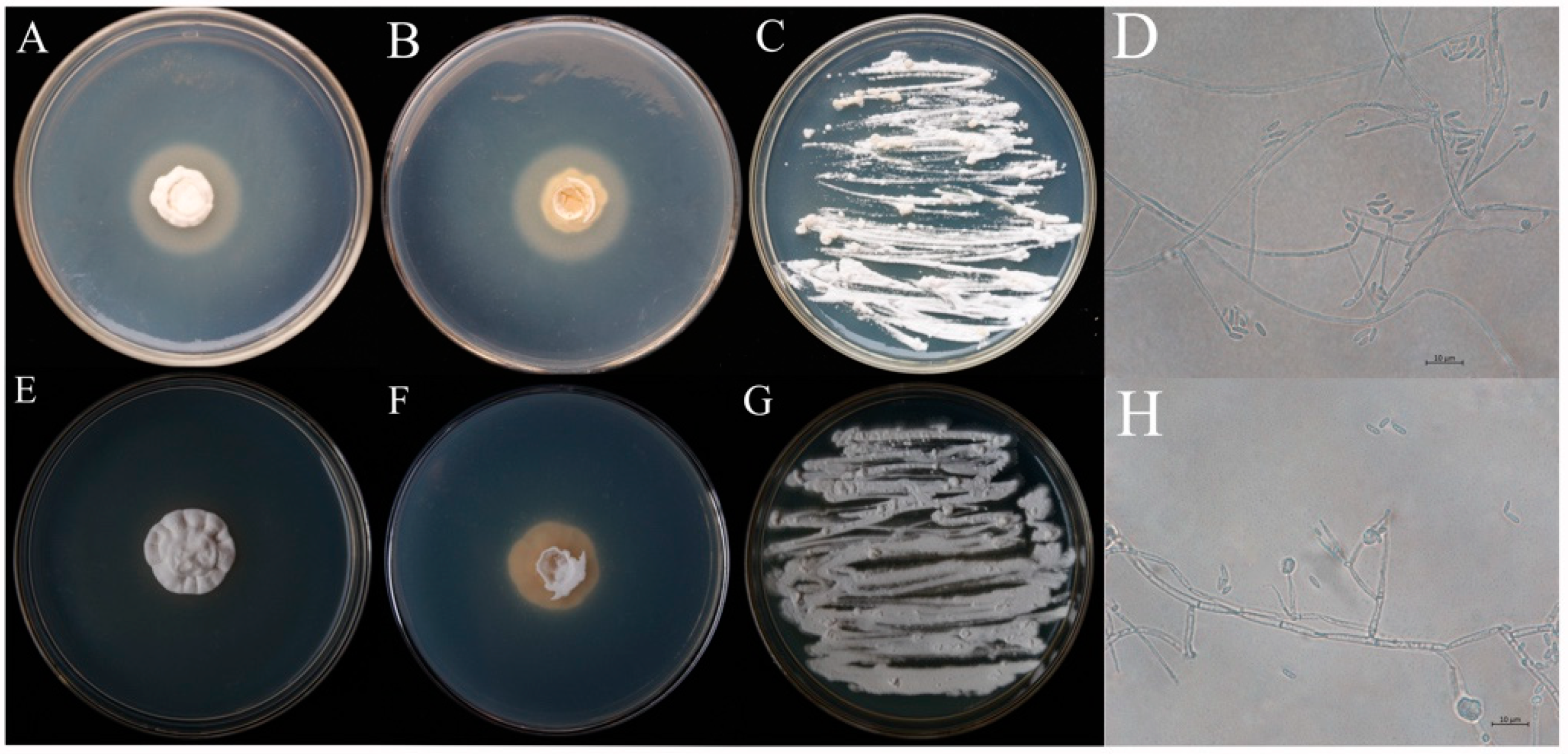
3.1. Morphological Identification of Fungi

Two strains of entomopathogenic fungi SCAUDCL-38 and SCAUDCL-56 were successfully isolated from soil during this study. Both strains grew well on PDA plates (Figure 1A,B,E,F). The two strains exhibited different morphological characteristics. The morphological characteristics of the two isolates are reported below.
SCAUDCL-38: The mycelia were hyaline or light-colored, with septate branching having the width of 1.2–2.0 μm and 1 or 2 branches. The mycelial joint was thick, while the tip was sharp. The total length was 14.5–23.0 × 0.9–1.6 μm. The conidia were either long or short, with an elliptical shape and were transparent and light (1.5–1.7 × 2.6–6.0 μm) (Figure 1D). The colony diameter after 10 days was 16 mm, and the conidial yield was 5.45 × 10⁶ conidia/mL.
SCAUDCL-56: The mycelia were hyaline or light-colored, with septate branching. The width of the mycelium was 1.4–2.1 μm, with 3–4 whorls. The mycelial joint was thick, while the tip was sharp. The total length was 10–20.7 × 1.1–2.3 μm. The conidia were long or short, had an elliptical shape, and were transparent and light (1.3–2.2 × 3.2–8.2 μm) (Figure 1H). After 10 days, the colony diameter was 22.5 mm, and the spore yield was 1.50 × 10⁷ conidia/mL.
After morphological observation, both strains (SCAUDCL-38 and SCAUDCL-56) were preliminarily identified as A. attenuatus.

3.2. Molecular Analyses

The purified DNA was amplified by PCR to obtain partial 18s rDNA, ITS, and EF 1-α sequences.

3.2.1. BLASTN Comparisons

The comparison of the results in GenBank showed that the ITS sequence of SCAUDCL-38 had 100%, 99.83%, and 99.48% similarity to Akanthomyces strain sequences in GenBank (GenBank Accession No.MH558279, LT992877, and MH231313). SCAUDCL-56 had 99.83–100% similarity to Akanthomyces strain sequences in GenBank (GenBank Accession No.MH558279, LT992877).The EF 1-α sequences of SCAUDCL-38 and SCAUDCL-56 had 98–100% similarity to Akanthomyces strains in GenBank. For details of the sequences in GenBank used above, see Table 2.

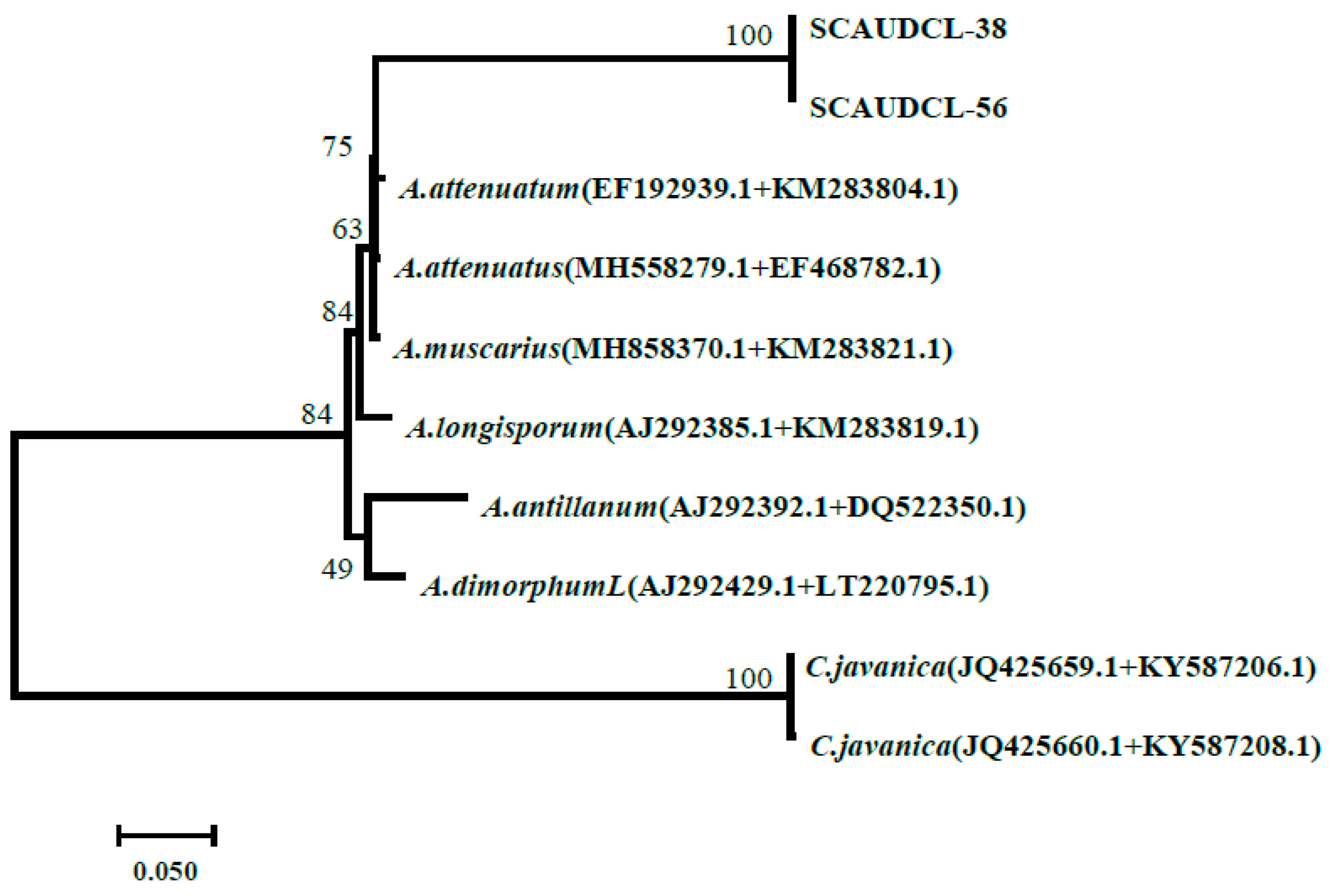
3.2.2. Phylogenetic Analysis

Our results showed that strains were closely similar to Akanthomyces spp. Both SCAUDCL-38 and SCAUDCL-56 clustered together with strains of A. attenuatus (GenBank Accession No.EF192939+KM283204) with a bootstrap value of 75% (Figure 2).

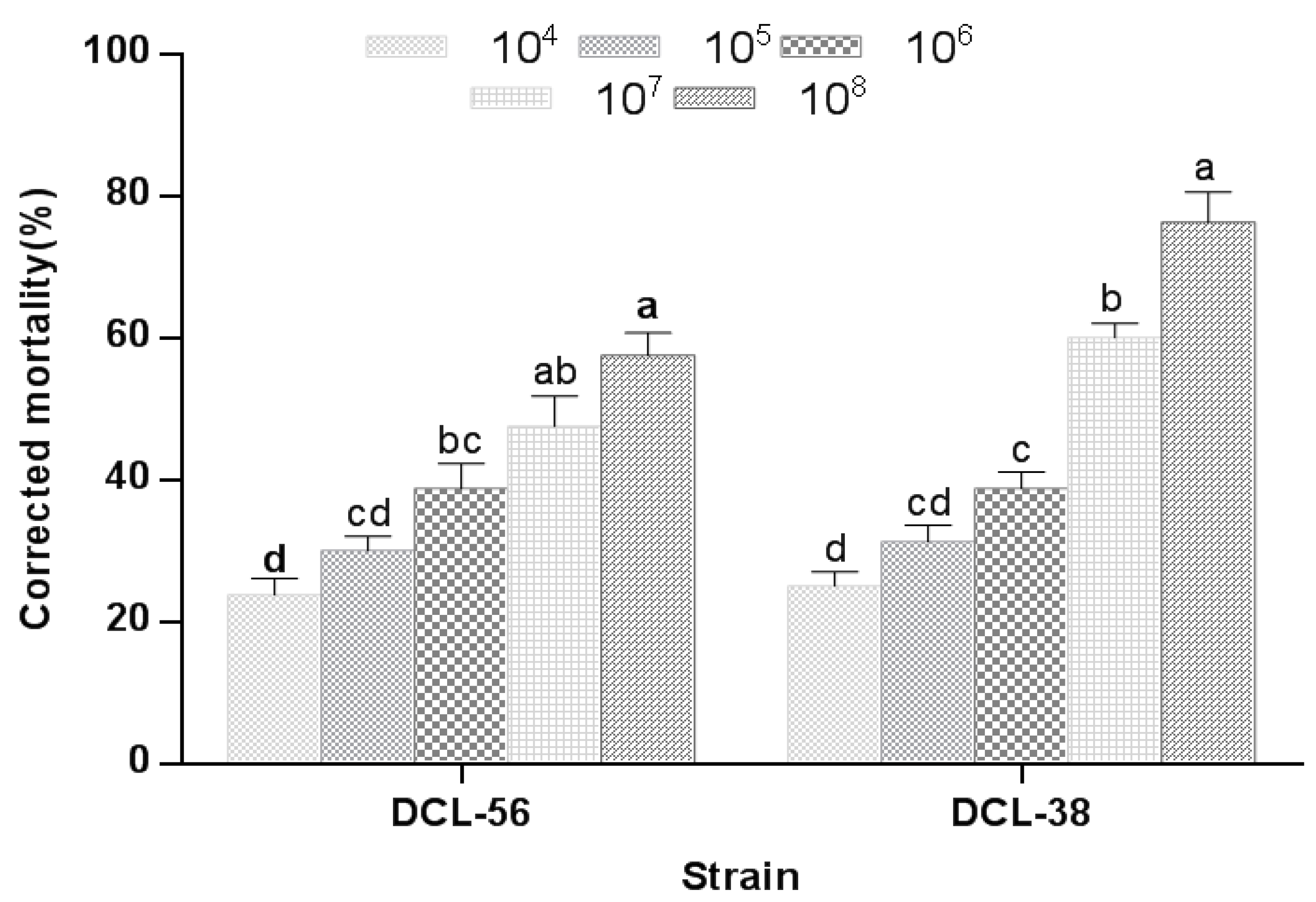
3.3. Virulence of A. attenuatus against M. usitatus

Both putative A. attenuatus strains (SCAUDCL-38 and SCAUDCL-56) were pathogenic to M. usitatus. The pathogenicity of the two strains against M. usitatus increased with increasing conidial concentration (Figure 3). A. attenuatus strain SCAUDCL-38 was more virulent than SCAUDCL-56 against M. usitatus. There was no significant difference in the adjusted mortality of M. usitatus caused by SCAUDCL-38 and SCAUDCL-56 when the insects were treated with lower conidial concentrations (1 × 10⁴, 1 × 10⁵ and 1 × 10⁶ conidia/mL). However, at higher concentrations (1 × 10⁷ and 1 × 10⁸ conidia/mL), the strain SCAUDCL-38 (76.25%) induced significantly higher M. usitatus mortality than the strain SCAUDCL-56 (57.5%) (Figure 3). LC50 values of SCAUDCL-38 and SCAUDCL-56 against M. usitatus were 1.9 × 10⁶ and 1.5 × 10⁷ conidia/mL, respectively. The LT50 values of SCAUDCL-38and SCAUDCL-56 (when 1 × 10⁸ conidia/mL was applied) were 3.5 and 4.9 days, respectively (Table 3 and Table 4).

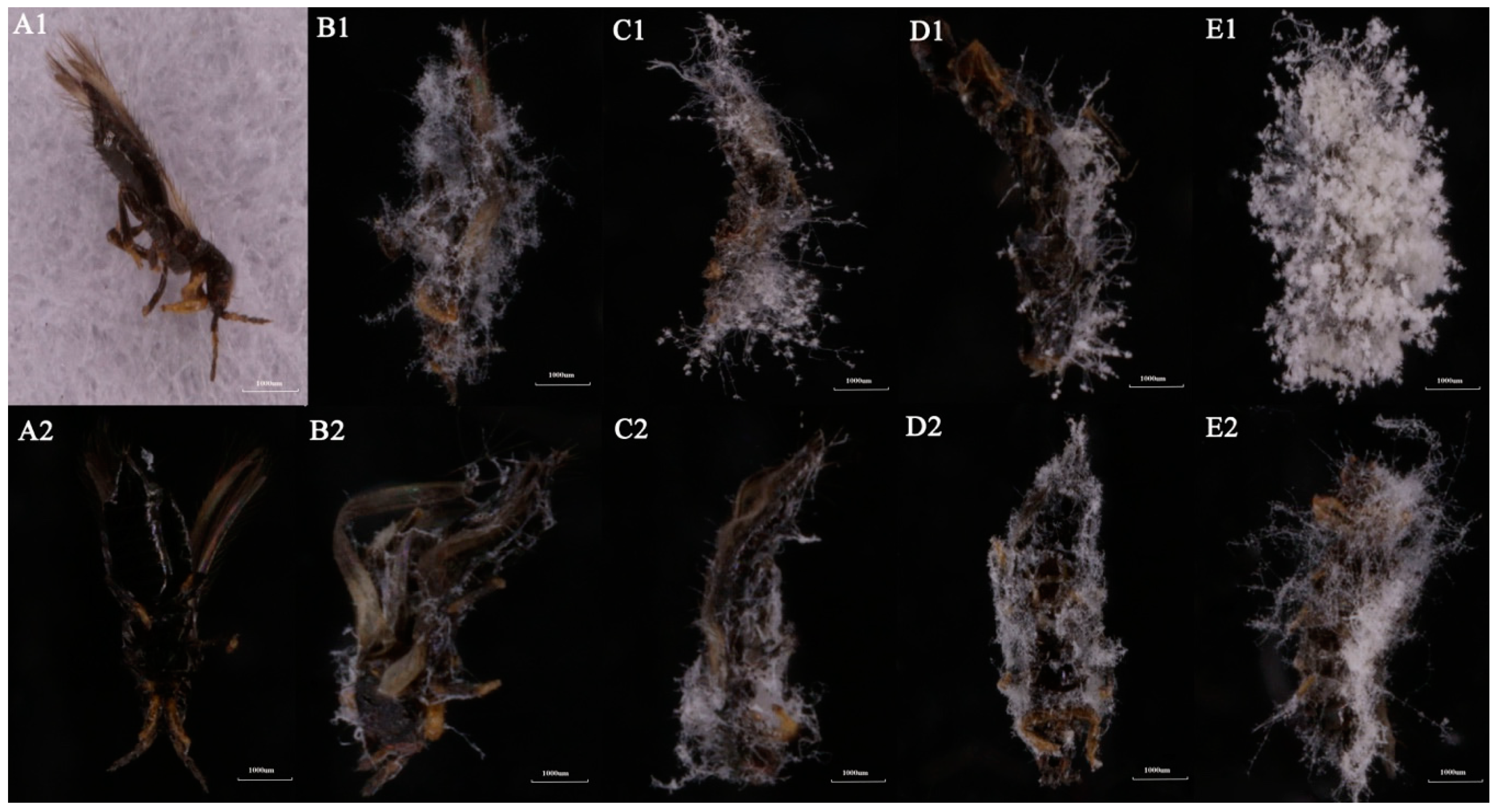
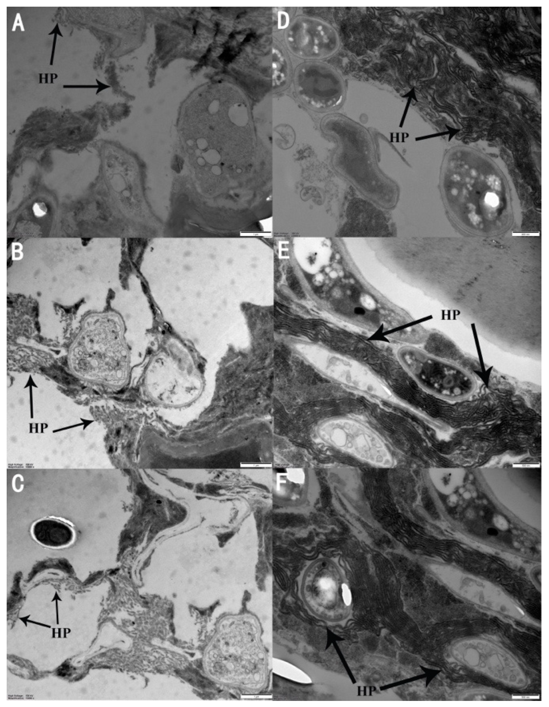
3.4. Microscopic Examination of A. attenuatus Infection

Both SCAUDLC-38 and SCAUDCL-56 induced similar symptoms in adult M. usitatus. At 24 h post-inoculation, white hyphae were produced by fungi around the anus and genitals of M. usitatus (Figure 4A1,A2). At 48 h post-inoculation, white hyphae covered the whole insect body; however, more white hyphae developed over the head, dorsal trunk, and ventral side of the wings (Figure 4B1,B2). At 72 h and 96 h post-inoculation, the insect behavior was abnormal, and hyphal growth extended over the whole body (Figure 4C1,C2,D1,D2). After 120 h, dense white hyphae completely covered the insects’ body (Figure 4E1,E2).
Scanning electron microscope images clearly showed the development of fungal hyphae throughout the body of M. usitatus (Figure 5). The results indicated that the adults were infected and killed by A. attenuatus.

4. Discussion

The development of biological control agents as an alternative to synthetic chemicals requires a clear understanding of the identification and pest control potential of biological control agents. In this study, two isolates of the entomopathogenic fungus A. attenuatus were identified and tested for their pathogenicity against M. usitatus. Our results revealed the successful isolation and purification of two A. attenuatus (SCAUDCL-38 and SCAUDCL-56) isolates from soil samples (collected at the South China Agricultural University in Guangzhou, China, during 2012). Furthermore, dose-dependent mortality studies of the A. attenuatus isolates (SCAUDCL-38 and SCAUDCL-56) showed considerable pathogenic potential against M. usitatus.
The size of the conidia produced by SCAUDCL-38 and SCAUDCL-56 was 1.5–1.7 × 2.6-6.0 μm and 1.3–2.2 × 3.2–8.2 μm, respectively. The size of the conidia is smaller than that observed for A. attenuatus strain ZJLA08 (1.5–2.5 × 3.5–7.0 μm) isolated in China by Lu et al. [20]. Such difference in conidial size may be the result of differences in the respective size of an insect host. Our strains were isolated from the soil, whereas strain ZJLA08 was directly isolated from the insect host (Diaphorina citri Kuwayama).
In the current phylogeny of hypocrealean entomopathogens, researchers have realized that the morphological features are not sufficient for the classification and identification of this large and complex fungal group [20,35]. Therefore, the genomic characterization of species can be used to determine the phylogenetic status as well as the identification of a species [36]. Molecular tools based on ITS rDNA genes have been used before to differentiate between morphologically similar Akanthomyces species [20]. Our results showed that differences in conidial morphology and size, as well as the homogeneity or variability of conidial size in Akanthomyces lecanii species complex (that also includes A. attenuatus), were highly correlated with the genomic identification results of the fungi of this species. The pairwise comparisons based on ITS rDNA genes indicated that strains SCAUDCL-38 and SCAUDCL-56 could not be distinguished from A. attenuatus (GenBank EF192939).
The ITS sequence has a rapid evolution rate, showing extremely wide sequence polymorphism, and is a highly conserved gene; therefore, ITS sequences are often used for intraspecies and subspecies classification and identification. [23,37]. However, this gene is not sufficient to clearly classify and identify species within the genus Akanthomyces, and the classification status of SCAUDCL-56 was not clear in the identification analysis of Akanthomyces. In this situation, other genes are usually chosen for sequence analysis of multiple gene loci, and the DNA sequence of the EF 1-α gene can successfully distinguish between species of Akanthomyces [38]. In this study, the EF 1-α sequences classified the Chinese isolates as A. attenuatus. Therefore, the phylogenetic tree also confirmed that the isolates SCAUDCL-38 and SCAUDCL-56 belong to A. attenuatus.
In the laboratory, A. attenuatus SCAUDCL-38 and SCAUDCL-56 both readily produced large quantities of conidia. This is an important reference for future large-scale production of innundative sprays. Our results successfully demonstrated that M. usitatus was suceptible to both A. attenuatus isolates (SCAUDCL-38 and SCAUDCL-56) in the study. The LC50 values of the A. attenuatus isolates used were a little higher than those observed by Montalva et al. [39]. These authors studied the toxicity of three strains of A. attenuatus (ARSEF13278, ARSEF13279, ARSEF13280) against Cinara cupressi (Buckton, 1881); they obtained LC50 values of 1.0 × 10⁶, 0.3 × 10⁶, 0.6 × 10⁶ conidia/mL. Kim et al. [40] conducted a virulence test of A. attenuatus against Aphis gossypii (Glover, 1877), which generated an LT50 value of 2.7 days for conidial concentration of 1 × 10⁸ conidia/mL. Our research showed that the highest concentration (1 × 10⁸ conidia/mL) produced LT50 values of 3.5 and 4.9 days for SCAUDCL-38 and SCAUDCL-56, respectively. The results of our research differ from those of previous studies, in part because of a different insect host [41,42]. On the basis of these initial research results, we believe that the isolates SCAUDCL-38 and SCAUDCL-56 may be useful candidates for the biological control of M. usitatus.

5. Conclusions

In summary, the newly identified strains of A. attenuatus (SCAUDCL-38 and SCAUDCL-56) were pathogenic to M. usitatus under laboratory conditions, having LC50 values of 1.9 × 10⁶ and 1.5 × 10⁷ conidia/mL, respectively after five days of fungal treatment. These strains may serve as alternative pest control agents for M. usitatus. Further studies are still required to confirm their efficacy under field conditions and to develop optimal formulations.

Author Contributions

C.D.: Data curation, Data analysis, writing—original draft. B.Y.: Data curation, Formal analysis, Writing—original draft. J.W.: Methodology, Writing—review and editing. S.A.: Conceptualization, Funding acquisition, Supervision; writing—original draft; writing—review and editing.

Funding

This research was funded by grants from the National Natural Science Foundation (31750110475); Gaungdong Province Science and Technology Innovation strategy special fund (2018B020206001); Science and Technology Programme of Guangzhou, P.R. China (201807010019); Special fund by South China Agricultural University for High-Level University Construction 2019.

Conflicts of Interest

The authors declare no conflict of interest.

References

  1. Tang, L.D.; Yan, K.L.; Fu, B.L. The life table parameters of Megalurothrips usitatus (Thysanopera: Thripidea) on four leguminous crops. Fla. Entomol. 2015, 98, 620–625. [Google Scholar] [CrossRef]
  2. Shipp, J.L.; Wang, K.; Binns, M.R. Economic injury levels for western flower thrips (Thysanoptera: Thrippidae) on greenhouse cucumber. J. Econ. Entomol. 2000, 93, 1732–1740. [Google Scholar] [CrossRef] [PubMed]
  3. Liu, P.P.; Jia, W.T.; Zheng, X.; Zhang, L.; Sangbaramou, R.; Tan, S.Q.; Liu, Y.Q.; Shi, W.P. Predation functional response and life table parameters of Orius sauter (Hemiptera: Antocoridae) feeding on Megalurothrips usitatus (Thysanoptera: Thripidae). Fla. Entomol. 2018, 101, 254–259. [Google Scholar] [CrossRef]
  4. Chen, W.S. A study on the relationship between thrips and the yield of peanut [Taeniothrips distalis Karny, Thrips spp. and Scirtothrips dorsalis]. Res. Bull. Taiwan Dais. 1980, 14, 51–57. [Google Scholar]
  5. Gao, Y.L.; Lei, Z.R.; Reitz, S.R. Western flower thrips resistance to insecticides: Detection, mechanisms and management strategies. Pest Manag. Sci. 2012, 68, 1111–1121. [Google Scholar] [CrossRef] [PubMed]
  6. Brownbridge, M.; Nelson, T.L.; Hackell, D.L. Field application of biopolymer-coated Beauveria bassiana F418 clover root weevil (Sitona Lepidus) control in Waikato and Manawatu. N. Z. Plant Prot. 2006, 59, 304–311. [Google Scholar]
  7. Jensen, S.E. Mechanisms associated with methiocarb resistance in Franklinie occidentalis (Thysanoptera: Thripidae). J. Econ. Entomol. 2000, 93, 464–471. [Google Scholar] [CrossRef]
  8. Fan, Y.M.; Tong, X.L.; Gao, L.I.; Wang, M.; Liu, Z.Q.; Zhang, Y.; Yang, Y. The spatial aggregation pattern of dominant species of thrips on cowpeain Hainan. J. Environ. Entomol. 2013, 35, 737–743. [Google Scholar]
  9. Wright, S.P.; Filotas, M.J.; Sanderson, J.P. Comparative efficacy of emulsifiable-oil, wettable-powder, and unformulated-powder preparations of Beauveria bassiana against the melon aphid Aphis gossypii. Biocontrol Sci. Technol. 2016, 26, 894–914. [Google Scholar] [CrossRef]
  10. Ansari, M.A.; Brownbridge, M.; Shah, F.A.; Butt, T.M. Efficacy of entomopathogenic fungi against soil-dwelling life stages of western flower thrips, Frankliniella occidentalis, in plant-growing media. Entomol. Exp. Appl. 2008, 127, 80–87. [Google Scholar] [CrossRef]
  11. Ansari, M.A.; Shah, M.A.; Whittaker, M.; Prasad, M.; Butt, T.M. Control of western flower thrips (Frankliniella occidentalis) pupae with Metarhizium anisopliae in peat and pear alternative growing media. Biol. Control 2007, 40, 293–297. [Google Scholar] [CrossRef]
  12. Saito, T.; Brownbrideg, M. Compatibility of soil-dwelling predators and microbial agents and their efficacy in controlling soil-dwelling stages of western flower thrips Frankliniella occdentalis. Biol. Control 2016, 92, 92–100. [Google Scholar] [CrossRef]
  13. Brownbridge, M.; Butt, T.M. Fungal Pathogens of Thrips, 1st ed.; CABI: Wallingford, UK, 1997; pp. 399–433. [Google Scholar]
  14. Skinner, M.; Gouli, S.; Frank, C.E.; Parker, B.L.; Kim, J.S. Management of Frankliniella occidentalis (Thysanoptera: Thripidae) with granular formulations of entomopathogenic fungi. Biol. Control 2012, 63, 246–252. [Google Scholar] [CrossRef]
  15. Kivett, J.M.; Cloyd, R.A.; Bello, N.M. Evaluation of entomopathogenic fungi against the western flower thrips (Thysanoptera: Thripidae) under laboratory conditions. J. Entomol. Sci. 2016, 51, 274–291. [Google Scholar] [CrossRef]
  16. Pong, K.K.; Ramle, M.; Wahizatul, A.A.; Norman, K.; Siti, R.A. Genetic variation of entomopathogenic fungi, Metarhizium anisopliae and Isaria amoenerosea and their pathogenicity against subterranean termite, Coptotermes curvignathus. J. Oil Palm Res. 2017, 29, 35–46. [Google Scholar]
  17. Ainsworth, G.C.; Bisby, G.R.; Kirk, P.M. Ainsworth & Bisby’s Dictionary of the Fungi, 5th ed.; CABI: Wallingford, UK, 2008; p. 771. [Google Scholar]
  18. Wang, T.J.; Huang, J.; You, M.; Guan, X.; Liu, B. Toxicity and feeding deterrence of crude toxin extracts of Lecanicillium (Verticillium) lecanii (Hyphomycetes) against sweet potato whitefly, Bemisia tabaci (Homoptera: Aleyrodidae). Pest Manag. Sci. 2007, 63, 381–387. [Google Scholar] [CrossRef]
  19. Gottel, M.S.; Koike, M.; Kim, J.J.; Aiuchi, D.; Shinya, R.; Brodeur, J. Potential of Leacanicillium spp. For management of insects nematodes and plant diseases. J. Invert. Pathol. 2008, 98, 256–261. [Google Scholar] [CrossRef]
  20. Lu, L.; Cheng, B.; Du, D.; Hu, X.; Peng, A.; Pu, Z.; Zhang, X.; Huang, Z.; Chen, G. Morphological, molecular and virulence characterization of three Lecanicillium species infecting Asian citrus psyllids in Huangyuan citrus groves. J. Invert. Pathol. 2015, 125, 45–55. [Google Scholar] [CrossRef]
  21. Inglis, G.D.; Johnson, D.L.; Kawchuk, L.M.; Goettel, M.S. Effect of soil texture and soil sterilization on susceptibility of ovipositing grasshoppers to Beauveria bassiana. J. Invert. Pathol. 1998, 71, 73–78. [Google Scholar] [CrossRef]
  22. Imoulan, A.; Alaoui, A.; Meziane, A. Natural occurrence of soil-borne entomopathogenic fungi in the Moroccan Endemic forest of Argania spinose and their pathogenicity to Ceratitis capitata. World J. Microbiol. Biotechnol. 2011, 27, 2619–2628. [Google Scholar] [CrossRef]
  23. Imoulan, A.; Wu, H.J.; Lu, W.L.; Li, Y.; Li, B.B.; Yand, R.H.; Wand, W.J.; Wand, X.J.; Kirk, P.M.; Yao, Y.J. Beauveria meogensis sp. nov., a new fungus of the entomopathogenic genus from China. J. Invert. Pathol. 2016, 139, 74–81. [Google Scholar] [CrossRef]
  24. Ali, S.; Huang, Z.; Ren, S.X. Media composition influences on growth, enzyme activity and virulence of the entomopathogen hyphomycete Isaria fumosorosea. Entomol. Exp. Appl. 2009, 131, 30–38. [Google Scholar] [CrossRef]
  25. Agrawal, Y.; Mual, P.; Shenoy, B.D. Multi-gene genealogies reveal cryptic species Beauveria rudraprayagi sp. nov. from India. Mycosphere 2014, 5, 719–736. [Google Scholar] [CrossRef]
  26. White, T.J.; Bruns, T.; Lee, S.; Taylor, J. Amplification and direct sequencing of fungal ribosomal RNA genes for phylogenetics. In PCR Protocols: Genes for Phylogenetics; Innis, M.A., Gelfand, D.H., Sninsky, J.J., White, T.J., Eds.; Academic Press: San Diego, CA, USA, 1990; pp. 315–322. [Google Scholar]
  27. Rehner, S.A.; Buckley, E. A Beauveria phylogeny inferred from nuclear ITS and EF 1—alpha sequences: Evidence for cryrtic diversification and links to Cordyceps teleomorphs. Mycologia 2005, 97, 84–98. [Google Scholar]
  28. Bischoff, J.F.; Rehne, S.A.; Humber, R.A. A multilocus phylogeny of the M. anisopliae. Mycologia 2009, 101, 512–530. [Google Scholar] [CrossRef]
  29. Goloboff, P.A.; Catalano, S.A. TNT version 1.5, including a full implementation of phylogenetic morphometrics. Cladisitics 2016, 32, 221–238. [Google Scholar] [CrossRef]
  30. Kimura, M. A simple method for estimating evolutionary rates of base substitutions through comparative studies of nucleotide sequences. J. Mol. Evol. 1980, 16, 111–120. [Google Scholar] [CrossRef]
  31. Ali, S.; Zhang, C.; Wang, Z.Q.; Wang, X.M.; Wu, J.H.; Cuthbertson, A.G.S.; Shao, Z.F.; Qiu, B.L. Toxicological and biochemical basis of synergism between the entomopathogenic fungus Lecanicillium muscarium and the insecticide matrine against Bemisia tabaci (Gennadius). Sci. Reports 2017, 7, 46558. [Google Scholar] [CrossRef] [PubMed]
  32. Wang, Y.; Lei, Z.R.; Zhang, Q.W. Observation of infection process of Metarhizium anisopliae on Plutella xylostella larvae with transmission electron microscopy. Acta Ent. Sin. 2006, 49, 1042–1045. [Google Scholar]
  33. Finney, J.G.; Smith, D.F.; Skeeters, D.E.; Auvenshine, C.D. MMPI alcoholism scales: Factor structure and content analysis. J. Stud. Alcohol 1971, 32, 1055–1060. [Google Scholar]
  34. SAS, T.C.J.; Gerver, W.J.M.; Bruin, R.D.; Mulder, P.G.H.; Cole, T.J.; Wall, W.D.; Hokken-Koelega, A.C.S. Body proportions during 6 years of GH treatment in children with short born small for gestational age participating in a randomised, double-blind, dose-response trial. Clin. Endocrinol. 2000, 53, 675–681. [Google Scholar] [CrossRef]
  35. Rehner, S.A.; Minnis, D.; Sung, G.H.; Luangsa-ard, J.J.; Devotto, L.; Humber, R.A. Phylogeny and systematics of the anamorphic, entomopathogenic genus Beauveria. Mycologia 2011, 103, 1055–1073. [Google Scholar] [CrossRef]
  36. Wang, D.K.; Deng, J.X.; Pei, Y.F.; Li, T.; Jin, Z.Y.; Liang, L.; Wang, W.K.; Li, L.D.; Dong, X.L. Identification and virulence characterization of entomopathogenic fungus Lecanicillium attenuatus against the pea aphid Acyrthos iphonpisum (Hemiptera: Aphididae). Appl. Entomol. Zool. 2017, 52, 511–518. [Google Scholar] [CrossRef]
  37. Vaidya, G.; Lohman, D.J.; Meier, R. Sequence Matrix: Concatenation software for the fast assembly of multi-gene data sets with character set and codon information. Cladistics 2011, 27, 171–180. [Google Scholar] [CrossRef]
  38. Bischoff, J.F.; Rehner, S.A.; Humber, R.A. Metarhizium frigidum sp. nov.: A cryptic species of M. anisopliae and a member of the M. flavoviride complex. Mycologia 2006, 98, 737–745. [Google Scholar] [CrossRef] [PubMed]
  39. Montalva, C.; Valenzuela, E.; Barta, M.; Rojas, E.; Arismendi, N.; Juscelino, R.; Humber, R.A. Lecanicillium attenuatus isolates affecting the invasive cypress aphid (Cinaracupressi) in Chile. Biol. Control 2017, 62, 625–637. [Google Scholar]
  40. Kim, J.J.; Goettel, M.S.; Gillespie, D.R. Potential of Lecanicillium species for dual microbial control of aphids and the cucumber powdery mildew fungus, Sphaerotheca fuliginea. Biol. Control 2007, 40, 327–332. [Google Scholar] [CrossRef]
  41. Jandricic, S.E.; Filotas, M.; Sanderson, J.P.; Wraight, S.P. Pathogenicity of conidia-based preparations of entomopathogenic fungi against the greenhouse pest aphids Myzus persicae, Aphis gossypii, and Aulacorthum solani (Hemiptera: Aphididae). J. Invertebr. Pathol. 2014, 118, 34–46. [Google Scholar] [CrossRef] [PubMed]
  42. Vu, V.; Hong, S.; Kim, K. Selection of entomopathogenic fungi for aphid control. Bio. Sci. Bioeng. 2007, 104, 498–504. [Google Scholar] [CrossRef]
Figure 1. Colony morphology and conidia morphology of two Akanthomyces attenuatus isolates (SCAUDCL-38 and SCAUDCL-56). (AC) Colony morphology of 10-day-old SCAUDCL-38; (D) conidia of isolate SCAUDCL-38; (EG) colony morphology of 10-day-old SCAUDCL-56; (H) conidia of isolate SCAUDCL-56.
Insects 10 00168 g001
Figure 2. Majority rule consensus phylogram from the Maximum Likelihood (ML) tree based on the sequences of the ITS region and of the protein-coding gene translation elongation factor 1 alpha. (EF1-α) for two A. attenuatus isolates (SCAUDCL-38 isolate, SCAUDCL-56). C. javanica was used as an outgroup.
Insects 10 00168 g002
Figure 3. Corrected mortality of Megalurothrips usitatus at five days post-inoculation with different conidial concentrations of A. attenuates isolates. Different lowercase letters above the bar for each isolate indicate significant differences at the level of p < 0.05 determined by Tukey’s HSD test.
Insects 10 00168 g003
Figure 4. Images of M. usitatus infected with strains SCAUDCL-38 and SCAUDCL-56, observed through a dissecting microscope. (A1,B1,C1,D1,E1) are SCAUDCL-38-infected M. usitatus at 24 h, 48 h, 72h, 96 h, and 120 h post-inoculation; (A2,B2,C2,D2,E2) are SCAUDCL-56-infected M. usitatus at 24 h, 48 h, 72 h, 96 h, and 120 eh post-inoculation.
Insects 10 00168 g004
Figure 5. Scanning electron microscope images of the cross sections of M. usitatus bodies showing the growth of A. attenuatus in M. usitatus. (AC) Five days post-infection with SCAUDCL-38; (DF) five days post-infection infection with SCAUDCL-56. HP: hyphae.
Insects 10 00168 g005
Table 1. Primers used in this study. ITS: internal transcribed spacer, EF 1-α: elongation factor 1 alpha.
Sr No.GenesPrimerPrimer SequenceReference
1ITSITS4FTCCTCCGCTTATTGATATGCWhite et al., 1990 [26]
ITS5RGGAAGTAAAAGTCGTAACAAGGWhite et al., 1990 [26]
2EF 1-α983FGCYCCYGGHCAYGGTGAYTTYATRehner and Buckley, 2005 [27]
2218RATGACACCRACRGCRACRGTYTGBischoff et al., 2009 [28]
Table 2. Reference entomopathogenic fungi from GeneBank used for the phylogenetic analysis.
SpeciesITSEF 1-α
Accession NumberStrain No.HostLocationAccession NumberStrain No.HostLocation
A. attenuatumEF192939CNU-23Green peach aphidKoreaKM283804KACC 42493/Korea
A. attenuatumMH558279MO315369Leaf RollerUSAEF468782CBS 402.78/USA
Akanthomyces muscariusMH858370CBS 641.63/AlbaniaKM283821CBS 143.62Trialeurodes vaporariorumKorea
Akanthomyces longisporumAJ292385IMI 021167VerticilliumUnited KingdomKM283819CBS 102072T. vaporariorumKorea
Akanthomyces antillanumAJ292392CBS 350.85VerticilliumUnited KingdomDQ522350CBS 350.85Animal pathogenUSA
Akanthomyces dimorphumAJ292429CBS 363.86VerticilliumUnited KingdomLT220795TMSL132SoilsPortugal
Cordyceps javanicaJQ425659BCC24976SpiderThailandKY587206CHE-CNRCB 357Diaphorina citriMexico
Cordyceps javanicaJQ425660BCC26304SpiderThailandKY587208CHE-CNRCB 363D. citriMexico
Table 3. Median lethal concentration (LC50) values for A. attenuatus isolates SCAUDCL-38 and SCAUDCL-56 against M. usitatus after five days of fungal treatment.
IsolatesRegression EquationLC50 (Conidia/mL)95% Confidence Limit
SCAUDCL-38Y = 0.352X–2.2171.9 × 106(3.2 × 10⁵–1.8 × 10⁷)
SCAUDCL-56Y = 0.229X–1.6511.5 × 107(1.2 × 10⁶–9.5 × 10¹¹)
Table 4. Median lethal time (LT50) values for 1 × 10⁸ conidal/mL of A. attenuatus isolates SCAUDCL-38 and SCAUDCL-56 against M. usitatus.
IsolatesRegression EquationLT50 (Days)95% Confidence Limit
SCAUDCL-38Y = 2.901X–5.9103.52(2.84–4.75)
SCAUDCL-56Y = 2.832X–5.8644.90(3.82–8.64)

