Review

Left Ventricular Noncompaction in Children: The Role of Genetics, Morphology, and Function for Outcome

by
Sabine Klaassen
1,2,3,4,*,
Jirko Kühnisch
1,2,3,
Alina Schultze-Berndt
1,2,4 and
Franziska Seidel
1,2,3,4,5
1
Max Delbrück Center for Molecular Medicine in the Helmholtz Association (MDC), 13125 Berlin, Germany
2
Experimental and Clinical Research Center, a Cooperation between the Max Delbrück Center for Molecular Medicine in the Helmholtz Association and Charité Universitätsmedizin Berlin, 13125 Berlin, Germany
3
DZHK (German Centre for Cardiovascular Research), Partner Site Berlin, 10785 Berlin, Germany
4
Department of Paediatric Cardiology, Charité Universitätsmedizin Berlin, Corporate Member of Freie Universität Berlin and Humboldt-Universität zu Berlin, 13353 Berlin, Germany
5
Department of Congenital Heart Disease-Paediatric Cardiology, German Heart Institute Berlin, 13353 Berlin, Germany
*
Author to whom correspondence should be addressed.
J. Cardiovasc. Dev. Dis. 2022, 9(7), 206; https://0-doi-org.brum.beds.ac.uk/10.3390/jcdd9070206
Submission received: 19 April 2022 / Revised: 24 June 2022 / Accepted: 27 June 2022 / Published: 30 June 2022
(This article belongs to the Special Issue Pediatric Cardiomyopathies: From Genotype to Phenotype)

Abstract

:
Left ventricular noncompaction (LVNC) is a ventricular wall anomaly morphologically characterized by numerous, excessively prominent trabeculations and deep intertrabecular recesses. Accumulating data now suggest that LVNC is a distinct phenotype but must not constitute a pathological phenotype. Some individuals fulfill the morphologic criteria of LVNC and are without clinical manifestations. Most importantly, morphologic criteria for LVNC are insufficient to diagnose patients with an associated cardiomyopathy (CMP). Genetic testing has become relevant to establish a diagnosis associated with CMP, congenital heart disease, neuromuscular disease, inborn error of metabolism, or syndromic disorder. Genetic factors play a more decisive role in children than in adults and severe courses of LVNC tend to occur in childhood. We reviewed the current literature and highlight the difficulties in establishing the correct diagnosis for children with LVNC. Novel insights show that the interplay of genetics, morphology, and function determine the outcome in pediatric LVNC.

1. Introduction

Cardiomyopathies (CMP) are myocardial diseases of which dilated (DCM) and hypertrophic (HCM) CMP are the most common subtypes. Restrictive (RCM), left ventricular noncompaction (LVNC), and mixed cardiomyopathies occur less frequently. Arrhythmogenic right ventricular cardiomyopathy (ARVC) is a rare disorder [1]. According to the American Heart Association (AHA), CMP are associated with mechanical and/or electrical dysfunction, and are classified into primary (genetic, nongenetic, acquired) and secondary forms [2]. The European Society of Cardiology groups CMP into the subtypes DCM, HCM, RCM, ARVC, and unclassified CMP, the latter includes LVNC. Further subclassification into familial/genetic and non-familial/non-genetic forms are part of the ESC classification [3]. This illustrates that classification of the LVNC phenotype is highly controversial and no general agreement exists. Examples of monogenic inheritance of the phenotype are frequent, but the association with CMP and other phenotypes rather suggest that the genetic architecture of LVNC is far more complex. Genetic background might be important for the development of noncompacted as well as compacted myocardium [4]. More recent data now point to isolated LVNC as a distinct phenotype which does not need to be pathologic [5]. LVNC has been shown to have a genetic cause in DCM, HCM, RCM, ARVC, the channelopathies, and congenital heart defects (CHD) [6,7,8]. More specifically in the pediatric population, LVNC has been identified with systemic entities such as Barth syndrome, chromosome 1p36 deletion syndrome, and CMP resulting from neuromuscular, metabolic, or mitochondrial disease [4,9,10]. We reviewed the literature to help identify the genetic and clinical features to determine outcome of LVNC in children.

2. Phenotype and Definition

There are many difficulties with the definition of the phenotype of LVNC. It is beyond the scope and not the intention of this review to define the history of several decades of the different definitions the phenotype of LVNC and we refer to several excellent previous reviews [4,8,11]. The most widely used definition of LVNC is still the one by Jenni et al. from 2001 [12]. The particular morphology, which was detected by echocardiography, mostly in adult patients with end-stage heart failure, with the technological advances in echocardiography raised much attention since then. As Jenni et al. described it, LVNC is a ventricular wall anomaly morphologically characterized by numerous, excessively prominent ventricular trabeculations and associated are deep intertrabecular recesses that are continuous with the left ventricular (LV) cavity [12]. A two-layer structure is present, with a compacted thin epicardial layer and a much thicker non-compacted endocardial layer. A maximal end systolic ratio of non-compacted to compacted layer of >2 is diagnostic (NC/C > 2) [12]. The extent of compacted to noncompacted myocardium does not lead to clinical manifestations and CMP per se. Regional ventricular hypokinesia was not restricted to the non-compacted segments in the original description [12]. Coexisting cardiac abnormalities are absent in isolated LVNC [12]. Of note, “isolated” in the original definition by Jenni et al. did not exclude the diagnosis of CMP, but was referring to the absence of CHD. Cardiac magnetic resonance (CMR) imaging was used in the following years to describe ratios between thickness, mass, or volume of NC/C [13,14]. The role of the thin compacted layer, which is used for the definition of LVNC, was not specifically defined. The compacted layer can be normal but also be disproportionately thinner than in controls [15]. Normal or abnormal thickness of the compact myocardium cannot be taken as a morphologic marker for LVNC. Rather, NC/C values (the ratio) need to reach cut-off values for the diagnosis [4,12]. Different other methods and normal reference values are available due to the lack of consensus criteria for LVNC [4,11]. The term “hypertrabeculation/noncompaction” is disputed due to interobserver disagreement [16]. Right ventricular noncompaction is a difficult morphology because an increased trabeculation is physiological in the right ventricle [11]. As for now, there are no morphologic markers or features that could predict the clinical impact of LVNC, whether it is a CMP or not.

3. Pathogenesis and Genetics

Many different terms and abbreviations used for noncompaction CMP and the noncompacted morphology are used and relate to the possible pathogenesis [11]. In 1990, Chin et al. suggested the term “isolated noncompaction of the left ventricular myocardium (INVM)”. It was assumed that the myocardial anomaly occurred as a result of an arrest of the normal compaction process during embryonal endomyocardial morphogenesis [17].

3.1. Developmental Aspects

Two developmental processes were described that govern the development of the normal myocardium. The first event is ventricular trabeculation which begins around week 4 of human development, the second is myocardial compaction during weeks 5 to 8 [18]. As demonstrated by several investigators, the Notch signaling pathway is critical for these developmental processes [19]. Most recently, two more unique genetic pathways were found in LVNC by genetic pathway analysis in humans: cardiomyocyte differentiation via BMP receptors and factors promoting cardiogenesis in vertebrates [20]. Quite the contrary was postulated by others, namely that LVNC may not be the result of an arrest in the compaction process at all. Instead LVNC could be the result from the compacted myocardium of the ventricular wall that grows into the ventricular lumen forming the trabeculae [21]. Jensen et al. found that left ventricular excessive trabeculations (LVET) are not of embryonic origin [21]. A reduction of the trabecular layer was never documented in humans [22]. Instead, they propose that the trabecular layer grows slower than the compact layer in later human development and this modulates wall morphology [22]. As there is no clear evidence for compaction on the developmental level the term “noncompaction” might need to be replaced [5]. Higher spatial resolution may clarify the prevalence and clinical significance of “LVET” [23].
What is the function of trabeculae in the adult heart, if we understand that excessive trabeculations might be detrimental? There is actually evidence that trabeculae are determinants of cardiac performance in adult hearts. The physiological function of trabeculae in the human heart was studied, using the UK Biobank, MRI data with fractal analysis, and modeling. The relationship of trabeculae-associated genomic loci with the risk of cardiovascular disorders was studied [24]. Reduced trabecular complexity was causally associated with the risk of heart failure. Trabeculae were important for cardiac contractility in both healthy and failing hearts.
Considerable evidence now favors the assignment of a mere phenotype to LVNC and genetic investigations are necessary to assign the possible pathogenicity of the phenotype [25,26].

3.2. Molecular Genetics

How could molecular genetic analysis help to determine the clinical outcome of the individuals affected with LVNC? The first genetic abnormality to cause LVNC without evidence of CHD was mutation in the X-linked gene TAFAZZIN [10]. Linkage analysis identified several autosomal dominant LVNC loci in families with LVNC phenotype, composed of individuals with and without signs of CMP [8,27]. Candidate gene analysis revealed that LVNC was associated with rare Mendelian variants in sarcomere genes [7,28]. In the majority of cases, LVNC is inherited in an autosomal dominant mode with heterozygous pathogenic variants leading to pediatric or adult LVNC [7]. Autosomal recessive inheritance or compound heterozygosity with early, severe disease phenotype were noted as well [29,30,31]. Compared with adults, children more frequently have X-linked disease, a mitochondrial inherited defect or chromosomal anomalies than adults [32].
In addition to genes coding for sarcomeric proteins, there are those coding for components of the nuclear envelope and Z-band, ion channels, sarcolemma proteins, and genes involved in signaling pathways or gene transcription. These genes associated with LVNC overlap with the genes causing CMP, therefore genetics suggest that LVNC-CMP may also be called HCM with LVNC or DCM with LVNC [33].
From the sarcomere genes, MYH7 was reported first and most frequently in adults and in children with LVNC. ACTC1, TTNT2, and TPM1 were also described as LVNC genes [7,34]. Later sequencing of TTN became feasible [35]. In individuals with HCM and LVNC, mutation of the gene MYBPC3 was frequent [28,33,34]. The incidence of MACE is overall comparable between genotype-positive and genotype-negative LVNC individuals [34,36]. The mechanisms by which defective sarcomere genes cause LVNC remain to be elucidated [4]. In a systematic review in adult LVNC, genotype-phenotype correlations were assessed [32]. Single missense mutations in sarcomere genes were the main causes for the phenotype, and 48% of the of the sarcomere mutations were in MYH7 [32]. MYH7 and ACTC1 mutations had a lower risk for MACE than MYBPC3 and TTN [32].
The spectrum of genetic variants identified in LVNC patients is heterogeneous because of the differences in enrolment criteria and site of investigation. The description of LVNC was first derived from a cohort of patients from tertiary centers with an obvious CMP. This would explain the relatively high yield in genetic testing in this preselected population [35,37]. The differences in genetic yield are caused by the current problems with the diagnosis of LVNC with clinical manifestations that range from asymptomatic CMP to heart failure with major adverse cardiac events (MACE). With next generation sequencing (NGS) a genetic cause is identified in overall up to 30% of adult index cases, but with up to 44% even more frequently found in children [32,38,39].
Especially in children, LVNC is often first detected as part of the clinical investigation into an unclear genetic syndrome, muscular dystrophy, or mitochondrial myopathy [4]. Genetic syndromes with LVNC have been identified in patients with chromosomal anomalies, aneuploidies, and microdeletion syndromes, and most represent isolated sporadic cases. Chromosome 1p36 deletion syndrome is the most common human terminal deletion syndrome and LVNC is identified in 23% of individuals. The CMP in 1p36 deletion syndrome and in a proportion of non-syndromic LVNC and DCM is caused by PRDM16 deletion or mutation, respectively [9].
In fact, to date more than hundred genes were described to be associated with LVNC [4,20]. From all the available literature on possible genetic associations with LVNC the study by Mazzarotto et al [40]. should be explained in more detail. It was determined whether or not LVNC was a distinct disease or a phenotype of CMP by using rare variant burden analysis. LVNC was characterized by a large overlap with DCM/HCM, but was also associated with distinct noncompaction and arrhythmia etiologies. LVNC-specific variant classes were truncating variants in MYH7, ACTN2, and PRDM16). Non-truncating (transmembrane) variants in HCN4 and structural variants in RYR2, both variant classes in genes involved in arrhythmia phenotypes were specific for LVNC. LVNC-specific variants probably explain 5–10% of cases. Significant gene-disease associations were detected, but no significant excess of rare variation was observed for the majority of the analyzed genes. These findings were very similar to previous findings for HCM and DCM [40,41]. Interestingly, a significant excess of rare variants in LVNC cases compared with gnomAD (https://gnomad.broadinstitute.org/; accessed on 18 April 2022) was observed for the variants in many of the sarcomere genes first reported to be associated with LVNC [7,28,34]. MYH7 (TV) with an anticipated mechanism of haploinsufficiency have generally been considered to be non-pathogenic in DCM or HCM, but was proposed as a pathomechanism in LVNC. The most frequent MYH7 (TV) variant in LVNC is the splice site variant c.732+1G>A which does not seem to be a founder variant [7,40]. It is important to mention that in ClinGen (https://clinicalgenome.org/; accessed on 18 April 2022) LVNC is not curated as a specific disease entity and it awaits further consideration if or how these specific LVNC variant classes will be assigned.

3.3. Functional Models
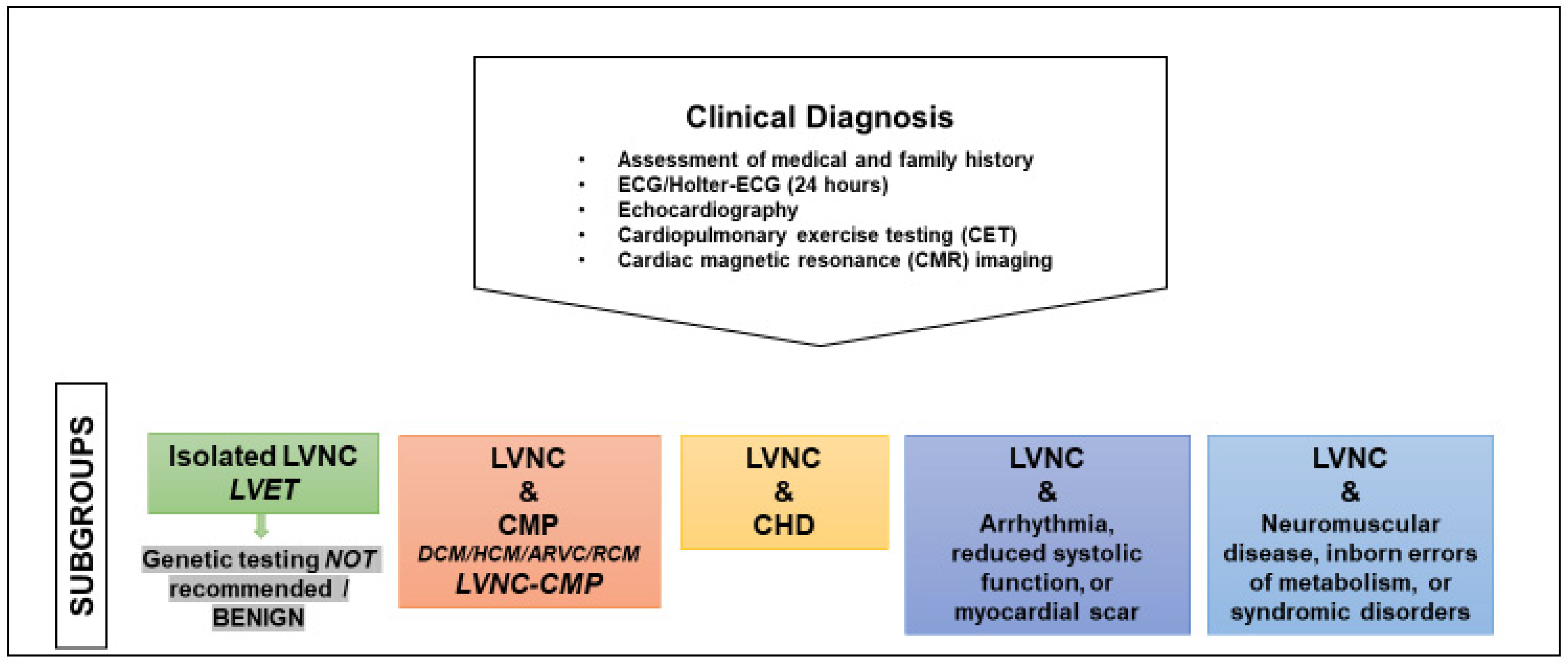
Several animal models were created to discover the underlying mechanisms of LVNC [8]. Unfortunately, only in a few genes investigated by these genetic models, pathogenic genetic variants were found in the corresponding human genes. The pathology of LVNC is difficult to recapitulate in murine models [42]. Genes which showed a LVNC phenotype in humans and demonstrated diverse cardiac phenotypes in murine models were PRDM16, MIB1, TBX20, NKX2-5, and TAFAZZIN. One of the CMP genes found by Mazzarotto et al. to be causative for LVNC only, and curated with limited evidence in ClinGen for DCM (https://clinicalgenome.org/; accessed on 18 April 2022) has recently been investigated in more detail, namely PRDM16. Both, contractile dysfunction and partial uncoupling of cardiomyocytes were the result of modelling of PRDM16 haploinsufficiency and a human truncation mutant in zebrafish [9]. There was also evidence of impaired cardiomyocyte proliferative capacity [9]. Genome editing of PRDM16 in human iPSC-derived cardiomyocytes (iPSC-CMs) recapitulated the proliferative defects associated with LVNC at the single-cell level [43]. Varying expression levels of mutant allele-specific mRNA expression between mild DCM and LVNC were used to explain the spectrum of clinical manifestations in LVNC [43]. Two murine genetic models with cardiomyocyte-specific Prdm16-null mutation were described [44,45]. In one model, using the Myh6-Cre driver, cardiac conduction abnormalities and a CMP phenotype were found [45]. Another mouse model deactivating Prdm16 with the Mesp1-Cre driver demonstrated predisposition to heart failure in response to metabolic stress [44]. Genetic lineage tracing revealed that compact myocardial myocytes are more proliferative than trabecular cardiomyocytes [46]. Impaired proliferation and expansion of the compact myocardium with premature cardiomyocyte maturation was suggested in the developing mouse heart to underlie LVNC [47]. In a more recent study, two new conditional Prdm16 knock out models recapitulated the LVNC phenotype with LV-specific dilatation and dysfunction [48]. In summary, the correct timing and spatial organization of cardiomyocyte proliferation and maturation is indispensable for normal left ventricular wall morphology and function.
Murine models were also used to test the hypothesis that LVNC may result from a combination of genetic variants. Evidence for oligogenic inheritance of LVNC was shown for a combination of three inherited heterozygous missense variants in the MKL/myocardin-like protein 2 (Mkl2), Myh7, and the transcription factor NK2 homeobox 5 (Nkx2-5) underlying familial LVNC. These triple compound heterozygous mice recapitulate signs of human LVNC [49]. Oligogenic inheritance was also supported by a study in which an increasing number of genetic variants in patients with LVNC strongly correlated with several markers of disease severity [50].

4. Diagnosis

The diagnosis of LVNC is made according to the morphologic criteria and made irrespective of the presence of LV systolic dysfunction or dilation [4,12]. This means that LVNC is a distinct but not necessarily pathological phenotype [51,52]. A wide spectrum of individuals meet the diagnostic criteria of LVNC without an underlying CMP. The genetic causes of LVNC are heterogeneous, reflect the underlying CMP such as DCM or HCM and most of them are not specific for the diagnosis of LVNC. Some acquired and reversible forms of LVNC strengthen that LVNC may be a physiologic, reversible phenotype [53,54,55]. LV trabeculations occur in response to increased LV loading conditions in pregnancy [56]. In one study, a high proportion of young athletes fulfil criteria for LVNC which suggests that the current diagnostic criteria, if used in an elite athletic population, are non-specific [57]. Ethnic considerations were described in which LVNC was more common in black individuals among an adult population fulfilling LVNC criteria supporting that LVNC might be a physiologic phenotype [26,58].
In an attempt to separate the individuals with pathologic from physiologic phenotype the term LVNC-CMP was introduced to specify those LVNC individuals with CMP [26]. When LV dilation and dysfunction are present, which are defining DCM, the diagnosis of LVNC-CMP may be more supported by using the DCM phenotype than by the trabecular morphology. In those cases, the diagnosis of DCM with LVNC morphology might be more suitable and practical. Both possibilities, either LVNC or CMP as the leading descriptor, provide the phenotype and diagnosis [4]. The morphology should be clearly described and practical to understand.

4.1. How to Reach and Confirm a Diagnosis

The different subtypes of pediatric LVNC are summarized in Figure 1. For the diagnosis of a CMP in the presence of LVNC, the following investigations are usually performed [26,36,38,59].
-
Assessment of medical and family history
-
Electrocardiography (ECG)
-
Echocardiography with functional assessment of LV size and function
-
Holter-ECG (24 h)
-
Cardiopulmonary exercise testing (CET)
-
Cardiac magnetic resonance (CMR) imaging
-
Genetic testing
Family history should include a three-generation pedigree. ECG changes are usually unspecific and arrhythmias detected by Holter-ECG may include ventricular ectopic beats and sustained or non-sustained ventricular tachycardia. Echocardiography data show LV dilatation and/or systolic/diastolic dysfunction. An early marker of systolic impairment is a reduced global longitudinal strain (GLS). CET is a measure for cardiovascular performance and possible arrhythmias. CMR imaging is used to characterize LV size and function and markers such as late gadolinium enhancement (LGE) are able to provide noninvasive tissue characterization and measure of fibrosis. Genetic testing should only be performed, if a CMP is diagnosed clinically.
With respect to subclinical CMP which can be detected by the newer imaging technologies there is still some uncertainty and no general agreement but the following is suggested. If strain is abnormal with normal LV-EF, the criteria for a CMP are not fulfilled according to the current AHA/ESC guidelines. These patients do not have CMP. LVNC with normal LVEF and without any other cardiovascular abnormalities, isolated LVNC, should be named LVET (Figure 1). Subclinical CMP (abnormal strain, clinically asymptomatic) requires clinical monitoring with echocardiography but genetic testing is not recommended.

4.2. The Importance of Imaging Modalities

Both imaging modalities, echocardiography and CMR imaging, are essential to determine the morphology (Figure 2). The echocardiographic criteria by Jenni et al [12]. remain the gold standard for the LV phenotype “noncompaction” and are most widely used [11].
Several studies have proposed CMR criteria to improve the diagnostic sensitivity and specificity [11]. Most of these studies have integrated Jenni’s criteria. Petersen et al. were the first to propose CMR criteria: “an NC/C ratio greater than 2.3 in a long axis end-diastolic image, in at least two consecutive segments” [14]. Further CMR studies demonstrated that the numerical cutoff may not be sufficient for the diagnosis and other biomarkers or deeper phenotyping was necessary [60]. Nevertheless, CMR provides important long-term prognostic information. CMR may differentiate patients with high rates of cardiac events from those with an excellent prognosis [54].
LV dilatation, systolic dysfunction, and late gadolinium enhancement (LGE) were independent predictors of poor outcome [54]. Interestingly, the extent of LV trabeculation failed to have a significant prognostic impact in this study [54]. In another study, at multivariate analysis LGE was the only independent predictor of impaired systolic LV function [61]. LGE was identified as an additional risk factor in adult LVNC patients without severe LV systolic dysfunction [36]. To guide the management of patients, a risk prediction model was developed and validated [36].
Myocardial extracellular volume (ECV) reflects diffuse myocardial fibrosis with CMR T1 mapping. Zhou et al. found elevated native T1 values in LVNC patients compared to normal controls [62]. LGE negative patients had elevated native T1 compared to normal controls which suggested that native T1 mapping can be applied to detect myocardial fibrosis in LVNC patients earlier than by LGE imaging [62]. In comparison, ECV was normal in most of the pediatric LVNC patients and was not related to cardiac function [63]. This was explained by their younger age and that the patients were clinically asymptomatic [63]. Midwall fibrosis (MWF) can be detected by LGE CMR and predicts adverse outcome in adults with non-ischemic DCM [64]. MWF was common in pediatric DCM, but was unable to predict heart failure in the pediatric study [65]. Data on MWF in LVNC would be of special interest and are not available so far.
Speckle-tracking echocardiography (STE) can detect subclinical changes in LV function, and may complement the echocardiographic assessment of LVNC patients in clinical routine. Although altered LV deformation in LVNC phenotype by STE does not enable the diagnosis of a definite CMP it can be helpful in clinical assessment. Particularly circumferential parameters are impaired in LVNC and can help to predict cardiovascular outcomes of LVNC [66].
LV trabeculation was found to be correlated with reduced myocardial deformation indexes, even if left ventricular ejection fraction (LV-EF) was normal [67]. Children with LVNC and normal LV function myocardial deformation patterns measured by tissue tracking analysis are significantly impaired compared to controls [68,69]. Rigid body rotation (RBR) is characterized by impaired LV twist with apex and base rotating in the same direction and can be used as a measure of cardiac mechanics in echocardiography and CMR. RBR was associated heart failure and MACE in pediatric DCM [65]. In adult LVNC patients, increased RBR was shown in LVNC with preserved EF when compared with controls [70]. In children, LV twist had good predictive value in diagnosing LVNC in young patients [71]. In summary, the findings indicate that ECV, RBR, and tissue tracking analysis may serve as novel markers of disease severity and permit earlier detection of LV functional impairment in pediatric LVNC.

4.3. Indication for Genetic Testing

With regard to genetic testing the American Heart Association, the Heart Failure Society of America, and the American College of Medical Genetics and Genomics propose guidelines following a common line [25,72,73]: “Genetic testing should be considered in individuals with CMP co-occurring with LVNC according to the recommendations for the associated CMP [73]. Genetic testing is not recommended when the LVNC phenotype is identified in asymptomatic individuals with otherwise normal cardiovascular phenotype (Figure 1) [25,72]. The same causes of familial HCM and DCM common in adults are also found throughout childhood and may be associated with LVNC [25]. Mitochondrial disease genes, metabolic causes of CMP, and genetic syndromes should be tested following the suspected underlying disorder according to the cardiovascular phenotype and additional extracardiac clinical features of the individual” [25].

5. Prognosis and Outcome

Because existing diagnostic criteria lack specificity prognosis and outcome are difficult to determine, LVNC includes both, physiological conditions or adjustments and pathological disease [74]. In cohorts retrieved from tertiary centers, it has repeatedly been demonstrated that LVNC was associated with an increased risk of heart failure, systemic embolisms, and ventricular arrhythmias, which are the clinical symptoms and events that guide therapy [38,75]. Reduced LV ejection fraction was the variable most strongly associated with MACE in adult and pediatric patients with LVNC [36,39,76,77].

5.1. Adult and Paediatric LVNC Outcome

Data on outcome of pediatric LVNC are scarce and therefore studies of adult patients are often taken into consideration. Overall survival was reduced in patients with LVNC compared with the expected survival of age- and sex-matched US population [76]. In this study by Vaidya et al. however, survival rate in the individuals with normal LV-EF and isolated apical noncompaction was comparable with that of the general population [76]. In multivariate cox regression analysis in adult LVNC, age, LV-EF < 50%, and extent of LVNC were significantly associated with all-cause mortality [76]. In a metanalysis Aung et al. systematically searched 28 eligible studies enrolling 2501 adult LVNC patients, mostly all studies with patients with reduced LV-EF, and reported the adverse outcomes [77]. Patients with LVNC had a similar outcome when compared with DCM patients [77]. LV-EF, not the extent of trabeculation, was a factor of adverse outcomes in LVNC patients [77]. In another large adult LVNC study population, the risk of MACE was related to factors such as familial aggregation, male sex, cardiovascular risk factors, older age at diagnosis, certain ECG abnormalities, and LGE on CMR, and like in so many other studies, to reduced LV-EF [36]. From all these studies, no concrete conclusions can be made for pediatric LVNC.
Genetic factors play a more important role in children than in adults with LVNC and severe forms of LVNC tend to be diagnosed in childhood [8,38]. LVNC had a high mortality rate in children in a tertiary center, resulting in an 18% incidence of death or transplantation in the total cohort [78]. Cardiac dysfunction or ventricular arrhythmias were associated with increased mortality, which was also significantly higher in children diagnosed in the first year of life [78]. Children with normal LV diameters and normal function were at low risk for sudden death [78]. Data from the Paediatric Cardiomyopathy Registry (PCMR) suggest the specific CMP phenotype that is associated with LVNC predicts outcome in children [79]. Pediatric patients with isolated LVNC have the best outcome compared to patients with LVNC and an associated DCM/HCM [79]. 20% of cases may be familial, therefore with the diagnosis of LVNC in a child family screening is recommended [79].
A systematic review by van Waning et al. in a large cohort of genetically confirmed 561 LVNC patients found worse clinical outcome in children compared to adults [32]. The study included children with genetic syndromes, chromosomal defects, and neuromuscular symptoms. The composition of the study cohort might explain the worse clinical outcome in children caused by the severe underlying disorder [32]. Non-sarcomere non-arrhythmia CMP genes, X-linked genes, arrhythmia genes, MYBPC3, and TTN were associated with MACE in multivariate analysis [32]. The severe outcome of children with multi-systemic disorders implies that diagnostic and therapeutic procedures should be guided by age at presentation [32].
Schultze-Berndt et al. detected a higher percentage of asymptomatic children than asymptomatic adult LVNC patients in their study cohort with primary CMP [39]. Reduced LV systolic function was identified as the main risk factor for MACE in the pediatric subcohort by multivariate analysis. The LV-EF reduction in children was the main risk factor, and LV-EF had a higher independent risk than a lower BSA or increased LVEDD [39].
In summary, in pediatric LVNC lower BSA and younger age are considered risk factors for MACE [32,39]. Congestive heart failure at diagnosis and reduced LV systolic function are independent risk factors in pediatric LVNC cohorts [30,39].

5.2. Genetic Variants and Risk of MACE

With respect to direct genotype-phenotype correlations in LVNC, several studies produced conflicting results. In a retrospective study of LVNC detected ≤21 years of age, it was less likely to find the genetic cause in isolated LVNC than in LVNC-CMP (0% versus 12%, respectively) [80]. Isolated LVNC individuals had a negative CMP gene panel result. This genetic yield in pediatric LVNC was lower compared with previous reports in adults (41% and 29%) [28,34] and in children (38% and 28%), respectively [30,39]. This low genetic yield is difficult to explain in the context of several other studies, but might be due to the imaging criteria for the diagnosis of LVNC. The severity of LVNC by imaging criteria was not associated with a positive genetic test result in this study [80]. Anyhow, children and young adults with isolated LVNC, in the absence of a family history of CMP, were not advised to undergo CMP gene panel testing [80]. No pathogenic variants were identified in another study in adult patients with isolated LVNC in the absence of cardiac dysfunction or syndromic features [81]. A study including pediatric LVNC patients (45% of the cohort were <18 years of age) suggested that LVNC might be driven by mutational burden [50]. The number of genetic variants was associated with the extent of noncompaction and LV systolic dysfunction. Left ventricular hypertrabeculation (LVHT) and LVNC might be the result of genotype accumulation which leads to differences in the expression of the phenotype [50]. It remains elusive, if and when a mutational burden might reach a threshold for the development of LVNC and after all, impaired cardiac function and clinical symptoms.
Variants in specific genes such as in TAFAZZIN, RBM20, Lamin A/C, TTN-truncating variants and non-sarcomere genes are associated with worse outcome in LVNC [30,35,82,83]. In the study by Wang et al. pathogenic variants were identified in 38% of patients (mean age 1.8 ± 0.4 years) and were independent risk factors for MACE such as death, heart transplantation, or implantable cardioverter-defibrillator implantation [30]. Sarcomere genes accounted for 63%. TAFAZZIN and MYH7 pathogenic variants were the most common. Patients with pathogenic variants showed poorer prognosis, especially those with multiple variants [30]. In the study by Schultze-Berndt et al. the genetic yield of (likely) pathogenic variants was 31%, 28% in pediatric patients, and 35% in adult patients, respectively. Mutations in MYH7, TTN, and MYBPC3 were most prevalent in the study, very comparable to other recent reports in adult and pediatric populations [36,38]. Risk stratification for MACE, depending on the individual mutation, was not possible, neither in the pediatric nor in the adult cohort [39]. Van Waning et al. showed that in children diagnosed <1 year of age, with multiple mutations in MYBPC3, and in (probable) genetic LVNC, an increased risk of MACE was observed [38]. The risk for adverse events in sporadic (non-genetic, non-familial) children was low [38]. In summary, there is evidence that specific genetic variants have a poorer prognosis but this knowledge is not ready to be transferred to clinical decision-making.

5.3. Paediatric LVNC Subtypes

The impact of LVNC subtypes was systematically reviewed in a study of Van Waning et al. [32]. Adult and pediatric cases were classified as isolated LVNC, LVNC with HCM, LVNC with DCM, and LVNC with DCM and HCM. Overall, the LVNC-DCM phenotype was present in 56% and therefore the most frequent subtype. LVNC-DCM was associated with a high risk for LV systolic dysfunction and an increased risk for MACE [32]. The LVNC-HCM/DCM phenotype was detected mostly in children, was not caused by mutations in CMP genes, and lead to severe outcome [32]. In the study by Schultze-Berndt et al. a higher prevalence of LVNC-HCM in the pediatric compared to the adult subcohort was shown [39]. Patients with LVNC-HCM were younger at diagnosis, more frequently affected by CHD, and at higher risk for MACE [39].
Genetic screening to identify family members at risk for developing CMP is useful and named “cascade screening” [59]. From investigating LVNC families, van Waning et al. could predict phenotype and outcome in relatives depending on the clinical features and genotype of LVNC index cases [33]. LVNC-DCM was associated with mutations in the tail of MYH7, LV systolic dysfunction, increased risk for MACE, and DCM without LVNC in relatives. Mutation in the head of MYH7, asymptomatic or mild disease, and isolated LVNC in relatives was associated with isolated LVNC. LVNC-HCM was associated with mutation of MYBPC3 and with HCM without LVNC in relatives [33]. These interesting data show that genetic background in families influences the cardiac phenotype associated with LVNC.
A recent study addressed the role of ion channel gene variants and correlation with arrhythmias in a large cohort of pediatric LVNC [84]. The LVNC patients with ion channel gene variants had nearly normal LV-EF and LVEDD, but showed a higher prevalence of arrhythmia phenotypes [84].
Patients with CHD and LVNC primarily present to the pediatric cardiologist and there are two possible etiologies. Pathologic remodeling induced by pressure or volume overload in CHD may lead to LVNC. Alternatively, genetic alteration may drive LVNC in CHD. In LVNC families, some relatives of affected members may have CHD or CMP with or without LVNC [26]. MYH7 mutation was found in Ebstein anomaly associated with LVNC [6]. In LVNC with or without CHD, there was no association between the likelihood of MACE and the NC/C ratio or the number of trabeculations [85]. Van Waning et al. reported on an increased risk in children for CHD and MACE [32].

6. Conclusions

LVNC is a phenotype that awaits classification and can be found in a spectrum from physiologic state to mild or severe disease. Adult individuals with isolated LVNC (LVET) seem to have the same outcome as the healthy population. The development of better imaging technologies, specifically CMR imaging, analyzes LVNC changes more easily and might lead to better diagnosis, if used with novel imaging biomarkers. We need larger systematic population-based studies and follow-up to identify individuals at risk for developing a CMP later in life specifically in the pediatric population.

Author Contributions

Writing—original draft preparation, S.K.; writing—review and editing, S.K., J.K., A.S.-B. and F.S. All authors have read and agreed to the published version of the manuscript.

Funding

This research received no external funding.

Data Availability Statement

Not applicable.

Conflicts of Interest

The authors declare no conflict of interest.

References

  1. Lee, T.M.; Hsu, D.T.; Kantor, P.; Towbin, J.A.; Ware, S.M.; Colan, S.D.; Chung, W.K.; Jefferies, J.L.; Rossano, J.W.; Castleberry, C.D.; et al. Pediatric Cardiomyopathies. Circ. Res. 2017, 121, 855–873. [Google Scholar] [CrossRef] [PubMed]
  2. Maron, B.J.; Towbin, J.A.; Thiene, G.; Antzelevitch, C.; Corrado, D.; Arnett, D.; Moss, A.J.; Seidman, C.E.; Young, J.B. Contemporary definitions and classification of the cardiomyopathies: An American Heart Association Scientific Statement from the Council on Clinical Cardiology, Heart Failure and Transplantation Committee; Quality of Care and Outcomes Research and Functional Genomics and Translational Biology Interdisciplinary Working Groups; and Council on Epidemiology and Prevention. Circulation 2006, 113, 1807–1816. [Google Scholar] [CrossRef] [PubMed] [Green Version]
  3. Elliott, P.; Andersson, B.; Arbustini, E.; Bilinska, Z.; Cecchi, F.; Charron, P.; Dubourg, O.; Kuhl, U.; Maisch, B.; McKenna, W.J.; et al. Classification of the cardiomyopathies: A position statement from the European Society of Cardiology Working Group on Myocardial and Pericardial Diseases. Eur. Heart J. 2008, 29, 270–276. [Google Scholar] [CrossRef] [PubMed] [Green Version]
  4. Arbustini, E.; Favalli, V.; Narula, N.; Serio, A.; Grasso, M. Left Ventricular Noncompaction: A Distinct Genetic Cardiomyopathy? J. Am. Coll. Cardiol. 2016, 68, 949–966. [Google Scholar] [CrossRef]
  5. Anderson, R.H.; Jensen, B.; Mohun, T.J.; Petersen, S.E.; Aung, N.; Zemrak, F.; Planken, R.N.; MacIver, D.H. Key Questions Relating to Left Ventricular Noncompaction Cardiomyopathy: Is the Emperor Still Wearing Any Clothes? Can. J. Cardiol. 2017, 33, 747–757. [Google Scholar] [CrossRef]
  6. Postma, A.V.; van Engelen, K.; van de Meerakker, J.; Rahman, T.; Probst, S.; Baars, M.J.; Bauer, U.; Pickardt, T.; Sperling, S.R.; Berger, F.; et al. Mutations in the sarcomere gene MYH7 in Ebstein anomaly. Circ. Cardiovasc. Genet. 2011, 4, 43–50. [Google Scholar] [CrossRef] [Green Version]
  7. Klaassen, S.; Probst, S.; Oechslin, E.; Gerull, B.; Krings, G.; Schuler, P.; Greutmann, M.; Hurlimann, D.; Yegitbasi, M.; Pons, L.; et al. Mutations in sarcomere protein genes in left ventricular noncompaction. Circulation 2008, 117, 2893–2901. [Google Scholar] [CrossRef]
  8. Towbin, J.A.; Lorts, A.; Jefferies, J.L. Left ventricular non-compaction cardiomyopathy. Lancet 2015, 386, 813–825. [Google Scholar] [CrossRef]
  9. Arndt, A.-K.; Schafer, S.; Drenckhahn, J.D.; Sabeh, M.K.; Plovie, E.R.; Caliebe, A.; Klopocki, E.; Musso, G.; Werdich, A.A.; Kalwa, H.; et al. Fine mapping of the 1p36 deletion syndrome identifies mutation of PRDM16 as a cause of cardiomyopathy. Am. J. Hum. Genet. 2013, 93, 67–77. [Google Scholar] [CrossRef] [Green Version]
  10. Bleyl, S.B.; Mumford, B.R.; Thompson, V.; Carey, J.C.; Pysher, T.J.; Chin, T.K.; Ward, K. Neonatal, lethal noncompaction of the left ventricular myocardium is allelic with Barth syndrome. Am. J. Hum. Genet. 1997, 61, 868–872. [Google Scholar] [CrossRef] [Green Version]
  11. Gerecke, B.J.; Engberding, R. Noncompaction Cardiomyopathy-History and Current Knowledge for Clinical Practice. J. Clin. Med. 2021, 10, 2457. [Google Scholar] [CrossRef] [PubMed]
  12. Jenni, R.; Oechslin, E.; Schneider, J.; Attenhofer Jost, C.; Kaufmann, P.A. Echocardiographic and pathoanatomical characteristics of isolated left ventricular non-compaction: A step towards classification as a distinct cardiomyopathy. Heart 2001, 86, 666–671. [Google Scholar] [CrossRef] [PubMed] [Green Version]
  13. Gati, S.; Rajani, R.; Carr-White, G.S.; Chambers, J.B. Adult left ventricular noncompaction: Reappraisal of current diagnostic imaging modalities. JACC Cardiovasc. Imaging 2014, 7, 1266–1275. [Google Scholar] [CrossRef] [Green Version]
  14. Petersen, S.E.; Selvanayagam, J.B.; Wiesmann, F.; Robson, M.D.; Francis, J.M.; Anderson, R.H.; Watkins, H.; Neubauer, S. Left ventricular non-compaction: Insights from cardiovascular magnetic resonance imaging. J. Am. Coll. Cardiol. 2005, 46, 101–105. [Google Scholar] [CrossRef] [Green Version]
  15. Gebhard, C.; Stahli, B.E.; Greutmann, M.; Biaggi, P.; Jenni, R.; Tanner, F.C. Reduced left ventricular compacta thickness: A novel echocardiographic criterion for non-compaction cardiomyopathy. J. Am. Soc. Echocardiogr. 2012, 25, 1050–1057. [Google Scholar] [CrossRef]
  16. Stollberger, C.; Gerecke, B.; Engberding, R.; Grabner, B.; Wandaller, C.; Finsterer, J.; Gietzelt, M.; Balzereit, A. Interobserver Agreement of the Echocardiographic Diagnosis of LV Hypertrabeculation/Noncompaction. JACC Cardiovasc. Imaging 2015, 8, 1252–1257. [Google Scholar] [CrossRef] [PubMed] [Green Version]
  17. Chin, T.K.; Perloff, J.K.; Williams, R.G.; Jue, K.; Mohrmann, R. Isolated noncompaction of left ventricular myocardium. A study of eight cases. Circulation 1990, 82, 507–513. [Google Scholar] [CrossRef] [Green Version]
  18. Grego-Bessa, J.; Luna-Zurita, L.; del Monte, G.; Bolos, V.; Melgar, P.; Arandilla, A.; Garratt, A.N.; Zang, H.; Mukouyama, Y.S.; Chen, H.; et al. Notch signaling is essential for ventricular chamber development. Dev. Cell 2007, 12, 415–429. [Google Scholar] [CrossRef] [Green Version]
  19. Misra, C.; Garg, V. Compacting the heart with Notch. Nat. Med. 2013, 19, 133–134. [Google Scholar] [CrossRef]
  20. Rojanasopondist, P.; Nesheiwat, L.; Piombo, S.; Porter, G.A., Jr.; Ren, M.; Phoon, C.K.L. Genetic Basis of Left Ventricular Noncompaction. Circ. Genom. Precis. Med. 2022, 15, e003517. [Google Scholar] [CrossRef]
  21. Jensen, B.; van der Wal, A.C.; Moorman, A.F.M.; Christoffels, V.M. Excessive trabeculations in noncompaction do not have the embryonic identity. Int. J. Cardiol. 2017, 227, 325–330. [Google Scholar] [CrossRef] [PubMed] [Green Version]
  22. Faber, J.W.; D’Silva, A.; Christoffels, V.M.; Jensen, B. Lack of morphometric evidence for ventricular compaction in humans. J. Cardiol. 2021, 78, 397–405. [Google Scholar] [CrossRef] [PubMed]
  23. Riekerk, H.C.E.; Coolen, B.F.; Strijkers, G.J.; van der Wal, A.C.; Petersen, S.E.; Sheppard, M.N.; Oostra, R.J.; Christoffels, V.M.; Jensen, B. Higher spatial resolution improves the interpretation of the extent of ventricular trabeculation. J Anat. 2021, 240, 357–375. [Google Scholar] [CrossRef] [PubMed]
  24. Meyer, H.V.; Dawes, T.J.W.; Serrani, M.; Bai, W.; Tokarczuk, P.; Cai, J.; de Marvao, A.; Henry, A.; Lumbers, R.T.; Gierten, J.; et al. Genetic and functional insights into the fractal structure of the heart. Nature 2020, 584, 589–594. [Google Scholar] [CrossRef]
  25. Hershberger, R.E.; Givertz, M.M.; Ho, C.Y.; Judge, D.P.; Kantor, P.F.; McBride, K.L.; Morales, A.; Taylor, M.R.G.; Vatta, M.; Ware, S.M. Genetic Evaluation of Cardiomyopathy-A Heart Failure Society of America Practice Guideline. J. Card. Fail. 2018, 24, 281–302. [Google Scholar] [CrossRef] [Green Version]
  26. Oechslin, E.; Jenni, R.; Klaassen, S. Left Ventricular Noncompaction Is a Myocardial Phenotype: Cardiomyopathy—Yes or No? Can. J. Cardiol. 2021, 37, 366–369. [Google Scholar] [CrossRef]
  27. Sasse-Klaassen, S.; Probst, S.; Gerull, B.; Oechslin, E.; Nurnberg, P.; Heuser, A.; Jenni, R.; Hennies, H.C.; Thierfelder, L. Novel gene locus for autosomal dominant left ventricular noncompaction maps to chromosome 11p15. Circulation 2004, 109, 2720–2723. [Google Scholar] [CrossRef] [Green Version]
  28. Hoedemaekers, Y.M.; Caliskan, K.; Michels, M.; Frohn-Mulder, I.; van der Smagt, J.J.; Phefferkorn, J.E.; Wessels, M.W.; ten Cate, F.J.; Sijbrands, E.J.; Dooijes, D.; et al. The importance of genetic counseling, DNA diagnostics, and cardiologic family screening in left ventricular noncompaction cardiomyopathy. Circ. Cardiovasc. Genet. 2010, 3, 232–239. [Google Scholar] [CrossRef] [Green Version]
  29. Kuhnisch, J.; Herbst, C.; Al-Wakeel-Marquard, N.; Dartsch, J.; Holtgrewe, M.; Baban, A.; Mearini, G.; Hardt, J.; Kolokotronis, K.; Gerull, B.; et al. Targeted panel sequencing in pediatric primary cardiomyopathy supports a critical role of TNNI3. Clin. Genet. 2019, 96, 549–559. [Google Scholar] [CrossRef] [Green Version]
  30. Wang, C.; Hata, Y.; Hirono, K.; Takasaki, A.; Ozawa, S.W.; Nakaoka, H.; Saito, K.; Miyao, N.; Okabe, M.; Ibuki, K.; et al. A Wide and Specific Spectrum of Genetic Variants and Genotype-Phenotype Correlations Revealed by Next-Generation Sequencing in Patients with Left Ventricular Noncompaction. J. Am. Heart Assoc. 2017, 6, e006210. [Google Scholar] [CrossRef]
  31. Kolokotronis, K.; Kuhnisch, J.; Klopocki, E.; Dartsch, J.; Rost, S.; Huculak, C.; Mearini, G.; Stork, S.; Carrier, L.; Klaassen, S.; et al. Biallelic mutation in MYH7 and MYBPC3 leads to severe cardiomyopathy with left ventricular noncompaction phenotype. Hum. Mutat. 2019, 40, 1101–1114. [Google Scholar] [CrossRef] [PubMed]
  32. Van Waning, J.I.; Moesker, J.; Heijsman, D.; Boersma, E.; Majoor-Krakauer, D. Systematic Review of Genotype-Phenotype Correlations in Noncompaction Cardiomyopathy. J. Am. Heart Assoc. 2019, 8, e012993. [Google Scholar] [CrossRef] [PubMed]
  33. van Waning, J.I.; Caliskan, K.; Michels, M.; Schinkel, A.F.L.; Hirsch, A.; Dalinghaus, M.; Hoedemaekers, Y.M.; Wessels, M.W.; AS, I.J.; Hofstra, R.M.W.; et al. Cardiac Phenotypes, Genetics, and Risks in Familial Noncompaction Cardiomyopathy. J. Am. Coll. Cardiol. 2019, 73, 1601–1611. [Google Scholar] [CrossRef]
  34. Probst, S.; Oechslin, E.; Schuler, P.; Greutmann, M.; Boye, P.; Knirsch, W.; Berger, F.; Thierfelder, L.; Jenni, R.; Klaassen, S. Sarcomere gene mutations in isolated left ventricular noncompaction cardiomyopathy do not predict clinical phenotype. Circ. Cardiovasc. Genet. 2011, 4, 367–374. [Google Scholar] [CrossRef] [PubMed] [Green Version]
  35. Sedaghat-Hamedani, F.; Haas, J.; Zhu, F.; Geier, C.; Kayvanpour, E.; Liss, M.; Lai, A.; Frese, K.; Pribe-Wolferts, R.; Amr, A.; et al. Clinical genetics and outcome of left ventricular non-compaction cardiomyopathy. Eur. Heart J. 2017, 38, 3449–3460. [Google Scholar] [CrossRef] [PubMed]
  36. Casas, G.; Limeres, J.; Oristrell, G.; Gutierrez-Garcia, L.; Andreini, D.; Borregan, M.; Larranaga-Moreira, J.M.; Lopez-Sainz, A.; Codina-Sola, M.; Teixido-Tura, G.; et al. Clinical Risk Prediction in Patients with Left Ventricular Myocardial Noncompaction. J. Am. Coll. Cardiol. 2021, 78, 643–662. [Google Scholar] [CrossRef]
  37. Klaassen, S. Ventricular noncompaction: An update. In Braunwald´s Heart Disease, A Textbook of Cardiovascular Medicine, 8th ed.; Libby, P., Bonow, R.O., Mann, D.L., Zipes, D.P., Eds.; Saunders Elsevier: Philadelphia, PA, USA, 2008. [Google Scholar]
  38. van Waning, J.I.; Caliskan, K.; Hoedemaekers, Y.M.; van Spaendonck-Zwarts, K.Y.; Baas, A.F.; Boekholdt, S.M.; van Melle, J.P.; Teske, A.J.; Asselbergs, F.W.; Backx, A.; et al. Genetics, Clinical Features, and Long-Term Outcome of Noncompaction Cardiomyopathy. J. Am. Coll. Cardiol. 2018, 71, 711–722. [Google Scholar] [CrossRef]
  39. Schultze-Berndt, A.; Kuhnisch, J.; Herbst, C.; Seidel, F.; Al-Wakeel-Marquard, N.; Dartsch, J.; Theisen, S.; Knirsch, W.; Jenni, R.; Greutmann, M.; et al. Reduced Systolic Function and Not Genetic Variants Determine Outcome in Pediatric and Adult Left Ventricular Noncompaction Cardiomyopathy. Front. Pediatr. 2021, 9, 722926. [Google Scholar] [CrossRef]
  40. Mazzarotto, F.; Hawley, M.H.; Beltrami, M.; Beekman, L.; de Marvao, A.; McGurk, K.A.; Statton, B.; Boschi, B.; Girolami, F.; Roberts, A.M.; et al. Systematic large-scale assessment of the genetic architecture of left ventricular noncompaction reveals diverse etiologies. Genet. Med. 2021, 23, 856–864. [Google Scholar] [CrossRef]
  41. Walsh, R.; Thomson, K.L.; Ware, J.S.; Funke, B.H.; Woodley, J.; McGuire, K.J.; Mazzarotto, F.; Blair, E.; Seller, A.; Taylor, J.C.; et al. Reassessment of Mendelian gene pathogenicity using 7,855 cardiomyopathy cases and 60,706 reference samples. Genet. Med. 2017, 19, 192–203. [Google Scholar] [CrossRef] [Green Version]
  42. Wilsbacher, L.; McNally, E.M. Genetics of Cardiac Developmental Disorders: Cardiomyocyte Proliferation and Growth and Relevance to Heart Failure. Annu Rev Pathol 2016, 11, 395–419. [Google Scholar] [CrossRef] [PubMed]
  43. Kodo, K.; Ong, S.G.; Jahanbani, F.; Termglinchan, V.; Hirono, K.; InanlooRahatloo, K.; Ebert, A.D.; Shukla, P.; Abilez, O.J.; Churko, J.M.; et al. iPSC-derived cardiomyocytes reveal abnormal TGF-beta signalling in left ventricular non-compaction cardiomyopathy. Nat. Cell Biol. 2016, 18, 1031–1042. [Google Scholar] [CrossRef] [PubMed] [Green Version]
  44. Cibi, D.M.; Bi-Lin, K.W.; Shekeran, S.G.; Sandireddy, R.; Tee, N.; Singh, A.; Wu, Y.; Srinivasan, D.K.; Kovalik, J.P.; Ghosh, S.; et al. Prdm16 Deficiency Leads to Age-Dependent Cardiac Hypertrophy, Adverse Remodeling, Mitochondrial Dysfunction, and Heart Failure. Cell Rep. 2020, 33, 108288. [Google Scholar] [CrossRef] [PubMed]
  45. Nam, J.M.; Lim, J.E.; Ha, T.W.; Oh, B.; Kang, J.O. Cardiac-specific inactivation of Prdm16 effects cardiac conduction abnormalities and cardiomyopathy-associated phenotypes. Am. J. Physiol. Heart Circ. Physiol. 2020, 318, H764–H777. [Google Scholar] [CrossRef]
  46. Tian, X.; Li, Y.; He, L.; Zhang, H.; Huang, X.; Liu, Q.; Pu, W.; Zhang, L.; Li, Y.; Zhao, H.; et al. Identification of a hybrid myocardial zone in the mammalian heart after birth. Nat. Commun. 2017, 8, 87. [Google Scholar] [CrossRef] [PubMed]
  47. Rhee, S.; Chung, J.I.; King, D.A.; D’Amato, G.; Paik, D.T.; Duan, A.; Chang, A.; Nagelberg, D.; Sharma, B.; Jeong, Y.; et al. Endothelial deletion of Ino80 disrupts coronary angiogenesis and causes congenital heart disease. Nat. Commun. 2018, 9, 368. [Google Scholar] [CrossRef] [Green Version]
  48. Wu, T.; Liang, Z.; Zhang, Z.; Liu, C.; Zhang, L.; Gu, Y.; Peterson, K.L.; Evans, S.M.; Fu, X.D.; Chen, J. PRDM16 Is a Compact Myocardium-Enriched Transcription Factor Required to Maintain Compact Myocardial Cardiomyocyte Identity in Left Ventricle. Circulation 2022, 145, 586–602. [Google Scholar] [CrossRef]
  49. Gifford, C.A.; Ranade, S.S.; Samarakoon, R.; Salunga, H.T.; de Soysa, T.Y.; Huang, Y.; Zhou, P.; Elfenbein, A.; Wyman, S.K.; Bui, Y.K.; et al. Oligogenic inheritance of a human heart disease involving a genetic modifier. Science 2019, 364, 865–870. [Google Scholar] [CrossRef]
  50. Miszalski-Jamka, K.; Jefferies, J.L.; Mazur, W.; Glowacki, J.; Hu, J.; Lazar, M.; Gibbs, R.A.; Liczko, J.; Klys, J.; Venner, E.; et al. Novel Genetic Triggers and Genotype-Phenotype Correlations in Patients with Left Ventricular Noncompaction. Circ. Cardiovasc. Genet. 2017, 10, e001763. [Google Scholar] [CrossRef] [Green Version]
  51. Hershberger, R.E.; Morales, A.; Cowan, J. Is Left Ventricular Noncompaction a Trait, Phenotype, or Disease? The Evidence Points to Phenotype. Circ. Cardiovasc. Genet. 2017, 10, e001968. [Google Scholar] [CrossRef]
  52. Oechslin, E.; Klaassen, S. Left Ventricular Noncompaction: Phenotype in an Integrated Model of Cardiomyopathy? J. Am. Coll. Cardiol. 2019, 73, 1612–1615. [Google Scholar] [CrossRef] [PubMed]
  53. Weir-McCall, J.R.; Yeap, P.M.; Papagiorcopulo, C.; Fitzgerald, K.; Gandy, S.J.; Lambert, M.; Belch, J.J.; Cavin, I.; Littleford, R.; Macfarlane, J.A.; et al. Left Ventricular Noncompaction: Anatomical Phenotype or Distinct Cardiomyopathy? J. Am. Coll. Cardiol. 2016, 68, 2157–2165. [Google Scholar] [CrossRef] [PubMed] [Green Version]
  54. Andreini, D.; Pontone, G.; Bogaert, J.; Roghi, A.; Barison, A.; Schwitter, J.; Mushtaq, S.; Vovas, G.; Sormani, P.; Aquaro, G.D.; et al. Long-Term Prognostic Value of Cardiac Magnetic Resonance in Left Ventricle Noncompaction: A Prospective Multicenter Study. J. Am. Coll. Cardiol. 2016, 68, 2166–2181. [Google Scholar] [CrossRef] [PubMed]
  55. Zemrak, F.; Ahlman, M.A.; Captur, G.; Mohiddin, S.A.; Kawel-Boehm, N.; Prince, M.R.; Moon, J.C.; Hundley, W.G.; Lima, J.A.; Bluemke, D.A.; et al. The relationship of left ventricular trabeculation to ventricular function and structure over a 9.5-year follow-up: The MESA study. J. Am. Coll. Cardiol. 2014, 64, 1971–1980. [Google Scholar] [CrossRef] [Green Version]
  56. Gati, S.; Papadakis, M.; Papamichael, N.D.; Zaidi, A.; Sheikh, N.; Reed, M.; Sharma, R.; Thilaganathan, B.; Sharma, S. Reversible de novo left ventricular trabeculations in pregnant women: Implications for the diagnosis of left ventricular noncompaction in low-risk populations. Circulation 2014, 130, 475–483. [Google Scholar] [CrossRef]
  57. Gati, S.; Chandra, N.; Bennett, R.L.; Reed, M.; Kervio, G.; Panoulas, V.F.; Ghani, S.; Sheikh, N.; Zaidi, A.; Wilson, M.; et al. Increased left ventricular trabeculation in highly trained athletes: Do we need more stringent criteria for the diagnosis of left ventricular non-compaction in athletes? Heart 2013, 99, 401–408. [Google Scholar] [CrossRef]
  58. Kohli, S.K.; Pantazis, A.A.; Shah, J.S.; Adeyemi, B.; Jackson, G.; McKenna, W.J.; Sharma, S.; Elliott, P.M. Diagnosis of left-ventricular non-compaction in patients with left-ventricular systolic dysfunction: Time for a reappraisal of diagnostic criteria? Eur. Heart J. 2008, 29, 89–95. [Google Scholar] [CrossRef] [Green Version]
  59. Al-Wakeel-Marquard, N.; Degener, F.; Herbst, C.; Kuhnisch, J.; Dartsch, J.; Schmitt, B.; Kuehne, T.; Messroghli, D.; Berger, F.; Klaassen, S. RIKADA Study Reveals Risk Factors in Pediatric Primary Cardiomyopathy. J. Am. Heart Assoc. 2019, 8, e012531. [Google Scholar] [CrossRef] [Green Version]
  60. Negri, F.; De Luca, A.; Fabris, E.; Korcova, R.; Cernetti, C.; Grigoratos, C.; Aquaro, G.D.; Nucifora, G.; Camici, P.G.; Sinagra, G. Left ventricular noncompaction, morphological, and clinical features for an integrated diagnosis. Heart Fail. Rev. 2019, 24, 315–323. [Google Scholar] [CrossRef]
  61. Nucifora, G.; Aquaro, G.D.; Pingitore, A.; Masci, P.G.; Lombardi, M. Myocardial fibrosis in isolated left ventricular non-compaction and its relation to disease severity. Eur. J. Heart Fail. 2011, 13, 170–176. [Google Scholar] [CrossRef] [Green Version]
  62. Zhou, H.; Lin, X.; Fang, L.; Zhao, X.; Ding, H.; Chen, W.; Xu, R.; Bai, X.; Wang, Y.; Fang, Q. Characterization of Compacted Myocardial Abnormalities by Cardiac Magnetic Resonance with Native T1 Mapping in Left Ventricular Non-Compaction Patients- A Comparison with Late Gadolinium Enhancement. Circ. J. 2016, 80, 1210–1216. [Google Scholar] [CrossRef] [PubMed] [Green Version]
  63. Al-Wakeel-Marquard, N.; Seidel, F.; Herbst, C.; Kuhnisch, J.; Kuehne, T.; Berger, F.; Klaassen, S.; Messroghli, D.R. Diffuse myocardial fibrosis by T1 mapping is associated with heart failure in pediatric primary dilated cardiomyopathy. Int. J. Cardiol. 2021, 333, 219–225. [Google Scholar] [CrossRef] [PubMed]
  64. Wang, J.; Yang, F.; Wan, K.; Mui, D.; Han, Y.; Chen, Y. Left ventricular midwall fibrosis as a predictor of sudden cardiac death in non-ischaemic dilated cardiomyopathy: A meta-analysis. ESC Heart Fail. 2020, 7, 2184–2192. [Google Scholar] [CrossRef] [PubMed]
  65. Al-Wakeel-Marquard, N.; Seidel, F.; Kuhnisch, J.; Kuehne, T.; Berger, F.; Messroghli, D.R.; Klaassen, S. Midwall Fibrosis and Cardiac Mechanics: Rigid Body Rotation Is a Novel Marker of Disease Severity in Pediatric Primary Dilated Cardiomyopathy. Front. Cardiovasc. Med. 2021, 8, 810005. [Google Scholar] [CrossRef] [PubMed]
  66. Anwer, S.; Heiniger, P.S.; Rogler, S.; Erhart, L.; Cassani, D.; Kuzo, N.; Rebellius, L.; Schoenenberger-Berzins, R.; Schmid, D.; Nussbaum, S.; et al. Left ventricular mechanics and cardiovascular outcomes in non-compaction phenotype. Int. J. Cardiol. 2021, 336, 73–80. [Google Scholar] [CrossRef]
  67. Cai, J.; Bryant, J.A.; Le, T.T.; Su, B.; de Marvao, A.; O’Regan, D.P.; Cook, S.A.; Chin, C.W. Fractal analysis of left ventricular trabeculations is associated with impaired myocardial deformation in healthy Chinese. J. Cardiovasc. Magn. Reson. 2017, 19, 102. [Google Scholar] [CrossRef] [Green Version]
  68. Nucifora, G.; Sree Raman, K.; Muser, D.; Shah, R.; Perry, R.; Awang Ramli, K.A.; Selvanayagam, J.B. Cardiac magnetic resonance evaluation of left ventricular functional, morphological, and structural features in children and adolescents vs. young adults with isolated left ventricular non-compaction. Int. J. Cardiol. 2017, 246, 68–73. [Google Scholar] [CrossRef]
  69. Sarnecki, J.; Paszkowska, A.; Petryka-Mazurkiewicz, J.; Kubik, A.; Feber, J.; Jurkiewicz, E.; Ziolkowska, L. Left and Right Ventricular Morphology, Function and Myocardial Deformation in Children with Left Ventricular Non-Compaction Cardiomyopathy: A Case-Control Cardiovascular Magnetic Resonance Study. J. Clin. Med. 2022, 11, 1104. [Google Scholar] [CrossRef]
  70. Guigui, S.A.; Horvath, S.A.; Arenas, I.A.; Mihos, C.G. Cardiac geometry, function and mechanics in left ventricular non-compaction cardiomyopathy with preserved ejection fraction. J. Echocardiogr. 2022, 1–7. [Google Scholar] [CrossRef]
  71. Sabatino, J.; Di Salvo, G.; Krupickova, S.; Fraisse, A.; Prota, C.; Bucciarelli, V.; Josen, M.; Paredes, J.; Sirico, D.; Voges, I.; et al. Left Ventricular Twist Mechanics to Identify Left Ventricular Noncompaction in Childhood. Circ. Cardiovasc. Imaging 2019, 12, e007805. [Google Scholar] [CrossRef] [Green Version]
  72. Hershberger, R.E.; Givertz, M.M.; Ho, C.Y.; Judge, D.P.; Kantor, P.F.; McBride, K.L.; Morales, A.; Taylor, M.R.G.; Vatta, M.; Ware, S.M.; et al. Genetic evaluation of cardiomyopathy: A clinical practice resource of the American College of Medical Genetics and Genomics (ACMG). Genet. Med. 2018, 20, 899–909. [Google Scholar] [CrossRef] [PubMed] [Green Version]
  73. Musunuru, K.; Hershberger, R.E.; Day, S.M.; Klinedinst, N.J.; Landstrom, A.P.; Parikh, V.N.; Prakash, S.; Semsarian, C.; Sturm, A.C. Genetic Testing for Inherited Cardiovascular Diseases: A Scientific Statement from the American Heart Association. Circ. Genom. Precis. Med. 2020, 13, e000067. [Google Scholar] [CrossRef]
  74. Ross, S.B.; Jones, K.; Blanch, B.; Puranik, R.; McGeechan, K.; Barratt, A.; Semsarian, C. A systematic review and meta-analysis of the prevalence of left ventricular non-compaction in adults. Eur. Heart J. 2020, 41, 1428–1436. [Google Scholar] [CrossRef]
  75. Oechslin, E.N.; Attenhofer Jost, C.H.; Rojas, J.R.; Kaufmann, P.A.; Jenni, R. Long-term follow-up of 34 adults with isolated left ventricular noncompaction: A distinct cardiomyopathy with poor prognosis. J. Am. Coll. Cardiol. 2000, 36, 493–500. [Google Scholar] [CrossRef] [Green Version]
  76. Vaidya, V.R.; Lyle, M.; Miranda, W.R.; Farwati, M.; Isath, A.; Patlolla, S.H.; Hodge, D.O.; Asirvatham, S.J.; Kapa, S.; Deshmukh, A.J.; et al. Long-Term Survival of Patients with Left Ventricular Noncompaction. J. Am. Heart Assoc. 2021, 10, e015563. [Google Scholar] [CrossRef]
  77. Aung, N.; Doimo, S.; Ricci, F.; Sanghvi, M.M.; Pedrosa, C.; Woodbridge, S.P.; Al-Balah, A.; Zemrak, F.; Khanji, M.Y.; Munroe, P.B.; et al. Prognostic Significance of Left Ventricular Noncompaction: Systematic Review and Meta-Analysis of Observational Studies. Circ. Cardiovasc. Imaging 2020, 13, e009712. [Google Scholar] [CrossRef]
  78. Brescia, S.T.; Rossano, J.W.; Pignatelli, R.; Jefferies, J.L.; Price, J.F.; Decker, J.A.; Denfield, S.W.; Dreyer, W.J.; Smith, O.; Towbin, J.A.; et al. Mortality and sudden death in pediatric left ventricular noncompaction in a tertiary referral center. Circulation 2013, 127, 2202–2208. [Google Scholar] [CrossRef] [Green Version]
  79. Jefferies, J.L.; Wilkinson, J.D.; Sleeper, L.A.; Colan, S.D.; Lu, M.; Pahl, E.; Kantor, P.F.; Everitt, M.D.; Webber, S.A.; Kaufman, B.D.; et al. Cardiomyopathy Phenotypes and Outcomes for Children with Left Ventricular Myocardial Noncompaction: Results from the Pediatric Cardiomyopathy Registry. J. Card. Fail. 2015, 21, 877–884. [Google Scholar] [CrossRef] [Green Version]
  80. Miller, E.M.; Hinton, R.B.; Czosek, R.; Lorts, A.; Parrott, A.; Shikany, A.R.; Ittenbach, R.F.; Ware, S.M. Genetic Testing in Pediatric Left Ventricular Noncompaction. Circ. Cardiovasc. Genet. 2017, 10, e001735. [Google Scholar] [CrossRef] [Green Version]
  81. Ross, S.B.; Singer, E.S.; Driscoll, E.; Nowak, N.; Yeates, L.; Puranik, R.; Sy, R.W.; Rajagopalan, S.; Barratt, A.; Ingles, J.; et al. Genetic architecture of left ventricular noncompaction in adults. Hum. Genome Var. 2020, 7, 33. [Google Scholar] [CrossRef]
  82. Li, S.; Zhang, C.; Liu, N.; Bai, H.; Hou, C.; Pu, J. Clinical implications of sarcomere and nonsarcomere gene variants in patients with left ventricular noncompaction cardiomyopathy. Mol. Genet. Genom. Med. 2019, 7, e874. [Google Scholar] [CrossRef] [Green Version]
  83. Li, S.; Zhang, C.; Liu, N.; Bai, H.; Hou, C.; Song, L.; Pu, J. Titin-truncating variants are associated with heart failure events in patients with left ventricular non-compaction cardiomyopathy. Clin. Cardiol. 2019, 42, 530–535. [Google Scholar] [CrossRef]
  84. Hirono, K.; Hata, Y.; Miyao, N.; Okabe, M.; Takarada, S.; Nakaoka, H.; Ibuki, K.; Ozawa, S.; Origasa, H.; Nishida, N.; et al. Increased Burden of Ion Channel Gene Variants Is Related to Distinct Phenotypes in Pediatric Patients with Left Ventricular Noncompaction. Circ. Genom. Precis. Med. 2020, 13, e002940. [Google Scholar] [CrossRef]
  85. Saleeb, S.F.; Margossian, R.; Spencer, C.T.; Alexander, M.E.; Smoot, L.B.; Dorfman, A.L.; Bergersen, L.; Gauvreau, K.; Marx, G.R.; Colan, S.D. Reproducibility of echocardiographic diagnosis of left ventricular noncompaction. J. Am. Soc. Echocardiogr. 2012, 25, 194–202. [Google Scholar] [CrossRef]
Figure 1. Clinical diagnosis and subgroups in pediatric LVNC. ARVC, arrhythmogenic right ventricular cardiomyopathy; CHD, congenital heart defects; CMP, cardiomyopathy; DCM, dilated cardiomyopathy; HCM, hypertrophic cardiomyopathy; ECG, electrocardiogram; LVET, left ventricular excessive trabeculation; LVNC, left ventricular noncompaction; LVNC-CMP, left ventricular noncompaction cardiomyopathy; RCM, restrictive cardiomyopathy.
Jcdd 09 00206 g001
Figure 2. Echocardiography and cardiovascular magnetic resonance (CMR) imaging in left ventricular noncompaction (LVNC) of a 14-year-old girl with LVNC. The upper and middle panel show 4, 2, and 3 chamber views (CV) of representative echocardiographic and CMR images, respectively. The lower panel depicts the CMR short axis view at the basis, midventricular site, and apex of the same heart.
Jcdd 09 00206 g002
Publisher’s Note: MDPI stays neutral with regard to jurisdictional claims in published maps and institutional affiliations.

