Candida sp. Infections in Patients with Diabetes Mellitus


1
CEB, Centre of Biological Engineering, LIBRO-Laboratório de Investigação em Biofilmes Rosário Oliveira, University of Minho, 4710-057 Braga, Portugal
2
LEPABE—Department of Chemical Engineering, Faculty of Engineering, University of Porto, Rua Dr. Roberto Frias, s/n, 4200-465 Porto, Portugal
*
Author to whom correspondence should be addressed.
J. Clin. Med. 2019, 8(1), 76; https://0-doi-org.brum.beds.ac.uk/10.3390/jcm8010076
Submission received: 14 December 2018
/
Revised: 27 December 2018
/
Accepted: 3 January 2019
/
Published: 10 January 2019
(This article belongs to the Special Issue Biomaterial-Related Infections)
Abstract
:Candidiasis has increased substantially worldwide over recent decades and is a significant cause of morbidity and mortality, especially among critically ill patients. Diabetes mellitus (DM) is a metabolic disorder that predisposes individuals to fungal infections, including those related to Candida sp., due to a immunosuppressive effect on the patient. This review aims to discuss the latest studies regarding the occurrence of candidiasis on DM patients and the pathophysiology and etiology associated with these co-morbidities. A comprehensive review of the literature was undertaken. PubMed, Scopus, Elsevier’s ScienceDirect, and Springer’s SpringerLink databases were searched using well-defined search terms. Predefined inclusion and exclusion criteria were applied to classify relevant manuscripts. Results of the review show that DM patients have an increased susceptibility to Candida sp. infections which aggravates in the cases of uncontrolled hyperglycemia. The conclusion is that, for these patients, the hospitalization periods have increased and are commonly associated with the prolonged use of indwelling medical devices, which also increase the costs associated with disease management.
Keywords:
Candida; biofilms; diabetes; medical devices; candidiasis; metabolic disorder; hyperglycemia; infection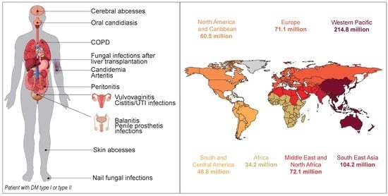
1. Introduction
Diabetes mellitus (DM) is a chronic metabolic and degenerative disorder that is characterized by chronic hyperglycemia and causes long-term complications like retinopathy, neuropathy, and nephropathy, generally accelerating macro- and micro-vascular changes. It is becoming one of the largest emerging threats to public health in the 21st century [1,2]. Several immune alterations have been described in diabetes with cellular immunity being more compromised and with changes in polymorphonuclear cells, monocytes, and lymphocytes [3]. DM individuals have higher glucose serum concentrations than healthy individuals (between 4.0 to 5.4 mmol/L or 72 to 99 mg/dL when fasting and up to 7.8 mmol/L or 140 mg/dL two hours after eating [4]; hemoglobin A1c (glycohemoglobin) ≤5.7%). In type 1 DM, the pathogenesis is multifactorial because of antibody-mediated autoimmunity, environmental toxins exposure, and major histocompatibility complex (MHC) Class II histocompatibility complex HLA-DR/DQ genetic polymorphisms. These features create an increased susceptibility to disease onset due to a continuous loss of insulin-producing β-cells in the pancreas, which is due to the T-cells’ infiltration through mitochondrial-driven apoptosis [5]. On the other hand, in type 2 DM, there is an insulin resistance that is associated with changes in the mitochondrial metabolism with reduced mitochondrial density, ATP production, and mitochondrial RNA (mtRNA) levels, as well as increased markers for oxidative stress. The chronic exposure of the circular mtDNA to these effects might trigger significant tissue modifications found in the pancreas and endothelial cells, leading to secondary vascular disease and causing cardiac, renal, ophthalmic, and neurological complications [5,6,7] (Figure 1).
In 2017, the worldwide prevalence of adult-onset diabetes (20–79 years) was nearly 425 million, and the World Health Organization and the International Diabetes Federation predicted that the number of adults in the world with diabetes will rise near 629 million by the year 2045 [8,9] (Figure 2). A higher prevalence of DM, cardiac, and pulmonary diseases can be found in senior patients with candidemia [10,11,12,13]. The relationship between diabetes and candidiasis has been widely studied [13,14,15,16], particularly due to the increased susceptibility of diabetic patients to fungal infections compared to those without DM [14,15,17,18].
Several mechanisms are attributed to higher Candida sp. predisposition among DM patients depending on the local or systemic infection. Among the recognized host conditions for candidal colonization and subsequent infection are yeast adhesion to epithelial cell surfaces [19], higher salivary glucose levels [15,20], reduced salivary flow [21], microvascular degeneration, and impaired candidacidal activity of neutrophils. These conditions are particularly serious in the presence of glucose [22,23], secretion of several degradative enzymes [24,25,26], or even a generalized immunosuppression state of the patient [8,27,28,29,30,31]. These factors have a major influence on the balance between host and yeasts, favoring the transition of Candida sp. from commensal to pathogen and causing infection. In fact, in a very recent study, Gürsoy et al. [32] suggested that there is a higher presence of intestinal Candida albicans colonization in diabetic patients. In fact, there may be a tendency of type 1 DM in patients with a high prevalence of intestinal C. albicans. C. albicans is also known to wait for a change in some aspect of the host physiology that normally suppress growth and invasiveness through a phenomenon called phenotypic switch system or white-opaque transition, described in 1987.
This involves reversible and heritable switching between alternative cellular phenotypes. It occurs at sites of infection and recurrently in episodes of infection in certain cases in diabetic patients [33].
Yeasts are part of the normal gut microflora, but cell counts do not normally surpass 10 colony forming units (CFU)/g feces [34,35]. Nevertheless, it has been described that Candida sp. is more widespread in the feces of patients with type 1 and type 2 DM with poor glycemic control as opposed to healthy subjects [36]. The main reasons for this colonization seem to be altered functions of the immune system in diabetic patients with poor glycemic control or a direct effect of elevated blood glucose levels, creating specific conditions for intensive fungal colonization [36]. In fact, another report [37] showed that in patients with type 1 DM, the total gut CFUs significantly rise up to 40% in C. albicans colonization compared to 14.3% in healthy individuals. This may be related to the decrease in commensal bacteria-probably the result of yeast-bacterial competition. Also, this higher growth may disrupt the ecological balance of intestinal flora, which occurs in type 1 DM [37]. Regarding the gastrointestinal colonization, Kowalewska et al. [38] studied the serum levels of interleukin-12 (IL12) in relation to the percentage of yeast-like fungi colonies residing in the gastrointestinal tract in children and adolescents with DM type 1. Results showed that high IL12 levels can inhibit infection with yeast-like fungal colonizing the gastrointestinal tract in children and adolescents with type 1 DM. However, further studies are needed to confirm the antifungal activity of IL12 [38].
The development of drug resistance among Candida sp. isolates allied to epidemiologic variations in Candida sp. natural flora has significant implications for morbidity and mortality [39,40,41,42]. The extensive use of medications, especially azoles, has promoted the selection of resistant species by shifting colonization to more naturally resistant Candida sp., such as C. glabrata, C. dubliniensis, and C. krusei [43,44,45,46]. Presently, the world distribution of Candida sp. is a feature of the epidemiology in the area, but it indicates a predominance of C. albicans, C. glabrata, C. tropicalis, C. parapsilosis, and C. krusei [45,47]. It has been confirmed that 90% of fungemia cases are attributed to Candida sp. [39,40], and the mortality has ranged from 40% to 80% in immunocompromised hosts [39,40,48]. Furthermore, a high mortality rate was also detected among non-immunocompromised patients (60%) [49] and those with diabetes (67%) [41].
The main pathophysiologic and nutritionally relevant sugars in diabetic patients are glucose and fructose, but other simple carbon sources also play an important part in the growth of Candida sp. in DM patients. Man et al. [50] evaluated the growth rate of C. albicans in the presence of different concentrations of glucose and fructose to obtain a better understanding of the nutrient acquisition strategy and its possible relation to the hyperglycemic status of diabetic patients. The authors determined that the glucose concentration is directly related to C. albicans growth, which may be linked to the frequent yeast infections that occur in non-controlled diabetic patients. Interestingly, fructose showed C. albicans inhibition capacities. This implies fructose-containing food may prevent the development of candidiasis. This is an important outcome in oral Candida sp. biofilms, especially for patients who use prosthesis [50]. In fact, other carbon compounds such as sucrose, maltose, and lactose increase the fungal population density [43,51,52] and decrease the activity of antifungal agents. A recent report explored the effects of glucose in diabetic mice on the susceptibility of Candida sp. to antifungal agents [53]. In that work, Mandal et al. [53] revealed that voriconazole (Vcz) has the greatest reduction in antifungal drug efficacy followed by amphotericin B (AmB). Glucose displayed a higher affinity to bind to Vcz through hydrogen bonding, decreasing the susceptibility of antifungal agents during chemotherapy. Additionally, Mandal et al. [53] confirmed that Vcz presented three important hydrogen bonds and AmB presented two hydrogen bonds that stabilized the glucose. In vivo results of the same study proposed that the physiologically relevant higher glucose level in the bloodstream of mice with DM might interact with the available selective agents during antifungal therapy, decreasing glucose activity by complex formation. Vcz-glucose and AmB-glucose complexes seem to present less effectiveness as their pure molecule. Accordingly, a proper selection of drugs for DM patients is important if we are to control infectious diseases [53]. Similarly, Rodaki et al. [54] studied the impact of glucose on C. albicans transcriptome for the modulation of carbon assimilatory pathways during pathogenesis. The elevated resistance to oxidative and cationic stresses and resistance to miconazole uncovered that glucose concentrations in the bloodstream have a significant impact upon C. albicans gene regulation. No significant susceptibility level was perceived for anidulafungin, while Vcz and AmB became less effective [54]. In another study, Rodrigues et al. [52] demonstrated that C. glabrata decreases its susceptibility to fluconazole when cultured in a medium enriched with glucose [52].
Accordingly, the aim of this review is to analyze the literature related to the occurrence of candidiasis in diabetic patients by discussing specific features of Candida sp. that relate directly to the occurrence of candidiasis in DM patients and related diseases, as well as by reviewing recent and relevant studies on the topic.
2. Particular Features of Candida sp. that Increase the Incidence of Candidiasis in Diabetic Patients
2.1. Enzymatic Activity
Several studies have established an association between hydrolytic enzymes activity and an increase in the pathogenic ability of Candida sp. [55,56].
It has been demonstrated that, due to higher blood glucose concentration, diabetic Candida sp. isolates present significantly higher hemolytic and esterase enzymatic activity, which may contribute to increased enzyme activity among diabetic patients [57,58,59,60]. The same authors also hypothesized that these species are more pathogenic under abnormal conditions such as DM [61,62]. Secreted aspartyl proteinases (SAP) capable of degrading numerous substrates that constitute host proteins in the oral cavity have also been studied. These enzymes are thought to help Candida sp. to acquire essential nitrogen for growth, to attach to and penetrate oral mucosa, or both [63,64]. They can also cause amplified vascular permeability, leading to inflammatory reactions [65] and clinical symptoms, which may disturb the humoral host defense [66]. Similarly, phospholipase (PL) targets the membrane phospholipids and digests these components, initiating cell lysis and facilitating the penetration of the infecting fungi [67]. This enzyme induces the accumulation of inflammatory cells and plasma proteins, releasing several inflammatory mediators in vivo [67].
Very recently, it was revealed that C. albicans hyphae induce both epithelial damage and innate immunity through the secretion of a cytolytic peptide toxin called candidalysin [68,69]. This enzyme is encoded by the hypha-associated ECE1 gene and is the first peptide toxin to be identified in any human fungal pathogen. Candidalysin induces calcium ion influx and lactate dehydrogenase (LDH) release in oral epithelial cells, which are features of cell damage and membrane destabilization. Importantly, the study also reported that C. albicans mutants where the entire ECE1 gene or the candidalysin-encoding region had been deleted have full invasive potential in vitro but are incapable of inducing tissue damage or cytokine release and are highly weakened in a mouse model of oropharyngeal candidiasis and a zebrafish swim bladder mucosal model [68].
2.2. Biofilm Formation
Biofilms are communities of microorganisms embedded in an extracellular matrix [70,71], which confer substantial resistance to antifungal therapy and increased host immune responses [72,73]. These communities can be formed in both biotic (e.g., mouth mucosae) or abiotic (e.g., catheters) surfaces [74,75]. In fact, candidemia are the most prevalent invasive mycoses worldwide with mortality rates close to 40% [76]. Candida sp. are often recognized as the origin of candidemia, urinary tract infections, and hospital pneumonia. In practically all of these cases, the infections are related with the use of a medical device and biofilm formation on its surface [20]. The most frequently colonized medical device is the central venous catheter used for administration of fluids, nutrients, and medicines [77]. The contamination of the catheter or the infusion fluid can arise from the skin of the patient, the hands of health professionals [77], or by migration into the catheter from a pre-existing lesion. Less commonly, if Candida sp. that colonize the gastrointestinal tract as a commensal start to develop a pathogenic behavior, they are able to infiltrate the intestinal mucosa and diffuse through the bloodstream. Consequently, circulating yeast may colonize the catheter endogenously. This is more common in cancer patients, as chemotherapy leads to damage to the intestinal mucosa [78]. In the other patients, infected catheters are the most significant source of bloodstream infections, followed by widespread invasive candidiasis. The catheter removal is recommended in patients with disseminated Candida sp. infection to enable disinfection of the blood and to increase prognosis [79,80].
Biofilm development of Candida sp. (Figure 3) can be explained in four chronological steps: adherence-initial phase in which the yeast in suspension and planktonic cells adhere to the surface (first 1–3 h); intermediate phase-development of biofilm (11–14 h); maturation phase-the polymeric matrix (PEM) completely penetrates all layers of the cells adhered to the surface in a three-dimensional structure (20–48 h); dispersion-the most superficial cells leave the biofilm and colonize areas surrounding the surface (after 24 h) [81]. Hence, a mature biofilm comprises of a dense network of cells in the form of yeasts, hyphae, or pseudohyphae (or not, depending on the Candida sp.) involved by PEM and with water channels between the cells. These help in the diffusion of nutrients from the environment through the biomass to the lower layers and also allow the removal of waste [81,82]. Formed using in vivo models, Candida sp. biofilms seem to follow the same sequence of in vitro formation [83]. Nonetheless, the maturation step happens more quickly, and the thickness is increased. The final architecture of the biofilm is variable and depends, in part, on the Candida sp. involved, the growing conditions, and the substrate on which it is formed [81,84].
High levels of glucose are thought to serve as the carbohydrate energy source necessitated by Candida sp. for the biofilm formation and are probably required to produce the polysaccharide matrix [85], which is secreted by sessile cells, providing protection against environmental challenges [86]. Biofilm formation has been shown to be dependent on the Candida sp. and its clinical origin. Biofilms are refractory to antifungal drugs and more difficult to treat than infections with planktonic cells [44]. Moreover, it has been verified that Candida sp. isolated from patients with DM have a higher pathogenic potential for biofilm-forming [87]. The communities are extremely common on medical devices.
2.3. Hydrophobicity
In Candida sp., the adhesion phenomenon is mediated by agglutinin-like (Als) sequence proteins [88], which are glycosylphosphatidylinositol (GPI)-linked to β-1-6 glucans in the fungal cell wall. Als-dependent cellular adhesion is connected with increases in cell surface hydrophobicity (CSH) [89]. The CSH of Candida sp. enhances virulence by promoting adhesion to host tissues [89,90]. C. albicans Als3p (hypha-associated) is a major epithelial adhesin that is strongly upregulated during epithelial infection in vitro [90], and the disruption of the ALS3 gene reduces epithelial adhesion in vitro. Likewise, decreasing the expression of the ALS2 gene also reduces adhesion [91,92]. On the other side, deletion of the ALS5, ALS6, or ALS7 genes increased adhesion [93], demonstrating that the Als proteins can have opposing roles in fungal attachment to surfaces. Putative homologues of Als proteins have also been identified in NCAC [94]. In C. glabrata, for example, epithelial adhesins (Epa) have a comparable structure to the Als proteins [95,96].
Together with adhesion ability, hydrophobicity is a virulent factor that is gene-regulated and usually positively correlated with biofilm metabolic activity [97,98] since hydrophobic interactions seem to be crucial in promoting tissue invasion by the mycelial phase of Candida sp. It is presumed that Candida sp. can grow under anaerobic conditions, although in these conditions fermentation is the dominant pathway for ATP production [99]. The results of Sardi et al. [100] indicated that 51.97% of diabetic patients’ isolates were highly hydrophobic under anaerobic conditions when compared to 21.90% under the aerobic atmosphere [100]. It is recognized that the germ tubes are able to adhere to fibronectin, fibrinogen, and complement via cell surface receptors [101], helping the attachment of filament yeasts to extracellular matrix components (ECM) [102] and producing impairment of phagocytosis, consequently increasing the resistance to blood clearance and the virulence of Candida sp. [103].
3. Candidiasis and DM
3.1. Oral Diseases
3.1.1. Oral Candidiasis
The prevalence of oral candidiasis is increasing, as it is one of the most common fungal infections [104]. Oral candidiasis can be diagnosed by the differential patterns of mucosal changes like erythematous, pseudomembranous, and curd-like plaques (biofilms) [105,106]. Higher Candida sp. colonization rates were reported in patients with DM type 1 when compared to DM type 2 patients (84% vs. 68%, respectively), while the percentage in nondiabetic subjects was around 27% [18]. The studies also describe how this colonization does not always lead to infection. Nonetheless, it is a prelude to infection when host immunity is compromised and the risk of a disseminated infection is high [107]. Such infections continue to be a major healthcare challenge [108].
The risk factors for oral candidal infection are complex, but it is known that tongue lesions, tobacco smoking, denture wearing, and immunosuppression (e.g., diabetes mellitus) clearly influence oral Candida sp. carriage and the upsurge of oral candidiasis [30,109,110,111,112,113,114]. The causes influencing the higher incidence of oral candidiasis in diabetic patients are presented in Table 1.
Higher expressions in enzymatic activity and the biofilm forming capacity of Candida sp. are two of the most important features in oral candidiasis. In a study by Sanitá et al. [115], the virulence of 148 clinical isolates of C. albicans from oral candidiasis was characterized by measuring the expression of PL and SAP in healthy subjects (HS), diabetics with oral candidiasis (DOC), and non-diabetics with oral candidiasis (NDOC). For PL, C. albicans from NDOC and DOC had the highest enzymatic activity (76.6%); for SAP, C. albicans from NDOC exhibited lower enzymatic activity (48.9%). Similar results have been reported in the past [59,87], with percentages greater than 90% for both PL and SAP activity among clinical isolates of C. albicans [26,116,117] found. Arlsan and colleagues found 12 different genotypes and compared the virulence factors of several Candida sp. isolated from the oral cavities of 142 healthy and diabetic individuals, with and without caries. Although the most isolated species was also C. albicans, there were statistical differences in terms of isolated Candida frequency between healthy subjects and diabetic patients. DM showed no effect on the activities of virulence factors (biofilm production, proteinase, and phospholipase activity) of Candida sp. Yet, different genotypes of C. albicans exhibited different virulence activities [118]. Other authors showed that the activity of SAPs suggestively rises in denture wearers with signs of candidiasis compared to denture wearers with a normal palatal mucosa [119]. The inconsistency of results of these reports can be explained by the large variation of intra-and interspecies of Candida and deviations in the methodology in most reports [24,26,59,87,116,117,119,120,121,122,123]. A longitudinal study by Sanchés-Vargas et al. [124] quantified biofilms in oral clinical isolates of Candida sp. from adults with local and systemic predisposing factors for candidiasis. Between the isolates, authors found C. albicans, C. tropicalis, C. glabrata, C. krusei, C. lusitaniae, C. kefyr, C. guilliermondii, and C. pulcherrima from the oral mucosa of totally and partially edentulous patients (62.3%) and the oral mucosa of diabetics (37.7%). On average, the oral isolates of C. glabrata were considered strong biofilm producers, whereas C. albicans (the most common species) and C. tropicalis were moderate producers. This might be because C. glabrata has been shown to have a higher aggressiveness, producing a great quantity of biofilm matrices yet also increasing chitin concentrations in the cell walls [125,126,127].
Additional important features are the oral pH and the glycemic control. A study performed by Samaranayake et al. [128] demonstrated that pyruvates and acetates are the major ionic species, generating a quick decrease in pH with Candida sp., as found in batch cultures of mixed saliva supplemented with glucose [128].
Other authors indicated that yeasts have a superior ability to adhere to epithelia and denture acrylic surfaces at a low pH of approximately 2–4.14 [31,129]. Balan and colleagues stated that during hyperglycemic episodes, the environmental alteration in the oral cavity increased salivary glucose and acid production, which favored the transition of Candida sp. from commensal to pathogen [31]. In another report, while comparing diabetics and non-diabetics, Pallavan et al. [130] verified that 70% of the healthy individuals had lower colonization and 43.3% of the diabetic patients had severe colonization by Candida sp., which was also observed in other studies [14,18,21,131]. Prediabetes is a condition where there is an elevation of plasma glucose above the normal range but below that of clinical diabetes [132]. Javed and their colleagues isolated oral Candida sp. from 100% of patients with prediabetes and from 65.7% of the controls, observing that the carriage of C. albicans was greater among patients with prediabetes (48.7%) than with controls (25.7%) [133]. They also observed that the colonization with Candida sp. reduced the salivary flow rate and was independent of glycemic status in patients with prediabetes [132].
In fact, while some studies found a direct significant association between glycosylated hemoglobin and oral Candida colonization [134,135], other authors found no relationship between glycosylated hemoglobin and high Candida-burden in patients with DM [14,18,136,137]. Furthermore, studies indicate that the concentration of glucose present in the gingival crevicular fluid is related to the blood glucose level [138]. The quality of glycemic control can also partially explain the presence of a significant relationship between subgingival plaque candidal colonization and higher concentrations of glucose [138].
Although most of the scientific community believes that diabetes is a risk factor associated with oral yeast infections, in a recent paper, Costa and colleagues [139] reported that the presence of yeasts in the oral cavity of patients with type 1 DM (60% of total) was not affected by diabetes, metabolic control, duration of the disease, salivary flow rate, or saliva buffer capacity by age, sex, place of residence, number of daily meals, consumption of sweets, or frequency of tooth brushing. Candida albicans was the most prevalent yeast species, but a higher number of yeast species was isolated in nondiabetics [139]. The fact that this study was developed exclusively in children may be related to this conclusion.
3.1.2. Antifungal Treatment of Oral Candidiasis
Importantly, several reports have stated the importance of the evaluation of the susceptibility of the oral isolates to the antifungal drugs in order to choose proper therapy in diabetic patients to control the fungal diseases. Aitken-Saavedra et al. [160] revealed that 66% of the yeasts isolated in diabetic patients were C. albicans, followed by C. glabrata (20.7%). In patients with decompensated type 2 DM, higher levels of salivary acidification and a greater diversity and quantity of yeasts of the genus Candida were observed. When nystatin was administered in these patients, higher inhibition was observed at a lower pH [160]. The study presented by Lydia Rajakumari and Saravana Kumari [161] showed a lower glycemic control leads to a higher candidal colonization in diabetic patients. The predominant species was C. albicans, but among denture wearers, C. glabrata was predominant. More importantly, ketoconazole, fluconazole, and itraconazole were effective against the isolated Candida sp. [161].
Similarly, Premkumar et al. [162] stated that, although C. albicans was the most predominantly isolated species, C. dubliniensis, C. tropicalis, and C. parapsilosis were also observed. The authors showed variable resistance toward amphotericin B, and fluconazole was observed in clinical isolates from diabetics but not from healthy patients. Again, a positive correlation was observed between glycemic control and candidal colonization [162]. In 2011, Bremenkamp et al. [163] found no significant differences in antifungal susceptibility to the tested agents between Candida sp. isolates from diabetic and non-diabetic subjects, which was consistent with the study by Manfredi et al. [164]. Furthermore, a high prevalence of C. dubliniensis in diabetic patients was found, which may suggest a potential misdiagnosis of its morphologically-related species, C. albicans. Other authors found the same two species in DM patients [1,16,57,135]. In yet another study, Sanitá and their colleagues [165] investigated the susceptibility of 198 oral clinical isolates of Candida sp. against caspofungin, amphotericin B, itraconazole, and fluconazole. Their findings confirmed the resistant profile of C. glabrata isolates against azole antifungals—especially itraconazole—in individuals with diabetes and denture stomatitis. The clinical sources of the isolates were shown to have no effect on the mininum inhibitory concentration (MIC) values obtained for all antifungals tested, which was in accordance with previous reports [26,119]. Additionally, Candida sp. isolates with higher rates of resistance to flucytosine, ketoconazole, miconazole, and econazole were confirmed in patients with diabetes when compared to healthy controls [163,166]. The increase in the environment glucose concentration may trigger the expression of several genes responsible for several carbohydrate cell wall and biofilm matrices components, consequently leading to resistant strains. This has been demonstrated before in gene and drug studies [126]. The variability in the susceptibility results may be related to the different antifungal drugs tested in those works.
Using a different approach, Mantri et al. [149] evaluated Candida sp. colonization in dentures with a silicone soft liner in diabetic and non-diabetic patients, assessing the antifungal efficacy of chlorhexidine gluconate [149]. The results showed normal oral flora in diabetics and non-diabetics and no difference between groups. They also showed a significant reduction of the colonization after cleaning the dentures with 4% chlorhexidine gluconate. This suggests that this drug has a good antiseptic effect on Candida sp. by killing it and preventing new adhesion. In 2015, Indira et al. [167] conducted a study that compared the common opportunistic infections (OIs) between 37 people living with HIV with DM (PLHIV-DM) and 37 people living with HIV without DM (PLHIV). Both of the groups were treated with anti-retroviral therapy (ART) [167]. The most common Ois included oral candidiasis (49% of PLHIV-DM and 35% of PLHIV) and C. krusei was the most common Candida sp. isolated (50%). No significant difference in the profile of Ois was found between PLHIV with and without DM.
3.1.3. Periodontal Diseases
Periodontal diseases of fungal origin are relatively unusual in healthy individuals but arise more often in immunocompromised people or in cases when normal oral flora has been distressed (e.g., the use of broad-spectrum antibiotics) [168,169]. Diabetes is a stated risk factor for periodontitis, which is the sixth-leading complication of diabetes [170,171]. Alterations in host response, collagen metabolism, vascularity, gingival crevicular fluid, heredity patterns, and immunosuppressive treatment (drugs, dosage, and treatment duration) are known factors that promote periodontitis in diabetes [172,173]. The etiology and pathogenesis of periodontitis is still imprecise, but it is recognized that Candida sp. is part of the oral and subgingival microbiota of individuals who suffer from severe periodontal inflammation [174]. The virulence factors of Candida sp. simplify the colonization and the proliferation in the periodontal pockets by co-aggregating with bacteria in dental biofilms and adhering to epithelial cells [30], which are essential in the microbial colonization, thereby contributing to the evolution of oral diseases [175,176]. As much as 20% of patients with chronic periodontitis have been shown to have periodontal pockets that are colonized by several species of Candida sp., predominantly C. albicans [177,178], but C. dubliniensis [174], C. glabrata, and C. tropicalis have been reported too [176,179]. Furthermore, C. albicans strains isolated from subgingival sites of diabetic and periodontal patients showed high PL in cases of chronic periodontitis. Environmental oxygen concentration demonstrated influence on the virulence factors [100,180,181]. Sardi et al. [100], in a study using PCR experiments, demonstrated that the quantities of several Candida sp. were higher in diabetic patients with a chronic periodontal disease than in patients without diabetes. C. albicans, C. dubliniensis, C. tropicalis, and C. glabrata were detected in 57%, 75%, 16%, and 5% of the periodontal pockets, respectively. In non-diabetic patients, C. albicans and C. dubliniensis were present in 20% and 14%, respectively. Periodontal inflammation has been described to be worse in prediabetics when compared to healthy controls [182,183,184,185], assuming that the oxidative stress induced by chronic hyperglycemia with a reduced unstimulated whole salivary flow rate (UWSFR) in these patients may contribute to deteriorating periodontal status [133]. Thus, it has been suggested that glycemic control enhances healing and reduces periodontal inflammation in patients with DM and prediabetes [134,182,185,186,187,188,189,190,191]. As a result, some authors believe that it may reduce oral Candida sp. carriage in patients with prediabetes [133]. The HbA1c concentration is an important diagnostic tool for monitoring long-term glycemic control [192]. Also, in these cases, higher Candida sp. infection levels have also been associated with low diabetic control (HbA1c > 9), occurring less frequently in subjects with well-controlled blood sugar levels (HbA1c < 6).
3.1.4. Denture Prosthetics and Candidiasis
Since the oral cavity is highly populated with several polymicrobial communities, each one occupies very precise niches that diverge in both anatomical location and as well as nutrient availability [193]. A consequence of strong commensal bacteria/yeast colonization is the inhibition of pathogenic microorganism colonization through resistance. The vital function of commensal yeast and bacteria and the harmful effects of commensal depletion through the use of broad spectrum antibiotics [194,195] are well recognized. Recent studies disclosed that commensal microorganisms not only protect the host by niche occupation, but also interact with host tissue, promoting the development of proper tissue structure and function [196,197].
Dentures represent a protective reservoir for oral microorganisms, mostly in biofilm form, favoring yeast proliferation, improving their infective potential, and protecting fungal cells against several medications [198,199,200,201,202]. Elderly edentulous denture wearers, patients with debilitating diseases, and users of acrylic prosthetics have a significant risk of virulent oral yeast infections [124]. Furthermore, elderly patients with diabetes have a 4.4 times higher estimated risk of developing oral candidiasis when compared with individuals without this disease. No statistically significant relation was determined between xerostomia, the use of prosthesis, and oral candidiasis [154], as suggested by some studies [203,204]. Sanitá et al. [199] studied the prevalence of Candida sp. in diabetics and non-diabetics with and without denture stomatitis (DS) and found that C. albicans was the predominant species isolated in the three groups (81.9%). They also detected C. tropicalis (15.71%) and C. glabrata (15.24%), as found in previous studies [10,14,15,21,24,57,136,144,173,205,206,207,208,209,210]. Interestingly, and contrary to other reports, the authors did not detect C. dubliniensis among any Candida sp. isolates. Even though these results confirm previous findings [10,14,16,21,24,26,136,144,173,207,209,210], this species has been isolated in both diabetic [12,57,206] and non-diabetic [208] patients. This discrepancy among studies may be related to problems with identification techniques, since C. dubliniensis and C. albicans have similar phenotypic characteristics. The same authors also found that the prevalence of C. tropicalis significantly increased, showing the highest degree of inflammation in DS, as observed in previous studies [12,16,24,136,144,173,208,209,210].
3.1.5. Co-Occurrence of Dental Plaque, Periodontitis, and Gingivitis
The existence of numerous different oral diseases in a single patient is frequent in diabetics. Hammad et al. [29] studied the relationship between the tongue and subgingival plaque Candida sp. colonization, as well as its relationship with the quality of glycemic control in type 2 diabetics with periodontitis. The results showed that Candida sp. colonized 59% and 48.7% of the patients’ tongues and subgingival plaque, respectively. In this cross-sectional study, the authors concluded that poorly controlled type 2 diabetics and female patients with periodontitis showed a higher prevalence of subgingival plaque Candida sp. colonization than men, regardless of oral hygiene, tobacco smoking, age, or duration of DM [29]. The authors could not correlate oral candidal colonization and the amount of dental plaque, a patient’s gingival status, or oral hygiene, as in other studies [211], but noticed Candida sp. present in the dental plaque in the form of biofilm. Remarkably, and compared with the control group, they found that C. albicans cells isolated from the subgingival plaque of patients with periodontitis adhered more to epithelial cells [212], suggesting that the oxygen concentration in the periodontal pockets affects the virulence of C. albicans [213]. A study that evaluated the effect of Candida sp. and general disease- or treatment-related factors on plaque-related gingivitis severity in children and adolescents with Nephrotic syndrome (NS, a clinical condition with a proteinuria level exceeding the body’s compensating abilities) and diabetes concluded that poor hygiene control was the main cause of gingivitis. Olczak-Kowalczyk and colleagues [172] showed in their work that Candida sp. often occurred in healthy patients, but oral candidiasis was found only in the NS and diabetes groups (9.37% and 11.43%). Their work also showed that gingivitis occurred more frequently in patients with NS/diabetes. Moreover, gingivitis severity was most likely correlated to age, lipid disorders, and an increase in body mass and Candida sp. In uncompensated diabetes and in those patients using immunosuppressive treatment, it was assumed that NS would increase the plaque-related gingivitis.
3.1.6. Esophagitis and Oropharyngeal Candidiasis
Candida sp. esophagitis and oropharyngeal are also oral complications found to moderately affect DM patients. Recently, Takahashi et al. [214] determined long-term trends in Candida sp. esophagitis (CE) prevalence and associated risk factors for patients with or without HIV infections. A risk analysis revealed that, among other factors, DM is associated with CE. Also, Mojazi and their colleagues [215] identified risk factors for oropharyngeal Candida sp. colonization in critically ill patients, with the results confirming DM as a risk factor [215]. Likewise, Owotade and colleagues [216] investigated the role of anti-retroviral (ARV) therapy and other factors related to oral candidiasis. Results demonstrated that 59.4% of the individuals were colonized by yeasts. C. albicans was the most common species (71%) and C. dubliniensis was the most frequent non-Candida albicans Candida species (NCAC). The probabilities of colonization were five times greater in patients with diabetes [216], confirming previous findings [16].
Oral and esophageal candidiasis sometimes leads to mucosal hyperplasia and progresses to carcinoma. There are many reports of the antibacterial effects of probiotics, but consensus about their antifungal effect has not been reached. In order to find alternative therapies, Terayama et al. [217] investigated whether probiotic (yogurt) containing Lactobacillus gasseri OLL2716 (LG21 yogurt) could prevent proliferative and inflammatory changes caused by C. albicans in a mucosal candidiasis animal model. Diabetes was induced in eight-week-old WBN/Kob rats by the intravenous administration of alloxan. The results suggested that probiotic (yogurt) containing L. gasseri OLL2716 can suppress squamous hyperplastic change and inflammation associated with C. albicans infection in the forestomach [217].
3.2. Vulvovaginal Candidiasis
The exact association between DM and vulvovaginal candidiasis (VVC) remains to be clarified, but some investigations propose that the general reduced immune response associated with DM is the main cause of recurrent VVC [218,219,220,221]. Additionally, diabetes type, severity, and degree of glucose control are probable risk factors associated with VVC prevalence, and it is acknowledged that the metabolic disorders that predispose to clinical vaginitis can be reduced by performing an appropriate diabetic control [222,223]. C. albicans is the most common species isolated, followed by C. glabrata in patients both with and without diabetes [219,224,225]. Studies have been reporting an increased frequency of infection by NCAC over time [226,227], especially C. glabrata, which is more recurrently associated with VVC in African and Asian countries [228,229,230,231]. Table 2 summarizes the metabolic disorders that can predispose one to VVC and those that can be decreased with proper diabetic control.
Several studies explored the association between VVC and DM. Gunther et al. [232] investigated the frequency of total isolation of vaginal Candida sp. and its different clinical profiles in women with type 2 DM compared to non-diabetic women in Brazil [232]. Vaginal yeast isolation occurred in 18.8% in the diabetic group and in 11.8% of women in the control group. The diabetic group was shown to be more symptomatic (VVC + recurrent VVC (RVVC) = 66.66%) than colonized women (33.33%), and indicated more colonization, VVC, and RVVC than the controls. Sherry et al. [233] studied the epidemiology of VVC in a cohort in order to find the causative organisms associated with VVC. The authors noticed a shifting prevalence of Candida sp. with C. albicans as the most common yeast but an increase of NCAC. A heterogeneous biofilm-forming capacity associated with lower antifungal drug sensitivity was also reported.
On the contrary, Ray et al. [234] stated that out of 11 diabetic patients, C. glabrata was isolated in 61.3% and C. albicans in 28.8% of VVC cases [234]. Other studies have shown similar results in diabetic women [221,235,236]. Results showed a persistent vaginal colonization with C. glabrata in estrogenized streptozotocin-induced type 1 diabetic mice, and vaginal polymorphonuclear (PMN) infiltration (constantly low) was found in a murine model study by Nash and colleagues [237]. Contrary to what happens in women and in other in vivo studies, in this case, curiously, biofilm formation was not detected, and co-infection of C. glabrata with C. albicans did not induce synergistic immunopathogenic effects [237].
In a different study, Nyirjesy and colleagues evaluated the effects of sodium glucose co-transporter 2 inhibitors (e.g., canagliflozin, dapagliflozin, sitagliptin) used for the treatment of type 2 DM. These drugs improve glycemic control by increasing urinary glucose excretion and are related to increased vaginal colonization with Candida sp. and in VVC adverse events in women with type 2 DM [252]. Of the nine subjects with VVC with a positive vaginal culture at the time of the adverse event, six cultures were positive for C. albicans, only one was positive for C. glabrata, and one was positive for C. tropicalis. These findings confirm previous suggestions that C. glabrata is less pathogenic than C. albicans and more often associated with asymptomatic colonization in VVC [261]. The investigation theorized that urinary glucose excretion and the subsequent deposition of urine on the vulvovaginal tissues with voiding are more significant factors in increasing the risk of VVC in diabetic women than overall glycemic control. In this study, women showed improved glycemic control due to the administration of canagliflozin [262,263] with a higher prevalence of Candida sp. colonization and symptomatic infection, which was also detected with dapaglifozin [264]. Also related to SGLT2 inhibitors, the prevalence and risk of VVC before SGLT2 inhibitors was carefully evaluated in real-world practice by Yokoyama and colleagues [265]. They reported that before starting SGLT2 inhibitors, 14.9% of the participants had positive vaginal Candida sp. colonization. Younger age and the presence of microangiopathy were significantly associated with the colonization. Moreover, of the 65 participants who were negative for Candida sp., 24 participants (36.9%) converted to a positive culture, and 18 participants (15.8%) developed symptomatic vaginitis. The authors concluded that the rates of developing positive colonization and symptomatic vaginitis after starting SGLT2 inhibitors appear to be higher in real-world practice than the rates of 31% and 5–10% in clinical trials, respectively. Risk factors of vaginal Candida colonization might be different before and after taking SGLT2 inhibitors [265].
The colonization of the vagina in prepubertal girls with Candida sp. is rare, as the low estrogen levels during childhood result in a rich anaerobic vaginal flora which inhibits Candida sp. Growth [266,267]. In a recent report, Atabek et al. [219] isolated Candida sp. in 39% of children with type 1 DM between 8–16 years in age. The subjects who had Candida sp. colonization and candidiasis were considered all acute. C. albicans was found in 50% of all cases, followed by C. glabrata (36.6%), C. krusei (3.3%), and C. dubliniensis (3.3%). Patients with VVC had a greater mean HbA1c when compared to those without such infections, and the authors thus suggested that patients with DM should undergo periodic screening for genital candidiasis [219]. Similarly, Sonck et al. [268] studied the anogenital yeast flora of 166 diabetic girls of less than 15 years of age with vulvitis, revealing that 55% were colonized, mostly by C. albicans.
Numerous studies have described the higher prevalence of asymptomatic vaginal colonization and symptomatic infection with Candida sp. in diabetic women, and some studies suggest pregnancy as an additional risk factor [254,255,256], although results are inconsistent [28,226].
Several studies have also shown that pregnancy and uncontrolled diabetes increase the infection risk. It is likely that reproductive hormone fluctuations during pregnancy and elevated glucose levels characteristic of diabetes provide the carbon needed for Candida overgrowth and infection. However, Sopian IL et al. [269] showed no relationship between diabetes and the occurrence of vaginal yeast infection in pregnant women, showing that there was no significant association between infection and age group, race, or education level [269]. In another report, Zheng et al. [254] studied the diversity of the vaginal fungal flora in healthy non-pregnant women, healthy pregnant women, women with gestational DM, and pregnant women with DM type 1 through an 18S rRNA gene clone library method [254]. Results showed that the most predominant vaginal fungal species belonged to the Candida and Saccharomyces genera. In a study of 251 women, Nowakowska et al. [270] demonstrated that the probability of vaginal mycosis was 4-fold greater in type 1 DM patients and nearly 2-fold greater in those with gestational DM when compared to healthy pregnant women. The report also highlighted the predominant role of poor glycemic control in the increased prevalence of vaginal candidiasis in pregnant women with type 1 DM [270]. In 2011, Masri et al. [271] determined the prevalence of Candida sp. in vaginal swabs of pregnant women from Serdang Hospital, Selangor, Malaysia, and their antifungal susceptibility. Results showed that 17.2% of the specimens were Candida sp., with C. albicans being the most common species detected (83.5%), followed by C. glabrata (16%) and C. famata (0.05%). All C. albicans and C. famata isolates were susceptible to fluconazole, whereas C. glabrata isolates had a dose-dependent susceptibility. The authors concluded that the first trimester, the second trimester, and DM were significant risk factors in patients for the vaginal candidiasis (p < 0.001). However, other studies noted that DM or impaired glucose tolerance during pregnancy was not connected with vaginal candidiasis [256]. Bassyouni et al. [242] explored the prevalence of VVC in diabetic women versus non-diabetic women and compared the ability of identified Candida sp. isolates to secrete PL and SAPs with the characterization of their genetic profile. The study involved 80 females with type 2 DM and 100 non-diabetics within the child-bearing period. Results revealed that VVC was significantly higher among the diabetic group (50%) versus the non-diabetic group (20%), and C. albicans was the predominant species in both groups (75% in non-diabetics and 50% in diabetics), followed by C. glabrata (20% in non-diabetics and 42.5% in diabetics). They also found that Candida sp. isolated from diabetics secreted higher quantities of proteinase than non-diabetics (87.7% and 65%, respectively), especially for C. albicans and C. glabrata, but non-significant associations between any of the tested proteinase or PL genes and DM were detected. These results were—by some means—in agreement with the ones from other reports [243,244] that also detected C. parapsilosis and C. tropicalis in a group of diabetic women. Kumari et al. [248] detected poor PL production in the isolated Candida sp. (causing vulvovaginitis), of which 81.25% were C. parapsilosis, 30.43% C. albicans, and 18.75% C. glabrata. Moreover, insignificant differences in the expression of Candida sp. PLB1-2 genes and SAP1–SAP8 genes between diabetic and non-diabetic women were reported by Bassyouni and colleagues [242]. Still, they concluded that the higher prevalence of VVC among diabetics could be directly correlated to increased SAPs production. The discrepancies between the results of different reports may be due to changes in growth conditions and host factors that alter the gene expression qualitatively and quantitatively [59,247], although findings suggest that the expression of hydrolytic enzymes by Candida sp. is a multifactorial process in patients with DM and the hyperglycemia level is thus not the only implicated factor.
Lastly, VVC is intimately related to vaginal mucosae biofilms. Mikamo and colleagues studied the involvement of Candida’s complement receptor (ICAM-1) in the adhesion of C. albicans or C. glabrata to the genitourinary epithelial cells in high glucose states. Their results demonstrated that, while the adhesion of C. albicans to human vaginal epithelial cells VK2/E6E7 significantly increased in the high glucose, human vulvovaginal epidermal cells A431 did not. ICAM-1 expression was increased in VK2/E6E7 cultured in the high glucose, but the expression level in A431 was not elevated. These data suggested that ICAM-1 is a ligand in the adhesion of C. albicans to the vaginal epithelial cells in an environment with high glucose concentration. Moreover, both host immune dysfunction and the adjustment in epithelial cells were considered responsible for VVC in diabetic patients [272].
3.3. Urinary Tract Candidiasis
Around 10% to 15% of in-hospital urinary tract infections (UTIs) are related to Candida sp. and the prevalence is still increasing [273]. Some predisposing factors such as DM, urinary retention, urinary stasis, renal transplantation, and hospitalization can increase the risk of candiduria. Specifically, DM has been known to cause severe complicated UTIs as a result of its various changes in the genitourinary system [274]. Since the 1980s, there has been a marked increase in opportunistic fungal infections involving the urinary tract, of which C. albicans is the most commonly isolated species, but NCAC sp. is now the majority in many countries worldwide [275,276]. Candiduria (presence of Candida sp. in urine) is an increasingly common finding in hospitalized patients [14], and subjects with DM are at a higher risk of developing fungal UTIs. Thus, reducing risk factors such as increasing glycemic control and the removal of urinary catheters can result in the remission of candiduria [273]. Physiopathology and etiology related to the occurrence of UTIs related to Candida sp. and DM are presented in Table 3.
According to the results of the Falahati et al. [277] study, there were significant associations between candiduria and the female gender, high fasting blood sugar and urine glucose, uncontrolled diabetes (HbA1c ≥ 8), and acidic urine pH (p < 0.05). Causative agents were identified as C. glabrata (n = 19, 50%), C. albicans (n = 12, 31.6%), C. krusei (n = 4, 10.5%), C. tropicalis (n = 2, 5.3%), and C. kefyr (n = 1, 2.6%). The study concluded that when considering the high incidence rate of candiduria in diabetic patients, the control of diabetes, predisposing factors, and causal relationships between diabetes and candiduria should be highlighted [277]. In a 2018 study, Esmailzadeh et al. [276] evaluated candiduria among type 2 diabetic patients. Indeed, the results showed that the rate of candiduria was relatively high in type 2 diabetic patients and they were also suffering from a lack of proper blood glucose control. Although the frequency of NCAC sp. was not significantly higher than C. albicans, they obtained more from those with symptomatic candiduria [276]. In a cross-sectional study, Yismaw et al. [273] determined the fungal causative agents of UTIs in asymptomatic and symptomatic diabetic patients and associated risk factors. Significant candiduria was detected in 7.5% and 17.1% of asymptomatic and symptomatic type 2 diabetic patients, respectively. Among the isolated Candida sp., 84.2% was observed in the asymptomatic diabetic patients and the remaining 15.8% in symptomatic patients. Rizzi and Trevisan studied the prevalence and significance of UTIs and genital infections (GI) in diabetes and the effects of sodium glucose cotransporter 2 (SGLT-2) inhibitors on these complications. Results presented that diabetic patients are at high risk of UTIs and of GIs. The authors concluded that only GIs were associated with poor glycemic control. Although patients treated with SGLT-2 inhibitors have an increased 3–5 fold risk of GIs, proper medical education may reduce this risk [278].
Diabetes mellitus, indwelling bladder catheter, sex (female), and the use of antibacterial agents have been found as the risk factors identified for both C. glabrata and C. albicans candiduria [275], as previously reported [27,279]. Emphysematous cystitis, which almost exclusively occurs in diabetic patients, is rare and is seldom the result of a fungal infection [280,281]. This condition is associated with a gas formation that may present itself as cystitis, pyelitis, or pyelonephritis. Uncontrolled DM is a major risk factor for this type of infection, as it provides a favorable microenvironment in which the gas-forming organisms can grow [282,283]. Alansari et al. [284] reported a case in which a patient with uncontrolled DM was diagnosed with emphysematous pyelitis by C. tropicalis, while Wang et al. [285] also reported the case of a 53-year-old male patient with fungus ball and emphysematous cystitis caused concurrently by C. tropicalis. The predisposing factors were DM and usage of broad-spectrum antibiotics. Garg et al. [286], in a one-year prospective single center study at Dayanand Medical College and Hospital, observed 151 diabetic and non-diabetic female patients diagnosed with UTIs. Uncontrolled diabetes was more commonly associated with severe UTIs like pyelonephritis and emphysematous pyelonephritis.
Escherichia coli was the most frequently isolated species in both groups, followed by Klebsiella, Pseudomonas, and Candida sp., and the latter was only isolated from the diabetic population. Tumbarello et al. [288] identified DM and urinary catheterization as features that are specifically associated with biofilm-forming Candida sp. bloodstream infections. Later, Vaidyanathan et al. [281] related a case of a 58-year-old diabetic paraplegic male with a long-term indwelling urethral catheter that developed a catheter block. Results showed an E. coli and C. albicans co-infection and HbA1c (glycosylated haemoglobin) was 111 mmol/mol, which is associated with uncontrolled DM. C. albicans later disseminated into the bloodstream through the damaged bladder and the urethral mucosa. Moreover, those isolates formed consistently high levels of biofilm formation in vitro and a resistance to voriconazole was also detected [281]. In another report, Suzuki et al. [287] investigated the relationship between UTIs and glucosuria, observing the effect of glucosuria induced by sodium-glucose cotransporter 2 (SGLT2) inhibitors on the progression of UTIs in mice. The results showed that in mice treated with dapagliflozin and canagliflozin (not tofogliflozin), the amount of C. albicans colony forming units (CFU) in kidneys increased in accordance with both treatment duration and dosage. The urine glucose concentration (UGC) significantly increased up to 12 (tofogliflozin) to 24 h (dapagliflozin and canagliflozin) after SGLT2 administration, indicating that a greater susceptibility to UTIs is associated with a persistent increase in UGC [287].
3.4. Systemic Candidiasis
It is recognized that DM predisposes one to systemic candidiasis for several factors [289]. Among these factors, the most important are the microvascular disease progression, the low host defense mechanisms, and the diabetic vasculopathy, which exacerbates hypoperfusion and hyperglycemia and may lead to neutrophil and lymphocyte dysfunction with impaired opsonization [11,42,290,291,292,293,294,295,296,297,298,299,300,301,302,303,304,305,306,307,308,309,310,311,312,313,314,315,316,317,318,319,320,321,322]. Catheter-associated candiduria is a common clinical finding in hospitalized patients, especially in intensive care units [295], and is intimately related to biofilms. Padawer et al. [295] studied demographic and clinical data at an Israeli hospital between 2011 and 2013 on the prevalence of Candida sp. in catheterized in-patients and the medical interventions provided to these patients. Their results showed that candiduria was observed in 146 catheterized in-patients out of the 1408 evaluated and was directly associated with DM. C. albicans was present in 69.1% of the subjects, followed by C. parapsilosis (9.58%), C. krusei (7.53%), C. tropicalis (6.16%), C. glabrata (4.79%), and other species (2.73%). DM was found to be a significant risk factor of infection by Candida sp. In another report, Padawer et al. [295] concluded that Candida sp. was the second leading pathogen causing catheter-associated urinary tract infections or asymptomatic colonization. Previously, Tambyah et al. [296], Makin and Tambyah [297], and Sievert et al. [298] found similar results.
Muskett et al. [299] performed a systematic review to identify the most prevalent risk factors, looking at published analyses, risk prediction models, and clinical decision rules for invasive fungal disease (IFD) in critically ill adult patients. The authors found studies that had a significant association of DM and IFD on both univariable and multivariable analyses. Paphitou and colleagues [300] established that patients with any combination of DM, new onset hemodialysis, use of total parenteral nutrition, or receipt of broad-spectrum antibiotics had an invasive candidiasis rate of 16.6% compared to a 5.1% rate in patients lacking these characteristics (p = 0.001). Also, Michalopuolos et al. [301], in a univariate regression analysis study between 1997 and 2002, confirmed that DM is a significant candidemia-associated factor and an independent candidemia predictor. C. albicans (70%), C. parapsilosis (10%), C. glabrata (6.7%), C. tropicalis (6.7%), and C. krusei (6.7%) were isolated in patients with candidemia. C. albicans was simultaneously isolated from blood (89.5%) and the central venous catheter tip. Among other factors, the authors found that DM was associated with a high 30-day mortality in candidemia. Candidemia due to C. parapsilosis was associated with high rates of survival [11], probably due to the fact that adherence and protein secretion do not correlate with strain pathogenicity in this species as opposed to the other Candida sp., as had been discussed [323]. Another retrospective study from 2007 to 2015 of candidemia in hospitalized adults was performed by Khatib et al. Most of the isolates (97.5%) were C. albicans. C. glabrata was more common in diabetics (52.9% vs. 32.0% in non-diabetics; p = 0.004) and in abdominal sources. The findings suggested possible species-related differences in colonization dynamics or pathogenicity [324].
Abad et al. [304] carried out a different study to investigate the susceptibilities of clinical fluconazole-resistant and fluconazole-susceptible dose-dependent to caspofungin of 207 Candida sp. in Iranian patients. Results showed that only 9.7% of the isolates were non-sensitive to caspofungin and that these isolates were observed in cancer, DM, and AIDS patients [304]. Wu et al. [305] investigated 238 candidemia hospitalized patients between 2009 and 2011 so as to study the incidence rates of candidemia and identify the differences in risk factors of patients with C. albicans and NCACs and with C. guilliermondii and non-C. guilliermondii candidemia. DM was identified as a significant risk factor in patients with candidemia due to C. albicans (35.2%) compared to candidemia related to NCACs (13.2%). Although C. guilliermondii is an uncommon cause of candidemia, even in immunocompromised hosts [306,307,308,309,310,311], it was also found to occur in a significant amount of the hospitalized patients. Over the three year period, the percentage of candidemia due to C. albicans decreased, while the percentage of candidemia due to C. parapsilosis and C. guilliermondii increased in more than 80% of all candidemia cases in 2011 [305].
Candida sp. bloodstream infections (CBSI) are the fourth leading cause of nosocomial bloodstream infections in the United States [316,318,325]. CBSIs occur in up to 10% of all bloodstream infections in hospitalized patients [313,314,315], and the mortality rate is about 40% [316,317]. Normally, this mortality is higher than in bloodstream infections involving bacteria [326,327]. Risk factors for CBSIs include critically ill patients in intensive care units, DM, immunosuppressive states, mechanical ventilation, neutropenia, recent surgical procedures, and prematurity [315,318,319]. In a study by Tumbarello and colleagues [328], it was found that DM is an independent predictor of biofilm-forming Candida sp. CBSIs. The use of total parenteral nutrition, hospital mortality, post-CBSI hospital length of stay (LOS), and the costs of antifungal therapy were all significantly greater among patients infected by biofilm-forming isolates when compared to those infected by non-biofilm-forming isolates. It was concluded that biofilm-forming CBSI was significantly related with a high risk of death compared to non-biofilm forming CBSI [328]. Corzo-Leon and colleagues [320] investigated the clinical and epidemiologic characteristics of patients with CBSI in two tertiary care reference medical institutions in Mexico City. Their results showed that CBSIs represented 3.8% of nosocomial bloodstream infections and C. albicans was the predominant species (46%), followed by C. tropicalis (26%). C. glabrata was isolated from 50% of patients with diabetes and elderly patients. Nucci et al. [321] published a paper reporting an incidence of 1.18 episodes of candidemia per 1000 admissions. C. albicans, C. tropicalis, and C. parapsilosis were isolated in 80% of cases and DM was also found in 11% of the total cases. Gupta et al. [322] reviewed the influence of C. glabrata candidemia in intensive care unit (ICU) patients between 2006 and 2010. Results showed that this species was the third most isolated, and DM was a risk factor among 50% of the total cases. Also, urine was the most common source of C. glabrata candidemia, while the overall mortality rate was 53.8% [322]. In another report, Wang et al. [42] observed no differences in the distribution of Candida sp. between elderly and young patients in China, but the resistance to fluconazole and voriconazole for NCAC in the first group was double the amount of the latter. Host-related risk factors included DM, mechanical ventilation, central vascular and urethral catheter placement, and were more common in elderly patients [42].
3.5. Other Candidiasis
Diabetic patients are highly predisposed to cutaneous fungal infections due to the higher blood sugar levels. These infections are frequently characterized by thick biofilms, and sometimes the use of medical devices to drain these lesions is mandatory. Foot infection (tinea pedis and toenail onychomycosis) is particularly important to diabetic individuals due to the high incidence of diabetic foot in these patients [329,330]. The most significant Candida sp. causing onychomycosis are C. albicans and C. parapsilosis and it is known that DM patients have a high rate of tinea pedis and onychomycosis. Thus, this infection is now considered to be a predictor of diabetic foot syndrome [331]. The predisposing factors for tinea pedis et unguium are presented in Table 4.
Diabetic foot ulcers are a serious cause of diagnostic and therapeutic concern, and Non-albicans Candida spp. with potential biofilm forming abilities are emerging as a predominant cause of this problematic condition. Indeed, in a recent study, the prevalence of different Candida sp. was identified as C. tropicalis (34.6%), C. albicans (29.3%), C. krusei (16.0%), C. parapsilosis (10.6%), and C. glabrata (9.33%) [332]. In order to find the frequency of fungal infections among cutaneous lesions of diabetic patients and to investigate azole antifungal agent susceptibility of the isolates, Raiesi et al. [333] studied type 1 and type 2 DM patients with foot ulcers (38.5%) and with skin and nail lesions (61.5%). Results showed that 24.5% had fungal infections and were at a higher frequency in patients with skin and nail lesions (28%) than in foot ulcers (19.1%). C. albicans and Aspergillus flavus were the most common species isolated, and a high prevalence of fluconazole-resistant Candida sp., particularly in diabetic foot ulcers, was determined [333].
Diabetics with onychomycosis have a greater risk of having a diabetic foot ulcer [336,337,339,340], as confirmed by numerous studies. In Germany, Eckhard and colleagues [337] found that out of 95 patients with type 1 DM, 84.6% had a fungal infection. The most frequent Candida sp. found were C. albicans, C. parapsilosis, and C. guilliermondii, followed by C. lipolytica, C. catenulate, and C. famata. In another study conducted at the Umm Al-Qura University, Makkah, Saudi Arabia from June 2011 to June 2012, the antimicrobial susceptibility of the most common bacterial and fungal infections among infected diabetic patients (foot infections) was determined. All Candida sp. showed susceptibility to amphotericin B, econazole, fluconazole, ketoconazole, and nystatin (100% each) [340]. Cases with Candida sp. co-infection were also observed in patients with fungal nail infections—both cutaneous and nail infections [337]. Lugo-Somolinos et al. [334] performed a cross-sectional study in Japan and revealed that 51.3% of patients with DM had onychomycosis of the toenails. In this particular case, C. albicans was more prevalent in the control group (24% vs. 15% in the DM patients) and nail thickness was significantly associated with an elevated HbA1c value [334]. Gupta et al. [341] reported that there was a 2.77-fold greater risk for diabetic subjects than for healthy individuals to have toenail onychomycosis. In the same year, a previous study in Taiwan reported that 60.5% of onychomycosis was caused by dermatophytes, 31.5% by Candida sp., and 8% by non-dermatophyte molds [342]. A total of 20 patients with onychomycosis had concomitant DM. Regarding gender, in diabetic males, the most common pathogens were dermatophytes (58.3%), while in diabetic females, Candida sp. was more prevalent (87.5%) [342]. However, on the contrary, Dogra and colleagues found that in Indian diabetics, yeasts were the most common isolate (48.1%), followed by dermatophytes (37%) and non-dermatophyte molds (14.8%) [336]. The authors concluded that diabetics had a 2.5 times greater probability of having onychomycosis when compared to the controls [336]. Chang et al. [331] studied 1245 Taiwanese patients with DM. Among them, 30.76% were reported to suffer from onychomycosis. In this investigation, the diagnosis of onychomycosis was limited to a general histopathologic examination (KOH stain) of the toenails. Therefore, the patients may have been affected by Candida sp. and by other fungi. Another study performed by Pierard et al. [335] investigated onychomycosis in 100 DM patients on chronic hemodialysis, showing that 39% of participants had a nail disease. Candida sp. was the second most prevalent pathogen (15%), and the authors concluded that diabetics on hemodialysis had about an 88% greater probability of acquiring onychomycosis than non-diabetics [335]. Another report by Wijesuriya and colleagues [338] described the etiological agents causing superficial fungal foot infections (SFFI) in patients with type 2 DM for one year. Their results demonstrated that 295 patients had SFFI and that, among patients with diabetes, more than 10 showed a prevalence of SFFI of 98%. Aspergillus niger was the most common pathogen, followed by C. albicans. Aging, gender, the duration of diabetes, and less-controlled glycemic levels were significantly associated with SFFIs [338].
A 2018 study explored the differential expression of toll-like receptor 2 (TLR2) and interleukin (IL)-8 secretion by keratinocytes in diabetic patients when challenged with C. albicans. Wang et al. [343] determined that the expression levels of both TLR2 and IL-8 increased and then decreased in the control and the diabetic groups, but in different dynamics. The observations revealed that TLR2 and IL-8 act on the keratinocytes interacting with C. albicans, and high glucose status can distress the function of HaCaT cells by reducing the secretion of IL-8 and TLR2. The study clearly supports the immunosuppression state that diabetic patients live in [343].
Adherence to the vascular endothelium, neutrophil chemotaxis, phagocytosis and opsonization, intracellular bactericidal activity, and cell-mediated immunity are all decreased in DM patients with hyperglycemia [344,345]. Regarding this matter, Souza et al. [346] treated diabetic rats with aminoguanidine (AMG, an inhibitor of protein glycation) and evaluated neutrophil reactive oxygen species (ROS) generation and C. albicans killing ability in order to evaluate the effects of hyperglycemia and the glycation of proteins on the NOX2 (phagocyte NADPH oxidase) activity of neutrophils and its implications for cellular physiology. The authors indicated that AMG increased the NOX2 response and microbicidal activity by neutrophils of the diabetic status. AMG seems to be a promising therapeutic answer for these patients [346].
In another interesting recent in vivo report, the interference of diabetic conditions in diabetic mice and the relation to the progress of C. albicans infection and anti-inflammatory response was evaluated. Compared to non-diabetic mice, diabetic mice indicated a significantly lower density of F4/80 and M2 macrophages, higher fungal burden, and deficiency in cytokine responses. C. albicans also increased tissue injury, highlighting significant deviations in diabetic animal responses to C. albicans infection that may be critical to the pathophysiological processes supporting cutaneous candidiasis in diabetic patients [347].
4. Diabetes Mellitus In Vivo Models
Diabetes mellitus is a serious epidemic disease, and the research for new therapies is becoming critical. Thus, the correct choice of the animal models is of vital importance for the validity of the reported studies.
In DM type 1, the choices range from chemical ablation of the pancreatic β-cells to animals with spontaneously developing autoimmune diabetes. In DM type 2, the animal models can be both obese and non-obese animals with varying degrees of insulin resistance and beta cell failure [376]. The animal models (e.g., species, strain, gender, genetic) of DM type 1 and type 2 have diverse purposes, and the choice of a model depends on the purpose of the study (e.g., applied for pharmacological or genetics studies and understanding disease mechanisms) [376,377,378,379,380,381]. Table 6 summarizes the main animal models used in diabetes mellitus in vivo studies.
5. Conclusions
Diabetes mellitus is a severe metabolic chronic disease that is most prevalent in developed countries. The general immunocompromised state with an often-poor glucose control often leads to secondary diseases in DM individuals. The biofilm fungal infections in diabetic patients are recognized to be more complicated to treat than they are in healthy patients, especially if related to medical devices. Among the candidiasis, oral diseases are the most frequent infections that occur in DM patients, as well as VVC and, more seriously, systemic candidiasis. The reports of these cases and the results of the elected therapeutic are extremely important if we are to continue to treat these patients in the most effective manner.
Author Contributions
C.F.R. and M.E.R. made the literature review and analyzed the data; C.F.R., M.E.R., and M.H. wrote the paper.
Funding
This study was supported by the Portuguese Foundation for Science and Technology (FCT) under the scope of: the strategic funding of UID/BIO/04469/2013 unit, COMPETE 2020 (POCI-01-0145-FEDER-006684) and BioTecNorte operation (NORTE-01-0145-FEDER-000004) funded by the European Regional Development Fund under the scope of Norte2020-Programa Operacional Regional do Norte, financially supported by project UID/EQU/00511/2019 - Laboratory for Process Engineering, Environment, Biotechnology and Energy – LEPABE funded by national funds through FCT/MCTES (PIDDAC), and by Célia F. Rodrigues’ [SFRH/BD/93078/2013] PhD grant and M. Elisa Rodrigues [SFRH/BPD/95401/2013] post-doc grant.
Acknowledgments
The authors would like to thank Liliana Araújo, from the Imperial College, London, for reviewing the English.
Conflicts of Interest
The authors declare no conflict of interest. The founding sponsors had no role in the design of the study, in the collection, analyses, or interpretation of data, in the writing of the manuscript, or in the decision to publish the results.
References
- Willis, A.M.; Coulter, W.A.; Fulton, C.R.; Hayes, J.R.; Bell, P.M.; Lamey, P.J. Oral candidal carriage and infection in insulin-treated diabetic patients. Diabet. Med. J. Br. Diabet. Assoc. 1999, 16, 675–679. [Google Scholar] [CrossRef]
- Karaa, A.; Goldstein, A. The spectrum of clinical presentation, diagnosis, and management of mitochondrial forms of diabetes. Pediatr. Diabetes 2015, 16, 1–9. [Google Scholar] [CrossRef] [PubMed]
- Calvet, H.M.; Yoshikawa, T.T. Infections in diabetes. Infect. Dis. Clin. N. Am. 2001, 15, 407–421. [Google Scholar] [CrossRef]
- Type 2 Diabetes: Prevention in People at High Risk|NICE Public Health Guideline 38—NICE. Available online: https://www.nice.org.uk/guidance/ph38/resources/type-2-diabetes-prevention-in-people-at-high-risk-pdf-1996304192197 (accessed on 11 September 2018).
- Blake, R.; Trounce, I.A. Mitochondrial dysfunction and complications associated with diabetes. Biochim. Biophys. Acta Gen. Subj. 2014, 1840, 1404–1412. [Google Scholar] [CrossRef] [PubMed]
- Tang, X.; Luo, Y.-X.; Chen, H.-Z.; Liu, D.-P. Mitochondria, endothelial cell function, and vascular diseases. Front. Physiol. 2014, 5, 175. [Google Scholar] [CrossRef] [PubMed]
- Martin, S.D.; McGee, S.L. The role of mitochondria in the aetiology of insulin resistance and type 2 diabetes. Biochim. Biophys. Acta Gen. Subj. 2014, 1840, 1303–1312. [Google Scholar] [CrossRef]
- King, H.; Aubert, R.E.; Herman, W.H. Global burden of diabetes, 1995-2025: Prevalence, numerical estimates, and projections. Diabetes Care 1998, 21, 1414–1431. [Google Scholar] [CrossRef]
- Agarwal, S.; Raman, R.; Paul, P.G.; Rani, P.K.; Uthra, S.; Gayathree, R.; McCarty, C.; Kumaramanickavel, G.; Sharma, T. Sankara Nethralaya—Diabetic Retinopathy Epidemiology and Molecular Genetic Study (SN—DREAMS 1): Study Design and Research Methodology. Ophthalmic Epidemiol. 2005, 12, 143–153. [Google Scholar] [CrossRef]
- De Resende, M.A.; de Sousa, L.V.N.F.; de Oliveira, R.C.B.W.; Koga-Ito, C.Y.; Lyon, J.P. Prevalence and Antifungal Susceptibility of Yeasts Obtained from the Oral Cavity of Elderly Individuals. Mycopathologia 2006, 162, 39–44. [Google Scholar] [CrossRef]
- Guimarães, T.; Nucci, M.; Mendonça, J.S.; Martinez, R.; Brito, L.R.; Silva, N.; Moretti, M.L.; Salomão, R.; Colombo, A.L. Epidemiology and predictors of a poor outcome in elderly patients with candidemia. Int. J. Infect. Dis. 2012, 16, 442–447. [Google Scholar] [CrossRef]
- Khosravi, A.R.; Yarahmadi, S.; Baiat, M.; Shokri, H.; Pourkabireh, M. Factors affecting the prevalence of yeasts in the oral cavity of patients with diabetes mellitus. J. Mycol. Médicale J. Med. Mycol. 2008, 18, 83–88. [Google Scholar] [CrossRef]
- Tang, H.J.; Liu, W.L.; Lin, H.L.; Lai, C.C. Epidemiology and prognostic factors of candidemia in elderly patients. Geriatr. Gerontol. Int. 2015, 15, 688–693. [Google Scholar] [CrossRef]
- Belazi, M.; Velegraki, A.; Fleva, A.; Gidarakou, I.; Papanaum, L.; Baka, D.; Daniilidou, N.; Karamitsos, D. Candidal overgrowth in diabetic patients: Potential predisposing factors. Mycoses 2005, 48, 192–196. [Google Scholar] [CrossRef] [PubMed]
- Darwazeh, A.M.G.; Lamey, P.-J.; Samaranayake, L.P.; Macfarlane, T.W.; Fisher, B.M.; Macrury, S.M.; Maccuish, A.C. The relationship between colonisation, secretor status and in-vitro adhesion of Candida albicans to buccal epithelial cells from diabetics. J. Med. Microbiol. 1990, 33, 43–49. [Google Scholar] [CrossRef] [PubMed] [Green Version]
- Gonçalves, R.H.P.; Miranda, E.T.; Zaia, J.E.; Giannini, M.J.S.M. Species diversity of yeast in oral colonization of insulin-treated diabetes mellitus patients. Mycopathologia 2006, 162, 83–89. [Google Scholar] [CrossRef] [PubMed]
- Gudlaugsson, O.; Gillespie, S.; Lee, K.; Vande Berg, J.; Hu, J.; Messer, S.; Herwaldt, L.; Pfaller, M.; Diekema, D. Attributable mortality of nosocomial candidemia, revisited. Clin. Infect. Dis. 2003, 37, 1172–1177. [Google Scholar] [CrossRef] [PubMed]
- Kumar, B.V.; Padshetty, N.S.; Bai, K.Y.; Rao, M.S. Prevalence of Candida in the oral cavity of diabetic subjects. J. Assoc. Physicians India 2005, 53, 599–602. [Google Scholar] [PubMed]
- Davenport, J.C. The oral distribution of candida in denture stomatitis. Br. Dent. J. 1970, 129, 151–156. [Google Scholar] [CrossRef] [Green Version]
- Flier, J.S.; Underhill, L.H.; Brownlee, M.; Cerami, A.; Vlassara, H. Advanced Glycosylation End Products in Tissue and the Biochemical Basis of Diabetic Complications. N. Engl. J. Med. 1988, 318, 1315–1321. [Google Scholar] [CrossRef]
- Kadir, T.; Pisiriciler, R.; Akyüz, S.; Yarat, A.; Emekli, N.; Ipbüker, A. Mycological and cytological examination of oral candidal carriage in diabetic patients and non-diabetic control subjects: Thorough analysis of local aetiologic and systemic factors. J. Oral Rehabil. 2002, 29, 452–457. [Google Scholar] [CrossRef]
- Wilson, R.M.; Reeves, W.G. Neutrophil phagocytosis and killing in insulin-dependent diabetes. Clin. Exp. Immunol. 1986, 63, 478–484. [Google Scholar] [PubMed]
- Duggan, S.; Essig, F.; Hünniger, K.; Mokhtari, Z.; Bauer, L.; Lehnert, T.; Brandes, S.; Häder, A.; Jacobsen, I.D.; Martin, R.; et al. Neutrophil activation by Candida glabrata but not Candida albicans promotes fungal uptake by monocytes. Cell. Microbiol. 2015, 17, 1259–1276. [Google Scholar] [CrossRef] [PubMed]
- Motta-Silva, A.C.; Aleva, N.A.; Chavasco, J.K.; Armond, M.C.; França, J.P.; Pereira, L.J. Erythematous Oral Candidiasis in Patients with Controlled Type II Diabetes Mellitus and Complete Dentures. Mycopathologia 2010, 169, 215–223. [Google Scholar] [CrossRef] [PubMed]
- Calderone, R.A.; Fonzi, W.A. Virulence factors of Candida albicans. Trends Microbiol. 2001, 9, 327–335. [Google Scholar] [CrossRef]
- Pinto, E.; Ribeiro, I.C.; Ferreira, N.J.; Fortes, C.E.; Fonseca, P.A.; Figueiral, M.H. Correlation between enzyme production, germ tube formation and susceptibility to fluconazole in Candida species isolated from patients with denture-related stomatitis and control individuals. J. Oral Pathol. Med. 2008, 37, 587–592. [Google Scholar] [CrossRef]
- Dorko, E.; Baranová, Z.; Jenča, A.; Kizek, P.; Pilipčinec, E.; Tkáčiková, L. Diabetes mellitus and candidiases. Folia Microbiol. 2005, 50, 255–261. [Google Scholar] [CrossRef]
- Nowakowska, D.; Kurnatowska, A.; Stray-Pedersen, B.; Wilczyński, J. Species distribution and influence of glycemic control on fungal infections in pregnant women with diabetes. J. Infect. 2004, 48, 339–346. [Google Scholar] [CrossRef]
- Hammad, M.M.; Darwazeh, A.M.G.; Idrees, M.M. The effect of glycemic control on Candida colonization of the tongue and the subgingival plaque in patients with type II diabetes and periodontitis. Oral Surg. Oral Med. Oral Pathol. Oral Radiol. 2013, 116, 321–326. [Google Scholar] [CrossRef]
- Al Mubarak, S.; Robert, A.A.; Baskaradoss, J.K.; Al-Zoman, K.; Al Sohail, A.; Alsuwyed, A.; Ciancio, S. The prevalence of oral Candida infections in periodontitis patients with type 2 diabetes mellitus. J. Infect. Public Health 2013, 6, 296–301. [Google Scholar]
- Balan, P.; Castelino, R.L.; Fazil Areekat, B.K. Candida Carriage Rate and Growth Characteristics of Saliva in Diabetes Mellitus Patients: A Case‒Control Study. J. Dent. Res. Dent. Clin. Dent. Prospect. 2015, 9, 274–279. [Google Scholar] [CrossRef]
- Gürsoy, S.; Koçkar, T.; Atik, S.U.; Önal, Z.; Önal, H.; Adal, E. Autoimmunity and intestinal colonization by Candida albicans in patients with type 1 diabetes at the time of the diagnosis. Korean J. Pediatr. 2018, 61, 217–220. [Google Scholar] [CrossRef] [PubMed]
- Bommanavar, S.; Gugwad, S.; Malik, N. Phenotypic switch: The enigmatic white-gray-opaque transition system of Candida albicans. J. Oral Maxillofac. Pathol. 2017, 21, 82. [Google Scholar] [CrossRef] [PubMed]
- Vaarala, O.; Atkinson, M.A.; Neu, J. The “Perfect Storm” for Type 1 Diabetes: The Complex Interplay Between Intestinal Microbiota, Gut Permeability, and Mucosal Immunity. Diabetes 2008, 57, 2555–2562. [Google Scholar] [CrossRef] [PubMed]
- Sapone, A.; de Magistris, L.; Pietzak, M.; Clemente, M.G.; Tripathi, A.; Cucca, F.; Lampis, R.; Kryszak, D.; Cartenì, M.; Generoso, M.; et al. Zonulin upregulation is associated with increased gut permeability in subjects with type 1 diabetes and their relatives. Diabetes 2006, 55, 1443–1449. [Google Scholar] [CrossRef] [PubMed]
- Gosiewski, T.; Salamon, D.; Szopa, M.; Sroka, A.; Malecki, M.T.; Bulanda, M. Quantitative evaluation of fungi of the genus Candida in the feces of adult patients with type 1 and 2 diabetes—A pilot study. Gut Pathog. 2014, 6, 43. [Google Scholar] [CrossRef]
- Soyucen, E.; Gulcan, A.; Aktuglu-Zeybek, A.C.; Onal, H.; Kiykim, E.; Aydin, A. Differences in the gut microbiota of healthy children and those with type 1 diabetes. Pediatr. Int. 2014, 56, 336–343. [Google Scholar] [CrossRef] [PubMed] [Green Version]
- Kowalewska, B.; Zorena, K.; Szmigiero-Kawko, M.; Wąż, P.; Myśliwiec, M. High Interleukin-12 Levels May Prevent an Increase in the Amount of Fungi in the Gastrointestinal Tract during the First Years of Diabetes Mellitus Type 1. Dis. Mark. 2016, 2016, 1–10. [Google Scholar] [CrossRef]
- Abelson, J.A.; Moore, T.; Bruckner, D.; Deville, J.; Nielsen, K. Frequency of Fungemia in Hospitalized Pediatric Inpatients Over 11 Years at a Tertiary Care Institution. Pediatrics 2005, 116, 61–67. [Google Scholar] [CrossRef]
- Costa, S.F.; Marinho, I.; Araújo, E.A.; Manrique, A.E.; Medeiros, E.A.; Levin, A.S. Nosocomial fungaemia: A 2-year prospective study. J. Hosp. Infect. 2000, 45, 69–72. [Google Scholar] [CrossRef]
- Lopes Colombo, A.; Nucci, M.; Salomão, R.; Branchini, M.L.M.; Richtmann, R.; Derossi, A.; Wey, S.B. High rate of non-albicans candidemia in Brazilian tertiary care hospitals. Diagn. Microbiol. Infect. Dis. 1999, 34, 281–286. [Google Scholar] [CrossRef]
- Wang, H.; Liu, N.; Yin, M.; Han, H.; Yue, J.; Zhang, F.; Shan, T.; Guo, H.; Wu, D. The epidemiology, antifungal use and risk factors of death in elderly patients with candidemia: A multicentre retrospective study. BMC Infect. Dis. 2014, 14, 609. [Google Scholar] [CrossRef] [PubMed]
- Rodrigues, C.F.; Rodrigues, M.; Silva, S.; Henriques, M. Candida glabrata Biofilms: How Far Have We Come? J. Fungi 2017, 3, 11. [Google Scholar] [CrossRef]
- Silva, S.; Rodrigues, C.F.; Araújo, D.; Rodrigues, M.; Henriques, M. Candida Species Biofilms’ Antifungal Resistance. J. Fungi 2017, 3, 8. [Google Scholar] [CrossRef] [PubMed]
- Pappas, P.G.; Kauffman, C.A.; Andes, D.R.; Clancy, C.J.; Marr, K.A.; Ostrosky-Zeichner, L.; Reboli, A.C.; Schuster, M.G.; Vazquez, J.A.; Walsh, T.J.; et al. Clinical Practice Guideline for the Management of Candidiasis: 2016 Update by the Infectious Diseases Society of America. Clin. Infect. Dis. 2015, 62, e1–e50. [Google Scholar] [CrossRef] [PubMed]
- Hedayati, M.T.; Tavakoli, M.; Zakavi, F.; Shokohi, T.; Mofarrah, R.; Ansari, S.; Armaki, M.T. In vitro antifungal susceptibility of Candida speciesisolated from diabetic patients. Rev. Soc. Bras. Med. Trop. 2018, 51, 542–545. [Google Scholar] [CrossRef] [PubMed]
- Puig-Asensio, M.; Padilla, B.; Garnacho-Montero, J.; Zaragoza, O.; Aguado, J.M.; Zaragoza, R.; Montejo, M.; Muñoz, P.; Ruiz-Camps, I.; Cuenca-Estrella, M.; et al. Epidemiology and predictive factors for early and late mortality in Candida bloodstream infections: A population-based surveillance in Spain. Clin. Infect. Dis. 2014, 20, O245–O254. [Google Scholar] [CrossRef] [PubMed]
- Meunier-Carpentier, F.; Kiehn, T.E.; Armstrong, D. Fungemia in the immunocompromised host. Changing patterns, antigenemia, high mortality. Am. J. Med. 1981, 71, 363–370. [Google Scholar] [CrossRef]
- Dimopoulos, G.; Karabinis, A.; Samonis, G.; Falagas, M.E. Candidemia in immunocompromised and immunocompetent critically ill patients: A prospective comparative study. Eur. J. Clin. Microbiol. Infect. Dis. 2007, 26, 377–384. [Google Scholar] [CrossRef]
- Man, A.; Ciurea, C.N.; Pasaroiu, D.; Savin, A.-I.; Toma, F.; Sular, F.; Santacroce, L.; Mare, A. New perspectives on the nutritional factors influencing growth rate of Candida albicans in diabetics. An in vitro study. Mem. Inst. Oswaldo Cruz 2017, 112, 587–592. [Google Scholar] [CrossRef] [Green Version]
- Barnett, J.A. The Utilization of Disaccharides and Some Other Sugars RY Yeasts. Adv. Carbohydr. Chem. Biochem. 1981, 39, 347–404. [Google Scholar]
- Rodrigues, C.F.; Henriques, M. Oral mucositis caused by Candida glabrata biofilms: Failure of the concomitant use of fluconazole and ascorbic acid. Ther. Adv. Infect. Dis. 2017, 1, 1–8. [Google Scholar] [CrossRef] [PubMed]
- Mandal, S.M.; Mahata, D.; Migliolo, L.; Parekh, A.; Addy, P.S.; Mandal, M.; Basak, A. Glucose directly promotes antifungal resistance in the fungal pathogen, Candida spp. J. Biol. Chem. 2014, 289, 25468–25473. [Google Scholar] [CrossRef] [PubMed]
- Rodaki, A.; Bohovych, I.M.; Enjalbert, B.; Young, T.; Odds, F.C.; Gow, N.A.R.; Brown, A.J.P. Glucose Promotes Stress Resistance in the Fungal Pathogen Candida albicans. Mol. Biol. Cell 2009, 20, 4845–4855. [Google Scholar] [CrossRef] [PubMed] [Green Version]
- Bramono, K.; Yamazaki, M.; Tsuboi, R.; Ogawa, H. Comparison of proteinase, lipase and alpha-glucosidase activities from the clinical isolates of Candida species. Jpn. J. Infect. Dis. 2006, 59, 73–76. [Google Scholar]
- Ingham, C.J.; Boonstra, S.; Levels, S.; de Lange, M.; Meis, J.F.; Schneeberger, P.M. Rapid Susceptibility Testing and Microcolony Analysis of Candida spp. Cultured and Imaged on Porous Aluminum Oxide. PLoS ONE 2012, 7, e33818. [Google Scholar] [CrossRef]
- Manfredi, M.; McCullough, M.J.; Al-Karaawi, Z.M.; Hurel, S.J.; Porter, S.R. The isolation, identification and molecular analysis of Candida spp. isolated from the oral cavities of patients with diabetes mellitus. Oral Microbiol. Immunol. 2002, 17, 181–185. [Google Scholar] [CrossRef]
- Soysa, N.S.; Samaranayake, L.P.; Ellepola, A.N.B. Diabetes mellitus as a contributory factor in oral candidosis. Diabet. Med. 2006, 23, 455–459. [Google Scholar] [CrossRef] [PubMed]
- Tsang, C.S.P.; Chu, F.C.S.; Leung, W.K.; Jin, L.J.; Samaranayake, L.P.; Siu, S.C. Phospholipase, proteinase and haemolytic activities of Candida albicans isolated from oral cavities of patients with type 2 diabetes mellitus. J. Med. Microbiol. 2007, 56, 1393–1398. [Google Scholar] [CrossRef] [PubMed] [Green Version]
- Manns, J.M.; Mosser, D.M.; Buckley, H.R. Production of a hemolytic factor by Candida albicans. Infect. Immun. 1994, 62, 5154–5156. [Google Scholar]
- Fatahinia, M.; Poormohamadi, F.; Mahmoudabadi, A.Z. Comparative study of esterase and hemolytic activities in clinically important Candida species, isolated from oral cavity of diabetic and non-diabetic individuals. Jundishapur J. Microbiol. 2015, 8, 3–6. [Google Scholar] [CrossRef]
- Shimizu, M.T.; Almeida, N.Q.; Fantinato, V.; Unterkircher, C.S. Studies on hyaluronidase, chondroitin sulphatase, proteinase and phospholipase secreted by Candida species. Mycoses 1996, 39, 161–167. [Google Scholar] [CrossRef] [PubMed]
- Naglik, J.R.; Challacombe, S.J.; Hube, B. Candida albicans secreted aspartyl proteinases in virulence and pathogenesis. Microbiol. Mol. Biol. Rev. 2003, 67, 400–428. [Google Scholar] [CrossRef] [PubMed]
- Naglik, J.; Albrecht, A.; Bader, O.; Hube, B. Candida albicans proteinases and host/pathogen interactions. Cell. Microbiol. 2004, 6, 915–926. [Google Scholar] [CrossRef] [PubMed] [Green Version]
- Kaminishi, H.; Tanaka, M.; Cho, T.; Maeda, H.; Hagihara, Y. Activation of the plasma kallikrein-kinin system by Candida albicans proteinase. Infect. Immun. 1990, 58, 2139–2143. [Google Scholar] [PubMed]
- Kaminishi, H.; Miyaguchi, H.; Tamaki, T.; Suenaga, N.; Hisamatsu, M.; Mihashi, I.; Matsumoto, H.; Maeda, H.; Hagihara, Y. Degradation of humoral host defense by Candida albicans proteinase. Infect. Immun. 1995, 63, 984–988. [Google Scholar]
- Ghannoum, M.A. Potential role of phospholipases in virulence and fungal pathogenesis. Clin. Microbiol. Rev. 2000, 13, 122–143. [Google Scholar] [CrossRef] [PubMed]
- Moyes, D.L.; Wilson, D.; Richardson, J.P.; Mogavero, S.; Tang, S.X.; Wernecke, J.; Höfs, S.; Gratacap, R.L.; Robbins, J.; Runglall, M.; et al. Candidalysin is a fungal peptide toxin critical for mucosal infection. Nature 2016, 532, 64. [Google Scholar] [CrossRef]
- Naglik, J.R.; König, A.; Hube, B.; Gaffen, S.L. Candida albicans–epithelial interactions and induction of mucosal innate immunity. Curr. Opin. Microbiol. 2017, 40, 104–112. [Google Scholar] [CrossRef]
- Costerton, J.W.; Lewandowski, Z.; Caldwell, D.E.; Korber, D.R.; Lappin-Scott, H.M. Microbial Biofilms. Annu. Rev. Microbiol. 1995, 49, 711–745. [Google Scholar] [CrossRef]
- Donlan, R.; Costerton, J. Biofilms: Survival mechanisms of clinically relevant microorganisms. Clin. Microbiol. Rev. 2002, 15, 167–193. [Google Scholar] [CrossRef]
- Fonseca, E.; Silva, S.; Rodrigues, C.F.; Alves, C.; Azeredo, J.; Henriques, M. Effects of fluconazole on Candida glabrata biofilms and its relationship with ABC transporter gene expression. Biofouling 2014, 30, 447–457. [Google Scholar] [CrossRef] [PubMed]
- Rodrigues, C.F.; Silva, S.; Henriques, M. Candida glabrata: A review of its features and resistance. Eur. J. Clin. Microbiol. Infect. Dis. 2014, 33, 673–688. [Google Scholar] [CrossRef] [PubMed]
- Chandra, J.; Mukherjee, P.K. Candida Biofilms: Development, Architecture, and Resistance. Microbiol. Spectr. 2015, 3, 157–176. [Google Scholar] [CrossRef] [PubMed]
- Kojic, E.M.E.M.; Darouiche, R.O.R.O. Candida infections of medical devices. Clin. Microbiol. Rev. 2004, 17, 255–267. [Google Scholar] [CrossRef] [PubMed]
- Falagas, M.E.; Roussos, N.; Vardakas, K.Z. Relative frequency of albicans and the various non-albicans Candida spp among candidemia isolates from inpatients in various parts of the world: A systematic review. Int. J. Infect. Dis. 2010, 14, e954–e966. [Google Scholar] [CrossRef] [PubMed]
- Seneviratne, C.J.; Jin, L.; Samaranayake, L.P. Biofilm lifestyle of Candida: A mini review. Oral Dis. 2008, 14, 582–590. [Google Scholar] [CrossRef] [PubMed]
- Douglas, L.J. Candida biofilms and their role in infection. Trends Microbiol. 2003, 11, 30–36. [Google Scholar] [CrossRef]
- Mermel, L.A.; Allon, M.; Bouza, E.; Craven, D.E.; Flynn, P.; O’Grady, N.P.; Raad, I.I.; Rijnders, B.J.A.; Sherertz, R.J.; Warren, D.K. Clinical practice guidelines for the diagnosis and management of intravascular catheter-related infection: 2009 Update by the Infectious Diseases Society of America. Clin. Infect. Dis. 2009, 49, 1–45. [Google Scholar] [CrossRef]
- Nucci, M.; Anaissie, E.; Betts, R.F.; Dupont, B.F.; Wu, C.; Buell, D.N.; Kovanda, L.; Lortholary, O. Early Removal of Central Venous Catheter in Patients with Candidemia Does Not Improve Outcome: Analysis of 842 Patients from 2 Randomized Clinical Trials. Clin. Infect. Dis. 2010, 51, 295–303. [Google Scholar] [CrossRef] [PubMed] [Green Version]
- Chandra, J.; Kuhn, D.; Mukherjee, P.; Hoyer, L.; McCormick, T.; Ghannoum, M. Biofilm formation by the fungal pathogen Candida albicans: Development, architecture, and drug resistance. J. Bacteriol. 2001, 183, 5385–5394. [Google Scholar] [CrossRef] [PubMed]
- Ramage, G.; Saville, S.P.; Thomas, D.P.; López-Ribot, J.L. Candida biofilms: An update. Eukaryot. Cell 2005, 4, 633–638. [Google Scholar] [CrossRef] [PubMed]
- Andes, D.; Nett, J.; Oschel, P.; Albrecht, R.; Marchillo, K.; Pitula, A. Development and characterization of an in vivo central venous catheter Candida albicans biofilm model. Infect. Immun. 2004, 72, 6023–6031. [Google Scholar] [CrossRef] [PubMed]
- Mukherjee, P.K.; Chandra, J. Candida biofilm resistance. Drug Resist. Updat. 2004, 7, 301–309. [Google Scholar] [CrossRef] [PubMed]
- Nett, J.; Lepak, A.; Marchillo, K.; Andes, D. Time course global gene expression analysis of an in vivo Candida biofilm. J. Infect. Dis. 2009, 200, 307–313. [Google Scholar] [CrossRef] [PubMed]
- Pierce, C.; Vila, T.; Romo, J.; Montelongo-Jauregui, D.; Wall, G.; Ramasubramanian, A.; Lopez-Ribot, J. The Candida albicans Biofilm Matrix: Composition, Structure and Function. J. Fungi 2017, 3, 14. [Google Scholar] [CrossRef] [PubMed]
- Rajendran, R.; Robertson, D.P.; Hodge, P.J.; Lappin, D.F.; Ramage, G. Hydrolytic Enzyme Production is Associated with Candida Albicans Biofilm Formation from Patients with Type 1 Diabetes. Mycopathologia 2010, 170, 229–235. [Google Scholar] [CrossRef]
- Hoyer, L.L.; Cota, E. Candida albicans agglutinin-like sequence (Als) family vignettes: A review of als protein structure and function. Front. Microbiol. 2016, 7, 280. [Google Scholar] [CrossRef]
- Rauceo, J.M.; Gaur, N.K.; Lee, K.-G.; Edwards, J.E.; Klotz, S.A.; Lipke, P.N. Global Cell Surface Conformational Shift Mediated by a Candida albicans Adhesin. Infect. Immun. 2004, 72, 4948–4955. [Google Scholar] [CrossRef] [Green Version]
- Zakikhany, K.; Naglik, J.R.; Schmidt-Westhausen, A.; Holland, G.; Schaller, M.; Hube, B. In vivo transcript profiling of Candida albicans identifies a gene essential for interepithelial dissemination. Cell. Microbiol. 2007, 9, 2938–2954. [Google Scholar] [CrossRef] [Green Version]
- Zhao, X.; Oh, S.-H.; Cheng, G.; Green, C.B.; Nuessen, J.A.; Yeater, K.; Leng, R.P.; Brown, A.J.P.; Hoyer, L.L. ALS3 and ALS8 represent a single locus that encodes a Candida albicans adhesin; functional comparisons between Als3p and Als1p. Microbiology 2004, 150, 2415–2428. [Google Scholar] [CrossRef] [Green Version]
- Zhao, X.; Oh, S.-H.; Yeater, K.M.; Hoyer, L.L. Analysis of the Candida albicans Als2p and Als4p adhesins suggests the potential for compensatory function within the Als family. Microbiology 2005, 151, 1619–1630. [Google Scholar] [CrossRef] [PubMed] [Green Version]
- Zhao, X.; Oh, S.-H.; Hoyer, L.L. Deletion of ALS5, ALS6 or ALS7 increases adhesion of Candida albicans to human vascular endothelial and buccal epithelial cells. Med. Mycol. 2007, 45, 429–434. [Google Scholar] [CrossRef] [PubMed]
- Richardson, J.; Ho, J.; Naglik, J. Candida–Epithelial Interactions. J. Fungi 2018, 4, 22. [Google Scholar] [CrossRef] [PubMed]
- De Las Peñas, A.; Pan, S.-J.; Castaño, I.; Alder, J.; Cregg, R.; Cormack, B.P. Virulence-related surface glycoproteins in the yeast pathogen Candida glabrata are encoded in subtelomeric clusters and subject to RAP1- and SIR-dependent transcriptional silencing. Genes Dev. 2003, 17, 2245–2258. [Google Scholar] [CrossRef] [PubMed] [Green Version]
- Castano, I.; Pan, S.; Zupancic, M.; Hennequin, C.; Dujon, B.; Cormack, B. Telomere length control and transcriptional regulation of subtelomeric adhesins in Candida glabrata. Mol. Microbiol. 2005, 55, 1246–1258. [Google Scholar] [CrossRef]
- Silva-Dias, A.; Miranda, I.M.; Branco, J.; Monteiro-Soares, M.; Pina-Vaz, C.; Rodrigues, A.G. Adhesion, biofilm formation, cell surface hydrophobicity, and antifungal planktonic susceptibility: Relationship among Candida spp. Front. Microbiol. 2015, 6, 205. [Google Scholar] [CrossRef] [PubMed]
- De Groot, P.W.J.; Kraneveld, E.A.; Yin, Q.Y.; Dekker, H.L.; Gross, U.; Crielaard, W.; de Koster, C.G.; Bader, O.; Klis, F.M.; Weig, M. The cell wall of the human pathogen Candida glabrata: Differential incorporation of novel adhesin-like wall proteins. Eukaryot. Cell 2008, 7, 1951–1964. [Google Scholar] [CrossRef] [PubMed]
- Ogasawara, A.; Odahara, K.; Toume, M.; Watanabe, T.; Mikami, T.; Matsumoto, T. Change in the respiration system of Candida albicans in the lag and log growth phase. Biol. Pharm. Bull. 2006, 29, 448–450. [Google Scholar] [CrossRef]
- Sardi, J.C.O.; Duque, C.; Höfling, J.F.; Gonçalves, R.B. Genetic and phenotypic evaluation of Candida albicans strains isolated from subgingival biofilm of diabetic patients with chronic periodontitis. Med. Mycol. 2012, 50, 467–475. [Google Scholar] [CrossRef] [Green Version]
- Calderone, R.A.; Braun, P.C. Adherence and receptor relationships of Candida albicans. Microbiol. Rev. 1991, 55, 1–20. [Google Scholar]
- Silva, T.M.; Glee, P.M.; Hazen, K.C. Influence of cell surface hydrophobicity on attachment of Candida albicans to extracellular matrix proteins. J. Med. Vet. Mycol. 1995, 33, 117–122. [Google Scholar] [CrossRef] [PubMed]
- Rodrigues, A.G.; Mårdh, P.A.; Pina-Vaz, C.; Martinez-de-Oliveira, J.; Fonseca, A.F. Germ tube formation changes surface hydrophobicity of Candida cells. Infect. Dis. Obstet. Gynecol. 1999, 7, 222–226. [Google Scholar] [CrossRef] [PubMed]
- Akpan, A.; Morgan, R. Oral candidiasis. Postgrad. Med. J. 2002, 78, 455–459. [Google Scholar] [CrossRef] [PubMed] [Green Version]
- Neville, B.W.; Damm, D.D.; Allen, C.M.; Chi, A.C. Oral and maxillofacial pathology. In In Fungal and Protozoal Diseases; Elsevier: London, UK, 2011; pp. 213–221. [Google Scholar]
- Regezi, J.A.; Sciubba, J.J.; Jordan, R.C.K. Oral pathology clinical pathologic correlations. In White Lesions; Elsevier: St. Louis, MO, USA, 2008; pp. 98–102. [Google Scholar]
- Lott, T.J.; Holloway, B.P.; Logan, D.A.; Fundyga, R.; Arnold, J. Towards understanding the evolution of the human commensal yeast Candida albicans. Microbiology 1999, 145, 1137–1143. [Google Scholar] [CrossRef] [PubMed] [Green Version]
- Van der Meer, J.W.M.; van de Veerdonk, F.L.; Joosten, L.A.B.; Kullberg, B.-J.; Netea, M.G. Severe Candida spp. infections: New insights into natural immunity. Int. J. Antimicrob. Agents 2010, 36, S58–S62. [Google Scholar] [CrossRef] [PubMed]
- Javed, F.; Klingspor, L.; Sundin, U.; Altamash, M.; Klinge, B.; Engström, P.-E. Periodontal conditions, oral Candida albicans and salivary proteins in type 2 diabetic subjects with emphasis on gender. BMC Oral Health 2009, 9, 12. [Google Scholar] [CrossRef]
- Lamey, P.J.; Darwaza, A.; Fisher, B.M.; Samaranayake, L.P.; Macfarlane, T.W.; Frier, B.M. Secretor status, candidal carriage and candidal infection in patients with diabetes mellitus. J. Oral Pathol. 1988, 17, 354–357. [Google Scholar] [CrossRef]
- Mulu, A.; Kassu, A.; Anagaw, B.; Moges, B.; Gelaw, A.; Alemayehu, M.; Belyhun, Y.; Biadglegne, F.; Hurissa, Z.; Moges, F.; et al. Frequent detection of ‘azole’ resistant Candida species among late presenting AIDS patients in northwest Ethiopia. BMC Infect. Dis. 2013, 13, 82. [Google Scholar] [CrossRef]
- Goregen, M.; Miloglu, O.; Buyukkurt, M.C.; Caglayan, F.; Aktas, A.E. Median rhomboid glossitis: A clinical and microbiological study. Eur. J. Dent. 2011, 5, 367–372. [Google Scholar]
- Arendorf, T.M.; Walker, D.M. Tobacco smoking and denture wearing as local aetiological factors in median rhomboid glossitis. Int. J. Oral Surg. 1984, 13, 411–415. [Google Scholar] [CrossRef]
- Flaitz, C.M.; Nichols, C.M.; Hicks, M.J. An overview of the oral manifestations of AIDS-related Kaposi’s sarcoma. Compend. Contin. Educ. Dent. 1995, 16, 136–138. [Google Scholar] [PubMed]
- Sanitá, P.V.; Zago, C.E.; Pavarina, A.C.; Jorge, J.H.; Machado, A.L.; Vergani, C.E. Enzymatic activity profile of a Brazilian culture collection of Candida albicans isolated from diabetics and non-diabetics with oral candidiasis. Mycoses 2014, 57, 351–357. [Google Scholar] [PubMed]
- Samaranayake, L.P.; Raeside, J.M.; MacFarlane, T.W. Factors affecting the phospholipase activity of Candida species in vitro. Sabouraudia 1984, 22, 201–207. [Google Scholar] [CrossRef]
- Lyon, J.P.; de Resende, M.A. Correlation between adhesion, enzyme production, and susceptibility to fluconazole in Candida albicans obtained from denture wearers. Oral Surg. Oral Med. Oral Pathol. Oral Radiol. Endodontol. 2006, 102, 632–638. [Google Scholar] [CrossRef]
- Arslan, S.; Koç, A.N.; Şekerci, A.E.; Tanriverdi, F.; Sav, H.; Aydemir, G.; Diri, H. Genotypes and virulence factors of Candida species isolated from oralcavities of patients with type 2 diabetes mellitus. Turkish J. Med. Sci. 2016, 46, 18–27. [Google Scholar] [CrossRef] [PubMed]
- Koga-Ito, C.Y.; Lyon, J.P.; Vidotto, V.; de Resende, M.A. Virulence Factors and Antifungal Susceptibility of Candida albicans Isolates from Oral Candidosis Patients and Control Individuals. Mycopathologia 2006, 161, 219–223. [Google Scholar] [CrossRef] [PubMed]
- D’Eça Júnior, A.; Silva, A.F.; Rosa, F.C.; Monteiro, S.G.; de Maria Silva Figueiredo, P.; de Andrade Monteiro, C. In vitro differential activity of phospholipases and acid proteinases of clinical isolates of Candida. Rev. Soc. Bras. Med. Trop. 2011, 44, 334–338. [Google Scholar] [CrossRef] [PubMed] [Green Version]
- Manfredi, M.; McCullough, M.J.; Al-Karaawi, Z.M.; Vescovi, P.; Porter, S.R. In vitro evaluation of virulence attributes of Candida spp. isolated from patients affected by diabetes mellitus. Oral Microbiol. Immunol. 2006, 21, 183–189. [Google Scholar] [CrossRef]
- De Menezes Thiele, M.C.; de Paula E Carvalho, A.; Gursky, L.C.; Rosa, R.T.; Samaranayake, L.P.; Rosa, E.A.R. The role of candidal histolytic enzymes on denture-induced stomatitis in patients living in retirement homes. Gerodontology 2008, 25, 229–236. [Google Scholar] [CrossRef]
- Negri, M.; Martins, M.; Henriques, M.; Svidzinski, T.I.E.; Azeredo, J.; Oliveira, R. Examination of Potential Virulence Factors of Candida tropicalis Clinical Isolates From Hospitalized Patients. Mycopathologia 2010, 169, 175–182. [Google Scholar] [CrossRef]
- Sánchez-Vargas, L.O.; Estrada-Barraza, D.; Pozos-Guillen, A.J.; Rivas-Caceres, R. Biofilm formation by oral clinical isolates of Candida species. Arch. Oral Biol. 2013, 58, 1318–1326. [Google Scholar] [CrossRef] [PubMed]
- Rodrigues, C.F.; Rodrigues, M.; Henriques, M. Susceptibility of Candida glabrata biofilms to echinocandins: Alterations in the matrix composition. Biofouling 2018, 34, 569–578. [Google Scholar] [CrossRef] [PubMed]
- Rodrigues, C.F.; Henriques, M. Portrait of Matrix Gene Expression in Candida glabrata Biofilms with Stress Induced by Different Drugs. Genes 2018, 9, 205. [Google Scholar] [CrossRef] [PubMed]
- Walker, L.A.; Gow, N.A.R.; Munro, C.A. Elevated chitin content reduces the susceptibility of Candida species to caspofungin. Antimicrob. Agents Chemother. 2013, 57, 146–154. [Google Scholar] [CrossRef] [PubMed]
- Samaranayake, L.P.; Hughes, A.; Weetman, D.A.; MacFarlane, T.W. Growth and acid production of Candida species in human saliva supplemented with glucose. J. Oral Pathol. 1986, 15, 251–254. [Google Scholar] [CrossRef] [PubMed]
- Samaranayake, L.P.; MacFarlane, T.W. An in-vitro study of the adherence of Candida albicans to acrylic surfaces. Arch. Oral Biol. 1980, 25, 603–609. [Google Scholar] [CrossRef]
- Pallavan, B.; Ramesh, V.; Dhanasekaran, B.P.; Oza, N.; Indu, S.; Govindarajan, V. Comparison and correlation of candidal colonization in diabetic patients and normal individuals. J. Diabetes Metab. Disord. 2014, 13, 66. [Google Scholar] [CrossRef] [Green Version]
- Zomorodian, K.; Kavoosi, F.; Pishdad, G.R.; Mehriar, P.; Ebrahimi, H.; Bandegani, A.; Pakshir, K. Prevalence of oral Candida colonization in patients with diabetes mellitus. J. Mycol. Med. 2016, 26, 103–110. [Google Scholar] [CrossRef]
- Buysschaert, M.; Medina, J.L.; Bergman, M.; Shah, A.; Lonier, J. Prediabetes and associated disorders. Endocrine 2015, 48, 371–393. [Google Scholar] [CrossRef]
- Javed, F.; Ahmed, H.B.; Mehmood, A.; Saeed, A.; Al-Hezaimi, K.; Samaranayake, L.P. Association between glycemic status and oral Candida carriage in patients with prediabetes. Oral Surg. Oral Med. Oral Pathol. Oral Radiol. 2014, 117, 53–58. [Google Scholar] [CrossRef]
- Darwazeh, A.M.; MacFarlane, T.W.; McCuish, A.; Lamey, P.J. Mixed salivary glucose levels and candidal carriage in patients with diabetes mellitus. J. Oral Pathol. Med. 1991, 20, 280–283. [Google Scholar] [CrossRef] [PubMed]
- Yar Ahmadi, S.; Khosravi, A.; Larijani, B.; Baiat, M.; Mahmoudi, M.; Baradar Jalili, R. Assessment of the fungal flora and the prevalence of fungal infections in the mouth of diabetics. Iran. J. Endocrinol. Metab. 2002, 4, 105–109. [Google Scholar]
- Fisher, B.M.; Lamey, P.J.; Samaranayake, L.P.; MacFarlane, T.W.; Frier, B.M. Carriage of Candida species in the oral cavity in diabetic patients: Relationship to glycaemic control. J. Oral Pathol. 1987, 16, 282–284. [Google Scholar] [CrossRef] [PubMed]
- Sahin, I.; Oksuz, S.; Sencan, I.; Gulcan, A.; Karabay, O.; Gulcan, E.; Yildiz, O. Prevalance and risk factors for yeast colonization in adult diabetic patients. Ethiop. Med. J. 2005, 43, 103–109. [Google Scholar] [PubMed]
- Kjellman, O. Secretion rate and buffering action of whole mixed saliva in subjects with insulin-treated diabetes mellitus. Odontol. Revy 1970, 21, 159–168. [Google Scholar]
- Costa, A.L.; Silva, B.M.A.; Soares, R.; Mota, D.; Alves, V.; Mirante, A.; Ramos, J.C.; Maló de Abreu, J.; Santos-Rosa, M.; Caramelo, F.; et al. Type 1 diabetes in children is not a predisposing factor for oral yeast colonization. Med. Mycol. 2016, 55, 358–367. [Google Scholar] [CrossRef]
- Reinhart, H.; Muller, G.; Sobel, J.D. Specificity and mechanism of in vitro adherence of Candida albicans. Ann. Clin. Lab. Sci. 1985, 15, 406–413. [Google Scholar]
- Samaranayake, L.P.; Macfarlane, T.W. The Effect of Dietary Carbohydrates on the In-vitro Adhesion of Candida Albicans to Epithelial Cells. J. Med. Microbiol. 1982, 15, 511–517. [Google Scholar] [CrossRef] [Green Version]
- Naik, R.; Ahmed Mujib, B.R.; Raaju, U.R.; Telagi, N. Assesing oral candidal carriage with mixed salivary glucose levels as non-invasive diagnostic tool in type-2 Diabetics of Davangere, Karnataka, India. J. Clin. Diagn. Res. 2014, 8, 69–72. [Google Scholar]
- Sashikumar, R.; Kannan, R. Salivary glucose levels and oral candidal carriage in type II diabetics. Oral Surg. Oral Med. Oral Pathol. Oral Radiol. Endodontol. 2010, 109, 706–711. [Google Scholar] [CrossRef]
- Geerlings, S.E.; Hoepelman, A.I. Immune dysfunction in patients with diabetes mellitus (DM). FEMS Immunol. Med. Microbiol. 1999, 26, 259–265. [Google Scholar] [CrossRef] [PubMed] [Green Version]
- Ferguson, D. The physiology and biology of saliva. In Color Atlas and Text of Salivary Gland: Disease, Disorders and Surgery; deBurgh Norman, J., McGurk, M., Eds.; Mosby-Wolfe: London, UK, 1995; pp. 40–48. [Google Scholar]
- Panchbhai, A.S.; Degwekar, S.S.; Bhowte, R.R. Estimation of salivary glucose, salivary amylase, salivary total protein and salivary flow rate in diabetics in India. J. Oral Sci. 2010, 52, 359–368. [Google Scholar] [CrossRef] [PubMed] [Green Version]
- Dorocka-Bobkowska, B.; Budtz-Jörgensen, E.; Włoch, S. Non-insulin-dependent diabetes mellitus as a risk factor for denture stomatitis. J. Oral Pathol. Med. 1996, 25, 411–415. [Google Scholar] [CrossRef] [PubMed]
- Samaranayake, L.P.; MacFarlane, T.W. Factors affecting the in-vitro adherence of the fungal oral pathogen Candida albicans to epithelial cells of human origin. Arch. Oral Biol. 1982, 27, 869–873. [Google Scholar] [CrossRef]
- Mantri, S.P.S.S.P.; Parkhedkar, R.D.; Mantri, S.P.S.S.P. Candida colonisation and the efficacy of chlorhexidine gluconate on soft silicone-lined dentures of diabetic and non-diabetic patients. Gerodontology 2013, 30, 288–295. [Google Scholar] [CrossRef] [PubMed]
- Knight, L.; Fletcher, J. Growth of Candida albicans in saliva: Stimulation by glucose associated with antibiotics, corticosteroids, and diabetes mellitus. J. Infect. Dis. 1971, 123, 371–377. [Google Scholar] [CrossRef] [PubMed]
- Malic, S.; Hill, K.E.; Ralphs, J.R.; Hayes, A.; Thomas, D.W.; Potts, A.J.; Williams, D.W. Characterization of Candida albicans infection of an in vitro oral epithelial model using confocal laser scanning microscopy. Oral Microbiol. Immunol. 2007, 22, 188–194. [Google Scholar] [CrossRef]
- Wang, J.; Ohshima, T.; Yasunari, U.; Namikoshi, S.; Yoshihara, A.; Miyazaki, H.; Maeda, N. The carriage of Candida species on the dorsal surface of the tongue: The correlation with the dental, periodontal and prosthetic status in elderly subjects. Gerodontology 2006, 23, 157–163. [Google Scholar] [CrossRef]
- Maria Beatriz Ribeiro Cardoso; Eliana Campêlo Lago. Oral Changes in Elderly from an Association Center. Rev. Para. Med. V 2010, 24, 35–41. [Google Scholar]
- Bianchi, C.M.; Bianchi, H.A.; Tadano, T.; Depaula, C.R.; Hoffmann-Santos, H.D.; Leite, D.P.; Hahn, R.C. Factors related to oral candidiasis in elderly users and non-users of removable dental prostheses. Rev. Inst. Med. Trop. Sao Paulo 2016, 58, 6–10. [Google Scholar] [CrossRef]
- Prado Leite, D.; Rabello Piva, M.; Ricardo Saquete Martins-Filho, P. Identification of Candida species in patients with denture stomatitis and evaluation of susceptibility to miconazole and photodynamic therapy. Rev. Odontol. UNESP 2015, 44, 12–17. [Google Scholar]
- Da Silva Santos, J.; Batista, S.A.; Silva, A., Jr.; Ferreira, M.F.; Agostini, M.; Torres, S.R. Oral candidiasis in patients admitted to ICU. Rev. Bras. Odontol. 2014, 71, 176–179. [Google Scholar]
- Scalercio, M.; Valente, T.; Israel, M.S.; Ramos, M.E. Denture stomatitis associated with candidiasis: Diagnosis and treatment. RGO 2007, 44, 395–398. [Google Scholar]
- Khovidhunkit, S.P.; Suwantuntula, T.; Thaweboon, S.; Mitrirattanakul, S.; Chomkhakhai, U.; Khovidhunkit, W. Xerostomia, hyposalivation, and oral microbiota in type 2 diabetic patients: A preliminary study. J. Med. Assoc. Thail. 2009, 92, 1220–1228. [Google Scholar]
- Sudbery, P.; Gow, N.; Berman, J. The distinct morphogenic states of Candida albicans. Trends Microbiol. 2004, 12, 317–324. [Google Scholar] [CrossRef] [PubMed]
- Aitken-Saavedra, J.; Lund, R.G.; González, J.; Huenchunao, R.; Perez-Vallespir, I.; Morales-Bozo, I.; Urzúa, B.; Tarquinio, S.C.; Maturana-Ramírez, A.; Martos, J.; et al. Diversity, frequency and antifungal resistance of Candida species in patients with type 2 diabetes mellitus. Acta Odontol. Scand. 2018, 76, 580–586. [Google Scholar] [CrossRef]
- Lydia Rajakumari, M.; Saravana Kumari, P. Prevalence of Candida species in the buccal cavity of diabetic and non-diabetic individuals in and around Pondicherry. J. Mycol. Med. 2016, 26, 359–367. [Google Scholar] [CrossRef]
- Premkumar, J.; Ramani, P.; Chandrasekar, T.; Natesan, A.; Premkumar, P. Detection of species diversity in oral candida colonization and anti-fungal susceptibility among non-oral habit adult diabetic patients. J. Nat. Sci. Biol. Med. 2014, 5, 148. [Google Scholar] [CrossRef] [Green Version]
- Bremenkamp, R.M.; Caris, A.R.; Jorge, A.O.C.; Back-Brito, G.N.; Mota, A.J.; Balducci, I.; Brighenti, F.L.; Koga-Ito, C.Y. Prevalence and antifungal resistance profile of Candida spp. oral isolates from patients with type 1 and 2 diabetes mellitus. Arch. Oral Biol. 2011, 56, 549–555. [Google Scholar] [CrossRef]
- Manfredi, M.; McCullough, M.J.; Polonelli, L.; Conti, S.; Al-Karaawi, Z.M.; Vescovi, P.; Porter, S.R. In vitro antifungal susceptibility to six antifungal agents of 229 Candida isolates from patients with diabetes mellitus. Oral Microbiol. Immunol. 2006, 21, 177–182. [Google Scholar] [CrossRef]
- Sanitá, P.V.; De Oliveira Mima, E.G.; Pavarina, A.C.; Jorge, J.H.; Machado, A.L.; Vergani, C.E. Susceptibility profile of a Brazilian yeast stock collection of Candida species isolated from subjects with Candida-associated denture stomatitis with or without diabetes. Oral Surg. Oral Med. Oral Pathol. Oral Radiol. 2013, 116, 562–569. [Google Scholar] [CrossRef] [PubMed] [Green Version]
- Al-Attas, S.; Amro, S. Candidal colonization, strain diversity, and antifungal susceptibility among adult diabetic patients. Ann. Saudi Med. 2010, 30, 101. [Google Scholar] [CrossRef] [PubMed]
- Indira, P.; Kumar, P.M.; Shalini, S.; Vaman, K. Opportunistic infections among People Living with HIV (PLHIV) with Diabetes Mellitus (DM) attending a tertiary care hospital in coastal city of South India. PLoS ONE 2015, 10, 4–11. [Google Scholar] [CrossRef]
- Scully, C.; Monteil, R.; Sposto, M.R. Infectious and tropical diseases affecting the human mouth. Periodontology 2000 1998, 18, 47–70. [Google Scholar] [CrossRef] [PubMed]
- Stanford, T.W.; Rivera-Hidalgo, F. Oral mucosal lesions caused by infective microorganisms. II. Fungi and parasites. Periodontology 2000 1999, 21, 125–144. [Google Scholar] [CrossRef] [PubMed]
- Löe, H. Periodontal disease. The sixth complication of diabetes mellitus. Diabetes Care 1993, 16, 329–334. [Google Scholar] [CrossRef] [PubMed]
- Cullinan, M.; Ford, P.; Seymour, G. Periodontal disease and systemic health: Current status. Aust. Dent. J. 2009, 54, S62–S69. [Google Scholar] [CrossRef] [PubMed]
- Olczak-Kowalczyk, D.; Pyrżak, B.; Dąbkowska, M.; Pańczyk-Tomaszewska, M.; Miszkurka, G.; Rogozińska, I.; Swoboda-Kopeć, E.; Gozdowski, D.; Kalińska, A.; Piróg, A.; et al. Candida spp. and gingivitis in children with nephrotic syndrome or type 1 diabetes. BMC Oral Health 2015, 15, 57. [Google Scholar] [CrossRef]
- Lotfi-Kamran, M.H.; Jafari, A.A.; Falah-Tafti, A.; Tavakoli, E.; Falahzadeh, M.H. Candida Colonization on the Denture of Diabetic and Non-diabetic Patients. Dent. Res. J. 2009, 6, 23–27. [Google Scholar]
- Urzúa, B.; Hermosilla, G.; Gamonal, J.; Morales-Bozo, I.; Canals, M.; Barahona, S.; Cóccola, C.; Cifuentes, V. Yeast diversity in the oral microbiota of subjects with periodontitis: Candida albicans and Candida dubliniensis colonize the periodontal pockets. Med. Mycol. 2008, 46, 783–793. [Google Scholar] [CrossRef]
- Sardi, J.; Duque, C.; Mariano, F.; Peixoto, I.; Hofling, J.; Gonçalves, R.B. Candida spp. in periodontal disease: A brief review. J. Oral Sci. 2010, 52, 177–185. [Google Scholar] [CrossRef] [PubMed]
- Järvensivu, A.; Hietanen, J.; Rautemaa, R.; Sorsa, T.; Richardson, M. Candida yeasts in chronic periodontitis tissues and subgingival microbial biofilms in vivo. Oral Dis. 2004, 10, 106–112. [Google Scholar] [CrossRef] [PubMed]
- Slots, J.; Rams, T.E.; Listgarten, M.A. Yeasts, enteric rods and pseudomonads in the subgingival flora of severe adult periodontitis. Oral Microbiol. Immunol. 1988, 3, 47–52. [Google Scholar] [CrossRef] [PubMed]
- Reynaud, A.H.; Nygaard-Østby, B.; Bøygard, G.K.; Eribe, E.R.; Olsen, I.; Gjermo, P. Yeasts in periodontal pockets. J. Clin. Periodontol. 2001, 28, 860–864. [Google Scholar] [CrossRef] [PubMed]
- Ergun, S.; Cekici, A.; Topcuoglu, N.; Migliari, D.-A.; Külekçi, G.; Tanyeri, H.; Isik, G. Oral status and Candida colonization in patients with Sjögren’s Syndrome. Med. Oral Patol. Oral Cir. Bucal 2010, 15, e310–e315. [Google Scholar] [CrossRef] [PubMed]
- Sardi, J.C.O.; Duque, C.; Camargo, G.A.C.G.; Hofling, J.F.; Gonçalves, R.B. Periodontal conditions and prevalence of putative periodontopathogens and Candida spp. in insulin-dependent type 2 diabetic and non-diabetic patients with chronic periodontitis—A pilot study. Arch. Oral Biol. 2011, 56, 1098–1105. [Google Scholar] [CrossRef] [PubMed]
- Barros, L.M.; Boriollo, M.F.G.; Alves, A.C.B.A.; Klein, M.I.; Gonçalves, R.B.; Höfling, J.F. Genetic diversity and exoenzyme activities of Candida albicans and Candida dubliniensis isolated from the oral cavity of Brazilian periodontal patients. Arch. Oral Biol. 2008, 53, 1172–1178. [Google Scholar] [CrossRef] [PubMed]
- Javed, F.; Thafeed AlGhamdi, A.S.; Mikami, T.; Mehmood, A.; Ahmed, H.B.; Samaranayake, L.P.; Tenenbaum, H.C. Effect of Glycemic Control on Self-Perceived Oral Health, Periodontal Parameters, and Alveolar Bone Loss Among Patients With Prediabetes. J. Periodontol. 2014, 85, 234–241. [Google Scholar] [CrossRef] [PubMed]
- Javed, F.; Samaranayake, L.P.; Al-Askar, M.; Al-Hezaimi, K. Periodontal Disease in Habitual Cigarette Smokers and Nonsmokers With and Without Prediabetes. Am. J. Med. Sci. 2013, 345, 94–98. [Google Scholar] [CrossRef] [PubMed]
- Javed, F.; Tenenbaum, H.C.; Nogueira-Filho, G.; Nooh, N.; O’Bello Correa, F.; Warnakulasuriya, S.; Dasanayake, A.P.; Al-Hezaimi, K. Periodontal Inflammatory Conditions Among Gutka Chewers and Non-chewers With and Without Prediabetes. J. Periodontol. 2013, 84, 1158–1164. [Google Scholar] [CrossRef] [PubMed]
- Javed, F.; Al-Askar, M.; Al-Rasheed, A.; Al-Hezaimi, K.; Babay, N.; Galindo-Moreno, P. Comparison of Self-Perceived Oral Health, Periodontal Inflammatory Conditions and Socioeconomic Status in Individuals With and Without Prediabetes. Am. J. Med. Sci. 2012, 344, 100–104. [Google Scholar] [CrossRef] [PubMed]
- Javed, F.; Näsström, K.; Benchimol, D.; Altamash, M.; Klinge, B.; Engström, P.-E. Comparison of Periodontal and Socioeconomic Status Between Subjects With Type 2 Diabetes Mellitus and Non-Diabetic Controls. J. Periodontol. 2007, 78, 2112–2119. [Google Scholar] [CrossRef]
- Javed, F.; Romanos, G.E. Impact of Diabetes Mellitus and Glycemic Control on the Osseointegration of Dental Implants: A Systematic Literature Review. J. Periodontol. 2009, 80, 1719–1730. [Google Scholar] [CrossRef] [PubMed]
- Bader, M.S.; Hinthorn, D.; Lai, S.M.; Ellerbeck, E.F. Hyperglycaemia and mortality of diabetic patients with candidaemia. Diabet. Med. 2005, 22, 1252–1257. [Google Scholar] [CrossRef]
- Oztürkcan, S.; Oztürkcan, S.; Topçu, S.; Akinci, S.; Bakici, M.Z.; Yalçin, N. Incidence of oral candidiasis in diabetic patients. Mikrobiyol. Bülteni 1993, 27, 352–356. [Google Scholar]
- Rodero, L.; Davel, G.; Soria, M.; Vivot, W.; Córdoba, S.; Canteros, C.E.; Saporiti, A.; EMIFN. [Multicenter study of fungemia due to yeasts in Argentina]. Rev. Argent. Microbiol. 2005, 37, 189–195. [Google Scholar] [PubMed]
- Saes Busato, I.M.; Bittencourt, M.S.; Machado, M.Â.N.; Grégio, A.M.T.; Azevedo-Alanis, L.R. Association between metabolic control and oral health in adolescents with type 1 diabetes mellitus. Oral Surg. Oral Med. Oral Pathol. Oral Radiol. Endodontol. 2010, 109, e51–e56. [Google Scholar] [CrossRef]
- Weykamp, C. HbA1c: A review of analytical and clinical aspects. Ann. Lab. Med. 2013, 33, 393–400. [Google Scholar] [CrossRef]
- Marsh, P.D. Microbial Ecology of Dental Plaque and its Significance in Health and Disease. Adv. Dent. Res. 1994, 8, 263–271. [Google Scholar] [CrossRef]
- Brook, I. Bacterial Interference. Crit. Rev. Microbiol. 1999, 25, 155–172. [Google Scholar] [CrossRef]
- He, X.; McLean, J.S.; Guo, L.; Lux, R.; Shi, W. The social structure of microbial community involved in colonization resistance. ISME J. 2014, 8, 564–574. [Google Scholar] [CrossRef]
- Roberts, F.A.; Darveau, R.P. Microbial protection and virulence in periodontal tissue as a function of polymicrobial communities: Symbiosis and dysbiosis. Periodontology 2000 2015, 69, 18–27. [Google Scholar] [CrossRef]
- Ley, R.E.; Hamady, M.; Lozupone, C.; Turnbaugh, P.J.; Ramey, R.R.; Bircher, J.S.; Schlegel, M.L.; Tucker, T.A.; Schrenzel, M.D.; Knight, R.; et al. Evolution of Mammals and Their Gut Microbes. Science 2008, 320, 1647–1651. [Google Scholar] [CrossRef] [PubMed]
- Mima, E.G.G.; Vergani, C.E.E.; Machado, A.L.L.; Massucato, E.M.S.M.S.; Colombo, A.L.L.; Bagnato, V.S.S.; Pavarina, A.C.C. Comparison of Photodynamic Therapy versus conventional antifungal therapy for the treatment of denture stomatitis: A randomized clinical trial. Clin. Microbiol. Infect. 2012, 18, E380–E388. [Google Scholar] [CrossRef] [PubMed]
- Sanit, P.V.; Pavarina, A.C.; Giampaolo, E.T.; Silva, M.M.; De Oliveira Mima, E.G.; Ribeiro, D.G.; Vergani, C.E. Candida spp. prevalence in well controlled type 2 diabetic patients with denture stomatitis. Oral Surg. Oral Med. Oral Pathol. Oral Radiol. Endodontol. 2011, 111, 726–733. [Google Scholar] [CrossRef] [PubMed] [Green Version]
- Sanita, P.V.; Machado, A.L.; Pavarina, A.C.; Massucato, E.M.S.; Colombo, A.L.; Vergani, C.E. Microwave denture disinfection versus nystatin in treating patients with well-controlled type 2 diabetes and denture stomatitis: A randomized clinical trial. Int. J. Prosthodont. 2012, 25, 232–244. [Google Scholar]
- Silva, M.M.; Mima, E.G.; Colombo, A.L.; Sanitá, P.V.; Jorge, J.H.; Massucato, E.M.S.; Vergani, C.E. Comparison of denture microwave disinfection and conventional antifungal therapy in the treatment of denture stomatitis: A randomized clinical study. Oral Surg. Oral Med. Oral Pathol. Oral Radiol. 2012, 114, 469–479. [Google Scholar] [CrossRef]
- Melo, A.S.; Bizerra, F.C.; Freymüller, E.; Arthington-Skaggs, B.A.; Colombo, A.L. Biofilm production and evaluation of antifungal susceptibility amongst clinical Candida spp. isolates, including strains of the Candida parapsilosis complex. Med. Mycol. 2011, 49, 253–262. [Google Scholar] [CrossRef]
- Iacopino, A.M.; Wathen, W.F. Oral candidal infection and denture stomatitis: A comprehensive review. J. Am. Dent. Assoc. 1992, 123, 46–51. [Google Scholar] [CrossRef]
- Artico, G.; Freitas, R.; Santos Filho, A.; Benard, G.; Romiti, R.; Migliari, D. Prevalence of Candida spp., xerostomia, and hyposalivation in oral lichen planus—A controlled study. Oral Dis. 2014, 20, e36–e41. [Google Scholar] [CrossRef]
- Willis, A.M.; Coulter, W.A.; Sullivan, D.J.; Coleman, D.C.; Hayes, J.R.; Bell, P.M.; Lamey, P.J. Isolation of C. dubliniensis from insulin-using diabetes mellitus patients. J. Oral Pathol. Med. 2000, 29, 86–90. [Google Scholar] [CrossRef] [PubMed]
- Coco, B.J.; Bagg, J.; Cross, L.J.; Jose, A.; Cross, J.; Ramage, G. Mixed Candida albicans and Candida glabrata populations associated with the pathogenesis of denture stomatitis. Oral Microbiol. Immunol. 2008, 23, 377–383. [Google Scholar] [CrossRef] [PubMed]
- Marcos-Arias, C.; Vicente, J.L.; Sahand, I.H.; Eguia, A.; De-Juan, A.; Madariaga, L.; Aguirre, J.M.; Eraso, E.; Quindós, G. Isolation of Candida dubliniensis in denture stomatitis. Arch. Oral Biol. 2009, 54, 127–131. [Google Scholar] [CrossRef] [PubMed]
- Vanden Abbeele, A.; de Meel, H.; Ahariz, M.; Perraudin, J.-P.; Beyer, I.; Courtois, P. Denture contamination by yeasts in the elderly. Gerodontology 2008, 25, 222–228. [Google Scholar] [CrossRef] [PubMed]
- Webb, B.C.; Thomas, C.J.; Whittle, T. A 2-year study of Candida-associated denture stomatitis treatment in aged care subjects. Gerodontology 2005, 22, 168–176. [Google Scholar] [CrossRef] [PubMed]
- Fongsmut, T.; Deerochanawong, C.; Prachyabrued, W. Intraoral candida in Thai diabetes patients. J. Med. Assoc. Thail. 1998, 81, 449–453. [Google Scholar]
- Darwazeh, A.-G.; Hammad, M.; Al-Jamaei, A. The relationship between oral hygiene and oral colonization with Candida species in healthy adult subjects. Int. J. Dent. Hyg. 2010, 8, 128–133. [Google Scholar] [CrossRef] [PubMed]
- Machado, A.G.; Komiyama, E.Y.; Dos Santos, S.S.F.; Jorge, A.O.C.; Brighenti, F.L.; Koga-Ito, C.Y. In vitro adherence of Candida albicans isolated from patients with chronic periodontitis. J. Appl. Oral Sci. 2011, 19, 384–387. [Google Scholar] [CrossRef] [Green Version]
- Rosa, E.A.R.; Rached, R.N.; Ignacio, S.A.; Rosa, R.T.; Jose da Silva, W.; Yau, J.Y.Y.; Samaranayake, L.P. Phenotypic evaluation of the effect of anaerobiosis on some virulence attributes of Candida albicans. J. Med. Microbiol. 2008, 57, 1277–1281. [Google Scholar] [CrossRef] [Green Version]
- Takahashi, Y.; Nagata, N.; Shimbo, T.; Nishijima, T.; Watanabe, K.; Aoki, T.; Sekine, K.; Okubo, H.; Watanabe, K.; Sakurai, T.; et al. Long-term trends in esophageal candidiasis prevalence and associated risk factors with or without HIV infection: Lessons from an endoscopic study of 80,219 patients. PLoS ONE 2015, 10, 1–13. [Google Scholar] [CrossRef]
- Mojazi Amiri, H.; Frandah, W.; Colmer-Hamood, J.; Raj, R.; Nugent, K. Risk factors of Candida colonization in the oropharynx of patients admitted to an intensive care unit. J. Mycol. Med. 2012, 22, 301–307. [Google Scholar] [CrossRef] [PubMed]
- Owotade, F.J.; Patel, M.; Ralephenya, T.R.M.D.R.; Vergotine, G. Oral candida colonization in HIV-positive women: Associated factors and changes following antiretroviral therapy. J. Med. Microbiol. 2013, 62, 126–132. [Google Scholar] [CrossRef] [PubMed]
- Terayama, Y.; Matsuura, T.; Uchida, M.; Narama, I.; Ozaki, K. Probiotic (yogurt) containing Lactobacillus gasseri OLL2716 is effective for preventing Candida albicans-induced mucosal inflammation and proliferation in the forestomach of diabetic rats. Histol. Histopathol. 2016, 31, 689–697. [Google Scholar] [PubMed]
- Malazy, O.T.; Shariat, M.; Heshmat, R.; Majlesi, F.; Alimohammadian, M.; Tabari, N.K.; Larijani, B. Vulvovaginal candidiasis and its related factors in diabetic women. Taiwan J. Obstet. Gynecol. 2007, 46, 399–404. [Google Scholar] [CrossRef]
- Atabek, M.E.; Akyürek, N.; Eklioglu, B.S. Frequency of Vaginal Candida Colonization and Relationship between Metabolic Parameters in Children with Type 1 Diabetes Mellitus. J. Pediatr. Adolesc. Gynecol. 2013, 26, 257–260. [Google Scholar] [CrossRef] [PubMed]
- Bohannon, N.J. V Treatment of Vulvovaginal Candidiasis in Patients With Diabetes. Diabetes Care 1998, 21, 451. [Google Scholar] [CrossRef] [PubMed]
- De Leon, E.M.; Jacober, S.J.; Sobel, J.D.; Foxman, B. Prevalence and risk factors for vaginal Candida colonization in women with type 1 and type 2 diabetes. BMC Infect. Dis. 2002, 2, 1. [Google Scholar] [CrossRef]
- Reed, B.D. Risk factors for Candida vulvovaginitis. Obstet. Gynecol. Surv. 1992, 47, 551–560. [Google Scholar] [CrossRef]
- Hoeltge, G. Clinical Laboratory Technical Procedure Manuals, 3rd ed.; NCCLS: Wayne, PA, USA, 1996; ISBN 9781562383152. [Google Scholar]
- Saporiti, A.M.; Gómez, D.; Levalle, S.; Galeano, M.; Davel, G.; Vivot, W.; Rodero, L. [Vaginal candidiasis: Etiology and sensitivity profile to antifungal agents in clinical use]. Rev. Argent. Microbiol. 2001, 33, 217–222. [Google Scholar]
- Sobel, J.D.; Chaim, W. Treatment of Torulopsis glabrata vaginitis: Retrospective review of boric acid therapy. Clin. Infect. Dis. 1997, 24, 649–652. [Google Scholar] [CrossRef]
- Goswami, R.; Dadhwal, V.; Tejaswi, S.; Datta, K.; Paul, A.; Haricharan, R.N.; Banerjee, U.; Kochupillai, N.P. Species-specific prevalence of vaginal candidiasis among patients with diabetes mellitus and its relation to their glycaemic status. J. Infect. 2000, 41, 162–166. [Google Scholar] [CrossRef] [PubMed]
- Nagesha, C.N.; Ananthakrishna, N.C. Clinical and laboratory study of monilial vaginitis. Am. J. Obstet. Gynecol. 1970, 107, 1267–1268. [Google Scholar] [CrossRef]
- Grigoriou, O.; Baka, S.; Makrakis, E.; Hassiakos, D.; Kapparos, G.; Kouskouni, E. Prevalence of clinical vaginal candidiasis in a university hospital and possible risk factors. Eur. J. Obstet. Gynecol. Reprod. Biol. 2006, 126, 121–125. [Google Scholar] [CrossRef] [PubMed]
- Achkar, J.M.; Fries, B.C. Candida infections of the genitourinary tract. Clin. Microbiol. Rev. 2010, 23, 253–273. [Google Scholar] [CrossRef] [PubMed]
- Deorukhkar, S.C.; Saini, S.; Mathew, S. Non-albicans Candida Infection: An Emerging Threat. Interdiscip. Perspect. Infect. Dis. 2014, 2014, 615958. [Google Scholar] [CrossRef] [PubMed]
- Lattif, A.A.; Mukhopadhyay, G.; Banerjee, U.; Goswami, R.; Prasad, R. Molecular typing and in vitro fluconazole susceptibility of Candida species isolated from diabetic and nondiabetic women with vulvovaginal candidiasis in India. J. Microbiol. Immunol. Infect. 2011, 44, 166–171. [Google Scholar] [CrossRef] [Green Version]
- Gunther, L.S.A.; Martins, H.P.R.; Gimenes, F.; De Abreu, A.L.P.; Consolaro, M.E.L.; Svidzinski, T.I.E. Prevalence of Candida albicans and non-albicans isolates from vaginal secretions: Comparative evaluation of colonization, vaginal candidiasis and recurrent vaginal candidiasis in diabetic and non-diabetic women. Sao Paulo Med. J. 2014, 132, 116–120. [Google Scholar] [CrossRef]
- Sherry, L.; Kean, R.; McKloud, E.; O’Donnell, L.E.; Metcalfe, R.; Jones, B.L.; Ramage, G. Biofilms Formed by Isolates from Recurrent Vulvovaginal Candidiasis Patients Are Heterogeneous and Insensitive to Fluconazole. Antimicrob. Agents Chemother. 2017, 61, e01065-17. [Google Scholar] [CrossRef]
- Ray, D.; Goswami, R.; Banerjee, U.; Dadhwal, V.; Goswami, D.; Mandal, P.; Sreenivas, V.; Kochupillai, N. Prevalence of Candida glabrata and Its Response to Boric Acid Vaginal Suppositories in Comparison With Oral Fluconazole in Patients With Diabetes and Vulvovaginal Candidiasis. Diabetes Care 2007, 30, 312–317. [Google Scholar] [CrossRef] [Green Version]
- Peer, A.K.; Hoosen, A.A.; Seedat, M.A.; van den Ende, J.; Omar, M.A. Vaginal yeast infections in diabetic women. S. Afr. Med. J. 1993, 83, 727–729. [Google Scholar]
- Nyirjesy, P.; Sobel, J.D. Genital Mycotic Infections in Patients With Diabetes. Postgrad. Med. 2013, 125, 33–46. [Google Scholar] [CrossRef] [PubMed]
- Nash, E.E.; Peters, B.M.; Lilly, E.A.; Noverr, M.C.; Fidel, P.L. A Murine Model of Candida glabrata Vaginitis Shows No Evidence of an Inflammatory Immunopathogenic Response. PLoS ONE 2016, 11, e0147969. [Google Scholar] [CrossRef] [PubMed]
- Corrêa, P.R.; David, P.R.; Peres, N.P.; da Cunha, K.C.; de Almeida, M.T.G. [Phenotypic characterization of yeasts isolated from the vaginal mucosa of adult women]. Rev. Bras. Ginecol. Obstet. 2009, 31, 177–181. [Google Scholar]
- Carrara, M.A.; Bazotte, R.B.; Donatti, L.; Svidzinski, T.I.E.; Consolaro, M.E.L.; Patussi, E.V.; Batista, M.R. Effect of experimental diabetes on the development and maintenance of vulvovaginal candidiasis in female rats. Am. J. Obstet. Gynecol. 2009, 200, 659.e1–659.e4. [Google Scholar] [CrossRef] [PubMed]
- Bassyouni, R.H.; Wegdan, A.A.; Abdelmoneim, A.; Said, W.; Aboelnaga, F. Phospholipase and aspartyl proteinase activities of candida species causing vulvovaginal candidiasis in patients with type 2 diabetes mellitus. J. Microbiol. Biotechnol. 2015, 25, 1734–1741. [Google Scholar] [CrossRef] [PubMed]
- Faraji, R.; Rahimi, A.; Rezvanmadani, F.; Hashemi, M. Prevalence of vaginal candidiasis infection in diabetic women. Afr. J. Microbiol. Res. 2012, 6, 2773–2778. [Google Scholar]
- Yildirim, Z.; Kilic, N.; Kalkanci, A. Fluorometric determination of acid proteinase activity in Candida albicans strains from diabetic patients with vulvovaginal candidiasis. Mycoses 2011, 54, e463–e467. [Google Scholar] [CrossRef]
- Chaffin, W.L. Candida albicans cell wall proteins. Microbiol. Mol. Biol. Rev. 2008, 72, 495–544. [Google Scholar] [CrossRef]
- Yang, Y.-L. Virulence factors of Candida species. J. Microbiol. Immunol. Infect. 2003, 36, 223–228. [Google Scholar]
- Samaranayake, Y.H.; Dassanayake, R.S.; Cheung, B.P.K.; Jayatilake, J.A.M.S.; Yeung, K.W.S.; Yau, J.Y.Y.; Samaranayake, L.P. Differential phospholipase gene expression by Candida albicans in artificial media and cultured human oral epithelium. APMIS 2006, 114, 857–866. [Google Scholar] [CrossRef]
- Tilak, R.; Kumari, V.; Banerjee, T.; Kumar, P.; Pandey, S. Emergence of non-albicans Candida among candidal vulvovaginitis cases and study of their potential virulence factors, from a tertiary care center, North India. Indian J. Pathol. Microbiol. 2013, 56, 144. [Google Scholar] [CrossRef] [PubMed]
- Kuştimur, S.; El-Nahi, H.; Altan, N. Virulence of Proteinase-Positive and Proteinase-Negative Candida Albicans to Mouse and Killing of the Yeast by Normal Human Leukocytes. In Candida and Candidamycosis; Springer US: Boston, MA, USA, 1991; pp. 159–166. [Google Scholar]
- Kendirci, M.; Koç, A.N.; Kurtoglu, S.; Keskin, M.; Kuyucu, T. Vulvovaginal candidiasis in children and adolescents with type 1 diabetes mellitus. J. Pediatr. Endocrinol. Metab. 2004, 17, 1545–1549. [Google Scholar] [CrossRef] [PubMed]
- Kelekci, S.; Kelekci, H.; Cetin, M.; Inan, I.; Tokucoglu, S. Glucose tolerance in pregnant women with vaginal candidiasis. Ann. Saudi Med. 2004, 24, 350–353. [Google Scholar] [CrossRef] [PubMed]
- Nyirjesy, P.; Zhao, Y.; Ways, K.; Usiskin, K. Evaluation of vulvovaginal symptoms and Candida colonization in women with type 2 diabetes mellitus treated with canagliflozin, a sodium glucose co-transporter 2 inhibitor. Curr. Med. Res. Opin. 2012, 28, 1173–1178. [Google Scholar] [CrossRef] [PubMed]
- Raith, L.; Csató, M.; Dobozy, A. Decreased Candida albicans killing activity of granulocytes from patients with diabetes mellitus. Mykosen 1983, 26, 557–564. [Google Scholar] [CrossRef]
- Paramythiotou, E.; Frantzeskaki, F.; Flevari, A.; Armaganidis, A.; Dimopoulos, G. Invasive Fungal Infections in the ICU: How to Approach, How to Treat. Molecules 2014, 19, 1085–1119. [Google Scholar] [CrossRef] [Green Version]
- Sobel, J.D.; Myers, P.G.; Kaye, D.; Levison, M.E. Adherence of Candida albicans to human vaginal and buccal epithelial cells. J. Infect. Dis. 1981, 143, 76–82. [Google Scholar] [CrossRef]
- Zheng, N.N.; Guo, X.C.; Lv, W.; Chen, X.X.; Feng, G.F. Characterization of the vaginal fungal flora in pregnant diabetic women by 18S rRNA sequencing. Eur. J. Clin. Microbiol. Infect. Dis. 2013, 32, 1031–1040. [Google Scholar] [CrossRef]
- Nowakowska, D.; Kurnatowska, A.; Stray-Pedersen, B.; Wilczyński, J. Activity of hydrolytic enzymes in fungi isolated from diabetic pregnant women: Is there any relationship between fungal alkaline and acid phosphatase activity and glycemic control? APMIS 2004, 112, 374–383. [Google Scholar] [CrossRef] [PubMed]
- Guzel, A.B.; Ilkit, M.; Burgut, R.; Urunsak, İ.F.; Ozgunen, F.T. An Evaluation of Risk Factors in Pregnant Women with Candida Vaginitis and the Diagnostic Value of Simultaneous Vaginal and Rectal Sampling. Mycopathologia 2011, 172, 25–36. [Google Scholar] [CrossRef] [PubMed]
- Hay, P.; Czeizel, A.E. Asymptomatic trichomonas and candida colonization and pregnancy outcome. Best Pract. Res. Clin. Obstet. Gynaecol. 2007, 21, 403–409. [Google Scholar] [CrossRef] [PubMed]
- Spinillo, A.; Capuzzo, E.; Acciano, S.; De Santolo, A.; Zara, F. Effect of antibiotic use on the prevalence of symptomatic vulvovaginal candidiasis. Am. J. Obstet. Gynecol. 1999, 180, 14–17. [Google Scholar] [CrossRef]
- Cotch, M.F.; Hillier, S.L.; Gibbs, R.S.; Eschenbach, D.A. Epidemiology and outcomes associated with moderate to heavy Candida colonization during pregnancy. Vaginal Infections and Prematurity Study Group. Am. J. Obstet. Gynecol. 1998, 178, 374–380. [Google Scholar] [CrossRef]
- French, W.; Gad, A. The frequency of Candida infections in pregnancy and their treatment with clotrimazole. Curr. Med. Res. Opin. 1977, 4, 640–644. [Google Scholar] [CrossRef] [PubMed]
- Huang, A.J.; Moore, E.E.; Boyko, E.J.; Scholes, D.; Lin, F.; Vittinghoff, E.; Fihn, S.D. Vaginal symptoms in postmenopausal women. Menopause 2010, 17, 121–126. [Google Scholar] [CrossRef] [Green Version]
- Rosenstock, J.; Polidori, D.; Zhao, Y.; Al, E. Canagliflozin, an inhibitor of sodium glucose co-transporter 2, improves glycemic control, lowers body weight, and improves beta-cell function in subjects with type 2 diabetes on background metformin. In Proceedings of the 46th Annual Meeting of the European Association for the Study of Diabetes, Stockholm, Sweden, 20–24 September 2010. [Google Scholar]
- Rosenstock, J.; Aggarwal, N.; Polidori, D.; Zhao, Y.; Arbit, D.; Usiskin, K.; Capuano, G.; Canovatchel, W. Canagliflozin DIA 2001 Study Group Dose-Ranging Effects of Canagliflozin, a Sodium-Glucose Cotransporter 2 Inhibitor, as Add-On to Metformin in Subjects With Type 2 Diabetes. Diabetes Care 2012, 35, 1232–1238. [Google Scholar] [CrossRef] [PubMed]
- Bailey, C.J.; Gross, J.L.; Pieters, A.; Bastien, A.; List, J.F. Effect of dapagliflozin in patients with type 2 diabetes who have inadequate glycaemic control with metformin: A randomised, double-blind, placebo-controlled trial. Lancet 2010, 375, 2223–2233. [Google Scholar] [CrossRef]
- Yokoyama, H.; Nagao, A.; Watanabe, S.; Honjo, J. Incidence and risk of vaginal candidiasis associated with sodium-glucose cotransporter 2 inhibitors in real-world practice for women with type 2 diabetes. J. Diabetes Investig. 2018. [Google Scholar] [CrossRef]
- Banerjee, K.; Curtis, E.; San Lazaro, C.; Graham, J.C. Low prevalence of genital candidiasis in children. Eur. J. Clin. Microbiol. Infect. Dis. 2004, 23, 696–698. [Google Scholar] [CrossRef]
- Schaaf, V.M.; Perez-Stable, E.J.; Borchardt, K. The limited value of symptoms and signs in the diagnosis of vaginal infections. Arch. Intern. Med. 1990, 150, 1929–1933. [Google Scholar] [CrossRef]
- Sonck, C.E.; Somersalo, O. The yeast flora of the anogenital region in diabetic girls. Arch. Dermatol. 1963, 88, 846–852. [Google Scholar] [CrossRef] [PubMed]
- Sopian, I.L.; Shahabudin, S.; Ahmed, M.A.; Lung, L.T.T.; Sandai, D. Yeast Infection and Diabetes Mellitus among Pregnant Mother in Malaysia. Malays. J. Med. Sci. 2016, 23, 27–34. [Google Scholar] [PubMed]
- Nowakowska, D.; Kurnatowska, A.; Stray-Pedersen, B.; Wilczynski, J. Prevalence of fungi in the vagina, rectum and oral cavity in pregnant diabetic women: Relation to gestational age and symptoms. Acta Obstet. Gynecol. Scand. 2004, 83, 251–256. [Google Scholar] [CrossRef] [PubMed]
- Masri, S.N.; Noor, S.M.; Mat Nor, L.A.; Osman, M.; Rahman, M.M. Candida isolates from pregnant women and their antifungal susceptibility in a Malaysian tertiary-care hospital. Pakistan J. Med. Sci. 2015, 31, 658–661. [Google Scholar]
- Mikamo, H.; Yamagishi, Y.; Sugiyama, H.; Sadakata, H.; Miyazaki, S.; Sano, T.; Tomita, T. High glucose-mediated overexpression of ICAM-1 in human vaginal epithelial cells increases adhesion of Candida albicans. J. Obstet. Gynaecol. 2018, 38, 226–230. [Google Scholar] [CrossRef]
- Yismaw, G.; Asrat, D.; Woldeamanuel, Y.; Unakal, C. Prevalence of candiduria in diabetic patients attending Gondar University Hospital, Gondar, Ethiopia. Iran. J. Kidney Dis. 2013, 7, 102–107. [Google Scholar] [PubMed]
- Mnif, M.F.; Kamoun, M.; Kacem, F.H.; Bouaziz, Z.; Charfi, N.; Mnif, F.; Ben Naceur, B.; Rekik, N.; Abid, M. Complicated urinary tract infections associated with diabetes mellitus: Pathogenesis, diagnosis and management. Indian J. Endocrinol. Metab. 2013, 17, 442–445. [Google Scholar]
- Sobel, J.D. Vaginitis. N. Engl. J. Med. 1997, 337, 1896–1903. [Google Scholar] [CrossRef]
- Esmailzadeh, A.; Zarrinfar, H.; Fata, A.; Sen, T. High prevalence of candiduria due to non- albicans Candida species among diabetic patients: A matter of concern? J. Clin. Lab. Anal. 2018, 32, e22343. [Google Scholar] [CrossRef]
- Falahati, M.; Farahyar, S.; Akhlaghi, L.; Mahmoudi, S.; Sabzian, K.; Yarahmadi, M.; Aslani, R. Characterization and identification of candiduria due to Candida species in diabetic patients. Curr. Med. Mycol. 2016, 2, 10–14. [Google Scholar] [CrossRef] [Green Version]
- Rizzi, M.; Trevisan, R. Genitourinary infections in diabetic patients in the new era of diabetes therapy with sodium-glucose cotransporter-2 inhibitors. Nutr. Metab. Cardiovasc. Dis. 2016, 26, 963–970. [Google Scholar] [CrossRef]
- Jarvis, W.R. Epidemiology of nosocomial fungal infections, with emphasis on Candida species. Clin. Infect. Dis. 1995, 20, 1526–1530. [Google Scholar] [CrossRef]
- Bartkowski, D.P.; Lanesky, J.R. Emphysematous prostatitis and cystitis secondary to Candida albicans. J. Urol. 1988, 139, 1063–1065. [Google Scholar] [CrossRef]
- Vaidyanathan, S.; Soni, B.; Hughes, P.; Ramage, G.; Sherry, L.; Singh, G.; Mansour, P. Candida albicans Fungaemia following Traumatic Urethral Catheterisation in a Paraplegic Patient with Diabetes Mellitus and Candiduria Treated by Caspofungin. Case Rep. Infect. Dis. 2013, 2013, 693480. [Google Scholar] [PubMed]
- Huang, J.J.; Tseng, C.C. Emphysematous pyelonephritis: Clinicoradiological classification, management, prognosis, and pathogenesis. Arch. Intern. Med. 2000, 160, 797–805. [Google Scholar] [CrossRef] [PubMed]
- Grupper, M.; Kravtsov, A.; Potasman, I. Emphysematous Cystitis. Medicine 2007, 86, 47–53. [Google Scholar] [CrossRef]
- Alansari, A.; Borras, M.D.; Boma, N. “I have chicken fat in my urine!” A case of Candida tropicalis induced emphysematous pyelitis. Med. Mycol. Case Rep. 2015, 10, 27–28. [Google Scholar] [CrossRef]
- Wang, L.; Ji, X.; Sun, G.; Qin, Y.; Gong, M.; Zhang, J.; Li, N.; Na, Y. Fungus ball and emphysematous cystitis secondary to Candida tropicalis: A case report. Can. Urol. Assoc. J. 2015, 9, E683–E686. [Google Scholar] [CrossRef]
- Garg, V. Comparison of Clinical Presentation and Risk Factors in Diabetic and Non- Diabetic Females with Urinary Tract Infection Assessed as Per the European Association of Urology Classification. J. Clin. Diagnostic Res. 2015, 9, PC12–PC14. [Google Scholar] [CrossRef] [PubMed]
- Suzuki, M.; Hiramatsu, M.; Fukazawa, M.; Matsumoto, M.; Honda, K.; Suzuki, Y.; Kawabe, Y. Effect of SGLT2 inhibitors in a murine model of urinary tract infection with Candida albicans. Diabetes Obes. Metab. 2014, 16, 622–627. [Google Scholar] [CrossRef] [PubMed]
- Tumbarello, M.; Posteraro, B.; Trecarichi, E.; Al, E. Biofilm production by Candida species and inadequate antifungal therapy as predictors of mortality for patients with candidemia. J. Clin. Microbiol. 2007, 45, 1843–1850. [Google Scholar] [CrossRef]
- Michalopoulos, A.; Kriaras, J.; Geroulanos, S. Systemic candidiasis in cardiac surgery patients. Eur. J. Cardiothorac. Surg. 1997, 11, 728–731. [Google Scholar] [CrossRef] [Green Version]
- Sievert, D.M.; Ricks, P.; Edwards, J.R.; Schneider, A.; Patel, J.; Srinivasan, A.; Kallen, A.; Limbago, B.; Fridkin, S.; National Healthcare Safety Network (NHSN) Team and Participating NHSN Facilities. Antimicrobial-Resistant Pathogens Associated with HealthcareAssociated Infections: Summary of Data Reported to the National Healthcare Safety Network at the Centers for Disease Control and Prevention, 2009–2010. Infect. Control Hosp. Epidemiol. 2013, 34, 1–14. [Google Scholar] [PubMed]
- Muskett, H.; Shahin, J.; Eyres, G.; Harvey, S.; Rowan, K.; Harrison, D. Risk factors for invasive fungal disease in critically ill adult patients: A systematic review. Crit. Care 2011, 15, R287. [Google Scholar] [CrossRef] [PubMed]
- Paphitou, N.I.; Ostrosky-Zeichner, L.; Rex, J.H. Rules for identifying patients at increased risk for candidal infections in the surgical intensive care unit: Approach to developing practical criteria for systematic use in antifungal prophylaxis trials. Med. Mycol. 2005, 43, 235–243. [Google Scholar] [CrossRef] [PubMed]
- Michalopoulos, A.S.; Geroulanos, S.; Mentzelopoulos, S.D. Determinants of Candidemia and Candidemia-Related Death in Cardiothoracic ICU Patients. Clin. Investig. Crit. Care 2003, 124, 2244–2255. [Google Scholar] [CrossRef]
- Wu, J.-Q.; Zhu, L.-P.; Ou, X.-T.; Xu, B.; Hu, X.-P.; Wang, X.; Weng, X.-H. Epidemiology and risk factors for non- Candida albicans candidemia in non-neutropenic patients at a Chinese teaching hospital. Med. Mycol. 2010, 49, 1–4. [Google Scholar] [CrossRef]
- Pfaller, M.; Jones, R.; Doern, G.; Fluit, A.; Verhoef, J.; Sader, H.; Messer, S.; Houston, A.; Coffman, S.; Hollis, R. International surveillance of blood stream infections due to Candida species in the European SENTRY Program: Species distribution and antifungal susceptibility including the investigational triazole and echinocandin agents. SENTRY Participant Group (Euro). Diagn. Microbiol. Infect. Dis. 1999, 35, 19–25. [Google Scholar] [CrossRef]
- Shekari Ebrahim Abad, H.; Zaini, F.; Kordbacheh, P.; Mahmoudi, M.; Safara, M.; Mortezaee, V. In Vitro Activity of Caspofungin Against Fluconazole-Resistant Candida Species Isolated From Clinical Samples in Iran. Jundishapur J. Microbiol. 2015, 8, 4–7. [Google Scholar] [CrossRef]
- Wu, Z.; Liu, Y.; Feng, X.; Liu, Y.; Wang, S.; Zhu, X.; Chen, Q.; Pan, S. Candidemia: Incidence rates, type of species, and risk factors at a tertiary care academic hospital in China. Int. J. Infect. Dis. 2014, 22, 4–8. [Google Scholar] [CrossRef]
- Barchiesi, F.; Spreghini, E.; Tomassetti, S.; Della Vittoria, A.; Arzeni, D.; Manso, E.; Scalise, G. Effects of caspofungin against Candida guilliermondii and Candida parapsilosis. Antimicrob. Agents Chemother. 2006, 50, 2719–2727. [Google Scholar] [CrossRef]
- Zepelin, M.B.-V.; Kunz, L.; Ruchel, R.; Reichard, U.; Weig, M.; Gross, U. Epidemiology and antifungal susceptibilities of Candida spp. to six antifungal agents: Results from a surveillance study on fungaemia in Germany from July 2004 to August 2005. J. Antimicrob. Chemother. 2007, 60, 424–428. [Google Scholar] [CrossRef] [PubMed]
- Golden, S.H.; Peart-Vigilance, C.; Kao, W.H.; Brancati, F.L. Perioperative glycemic control and the risk of infectious complications in a cohort of adults with diabetes. Diabetes Care 1999, 22, 1408–1414. [Google Scholar] [CrossRef] [PubMed]
- Desnos-Ollivier, M.; Ragon, M.; Robert, V.; Raoux, D.; Gantier, J.-C.; Dromer, F. Debaryomyces hansenii (Candida famata), a Rare Human Fungal Pathogen Often Misidentified as Pichia guilliermondii (Candida guilliermondii). J. Clin. Microbiol. 2008, 46, 3237–3242. [Google Scholar] [CrossRef] [PubMed] [Green Version]
- Savini, V.; Catavitello, C.; Di Marzio, I.; Masciarelli, G.; Astolfi, D.; Balbinot, A.; Bianco, A.; Pompilio, A.; Di Bonaventura, G.; D’Amario, C.; et al. Pan-azole-Resistant Candida guilliermondii from a Leukemia Patient’s Silent Funguria. Mycopathologia 2010, 169, 457–459. [Google Scholar] [CrossRef] [PubMed]
- Savini, V.; Catavitello, C.; Onofrillo, D.; Masciarelli, G.; Astolfi, D.; Balbinot, A.; Febbo, F.; D’Amario, C.; D’Antonio, D. What do we know about Candida guilliermondii? A voyage throughout past and current literature about this emerging yeast. Mycoses 2011, 54, 434–441. [Google Scholar] [CrossRef]
- Hamilton, H.C.; Foxcroft, D. Central venous access sites for the prevention of venous thrombosis, stenosis and infection in patients requiring long-term intravenous therapy. In Cochrane Database of Systematic Reviews; Hamilton, H.C., Ed.; John Wiley & Sons, Ltd.: Chichester, UK, 2007; p. CD004084. [Google Scholar]
- Ma, X.; Sun, W.; Liu, T. [Clinical characteristics of Candida septicemia seen in a neonatal intensive care unit: Analysis of 9 cases]. Zhonghua er ke za zhi = Chin. J. Pediatr. 2006, 44, 694–697. [Google Scholar]
- Patel, G.P.; Simon, D.; Scheetz, M.; Crank, C.W.; Lodise, T.; Patel, N. The Effect of Time to Antifungal Therapy on Mortality in Candidemia Associated Septic Shock. Am. J. Ther. 2009, 16, 508–511. [Google Scholar] [CrossRef] [PubMed]
- Colombo, A.L.; Nucci, M.; Park, B.J.; Nouér, S.A.; Arthington-Skaggs, B.; da Matta, D.A.; Warnock, D.; Morgan, J.; Brazilian Network Candidemia Study, for the B.N.C. Epidemiology of candidemia in Brazil: A nationwide sentinel surveillance of candidemia in eleven medical centers. J. Clin. Microbiol. 2006, 44, 2816–2823. [Google Scholar] [CrossRef] [PubMed]
- Horn, D.L.; Neofytos, D.; Anaissie, E.J.; Fishman, J.A.; Steinbach, W.J.; Olyaei, A.J.; Marr, K.A.; Pfaller, M.A.; Chang, C.; Webster, K.M. Epidemiology and Outcomes of Candidemia in 2019 Patients: Data from the Prospective Antifungal Therapy Alliance Registry. Clin. Infect. Dis. 2009, 48, 1695–1703. [Google Scholar] [CrossRef] [PubMed] [Green Version]
- Pfaller, M.A.; Diekema, D.J. Epidemiology of invasive candidiasis: A persistent public health problem. Clin. Microbiol. Rev. 2007, 20, 133–163. [Google Scholar] [CrossRef] [PubMed]
- Nucci, M.; Queiroz-Telles, F.; Tobón, A.M.; Restrepo, A.; Colombo, A.L. Epidemiology of Opportunistic Fungal Infections in Latin America. Clin. Infect. Dis. 2010, 51, 561–570. [Google Scholar] [CrossRef] [PubMed] [Green Version]
- Ryan, T.; Mc Carthy, J.F.; Rady, M.Y.; Serkey, J.; Gordon, S.; Starr, N.J.; Cosgrove, D.M. Early bloodstream infection after cardiopulmonary bypass: Frequency rate, risk factors, and implications. Crit. Care Med. 1997, 25, 2009–2014. [Google Scholar] [CrossRef] [PubMed]
- Leroy, O.; Gangneux, J.-P.; Montravers, P.; Mira, J.-P.; Gouin, F.; Sollet, J.-P.; Carlet, J.; Reynes, J.; Rosenheim, M.; Regnier, B.; et al. Epidemiology, management, and risk factors for death of invasive Candida infections in critical care: A multicenter, prospective, observational study in France (2005–2006). Crit. Care Med. 2009, 37, 1612–1618. [Google Scholar] [CrossRef] [PubMed]
- Shorr, A.F.; Lazarus, D.R.; Sherner, J.H.; Jackson, W.L.; Morrel, M.; Fraser, V.J.; Kollef, M.H. Do clinical features allow for accurate prediction of fungal pathogenesis in bloodstream infections? Potential implications of the increasing prevalence of non-albicans candidemia. Crit. Care Med. 2007, 35, 1077–1083. [Google Scholar] [CrossRef] [PubMed]
- Corzo-leon, D.E.; Alvarado-matute, T.; Colombo, A.L.; Cornejo-juarez, P.; Cortes, J.; Echevarria, J.I.; Macias, A.E.; Nucci, M.; Ostrosky-Zeichner, L.; Ponce-de-Leon, A.; et al. Surveillance of Candida spp Bloodstream Infections: Epidemiological Trends and Risk Factors of Death in Two Mexican Tertiary Care Hospitals. PLoS ONE 2014, 9, 1–6. [Google Scholar] [CrossRef] [PubMed]
- Nucci, M.; Queiroz-Telles, F.; Alvarado-Matute, T.; Tiraboschi, I.N.; Cortes, J.; Zurita, J.; Guzman-Blanco, M.; Santolaya, M.E.; Thompson, L.; Sifuentes-Osornio, J.; et al. Epidemiology of Candidemia in Latin America: A Laboratory-Based Survey. PLoS ONE 2013, 8, e59373. [Google Scholar] [CrossRef] [PubMed]
- Gupta, A.; Gupta, A.; Varma, A. Candida glabrata candidemia: An emerging threat in critically ill patients. Indian J. Crit. Care Med. 2015, 19, 151–154. [Google Scholar] [CrossRef] [PubMed]
- Pozzilli, P.; Leslie, R.D. Infections and diabetes: Mechanisms and prospects for prevention. Diabet. Med. 1994, 11, 935–941. [Google Scholar] [CrossRef] [PubMed]
- MacCuish, A.C.; Urbaniak, S.J.; Campbell, C.J.; Duncan, L.J.; Irvine, W.J. Phytohemagglutinin transformation and circulating lymphocyte subpopulations in insulin-dependent diabetic patients. Diabetes 1974, 23, 708–712. [Google Scholar] [CrossRef] [PubMed]
- Hostetter, M.K. Handicaps to host defense. Effects of hyperglycemia on C3 and Candida albicans. Diabetes 1990, 39, 271–275. [Google Scholar] [CrossRef] [PubMed]
- Padawer, D.; Pastukh, N.; Nitzan, O.; Labay, K.; Aharon, I.; Brodsky, D.; Glyatman, T.; Peretz, A. Catheter-associated candiduria: Risk factors, medical interventions, and antifungal susceptibility. Am. J. Infect. Control 2015, 43, e19–e22. [Google Scholar] [CrossRef] [PubMed]
- Tambyah, P.A.; Halvorson, K.T.; Maki, D.G. A Prospective Study of Pathogenesis of Catheter-Associated Urinary Tract Infections. Mayo Clin. Proc. 1999, 74, 131–136. [Google Scholar] [CrossRef] [PubMed]
- Maki, D.G.; Tambyah, P.A. Engineering out the risk for infection with urinary catheters. Emerg. Infect. Dis. 2001, 7, 342–347. [Google Scholar] [CrossRef] [PubMed]
- Kuhn, D.M.; Mikherjee, P.K.; Clark, T.A.; Pujol, C.; Chandra, J.; Hajjeh, R.A.; Warnock, D.W.; Soil, D.R.; Ghannoum, M.A. Candida parapsilosis characterization in an outbreak setting. Emerg. Infect. Dis. 2004, 10, 1074–1081. [Google Scholar] [CrossRef] [PubMed]
- Khatib, R.; Johnson, L.B.; Fakih, M.G.; Riederer, K.; Briski, L. Current trends in candidemia and species distribution among adults: Candida glabrata surpasses C. albicans in diabetic patients and abdominal sources. Mycoses 2016, 59, 781–786. [Google Scholar] [CrossRef] [PubMed]
- Lipsett, P.A. Surgical critical care: Fungal infections in surgical patients. Crit. Care Med. 2006, 34, S215–S224. [Google Scholar] [CrossRef]
- Hattori, H.; Maeda, M.; Nagatomo, Y.; Takuma, T.; Niki, Y.; Naito, Y.; Sasaki, T.; Ishino, K. Epidemiology and risk factors for mortality in bloodstream infections: A single-center retrospective study in Japan. Am. J. Infect. Control 2018, 46, e75–e79. [Google Scholar] [CrossRef]
- Zhao, S.J.; Fu, Y.Q.; Zhu, M.X.; Zhou, H.; Xu, M.; Yan, R.L.; Shui, Y.X.; Zhou, J.Y. [Patients of Escherichia coli bloodstream infection: Analysis of antibiotic resistance and predictors of mortality]. Zhonghua Yi Xue Za Zhi 2017, 97, 2496–2500. [Google Scholar]
- Tumbarello, M.; Fiori, B.; Trecarichi, E.M.; Posteraro, P.; Losito, A.R.; de Luca, A.; Sanguinetti, M.; Fadda, G.; Cauda, R.; Posteraro, B. Risk factors and outcomes of candidemia caused by biofilm-forming isolates in a tertiary care hospital. PLoS ONE 2012, 7, 1–9. [Google Scholar] [CrossRef]
- Bristow, I.R.; Spruce, M.C. Fungal foot infection, cellulitis and diabetes: A review. Diabet. Med. 2009, 26, 548–551. [Google Scholar] [CrossRef] [PubMed]
- Tchernev, G.; Penev, P.K.; Nenoff, P.; Zisova, L.G.; Cardoso, J.C.; Taneva, T.; Ginter-Hanselmayer, G.; Ananiev, J.; Gulubova, M.; Hristova, R.; et al. Onychomycosis: Modern diagnostic and treatment approaches. Wiener Medizinische Wochenschrift 2013, 163, 1–12. [Google Scholar] [CrossRef] [PubMed]
- Chang, S.-J.; Hsu, S.-C.; Tien, K.-J.; Hsiao, J.-Y.; Lin, S.-R.; Chen, H.-C.; Hsieh, M.-C. Metabolic syndrome associated with toenail onychomycosis in Taiwanese with diabetes mellitus. Int. J. Dermatol. 2008, 47, 467–472. [Google Scholar] [CrossRef] [PubMed]
- Kumar, D.; Banerjee, T.; Chakravarty, J.; Singh, S.; Dwivedi, A.; Tilak, R. Identification, antifungal resistance profile, in vitro biofilm formation and ultrastructural characteristics of Candida species isolated from diabetic foot patients in Northern India. Indian J. Med. Microbiol. 2016, 34, 308. [Google Scholar]
- Raiesi, O.; Siavash, M.; Mohammadi, F.; Chabavizadeh, J.; Mahaki, B.; Maherolnaghsh, M.; Dehghan, P. Frequency of Cutaneous Fungal Infections and Azole Resistance of the Isolates in Patients with Diabetes Mellitus. Adv. Biomed. Res. 2017, 6, 71. [Google Scholar] [PubMed]
- Lugo-Somolinos, A.; Sánchez, J.L. Prevalence of dermatophytosis in patients with diabetes. J. Am. Acad. Dermatol. 1992, 26, 408–410. [Google Scholar] [CrossRef]
- Pierard, G.E.; Pierard-Franchimont, C. The nail under fungal siege in patients with type II diabetes mellitus. Mycoses 2005, 48, 339–342. [Google Scholar] [CrossRef]
- Dogra, S.; Kumar, B.; Bhansali, A.; Chakrabarty, A. Epidemiology of onychomycosis in patients with diabetes mellitus in India. Int. J. Dermatol. 2002, 41, 647–651. [Google Scholar] [CrossRef]
- Eckhard, M.; Lengler, A.; Liersch, J.; Bretzel, R.G.; Mayser, P. Fungal foot infections in patients with diabetes mellitus-results of two independent investigations. Mycoses 2007, 50, 14–19. [Google Scholar] [CrossRef]
- Wijesuriya, T.M.; Weerasekera, M.M.; Kottahachchi, J.; Ranasinghe, K.N.P.; Dissanayake, M.S.S.; Prathapan, S.; Gunasekara, T.D.C.P.; Nagahawatte, A.; Guruge, L.D.; Bulugahapitiya, U.; et al. Proportion of lower limb fungal foot infections in patients with type 2 diabetes at a tertiary care hospital in Sri Lanka. Indian J. Endocrinol. Metab. 2014, 18, 63–69. [Google Scholar]
- Boyko, E.J.; Ahroni, J.H.; Cohen, V.; Nelson, K.M.; Heagerty, P.J. Prediction of Diabetic Foot Ulcer Occurrence Using Commonly Available Clinical Information: The Seattle Diabetic Foot Study. Diabetes Care 2006, 29, 1202–1207. [Google Scholar] [CrossRef] [PubMed] [Green Version]
- Johargy, A.K. Antimicrobial susceptibility of bacterial and fungal infections among infected diabetic patients. J. Pak. Med. Assoc. 2016, 66, 1291–1295. [Google Scholar] [PubMed]
- Gupta, A.K.; Konnikov, N.; Macdonald, P.; Rich, P.; Rodger, N.W.; Edmonds, M.W.; Mcmanus, R.; Summerbell, R.C. Prevalence and epidemiology of toenail onychomycosis in diabetic subjects: A multicentre survey. Br. J. Dermatol. 1998, 139, 665–671. [Google Scholar] [CrossRef]
- Chi, C.-C.; Wang, S.-H.; Chou, M.-C. The causative pathogens of onychomycosis in southern Taiwan. Mycoses 2005, 48, 413–420. [Google Scholar] [CrossRef] [PubMed]
- Wang, D.; Jiang, Y.; Li, Z.; Xue, L.; Li, X.; Liu, Y.; Li, C.; Wang, H. The Effect of Candida albicans on the Expression Levels of Toll-like Receptor 2 and Interleukin-8 in HaCaT Cells Under High- and Low-glucose Conditions. Indian J. Dermatol. 2018, 63, 201–207. [Google Scholar] [PubMed]
- Delamaire, M.; Maugendre, D.; Moreno, M.; Le Goff, M.-C.; Allannic, H.; Genetet, B. Impaired Leucocyte Functions in Diabetic Patients. Diabet. Med. 1997, 14, 29–34. [Google Scholar] [CrossRef]
- Llorente, L.; De La Fuente, H.; Richaud-Patin, Y.; Alvarado-De La Barrera, C.; Diaz-Borjón, A.; López-Ponce, A.; Lerman-Garber, I.; Jakez-Ocampo, J. Innate immune response mechanisms in non-insulin dependent diabetes mellitus patients assessed by flow cytoenzymology. Immunol. Lett. 2000, 74, 239–244. [Google Scholar] [CrossRef]
- De Souza Ferreira, C.; Pennacchi, P.C.; Araújo, T.H.; Taniwaki, N.N.; De Araújo Paula, F.B.; Da Silveira Duarte, S.M.; Rodrigues, M.R. Aminoguanidine treatment increased NOX2 response in diabetic rats: Improved phagocytosis and killing of Candida albicans by neutrophils. Eur. J. Pharmacol. 2016, 772, 83–91. [Google Scholar] [CrossRef]
- Pupim, A.C.E.; Campois, T.G.; Araújo, E.J.A.; Svidizinski, T.I.E.; Felipe, I. Infection and tissue repair of experimental cutaneous candidiasis in diabetic mice. J. Med. Microbiol. 2017, 66, 808–815. [Google Scholar] [CrossRef]
- Rubin, B.G.; German, M.L.; Louis, S. Candida infection with aneurysm formation in the juxtarenal aorta. J. Vasc. Surg. 1994, 20, 311–314. [Google Scholar] [CrossRef]
- King, A.J.F. The use of animal models in diabetes research. Br. J. Pharmacol. 2012, 166, 877–894. [Google Scholar] [CrossRef] [PubMed] [Green Version]
- Brosius, F.C.; Alpers, C.E.; Bottinger, E.P.; Breyer, M.D.; Coffman, T.M.; Gurley, S.B.; Harris, R.C.; Kakoki, M.; Kretzler, M.; Leiter, E.H.; et al. Mouse Models of Diabetic Nephropathy. J. Am. Soc. Nephrol. 2009, 20, 2503–2512. [Google Scholar] [CrossRef] [PubMed] [Green Version]
- Sullivan, K.A.; Hayes, J.M.; Wiggin, T.D.; Backus, C.; Su Oh, S.; Lentz, S.I.; Brosius, F.; Feldman, E.L. Mouse models of diabetic neuropathy. Neurobiol. Dis. 2007, 28, 276–285. [Google Scholar] [CrossRef] [PubMed] [Green Version]
- Sullivan, K.A.; Lentz, S.I.; Roberts, J.L., Jr.; Feldman, E.L. Criteria for Creating and Assessing Mouse Models of Diabetic Neuropathy. Curr. Drug Targets 2008, 9, 3. [Google Scholar] [PubMed]
- Franconi, F.; Seghieri, G.; Canu, S.; Straface, E.; Campesi, I.; Malorni, W. Are the available experimental models of type 2 diabetes appropriate for a gender perspective? Pharmacol. Res. 2008, 57, 6–18. [Google Scholar] [CrossRef]
- Inada, A.; Arai, H.; Nagai, K.; Miyazaki, J.; Yamada, Y.; Seino, Y.; Fukatsu, A. Gender Difference in ICER Iγ Transgenic Diabetic Mouse. Biosci. Biotechnol. Biochem. 2007, 71, 1920–1926. [Google Scholar] [CrossRef] [Green Version]
- Hassan, A.; Poon, W.; Baker, M.; Linton, C.; Mühlschlegel, F.A. Confirmed Candida albicans endogenous fungal endophthalmitis in a patient with chronic candidiasis. Med. Mycol. Case Rep. 2012, 1, 42–44. [Google Scholar] [CrossRef] [PubMed]
- Woodrum, D.T.; Welke, K.F.; Fillinger, M.F. Candida infection associated with a solitary mycotic common iliac artery aneurysm. J. Vasc. Surg. 2001, 34, 166–168. [Google Scholar] [CrossRef] [Green Version]
- Dowling, R.D.; Baladi, N.; Zenati, M.; Dummer, J.S.; Kormos, R.L.; Armitage, J.M.; Yousem, S.A.; Hardesty, R.L.; Griffith, B.P. Disruption of the aortic anastomosis after heart-lung transplantation. Ann. Thorac. Surg. 1990, 49, 118–122. [Google Scholar] [CrossRef]
- Laouad, I.; Buchler, M.; Noel, C.; Sadek, T.; Maazouz, H.; Westeel, P.F.; Lebranchu, Y. Renal Artery Aneurysm Secondary to Candida albicans in Four Kidney Allograft Recipients. Transplant. Proc. 2005, 37, 2834–2836. [Google Scholar] [CrossRef]
- Mai, H.; Champion, L.; Ouali, N.; Hertig, A.; Peraldi, M.-N.; Glotz, D.; Rondeau, E.; Costa, M.-A.; Snanoudj, R.; Benoit, G.; et al. Candida albicans Arteritis Transmitted by Conservative Liquid After Renal Transplantation: A Report of Four Cases and Review of the Literature. Transplantation 2006, 82, 1163–1167. [Google Scholar] [CrossRef] [PubMed] [Green Version]
- Valentine, R.J.; Chung, J. Primary Vascular Infection. Curr. Probl. Surg. 2012, 49, 128–182. [Google Scholar] [CrossRef] [PubMed]
- Chapuis-Taillard, C.; Manuel, O.; Bille, J.; Calandra, T.; Rotman, S.; Tarr, P.E. Candida Arteritis in Patients Who Have Not Received Organ Transplants: Case Report and Review of the Literature. Clin. Infect. Dis. 2008, 46, e106–e111. [Google Scholar] [CrossRef] [PubMed]
- Brown, S.L.; Busuttil, R.W.; Baker, J.D.; Machleder, H.I.; Moore, W.S.; Barker, W.F. Bacteriologic and surgical determinants of survival in patients with mycotic aneurysms. J. Vasc. Surg. 1984, 1, 541–547. [Google Scholar] [CrossRef]
- Potti, A.; Danielson, B.; Sen, K. “True” mycotic aneurysm of a renal artery allograft. Am. J. Kidney Dis. 1998, 31, E3. [Google Scholar] [CrossRef] [PubMed]
- Oderich, G.S.; Panneton, J.M.; Bower, T.C.; Cherry, K.J., Jr.; Rowland, C.M.; Noel, A.A.; Hallett, J.W., Jr.; Gloviczk, P. Infected aortic aneurysms: Aggressive presentation, complicated early outcome, but durable results. J. Vasc. Surg. 2001, 34, 900–908. [Google Scholar] [CrossRef] [Green Version]
- Sergio, P.; De Araujo, R.; Medeiros, Z.; Melo, F.L. De Case Report Candida famata- induced fulminating cholecystitis. Rev Soc Bras Med Trop. 2013, 46, 795–796. [Google Scholar]
- Kakeya, H.; Izumikawa, K.; Yamada, K.; Narita, Y.; Nishino, T.; Obata, Y.; Takazono, T.; Kurihara, S.; Kosai, K.; Morinaga, Y.; et al. Concurrent subcutaneous candidal abscesses and pulmonary cryptococcosis in a patient with diabetes mellitus and a history of corticosteroid therapy. Intern Med. 2014, 53, 1385–1390. [Google Scholar] [CrossRef]
- Florescu, D.F.; Brostrom, S.E.; Dumitru, I.; Kalil, A.C. Candida albicans Skin Abscess in a Heart Transplant Recipient. Infect. Dis. Clin. Pract. 2010, 18, 243–246. [Google Scholar] [CrossRef]
- Neves, N.; Santos, L.; Reis, C.; Sarmento, A. Candida albicans brain abscesses in an injection drug user patient: A case report. BMC Res. Notes 2014, 7, 837. [Google Scholar] [CrossRef]
- Honda, H.; Warren, D.K. Central Nervous System Infections: Meningitis and Brain Abscess. Infect. Dis. Clin. N. Am. 2009, 23, 609–623. [Google Scholar] [CrossRef] [PubMed]
- Fennelly, A.M.; Slenker, A.K.; Murphy, L.C.; Moussouttas, M.; DeSimone, J.A. Candida cerebral abscesses: A case report and review of the literature. Med. Mycol. 2013, 51, 779–784. [Google Scholar] [CrossRef] [PubMed]
- Yang, C.-H.; He, X.-S.; Chen, J.; Ouyang, B.; Zhu, X.-F.; Chen, M.-Y.; Xie, W.-F.; Chen, L.; Zheng, D.-H.; Zhong, Y.; et al. Fungal infection in patients after liver transplantation in years 2003 to 2012. Ann. Transplant. 2012, 17, 59–63. [Google Scholar] [PubMed]
- Abraham, G.; Kumar, V.; Nayak, K.S.; Ravichandran, R.; Srinivasan, G.; Krishnamurthy, M.; Prasath, A.K.; Kumar, S.; Thiagarajan, T.; Mathew, M.; et al. Predictors of long-term survival on peritoneal dialysis in south india: A multicenter study. Perit. Dial. Int. 2010, 30, 29–34. [Google Scholar] [CrossRef]
- Yuvaraj, A.; Rohit, A.; Koshy, P.J.; Nagarajan, P.; Nair, S.; Abraham, G. Rare occurrence of fatal Candida haemulonii peritonitis in a diabetic CAPD patient. Ren. Fail. 2014, 36, 1466–1467. [Google Scholar] [CrossRef] [PubMed] [Green Version]
- Zappella, N.; Desmard, M.; Chochillon, C.; Ribeiro-Parenti, L.; Houze, S.; Marmuse, J.P.; Montravers, P. Positive peritoneal fluid fungal cultures in postoperative peritonitis after bariatric surgery. Clin. Microbiol. Infect. 2015, 21, 853.e1–853.e3. [Google Scholar] [CrossRef] [PubMed] [Green Version]
- Mulcahy, J.J. Long-term experience with salvage of infected penile implants. J. Urol. 2000, 163, 481–482. [Google Scholar] [CrossRef]
- Peppas, D.S.; Moul, J.W.; McLeod, D.G. Candida albicans corpora abscess following penile prosthesis placement. J. Urol. 1988, 140, 1541–1542. [Google Scholar] [CrossRef]
- Mulcahy, J.J.; Carson, C.C. Long-Term Infection Rates in Diabetic Patients Implanted With Antibiotic-Impregnated Versus Nonimpregnated Inflatable Penile Prostheses: 7-Year Outcomes. Eur. Urol. 2011, 60, 167–172. [Google Scholar] [CrossRef]
- Cotta, B.H.; Butcher, M.; Welliver, C.; Mcvary, K.; Köhler, T. Two Fungal Infections of Inflatable Penile Prostheses in Diabetics. Sex. Med. 2015, 3, 339–342. [Google Scholar] [CrossRef] [Green Version]
- Maatouk, I.; Hajjar, M.; Moutran, R. Candida albicans and Streptococcus pyogenes balanitis: Diabetes or STI? Int. J. Std Aids 2015, 26, 755–756. [Google Scholar] [CrossRef] [PubMed]
- Wróblewska, M.; Kuzaka, B.; Borkowski, T.; Kuzaka, P.; Kawecki, D.; Radziszewski, P. Fournier’s Gangrene—Current Concepts. Polish J. Microbiol. 2014, 63, 267–273. [Google Scholar]
- Saha, K.; Sit, N.K.; Maji, A.; Jash, D. Recovery of fluconazole sensitive Candida ciferrii in a diabetic chronic obstructive pulmonary disease patient presenting with pneumonia. Lung India 2013, 30, 338–340. [Google Scholar] [CrossRef] [PubMed]
Figure 1.
Main diseases related to Candida sp. occurring with higher incidence in patients with diabetes mellitus type 1 or type 2 (adapted image from GraphicsRF on stock.adobe.com).
Figure 1.
Main diseases related to Candida sp. occurring with higher incidence in patients with diabetes mellitus type 1 or type 2 (adapted image from GraphicsRF on stock.adobe.com).

Figure 2.
The estimated number of people with diabetes worldwide and per region in 2045 between 20–79 years in age, with a total of 629 million (source: International Diabetes Federation) (adapted image from GraphicsRF on stock.adobe.com).
Figure 2.
The estimated number of people with diabetes worldwide and per region in 2045 between 20–79 years in age, with a total of 629 million (source: International Diabetes Federation) (adapted image from GraphicsRF on stock.adobe.com).

Figure 3.
Development of a Candida sp. biofilm in a surface.


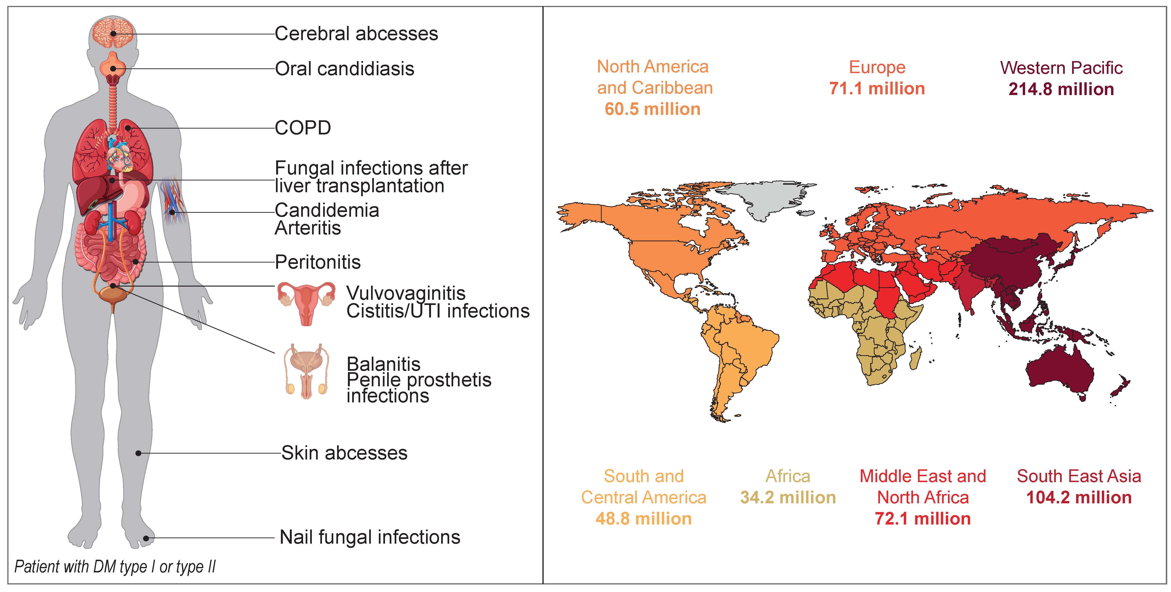
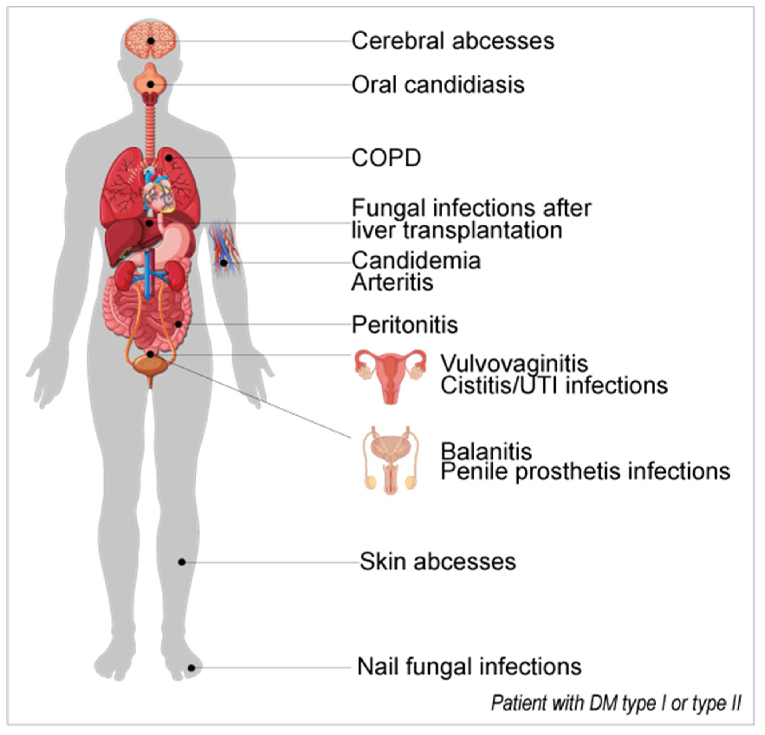
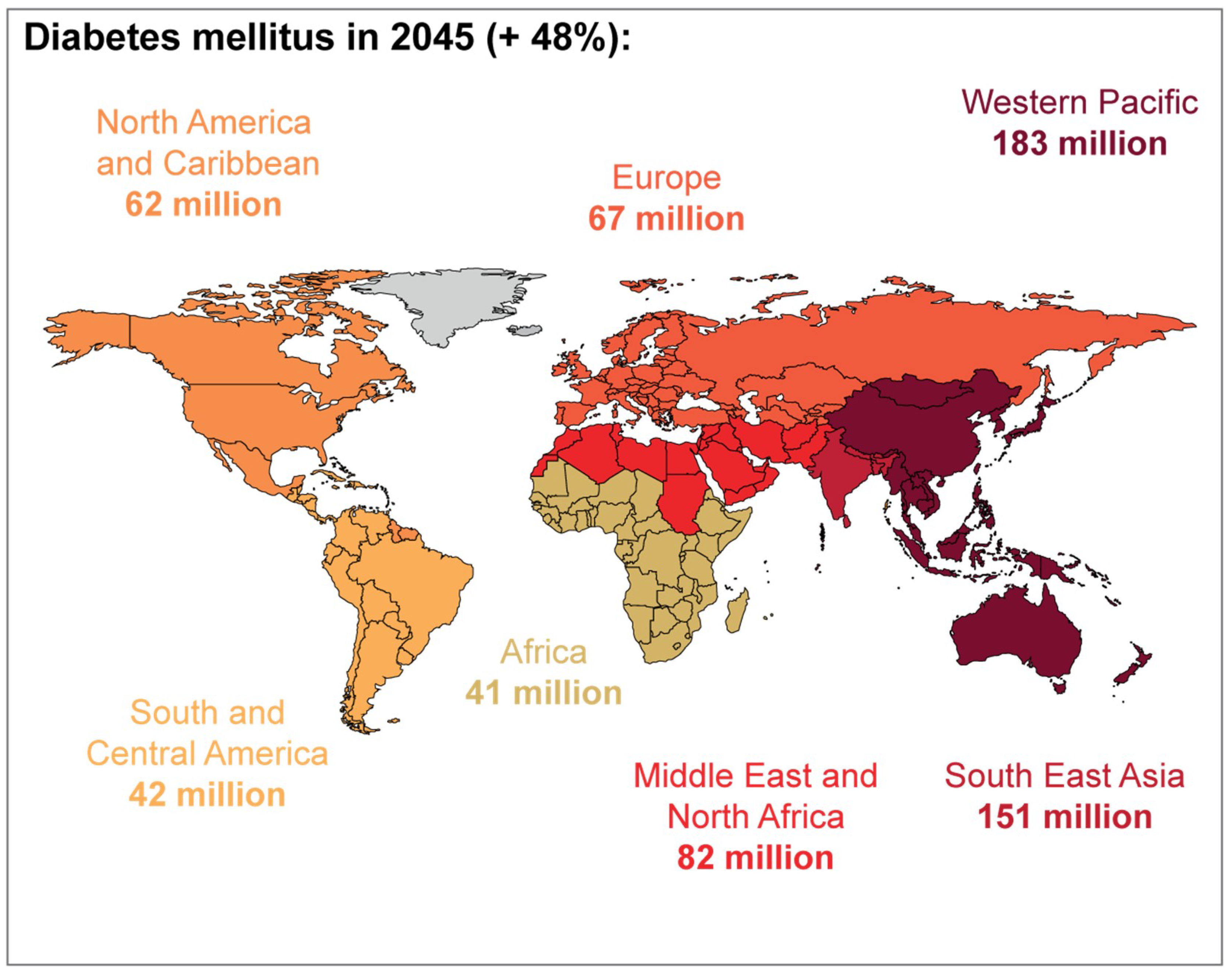
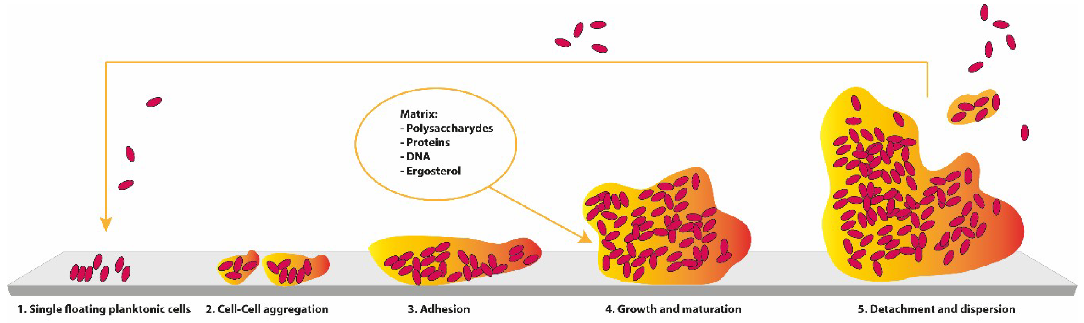
Table 1.
Physiopathology and etiology related to the occurrence of oral candidiasis in diabetics.
| Physiopathology | Reference (s) | |
|---|---|---|
| Uncontrolled hyperglycemia (high HbA1c) and high glucose levels in saliva | -Uncontrolled hyperglycemia may cause intensification in salivary glucose levels because in diabetics the basement membrane of the parotid salivary gland is more permeable -High glucose levels allow Candida sp. to multiply, even in the presence of normal bacterial flora -During hyperglycemic episodes, the chemically reversible glycosylation products with proteins in tissues and the accumulation of glycosylation products on buccal epithelial cells may sequentially increase the number of available receptors for Candida sp. -Glucose suppression of the killing capacity of neutrophils, emphasizing colonization (immunosuppression) -Glucose, maltose, and sucrose boost the adhesion of Candida to buccal epithelial cells | [15,29,31,52,134,140,141,142,143,144,145,146,147] |
| Lower salivary pH | -The growth of Candida in saliva is accompanied by a rapid decline in pH, which favors their growth and triggers the extracellular phospholipase (PL) and acid proteases, increasing the yeast adhesion to oral mucosal surfaces | [31,128,148] |
| Tissue response to injury is diminished | -Diabetes mellitus (DM) is known to diminish the host resistance and modify the tissue response to injury. This can result in severe colonization, even in the absence of any clinically evident oral candidiasis and possibly with further dissemination via the blood. | [130,149] |
| Oral epithelium | -It is most probable that the host oral epithelium of patients with diabetes favors the adhesion of colonization and subsequent infection. | [150,151] |
| Poor oral hygiene | The lack of control of the oral environment, especially concerning the prevention of dental caries (coronary, root, and periodontal), leads to a higher rate of oral candidiasis, especially in DM older patients | [152,153,154] |
| Aging | Diabetic women, orally colonized with Candida sp. have higher oral glucose levels than diabetics without oral Candida sp. | [134] |
| Gender | ||
| Prostheses | Inadequate use of prostheses, together with inadequate hygienization, favours the growth of Candida sp. | [155,156,157] |
| Drugs | Xerostomia (abnomal lack if saliva): Candida sp. stagnation and growth on oral tissues | [30,109,110,111,136,158,159] |
Table 2.
Physiopathology and etiology related to the occurrence of vulvovaginitis in diabetics.
| Condition | Physiopathology | Reference (s) |
|---|---|---|
| Uncontrolled hyperglycemia (high HbA1c) and high glucose levels in vaginal mucosae | - The increased serum glucose level is thought to lead to impaired monocyte, granulocyte, and neutrophil adherence, as well as reduced chemotaxis, phagocytosis, and pathogen killing - Diabetics secretions contain glucose, which can be used as a nutrient by yeasts - pH, nutritional substance, temperature, and adherence capacity in the vulvovaginal tissue may induce Candida sp. virulence. The vaginal epithelial cell receptor of fucose supports the adhesion of Candida sp. to vaginal epithelial cells -The rise in vaginal glucose and secretions and activities of hydrolytic enzymes [e.g., secreted aspartyl proteinases (SAPs), PL] increases the pathogenicity of Candida sp. -Increased levels of glycogen increase colonization and infection by Candida sp. by lowering the vaginal pH, facilitating the development of vulvovaginal candidiasis (VVC) | [55,56,59,219,222,224,225,231,238,239,240,241,242,243,244,245,246,247,248,249,250,251,252,253] |
| Pregnancy | -The increased circulation of estrogen levels and the deposition of glycogen and other substrates in the vagina leads to a 10–50% higher incidence of vaginal colonization by Candida sp. -Variability of constitutive defensins (e.g., lactoferrins, peptides) and lysozyme, leading to a poor innate immune response -Hyperglycemia can rise the anaerobic glycolysis in vaginal epithelial cells, increasing lactic acid and acetone production, decreasing the vaginal pH, thus enabling fungal colonization and proliferation | [241,254,255,256,257,258,259,260] |
| Diabetes type | - The incidence of VVC related to the type 1 DM or type 2 DM and is variable among studies | [218,221,222,223] |
| Aging | - The older one is, the higher VVC prevalence is | [222,223] |
Table 3.
Physiopathology and etiology related to the occurrence of urinary tract infections and systemic candidiasis in diabetics.
Table 3.
Physiopathology and etiology related to the occurrence of urinary tract infections and systemic candidiasis in diabetics.
| Condition | Physiopathology | Reference (s) |
|---|---|---|
| Uncontrolled hyperglycemia (high HbA1c) and high glucose levels in urinary tract (UT) mucosae or blood | - Favorable microenvironment for the gas-forming organisms, such as Candida sp., to grow | [282,283] |
| Gender | -An association between candiduria and being female | [27,273,279] |
| Drugs | -SGLT2 inhibitors (e.g., dapagliflozin, canagliflozin, tofogliflozin) administration leads to a greater susceptibility to urinary tract infection (UTI) -Association with a persistent increase in urine glucose concentration | [287] |
Table 4.
Physiopathology and etiology related to the occurrence of nail fungal diseases linked to Candida sp. in diabetics.
Table 4.
Physiopathology and etiology related to the occurrence of nail fungal diseases linked to Candida sp. in diabetics.
| Condition | Physiopathology | Reference (s) |
|---|---|---|
| Uncontrolled hyperglycemia (high HbA1c) and high glucose levels in vaginal mucosae | -Circulatory disorders affecting the lower extremities (peripheral circulation), peripheral neuropathy, and retinopathy - Nail thickness is associated with an elevated HbA1c value - Diabetics using hemodialysis exhibit a higher probability of onychomycosis | [329,330,334,335,336,337,338] |
| Duration of DM | -More time leads to a higher probability of onychomycosis | [336] |
| Gender Aging | -Being male and being older are directly associated with onychomycosis in diabetics | [331,336] |
Table 5.
Physiopathology and etiology related to the occurrence of other diseases linked to Candida sp. in diabetics.
Table 5.
Physiopathology and etiology related to the occurrence of other diseases linked to Candida sp. in diabetics.
| Condition | Physiopathology | Candida sp. Found in the Study | Reference (s) |
|---|---|---|---|
| Arterial infection | -Direct invasion or haematogenous spread of Candida sp. to the vessels wall (less common): a. infection of a pre-existing aneurysm (most possible from atherosclerotic) with fungi after an episode of candidemia b. contained fungal infection of the arterial wall (linked mostly to intravenous drug abuse). | C. albicans C. tropicalis C. parapsilosis Other Candida sp. (not specified) | [348,349,350,351,352,353,354,355,356,357] |
| Endophthalmitis | - Uncontrolled DM concomitant with other co-morbidities | C. albicans | [358] |
| Cholecystitis | - Uncontrolled DM concomitant with other co-morbidities | C. famata | [359] |
| Skin abscesses | - Uncontrolled DM concomitant with other co-morbidities | C. albicans | [360,361] |
| Brain abscesses | -Immunocompromised states and poorly-controlled diabetes (the role of DM as an immunosuppressive condition in this particular case is uncertain) | C. albicans C. parapsilosis C. tropicalis C. guilliermondi Other Candida sp. (not specified) | [362,363,364] |
| Fungal infections after liver transplantation | - Chronically high blood sugar may be a predisposing factor (among other factors) | C. albicans | [365] |
| Peritonitis | - Uncontrolled DM concomitant with other co-morbidities | C. haemulonii C. glabrata Other Candida sp. (not specified) | [366,367,368] |
| Penile prosthesis Infections | Other Candida sp. (not specified) | [369,370,371,372] | |
| Balanitis | C. albicans | [373] | |
| Fournier’s gangrene | Candida sp. (not specified) | [374] | |
| Chronic obstructive pulmonary disease | C. ciferrii | [375] |
Table 6.
Main animal models used in diabetes mellitus studies [376].
Table 6.
Main animal models used in diabetes mellitus studies [376].
| Diabetes Mellitus | Model/Induction | Studies | Features |
|---|---|---|---|
| Type 1 | Chemical induction | ||
| High dose streptozotocin | New formulations of insulin | Hyperglicaemia model | |
| Alloxan | Transplantation | Induced insulitis | |
| Multiple low dose streptozotocin | Treatment to prevent β-cell death | ||
| Spontaneous autoimmune | |||
| NOD mice | Understanding genetic mechanisms | Autoimmune β-cell destruction | |
| BB rats | |||
| LEW.1AR1/-iddm rats | Treatment to prevent β-cell death or to manipulate autoimmune processes | ||
| Genetically Induced | |||
| AKITA rats* | New formulations of insulin Transplantation Treatment to prevent ER stress | β-cell destruction due to ER + Insulin dependent | |
| Virally induction | |||
| Coxsackie B virus | Study of potencial role of viruses in DM type 1 | Viral β-cell destruction | |
| Encephalomyocarditis virus | |||
| Kilham rat virus | |||
| Lymphocytic choriomeningitis virus (LCMV) under insulin promoter | |||
| Type 2 | Monogenic—obese models | ||
| Obese-induced hyperglycemia | |||
| Lepob/ob mice | Treatment to improve insulin resistance | ||
| Leprdb/db mice | Treatment to improve β-cell resistance | ||
| KDF rats | |||
| Polygenic—obese models | |||
| KK mice | Treatment to insulin resistance | ||
| OLEFT rat | Treatment to improve β-cell function | ||
| NZO mice | Some models show diabetic complications | ||
| TallyHO/Jng mice | |||
| NoncNZO10/|LtJ mice | |||
| Induced obesity | |||
| High fat feeding (mice or rats) | Treatment to insulin resistance | ||
| Nile grass rat | Treatment to prevent diet-induced obesity | ||
| Desert gerbil | Treatment to improve β-cell function | ||
* can be also used as a DM type 2 model.
© 2019 by the authors. Licensee MDPI, Basel, Switzerland. This article is an open access article distributed under the terms and conditions of the Creative Commons Attribution (CC BY) license (http://creativecommons.org/licenses/by/4.0/).
Share and Cite
MDPI and ACS Style
Rodrigues, C.F.; Rodrigues, M.E.; Henriques, M. Candida sp. Infections in Patients with Diabetes Mellitus. J. Clin. Med. 2019, 8, 76. https://0-doi-org.brum.beds.ac.uk/10.3390/jcm8010076
AMA Style
Rodrigues CF, Rodrigues ME, Henriques M. Candida sp. Infections in Patients with Diabetes Mellitus. Journal of Clinical Medicine. 2019; 8(1):76. https://0-doi-org.brum.beds.ac.uk/10.3390/jcm8010076
Chicago/Turabian StyleRodrigues, Célia F., Maria Elisa Rodrigues, and Mariana Henriques. 2019. "Candida sp. Infections in Patients with Diabetes Mellitus" Journal of Clinical Medicine 8, no. 1: 76. https://0-doi-org.brum.beds.ac.uk/10.3390/jcm8010076
Note that from the first issue of 2016, this journal uses article numbers instead of page numbers. See further details here.