Review

The Role of Neutrophils and Neutrophil Extracellular Traps in Vascular Damage in Systemic Lupus Erythematosus

by
Liam J. O’Neil
,
Mariana J. Kaplan
and
Carmelo Carmona-Rivera
*
Systemic Autoimmunity Branch, National Institute of Arthritis and Musculoskeletal and Skin Diseases, National Institutes of Health, Bethesda, MD 20892, USA
*
Author to whom correspondence should be addressed.
Submission received: 25 July 2019 / Revised: 16 August 2019 / Accepted: 25 August 2019 / Published: 28 August 2019

Abstract

:
Systemic lupus erythematosus (SLE) is an autoimmune syndrome of unknown etiology, characterized by multi-organ inflammation and clinical heterogeneity. SLE affects mostly women and is associated with a high risk of cardiovascular disease. As the therapeutic management of SLE improved, a pattern of early atherosclerotic disease became one of the hallmarks of late disease morbidity and mortality. Neutrophils emerged as important players in SLE pathogenesis and they are associated with increased risk of developing atherosclerotic disease and vascular damage. Enhanced neutrophil extracellular trap (NET) formation was linked to vasculopathy in both SLE and non-SLE subjects and may promote enhanced coronary plaque formation and lipoprotein dysregulation. Foundational work provided insight into the complex relationship between NETs and immune and tissue resident cells within the diseased artery. In this review, we highlight the mechanistic link between neutrophils, NETs, and atherosclerosis within the context of both SLE and non-SLE subjects. We aim to identify actionable pathways that will drive future research toward translational therapeutics, with the ultimate goal of preventing early morbidity and mortality in SLE.

1. Introduction

Atherosclerosis, a common cause of morbidity and mortality in the general population [1], continues to be a hotbed of translational and clinical research. Once thought to be simply the result of vascular accumulation of lipids, the understanding of atherosclerosis greatly evolved over time. Inflammation plays a fundamental role in the evolution of this disease process [2]. Consistent with this, patients with various autoimmune diseases have a clear predisposition toward the development of atherosclerosis [3]. This association is particularly striking in systemic lupus erythematosus (SLE), where observational and epidemiologic studies described up to 50-fold increased risk of developing cardiovascular disease compared with non-SLE patients [4,5]. In recent years, neutrophils were implicated in the inflammation observed in the pathogenesis of both athero-embolic disease and SLE pathophysiology [6]. Understanding how neutrophils mediate atherosclerosis may provide an understanding of actionable pathways in both SLE and non-SLE subjects and, ultimately, could contribute to improving patient management and clinical outcomes. In this review, we provide an overview of SLE and atherosclerosis, discuss the role of neutrophils and neutrophil extracellular traps (NETs) in atherosclerosis, and provide SLE-specific evidence of NETs mediating atherosclerotic disease.

2. SLE and Atherosclerosis: Epidemiology

SLE is a systemic autoimmune syndrome with profound disease heterogeneity, characterized by dysregulated innate and adaptive immune responses and the presence of a type 1 interferon signature in a significant number of patients. Individuals affected by SLE also have enhanced cardiovascular risk due to the development of premature atherosclerosis, although the magnitude of the risk reported in the literature varies widely [7]. Indeed, the elevated risk of atherosclerosis in SLE ranges from 10–50-fold, while the risk of myocardial infarction is 5–8-fold. Women with SLE aged 45 to 54 appear to have the greatest fold difference from their healthy counterparts after controlling for traditional risk factors [8,9]. SLE has a bimodal pattern of mortality, and atherosclerosis is likely the most important cause of late death in these patients [10]. Similar to most associative findings in SLE, disease heterogeneity clouds definitive conclusions from well-designed epidemiological studies, and atherosclerosis is no exception. Disease activity [11], disease duration [12], clinical damage scores [13], and renal disease [14] are all linked to the development of atherosclerosis. Traditional Framingham cardiac risk factors (hypertension, dyslipidemia, smoking, etc.) are all associated with atherosclerosis in SLE, but cannot fully explain the increased risk. Indeed, immune dysregulation characteristic of SLE appears to play a key role in driving premature atherogenesis. What remains evident is that atherosclerosis represents an important factor for SLE morbidity and mortality, and understanding the inter-relations between atherosclerosis, autoimmunity, and systemic inflammation may uncover key actionable pathways for improvement in patient outcomes.

Lupus-Specific Mechanisms of Enhanced Atherosclerosis

Standardized prediction models underestimate the incidence of cardiovascular events in SLE patients, suggesting that there are unique pathways that contribute to the development of early atherosclerosis. Dysfunctional high-density lipoprotein (HDL), a proinflammatory biomarker for atherosclerosis, is elevated in SLE patients [15]. SLE patients also have antibodies against apolipoprotein A1 (ApoA1) and HDL, which may neutralize their atheroprotective effects [16]. Invariant natural killer T cells (iNKTs) respond to lipid antigens and modulate the inflammatory response observed in autoimmune atherosclerosis [17]. SLE patients with atherosclerosis have increased numbers of iNKT cells which likely have a protective role. However, iNKT functionality is altered in patients who transition from subclinical to overt cardiovascular disease—suggesting their role in the establishment of clinical disease [18]. Elevated levels of homocysteine may also play an important role in SLE-related premature atherosclerosis. Homocysteine levels are increased in lupus [19], which has adverse effects on smooth muscle cells and platelet activation [20]. Lupus is strongly associated with the expression of type 1 interferon-regulated genes [21], and this pathway likely also contributes to atherosclerotic risk [22]. Type 1 interferon modulates macrophages and cytotoxic T cells to promote atherosclerotic plaque formation. Finally, a clear reduction in atheroprotective factors in SLE, such as endothelial progenitor cells [23] and immunoglobulin M (IgM) [24], also likely plays an important role in the development of disease.

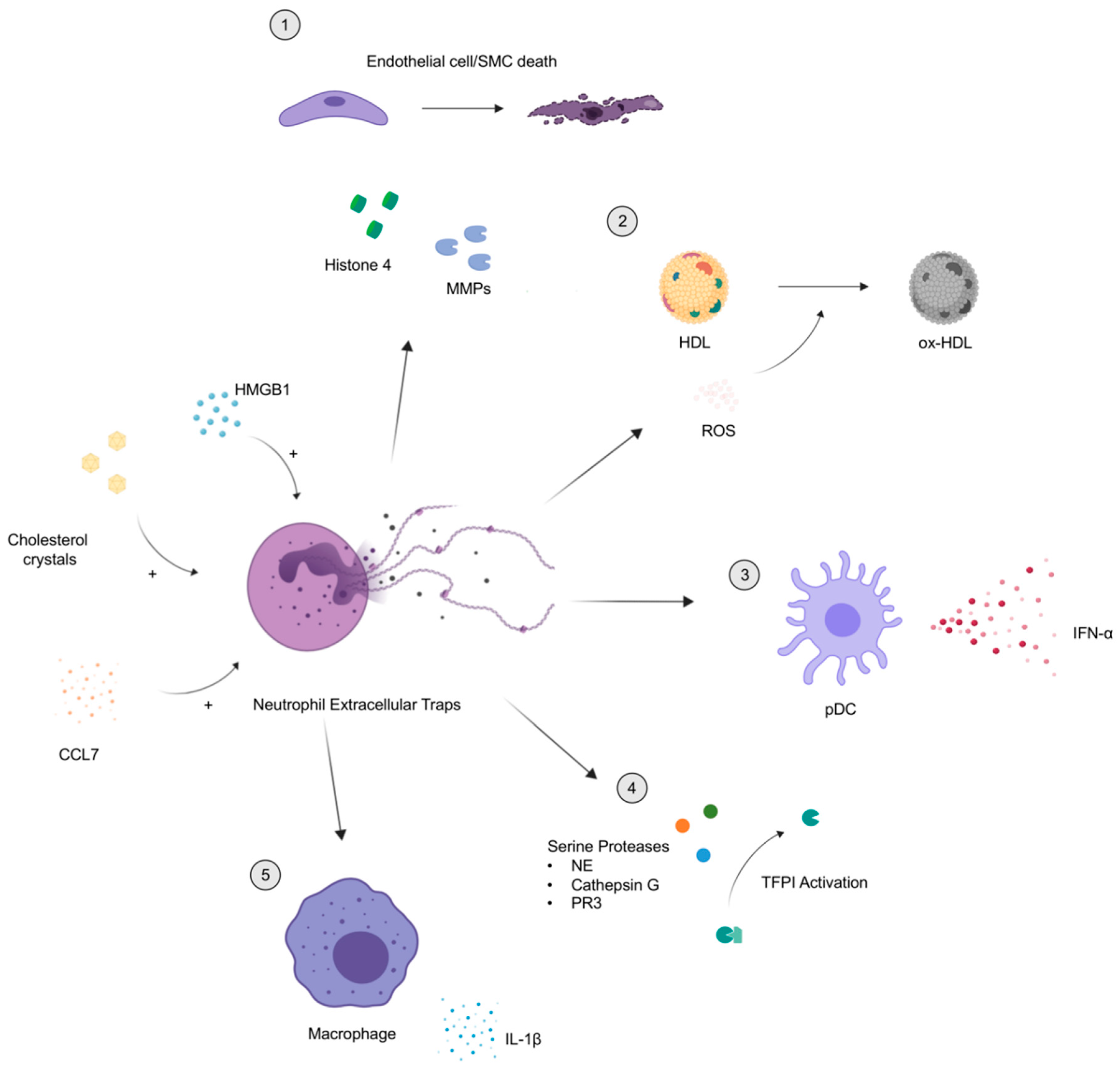
3. Inflammation, Neutrophil Extracellular Traps (NETs), and Atherosclerosis

The landmark study associating systemic elevation of C-reactive protein (CRP) and the development of cardiovascular disease was the first major insight into the importance of inflammation in atherosclerosis [25]. Replication of these findings [26], and the clinical efficacy of canakinumab, a monoclonal antibody therapy that neutralizes the pro-inflammatory cytokine interleukin (IL)-1β [27], provided clarity on the important role that inflammation plays in the development of atherosclerosis. However, the complex pathophysiology of atherosclerosis and the heterogeneity of patient presentation made identifying other inflammatory markers a more difficult task. It appears that both humoral and cellular immunity contribute in meaningful ways to atherogenesis at different stages of this process [28]. Given the abundance of neutrophils and their armamentarium of cytotoxic mediators, it is clear that they have the capacity to substantially harm tissue and promote inflammatory responses in a diseased artery [29]. Indeed, research over the last decade implicated neutrophils in the development of atherosclerosis [30].
In humans, neutrophils are the most abundant leukocyte in circulation. These sentinel cells are short-lived (although they may live longer in inflammatory environments [31]), and they are well equipped to kill infiltrating pathogens [32]. They possess a broad spectrum of cytotoxic mediators and respond to invading microbes using phagocytosis, generation of reactive oxygen species, and degranulation [33]. A unique feature of neutrophils is that they synthesize their granules prior to their release from the bone marrow into circulation [34]. This allows a rapid and robust response to pathogens without the need for transcriptional machinery. Perhaps the most distinctive cytotoxic function of neutrophils is their ability to undergo a distinct form of cell death that leads to the release of neutrophil extracellular traps (NETs). Vital NETosis is an alternate form of DNA release that does not result in cell death, whereby a neutrophil maintains its phagocytic function despite compromise of its cellular membrane [35]. During most, but not all forms of NET formation, peptidylarginine deiminase-4 (PAD4) enzymatically converts arginine into citrulline on histone tails, promoting chromatin decondensation in the nucleus [36]. Neutrophil elastase (NE) and myeloperoxidase (MPO) are translocated into the nucleus and further promote chromatin release and nuclear membrane dissolution. In later stages, the plasma membrane is disrupted, and chromatin decorated with granule proteins is released in web-like structures to the extracellular space. It is thought that chromatin is used by the host immune system to entrap microbes and expose them to cytotoxic compounds such as proteases [37]. Importantly, during some forms of NET formation, mitochondrial DNA may also be incorporated into these structures, which adds to their pathogen-fighting abilities [38].
Dysregulated NET formation was recently implicated in a variety of inflammatory conditions including autoimmunity, autoinflammation, thrombosis, malignancy, and sepsis [39,40]. Intriguingly, NETs are proposed to play a fundamental role in SLE pathophysiology and to be a primary driver of increased vasculopathy in these patients. Enhanced NET formation and impaired NET clearance are described in peripheral blood and various tissues as hallmarks of SLE [41]. Reduced NET clearance is partially explained by impaired DNAse-1 activity [41], the key enzyme for NET degradation. Neutrophils respond to, and produce [42] type-1 interferon, a key cytokine in SLE pathogenesis. Furthermore, subsets of lupus neutrophils readily undergo enhanced spontaneous NET formation [43]. NETs may also have immunoregulatory properties; for example, NETs may reduce the release of proinflammatory cytokines in macrophages [44]. Unraveling the connection between SLE, NET formation, and atherosclerosis may allow for the discovery of new, actionable pathways that are relevant to not only SLE, but also to the general population.

4. Neutrophils Facilitate Atherosclerosis Development and Progression

The process of atherosclerosis generally evolves over many years, and its initiation is a quiescent, subclinical event. Selectins are proteins which allow circulating neutrophils to slow, roll, and tether to the endothelium [45]. These cell adhesion molecules are expressed by endothelial cells as the initiating event in atherosclerosis, particularly in an environment induced by dyslipidemia. Indeed, in mice that are fed high-fat diets, neutrophil accumulation within the arteries [46] is a very early event. C–C motif chemokine ligand 5 (CCL5) allows neutrophils to firmly attach to the endothelium, where it begins to promote pathogenic changes to initiate atherogenesis. Granule proteins and radicals promote endothelial dysfunction [47] and monocyte recruitment [48], as well as increase vascular permeability [49]. The recruitment of monocytes is of particular importance, as these cells provide the precursors to endothelial macrophages, which leads to the development of proinflammatory M1 cells and foam cells [50,51,52], the latter of which are crucial for the development of plaque. Finally, plaques that form over several years to decades have the propensity to rupture or destabilize, resulting in thrombus formation. Neutrophils express a variety of proteases such as matrix metalloproteinases or serine proteases. These enzymes appear to be important in the development of intraplaque hemorrhage [53] and degradation of the fibrous cap [54].
Activated neutrophils are the predominant cell type found within the thrombus of patients having a coronary event [55]. Furthermore, NETs within the thrombus significantly correlate with cardiac infarct size and, inversely, with electrocardiogram (EKG) ST-segment resolution. These findings implicate NETs in the clinical severity of a coronary lesion. Thrombus specimens following percutaneous coronary intervention were found to be infiltrated with neutrophils, with a subset of lesions containing granule–DNA complexes, suggestive of NETs [56]. Moreover, granule–DNA complexes are also found in coronary plaque erosions and intra-plaque hemorrhages, and they are not only detectable in the interstitial adventitia but also in the lumen of microvessels [57]. NETs were also found shown in monocyte-depleted Lysmegfp/egfp atherosclerosis-prone apolipoprotein-E−/− mice within the carotid bifurcation using intravital microscopy [58]. High-mobility group box 1 (HMGB1) synthesized by activated platelets was described as an inducer of NET formation by coronary artery neutrophils (Figure 1) [59]. Cholesterol crystals, known to play an important role in plaque formation, also induce NET formation. The interplay between NETs, cholesterol crystals, and macrophages can promote pro-inflammatory cytokine release such as IL-1β [60]. NETs isolated from coronary thrombectomy specimens also appear to be coated with IL-17A and IL-17F, which may contribute to inflammation, platelet aggregation, and thrombus expansion [61].

4.1. The Role of Serine Proteases

Apolipoprotein-E−/− and the low-density lipoprotein (LDL)−/− mice are important models for studying the pathogenesis of atherosclerosis. Deletion of neutrophil elastase (NE) and proteinase-3 (PR3) in the apolipoprotein-E−/− mouse leads to reduced NET formation, along with a reduction in atherosclerotic lesions and systemic inflammation [60]. In the same mouse model, NETs within atherosclerotic plaques can stimulate plasmacytoid dendritic cells (pDCs) to produce type 1 interferons and contribute to the development of plaque [65]. Indeed, depletion of pDCs leads to a substantial reduction in plaque formation in this model, highlighting an important role for type 1 interferons in plaque formation. A triple knockout mouse model (ApoE/PR3/NE KO) displays substantially reduced atherosclerotic plaque formation as well, a finding that appears to be at least in part mediated by the intact DNA within NETs. In a separate study, loss of NE alone did not affect lesional formation without the concurrent loss of PR3 [67]. Neutrophil serine proteases (NE and cathepsin G), found in high quantity in NETs [68], were described as important mediators of coagulation. In a coagulation murine model, elastase (ELANE)−/−/Cathepsin G (CTSG)−/− mice have impaired coagulation when compared to wild-type mice, with more fragile and smaller thrombi, along with heightened bleeding times [66]. NE was described as a predominant protease in clotting mechanisms, with near complete recovery of normal phenotype in this mouse model after injection of recombinant NE. NE cleaves tissue factor pathway inhibitor (TFPI), a key anticoagulant found in platelets. NETs are required for the co-localization of TFPI and NE, as this effect is ameliorated by treatment with DNase-1, a nuclease that disrupts NET architecture, both in vitro and in vivo.

4.2. Cellular Toxicity by Histone H4 and Matrix Metalloproteinases

NETs may promote arterial dysfunction by directly interacting with endothelial cells. In ApoE−/− mice, neutrophil depletion resulted in increased smooth muscle cells (SMC), while neutrophilic mice generated by C–X–C motif chemokine receptor 4 (CXCR4) deletion had heightened SMC death [62], suggesting that neutrophils regulate SMC survival. In this study, authors also reported that C–C motif chemokine ligand 7 (CCL7) synthesized by SMC enhances NET formation that then triggers SMC cell death. Histone H4 contained in NETs is a key mediator in the induction of SMC cell death through the formation of cell membrane pores and membrane destabilization. The use of histone inhibitory protein (HIP) in mice with pre-existing atherosclerotic lesions increased the stability of these lesions without altering neutrophil recruitment. In conjunction with this mechanism, matrix metalloproteinases (MMPs, specifically MMP9) within NETs induce endothelial cell cytotoxicity and promote dysfunctional vascular relaxation [63]. Importantly, this is enhanced when assessing NETs derived from lupus neutrophils. Histones can also induce endothelial dysfunction [69]; however, whether histone modification alters the capacity to damage the endothelium remains to be systematically examined. Whether or not MMPs can promote SMC death remains to be determined, but there may be synergism between NET molecules that drives artery-specific cellular toxicity.

4.3. PAD-4, a Potential Target for Therapy

PAD4, one of the enzymes that mediates the citrullination of arginine, appears to play an important role in NET formation following certain types of stimulation [70]. Although the role of PAD4 in the clinical and immunological manifestations of murine SLE is debated [71,72], studies investigating the role of this molecule in NET formation and atherosclerosis are much clearer. Inhibition of PAD4 by the pan-PAD inhibitor Cl-amidine in ApoE−/− mice significantly reduced atherosclerotic lesion size and prolonged time to thrombosis in a photochemical injury model [73]. This model confirmed enhanced systemic NET formation in murine atherosclerosis and a type 1 interferon response within the aortic tissue, all of which were mitigated by Cl-amidine therapy. These findings were recapitulated in a PAD4−/− myocardial infarction mouse model, which displayed improved ejection fraction percentage and reduced infarct size compared to wild-type animals, with no additional benefit of DNase I treatment. Furthermore, obesity-induced endothelial dysfunction is also ameliorated by Cl-amidine [74]. PAD4 is also responsible for promoting von Willebrand factor–platelet string formation, which accelerates thrombosis upon endothelial injury in mice [75]. In human carotid specimens, NETs were shown to be more closely associated with erosion-prone rather than rupture-prone pathology. Indeed, there is clear evidence of neutrophil infiltration and NET formation in mice exposed to flow perturbation at sites of intimal expansion [76]. Deficiency of PAD4 in a murine model of endothelial injury revealed reduced NET formation, preservation of endothelial continuity, and smaller intraluminal thrombus. Indeed, protection through a myeloid-specific deletion of PAD4 confirms the importance of NET formation in murine atherosclerosis [77]. Although some debate exists over the role of NET formation in modulating SLE murine manifestations [71,72], PAD4 inhibition in humans could have a variety of consequential effects. Nonetheless, PAD4 may represent an important drug target via which NET-induced atherogenesis could be significantly altered.

4.4. Mitochondrial DNA and Reactive Oxygen Species

Mitochondrial DNA and oxidative stress may play an important role in the pathogenic effect of NETs in atherosclerotic disease [78]. There is correlation between atherosclerosis, aging, and mitochondrial ROS generation [79]. Bone marrow transplantation from mitochondrial catalase transgenic mice into low density lipoprotein receptor (Ldlr)−/− mice resulted in reduced lesional mitochondrial oxidative stress, decreased NET formation, and protection from plaque development [80]. This observation was not apparent in younger mice, but rather in an aging mouse model of atherosclerosis. Whether these differential findings in varying age groups occur in humans remains to be determined.
High-density lipoprotein (HDL)-mediated cholesterol efflux is an important factor that protects from the generation of foam cells and atherosclerosis in the general population and in autoimmunity [81]. Nicotinamide adenine dinucleotide phosphate hydrogen (NADPH) oxidase (NOX) and nitric oxide synthase activity within NETs can induce nitrogen and reactive oxygen species in the extracellular space [82]. NETs can mediate HDL oxidation, which drives mishandling of lipoprotein homeostasis, transforming HDL into a proatherogenic, proinflammatory form that can promote proinflammatory responses in macrophages [64]. Low-density granulocytes (LDGs), a subset of neutrophils found in high abundance in SLE patients [6], may also play an important role in atherogenesis. LDGs have a propensity to form NETs spontaneously in vitro (Figure 2). These NETs also have a higher degree of mitochondrial ROS and oxidized mitochondrial DNA (mtDNA) compared to their normal dense counterparts [38]. Oxidized mitochondrial DNA released from neutrophils can drive type 1 interferon in lupus [38,83]. Furthermore, LDG gene signatures correlate with non-calcified plaque burden and vascular inflammation in SLE patients [84]. Similar findings were also shown in psoriasis, another systemic autoimmune disease [85]. Thus, there may be a link between LDG NETs, mitochondrial ROS, and lipoprotein dysfunction that drives autoimmune-specific heightened atherosclerosis.

4.5. NETs as a Biomarker for Subclinical Atherosclerosis

Despite a clear role for NETs in the pathogenesis of atherosclerosis, the utility of cell-free DNA and related biomarkers still remains relatively understudied. In a cohort of patients with coronary artery disease, extracellular double-stranded DNA (dsDNA), nucleosomes, and MPO–DNA complexes are higher in patients with severe coronary artery disease (CAD) compared to those without CAD. These parameters also correlated with the number of calcified plaques, in vitro thrombin generation, and future major cardiac events [86]. Despite NET complexes being a putative relevant biomarker in SLE [63], it is unclear if these correlate with measures of subclinical atherosclerosis or patient-related outcomes. Furthermore, there remain methodological inconsistencies by which NETs are measured from human specimens. NET proteins bound to DNA (citrullinated histones, MPO, and NE most commonly) are used as a surrogate for measuring NET formation. More broadly, circulating nucleosomes and cell-free DNA are much less specific, as these remnants could be derived from a variety of sources.

5. Concluding Remarks

Recent studies emphasized the role of a pathogenic interplay between neutrophils and the type 1 IFN response as important drivers of atherosclerosis and vasculopathy in SLE. While the potential for intervening in these pathways remains an intriguing concept, much work is needed to translate these findings into clinical practice. The role of PAD inhibition in atherosclerosis is an attractive approach but needs to be better investigated in pre-clinical models before being investigated in human trials. Canakinumab therapy reduces athero-embolic disease in non-SLE patients; however, blocking this cytokine is not routine therapy in SLE and may have deleterious consequences given that vasculogenesis in SLE is impaired [87]. Furthermore, CRP, an important inclusion factor for patients in the representative clinical trial, is not a known biomarker of SLE inflammation, and CRP responses may be decreased in this disease due to type 1 IFN effects [88]. Inhibiting the toxic and potentially synergistic effects of histones and MMPs on structural arterial cells may also be an important pathway to target. Future studies will hopefully shed light on this important question in the context of both SLE and non-SLE atherosclerosis. Ultimately, research in SLE atherosclerosis remains in its infancy. There appears to be reason for optimism, as research highlighted in this review suggests there was important progress in the understanding of the pathogenesis of lupus vasculopathy. Carrying this momentum toward translatable clinical solutions remains a lofty, but attainable goal.

Author Contributions

Conceptualization, L.J.O., M.J.K., and C.C.-R.; writing—review and editing, L.J.O., M.J.K., and C.C.-R.; supervision, C.C.-R.

Funding

This work was supported by the Intramural Research Program, NIAMS/NIH, ZIA AR041199.

Conflicts of Interest

The authors declare no conflicts of interest.

References

  1. Herrington, W.; Lacey, B.; Sherliker, P.; Armitage, J.; Lewington, S. Epidemiology of Atherosclerosis and the Potential to Reduce the Global Burden of Atherothrombotic Disease. Circ. Res. 2016, 118, 535–546. [Google Scholar] [CrossRef] [PubMed]
  2. Gistera, A.; Hansson, G.K. The immunology of atherosclerosis. Nat. Rev. Nephrol. 2017, 13, 368–380. [Google Scholar] [CrossRef] [PubMed]
  3. Sima, P.; Vannucci, L.; Vetvicka, V. Atherosclerosis as autoimmune disease. Ann. Transl. Med. 2018, 6, 116. [Google Scholar] [CrossRef] [PubMed]
  4. Frieri, M.; Stampfl, H. Systemic lupus erythematosus and atherosclerosis: Review of the literature. Autoimmun. Rev. 2016, 15, 16–21. [Google Scholar] [CrossRef] [PubMed]
  5. Turano, L. Premature atherosclerotic cardiovascular disease in systemic lupus erythematosus: Understanding management strategies. J. Cardiovasc. Nurs. 2013, 28, 48–53. [Google Scholar] [CrossRef]
  6. Kaplan, M.J. Neutrophils in the pathogenesis and manifestations of SLE. Nat. Rev. Rheumatol. 2011, 7, 691–699. [Google Scholar] [CrossRef] [PubMed] [Green Version]
  7. Gustafsson, J.T.; Herlitz Lindberg, M.; Gunnarsson, I.; Pettersson, S.; Elvin, K.; Ohrvik, J.; Larsson, A.; Jensen-Urstad, K.; Svenungsson, E. Excess atherosclerosis in systemic lupus erythematosus,-A matter of renal involvement: Case control study of 281 SLE patients and 281 individually matched population controls. PLoS ONE 2017, 12, e0174572. [Google Scholar] [CrossRef]
  8. Kiani, A.N.; Magder, L.S.; Post, W.S.; Szklo, M.; Bathon, J.M.; Schreiner, P.J.; O’Leary, D.; Petri, M. Coronary calcification in SLE: Comparison with the Multi-Ethnic Study of Atherosclerosis. Rheumatol. (Oxf.) 2015, 54, 1976–1981. [Google Scholar] [CrossRef]
  9. Manzi, S.; Meilahn, E.N.; Rairie, J.E.; Conte, C.G.; Medsger, T.A., Jr.; Jansen-McWilliams, L.; D’Agostino, R.B.; Kuller, L.H. Age-specific incidence rates of myocardial infarction and angina in women with systemic lupus erythematosus: Comparison with the Framingham Study. Am. J. Epidemiol. 1997, 145, 408–415. [Google Scholar] [CrossRef]
  10. Rubin, L.A.; Urowitz, M.B.; Gladman, D.D. Mortality in systemic lupus erythematosus: The bimodal pattern revisited. Q. J. Med. 1985, 55, 87–98. [Google Scholar]
  11. Bengtsson, C.; Ohman, M.L.; Nived, O.; Rantapaa Dahlqvist, S. Cardiovascular event in systemic lupus erythematosus in northern Sweden: Incidence and predictors in a 7-year follow-up study. Lupus 2012, 21, 452–459. [Google Scholar] [CrossRef] [PubMed]
  12. Manzi, S.; Selzer, F.; Sutton-Tyrrell, K.; Fitzgerald, S.G.; Rairie, J.E.; Tracy, R.P.; Kuller, L.H. Prevalence and risk factors of carotid plaque in women with systemic lupus erythematosus. Arthritis Rheum 1999, 42, 51–60. [Google Scholar] [CrossRef]
  13. Haque, S.; Gordon, C.; Isenberg, D.; Rahman, A.; Lanyon, P.; Bell, A.; Emery, P.; McHugh, N.; Teh, L.S.; Scott, D.G.; et al. Risk factors for clinical coronary heart disease in systemic lupus erythematosus: The lupus and atherosclerosis evaluation of risk (LASER) study. J. Rheumatol. 2010, 37, 322–329. [Google Scholar] [CrossRef] [PubMed]
  14. Sule, S.; Fivush, B.; Neu, A.; Furth, S. Increased risk of death in pediatric and adult patients with ESRD secondary to lupus. Pediatr. Nephrol. 2011, 26, 93–98. [Google Scholar] [CrossRef] [PubMed]
  15. McMahon, M.; Grossman, J.; FitzGerald, J.; Dahlin-Lee, E.; Wallace, D.J.; Thong, B.Y.; Badsha, H.; Kalunian, K.; Charles, C.; Navab, M.; et al. Proinflammatory high-density lipoprotein as a biomarker for atherosclerosis in patients with systemic lupus erythematosus and rheumatoid arthritis. Arthritis Rheum. 2006, 54, 2541–2549. [Google Scholar] [CrossRef] [PubMed]
  16. O’Neill, S.G.; Giles, I.; Lambrianides, A.; Manson, J.; D’Cruz, D.; Schrieber, L.; March, L.M.; Latchman, D.S.; Isenberg, D.A.; Rahman, A. Antibodies to apolipoprotein A-I, high-density lipoprotein, and C-reactive protein are associated with disease activity in patients with systemic lupus erythematosus. Arthritis Rheum. 2010, 62, 845–854. [Google Scholar] [CrossRef] [PubMed]
  17. Tupin, E.; Nicoletti, A.; Elhage, R.; Rudling, M.; Ljunggren, H.G.; Hansson, G.K.; Berne, G.P. CD1d-dependent activation of NKT cells aggravates atherosclerosis. J. Exp. Med. 2004, 199, 417–422. [Google Scholar] [CrossRef]
  18. Smith, E.; Croca, S.; Waddington, K.E.; Sofat, R.; Griffin, M.; Nicolaides, A.; Isenberg, D.A.; Torra, I.P.; Rahman, A.; Jury, E.C. Cross-talk between iNKT cells and monocytes triggers an atheroprotective immune response in SLE patients with asymptomatic plaque. Sci. Immunol. 2016, 1. [Google Scholar] [CrossRef]
  19. Bonciani, D.; Antiga, E.; Bonciolini, V.; Verdelli, A.; Del Bianco, E.; Volpi, W.; Caproni, M. Homocysteine serum levels are increased and correlate with disease severity in patients with lupus erythematosus. Clin. Exp. Rheumatol. 2016, 34, 76–81. [Google Scholar]
  20. Clarke, R.; Daly, L.; Robinson, K.; Naughten, E.; Cahalane, S.; Fowler, B.; Graham, I. Hyperhomocysteinemia: An independent risk factor for vascular disease. N. Engl. J. Med. 1991, 324, 1149–1155. [Google Scholar] [CrossRef]
  21. Crow, M.K. Type I interferon in the pathogenesis of lupus. J. Immunol. 2014, 192, 5459–5468. [Google Scholar] [CrossRef] [PubMed]
  22. Lood, C.; Amisten, S.; Gullstrand, B.; Jonsen, A.; Allhorn, M.; Truedsson, L.; Sturfelt, G.; Erlinge, D.; Bengtsson, A.A. Platelet transcriptional profile and protein expression in patients with systemic lupus erythematosus: Up-regulation of the type I interferon system is strongly associated with vascular disease. Blood 2010, 116, 1951–1957. [Google Scholar] [CrossRef] [PubMed]
  23. Schmidt-Lucke, C.; Rossig, L.; Fichtlscherer, S.; Vasa, M.; Britten, M.; Kamper, U.; Dimmeler, S.; Zeiher, A.M. Reduced number of circulating endothelial progenitor cells predicts future cardiovascular events: Proof of concept for the clinical importance of endogenous vascular repair. Circulation 2005, 111, 2981–2987. [Google Scholar] [CrossRef] [PubMed]
  24. Anania, C.; Gustafsson, T.; Hua, X.; Su, J.; Vikstrom, M.; de Faire, U.; Heimburger, M.; Jogestrand, T.; Frostegard, J. Increased prevalence of vulnerable atherosclerotic plaques and low levels of natural IgM antibodies against phosphorylcholine in patients with systemic lupus erythematosus. Arthritis Res. 2010, 12, R214. [Google Scholar] [CrossRef] [PubMed]
  25. Ridker, P.M.; Cushman, M.; Stampfer, M.J.; Tracy, R.P.; Hennekens, C.H. Inflammation, aspirin, and the risk of cardiovascular disease in apparently healthy men. N. Engl. J. Med. 1997, 336, 973–979. [Google Scholar] [CrossRef] [PubMed]
  26. Pradhan, A.D.; Manson, J.E.; Rossouw, J.E.; Siscovick, D.S.; Mouton, C.P.; Rifai, N.; Wallace, R.B.; Jackson, R.D.; Pettinger, M.B.; Ridker, P.M. Inflammatory biomarkers, hormone replacement therapy, and incident coronary heart disease: Prospective analysis from the Women’s Health Initiative observational study. JAMA 2002, 288, 980–987. [Google Scholar] [CrossRef]
  27. Ridker, P.M.; Everett, B.M.; Thuren, T.; MacFadyen, J.G.; Chang, W.H.; Ballantyne, C.; Fonseca, F.; Nicolau, J.; Koenig, W.; Anker, S.D.; et al. Antiinflammatory Therapy with Canakinumab for Atherosclerotic Disease. N. Engl. J. Med. 2017, 377, 1119–1131. [Google Scholar] [CrossRef]
  28. Taleb, S. Inflammation in atherosclerosis. Arch. Cardiovasc. Dis. 2016, 109, 708–715. [Google Scholar] [CrossRef]
  29. Moreno, J.A.; Ortega-Gomez, A.; Delbosc, S.; Beaufort, N.; Sorbets, E.; Louedec, L.; Esposito-Farese, M.; Tubach, F.; Nicoletti, A.; Steg, P.G.; et al. In vitro and in vivo evidence for the role of elastase shedding of CD163 in human atherothrombosis. Eur. Heart J. 2012, 33, 252–263. [Google Scholar] [CrossRef]
  30. Doring, Y.; Drechsler, M.; Soehnlein, O.; Weber, C. Neutrophils in atherosclerosis: From mice to man. Arter. Thromb. Vasc. Biol. 2015, 35, 288–295. [Google Scholar] [CrossRef]
  31. Pillay, J.; den Braber, I.; Vrisekoop, N.; Kwast, L.M.; de Boer, R.J.; Borghans, J.A.; Tesselaar, K.; Koenderman, L. In vivo labeling with 2H2O reveals a human neutrophil lifespan of 5.4 days. Blood 2010, 116, 625–627. [Google Scholar] [CrossRef] [PubMed]
  32. Kolaczkowska, E.; Kubes, P. Neutrophil recruitment and function in health and inflammation. Nat. Rev. Immunol. 2013, 13, 159–175. [Google Scholar] [CrossRef] [PubMed]
  33. O’Neil, L.J.; Kaplan, M.J. Neutrophils in Rheumatoid Arthritis: Breaking Immune Tolerance and Fueling Disease. Trends Mol. Med. 2019, 25, 215–227. [Google Scholar] [CrossRef]
  34. Borregaard, N.; Cowland, J.B. Granules of the human neutrophilic polymorphonuclear leukocyte. Blood 1997, 89, 3503–3521. [Google Scholar] [PubMed]
  35. Yipp, B.G.; Kubes, P. NETosis: How vital is it? Blood 2013, 122, 2784–2794. [Google Scholar] [CrossRef] [PubMed]
  36. Papayannopoulos, V. Neutrophil extracellular traps in immunity and disease. Nat. Rev. Immunol. 2018, 18, 134–147. [Google Scholar] [CrossRef] [PubMed]
  37. Lightfoot, Y.L.; Kaplan, M.J. Disentangling the role of neutrophil extracellular traps in rheumatic diseases. Curr. Opin. Rheumatol. 2017, 29, 65–70. [Google Scholar] [CrossRef]
  38. Lood, C.; Blanco, L.P.; Purmalek, M.M.; Carmona-Rivera, C.; de Ravin, S.S.; Smith, C.K.; Malech, H.L.; Ledbetter, J.A.; Elkon, K.B.; Kaplan, M.J. Neutrophil extracellular traps enriched in oxidized mitochondrial DNA are interferogenic and contribute to lupus-like disease. Nat. Med. 2016, 22, 146–153. [Google Scholar] [CrossRef] [Green Version]
  39. Boeltz, S.; Amini, P.; Anders, H.J.; Andrade, F.; Bilyy, R.; Chatfield, S.; Cichon, I.; Clancy, D.M.; Desai, J.; Dumych, T.; et al. To NET or not to NET:current opinions and state of the science regarding the formation of neutrophil extracellular traps. Cell Death Differ. 2019, 26, 395–408. [Google Scholar] [CrossRef] [Green Version]
  40. Jorch, S.K.; Kubes, P. An emerging role for neutrophil extracellular traps in noninfectious disease. Nat. Med. 2017, 23, 279–287. [Google Scholar] [CrossRef]
  41. Hakkim, A.; Furnrohr, B.G.; Amann, K.; Laube, B.; Abed, U.A.; Brinkmann, V.; Herrmann, M.; Voll, R.E.; Zychlinsky, A. Impairment of neutrophil extracellular trap degradation is associated with lupus nephritis. Proc. Natl. Acad. Sci. USA 2010, 107, 9813–9818. [Google Scholar] [CrossRef] [PubMed] [Green Version]
  42. Lindau, D.; Mussard, J.; Rabsteyn, A.; Ribon, M.; Kotter, I.; Igney, A.; Adema, G.J.; Boissier, M.C.; Rammensee, H.G.; Decker, P. TLR9 independent interferon alpha production by neutrophils on NETosis in response to circulating chromatin, a key lupus autoantigen. Ann. Rheum. Dis. 2014, 73, 2199–2207. [Google Scholar] [CrossRef] [PubMed]
  43. Villanueva, E.; Yalavarthi, S.; Berthier, C.C.; Hodgin, J.B.; Khandpur, R.; Lin, A.M.; Rubin, C.J.; Zhao, W.; Olsen, S.H.; Klinker, M.; et al. Netting neutrophils induce endothelial damage, infiltrate tissues, and expose immunostimulatory molecules in systemic lupus erythematosus. J. Immunol. 2011, 187, 538–552. [Google Scholar] [CrossRef] [PubMed]
  44. Ribon, M.; Seninet, S.; Mussard, J.; Sebbag, M.; Clavel, C.; Serre, G.; Boissier, M.C.; Semerano, L.; Decker, P. Neutrophil extracellular traps exert both pro- and anti-inflammatory actions in rheumatoid arthritis that are modulated by C1q and LL-37. J. Autoimmun. 2019, 98, 122–131. [Google Scholar] [CrossRef] [PubMed]
  45. Weil, B.R.; Neelamegham, S. Selectins and Immune Cells in Acute Myocardial Infarction and Post-infarction Ventricular Remodeling: Pathophysiology and Novel Treatments. Front. Immunol. 2019, 10, 300. [Google Scholar] [CrossRef] [PubMed] [Green Version]
  46. Drechsler, M.; Megens, R.T.; van Zandvoort, M.; Weber, C.; Soehnlein, O. Hyperlipidemia-triggered neutrophilia promotes early atherosclerosis. Circulation 2010, 122, 1837–1845. [Google Scholar] [CrossRef] [PubMed]
  47. Jacobi, J.; Sela, S.; Cohen, H.I.; Chezar, J.; Kristal, B. Priming of polymorphonuclear leukocytes: A culprit in the initiation of endothelial cell injury. Am. J. Physiol. Heart. Circ. Physiol. 2006, 290, H2051–H2058. [Google Scholar] [CrossRef] [PubMed]
  48. Taekema-Roelvink, M.E.; Kooten, C.; Kooij, S.V.; Heemskerk, E.; Daha, M.R. Proteinase 3 enhances endothelial monocyte chemoattractant protein-1 production and induces increased adhesion of neutrophils to endothelial cells by upregulating intercellular cell adhesion molecule-1. J. Am. Soc. Nephrol. 2001, 12, 932–940. [Google Scholar]
  49. Wedmore, C.V.; Williams, T.J. Control of vascular permeability by polymorphonuclear leukocytes in inflammation. Nature 1981, 289, 646–650. [Google Scholar] [CrossRef] [PubMed]
  50. Soehnlein, O.; Kai-Larsen, Y.; Frithiof, R.; Sorensen, O.E.; Kenne, E.; Scharffetter-Kochanek, K.; Eriksson, E.E.; Herwald, H.; Agerberth, B.; Lindbom, L. Neutrophil primary granule proteins HBP and HNP1-3 boost bacterial phagocytosis by human and murine macrophages. J. Clin. Investig. 2008, 118, 3491–3502. [Google Scholar] [CrossRef]
  51. Gombart, A.F.; Krug, U.; O’Kelly, J.; An, E.; Vegesna, V.; Koeffler, H.P. Aberrant expression of neutrophil and macrophage-related genes in a murine model for human neutrophil-specific granule deficiency. J. Leukoc. Biol. 2005, 78, 1153–1165. [Google Scholar] [CrossRef] [PubMed] [Green Version]
  52. Higazi, A.A.; Lavi, E.; Bdeir, K.; Ulrich, A.M.; Jamieson, D.G.; Rader, D.J.; Usher, D.C.; Kane, W.; Ganz, T.; Cines, D.B. Defensin stimulates the binding of lipoprotein (a) to human vascular endothelial and smooth muscle cells. Blood 1997, 89, 4290–4298. [Google Scholar] [PubMed]
  53. Leclercq, A.; Houard, X.; Philippe, M.; Ollivier, V.; Sebbag, U.; Meilhac, O.; Michel, J.B. Involvement of intraplaque hemorrhage in atherothrombosis evolution via neutrophil protease enrichment. J. Leukoc. Biol. 2007, 82, 1420–1429. [Google Scholar] [CrossRef] [PubMed]
  54. Laxton, R.C.; Hu, Y.; Duchene, J.; Zhang, F.; Zhang, Z.; Leung, K.Y.; Xiao, Q.; Scotland, R.S.; Hodgkinson, C.P.; Smith, K.; et al. A role of matrix metalloproteinase-8 in atherosclerosis. Circ. Res. 2009, 105, 921–929. [Google Scholar] [CrossRef] [PubMed]
  55. Mangold, A.; Alias, S.; Scherz, T.; Hofbauer, T.; Jakowitsch, J.; Panzenbock, A.; Simon, D.; Laimer, D.; Bangert, C.; Kammerlander, A.; et al. Coronary neutrophil extracellular trap burden and deoxyribonuclease activity in ST-elevation acute coronary syndrome are predictors of ST-segment resolution and infarct size. Circ. Res. 2015, 116, 1182–1192. [Google Scholar] [CrossRef] [PubMed]
  56. Riegger, J.; Byrne, R.A.; Joner, M.; Chandraratne, S.; Gershlick, A.H.; Ten Berg, J.M.; Adriaenssens, T.; Guagliumi, G.; Godschalk, T.C.; Neumann, F.J.; et al. Histopathological evaluation of thrombus in patients presenting with stent thrombosis. A multicenter European study: A report of the prevention of late stent thrombosis by an interdisciplinary global European effort consortium. Eur. Heart J. 2016, 37, 1538–1549. [Google Scholar] [CrossRef] [PubMed]
  57. Pertiwi, K.R.; de Boer, O.J.; Mackaaij, C.; Pabittei, D.R.; de Winter, R.J.; Li, X.; van der Wal, A.C. Extracellular traps derived from macrophages, mast cells, eosinophils and neutrophils are generated in a time-dependent manner during atherothrombosis. J. Pathol. 2019, 247, 505–512. [Google Scholar] [CrossRef] [PubMed]
  58. Megens, R.T.; Vijayan, S.; Lievens, D.; Doring, Y.; van Zandvoort, M.A.; Grommes, J.; Weber, C.; Soehnlein, O. Presence of luminal neutrophil extracellular traps in atherosclerosis. Thromb. Haemost. 2012, 107, 597–598. [Google Scholar] [CrossRef] [PubMed]
  59. Maugeri, N.; Campana, L.; Gavina, M.; Covino, C.; de Metrio, M.; Panciroli, C.; Maiuri, L.; Maseri, A.; D’Angelo, A.; Bianchi, M.E.; et al. Activated platelets present high mobility group box 1 to neutrophils, inducing autophagy and promoting the extrusion of neutrophil extracellular traps. J. Thromb. Haemost. 2014, 12, 2074–2088. [Google Scholar] [CrossRef]
  60. Warnatsch, A.; Ioannou, M.; Wang, Q.; Papayannopoulos, V. Inflammation. Neutrophil extracellular traps license macrophages for cytokine production in atherosclerosis. Science 2015, 349, 316–320. [Google Scholar] [CrossRef]
  61. De Boer, O.J.; Li, X.; Teeling, P.; Mackaay, C.; Ploegmakers, H.J.; van der Loos, C.M.; Daemen, M.J.; de Winter, R.J.; van der Wal, A.C. Neutrophils, neutrophil extracellular traps and interleukin-17 associate with the organisation of thrombi in acute myocardial infarction. Thromb. Haemost. 2013, 109, 290–297. [Google Scholar] [CrossRef] [PubMed]
  62. Silvestre-Roig, C.; Braster, Q.; Wichapong, K.; Lee, E.Y.; Teulon, J.M.; Berrebeh, N.; Winter, J.; Adrover, J.M.; Santos, G.S.; Froese, A.; et al. Externalized histone H4 orchestrates chronic inflammation by inducing lytic cell death. Nature 2019, 569, 236–240. [Google Scholar] [CrossRef] [PubMed]
  63. Carmona-Rivera, C.; Zhao, W.; Yalavarthi, S.; Kaplan, M.J. Neutrophil extracellular traps induce endothelial dysfunction in systemic lupus erythematosus through the activation of matrix metalloproteinase-2. Ann. Rheum. Dis. 2015, 74, 1417–1424. [Google Scholar] [CrossRef] [PubMed]
  64. Smith, C.K.; Vivekanandan-Giri, A.; Tang, C.; Knight, J.S.; Mathew, A.; Padilla, R.L.; Gillespie, B.W.; Carmona-Rivera, C.; Liu, X.; Subramanian, V.; et al. Neutrophil extracellular trap-derived enzymes oxidize high-density lipoprotein: An additional proatherogenic mechanism in systemic lupus erythematosus. Arthritis Rheumatol. 2014, 66, 2532–2544. [Google Scholar] [CrossRef] [PubMed]
  65. Doring, Y.; Manthey, H.D.; Drechsler, M.; Lievens, D.; Megens, R.T.; Soehnlein, O.; Busch, M.; Manca, M.; Koenen, R.R.; Pelisek, J.; et al. Auto-antigenic protein-DNA complexes stimulate plasmacytoid dendritic cells to promote atherosclerosis. Circulation 2012, 125, 1673–1683. [Google Scholar] [CrossRef] [PubMed]
  66. Massberg, S.; Grahl, L.; von Bruehl, M.L.; Manukyan, D.; Pfeiler, S.; Goosmann, C.; Brinkmann, V.; Lorenz, M.; Bidzhekov, K.; Khandagale, A.B.; et al. Reciprocal coupling of coagulation and innate immunity via neutrophil serine proteases. Nat. Med. 2010, 16, 887–896. [Google Scholar] [CrossRef]
  67. Soehnlein, O.; Ortega-Gomez, A.; Doring, Y.; Weber, C. Neutrophil-macrophage interplay in atherosclerosis: Protease-mediated cytokine processing versus NET release. Thromb. Haemost. 2015, 114, 866–867. [Google Scholar] [CrossRef] [PubMed]
  68. Petretto, A.; Bruschi, M.; Pratesi, F.; Croia, C.; Candiano, G.; Ghiggeri, G.; Migliorini, P. Neutrophil extracellular traps (NET) induced by different stimuli: A comparative proteomic analysis. PLoS ONE 2019, 14, e0218946. [Google Scholar] [CrossRef]
  69. Perez-Cremades, D.; Bueno-Beti, C.; Garcia-Gimenez, J.L.; Ibanez-Cabellos, J.S.; Hermenegildo, C.; Pallardo, F.V.; Novella, S. Extracellular histones disarrange vasoactive mediators release through a COX-NOS interaction in human endothelial cells. J. Cell. Mol. Med. 2017, 21, 1584–1592. [Google Scholar] [CrossRef]
  70. Li, P.; Li, M.; Lindberg, M.R.; Kennett, M.J.; Xiong, N.; Wang, Y. PAD4 is essential for antibacterial innate immunity mediated by neutrophil extracellular traps. J. Exp. Med. 2010, 207, 1853–1862. [Google Scholar] [CrossRef]
  71. Gordon, R.A.; Herter, J.M.; Rosetti, F.; Campbell, A.M.; Nishi, H.; Kashgarian, M.; Bastacky, S.I.; Marinov, A.; Nickerson, K.M.; Mayadas, T.N.; et al. Lupus and proliferative nephritis are PAD4 independent in murine models. JCI Insight 2017, 2. [Google Scholar] [CrossRef] [PubMed] [Green Version]
  72. Knight, J.S.; Subramanian, V.; O’Dell, A.A.; Yalavarthi, S.; Zhao, W.; Smith, C.K.; Hodgin, J.B.; Thompson, P.R.; Kaplan, M.J. Peptidylarginine deiminase inhibition disrupts NET formation and protects against kidney, skin and vascular disease in lupus-prone MRL/lpr mice. Ann. Rheum. Dis. 2015, 74, 2199–2206. [Google Scholar] [CrossRef] [PubMed]
  73. Knight, J.S.; Luo, W.; O’Dell, A.A.; Yalavarthi, S.; Zhao, W.; Subramanian, V.; Guo, C.; Grenn, R.C.; Thompson, P.R.; Eitzman, D.T.; et al. Peptidylarginine deiminase inhibition reduces vascular damage and modulates innate immune responses in murine models of atherosclerosis. Circ. Res. 2014, 114, 947–956. [Google Scholar] [CrossRef] [PubMed]
  74. Wang, H.; Wang, Q.; Venugopal, J.; Wang, J.; Kleiman, K.; Guo, C.; Eitzman, D.T. Obesity-induced Endothelial Dysfunction is Prevented by Neutrophil Extracellular Trap Inhibition. Sci. Rep. 2018, 8, 4881. [Google Scholar] [CrossRef] [PubMed]
  75. Sorvillo, N.; Mizurini, D.; Coxon, C.; Martinod, K.; Tilvawala, R.; Cherpokova, D.; Salinger, A.J.; Seward, R.J.; Staudinger, C.; Weerapana, E.; et al. Plasma Peptidylarginine Deiminase IV Promotes VWF-Platelet String Formation and Accelerates Thrombosis after Vessel Injury. Circ. Res. 2019. [Google Scholar] [CrossRef] [PubMed]
  76. Franck, G.; Mawson, T.L.; Folco, E.J.; Molinaro, R.; Ruvkun, V.; Engelbertsen, D.; Liu, X.; Tesmenitsky, Y.; Shvartz, E.; Sukhova, G.K.; et al. Roles of PAD4 and NETosis in Experimental Atherosclerosis and Arterial Injury: Implications for Superficial Erosion. Circ. Res. 2018, 123, 33–42. [Google Scholar] [CrossRef] [PubMed]
  77. Liu, Y.; Carmona-Rivera, C.; Moore, E.; Seto, N.L.; Knight, J.S.; Pryor, M.; Yang, Z.H.; Hemmers, S.; Remaley, A.T.; Mowen, K.A.; et al. Myeloid-Specific Deletion of Peptidylarginine Deiminase 4 Mitigates Atherosclerosis. Front. Immunol. 2018, 9, 1680. [Google Scholar] [CrossRef] [PubMed] [Green Version]
  78. Madamanchi, N.R.; Zhou, R.H.; Vendrov, A.E.; Niu, X.L.; Runge, M.S. Does oxidative DNA damage cause atherosclerosis and metabolic syndrome?: New insights into which came first: The chicken or the egg. Circ. Res. 2010, 107, 940–942. [Google Scholar] [CrossRef]
  79. Dorighello, G.G.; Paim, B.A.; Leite, A.C.R.; Vercesi, A.E.; Oliveira, H.C.F. Spontaneous experimental atherosclerosis in hypercholesterolemic mice advances with ageing and correlates with mitochondrial reactive oxygen species. Exp. Gerontol. 2018, 109, 47–50. [Google Scholar] [CrossRef]
  80. Wang, Y.; Wang, W.; Wang, N.; Tall, A.R.; Tabas, I. Mitochondrial Oxidative Stress Promotes Atherosclerosis and Neutrophil Extracellular Traps in Aged Mice. Arter. Thromb. Vasc. Biol. 2017, 37, e99–e107. [Google Scholar] [CrossRef] [Green Version]
  81. Ronda, N.; Favari, E.; Borghi, M.O.; Ingegnoli, F.; Gerosa, M.; Chighizola, C.; Zimetti, F.; Adorni, M.P.; Bernini, F.; Meroni, P.L. Impaired serum cholesterol efflux capacity in rheumatoid arthritis and systemic lupus erythematosus. Ann. Rheum. Dis. 2014, 73, 609–615. [Google Scholar] [CrossRef] [PubMed]
  82. Hirschfeld, J.; White, P.C.; Milward, M.R.; Cooper, P.R.; Chapple, I.L.C. Modulation of Neutrophil Extracellular Trap and Reactive Oxygen Species Release by Periodontal Bacteria. Infect. Immun. 2017, 85. [Google Scholar] [CrossRef] [PubMed]
  83. Garcia-Romo, G.S.; Caielli, S.; Vega, B.; Connolly, J.; Allantaz, F.; Xu, Z.; Punaro, M.; Baisch, J.; Guiducci, C.; Coffman, R.L.; et al. Netting neutrophils are major inducers of type I IFN production in pediatric systemic lupus erythematosus. Sci. Transl. Med. 2011, 3, 73ra20. [Google Scholar] [CrossRef] [PubMed]
  84. Carlucci, P.M.; Purmalek, M.M.; Dey, A.K.; Temesgen-Oyelakin, Y.; Sakhardande, S.; Joshi, A.A.; Lerman, J.B.; Fike, A.; Davis, M.; Chung, J.H.; et al. Neutrophil subsets and their gene signature associate with vascular inflammation and coronary atherosclerosis in lupus. JCI Insight 2018, 3. [Google Scholar] [CrossRef] [PubMed] [Green Version]
  85. Teague, H.L.; Varghese, N.J.; Tsoi, L.C.; Dey, A.K.; Garshick, M.S.; Silverman, J.I.; Baumer, Y.; Harrington, C.L.; Stempinski, E.; Elnabawi, Y.A.; et al. Neutrophil Subsets, Platelets, and Vascular Disease in Psoriasis. JACC Basic. Transl. Sci. 2019, 4, 1–14. [Google Scholar] [CrossRef] [PubMed]
  86. Borissoff, J.I.; Joosen, I.A.; Versteylen, M.O.; Brill, A.; Fuchs, T.A.; Savchenko, A.S.; Gallant, M.; Martinod, K.; Ten Cate, H.; Hofstra, L.; et al. Elevated levels of circulating DNA and chromatin are independently associated with severe coronary atherosclerosis and a prothrombotic state. Arter. Thromb. Vasc. Biol. 2013, 33, 2032–2040. [Google Scholar] [CrossRef]
  87. Thacker, S.G.; Berthier, C.C.; Mattinzoli, D.; Rastaldi, M.P.; Kretzler, M.; Kaplan, M.J. The detrimental effects of IFN-alpha on vasculogenesis in lupus are mediated by repression of IL-1 pathways: Potential role in atherogenesis and renal vascular rarefaction. J. Immunol. 2010, 185, 4457–4469. [Google Scholar] [CrossRef] [PubMed]
  88. Enocsson, H.; Sjowall, C.; Skogh, T.; Eloranta, M.L.; Ronnblom, L.; Wettero, J. Interferon-alpha mediates suppression of C-reactive protein: Explanation for muted C-reactive protein response in lupus flares? Arthritis Rheum. 2009, 60, 3755–3760. [Google Scholar] [CrossRef]
Figure 1. Mechanistic overview of the atherosclerosis pathways mediated by neutrophil extracellular traps (NETs). Molecules such as high-mobility group box 1 (HMGB1) [59], cholesterol crystals [60] and C–C chemokine ligand 7 (CCL7) [62] can exacerbate NET formation. (1) During this event, Histone H4 and matrix metalloproteinases (MMPs) [63] are externalized and induce cell death in smooth muscle cells and endothelial cells (key structural cells in the artery), respectively. (2) Also, reactive oxygen species (ROS) are released during NET formation, which triggers oxidation of high-density lipoprotein (HDL), impaired cholesterol efflux capacity [64], and formation of foam cells. (3) NETs also induce synthesis of interferon alpha (IFN-α) by plasmacytoid dendritic cells [65], a mechanism linked to both systemic lupus erythematosus (SLE) and atherosclerosis. (4) NETs are decorated by serine proteases that were shown to deactivate tissue factor pathway inhibitor (TFPI), an inhibitor of thrombosis [66]. (5) NETs lead to activation of macrophages and, ultimately, the release of interleukin 1 beta (IL-1β) [60], a key inflammatory factor in atherosclerosis.
Jcm 08 01325 g001
Figure 2. Low-density granulocytes (LDGs) from SLE patients display enhanced NET formation. Immunofluorescence analysis shows that LDGs isolated from a lupus patient have the propensity to form exuberant NETs in vitro. Red is myeloperoxidase (MPO); blue is DNA. Original magnification 400×.
Jcm 08 01325 g002

