Review

Liquid Biopsies in Hepatocellular Carcinoma: Are We Winning?

1
Octavian Fodor Institute for Gastroenterology and Hepatology, Iuliu Haţieganu, University of Medicine and Pharmacy, 400162 Cluj-Napoca, Romania
2
Research Institute for Medicines (iMed.ULisboa), Faculty of Pharmacy, Universidade de Lisboa, 1649-003 Lisbon, Portugal
3
Department of Pathophysiology, Nicolaus Copernicus University in Toruń, Ludwik Rydygier Collegium Medicum, 85-094 Bydgoszcz, Poland
4
Department of Internal Medicine I, University Hospital of the Rheinische Friedrich-Wilhelms-University, 53127 Bonn, Germany
5
Department of General, Visceral and Thoracic Surgery, German Armed Forces Central Hospital Koblenz, 56072 Koblenz, Germany
*
Author to whom correspondence should be addressed.
Submission received: 28 March 2020 / Revised: 18 April 2020 / Accepted: 11 May 2020 / Published: 20 May 2020

Abstract

:
Hepatocellular carcinoma (HCC) represents the sixth most common cancer worldwide and the third most common cause of cancer-related death. One of the major problems faced by researchers and clinicians in this area is the lack of reliable disease biomarkers, which would allow for an earlier diagnosis, follow-up or prediction of treatment response, among others. In this regard, the “HCC circulome”, defined as the pool of circulating molecules in the bloodstream derived from the primary tumor, represents an appealing target, the so called liquid biopsy. Such molecules encompass circulating tumor proteins, circulating tumor cells (CTCs), extracellular vesicles (EVs), tumor-educated platelets (TEPs), and circulating tumor nucleic acids, namely circulating tumor DNA (ctDNA) and circulating tumor RNA (ctRNA). In this article, we summarize recent findings highlighting the promising role of liquid biopsies as novel potential biomarkers in HCC, emphasizing on its clinical performance.

1. Introduction

Between 1990 and 2015, the incidence of hepatocellular carcinoma (HCC) increased by 75%, with this type of cancer being accountable roughly for more than 810,000 deaths in 2015 [1,2]. At around the same time, the Italian Liver Cancer Group showed that the 5-year overall survival rate in HCC was 32.7% in semi-annually surveilled patients, 25.2% in annually surveilled patients, and 12.2% in symptomatic patients [3]. These results strongly suggest that a semi-annual HCC screening might prolong lifetime as a result of early HCC monitoring/surveillance [3]. Hence, the European Association for the Study of the Liver (EASL) and other liver cancer interest groups emphasized the urgent need of suitable hepatic cancer biomarkers. In fact, while HCC management has changed dramatically in past recent years, there has not been much progress in the discovery of reliable and predictive biomarkers obtained through liquid biopsies in HCC.
Alpha-fetoprotein (AFP) was one of the first biomarkers of diagnosis evaluated in the blood of HCC patients. Due to the promising results, the 2003 HCC guidelines from the British Society of Gastroenterology recommended both AFP and abdominal ultrasound for HCC surveillance [4]. Nonetheless, it has since been shown that AFP is relatively insensitive under certain circumstances, as it is only elevated in the blood in 40%–60% of HCC cases. Further, AFP levels can also be found elevated in non-HCC patients, including non-cancerous chronic liver diseases, intrahepatic cholangiocarcinoma and metastatic colon cancer [5]. Additional potential diagnostic biomarkers for HCC have also been studied in HCC, singularly or in combination, including des-γ-carboxyprothrombin (DCP), AFP lectin fraction (AFP-L3), and glypican 3, among others [6]. Unfortunately, available data has shown similar or only slightly better sensitivities and specificities of these biomarkers when compared to AFP, with none being used in clinical practice.
The indications for tissue biopsy in HCC have evolved over time [7]. The development of high-throughput technologies has allowed the detection of molecular drivers of tumorigenesis and of specific DNA mutations in tumor biopsy samples, which may be able to predict response or resistance to targeted therapies [8]. The same technologies have also highlighted the significance of morphological and genetic tumor heterogeneity when considering a liver biopsy for diagnostic or prognostic purposes. For instance, it was shown that somatic mutations generally occur early during tumorigenesis and tend to propagate in many clones, whereas later mutations exist only in some clones (spatial heterogeneity) [9]. Additionally, treatment initiation (e.g., sorafenib or radiofrequency ablation) induces the selection of rare mutants and treatment-resistant sub-clones, culminating in the development of different genetic backgrounds at different times (temporal heterogeneity) [10,11]. As such, a particular tissue biopsy only mirrors a single spot of the tumor and is unlikely to represent the whole spectrum of somatic tumoral mutations due to the high degree of spatial and temporal heterogeneities. Furthermore, there are several problems associated with performing a liver biopsy, including the risk of spreading the tumor along the needle tract. In fact, biopsy in HCC is mostly recommended when radiological diagnosis is not conclusive and suspicion is high, but not for diagnostic or prognostic purposes.
Altogether, the concept of liquid biopsy was developed to address the need for reliable, minimally invasive methods of diagnosis, prognosis and overall disease monitoring. It is a modality where body fluids samples, instead of solid tissue, are used for pathophysiological or molecular analyses. It has been introduced for many clinically relevant fields, including cancer research and, in general, any body fluids can be used as potential samples for liquid biopsy [12]. The term liquid biopsy can apply to cancer by-products including circulating tumor cells (CTC), cell-free DNA (cfDNA), cell-free RNA (cfRNA), microRNA (miRNA), extracellular vesicles (EVs), and tumor-derived metabolites (Figure 1). Others have reviewed the general role of liquid biopsies in HCC [13,14,15]. Nonetheless, the liquid biopsy research is dynamic and rapidly evolving, with many new data becoming available every year. The purpose of this article is to provide a comprehensive overview of the latest available data on the diagnosis, prognosis and predictive role of liquid biopsy in HCC. Finally, we will interpret available results as discussed in the text and as summarized briefly in Tables 1–3 after its associated text part.

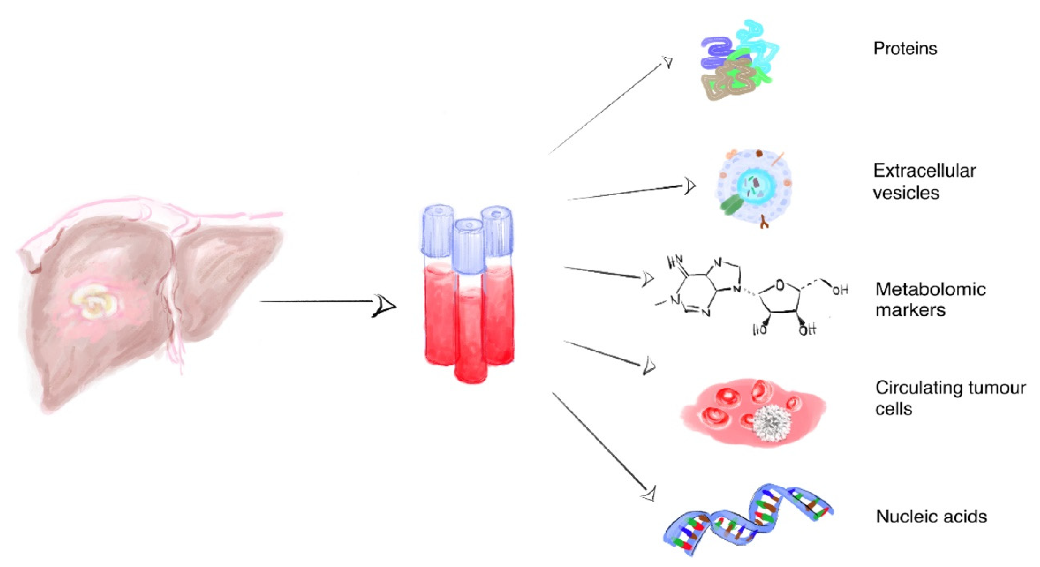
2. Liquid Biopsy Biomarkers in HCC

2.1. Proteins

Tumor protein biomarkers include cytokines and proteins produced by both tumor cells and non-tumoral cellular components. The most commonly used and studied HCC biomarker is AFP. In brief, AFP has a similar molecular structure to albumin and contains one glycoprotein consisting of 591 amino acids. The gene for AFP is localized in chromosome 4 and its expression is elevated in the fetal period of human development as well as in adults with liver disease, including HCC [16]. According to its reactivity to lens culinaris agglutinin (LCA), AFP can be classified as AFP-L1, AFP-L2 and AFP L-3. AFP-L1 is mostly found in patients with chronic hepatitis and liver cirrhosis, while AFP-L3 is more commonly found in HCC patients [17,18,19]. Still, many recent studies have challenged the utility of AFP as an HCC biomarker, arguing for its rather low sensitivity and specificity [18,19,20,21,22,23]. Further, many HCC patients exhibit normal AFP serum levels, particularly during early stage disease [19,22]. As such, while AFP is sometimes still used in clinical practice as a risk assessment tool in cirrhotic patients with a high risk to develop HCC, clinical practice guidelines do not recommend AFP (or any other biomarker for that matter) for the diagnosis of HCC [24].
Des-gamma-carboxy-prothrombin (DCP) represents another potential protein-based circulating biomarker in HCC. In a phase 2 multicenter case-controlled study, early-stage HCC patients were shown to exhibit significant increases in total AFP, AFP-L3 and DCP. In particular, DCP was reported to showcase higher sensitivity and specificity in distinguishing HCC from other chronic liver diseases, including cirrhosis, when compared to AFP [18,22]. In terms of tumor volume, Nakamura et al. reported a better DCP-based predictive outcome, compared to AFP, in a case of HCC characterized by a massive tumor mass [25]. Interestingly, DCP performance is also superior to the one of AFP for HCC tumors with a diameter under 5 cm indicating a possible limitations since its reliability appears to greatly depend on tumor stage [25]. Parallel to Nakamura et al., Toyoda et al. compared AFP, AFP-L3 and DCP regarding HCC prognosis and reported that AFP-L3 and DCP likely mirror different features of HCC progression, prediction of patient outcome, and treatment efficacy [26]. HCC patients positive for all three markers were associated with the shortest survival and vice versa, patients were negative for AFP, AFP-L3 and DCP associated with increased survival [26].
Other circulating protein biomarkers studied in HCC patients include glypican-3 (GPC3), osteopontin, and vascular endothelial growth factor (VEGF). As a heparan sulfate proteoglycan, GPC3 interacts with different growth factors and participates in cell differentiation and migration, including of HCC tumor cells. GPC3 expression has been reported not to depend on HCC tumor size, suggesting a potential role as an early stage diagnostic HCC biomarker [27,28]. In addition, plasma levels of GPC3 have been suggested to predict HCC tumor recurrence [29]. Shimizu et al. also illustrated the utility of GPC3 as a biomarker and as a potential HCC vaccine immunotherapy target [30]. The combination of GPC3 with additional biomarkers, namely AFP, appears to further increase its diagnostic sensitivity and specificity [27,31]. Unfortunately, not all HCC cells express GPC3, while increased GPC3 expression is also found in squamous cell- and lung-carcinomas, clear cell tumors of the ovary, testicular germ cell tumors and melanomas [27,32,33]. In addition, significantly increased serum GPC3 levels have also been reported in patients with non-malignant liver diseases [19].
Neovascularization and angiogenesis are essential for tumor growth. Typically, tumors cannot grow over 2 mm in diameter without any neovascularization support. Angiogenic proteins, including members of the vascular endothelial growth factor (VEGF) family, like VEGF-A, play a vital role in tumor angiogenesis and tumor neovascularization. In fact, VEGF expression is found increased in HCC liver tissue. Of note, increased serum levels of soluble VEGF-A were associated with advanced HCC and shorter overall survival as discussed briefly by Zhou et al. [19]. Interestingly, liver VEGF was capable to indicate the presence of cancer with high accuracy, whereas AFP could identify the type of cancer as HCC, accordingly to Mukozu T. et al. [34].
In combination with AFP, the low specificity of VEGF in the diagnosis of advanced HCC has been shown to increase from 60% to 85%. The triple combination of AFP/VEGF and α-L-fucosidase (AFU) was associated in this pre-clinical study with a sensitivity of 100% and a specificity of 95% [35]. Of note this promising result is lacking so far the needed power, and it could be interesting that it is validated in larger patient cohorts. Interestingly, cancer cells appear not to represent the largest reservoir for VEGF, but rather the skeletal muscle [36]. As such, the combination of AFP and VEGF is prone to the same diagnostic drawbacks reported for AFP alone. VEGF was able to predict the overall survival in the SHARP study as an independent biomarker besides other, where 602 patients with advanced HCC receiving either oral sorafenib 400 mg twice a day or placebo were enrolled [37]. VEGF’s role as a biomarker for HCC lethality should be acknowledged based on additional studies like the SHARP study [37] and as recently published by Joo et al., underlining VEGF as a guide in future treatment decision-making [38].
Finally, glycoprotein osteopontin (OPN) represents another potential experimental HCC biomarker. Osteopontin, ubiquitously expressed in bone and epithelial cells, is also highly expressed by tumor cells, including in HCC. In this regard, Shang et al. reported that OPN alone was associated with good sensitivity and specificity values, hence the combination of AFP and OPN did in fact slightly improve the predictive sensitivity specificity to 95% and of 96%, respectively. The authors further demonstrated that OPN has high sensitivity in AFP-negative HCC [39].
Among the circulating protein biomarkers, AFP and AFP-L3, besides DCP and AFP in combination with OPN, represent widely used biomarkers in clinical use, despite some known limitations. A. Mohammed F.H. and L.R. Roberts discussed the drawbacks of AFP and why the AASLD is not in favor of endorsing AFP as a standalone biomarker in their guidelines, partly based on still missing evidence supporting its high sensitivity and specificity as an effective surveillance and diagnostic tool for HCC [40].

2.2. Metabolomic Markers

The development of “omics” technologies has made possible the study of hundreds of putative biomarkers simultaneously. Metabolomics is considered to be a powerful high-throughput platform to measure low molecular weight metabolites in biological samples (e.g., blood, urine, bile, ascites, tissue etc.). Thus far, it has already contributed to elucidate biochemical pathways playing a role in multiple human cancers, also offering a unique opportunity to discover novel biomarkers in this field [41,42]. Several analytical techniques can be employed, including nuclear magnetic resonance (NMR) [43], gas chromatography-mass spectrometry (GC-MS) [44], liquid chromatography-mass spectrometry (LC-MS) [45], capillary electrophoresis-mass spectrometry (CE-MS) [46], or various combinations of these analytical techniques [47].
In recent years, metabolomics has provided a large list of potentially novel biomarkers for HCC. A comprehensive analysis of all these biomarkers is beyond the purpose of this review and has already been compiled by others [48]. Overall, among the several metabolomic studies performed in HCC, few have assessed the diagnostic performances of each specific biomarker. Still, it appears that metabolomic biomarkers perform statistically better comparing with AFP in discriminating HCC from cirrhosis or HCC from healthy volunteers. In one of the first studies, a diagnostic panel of 18 metabolites was found to be able to differentiate HCC patients from healthy volunteers with an area under the curve (AUC) of 0.9275 [44]. In particular, the best model for discriminating between HCC and healthy controls was based on serum levels of 1-methyladenosine in combination with AFP, with an AUC of 0.95 [49]. Another model included endocannabinoids anandamide and palmitylethanolamide, with an AUC of 0.94 [50]. When discriminating between HCC and cirrhosis, a panel composed of formate, phytosphingosine and 3a, 6a, 7a, 12a-tetrahydroxy-5b-cholan-24-oic acid, exhibited an AUC of 1.000 and 0.995 for the identification and validation set, respectively [47]. Another model based on acetylcarnitine C3 and betaine was shown to exhibit an AUC of 0.98 in discriminating between HCC and cirrhosis [45]. Finally, a validation-designed study using a serum biomarker panel composed of tryptophan, arginine, glycine, and 2-hydroxybutiric acid yielded an AUC of 0.976 in discriminating between small HCC from pre-cancer cirrhosis [46].
Overall, while many metabolites appear to display excellent diagnostic performances in HCC, reproducibility stands as a major issue to address. The same holds true for the reporting of compensated or decompensated cirrhosis. In addition, validation cohorts have been used in a very small number of studies, most of them retrospective on nature and with no statistical adjustments for important confounding variables such as smoking status, alcohol consumption, lifestyle habits, physical activity, body mass index or waist circumference. Even so, a multi-centric, prospective study, using an NMR metabolomic approach, showed that when adjustments for these cofounders was performed, the AUC for discriminating HCC from healthy controls was only slightly better than AFP (75% vs. 73%) [43]. Nonetheless, differences in lifestyle habits, as well as environmental factors, appear to have a huge impact on the utility of metabolomic results and should be carefully balanced in future studies.
Similarly, in order to accelerate translation of metabolomic biomarkers into the clinics, future research should focus on pseudo-targeted and targeted approaches rather than the more commonly performed untargeted metabolomics experiments. As an example, in a urinary pseudo-targeted metabolomic approach, butyrylcarnitine and hydantoin-5-propionic acid yielded an AUC of 0.786 and 0.773 for the diagnosis of HCC in the discovery and validation cohorts, respectively [51]. In addition, in a single targeted metabolomic study, serum acetylcarnitine showed good potential in discriminating HCC patients from both healthy individuals (AUC of 0.803) and liver cirrhosis patients (AUC of 0.808) [52].
An ideal biomarker should also be able to discriminate between HCC and other types of liver cancer such as intrahepatic cholangiocarcinoma (CCA). However, the real challenge lies in correctly assessing combined hepatocellular-cholangiocarcinoma (cHCC-CCA) as cholangiolocarcinoma (CLC) and as intermediate cell carcinoma, potentially sharing features of both cell types [53]. However, a recent transcriptomic and metabolomic study found that a panel of 14 compounds could discriminate between intrahepatic cholangiocarcinoma and HCC with an accuracy of 78.13% [54], a performance value close to those of currently available imaging techniques. Recently, a urinary metabolic signature was proposed to associate with CCA. This signature was capable of distinguishing CCA among other cancer entities including hepatobiliary cancers [55]. It was also recently shown that in the serum, specific metabolites might also embody novel biomarkers to distinguish intrahepatic CCA from HCC, with an AUC of 0.89, 75% sensitivity, and 90% specificity, underlining the power of metabolomic biomarkers in biliary cancer per se [56].

2.3. Circulating Tumor Cells and Extracellular Vesicles

The use of circulating tumor cells (CTCs) for the diagnosis of cancer was approved in 2004 by the U.S. Food and Drug Administration (FDA). Since this method heavily relies on the availability of tumor-disconnected tumor cells in the peripheral blood circulation [57], FDA approval was restricted for the diagnosis of certain epithelial tumor entities, such as prostate, breast and colorectal cancer and limited to malign/metastatic tumor entities or cancer subtypes. Given the highly vascularized liver environment, the use of CTCs as biomarkers for HCC has been under scrutiny. For instance, two different meta-analyses have shown that CTCs associate with a poor prognosis and poor clinicopathologic parameters in HCC [58,59]. Epithelial-mesenchymal-mixed CTCs seem to play an important role in epithelial-mesenchymal transition in HCC, mixed CTCs appearing to play a vital factor for intrahepatic metastasis, and mesenchymal CTCs having the potential to predict extrahepatic metastasis [60]. In addition, both the positive rate and number of CTCs have been significantly correlated with tumor size and TNM staging in HCC patients [61]. Interestingly, CTC-based HCC surveillance could benefit from its combination with other techniques such as high-throughput microfluidic CTC-iChip—RNA-based digital PCR, to detect CTC-derived signatures. Kalinich et al. demonstrated such approach, where CTCs were first isolated via CTC-iCHip technology followed by RNA-based dPCR profiling yielding several targets that pointed towards the presence of HCC. This study reached promising predictive values of 83% (negative) and 86% (positive) in patients with underlying cirrhosis (no sensitivity and specificity was provided) [62]. It has also been suggested that AFP mRNA-positive CTCs may represent a novel predictor for HCC metastasis before and after hepatectomy [63]. Recently, single-cell mRNAseq in HCC patients yielded a genome-wide transcriptome profiling to confidently detect CTCs, while highlighting its potential role to monitor HCC heterogeneity and its ability to detect HCC driver genes [64]. These studies underline the potential synergistic effects of mRNA screening and CTC detection in HCC diagnosis and monitoring and hold potential for further exploitation.
A drawback of using CTCs as diagnostic biomarkers is their apparent lack of specificity, as its detection relies on the use of pan-cancer markers such as epithelial cell adhesion molecule (EpCAM) and creatine kinase (CK), among others [57]. Furthermore, only a small proportion (20%–35%) of HCCs express EpCAM [65], although at least a weak EpCAM expression in >10% of tumors was observed in 87 of 131 different tumor categories [66]. In this regard, the use of CTCs for monitoring anti-tumor therapy response and tumor recurrence is, perhaps, more promising. Shen et al. showed that elevated EpCAM+ CTCs correlated with poor survival of patients with unresectable HCC tumor after transcatheter arterial chemoembolization (TACE) [67]. In addition, detection of EpCAM+ CTCs in bloodstream prior to surgery predicts an elevated risk of HCC recurrence and shorter recurrence free survival after curative resection [68]. Similarly, following radical hepatic resection, elevated EpCAM+ CTCs and Treg/CD4+ cell levels were found to associate with a worst prognosis [69].
A somewhat newer concept, still in earlier experimental phases, suggests taking benefit of CTC recovery, followed by CTC expansion ex vivo. After sufficient expansion/proliferation, anti-tumoral drugs, as approved by the FDA, might be screened under standardized conditions to rule out unsuitable drugs, namely those with persisting resistance in patient’s derived CTCs [70,71]. This basic approach could (a) contribute to reduce public healthcare costs by avoiding the use of inefficient and expensive anti-tumoral drugs (b) lead to the identification of the most effective personalized anti-cancer drug and (c) decrease the amount of time until effective treatment for the benefit of the patient. Unfortunately, the major limiting factor for the use of these methodologies lies on how to induce a timely CTC expansion. In this regard, Khoo et al. recently published an experimental protocol aiming to reduce the time needed to expand CTCs, although most protocols still require months [71]. Of note, and until now, CTCs have not been recognised as a possible diagnostic tool for HCC by either AASLD or EASL.
When firstly identified, extracellular vesicles (EVs) were regarded as cellular waste or debris, without any biological function [72]. The current understanding is that EVs are a new cellular dimension of the so-called horizontal cell-cell communication, traveling to proximal and distal spaces with the ability of altering intracellular pathways with pathophysiological consequences [73,74,75,76]. For instance, tumor cells may use EVs to its advantage by enforcing tumor-induced immune suppression via the release of EVs [77]. Given the nature of EVs, particularly their nano sizes, a number of detection and analysis methods have been reported, with little standardization across studies. As a consequence, the International Society for Extracellular Vesicles (ISEV) released their first guidelines regarding experimental work on EVs in 2014 [78]. It was the first step for the growing EV community to standardize guidelines for EV characterization, addressing purity, desired EV isolation methods, and quantification procedures. The latest ISEV guideline MISEV2018 argues against the common use of the term exosomes and microvesicles (MVs)/microparticles (MPs) in favour of small EVs and large EVs, towards a more simplified nomenclature [79]. According to the ISEV guidelines, two major release mechanisms distinguish these two types of vesicles, namely EV shedding (large EVs) and the bulk release via exocytosis of multi-vesicular bodies (small EVs). Briefly, small EVs typically range in sizes between 30 nm and 100 nm in diameter, while large EVs measure above 100 nm, up to 1000 nm. Oncosomes, a third subtype of EVs, may eventually surpass them in size (up to 10 μm in diameter). Some EV markers have been postulated to be able to distinguish between small and large EVs, including CD63, HSP70, CD9, CD81 and integrins [80,81]. Nonetheless, it should be noted that the border of distinction between these EV populations is not sharp but rather fluent [78]. EVs possess a typical density of 1.13–1.19 g/mL [78], allowing for its separation and enrichment via sequential ultracentrifugation, among other methods [82]. EVs might be phenotypically characterized by their surface membrane proteins or their vesicular content, namely mRNA, lncRNA or proteins, by means of different commercially available kits [78].
Concerning the role of EVs as biomarkers in HCC, it was recently shown that surface marker HepPar1-positive EVs are found increased in HCC patients [83]. In addition, Julich-Haertel et al. showed that AnnexinV, EpCAM, ASGPR1 and CD133 simultaneously positive EVs could be detected in HCC and CCA, being further able to differentiate these two types of liver tumors from other cancer types, as well as patients with cirrhosis and healthy individuals, with a sensitivity of 81% and positive predictive value (PPV) of 73% (cirrhosis vs. HCC) [84]. In parallel, other studies have been focusing on EV content. For instance, it was recently shown that exosome-associated galectin-3-binding protein (LG3BP) provides higher AUC values for the diagnosis of HCC when comparing to AFP [85]. Another noteworthy publication showed a similar diagnostic value of EV hsa-miR-144-3p/hsa-miR-21-5p, found elevated in HCC (AUC of 0.78), compared to AFP (AUC of 0.626) [86]. Similarly, in a highly reproducible rodent HCC model, a panel of 4 miRNAs, namely miRNA-10b, miRNA-21, miRNA-122 and miRNA-200a, exhibited a higher AUC value in diagnosing HCC, comparing to AFP (0.943 vs. 0.826) [87]. In addition, although miR-148a (AUC of 0.860) alone was significantly superior to AFP (AUC of 0.665), the combination of miR-122, miR-148a, and AFP exhibited a higher diagnostic power in discriminating early HCC from liver cirrhosis in humans, with an AUC of 0.947 [88]. Additional and larger patient studies should ideally now be performed in order to fully establish the potential of EVs as reliable and reproducible biomarkers for HCC. Selected and associated publications are systematically summarized in Table 1.

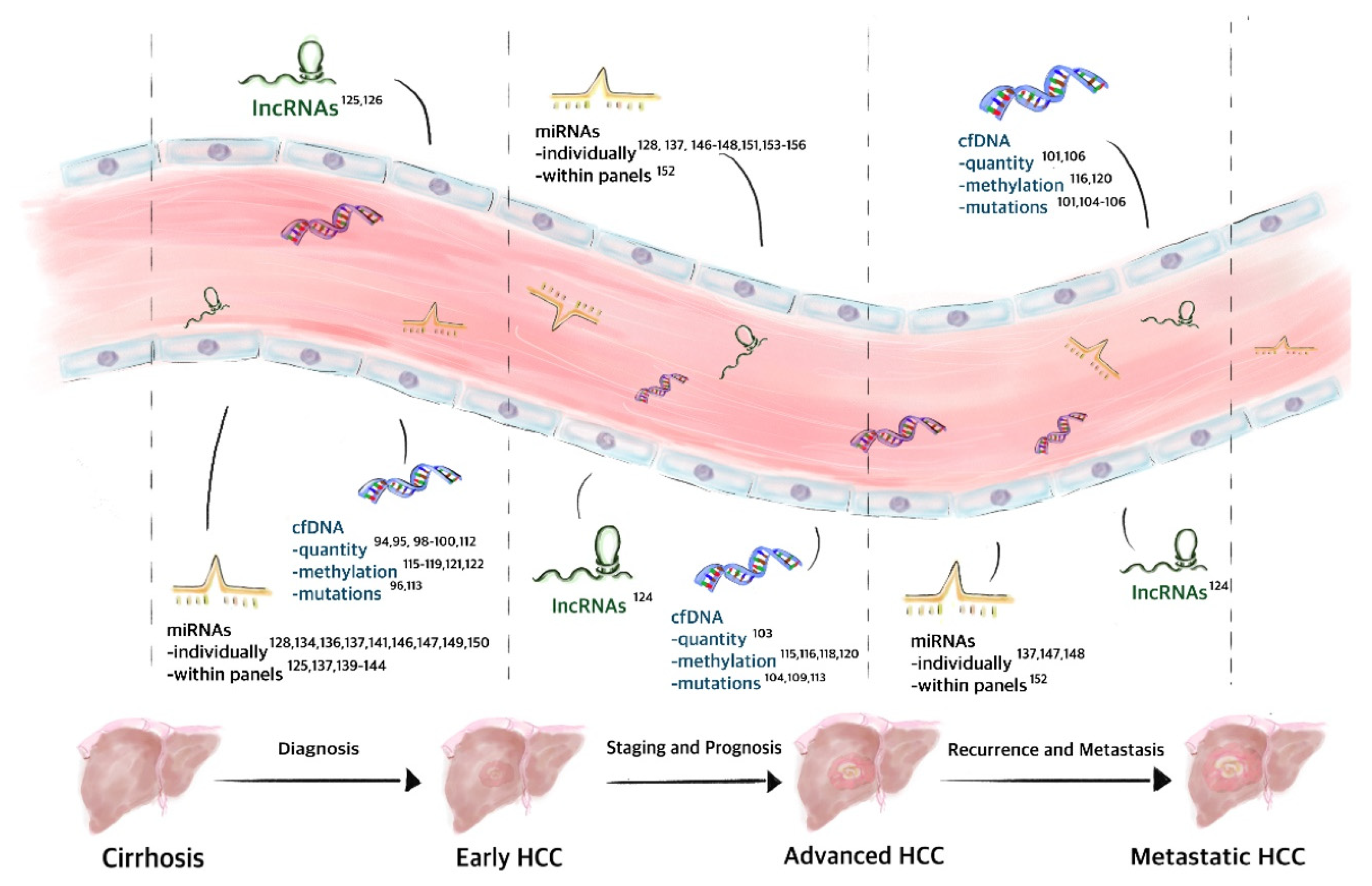
2.4. Circulating Nucleic Acids in HCC

Cell-free nucleic acids are generally divided into “cell-free DNA” (cfDNA) and “cell-free RNA” (cfRNA), two broader terms that also account for circulating tumor DNA (ctDNA) and circulating tumor RNA (ctRNA). Both cfDNA and cfRNA are released into the blood stream through different mechanisms, including active secretion or through means of the apoptosis, necrosis, and lysis of CTCs. Once released into circulation, cfDNA forms short nucleosome-associated fragments or long fragments encapsulated within EVs. Given the relative instability of RNA, cfRNA is usually detected in association with proteins, proteolipid complexes and EVs [89].
The detection and analysis of circulating nucleic acids in blood may aid not only in disease diagnosis but also in guiding molecular targeted therapy, monitoring response to such or other treatments, as well as identifying mutations associated with drug resistance. Furthermore, unlike a tumor biopsy, circulating nucleic acids may provide useful information regarding tumor heterogeneity. The recent approval of the first companion diagnostic test based on the ctDNA content of a liquid biopsy for lung cancer by the FDA constituted a landmark in this field and is paving the way for the further exploitation of circulating nucleic acids as novel and minimally invasive disease biomarkers [90]. This approach is being evaluated through the quantification of cfDNA but more particularly through the detection of point mutations, indels, aberrant DNA methylation, and chromosomal aberrations, as well as cfRNA profiling and quantification [91] (Figure 2).

2.5. cfDNA

In 1948, Mandel and Metais first reported the detection of cfDNA in the serum of healthy individuals [92]. It then took approximately 50 years until the tumor biomarker potential of cfDNA started to emerge, with the identification of KRAS mutations in ctDNA isolated from patients with colorectal and pancreatic cancer [93]. Since then, many studies have been conducted to find the utility of circulating nucleic acids as liquid biopsies, including in the diagnosis of HCC. Earlier studies have focused on establishing the levels of cfDNA as a biomarker for detecting and staging HCC. For instance, in 2006, Iizuka et al. described that cfDNA levels were elevated in patients with hepatitis C virus (HCV)-associated HCC, and its diagnostic power was superior to that of serum AFP [94]. More recently, and because cfDNA levels can originate from multiple types of cancer, arguing against the specificity of single quantitation, the combination with additional markers has been reported, which appears to increase specificity. For instance, two mathematical models including cfDNA and AFP levels had a higher discriminative power in diagnosing HCC than that of a model with cfDNA or AFP alone [95,96]. This is particularly relevant considering that the predictive value of AFP alone (>20 ng/mL) has a sensitivity of ~60%, which is low [97].
Interestingly, a previous study revealed that the size profile of cfDNA from patients with HCC is shorter than that of non-tumor DNA [98]. Furthermore, this profile reverses to less fragmented cfDNA after HCC tumor resection [99]. It is noteworthy that cfDNA fragmentation also occurs in other types of cancer. With respect to this, a study found evidence that hepatic cfDNA showcases ends at specific genomic coordinates, reinforcing the notion that cfDNA fragmentation is a nonrandom process. Furthermore, quantitative assessment of cfDNA molecules has been shown to correlate with the relative abundance of tumor- or liver-derived DNA in plasma [100]. Nonetheless, and for the time being, the analysis of cfDNA methylation and the mutational landscape appears to offer more specificity.
An exome sequencing study showed that 83% of the mutations identified in the liver were also detected in cfDNA, which highlights the usefulness of cfDNA in the diagnosis of HCC [101]. In this regard, ultra-deep targeted sequencing for all exons of the 58 most frequently mutated genes in HCC reported an overall detection rate for tissue mutations in plasma cfDNA of 43% (9/21) [102]. A more recent study showed that all the mutations detected in plasma cfDNA matched HCC tissue DNA but not the opposite, pointing towards a high specificity but low sensitivity of plasma cfDNA as an HCC biomarker [103].
The ability to detect cfDNA mutations in patients with HCC also relates to clinical features of the disease. An earlier study showed that mutations typically detected in the hepatic tissue of patients with HCC were also detected in cfDNA (27% of the patients showed the presence of cfDNA and almost all patients (6/7) had either large tumor (≥5 cm diameter) or metastatic disease) [104]. Targeted deep sequencing, in turn, identified tumor-associated mutations for HCC in plasma cfDNA associating with vascular invasion [105]. Interestingly, cfDNA levels and the identification of 28 mutation variants correlated with the presence of metastases and survival in a cohort of 13 patients with advanced HCC [106].
Finally, the level of cfDNA as a liquid biopsy for HCC should ideally also arise from its ability to address intratumoral heterogeneity. This question was recently addressed through whole-exome sequencing and targeted deep sequencing in 32 multi-regional tumor samples and cfDNAs from five patients with HCC. The results showed that cfDNA was not as efficient as tissue samples in revealing disease heterogeneity [107]. Further studies using a large cohort and/or late-stage patients should be conducted to help shed more light into this issue.
TP53 mutations are the most commonly found mutations in HCC tissue and cfDNA, with more than 120 non-unique alterations [108,109]. Using digital droplet PCR targeting, the TP53 c.747G > T (p.R249S) mutation, as well as CTNNB1 c.121A > G (p.T41A), CTNNB1 c.133T > C (p.S45A), and TERT c.-124C > T mutations, were identified in at least one mutation in 56.3% (27/48) of the cfDNAs obtained from patients with HCC. None of the aforementioned mutations were detected in the non-tumoral HCC tissue or in peripheral blood mononuclear cells (PBMCs) [110]. In particular, the TP53 mutation at codon 249 appears to be highly specific. A recent study, making use of an experimental blood test, analyzed both genetic alterations and protein biomarkers in different types of stage I cancer. Their results showed that the TP53 c.747G > T (p.R249S) mutation was found in ~20% of the HCC plasma samples and in only ~3–4% of the pancreatic and stomach cancer samples, and it was not detected in a large cohort of 812 healthy controls. Mutations in KRAS and CTNNB1 were also concomitantly found in HCC tissue and plasma samples [111]. Moreover, mutations in ERBB2 occurred more frequently in Hepatitis B (HBV)-positive patients [109]. A similar test, combining the analysis of cfDNA mutations with protein markers, was shown to be particularly useful in detecting early stage HCC in a high-risk asymptomatic population who were seropositive for the surface antigen of hepatitis B virus (HBsAg). In the training cohort, the HCC screen assay exhibited a sensitivity of 85% and specificity of 93% in diagnosing HCC. In the validation cohort, the HCC screen assay exhibited 100% sensitivity and 94% specificity [112].
The human telomerase reverse transcriptase (TERT) gene encodes for the catalytic subunit of telomerase that will act together with multiple molecular partners to maintain telomere homeostasis and, as such, chromosomal integrity. Highly recurrent mutations have been found in in the promoter of TERT, providing a mechanism for TERT reactivation and oncogenic transformation. Of note, TERT promoter mutations is one of the most common genetic alterations in HCC, also found in cfDNA of patients with HCC, with similar prevalence. Male patients with HCV and/or alcohol-induced cirrhosis were identified as the patient group with the highest prevalence of TERT promoter mutations, either within the tumor or in the cfDNA [113]. As such, querying for TERT promoter mutations in cfDNA may allow for the early detection of HCC in populations at risk of HCC.
Since tumor suppressor genes are methylated early during tumorigenesis, analysis of DNA methylation patterns may embody key diagnostic value. With respect to this, and similar to the cfDNA quantification and mutational landscape analysis, cfDNA aberrant methylation allows for diagnostic and prognostic insights in HCC, and might reveal information regarding the tumor size and risk of metastasis or recurrence [114]. A recent study identified a DNA methylation marker panel enriched in HCC tissues, which was replicated in cfDNA of the same patients. A diagnostic prediction model with 10 selected methylation markers showed high specificity (>83%) and sensitivity (>90%) correlating with tumor burden, treatment response, and disease stage. The model further differentiated patients with HCC from those with hepatitis B virus (HBV)/HCV infection and fatty liver disease [115]. Suppressor of cytokine signaling 3 (SOCS3) methylation in both HCC tissue and plasma was also found to be associated with tumor size and differentiation, metastasis, and recurrence, as well as with poorer prognosis [116].
Analysis of DNA methylation patterns could also be used to screen populations at risk of HCC. In a recent study, RAS association domain family protein 1A (RASSF1A) methylation was largely detected in the cfDNA of patients with HCC (52.04%), but not as much in patients with liver cirrhosis or chronic hepatitis B or in healthy controls (13.33%, 4.44%, and 3.75%, respectively). Of note, the RASSF1A methylation exhibited better sensitivity and specificity (52.0% and 91.5%, respectively) in distinguishing HCC from chronic hepatitis B compared with AFP (48.0% and 73.9%, respectively) [117]. Interestingly, RASSF1A has been combined with three additional candidates (APC, COX2, and miR-203) in a methylation status prediction model (AUC of 0.87) with a sensitivity and specificity of 84.2% and 83%, respectively, in the diagnosis of HBV-related HCC, which is, again, superior to the currently used AFP detection model [118]. Likewise, a 32-gene diagnostic model, detecting genome-wide 5-hydroxymethylcytosines, accurately distinguished patients with early HCC or small tumours (e.g., ≤2.0 cm) from non-HCC subjects (AUC of 0.884), including high risk patients with liver cirrhosis or chronic hepatitis B (AUC of 0.846) [119].
Hypomethylation of cfDNA may also serve as a biomarker of HCC. In particular, several studies have reported increased levels of hypomethylated-type transposase domain containing long interspersed nuclear element 1 (LINE-1) in cfDNA of patients with HCC. This is associated with clinical pathologic features such as HBsAg positivity, tumor size, or AFP levels. LINE-1 hypomethylation is also associated with poor survival of patients with HCC and with early recurrence and poor prognosis upon curative resection when combined with the measurement of RASSF1A hypermethylation [120].
Alterations in cfDNA methylation, associated with the initiation and progression of HCC, have been described for many other genes, including p15, p16, APC, SPINT2, SFRP1, p16INK4a, TFPI2, GSTP1, SEPT9, VIM, and FBLN1, as well as in CpGs sites [14,121,122,123]. Altogether, these results support the hypothesis that analysis of ctDNA methylation status embodies novel putative biomarker tests for HCC.
The use of ctDNA as a liquid biopsy still poses several limitations, particularly in the early detection of cancer, where ctDNA levels are low. However, the lack of standardized protocols for preanalytical sample preparation and ctDNA purification and analysis is the major limitation in the diagnosis of HCC. While large prospective, well-controlled studies on HCC cfDNA profiling are highly warranted, the promising results obtained so far testify to the potential of cfDNA as a superior tool for tumor diagnosis and monitoring. Selected and associated publications are systematically summarized in Table 2.

2.6. cfRNA

Most known classes of RNA have been detected in circulation, with long noncoding RNAs (lncRNAs) and microRNAs (miRNAs) being the most studied as putative disease biomarkers. LncRNAs are typically >200 nucleotides in length and play a key role in epigenetic regulation. Due to their ability to form stable secondary structures, lncRNAs can be found in body fluids and have been studied as potential biomarkers for different diseases, including HCC. The long intergenic nonprotein coding RNA 974 (Linc00974) has been found increased in the serum of patients with HCC [124], whereas lncRNA–CTBP exhibited high sensitivity and specificity in differentiating HCC from both healthy controls and patients with cirrhosis [125]. In a recent study, three circulating lncRNAs, namely LINC00152, RP11-160H22.5, and XLOC014172, combined with AFP, were able to distinguish HCC from chronic hepatitis patients or healthy controls with an AUC of 0.986 and 0.985, respectively [126].

2.7. miRNA

miRNAs are a class of short, single-stranded, noncoding RNAs (18–24 nt) that play a key role in the posttranscriptional regulation of gene expression. By doing so, miRNAs modulate many pathophysiological processes, including cancer. They can be found in circulation enclosed in EVs or associated with proteins (typically argonaute 2 (AGO2) and nucleomorphin 1 (NPM1)) or lipoproteins (e.g., HDL). Many circulating miRNAs, including miR-106b, miR-21, miR-200a, miR-122, miR-223, let-7f, and miR-155 have already been described to associate with HCC, with promising diagnostic and prognostic value [127,128]. Overall, more than 70 miRNAs have already been suggested as candidate biomarkers for HCC [129]. Of note, special attention has been paid to miR-122 and miR-192 because of their liver specificity and enrichment. In fact, both these markers account for over 70% of the total miRNA pool in the liver [130]. While this could mean that miR-122 and miR-192 would lack specificity, as any type of liver disease will affect their release into circulation, it is becoming apparent that partitioning of the release of miR-122, that is, its incorporation into EVs or association with carrying proteins, might be specific to the type of liver tissue damage [131,132,133]. Future studies should elucidate whether the combination of data addressing the partitioning of miR-122 with its expression might improve its relevance as a biomarker for HCC.
A meta-analysis based on 24 articles revealed that the expression levels of miR-21, miR-122, and miR-199 appear to rank among the most selective for the diagnosis of HCC [134]. However, it should be noted that conflicting results on circulating miR-21 have been published, with some suggesting upregulation [135,136] and others suggesting downregulation in HCC [110,137]. Nonetheless, serum miR-21 levels might have a better predictive role as a putative biomarker for HCC when used in conjunction with additional biomarkers. In fact, similar to cfDNA, the combination of different miRNAs into a biomarker panel, or its combination with select clinical parameters, appears to increase sensitivity for HCC diagnosis. This is the case, for instance, when combining miR-21 with miR-26a, miR-27a, miR-122, miR-192, miR-223 and miR-801, which altogether exhibit a high diagnostic accuracy for HCC. Furthermore, this miRNA panel was able to differentiate between patients with HBV and cirrhosis and healthy subjects, with an AUC of 0.84, 0.88, and 0.94, respectively [138,139]. Other examples of combined miRNA panels include serum levels of miR-375, miR-10a, miR-122, and miR-423, found elevated in patients with HCC comparing with healthy controls (AUROC of 0.995) [140]; miR-101-3p, miR-1246, and miR-106b-3p, which exhibited high diagnostic accuracy for HCC when patients with cirrhosis (AUC of 0.99) were compared with control subjects (AUC of 1.00) [141]; or miR-122, miR-142-3p and miR-486 in distinguishing HCC from cirrhosis in patients with chronic Hepatitis C (AUC = 0.94) [142]. Interestingly, a recent study combining eight miRNAs showed a sensitivity of 97.7% and a specificity of 94.7% in discriminating the presence of HCC in high risk patients, allowing the detection of 98% stage I HCC cases [143].
Of note, miR-26a and miR-101, combined with AFP, exhibited a better sensitivity for HCC than AFP alone [110]. Similarly, the combination of miR-92-3p, miR-107, and miR-3126-5p with AFP was highly effective in differentiating patients with early stage HCC (AUC of 0.988) and patients with a low level of AFP in HCC (AUC of 0.989) from healthy individuals, even more so than the three miRNAs panel alone [144]. Last but not least, a panel of miR-122, miR-885-5p, and miR-29b, associated with AFP testing, was shown to exhibit high diagnostic accuracy for the early detection of HCC in a normal population, whereas combining miR-122, miR-885-5p, miR-221, and miR-22 with AFP allowed for the accurate diagnosis of early HCC in cirrhosis [139]. A recent meta-analysis evaluating the significance of circulating miRNAs in the diagnosis of HCC indeed suggests that the combination of miRNAs with AFP elicits superior diagnostic performance [145].
Circulating levels of select miRNAs also correlate with different clinical features and tumor properties. For instance, increased plasma levels of miR-21 and miR-224 in patients with HCC was found to correlate with clinical stage and distant metastasis and larger tumor size and recurrence, respectively [146,147]. Moreover, serum miR-1246 was found increased in HCC patients with early tumor recurrence within 12 months after hepatic resection, exhibiting high potential as a recurrence and prognostic biomarker of HCC [148]. Indeed, several studies have revealed that cfRNA levels reflect tumor dynamics and burden, with important implications for disease monitoring. For instance, both miR-224 and miR-500 are highly expressed and detected in the plasma of patients with HCC, but its circulating levels significantly decreased following resection [146,149]. On the contrary, miR-148a is downregulated and therefore detected in low quantities in the plasma of patients with HCC compared with patients with cirrhosis and healthy controls, and tumor resection upregulated the expression of miR-148a [150].
Recent studies have focused on the role of circulating miRNAs in HCC as predictors of clinical outcomes after treatment, thus helping in finding a personalized therapy for individual patients. A recent study showed that serum levels of miR-106b, miR-107, and miR-133b were elevated in patients with HCC who responded to TACE, with miR-26a being elevated in nonresponders. miR-26a and miR-133b exhibited the highest diagnostic performance, with miR-133b further distinguishing complete responders from partial responders and non-responders (AUC ≥ 0.90) [151]. Other studies have indicated that a combinatory analysis of plasma levels of miR-21, miR-26a, and miR-29a-3p, or even miR-122 alone, may predict early TACE refractoriness in patients with TACE-treated HCC [152]. It was also suggested that high expression levels of miR-122, as well as low expression levels of miR-26a and miR-29a, could constitute a poor prognostic marker for patients with HCC undergoing radiofrequency ablation [153,154]. With respect to chemotherapeutic drugs, circulating miR-221 levels have been reported to associate with sorafenib treatment response in patients with HCC [155], whereas recent results from the RESORCE (NCT01774344) clinical trial, identified several miRNAs, namely miR-30a, miR-22, miR-125b, miR-200a, miR-374b, miR-15b, miR-107, miR-320, and miR-645, associated with increased overall survival of patients with HCC who are treated with regorafenib [156]. This highlights its possible usefulness in identifying patients most likely to respond to treatment. Nonetheless, and similar to cfDNA, the most important limitation to the implementation of cfRNAs in the clinical setting still pertains to the pre-analytical and analytical steps. Selected and associated publications are systematically summarized in Table 3.

3. Further Directions

Given the high incidence of HCC, the search for non-invasive biomarkers is crucial in modern diagnostics of this disease. Such markers should not only be useful in very early diagnosis of the disease but also in prognosis and monitoring of its course and treatment. Based on a review of the literature, it appears that such features are present within a liquid biopsy; however, this is not a flawless method. First off, methodological difficulties in biomarker determination loom large for scientists. The currently used methods require special preparation of biological material and highly specialized laboratory equipment. For this reason, there is a need to intensify efforts to develop a fast, simple, and inexpensive analytical technique. In light of the experimental and clinical studies available, the lack of such a method is a serious limitation on the use of liquid biopsy in clinical practice. For this reason, we have formed the opinion that future research should focus on this topic and this is supported by the fact that many groups of researchers have demonstrated the high specificity and sensitivity of circulating markers, but their introduction for use in the clinic is highly unlikely until a simple analytical method has been developed.

4. Conclusions

As discussed in this review, there are some good preclinical arguments for the urgent need of liquid biopsy in HCC diagnosis, screening and surveillance. Some of the reviewed techniques will likely help physicians to perform a more accurate diagnosis through synergistic effects base on novel liquid biopsy based methods and of previously established techniques such as ultrasound, CT, and MRI. The image-based analysis provides the operator with visible information of a lesion. However, it requires an experienced professional to interpret the seeable lesion. However, subjectivity could potentially be an issue. Additional tests based on liquid biopsy, as discussed, might led to a synergy to overcome image-based drawbacks. Ideally, in liquid biopsy, only the cut-off value should determine the diagnosis and increase image based diagnosis confidence.
Recently, the term “liquid biopsy” has evolved to include a variety of newly developed methods used for HCC diagnosis. As discussed above, some of the techniques show good preliminary results, whereas others appear promising but have not yet reached conclusive results. Additional biomarkers, capable of being detected by mass spectrometry or RNA arrays will probably be established soon. Altogether, screening for various types of nucleotides, such as cfRNA and miRNA, could soon be incorporated into different clinical practice guideline, including those of EASL and AASLD. In addition, a conceptual new direction has recently emerged, namely EVs. Only a few studies have been conducted so far promoting the idea to use EVs as a novel liquid biopsy marker for HCC, but the field is promising.
Overall, we cannot predict the technique that will finally hit the market and satisfy the demand for non-invasive HCC biomarkers in the form of a liquid biopsy. It could be very well that the combination of EV technology and screening of nucleotides within EVs, as discussed for EV RNA, could be a game-changer. Of note, reliably working liquid biopsies will have many advantages for the patient’s welfare and will likely be associated with lower healthcare costs.

Funding

This research was funded by the Deutsche Forschungsgemeinschaft (DFG, German Research Foundation), grant number 410853455; and by Fundação para a Ciência e a Tecnologia, Portugal, grant number PTDC/MED-PAT/31882/2017; and by “la Caixa” Foundation, Spain, grant number LCF/PR/HP17/52190004. And APC was funded by the Deutsche Forschungsgemeinschaft (DFG, German Research Foundation).

Acknowledgments

We are remarkably grateful to Wioleta Chomko for assistance with figures.

Conflicts of Interest

The authors declare no potential conflicts of interest with respect to research, authorship and/or publication of this article.

References

  1. Forner, A.; Reig, M.; Bruix, J. Hepatocellular carcinoma. Lancet 2018, 391, 1301–1314. [Google Scholar] [CrossRef]
  2. Akinyemiju, T.; Abera, S.; Ahmed, M.; Alam, N.; Alemayohu, M.A.; Allen, C.; Al-Raddadi, R.; Alvis-Guzman, N.; Amoako, Y.; Artaman, A.; et al. The Burden of Primary Liver Cancer and Underlying Etiologies From 1990 to 2015 at the Global, Regional, and National Level: Results From the Global Burden of Disease Study 2015. JAMA Oncol. 2017, 3, 1683–1691. [Google Scholar] [PubMed]
  3. Cucchetti, A.; Trevisani, F.; Pecorelli, A.; Erroi, V.; Farinati, F.; Ciccarese, F.; Rapaccini, G.L.; Di Marco, M.; Caturelli, E.; Giannini, E.G.; et al. Estimation of lead-time bias and its impact on the outcome of surveillance for the early diagnosis of hepatocellular carcinoma. J. Hepatol. 2014, 61, 333–341. [Google Scholar] [CrossRef] [PubMed]
  4. Ryder, S.D. Guidelines for the diagnosis and treatment of hepatocellular carcinoma (HCC) in adults. Gut 2003, 52, iii1–iii8. [Google Scholar] [CrossRef] [Green Version]
  5. Zamcheck, N.; Pusztaszeri, G. CEA, AFP and other potential tumor markers. CA Cancer J. Clin. 1975, 25, 204–214. [Google Scholar] [CrossRef]
  6. Rich, N.; Singal, A.G. Hepatocellular carcinoma tumour markers: Current role and expectations. Best Pract. Res. Clin. Gastroenterol. 2014, 28, 843–853. [Google Scholar] [CrossRef]
  7. Sparchez, Z.; Mocan, T. Contemporary role of liver biopsy in hepatocellular carcinoma. World J. Hepatol. 2018, 10, 452–461. [Google Scholar] [CrossRef]
  8. Gerlinger, M.; Rowan, A.J.; Horswell, S.; Math, M.; Larkin, J.; Endesfelder, D.; Gronroos, E.; Martinez, P.; Matthews, N.; Stewart, A.; et al. Intratumor heterogeneity and branched evolution revealed by multiregion sequencing. N. Engl. J. Med. 2012, 366, 883–892. [Google Scholar] [CrossRef] [Green Version]
  9. Jamal-Hanjani, M.; Quezada, S.A.; Larkin, J.; Swanton, C. Translational implications of tumor heterogeneity. Clin. Cancer Res. 2015, 21, 1258–1266. [Google Scholar] [CrossRef] [Green Version]
  10. McGranahan, N.; Swanton, C. Biological and therapeutic impact of intratumor heterogeneity in cancer evolution. Cancer Cell 2015, 27, 15–26. [Google Scholar] [CrossRef] [Green Version]
  11. Yoshida, S.; Kornek, M.; Ikenaga, N.; Schmelzle, M.; Masuzaki, R.; Csizmadia, E.; Wu, Y.; Robson, S.C.; Schuppan, D. Sublethal heat treatment promotes epithelial-mesenchymal transition and enhances the malignant potential of hepatocellular carcinoma. Hepatology 2013, 58, 1667–1680. [Google Scholar] [CrossRef]
  12. Zhou, J.; Huang, A.; Yang, X.R. Liquid Biopsy and its Potential for Management of Hepatocellular Carcinoma. J. Gastrointest. Cancer 2016, 47, 157–167. [Google Scholar] [CrossRef]
  13. Labgaa, I.; Villanueva, A. Liquid biopsy in liver cancer. Discov. Med. 2015, 19, 263–273. [Google Scholar]
  14. Li, J.; Han, X.; Yu, X.; Xu, Z.; Yang, G.; Liu, B.; Xiu, P. Clinical applications of liquid biopsy as prognostic and predictive biomarkers in hepatocellular carcinoma: Circulating tumor cells and circulating tumor DNA. J. Exp. Clin. Cancer Res. 2018, 37, 213. [Google Scholar] [CrossRef] [PubMed] [Green Version]
  15. Yin, C.Q.; Yuan, C.H.; Qu, Z.; Guan, Q.; Chen, H.; Wang, F.B. Liquid Biopsy of Hepatocellular Carcinoma: Circulating Tumor-Derived Biomarkers. Dis. Markers 2016, 2016, 1427849. [Google Scholar] [CrossRef] [PubMed] [Green Version]
  16. Nikulina, D.; Terentyev, A.; Galimzyanov, K.; Jurisic, V. Fifty years of discovery of alpha-fetoprotein as the first tumor marker. Srp. Arh. Celok. Lek. 2015, 143, 100–104. [Google Scholar] [CrossRef] [PubMed]
  17. Li, D.; Mallory, T.; Satomura, S. AFP-L3: A new generation of tumor marker for hepatocellular carcinoma. Clin. Chim. Acta 2001, 313, 15–19. [Google Scholar] [CrossRef]
  18. Marrero, J.A.; Feng, Z.; Wang, Y.; Nguyen, M.H.; Befeler, A.S.; Roberts, L.R.; Reddy, K.R.; Harnois, D.; Llovet, J.M.; Normolle, D.; et al. Alpha-fetoprotein, des-gamma carboxyprothrombin, and lectin-bound alpha-fetoprotein in early hepatocellular carcinoma. Gastroenterology 2009, 137, 110–118. [Google Scholar] [CrossRef] [PubMed] [Green Version]
  19. Zhou, L.; Liu, J.; Luo, F. Serum tumor markers for detection of hepatocellular carcinoma. World J. Gastroenterol. 2006, 12, 1175–1181. [Google Scholar] [CrossRef] [Green Version]
  20. Flecken, T.; Schmidt, N.; Spangenberg, H.C.; Thimme, R. Hepatocellular carcinoma—From immunobiology to immunotherapy. Z. Gastroenterol. 2012, 50, 47–56. [Google Scholar]
  21. Chaiteerakij, R.; Addissie, B.D.; Roberts, L.R. Update on biomarkers of hepatocellular carcinoma. Clin. Gastroenterol. Hepatol. 2015, 13, 237–245. [Google Scholar] [CrossRef] [PubMed] [Green Version]
  22. Marrero, J.A.; Su, G.L.; Wei, W.; Emick, D.; Conjeevaram, H.S.; Fontana, R.J.; Lok, A.S. Des-gamma carboxyprothrombin can differentiate hepatocellular carcinoma from nonmalignant chronic liver disease in american patients. Hepatology 2003, 37, 1114–1121. [Google Scholar] [CrossRef] [PubMed]
  23. Galle, P.R.; Foerster, F.; Kudo, M.; Chan, S.L.; Llovet, J.M.; Qin, S.; Schelman, W.R.; Chintharlapalli, S.; Abada, P.B.; Sherman, M.; et al. Biology and significance of alpha-fetoprotein in hepatocellular carcinoma. Liver Int. 2019, 39, 2214–2229. [Google Scholar] [CrossRef] [PubMed] [Green Version]
  24. European Association for the Study of the Liver. EASL Clinical Practice Guidelines: Management of hepatocellular carcinoma. J. Hepatol. 2018, 69, 182–236. [Google Scholar] [CrossRef] [Green Version]
  25. Nakamura, S.; Nouso, K.; Sakaguchi, K.; Ito, Y.M.; Ohashi, Y.; Kobayashi, Y.; Toshikuni, N.; Tanaka, H.; Miyake, Y.; Matsumoto, E.; et al. Sensitivity and specificity of des-gamma-carboxy prothrombin for diagnosis of patients with hepatocellular carcinomas varies according to tumor size. Am. J. Gastroenterol. 2006, 101, 2038–2043. [Google Scholar] [CrossRef]
  26. Toyoda, H.; Kumada, T.; Kiriyama, S.; Sone, Y.; Tanikawa, M.; Hisanaga, Y.; Yamaguchi, A.; Isogai, M.; Kaneoka, Y.; Washizu, J. Prognostic significance of simultaneous measurement of three tumor markers in patients with hepatocellular carcinoma. Clin. Gastroenterol. Hepatol. 2006, 4, 111–117. [Google Scholar] [CrossRef]
  27. Kandil, D.H.; Cooper, K. Glypican-3: A novel diagnostic marker for hepatocellular carcinoma and more. Adv. Anat. Pathol. 2009, 16, 125–129. [Google Scholar] [CrossRef]
  28. Zhou, F.; Shang, W.; Yu, X.; Tian, J. Glypican-3: A promising biomarker for hepatocellular carcinoma diagnosis and treatment. Med. Res. Rev. 2018, 38, 741–767. [Google Scholar] [CrossRef]
  29. Ofuji, K.; Saito, K.; Suzuki, S.; Shimomura, M.; Shirakawa, H.; Nobuoka, D.; Sawada, Y.; Yoshimura, M.; Tsuchiya, N.; Takahashi, M.; et al. Perioperative plasma glypican-3 level may enable prediction of the risk of recurrence after surgery in patients with stage I hepatocellular carcinoma. Oncotarget 2017, 8, 37835–37844. [Google Scholar] [CrossRef] [Green Version]
  30. Shimizu, Y.; Suzuki, T.; Yoshikawa, T.; Endo, I.; Nakatsura, T. Next-Generation Cancer Immunotherapy Targeting Glypican-3. Front. Oncol. 2019, 9, 248. [Google Scholar] [CrossRef]
  31. Capurro, M.; Wanless, I.R.; Sherman, M.; Deboer, G.; Shi, W.; Miyoshi, E.; Filmus, J. Glypican-3: A novel serum and histochemical marker for hepatocellular carcinoma. Gastroenterology 2003, 125, 89–97. [Google Scholar] [CrossRef]
  32. Wang, S.K.; Zynger, D.L.; Hes, O.; Yang, X.J. Discovery and diagnostic value of a novel oncofetal protein: Glypican 3. Adv. Anat. Pathol. 2014, 21, 450–460. [Google Scholar] [CrossRef] [PubMed]
  33. Nakatsura, T.; Nishimura, Y. Usefulness of the novel oncofetal antigen glypican-3 for diagnosis of hepatocellular carcinoma and melanoma. BioDrugs 2005, 19, 71–77. [Google Scholar] [CrossRef] [PubMed]
  34. Mukozu, T.; Nagai, H.; Matsui, D.; Kanekawa, T.; Sumino, Y. Serum VEGF as a tumor marker in patients with HCV-related liver cirrhosis and hepatocellular carcinoma. Anticancer Res. 2013, 33, 1013–1021. [Google Scholar] [CrossRef]
  35. el-Houseini, M.E.; Mohammed, M.S.; Elshemey, W.M.; Hussein, T.D.; Desouky, O.S.; Elsayed, A.A. Enhanced detection of hepatocellular carcinoma. Cancer Control 2005, 12, 248–253. [Google Scholar] [CrossRef] [Green Version]
  36. Kut, C.; Mac Gabhann, F.; Popel, A.S. Where is VEGF in the body? A meta-analysis of VEGF distribution in cancer. Br. J. Cancer 2007, 97, 978–985. [Google Scholar] [CrossRef] [Green Version]
  37. Llovet, J.M.; Pena, C.E.; Lathia, C.D.; Shan, M.; Meinhardt, G.; Bruix, J.; Group, S.I.S. Plasma biomarkers as predictors of outcome in patients with advanced hepatocellular carcinoma. Clin. Cancer Res. 2012, 18, 2290–2300. [Google Scholar] [CrossRef] [Green Version]
  38. Joo, Y.Y.; Jang, J.W.; Lee, S.W.; Yoo, S.H.; Kwon, J.H.; Nam, S.W.; Bae, S.H.; Choi, J.Y.; Yoon, S.K. Circulating pro- and anti-angiogenic factors in multi-stage liver disease and hepatocellular carcinoma progression. Sci. Rep. 2019, 9, 9137. [Google Scholar] [CrossRef]
  39. Shang, S.; Plymoth, A.; Ge, S.; Feng, Z.; Rosen, H.R.; Sangrajrang, S.; Hainaut, P.; Marrero, J.A.; Beretta, L. Identification of osteopontin as a novel marker for early hepatocellular carcinoma. Hepatology 2012, 55, 483–490. [Google Scholar] [CrossRef] [Green Version]
  40. Ahmed Mohammed, H.F.; Roberts, L.R. Should AFP (or any biomarkers) be used for HCC surveillance? Curr. Hepatol. Rep. 2017, 16, 137–145. [Google Scholar] [CrossRef]
  41. Nicholson, J.K.; Holmes, E.; Kinross, J.M.; Darzi, A.W.; Takats, Z.; Lindon, J.C. Metabolic phenotyping in clinical and surgical environments. Nature 2012, 491, 384–392. [Google Scholar] [CrossRef] [PubMed]
  42. Wang, X.; Zhang, A.; Sun, H. Power of metabolomics in diagnosis and biomarker discovery of hepatocellular carcinoma. Hepatology 2013, 57, 2072–2077. [Google Scholar] [CrossRef] [PubMed]
  43. Fages, A.; Duarte-Salles, T.; Stepien, M.; Ferrari, P.; Fedirko, V.; Pontoizeau, C.; Trichopoulou, A.; Aleksandrova, K.; Tjonneland, A.; Olsen, A.; et al. Metabolomic profiles of hepatocellular carcinoma in a European prospective cohort. BMC Med. 2015, 13, 242. [Google Scholar] [CrossRef] [PubMed]
  44. Wu, H.; Xue, R.; Dong, L.; Liu, T.; Deng, C.; Zeng, H.; Shen, X. Metabolomic profiling of human urine in hepatocellular carcinoma patients using gas chromatography/mass spectrometry. Anal. Chim. Acta 2009, 648, 98–104. [Google Scholar] [CrossRef] [PubMed]
  45. Huang, Q.; Tan, Y.; Yin, P.; Ye, G.; Gao, P.; Lu, X.; Wang, H.; Xu, G. Metabolic characterization of hepatocellular carcinoma using nontargeted tissue metabolomics. Cancer Res. 2013, 73, 4992–5002. [Google Scholar] [CrossRef] [Green Version]
  46. Zeng, J.; Yin, P.; Tan, Y.; Dong, L.; Hu, C.; Huang, Q.; Lu, X.; Wang, H.; Xu, G. Metabolomics study of hepatocellular carcinoma: Discovery and validation of serum potential biomarkers by using capillary electrophoresis-mass spectrometry. J. Proteome Res. 2014, 13, 3420–3431. [Google Scholar] [CrossRef]
  47. Liu, Y.; Hong, Z.; Tan, G.; Dong, X.; Yang, G.; Zhao, L.; Chen, X.; Zhu, Z.; Lou, Z.; Qian, B.; et al. NMR and LC/MS-based global metabolomics to identify serum biomarkers differentiating hepatocellular carcinoma from liver cirrhosis. Int. J. Cancer 2014, 135, 658–668. [Google Scholar] [CrossRef]
  48. Kimhofer, T.; Fye, H.; Taylor-Robinson, S.; Thursz, M.; Holmes, E. Proteomic and metabonomic biomarkers for hepatocellular carcinoma: A comprehensive review. Br. J. Cancer 2015, 112, 1141–1156. [Google Scholar] [CrossRef] [Green Version]
  49. Chen, F.; Xue, J.; Zhou, L.; Wu, S.; Chen, Z. Identification of serum biomarkers of hepatocarcinoma through liquid chromatography/mass spectrometry-based metabonomic method. Anal. Bioanal. Chem. 2011, 401, 1899–1904. [Google Scholar] [CrossRef] [Green Version]
  50. Zhou, L.; Ding, L.; Yin, P.; Lu, X.; Wang, X.; Niu, J.; Gao, P.; Xu, G. Serum metabolic profiling study of hepatocellular carcinoma infected with hepatitis B or hepatitis C virus by using liquid chromatography-mass spectrometry. J. Proteome Res. 2012, 11, 5433–5442. [Google Scholar] [CrossRef]
  51. Shao, Y.; Zhu, B.; Zheng, R.; Zhao, X.; Yin, P.; Lu, X.; Jiao, B.; Xu, G.; Yao, Z. Development of urinary pseudotargeted LC-MS-based metabolomics method and its application in hepatocellular carcinoma biomarker discovery. J. Proteome Res. 2015, 14, 906–916. [Google Scholar] [CrossRef] [PubMed]
  52. Lu, Y.; Li, N.; Gao, L.; Xu, Y.J.; Huang, C.; Yu, K.; Ling, Q.; Cheng, Q.; Chen, S.; Zhu, M.; et al. Acetylcarnitine Is a Candidate Diagnostic and Prognostic Biomarker of Hepatocellular Carcinoma. Cancer Res. 2016, 76, 2912–2920. [Google Scholar] [CrossRef] [PubMed] [Green Version]
  53. Brunt, E.; Aishima, S.; Clavien, P.A.; Fowler, K.; Goodman, Z.; Gores, G.; Gouw, A.; Kagen, A.; Klimstra, D.; Komuta, M.; et al. cHCC-CCA: Consensus terminology for primary liver carcinomas with both hepatocytic and cholangiocytic differentation. Hepatology 2018, 68, 113–126. [Google Scholar] [CrossRef] [Green Version]
  54. Murakami, Y.; Kubo, S.; Tamori, A.; Itami, S.; Kawamura, E.; Iwaisako, K.; Ikeda, K.; Kawada, N.; Ochiya, T.; Taguchi, Y.H. Comprehensive analysis of transcriptome and metabolome analysis in Intrahepatic Cholangiocarcinoma and Hepatocellular Carcinoma. Sci. Rep. 2015, 5, 16294. [Google Scholar] [CrossRef] [Green Version]
  55. Alsaleh, M.; Barbera, T.A.; Reeves, H.L.; Cramp, M.E.; Ryder, S.; Gabra, H.; Nash, K.; Shen, Y.L.; Holmes, E.; Williams, R.; et al. Characterization of the urinary metabolic profile of cholangiocarcinoma in a United Kingdom population. Hepat. Med. 2019, 11, 47–67. [Google Scholar] [CrossRef] [Green Version]
  56. Banales, J.M.; Inarrairaegui, M.; Arbelaiz, A.; Milkiewicz, P.; Muntane, J.; Munoz-Bellvis, L.; La Casta, A.; Gonzalez, L.M.; Arretxe, E.; Alonso, C.; et al. Serum Metabolites as Diagnostic Biomarkers for Cholangiocarcinoma, Hepatocellular Carcinoma, and Primary Sclerosing Cholangitis. Hepatology 2018, 70, 547–562. [Google Scholar] [CrossRef] [Green Version]
  57. Plaks, V.; Koopman, C.D.; Werb, Z. Circulating tumor cells. Science 2013, 341, 1186–1188. [Google Scholar] [CrossRef]
  58. Fan, J.L.; Yang, Y.F.; Yuan, C.H.; Chen, H.; Wang, F.B. Circulating Tumor Cells for Predicting the Prognostic of Patients with Hepatocellular Carcinoma: A Meta Analysis. Cell. Physiol. Biochem. 2015, 37, 629–640. [Google Scholar] [CrossRef]
  59. Sun, C.; Liao, W.; Deng, Z.; Li, E.; Feng, Q.; Lei, J.; Yuan, R.; Zou, S.; Mao, Y.; Shao, J.; et al. The diagnostic value of assays for circulating tumor cells in hepatocellular carcinoma: A meta-analysis. Medicine 2017, 96, e7513. [Google Scholar] [CrossRef]
  60. Liu, Y.K.; Hu, B.S.; Li, Z.L.; He, X.; Li, Y.; Lu, L.G. An improved strategy to detect the epithelial-mesenchymal transition process in circulating tumor cells in hepatocellular carcinoma patients. Hepatol. Int. 2016, 10, 640–646. [Google Scholar] [CrossRef]
  61. Wang, S.; Zhang, C.; Wang, G.; Cheng, B.; Wang, Y.; Chen, F.; Chen, Y.; Feng, M.; Xiong, B. Aptamer-Mediated Transparent-Biocompatible Nanostructured Surfaces for Hepotocellular Circulating Tumor Cells Enrichment. Theranostics 2016, 6, 1877–1886. [Google Scholar] [CrossRef] [PubMed]
  62. Kalinich, M.; Bhan, I.; Kwan, T.T.; Miyamoto, D.T.; Javaid, S.; LiCausi, J.A.; Milner, J.D.; Hong, X.; Goyal, L.; Sil, S.; et al. An RNA-based signature enables high specificity detection of circulating tumor cells in hepatocellular carcinoma. Proc. Natl. Acad. Sci. USA 2017, 114, 1123–1128. [Google Scholar] [CrossRef] [PubMed] [Green Version]
  63. Jin, J.; Niu, X.; Zou, L.; Li, L.; Li, S.; Han, J.; Zhang, P.; Song, J.; Xiao, F. AFP mRNA level in enriched circulating tumor cells from hepatocellular carcinoma patient blood samples is a pivotal predictive marker for metastasis. Cancer Lett. 2016, 378, 33–37. [Google Scholar] [CrossRef] [PubMed] [Green Version]
  64. D’Avola, D.; Villacorta-Martin, C.; Martins-Filho, S.N.; Craig, A.; Labgaa, I.; von Felden, J.; Kimaada, A.; Bonaccorso, A.; Tabrizian, P.; Hartmann, B.M.; et al. High-density single cell mRNA sequencing to characterize circulating tumor cells in hepatocellular carcinoma. Sci. Rep. 2018, 8, 11570. [Google Scholar] [CrossRef]
  65. Court, C.M.; Hou, S.; Winograd, P.; Segel, N.H.; Li, Q.W.; Zhu, Y.; Sadeghi, S.; Finn, R.S.; Ganapathy, E.; Song, M.; et al. A novel multimarker assay for the phenotypic profiling of circulating tumor cells in hepatocellular carcinoma. Liver Transpl. 2018, 24, 946–960. [Google Scholar] [CrossRef]
  66. Went, P.T.; Lugli, A.; Meier, S.; Bundi, M.; Mirlacher, M.; Sauter, G.; Dirnhofer, S. Frequent EpCam protein expression in human carcinomas. Hum. Pathol. 2004, 35, 122–128. [Google Scholar] [CrossRef]
  67. Shen, J.; Wang, W.S.; Zhu, X.L.; Ni, C.F. High Epithelial Cell Adhesion Molecule-Positive Circulating Tumor Cell Count Predicts Poor Survival of Patients with Unresectable Hepatocellular Carcinoma Treated with Transcatheter Arterial Chemoembolization. J. Vasc. Interv. Radiol. 2018, 29, 1678–1684. [Google Scholar] [CrossRef]
  68. von Felden, J.; Schulze, K.; Krech, T.; Ewald, F.; Nashan, B.; Pantel, K.; Lohse, A.W.; Riethdorf, S.; Wege, H. Circulating tumor cells as liquid biomarker for high HCC recurrence risk after curative liver resection. Oncotarget 2017, 8, 89978–89987. [Google Scholar] [CrossRef] [Green Version]
  69. Zhou, Y.; Wang, B.; Wu, J.; Zhang, C.; Zhou, Y.; Yang, X.; Zhou, J.; Guo, W.; Fan, J. Association of preoperative EpCAM Circulating Tumor Cells and peripheral Treg cell levels with early recurrence of hepatocellular carcinoma following radical hepatic resection. BMC Cancer 2016, 16, 506. [Google Scholar] [CrossRef] [Green Version]
  70. Yu, M.; Bardia, A.; Aceto, N.; Bersani, F.; Madden, M.W.; Donaldson, M.C.; Desai, R.; Zhu, H.; Comaills, V.; Zheng, Z.; et al. Cancer therapy. Ex vivo culture of circulating breast tumor cells for individualized testing of drug susceptibility. Science 2014, 345, 216–220. [Google Scholar] [CrossRef] [Green Version]
  71. Khoo, B.L.; Grenci, G.; Lim, Y.B.; Lee, S.C.; Han, J.; Lim, C.T. Expansion of patient-derived circulating tumor cells from liquid biopsies using a CTC microfluidic culture device. Nat. Protoc. 2018, 13, 34–58. [Google Scholar] [CrossRef]
  72. Wolf, P. The nature and significance of platelet products in human plasma. Br. J. Haematol. 1967, 13, 269–288. [Google Scholar] [CrossRef]
  73. Slomka, A.; Urban, S.K.; Lukacs-Kornek, V.; Zekanowska, E.; Kornek, M. Large Extracellular Vesicles: Have We Found the Holy Grail of Inflammation? Front. Immunol. 2018, 9, 2723. [Google Scholar] [CrossRef]
  74. Mathieu, M.; Martin-Jaular, L.; Lavieu, G.; Thery, C. Specificities of secretion and uptake of exosomes and other extracellular vesicles for cell-to-cell communication. Nat. Cell Biol. 2019, 21, 9–17. [Google Scholar] [CrossRef]
  75. Valadi, H.; Ekstrom, K.; Bossios, A.; Sjostrand, M.; Lee, J.J.; Lotvall, J.O. Exosome-mediated transfer of mRNAs and microRNAs is a novel mechanism of genetic exchange between cells. Nat. Cell Biol. 2007, 9, 654–659. [Google Scholar] [CrossRef] [Green Version]
  76. Thery, C.; Zitvogel, L.; Amigorena, S. Exosomes: Composition, biogenesis and function. Nat. Rev. Immunol. 2002, 2, 569–579. [Google Scholar] [CrossRef]
  77. Whiteside, T.L. Exosomes in Cancer: Another Mechanism of Tumor-Induced Immune Suppression. Adv. Exp. Med. Biol. 2017, 1036, 81–89. [Google Scholar]
  78. Lotvall, J.; Hill, A.F.; Hochberg, F.; Buzas, E.I.; Di Vizio, D.; Gardiner, C.; Gho, Y.S.; Kurochkin, I.V.; Mathivanan, S.; Quesenberry, P.; et al. Minimal experimental requirements for definition of extracellular vesicles and their functions: A position statement from the International Society for Extracellular Vesicles. J. Extracell. Vesicles 2014, 3, 26913. [Google Scholar] [CrossRef]
  79. Thery, C.; Witwer, K.W.; Aikawa, E.; Alcaraz, M.J.; Anderson, J.D.; Andriantsitohaina, R.; Antoniou, A.; Arab, T.; Archer, F.; Atkin-Smith, G.K.; et al. Minimal information for studies of extracellular vesicles 2018 (MISEV2018): A position statement of the International Society for Extracellular Vesicles and update of the MISEV2014 guidelines. J. Extracell. Vesicles 2018, 7, 1535750. [Google Scholar] [CrossRef] [Green Version]
  80. Banales, J.M.; Feldstein, A.E.; Sanger, H.; Lukacs-Kornek, V.; Szabo, G.; Kornek, M. Extracellular Vesicles in Liver Diseases: Meeting Report from the International Liver Congress 2018. Hepatol. Commun. 2019, 3, 305–315. [Google Scholar] [CrossRef] [Green Version]
  81. Urban, S.K.; Mocan, T.; Sanger, H.; Lukacs-Kornek, V.; Kornek, M. Extracellular Vesicles in Liver Diseases: Diagnostic, Prognostic, and Therapeutic Application. Semin. Liver Dis. 2019, 39, 70–77. [Google Scholar] [CrossRef] [PubMed]
  82. Colombo, M.; Raposo, G.; Thery, C. Biogenesis, secretion, and intercellular interactions of exosomes and other extracellular vesicles. Annu. Rev. Cell Dev. Biol. 2014, 30, 255–289. [Google Scholar] [CrossRef] [PubMed]
  83. Abbate, V.; Marcantoni, M.; Giuliante, F.; Vecchio, F.M.; Gatto, I.; Mele, C.; Saviano, A.; Arciuolo, D.; Gaetani, E.; Ferrari, M.C.; et al. HepPar1-Positive Circulating Microparticles Are Increased in Subjects with Hepatocellular Carcinoma and Predict Early Recurrence after Liver Resection. Int. J. Mol. Sci. 2017, 18, 1043. [Google Scholar] [CrossRef] [PubMed] [Green Version]
  84. Julich-Haertel, H.; Urban, S.K.; Krawczyk, M.; Willms, A.; Jankowski, K.; Patkowski, W.; Kruk, B.; Krasnodebski, M.; Ligocka, J.; Schwab, R.; et al. Cancer-associated circulating large extracellular vesicles in cholangiocarcinoma and hepatocellular carcinoma. J. Hepatol. 2017, 67, 282–292. [Google Scholar] [CrossRef] [PubMed]
  85. Arbelaiz, A.; Azkargorta, M.; Krawczyk, M.; Santos-Laso, A.; Lapitz, A.; Perugorria, M.J.; Erice, O.; Gonzalez, E.; Jimenez-Aguero, R.; Lacasta, A.; et al. Serum extracellular vesicles contain protein biomarkers for primary sclerosing cholangitis and cholangiocarcinoma. Hepatology 2017, 66, 1125–1143. [Google Scholar] [CrossRef] [PubMed]
  86. Pu, C.; Huang, H.; Wang, Z.; Zou, W.; Lv, Y.; Zhou, Z.; Zhang, Q.; Qiao, L.; Wu, F.; Shao, S. Extracellular Vesicle-Associated mir-21 and mir-144 Are Markedly Elevated in Serum of Patients with Hepatocellular Carcinoma. Front. Physiol. 2018, 9, 930. [Google Scholar] [CrossRef]
  87. Liu, W.H.; Ren, L.N.; Wang, X.; Wang, T.; Zhang, N.; Gao, Y.; Luo, H.; Navarro-Alvarez, N.; Tang, L.J. Combination of exosomes and circulating microRNAs may serve as a promising tumor marker complementary to alpha-fetoprotein for early-stage hepatocellular carcinoma diagnosis in rats. J. Cancer Res. Clin. Oncol. 2015, 141, 1767–1778. [Google Scholar] [CrossRef]
  88. Wang, Y.; Zhang, C.; Zhang, P.; Guo, G.; Jiang, T.; Zhao, X.; Jiang, J.; Huang, X.; Tong, H.; Tian, Y. Serum exosomal microRNAs combined with alpha-fetoprotein as diagnostic markers of hepatocellular carcinoma. Cancer Med. 2018, 7, 1670–1679. [Google Scholar] [CrossRef] [Green Version]
  89. De Rubis, G.; Rajeev Krishnan, S.; Bebawy, M. Liquid Biopsies in Cancer Diagnosis, Monitoring, and Prognosis. Trends Pharmacol. Sci. 2019, 40, 172–186. [Google Scholar] [CrossRef]
  90. Kwapisz, D. The first liquid biopsy test approved. Is it a new era of mutation testing for non-small cell lung cancer? Ann. Transl. Med. 2017, 5, 46. [Google Scholar] [CrossRef] [Green Version]
  91. Chan, S.L.; Wong, A.M.; Lee, K.; Wong, N.; Chan, A.K. Personalized therapy for hepatocellular carcinoma: Where are we now? Cancer Treat. Rev. 2016, 45, 77–86. [Google Scholar] [CrossRef]
  92. Mandel, P.; Metais, P. Les acides nucleiques du plasma sanguin chez l’homme. C. R. Seances Soc. Biol. Fil. 1948, 142, 241–243. [Google Scholar]
  93. De Rubis, G.; Krishnan, S.R.; Bebawy, M. Circulating tumor DNA–Current state of play and future perspectives. Pharmacol. Res. 2018, 136, 35–44. [Google Scholar] [CrossRef]
  94. Iizuka, N.; Sakaida, I.; Moribe, T.; Fujita, N.; Miura, T.; Stark, M.; Tamatsukuri, S.; Ishitsuka, H.; Uchida, K.; Terai, S.; et al. Elevated levels of circulating cell-free DNA in the blood of patients with hepatitis C virus-associated hepatocellular carcinoma. Anticancer Res. 2006, 26, 4713–4719. [Google Scholar]
  95. Yan, L.; Chen, Y.; Zhou, J.; Zhao, H.; Zhang, H.; Wang, G. Diagnostic value of circulating cell-free DNA levels for hepatocellular carcinoma. Int. J. Infect. Dis. 2018, 67, 92–97. [Google Scholar] [CrossRef] [Green Version]
  96. Xiong, Y.; Xie, C.R.; Zhang, S.; Chen, J.; Yin, Z.Y. Detection of a novel panel of somatic mutations in plasma cell-free DNA and its diagnostic value in hepatocellular carcinoma. Cancer Manag. Res. 2019, 11, 5745–5756. [Google Scholar] [CrossRef]
  97. Marrero, J.A.; Kulik, L.M.; Sirlin, C.B.; Zhu, A.X.; Finn, R.S.; Abecassis, M.M.; Roberts, L.R.; Heimbach, J.K. Diagnosis, Staging, and Management of Hepatocellular Carcinoma: 2018 Practice Guidance by the American Association for the Study of Liver Diseases. Hepatology 2018, 68, 723–750. [Google Scholar] [CrossRef] [Green Version]
  98. Jiang, P.; Chan, C.W.; Chan, K.C.; Cheng, S.H.; Wong, J.; Wong, V.W.; Wong, G.L.; Chan, S.L.; Mok, T.S.; Chan, H.L.; et al. Lengthening and shortening of plasma DNA in hepatocellular carcinoma patients. Proc. Natl. Acad. Sci. USA 2015, 112, E1317–E1325. [Google Scholar] [CrossRef] [Green Version]
  99. Huang, A.; Zhang, X.; Zhou, S.L.; Cao, Y.; Huang, X.W.; Fan, J.; Yang, X.R.; Zhou, J. Plasma Circulating Cell-free DNA Integrity as a Promising Biomarker for Diagnosis and Surveillance in Patients with Hepatocellular Carcinoma. J. Cancer 2016, 7, 1798–1803. [Google Scholar] [CrossRef] [Green Version]
  100. Jiang, P.; Sun, K.; Tong, Y.K.; Cheng, S.H.; Cheng, T.H.T.; Heung, M.M.S.; Wong, J.; Wong, V.W.S.; Chan, H.L.Y.; Chan, K.C.A.; et al. Preferred end coordinates and somatic variants as signatures of circulating tumor DNA associated with hepatocellular carcinoma. Proc. Natl. Acad. Sci. USA 2018, 115, E10925–E10933. [Google Scholar] [CrossRef] [Green Version]
  101. Ono, A.; Fujimoto, A.; Yamamoto, Y.; Akamatsu, S.; Hiraga, N.; Imamura, M.; Kawaoka, T.; Tsuge, M.; Abe, H.; Hayes, C.N.; et al. Circulating Tumor DNA Analysis for Liver Cancers and Its Usefulness as a Liquid Biopsy. Cell. Mol. Gastroenterol. Hepatol. 2015, 1, 516–534. [Google Scholar] [CrossRef] [PubMed] [Green Version]
  102. Labgaa, I.; Villacorta-Martin, C.; D’Avola, D.; Craig, A.J.; von Felden, J.; Martins-Filho, S.N.; Sia, D.; Stueck, A.; Ward, S.C.; Fiel, M.I.; et al. A pilot study of ultra-deep targeted sequencing of plasma DNA identifies driver mutations in hepatocellular carcinoma. Oncogene 2018, 37, 3740–3752. [Google Scholar] [CrossRef] [PubMed]
  103. Howell, J.; Atkinson, S.R.; Pinato, D.J.; Knapp, S.; Ward, C.; Minisini, R.; Burlone, M.E.; Leutner, M.; Pirisi, M.; Buttner, R.; et al. Identification of mutations in circulating cell-free tumour DNA as a biomarker in hepatocellular carcinoma. Eur. J. Cancer 2019, 116, 56–66. [Google Scholar] [CrossRef] [PubMed]
  104. Ng, C.K.Y.; Di Costanzo, G.G.; Tosti, N.; Paradiso, V.; Coto-Llerena, M.; Roscigno, G.; Perrina, V.; Quintavalle, C.; Boldanova, T.; Wieland, S.; et al. Genetic profiling using plasma-derived cell-free DNA in therapy-naive hepatocellular carcinoma patients: A pilot study. Ann. Oncol. 2018, 29, 1286–1291. [Google Scholar] [CrossRef] [PubMed] [Green Version]
  105. Liao, W.; Yang, H.; Xu, H.; Wang, Y.; Ge, P.; Ren, J.; Xu, W.; Lu, X.; Sang, X.; Zhong, S.; et al. Noninvasive detection of tumor-associated mutations from circulating cell-free DNA in hepatocellular carcinoma patients by targeted deep sequencing. Oncotarget 2016, 7, 40481–40490. [Google Scholar] [CrossRef] [PubMed] [Green Version]
  106. Alunni-Fabbroni, M.; Ronsch, K.; Huber, T.; Cyran, C.C.; Seidensticker, M.; Mayerle, J.; Pech, M.; Basu, B.; Verslype, C.; Benckert, J.; et al. Circulating DNA as prognostic biomarker in patients with advanced hepatocellular carcinoma: A translational exploratory study from the SORAMIC trial. J. Transl. Med. 2019, 17, 328. [Google Scholar] [CrossRef]
  107. Huang, A.; Zhao, X.; Yang, X.R.; Li, F.Q.; Zhou, X.L.; Wu, K.; Zhang, X.; Sun, Q.M.; Cao, Y.; Zhu, H.M.; et al. Circumventing intratumoral heterogeneity to identify potential therapeutic targets in hepatocellular carcinoma. J. Hepatol. 2017, 67, 293–301. [Google Scholar] [CrossRef]
  108. Su, Y.H.; Kim, A.K.; Jain, S. Liquid biopsies for hepatocellular carcinoma. Transl. Res. 2018, 201, 84–97. [Google Scholar] [CrossRef]
  109. Kaseb, A.O.; Sanchez, N.S.; Sen, S.; Kelley, R.K.; Tan, B.; Bocobo, A.G.; Lim, K.H.; Abdel-Wahab, R.; Uemura, M.; Pestana, R.C.; et al. Molecular Profiling of Hepatocellular Carcinoma Using Circulating Cell-Free DNA. Clin. Cancer Res. 2019, 25, 6107–6118. [Google Scholar] [CrossRef] [Green Version]
  110. Huang, A.; Zhang, X.; Zhou, S.L.; Cao, Y.; Huang, X.W.; Fan, J.; Yang, X.R.; Zhou, J. Detecting Circulating Tumor DNA in Hepatocellular Carcinoma Patients Using Droplet Digital PCR Is Feasible and Reflects Intratumoral Heterogeneity. J. Cancer 2016, 7, 1907–1914. [Google Scholar] [CrossRef] [Green Version]
  111. Cohen, J.D.; Li, L.; Wang, Y.; Thoburn, C.; Afsari, B.; Danilova, L.; Douville, C.; Javed, A.A.; Wong, F.; Mattox, A.; et al. Detection and localization of surgically resectable cancers with a multi-analyte blood test. Science 2018, 359, 926–930. [Google Scholar] [CrossRef] [Green Version]
  112. Qu, C.; Wang, Y.; Wang, P.; Chen, K.; Wang, M.; Zeng, H.; Lu, J.; Song, Q.; Diplas, B.H.; Tan, D.; et al. Detection of early-stage hepatocellular carcinoma in asymptomatic HBsAg-seropositive individuals by liquid biopsy. Proc. Natl. Acad. Sci. USA 2019, 116, 6308–6312. [Google Scholar] [CrossRef] [PubMed] [Green Version]
  113. Jiao, J.; Watt, G.P.; Stevenson, H.L.; Calderone, T.L.; Fisher-Hoch, S.P.; Ye, Y.; Wu, X.; Vierling, J.M.; Beretta, L. Telomerase reverse transcriptase mutations in plasma DNA in patients with hepatocellular carcinoma or cirrhosis: Prevalence and risk factors. Hepatol. Commun. 2018, 2, 718–731. [Google Scholar] [CrossRef]
  114. Ng, C.K.Y.; Di Costanzo, G.G.; Terracciano, L.M.; Piscuoglio, S. Circulating Cell-Free DNA in Hepatocellular Carcinoma: Current Insights and Outlook. Front. Med. 2018, 5, 78. [Google Scholar] [CrossRef] [PubMed] [Green Version]
  115. Xu, R.H.; Wei, W.; Krawczyk, M.; Wang, W.; Luo, H.; Flagg, K.; Yi, S.; Shi, W.; Quan, Q.; Li, K.; et al. Circulating tumour DNA methylation markers for diagnosis and prognosis of hepatocellular carcinoma. Nat. Mater. 2017, 16, 1155–1161. [Google Scholar] [CrossRef] [PubMed]
  116. Wei, L.; Huang, Y.; Zhao, R.; Zhang, J.; Liu, Q.; Liang, W.; Ding, X.; Gao, B.; Li, B.; Sun, C.; et al. Detection of promoter methylation status of suppressor of cytokine signaling 3 (SOCS3) in tissue and plasma from Chinese patients with different hepatic diseases. Clin. Exp. Med. 2018, 18, 79–87. [Google Scholar] [CrossRef] [PubMed]
  117. Dong, X.; Hou, Q.; Chen, Y.; Wang, X. Diagnostic Value of the Methylation of Multiple Gene Promoters in Serum in Hepatitis B Virus-Related Hepatocellular Carcinoma. Dis. Markers 2017, 2017, 2929381. [Google Scholar] [CrossRef] [Green Version]
  118. Lu, C.Y.; Chen, S.Y.; Peng, H.L.; Kan, P.Y.; Chang, W.C.; Yen, C.J. Cell-free methylation markers with diagnostic and prognostic potential in hepatocellular carcinoma. Oncotarget 2017, 8, 6406–6418. [Google Scholar] [CrossRef] [Green Version]
  119. Cai, J.; Chen, L.; Zhang, Z.; Zhang, X.; Lu, X.; Liu, W.; Shi, G.; Ge, Y.; Gao, P.; Yang, Y.; et al. Genome-wide mapping of 5-hydroxymethylcytosines in circulating cell-free DNA as a non-invasive approach for early detection of hepatocellular carcinoma. Gut 2019, 68, 2195–2205. [Google Scholar] [CrossRef] [Green Version]
  120. Liu, Z.J.; Huang, Y.; Wei, L.; He, J.Y.; Liu, Q.Y.; Yu, X.Q.; Li, Z.L.; Zhang, J.; Li, B.; Sun, C.J.; et al. Combination of LINE-1 hypomethylation and RASSF1A promoter hypermethylation in serum DNA is a non-invasion prognostic biomarker for early recurrence of hepatocellular carcinoma after curative resection. Neoplasma 2017, 64, 795–802. [Google Scholar] [CrossRef] [Green Version]
  121. Holmila, R.; Sklias, A.; Muller, D.C.; Degli Esposti, D.; Guilloreau, P.; McKay, J.; Sangrajrang, S.; Srivatanakul, P.; Hainaut, P.; Merle, P.; et al. Targeted deep sequencing of plasma circulating cell-free DNA reveals Vimentin and Fibulin 1 as potential epigenetic biomarkers for hepatocellular carcinoma. PLoS ONE 2017, 12, e0174265. [Google Scholar] [CrossRef] [PubMed] [Green Version]
  122. Oussalah, A.; Rischer, S.; Bensenane, M.; Conroy, G.; Filhine-Tresarrieu, P.; Debard, R.; Forest-Tramoy, D.; Josse, T.; Reinicke, D.; Garcia, M.; et al. Plasma mSEPT9: A Novel Circulating Cell-free DNA-Based Epigenetic Biomarker to Diagnose Hepatocellular Carcinoma. EBioMedicine 2018, 30, 138–147. [Google Scholar] [CrossRef] [Green Version]
  123. Hlady, R.A.; Zhao, X.; Pan, X.; Yang, J.D.; Ahmed, F.; Antwi, S.O.; Giama, N.H.; Patel, T.; Roberts, L.R.; Liu, C.; et al. Genome-wide discovery and validation of diagnostic DNA methylation-based biomarkers for hepatocellular cancer detection in circulating cell free DNA. Theranostics 2019, 9, 7239–7250. [Google Scholar] [CrossRef] [PubMed]
  124. Tang, J.; Zhuo, H.; Zhang, X.; Jiang, R.; Ji, J.; Deng, L.; Qian, X.; Zhang, F.; Sun, B. A novel biomarker Linc00974 interacting with KRT19 promotes proliferation and metastasis in hepatocellular carcinoma. Cell Death Dis. 2014, 5, e1549. [Google Scholar] [CrossRef] [PubMed] [Green Version]
  125. El-Tawdi, A.H.; Matboli, M.; Shehata, H.H.; Tash, F.; El-Khazragy, N.; Azazy Ael, S.; Abdel-Rahman, O. Evaluation of Circulatory RNA-Based Biomarker Panel in Hepatocellular Carcinoma. Mol. Diagn. Ther. 2016, 20, 265–277. [Google Scholar] [CrossRef] [PubMed]
  126. Yuan, W.; Sun, Y.; Liu, L.; Zhou, B.; Wang, S.; Gu, D. Circulating LncRNAs Serve as Diagnostic Markers for Hepatocellular Carcinoma. Cell. Physiol. Biochem. 2017, 44, 125–132. [Google Scholar] [CrossRef]
  127. Pezzuto, F.; Buonaguro, L.; Buonaguro, F.M.; Tornesello, M.L. The Role of Circulating Free DNA and MicroRNA in Non-Invasive Diagnosis of HBV- and HCV-Related Hepatocellular Carcinoma. Int. J. Mol. Sci. 2018, 19, 1007. [Google Scholar] [CrossRef] [Green Version]
  128. Shi, B.M.; Lu, W.; Ji, K.; Wang, Y.F.; Xiao, S.; Wang, X.Y. Study on the value of serum miR-106b for the early diagnosis of hepatocellular carcinoma. World J. Gastroenterol. 2017, 23, 3713–3720. [Google Scholar] [CrossRef]
  129. Okajima, W.; Komatsu, S.; Ichikawa, D.; Miyamae, M.; Ohashi, T.; Imamura, T.; Kiuchi, J.; Nishibeppu, K.; Arita, T.; Konishi, H.; et al. Liquid biopsy in patients with hepatocellular carcinoma: Circulating tumor cells and cell-free nucleic acids. World J. Gastroenterol. 2017, 23, 5650–5668. [Google Scholar] [CrossRef]
  130. Szabo, G.; Bala, S. MicroRNAs in liver disease. Nat. Rev. Gastroenterol. Hepatol. 2013, 10, 542–552. [Google Scholar] [CrossRef] [Green Version]
  131. Povero, D.; Eguchi, A.; Li, H.; Johnson, C.D.; Papouchado, B.G.; Wree, A.; Messer, K.; Feldstein, A.E. Circulating extracellular vesicles with specific proteome and liver microRNAs are potential biomarkers for liver injury in experimental fatty liver disease. PLoS ONE 2014, 9, e113651. [Google Scholar] [CrossRef] [PubMed] [Green Version]
  132. Pirola, C.J.; Fernandez Gianotti, T.; Castano, G.O.; Mallardi, P.; San Martino, J.; Mora Gonzalez Lopez Ledesma, M.; Flichman, D.; Mirshahi, F.; Sanyal, A.J.; Sookoian, S. Circulating microRNA signature in non-alcoholic fatty liver disease: From serum non-coding RNAs to liver histology and disease pathogenesis. Gut 2015, 64, 800–812. [Google Scholar] [CrossRef] [PubMed] [Green Version]
  133. Bala, S.; Petrasek, J.; Mundkur, S.; Catalano, D.; Levin, I.; Ward, J.; Alao, H.; Kodys, K.; Szabo, G. Circulating microRNAs in exosomes indicate hepatocyte injury and inflammation in alcoholic, drug-induced, and inflammatory liver diseases. Hepatology 2012, 56, 1946–1957. [Google Scholar] [CrossRef] [PubMed] [Green Version]
  134. Ding, Y.; Yan, J.L.; Fang, A.N.; Zhou, W.F.; Huang, L. Circulating miRNAs as novel diagnostic biomarkers in hepatocellular carcinoma detection: A meta-analysis based on 24 articles. Oncotarget 2017, 8, 66402–66413. [Google Scholar] [CrossRef]
  135. Zhou, L.; Yang, Z.X.; Song, W.J.; Li, Q.J.; Yang, F.; Wang, D.S.; Zhang, N.; Dou, K.F. MicroRNA-21 regulates the migration and invasion of a stem-like population in hepatocellular carcinoma. Int. J. Oncol. 2013, 43, 661–669. [Google Scholar] [CrossRef] [PubMed] [Green Version]
  136. Amr, K.S.; Ezzat, W.M.; Elhosary, Y.A.; Hegazy, A.E.; Fahim, H.H.; Kamel, R.R. The potential role of miRNAs 21 and 199-a in early diagnosis of hepatocellular carcinoma. Gene 2016, 575, 66–70. [Google Scholar] [CrossRef]
  137. Zhuang, C.; Jiang, W.; Huang, D.; Xu, L.; Yang, Q.; Zheng, L.; Wang, X.; Hu, L. Serum miR-21, miR-26a and miR-101 as potential biomarkers of hepatocellular carcinoma. Clin. Res. Hepatol. Gastroenterol. 2016, 40, 386–396. [Google Scholar] [CrossRef]
  138. Romano, G.; Veneziano, D.; Acunzo, M.; Croce, C.M. Small non-coding RNA and cancer. Carcinogenesis 2017, 38, 485–491. [Google Scholar] [CrossRef] [Green Version]
  139. Zekri, A.N.; Youssef, A.S.; El-Desouky, E.D.; Ahmed, O.S.; Lotfy, M.M.; Nassar, A.A.; Bahnassey, A.A. Serum microRNA panels as potential biomarkers for early detection of hepatocellular carcinoma on top of HCV infection. Tumour Biol. 2016, 37, 12273–12286. [Google Scholar] [CrossRef]
  140. An, Y.; Gao, S.; Zhao, W.C.; Qiu, B.A.; Xia, N.X.; Zhang, P.J.; Fan, Z.P. Novel serum microRNAs panel on the diagnostic and prognostic implications of hepatocellular carcinoma. World J. Gastroenterol. 2018, 24, 2596–2604. [Google Scholar] [CrossRef]
  141. Moshiri, F.; Salvi, A.; Gramantieri, L.; Sangiovanni, A.; Guerriero, P.; De Petro, G.; Bassi, C.; Lupini, L.; Sattari, A.; Cheung, D.; et al. Circulating miR-106b-3p, miR-101-3p and miR-1246 as diagnostic biomarkers of hepatocellular carcinoma. Oncotarget 2018, 9, 15350–15364. [Google Scholar] [CrossRef] [PubMed]
  142. Weis, A.; Marquart, L.; Calvopina, D.A.; Genz, B.; Ramm, G.A.; Skoien, R. Serum MicroRNAs as Biomarkers in Hepatitis C: Preliminary Evidence of a MicroRNA Panel for the Diagnosis of Hepatocellular Carcinoma. Int. J. Mol. Sci. 2019, 20, 864. [Google Scholar] [CrossRef] [PubMed] [Green Version]
  143. Yamamoto, Y.; Kondo, S.; Matsuzaki, J.; Esaki, M.; Okusaka, T.; Shimada, K.; Murakami, Y.; Enomoto, M.; Tamori, A.; Kato, K.; et al. Highly Sensitive Circulating MicroRNA Panel for Accurate Detection of Hepatocellular Carcinoma in Patients With Liver Disease. Hepatol. Commun. 2020, 4, 284–297. [Google Scholar] [CrossRef] [PubMed]
  144. Zhang, Y.; Li, T.; Qiu, Y.; Zhang, T.; Guo, P.; Ma, X.; Wei, Q.; Han, L. Serum microRNA panel for early diagnosis of the onset of hepatocellular carcinoma. Medicine 2017, 96, e5642. [Google Scholar] [CrossRef]
  145. Peng, C.; Ye, Y.; Wang, Z.; Guan, L.; Bao, S.; Li, B.; Li, W. Meta-analysis of circulating microRNAs for the diagnosis of hepatocellular carcinoma. Dig. Liver Dis. 2018, 51, 621–631. [Google Scholar] [CrossRef]
  146. Okajima, W.; Komatsu, S.; Ichikawa, D.; Miyamae, M.; Kawaguchi, T.; Hirajima, S.; Ohashi, T.; Imamura, T.; Kiuchi, J.; Arita, T.; et al. Circulating microRNA profiles in plasma: Identification of miR-224 as a novel diagnostic biomarker in hepatocellular carcinoma independent of hepatic function. Oncotarget 2016, 7, 53820–53836. [Google Scholar] [CrossRef] [Green Version]
  147. Guo, X.; Lv, X.; Lv, X.; Ma, Y.; Chen, L.; Chen, Y. Circulating miR-21 serves as a serum biomarker for hepatocellular carcinoma and correlated with distant metastasis. Oncotarget 2017, 8, 44050–44058. [Google Scholar] [CrossRef] [Green Version]
  148. Chuma, M.; Toyoda, H.; Matsuzaki, J.; Saito, Y.; Kumada, T.; Tada, T.; Kaneoka, Y.; Maeda, A.; Yokoo, H.; Ogawa, K.; et al. Circulating microRNA-1246 as a possible biomarker for early tumor recurrence of hepatocellular carcinoma. Hepatol. Res. 2019, 49, 810–822. [Google Scholar] [CrossRef]
  149. Yamamoto, Y.; Kosaka, N.; Tanaka, M.; Koizumi, F.; Kanai, Y.; Mizutani, T.; Murakami, Y.; Kuroda, M.; Miyajima, A.; Kato, T.; et al. MicroRNA-500 as a potential diagnostic marker for hepatocellular carcinoma. Biomarkers 2009, 14, 529–538. [Google Scholar] [CrossRef]
  150. Han, J.; Li, J.; Qian, Y.; Liu, W.; Liang, J.; Huang, Z.; Wang, S.; Zhao, C. Identification of plasma miR-148a as a noninvasive biomarker for hepatocellular carcinoma. Clin. Res. Hepatol. Gastroenterol. 2019, 43, 585–593. [Google Scholar] [CrossRef]
  151. Ali, H.E.A.; Emam, A.A.; Zeeneldin, A.A.; Srour, R.; Tabashy, R.; El-Desouky, E.D.; Abd Elmageed, Z.Y.; Abdel-Wahab, A.A. Circulating miR-26a, miR-106b, miR-107 and miR-133b stratify hepatocellular carcinoma patients according to their response to transarterial chemoembolization. Clin. Biochem. 2019, 65, 45–52. [Google Scholar] [CrossRef] [PubMed]
  152. Kim, S.S.; Cho, H.J.; Nam, J.S.; Kim, H.J.; Kang, D.R.; Won, J.H.; Kim, J.; Kim, J.K.; Lee, J.H.; Kim, B.H.; et al. Plasma MicroRNA-21, 26a, and 29a-3p as Predictive Markers for Treatment Response Following Transarterial Chemoembolization in Patients with Hepatocellular Carcinoma. J. Korean Med. Sci. 2018, 33, e6. [Google Scholar] [CrossRef] [PubMed] [Green Version]
  153. Cho, H.J.; Kim, J.K.; Nam, J.S.; Wang, H.J.; Lee, J.H.; Kim, B.W.; Kim, S.S.; Noh, C.K.; Shin, S.J.; Lee, K.M.; et al. High circulating microRNA-122 expression is a poor prognostic marker in patients with hepatitis B virus-related hepatocellular carcinoma who undergo radiofrequency ablation. Clin. Biochem. 2015, 48, 1073–1078. [Google Scholar] [CrossRef] [PubMed]
  154. Cho, H.J.; Kim, S.S.; Nam, J.S.; Kim, J.K.; Lee, J.H.; Kim, B.; Wang, H.J.; Kim, B.W.; Lee, J.D.; Kang, D.Y.; et al. Low levels of circulating microRNA-26a/29a as poor prognostic markers in patients with hepatocellular carcinoma who underwent curative treatment. Clin. Res. Hepatol. Gastroenterol. 2017, 41, 181–189. [Google Scholar] [CrossRef]
  155. Fornari, F.; Pollutri, D.; Patrizi, C.; La Bella, T.; Marinelli, S.; Casadei Gardini, A.; Marisi, G.; Baron Toaldo, M.; Baglioni, M.; Salvatore, V.; et al. In Hepatocellular Carcinoma miR-221 Modulates Sorafenib Resistance through Inhibition of Caspase-3-Mediated Apoptosis. Clin. Cancer Res. 2017, 23, 3953–3965. [Google Scholar] [CrossRef] [Green Version]
  156. Teufel, M.; Seidel, H.; Kochert, K.; Meinhardt, G.; Finn, R.S.; Llovet, J.M.; Bruix, J. Biomarkers Associated With Response to Regorafenib in Patients With Hepatocellular Carcinoma. Gastroenterology 2019, 156, 1731–1741. [Google Scholar] [CrossRef] [Green Version]
Figure 1. Liquid biopsy in HCC. Various forms of liquid biopsy, aiming at different tumor tracers, have been investigated in HCC. Some are still not commercially available or approved for HCC, or are still under experimental research, including cell-free DNA and cell-free RNA (cfDNA and cfRNA, respectively), circulating tumor cells (CTCs), extracellular vesicles (EVs), and metabolomics.
Jcm 09 01541 g001
Figure 2. Role of circulating nucleic acids as biomarkers in HCC. Different types of circulating nucleic acids have already been described as potential biomarkers for the diagnosis and staging of HCC, as well as predictors of progression, recurrence and metastasis, among others, like treatment response. These circulating nucleic acids include lncRNAs; miRNAs used alone or in combinatory panels with other miRNAs or other molecules; as well as the detection of the quantity, hyper/hypomethylation status and different mutations in cfDNA.
Jcm 09 01541 g002
Table 1. HCC: hepatocellular carcinoma, AUC: area under the curve; AFP: alpha-fetoprotein; DCP: Des-gamma-carboxy-prothrombin; * clinical AFP cut-off; ** 40 mAU/mL cut-off DCP; CHB: chronic hepatitis B; CTC: circulating tumor cells; EVs: extracellular vesicles; OPN: osteopontin; # depicted ROC provided, but no number given; § pooled sensitivity and pooled specificity; ° was not able to differentiate between HCC and CCA; ″ values not reported in the studies but estimated from AUC curves; Ref.: references.
Type of BiomarkerSampleNumber of Patients (HCC)Control (Number)Sensitivity and Specificity (AUC)Study DesignRef.
AFP-L3Serum836 (419)Compensated cirrhosis controls (417)42%; 97% (0.72) * Phase 2 biomarker case-controlled[18]
AFPSerum836 (419)Compensated cirrhosis controls (417)59%; 90% (0.83) *Phase 2 biomarker case-controlled[18]
DCP and AFPSerum836 (419)Compensated cirrhosis controls (417)86%; 63% (0.XX) * Phase 2 biomarker case-controlled[18]
DCP and AFPSerum207 (55)G1: normal liver biochemistry (48)
G2: confirmed non-cirrhotic chronic hepatitis (51)
G3: proven cirrhosis and compensated liver disease (53)
90%; 95% (0.92) Retrospective[22]
AFPSerum1709 (1361)Hepatitis C common etiologic factor (348)62%; 97% (0.93) *Retrospective[25]
DCPSerum1709 (1361)Hepatitis C common etiologic factor (348)58%; 97% (0.81) **Retrospective[25]
DCP and AFPSerum1709 (1361)Hepatitis C common etiologic factor (348)82%; 91% (0.81) *, **Retrospective[25]
VEGFSerum124 (59)Hepatitis C virus related liver cirrhosis (28) 98%; 46% (XX) #Retrospective[34]
VEGF, AFP and AFUSerum124 (59)Hepatitis C virus related liver cirrhosis (28)100%; 95% (XX) #Retrospective[34]
OPNPlasma312 (131)Cirrhosis/CHB (96)82%; 96% (XX)Retrospective[39]
OPN and AFPPlasma312 (131)Cirrhosis/CHB (96)95%; 96% (XX)Retrospective[39]
16 metabolitesSerum336 (114)Healthy individuals (222)75.4%; 80.6% (XX)Prospective [43]
18 metabolites and AFPUrine40 (20)Healthy male cases (20)90%; 85% (0.92) ″Retrospective[44]
acetylcarnitine C3 and betaineTissue50 (50)Adjacent noncancerous (50)
Distal noncancerous (50)
97%; 84% (0.98) ″Retrospective[45]
tryptophan, arginine, glycine, and 2-hydroxybutiric acidSerum106 (50)Liver cirrhosis (26)
Healthy individuals (31)
98%; 97% (0.99)Retrospective[46]
formate, phytosphingosine and 3a,6a,7a,12a tetrahydroxy-5b-cholan-24-oic acidSerum103 (43)Liver cirrhosis (42)
Healthy individuals (18)
100%; 100% (1.00)Retrospective[47]
1-methyladenosine and AFPPlasma79 (41)Healthy individuals (38)92%; 88% (0.95) ″Retrospective[49]
endocannabinoids anandamide and palmitylethanolamideSerum128 (69)Healthy individuals (31)
Cirrhosis (28)
84%; 90% (0.94)Retrospective[50]
butyrylcarnitine and hydantoin-5-propionic acidUrine54 (33)Liver cirrhosis (21)91%; 52% (0.77)Retrospective[51]
acetylcarnitineSerum58 (18)Liver cirrhosis (20)
Healthy individuals (22)
74%; 79% (0.80)Prospective[52]
glycine, aspartic acid, SM (42:3) and SM (43:2)Serum40 (20)Cholangiocarcinoma (20)75%; 90% (0.89)Retrospective[56]
CTCPeripheral Blood2256 (998)Healthy and various hepatic and tumorous diseases (1258)67%; 98% (XX) §Meta-analysis of total 20 studies[59]
large EVsSerum214 (86)Cirrhosis (49)81%; 47% ° (0.73)Retrospective[84]
small EVsSerum57 (24)CHB (16)90%; 80% (0.78) ″Retrospective[86]
small EVs and AFPSerum180 (50)Cirrhosis (40)86%; 88% (0.93)Retrospective[88]
Table 2. HCC: hepatocellular carcinoma, AUC: area under the curve, cfDNA: “cell-free DNA”, HCV: hepatitis C virus, AFP: alpha-fetoprotein, HBV Hepatitis B, HBsAg: surface antigen of hepatitis B virus, US: ultrasonography, RASSF1A: RAS association domain family protein 1A, SEPT9, Septin 9 gene.
Type of DNA BiomarkerSampleNumber of Patients (HCC)Control (Number)Sensitivity and Specificity (AUC)Study DesignRef.
cfDNA quantitySerum98 (52)HCV carriers without HCC (30) and HCV-negative non-cancer patients (16)69.2%; 93.3% (0.90)Retrospective[94]
cfDNA quantity + AFPPlasma86 (24)HBV-related liver fibrosis patients (62)87%; 100% (0.98)Retrospective[95]
cfDNA sizePlasma225 (90)healthy individuals (32)80%; 94% (0.93)Retrospective[98]
cfDNA sizePlasma106 (53)healthy individuals (22)43.4%; 100% (0.705)Retrospective[99]
cfDNA size + AFPPlasma106 (53)healthy individuals (22)79.2%; 100%Retrospective[99]
cfDNA mutationsPlasma43 (37)healthy individuals (6)65%; 100% (0.92)Retrospective[96]
cfDNA mutationsPlasma8 (8)matched HCC tissue DNA (8)30%; 100%Retrospective[103]
cfDNA mutations + AFP (+)Plasma43 (37)healthy individuals (6)53%; 100% (0.86)Retrospective[96]
cfDNA mutations + AFP (−)Plasma43 (37)healthy individuals (6)73%; 100% (0.96)Retrospective[96]
cfDNA mutations + protein markersPlasma135 (65)non-HCC cases in HBsAg-positive patients with AFP/US positive results (70)85%; 93% (0.928)Retrospective[112]
cfDNA mutations + protein markersPlasma331Prospective study100%; 94%Prospective[112]
cfDNA methylationPlasma1275 (715)healthy individuals (560)85.7%; 94.3% (0.966)Retrospective[115]
cfDNA methylationPlasma658 (383)healthy individuals (275)83.3%; 90.5% (0.944)Retrospective[115]
cfDNA methylation (RASSF1A)Serum188 (98)chronic hepatitis B patients (90)52%; 91.5% (0.718)Retrospective[117]
cfDNA methylationPlasma1120 (335)non-HCC cases (785)89.6%; 78.9% (0.923)Retrospective[119]
cfDNA methylationPlasma1194 (220)non-HCC cases (385)82.7%; 76.4% (0.884)Retrospective[119]
cfDNA methylation (SEPT9)Plasma186 (51)cirrhotic patients without HCC (135)98%; 64.4%Retrospective[122]
cfDNA methylation (SEPT9)Plasma103 (47)cirrhotic patients without HCC (56)93.6%; 75%Retrospective[122]
cfDNA methylation + AFPSerum188 (98)chronic hepatitis B patients (90)83.7%; 78.9% (0.852)Retrospective[117]
cfDNA methylation + miRNAPlasma226 (123)chronic hepatitis B with and without cirrhosis (53) and healthy individuals (50)84.2%; 83% (0.87)Retrospective[118]
Table 3. HCC: hepatocellular carcinoma, AUC: area under the curve, lncRNA–CTBP: long non-coding RNA-C terminal binding protein, androgen responsive, LAMP2: lysosomal-associated membrane protein 2, ETR: early tumour recurrence, TACE: transcatheter arterial chemoembolization, AFP: alpha-fetoprotein.
Type of RNA BiomarkerSampleNumber of Patients (HCC)Control (Number)Sensitivity and Specificity (AUC)Study DesignRef.
lncRNA–CTBP + miR-16-2 + miR-21-5p + LAMP2Serum158 (78)healthy individuals (44) and chronic hepatitis B patients (36)79.5%; 100% (0.938)Retrospective[125]
miR-106bSerum335non-HCC cases (310)90%; 66.7% (0.855)Retrospective[128]
miR 21-5pSerum40 (23)chronic hepatitis patients (17)100%; 81.2% (0.943)Retrospective[136]
miR 21-5pSerum453 (175)healthy individuals (136) and chronic hepatitis B/liver cirrhosis patients (142)82.1%; 83.9% (0.849)Retrospective[147]
miR-199-aSerum40 (23)chronic hepatitis patients (17)54.5%; 100% (0.856)Retrospective[136]
miR-224Plasma142 (87)healthy individuals (55)93.1%; 80% (0.908)Retrospective[146]
miR-1246Serum209 (121)HCC without ETR (31)54.1%; 77.4% (0.762)Retrospective[148]
miR-148aPlasma346 (155)healthy individuals (95)97.9%; 92.7% (0.98)Retrospective[150]
miR-148aPlasma346 (155)liver cirrhosis patients (96)89.6%; 89% (0.919)Retrospective[150]
miR-133bSerum51 (51)Patients with HCC (partial responders (17) and non-responders (18) to TACE)93.8%; 88.2% (0.919) and 100%; 94.4% (0.997)Prospective[151]
miR-21, miR-26a and miR-101 + AFPSerum137 (52)healthy individuals (43)87%; 78% (0.914)Retrospective[137]
miR-26a and miR-101 + AFPSerum137 (52)chronic hepatitis patients (42)72.5%; 86.7% (0.854)Retrospective[137]
miR-101-3p + miR-106b-3p and miR-1246Plasma128 (62)healthy individuals (25)100%; 100% (1)Retrospective[141]
miR-101-3p + miR-106b-3p and miR-1246Plasma128 (62)liver cirrhosis patients (41)100%; 92.9% (0.99)Retrospective[141]
miR-122, miR-142-3p and miR-486Serum60 (20)liver cirrhosis patients (20)80%; 95% (0.94)Retrospective[142]
Panel of 8 miRNAsSerum1517 (345)healthy individuals (1033) and chronic hepatitis/liver cirrhosis patients (139)97.7%; 94.7% (0.99)Retrospective[143]

