
Biphasic Bioceramic Obtained from Byproducts of Sugar Beet Processing for Use in Bioactive Coatings and Bone Fillings

 , , , ,
Abstract
:1. Introduction
2. Materials and Methods
2.1. Materials
2.2. Methodology
- Microstructural Properties, Chemical Characterization, Structural Properties, and Thermogravimetric Analysis
- B.
- Density, Relative Density, and Textural Properties
- C.
- Cell Viability Assay
Statistical Analysis
3. Results and Discussion
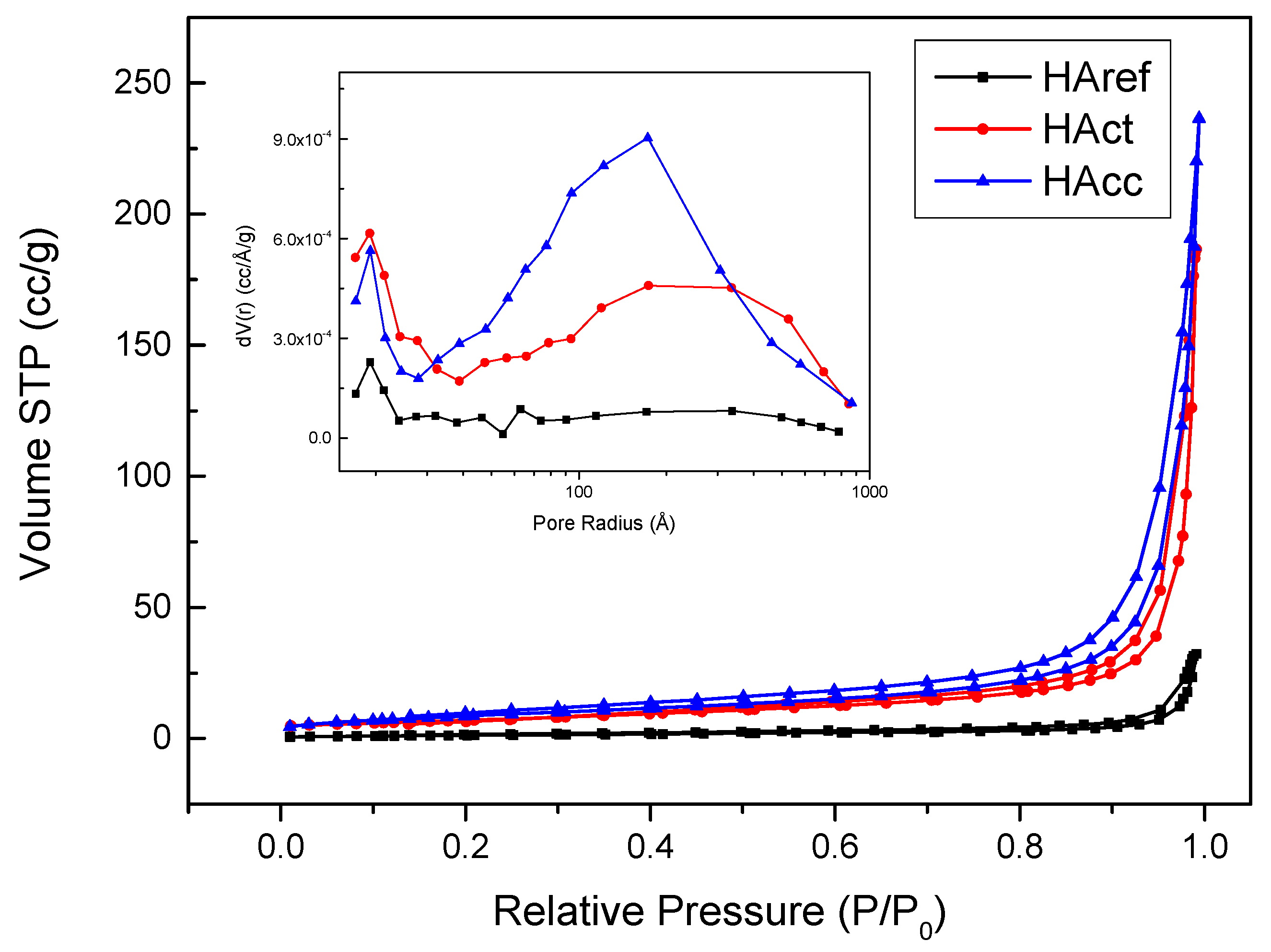
3.1. Density, Relative Density, and Textural Properties
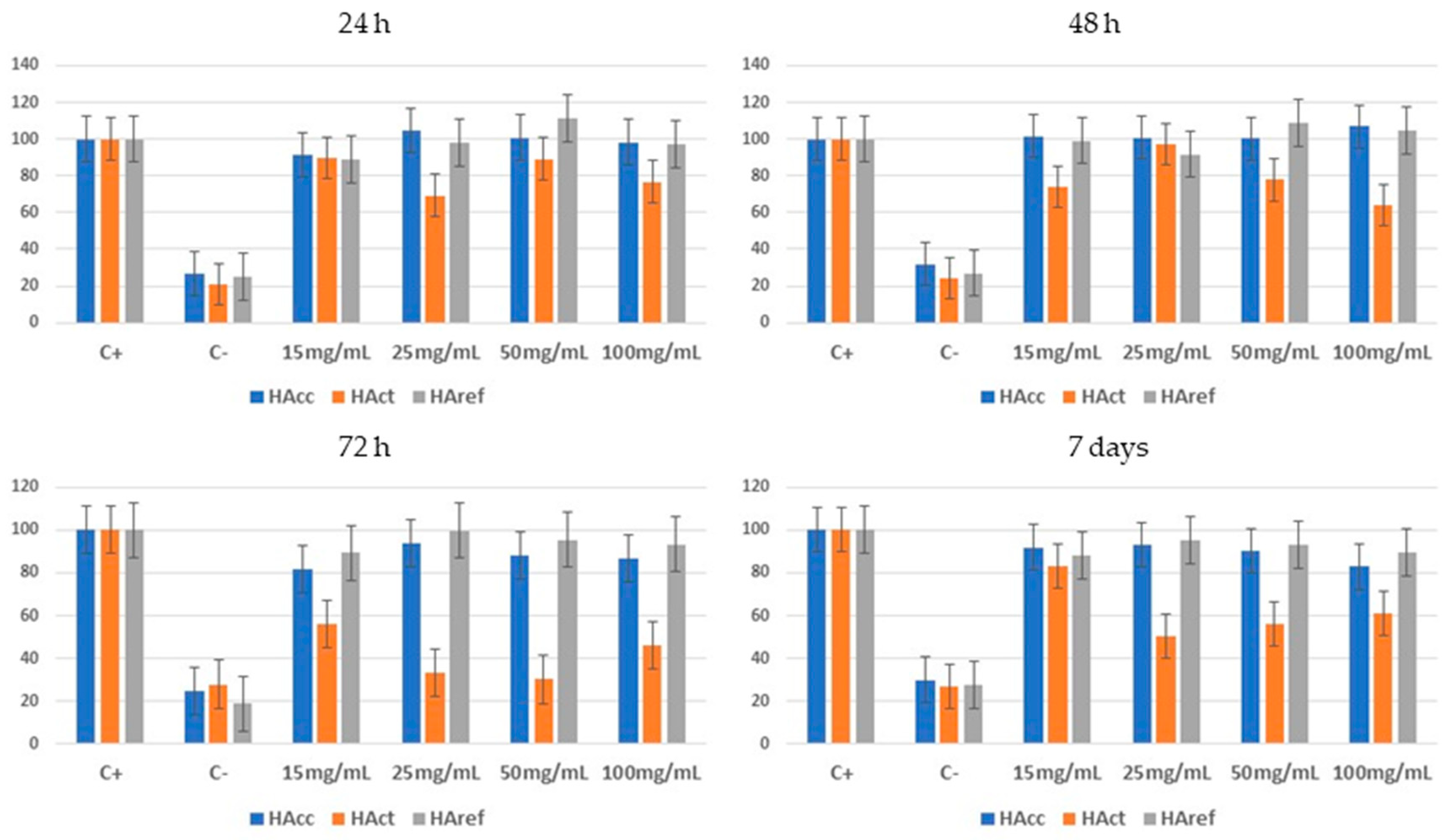
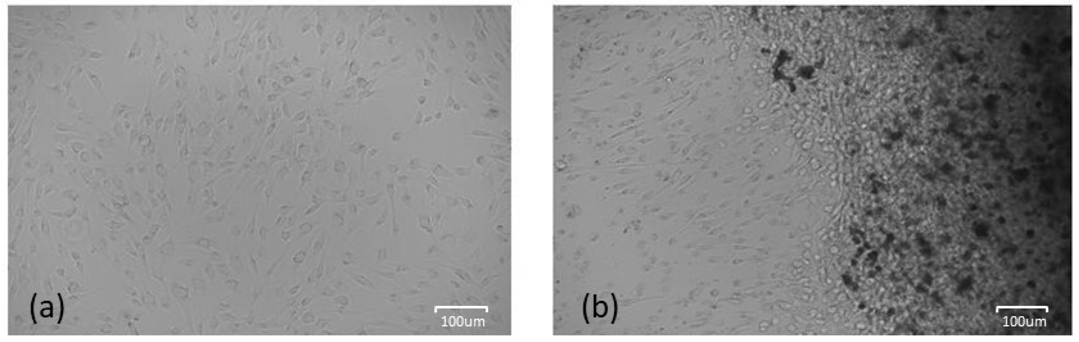
3.2. Cell Viability Assay
4. Conclusions
5. Patents
Author Contributions
Funding
Data Availability Statement
Conflicts of Interest
References
- Wood, N.J.; Jenkinson, H.F.; Davis, S.A.; Mann, S.; O’Sullivan, D.J.; Barbour, M.E. Chlorhexidine hexametaphosphate nanoparticles as a novel antimicrobial coating for dental implants. J. Mater. Sci. Mater. Med. 2015, 26, 201. [Google Scholar] [CrossRef]
- Siqueira, W.L.; Custodio, W.; McDonald, E.E. New insights into the composition and functions of the acquired enamel pellicle. J. Dent. Res. 2012, 91, 1110–1118. [Google Scholar] [CrossRef]
- Subramani, K.; Jung, R.E.; Molenberg, A.; Hammerle, C.H.F. Biofilm on dental implants: A review of the literature. Int. J. Oral Maxillofac. Implants. 2009, 24, 616–626. [Google Scholar] [CrossRef]
- Keegan, G.M.; Learmonth, I.D.; Case, C.P. Orthopaedic metals and their potential toxicity in the arthroplasty patient. A review of current knowledge and future strategies. J. Bone Jt. Surg. Ser. B 2007, 89, 567–573. [Google Scholar] [CrossRef]
- Bourne, R.B.; Chesworth, B.M.; Davis, A.M.; Mahomed, N.N.; Charron, K.D.J.; Met, D. Patient Satisfaction after Total Knee Arthroplasty Who is Satisfied and Who is Not ? Clin. Orthop. Relat. Res. 2010, 468, 57–63. [Google Scholar] [CrossRef]
- Roy, D.M.; Eysel, W.; Dinger, D.R. Hydrothermal synthesis of various carbonate containing calcium hydroxyapatites. Mater. Res. Bull. 1974, 9, 35–39. [Google Scholar] [CrossRef]
- Tampieri, A.; Sprio, S.; Ruffini, A.; Celotti, G.; Lesci, I.G.; Roveri, N. From wood to bone: Multi-step process to convert wood hierarchical structures into biomimetic hydroxyapatite scaffolds for bone tissue engineering. J. Mater. Chem. 2009, 19, 4973–4980. [Google Scholar] [CrossRef]
- Parfen’eva, L.S.; Orlova, T.S.; Kartenko, N.F.; Sharenkova, N.V.; Smirnov, B.I.; Smirnov, I.A.; Misiorek, H.; Jezowski, A.; Mucha, J.; de Arellano-Lopez, A.R.; et al. Thermal and electrical properties of a white-eucalyptus carbon preform for SiC/Si ecoceramics. Phys. Solid State 2006, 48, 441–446. [Google Scholar] [CrossRef]
- Ruffini, A.; Sprio, S.; Tampieri, A. Study of the hydrothermal transformation of wood-derived calcium carbonate into 3D hierarchically organized hydroxyapatite. Chem. Eng. J. 2013, 217, 150–158. [Google Scholar] [CrossRef]
- Roy, D.M.; Linnehan, S.K. Hydroxyapatite formed from Coral Skeletal Carbonate by Hydrothermal Exchange. Nature 1974, 247, 220–222. [Google Scholar] [CrossRef]
- Han, K.-S.; Sathiyaseelan, A.; Saravanakumar, K.; Wang, M.-H. Wound healing efficacy of biocompatible hydroxyapatite from bovine bone waste for bone tissue engineering application. J. Environ. Chem. Eng. 2022, 10, 106888. [Google Scholar] [CrossRef]
- Ashrit, S.S.; Chatti, R.V.; Sarkar, S. Synthesis and characterization of hematite based calcium rich hydroxyapatite—A nano material from LD slag fines. J. Environ. Chem. Eng. 2020, 8, 103581. [Google Scholar] [CrossRef]
- Heimann, R.B. Plasma-Sprayed Hydroxylapatite-Based Coatings: Chemical, Mechanical, Microstructural, and Biomedical Properties. J. Therm. Spray Technol. 2016, 25, 827–850. [Google Scholar] [CrossRef]
- Scalera, F.; Pereira, S.I.A.; Bucciarelli, A.; Tobaldi, D.M.; Quarta, A.; Gervaso, F.; Castro, P.M.L.; Polini, A.; Piccirillo, C. Chitosan-hydroxyapatite composites made from sustainable sources: A morphology and antibacterial study. Mater. Today Sustain. 2023, 21, 100334. [Google Scholar] [CrossRef]
- García, V.S.B.; Mujica, K.H.; Castañeda-Vía, J.A.; Landauro, C.V.; Quispe, J.; Jon, L.Y.T.C. Obtención de hidroxiapatita a través de residuos biológicos para injertos óseos dentales. Rev. Estomatológica Hered. 2021, 31, 111–116. [Google Scholar] [CrossRef]
- Soydal, U.; Marti, M.E.; Ahmetli, G. Using Sugar Mill Waste for Biobased Epoxy Composites. Int. J. Chem. Mol. Eng. 2016, 10, 4. [Google Scholar] [CrossRef]
- Boyetey, M.J.B.; Torgbo, S.; Sukyai, P.; Watthanasakphuban, N.; Kamonsutthipaijit, N. Filter cake-derived calcium carbonate polymorphs from sugar refinery for hydroxyapatite production as a sustainable material for biomedical application. Ceram. Int. 2023, 49, 23417–23425. [Google Scholar] [CrossRef]
- Mahmoud, E.-S.A.; Hassanin, M.A.; Borham, T.I.; Emara, E.I.R. Tolerance of some sugar beet varieties to water stress. Agric. Water Manag. 2018, 201, 144–151. [Google Scholar] [CrossRef]
- Thavornyutikarn, B.; Chantarapanich, N.; Sitthiseripratip, K.; Thouas, G.A.; Chen, Q. Bone tissue engineering scaffolding: Computer-aided scaffolding techniques. Prog. Biomater. 2014, 3, 61–102. [Google Scholar] [CrossRef]
- Nanou, C.P.; De Fuentes, L. Awarenet: Agro-Food Wastes Minimisation and Reduction Network. Waste Manag. Environ. 2002, 56, 1877–1880. [Google Scholar]
- Canales, C.; Cortés, V.; Martínez, J.A. Guía de Mejores Técnicas Disponibles en España del Sector Refino de Petróleo; Revista VirtualPRO: Bogotá, Colombia, 2004. [Google Scholar]
- Suffo, M.; Mata, M.D.L.; Molina, S.I. A sugar-beet waste based thermoplastic agro-composite as substitute for raw materials. J. Clean. Prod. 2020, 257, 120382. [Google Scholar] [CrossRef]
- Yoshimura, M.; Sujaridworakun, P.; Koh, F.; Fujiwara, T.; Pongkao, D.; Ahniyaz, A. Hydrothermal conversion of calcite crystals to hydroxyapatite. Mater. Sci. Eng. C 2004, 24, 521–525. [Google Scholar] [CrossRef]
- Doebelin, N.; Kleeberg, R. Profex: A graphical user interface for the Rietveld refinement program BGMN. J. Appl. Crystallogr. 2015, 48, 1573–1580. [Google Scholar] [CrossRef] [PubMed]
- ISO 14044:2020/Amd 2:2020; Environmental Management—Life Cycle Assessment—Requirements and Guide-Lines—Amendment 2. International Organization for Standardization: Geneva, Switzerland, 2020.
- Wang, M.; Joseph, R.; Bonfield, W. Hydroxyapatite-polyethylene composites for bone substitution: Effects of ceramic particle size and morphology. Biomaterials 1998, 19, 2357–2366. [Google Scholar] [CrossRef] [PubMed]
- Padilla, S.; Izquierdo-Barba, I.; Vallet-Regí, M. High Specific Surface Area in Nanometric Carbonated Hydroxyapatite. Chem. Mater. 2008, 20, 5942–5944. [Google Scholar] [CrossRef]
- Sassoni, E. Hydroxyapatite And Other calcium phosphates for the conservation of cultural heritage: A review. Materials 2018, 11, 557. [Google Scholar] [CrossRef]
- Leena, M.; Rana, D.; Webster, T.J.; Ramalingam, M. Accelerated synthesis of biomimetic nano hydroxyapatite using simulated body fluid. Mater. Chem. Phys. 2016, 180, 166–172. [Google Scholar] [CrossRef]
- Fathi, M.H.; Hanifi, A.; Mortazavi, V. Preparation and bioactivity evaluation of bone-like hydroxyapatite nanopowder. J. Mater. Process. Technol. 2008, 202, 536–542. [Google Scholar] [CrossRef]
- Kong, W.; Zhao, K.; Gao, C.; Zhu, P. Synthesis and characterization of carbonated hydroxyapatite with layered structure. Mater. Lett. 2019, 255, 126552. [Google Scholar] [CrossRef]
- Xu, J.; Yang, Y.; Wan, R.; Shen, Y.; Zhang, W. Hydrothermal preparation and characterization of ultralong strontium-substituted hydroxyapatite whiskers using acetamide as homogeneous precipitation reagent. Sci. World J. 2014, 2014, 863137. [Google Scholar] [CrossRef]
- Ślósarczyk, A.; Paszkiewicz, Z.; Paluszkiewicz, C. FTIR and XRD evaluation of carbonated hydroxyapatite powders synthesized by wet methods. J. Mol. Struct. 2005, 744–747, 657–661. [Google Scholar] [CrossRef]
- Dorozhkin, S.V. Calcium orthophosphates in nature, biology and medicine. Materials 2009, 2, 399–498. [Google Scholar] [CrossRef]
- Hughes, J.M. Structure and Chemistry of the Apatites and Other Calcium Orthophosphates; Elliot, J.C., Ed.; Elsevier: Amsterdam, The Netherlands, 1994; pp. xii + 389. ISBN 0-444-81582-1. [Google Scholar] [CrossRef]
- Murphy, W.; Black, J.; Hastings, G. Handbook of Biomaterial Properties, 2nd ed.; Springer: New York, NY, USA, 2016; Volume 676. [Google Scholar] [CrossRef]
- Daculsi, G.; LeGeros, R.Z.; Nery, E.; Lynch, K.; Kerebel, B. Transformation of biphasic calcium phosphate ceramics in vivo: Ultrastructural and physicochemical characterization. J. Biomed. Mater. Res. 1989, 23, 883–894. [Google Scholar] [CrossRef]
- Liao, C.-J.; Lin, F.-H.; Chen, K.-S.; Sun, J.-S. Thermal decomposition and reconstitution of hydroxyapatite in air atmosphere. Biomaterials 1999, 20, 1807–1813. [Google Scholar] [CrossRef]
- Agbabiaka, O.G.; Oladele, I.O.; Akinwekomi, A.D.; Adediran, A.A.; Balogun, A.O.; Olasunkanm, O.G.; Olayanju, T.M.A. Effect of calcination temperature on hydroxyapatite developed from waste poultry eggshell. Sci. Afr. 2020, 8, e00452. [Google Scholar] [CrossRef]
- Abifarin, J.K.; Obada, D.O.; Dauda, E.T.; Dodoo-Arhin, D. Experimental data on the characterization of hydroxyapatite synthesized from biowastes. Data Br. 2019, 26, 104485. [Google Scholar] [CrossRef] [PubMed]
- Ofudje, E.A.; Rajendran, A.; Adeogun, A.I.; Idowu, M.A.; Kareem, S.O.; Pattanayak, D.K. Synthesis of organic derived hydroxyapatite scaffold from pig bone waste for tissue engineering applications. Adv. Powder Technol. 2018, 29, 1–8. [Google Scholar] [CrossRef]
- Saharudin, S.H.; Shariffuddin, J.H.; Ismail, A.M.; Mah, J.-H. Recovering value from waste: Biomaterials production from marine shell waste. Bull. Mater. Sci. 2018, 41, 162. [Google Scholar] [CrossRef]
- Adenan, N.H.; Zainol, I.; Rahim, N.A.; Jaafar, C.N.A. Extraction of Nanohydroxyapatite from Waste Bovine Bone Using Alkaline Digestion Method. J. Phys. Conf. Ser. 2018, 1082, 12005. [Google Scholar] [CrossRef]
- Zhu, F.; Qiu, Y.; Yeung, H.Y.; Lee, K.M.; Cheng, C.Y.J. Trabecular bone micro-architecture and bone mineral density in adolescent idiopathic and congenital scoliosis. Orthop. Surg. 2009, 1, 78–83. [Google Scholar] [CrossRef]
- Zhu, K.; Hunter, M.; Stuckey, B.G.A.; Walsh, J.P. Establishing a Total Hip T-Score Threshold to Measure Contralateral Hip Bone Mineral Density: Avoiding Missed Diagnosis of Osteoporosis. J. Clin. Densitom. 2022, 25, 577–586. [Google Scholar] [CrossRef]
- Gaujac, N.; Sariali, E.; Grimal, Q. Does the bone mineral density measured on a preoperative CT scan before total hip arthroplasty reflect the bone’s mechanical properties? Orthop. Traumatol. Surg. Res. 2022, 109, 103348. [Google Scholar] [CrossRef]
- Mancini, A.; Frondini, F.; Capezzuoli, E.; Mejia, E.G.; Lezzi, G.; Matarazzi, D.; Brogi, A.; Swennen, R. Porosity, bulk density and CaCO3 content of travertines. A new dataset from Rapolano, Canino and Tivoli travertines (Italy). Data Br. 2019, 25, 104158. [Google Scholar] [CrossRef] [PubMed]
- Youness, R.A.; Taha, M.A.; Elhaes, H.; Ibrahim, M. Molecular modeling, FTIR spectral characterization and mechanical properties of carbonated-hydroxyapatite prepared by mechanochemical synthesis. Mater. Chem. Phys. 2017, 190, 209–218. [Google Scholar] [CrossRef]
- Sing, K.S.W. Reporting physisorption data for gas/solid systems with special reference to the determination of surface area and porosity (Recommendations 1984). Pure Appl. Chem. 1985, 57, 603–619. [Google Scholar] [CrossRef]
- Thommes, M.; Kaneko, K.; Neimark, A.V.; Olivier, J.P.; Rodriguez-Reinoso, F.; Rouquerol, J.; Sing, K.S.W. Physisorption of gases, with special reference to the evaluation of surface area and pore size distribution (IUPAC Technical Report). Pure Appl. Chem. 2015, 87, 1051–1069. [Google Scholar] [CrossRef]
- Minh, D.P.; Lyczko, N.; Sebei, H.; Nzihou, A.; Sharrock, P. Synthesis of calcium hydroxyapatite from calcium carbonate and different orthophosphate sources: A comparative study. Mater. Sci. Eng. B 2012, 177, 1080–1089. [Google Scholar] [CrossRef]
- Bijukumar, D.R.; Segu, A.; Souza, J.C.M.; Li, X.; Barba, M.; Mercuri, L.G.; Jacobs, J.J.; Mathew, M.T. Systemic and local toxicity of metal debris released from hip prostheses: A review of experimental approaches. Nanomedicine 2018, 14, 951–963. [Google Scholar] [CrossRef]


| Morphology | Powder |
|---|---|
| Genesis | Purification process of juice sweetened with lime hydroxide and CO2 |
| Chemical composition | >80% CaCO3; 7% organic matter; oligo-elements (N, K2O, P2O5 and Mg); assimilable organic acids |
| Humidity | <35% |
| Production | 20,000 average annual tons |
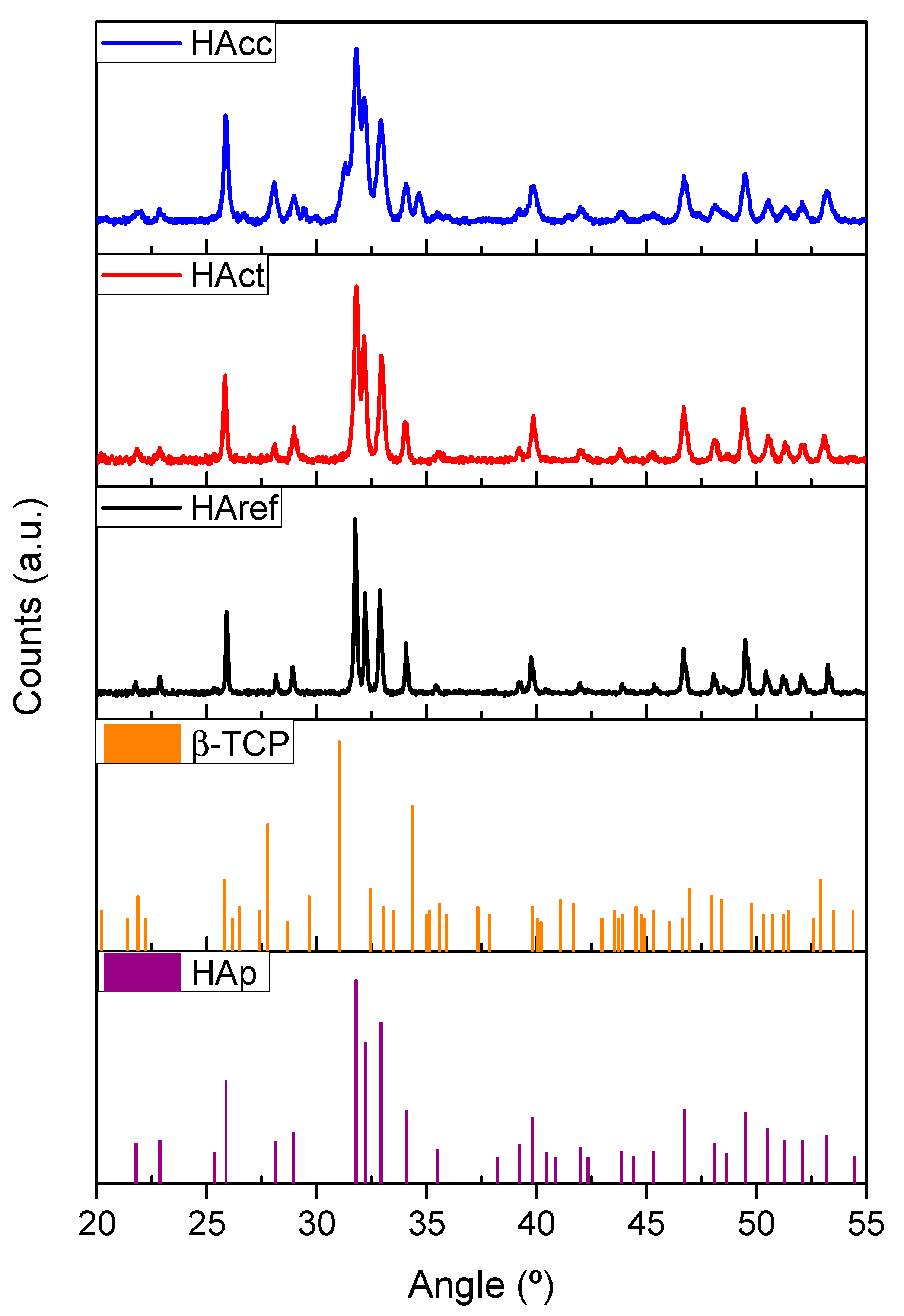
| Sample | Hydroxyapatite (HA) | β-Tricalcium Phosphate (TCP) | ||||
|---|---|---|---|---|---|---|
| % w/w | Lattice Parameters (Å) | Crystallite Sizes (nm) | % w/w | Lattice Parameters (Å) | Crystallite Sizes (nm) | |
| HAcc | 75.17 | a: 9.4160 | (1,0,0):22.2 | 24.83 | a:10.3523 c: 37.1275 | (1,1,1):42.4 |
| c: 6.8877 | (0,0,1):39.5 | |||||
| HAct | 100 | a: 9.4112 | (1,0,0):39.8 | 0 | -- | -- |
| c: 6.8982 | (0,0,1):58.2 | |||||
| HAref | 100 | a: 9.4288 | (1,0,0):85.9 | 0 | -- | -- |
| c: 6.8728 | (0,0,1):150.8 | |||||
Disclaimer/Publisher’s Note: The statements, opinions and data contained in all publications are solely those of the individual author(s) and contributor(s) and not of MDPI and/or the editor(s). MDPI and/or the editor(s) disclaim responsibility for any injury to people or property resulting from any ideas, methods, instructions or products referred to in the content. |
© 2023 by the authors. Licensee MDPI, Basel, Switzerland. This article is an open access article distributed under the terms and conditions of the Creative Commons Attribution (CC BY) license (https://creativecommons.org/licenses/by/4.0/).
Share and Cite
Suffo-Pino, M.; Cauqui-López, M.Á.; Pérez-Muñoz, C.; Goma-Jiménez, D.; Fernández-Delgado, N.; Herrera-Collado, M. Biphasic Bioceramic Obtained from Byproducts of Sugar Beet Processing for Use in Bioactive Coatings and Bone Fillings. J. Funct. Biomater. 2023, 14, 499. https://0-doi-org.brum.beds.ac.uk/10.3390/jfb14100499
Suffo-Pino M, Cauqui-López MÁ, Pérez-Muñoz C, Goma-Jiménez D, Fernández-Delgado N, Herrera-Collado M. Biphasic Bioceramic Obtained from Byproducts of Sugar Beet Processing for Use in Bioactive Coatings and Bone Fillings. Journal of Functional Biomaterials. 2023; 14(10):499. https://0-doi-org.brum.beds.ac.uk/10.3390/jfb14100499
Chicago/Turabian StyleSuffo-Pino, Miguel, Miguel Ángel Cauqui-López, Celia Pérez-Muñoz, Daniel Goma-Jiménez, Natalia Fernández-Delgado, and Miriam Herrera-Collado. 2023. "Biphasic Bioceramic Obtained from Byproducts of Sugar Beet Processing for Use in Bioactive Coatings and Bone Fillings" Journal of Functional Biomaterials 14, no. 10: 499. https://0-doi-org.brum.beds.ac.uk/10.3390/jfb14100499