Article

Path Tracing vs. Volume Rendering Technique in Post-Surgical Assessment of Bone Flap in Oncologic Head and Neck Reconstructive Surgery: A Preliminary Study

1
Radiology Unit, Department of Pathology and Diagnostics, University Hospital of Verona, Piazzale Aristide Stefani, 1, 37126 Verona, Italy
2
Otolaryngology-Head and Neck Surgery Department, University Hospital of Verona, Piazzale Aristide Stefani, 1, 37126 Verona, Italy
3
Unit of Maxillo-Facial Surgery and Dentistry, University of Verona, P.le L.A. Scuro 10, 37134 Verona, Italy
4
Department of Radiology, G.B. Rossi University Hospital, University of Verona, 37134 Verona, Italy
*
Author to whom correspondence should be addressed.
Submission received: 21 September 2022 / Revised: 15 November 2022 / Accepted: 26 December 2022 / Published: 20 January 2023

Abstract

:
This study aims to compare a relatively novel three-dimensional rendering called Path Tracing (PT) to the Volume Rendering technique (VR) in the post-surgical assessment of head and neck oncologic surgery followed by bone flap reconstruction. This retrospective study included 39 oncologic patients who underwent head and neck surgery with free bone flap reconstructions. All exams were acquired using a 64 Multi-Detector CT (MDCT). PT and VR images were created on a dedicated workstation. Five readers, with different expertise in bone flap reconstructive surgery, independently reviewed the images (two radiologists, one head and neck surgeon and two otorhinolaryngologists, respectively). Every observer evaluated the images according to a 5-point Likert scale. The parameters assessed were image quality, anatomical accuracy, bone flap evaluation, and metal artefact. Mean and median values for all the parameters across the observer were calculated. The scores of both reconstruction methods were compared using a Wilcoxon matched-pairs signed rank test. Inter-reader agreement was calculated using Spearman’s rank correlation coefficient. PT was considered significantly superior to VR 3D reconstructions by all readers (p < 0.05). Inter-reader agreement was moderate to strong across four out of five readers. The agreement was stronger with PT images compared to VR images. In conclusion, PT reconstructions are significantly better than VR ones. Although they did not modify patient outcomes, they may improve the post-surgical evaluation of bone-free flap reconstructions following major head and neck surgery.

1. Introduction

Modern multi-detector computed tomography (MDCT) can acquire high-resolution images in a relatively low acquisition time [1,2]. In addition, the resulting isotropic millimetric or sub millimetric voxels allow multiplanar image reformatting without loss of spatial resolution. Although high quality, the acquired images and derived bi-dimensional reconstruction may not be as informative as a three-dimensional volume that radiologists and other physicians can rotate, zoom in and out or cut, to highlight or reveal structures of interest [3]. For these reasons, 3D reconstructions may be helpful to visualise complex anatomy clearly and synthetically.
The Volume Rendering (VR) technique represents today the standard for 3D visualisation, transforming cross-sectional imaging into 3D images. This technique relies on a cast of a single simulated ray of light across the volume for each pixel image. The ray does not stop on the surface, and, going through the volume, it samples the volume data one way. Therefore, this technique does not consider the ray scatter or ray extinction once the ray hits the surface of the volume. To overcome this, a non-realistic shading model simulates the scatter effects, if needed [4].
Another recent approach to visualise 3D volume data is Path Tracing (PT) [5] or its similar commercial implementation, Cinematic Rendering (CR) [6]. In these rendering algorithms, the light model is far more complex. PT includes light scattering and extinction, simulating how the natural light ray behave. The resultant model has many light rays, most of them with complex paths and multiple reflections. The calculation of light scatters and extinctions is computationally expensive, so the light rays and reflections are usually artificially limited. Moreover, the lighting sources are very different compared to VR. There is a global illumination approach in PT or CR, while in VR, there are a finite number of synthetic light sources [7].
As a result, PT or CR images look more natural and physically accurate than VR reconstructions, with more realistic lighting and shadows, which leads to a better depth of field perception [8]. This kind of “photorealistic” appearance may be helpful in post-surgical imaging to better evaluate the immediate outcome of the surgery.
The primary endpoint of this paper is to investigate if these reconstructions are superior to traditional VR images in evaluating complex post-surgical head and neck anatomy with bone flap reconstruction.

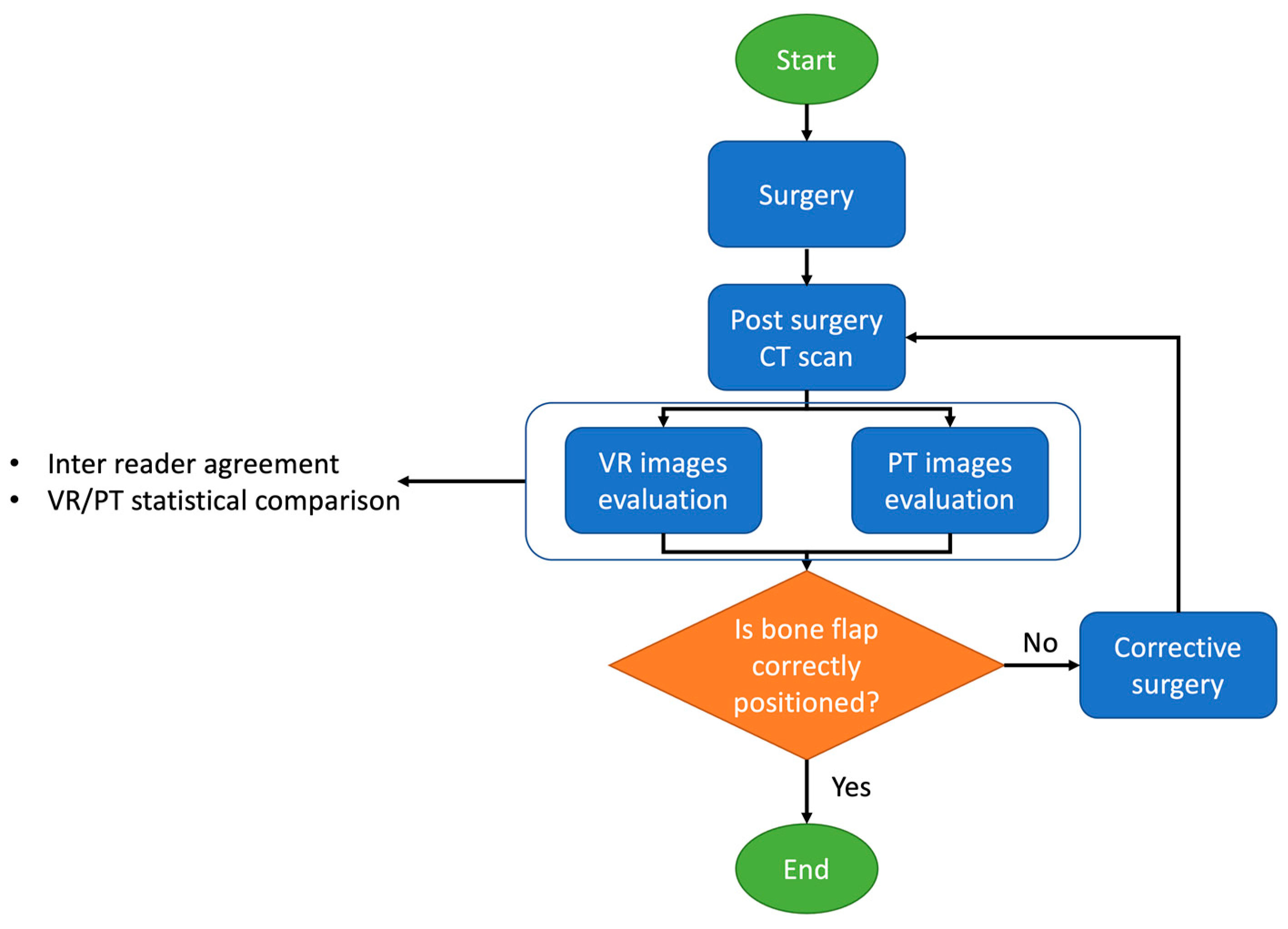
2. Materials and Methods

2.1. Patients Selection

To investigate the hypothesis, a retrospective study of a consecutive series of patients who underwent head and neck reconstructive surgery comprehensive of bony reconstruction with free flaps was performed. Informed consent to perform CT scan was waived. Patients were treated in a tertiary center from 2017 to 2021 (a mean of 7.8 patients/year). Inclusion and exclusion criteria are listed in Table 1.
Post-traumatic bone reconstructions were excluded from this study to maintain the study group homogeneous from a surgical point of view, especially for bone flap evaluation.

2.2. CT Acquisition and Post Elaboration

To assess the bone flap, we acquired a CT scan 24 h after surgery, as our head and neck surgeons routinely required. The rationale of the post-surgery CT is to check eventual bone flap and osteosynthesis material mispositioning that may benefit from immediate surgical correction. Fortunately, no patients required a revision of bone flap positioning after the post-surgery CT in our series. CT scans were acquired on 64 rows of multi-detector equipment (Philips Brilliance 64, Philips, Eindhoven, Nederland). Parameters were as follows: 120 kV, 200 mAS, slice thickness 0.8 mm, pitch 0.4, bone and soft tissue kernel reconstruction.
For every patient, two VR and two PT 3D reconstructions, with the same angle of view, were elaborated on a workstation with an Nvidia RTX 2080 Ti graphic processing unit (GPU), respectively with a free dicom viewer (Medixant. RadiAnt DICOM Viewer. Version 2020.2. 19 July 2020. URL: https://www.radiantviewer.com, accessed on 28 August 2020) for VR and a custom MevisLab script (MeVisLab version 3.4, MeVis Medical Solutions AG) and his PathTracing module. All the anonymised images, in the form of screenshots, were arranged randomly in two separate files for VR and PT.

2.3. Images Evaluations and Statistical Analysis

The images were then evaluated independently by five observers, two radiologists with 2 and 10 years of expertise in the field (NC and MDO), one head and neck surgeon with 10 years of experience (VF) and two otorhinolaryngologists with 2 (RN) and 15 years (GM) of experience in bone flap reconstructions. If requested, the observers may require additional images to highlight the surgical reconstruction better. No observers required more projections. All the observers were blinded from the surgical procedure of the patients. Moreover, the head and neck surgeons and otorhinolaryngologists who evaluated the images did not perform the surgery to avoid patients’ recognition bias.
Every observer evaluated the images using a 5-point Likert scale reported in Table 2. For each parameter, mean and median values were calculated. A Wilcoxon matched-pairs signed-rank test was performed to compare the reconstructions. Interobserver agreement was assessed using Spearman’s rank correlation coefficient. A p-value of less than 0.05 indicated statistical significance for both tests.
In Figure 1 is summarised the flowchart of the study.

3. Results

3.1. Patients

According to the inclusion and exclusion criteria listed previously, 39 patients (25 males and 15 females; mean age 58.18 ± 12.10 years) were enrolled in the study. Among bone flap reconstructions, there were 10 scapular (Figure 2 and Figure 3), 28 fibular (Figure 4) and one medial femoral condyle flap. There were 25 squamous cell carcinoma patients (19 primary and six recurrent. According to TNM classification, there were 23 T4 and two T3 tumors), three ameloblastomas, one ameloblastic carcinoma (T4), one poorly differentiated carcinoma (T4), one basal cell carcinoma (T4), one osteosarcoma (T2 according to Enneking classification), one metastasis from breast adenocarcinoma, one inverted papilloma, one myxoid haemangioma, one radio-induced osteoblastic osteosarcoma, two bone radio-necrosis and one osteomyelitis.

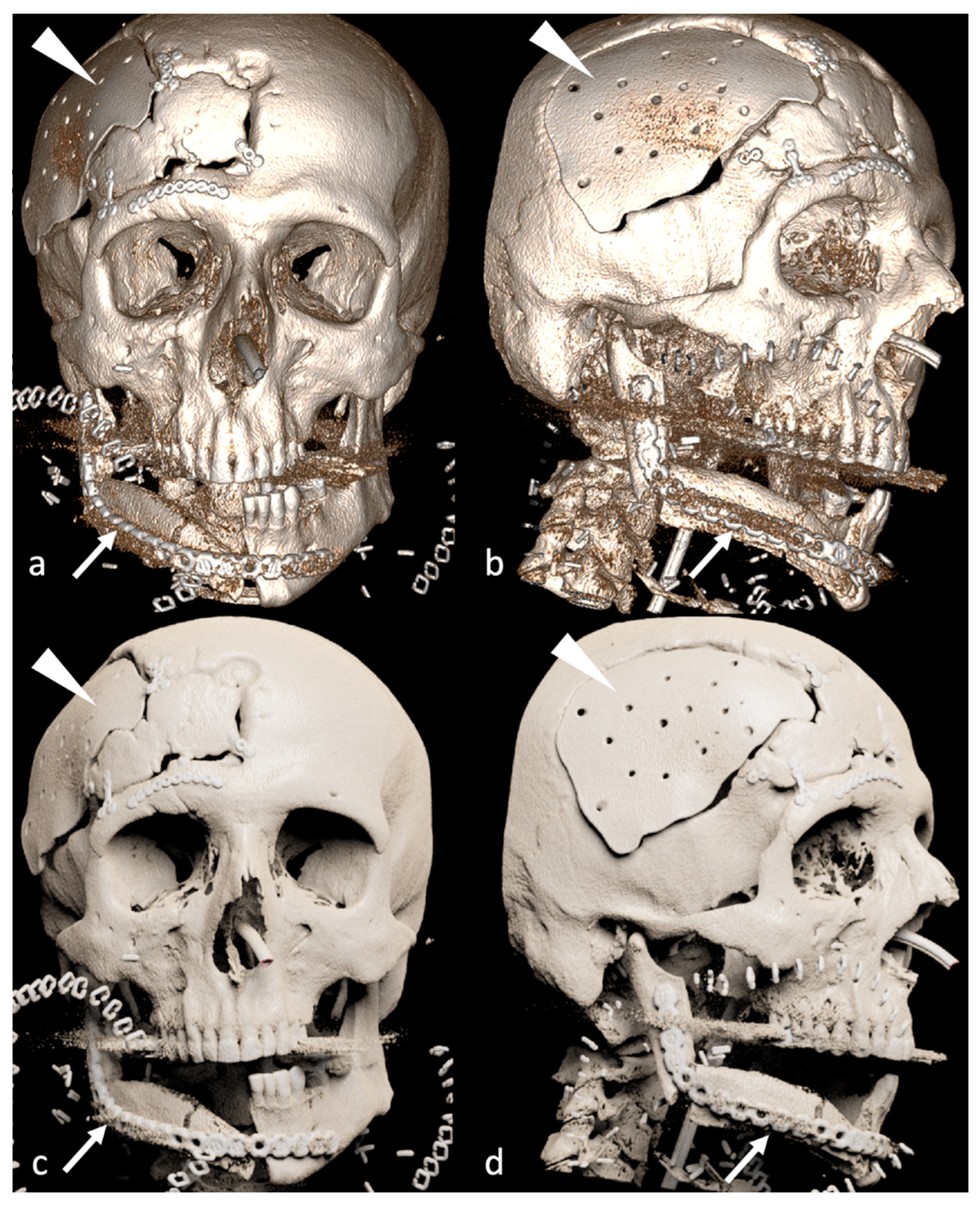
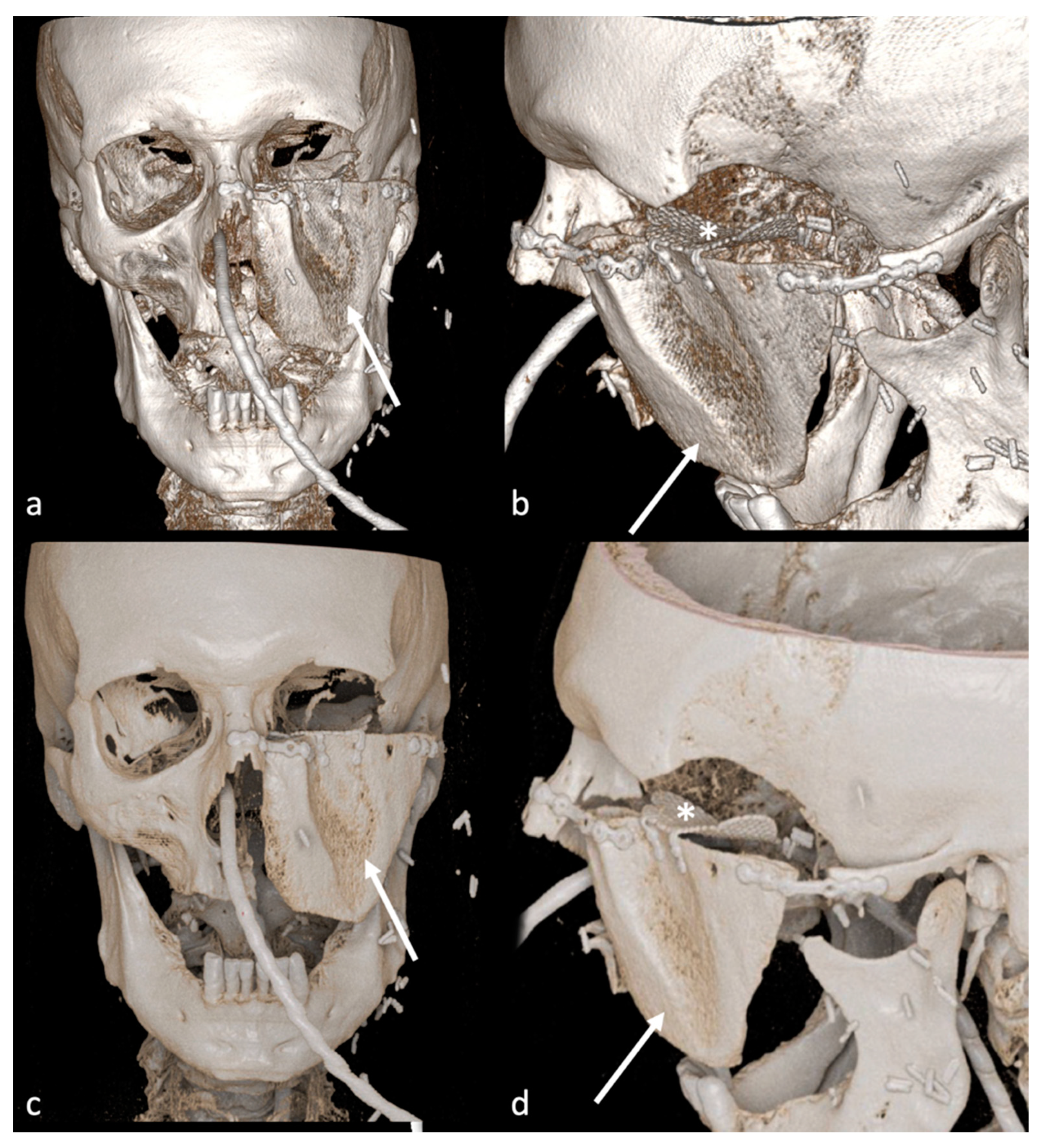
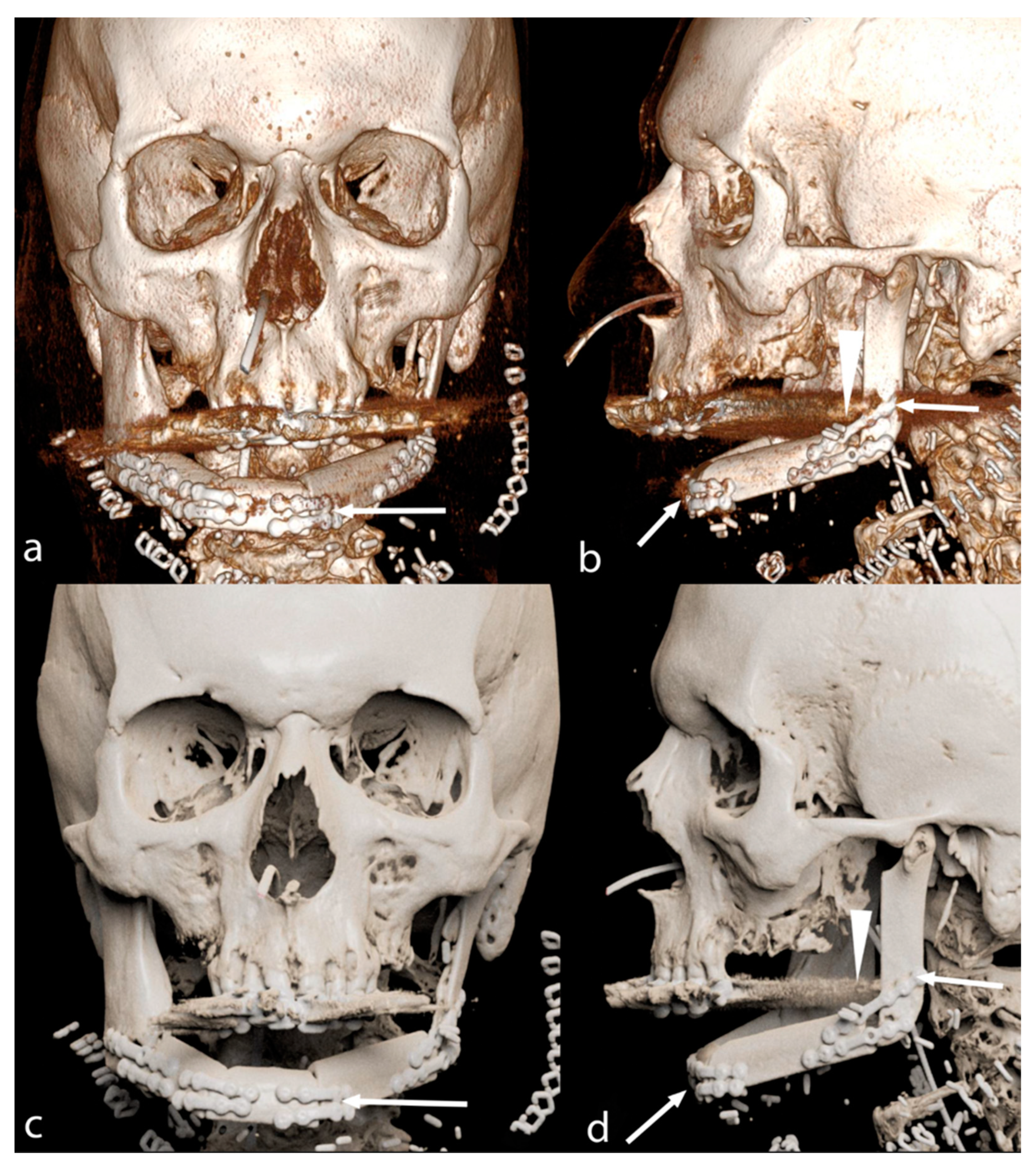
3.2. Image Analysis—VR vs. PT

PT images were considered significantly superior to VR 3D reconstructions by all readers (p < 0.05), with similar evaluation across all the four parameters considered, except for metal artefacts for reader RN, which didn’t find statistically significant differences between VR and PT images (Table 3).
Similar results were found if all readers’ median and mean values were considered together. In particular, the general image quality had a median and mean score of 4.00 and 3.99 in PT compared to 3.00 and 3.17 in VR reconstructions. PT images retained higher anatomical accuracy (4.25 and 4.09 median and mean value, respectively), than VR images. The bone flap was better depicted in PT images, with a median and mean score of 4.25 and 4.11, compared to 3.25 and 3.11 in VR images. Furthermore, PT reconstructions are less prone to metal artefacts than VR reconstructions, with a median and mean score of 3.75 and 3.69 (Table 4).

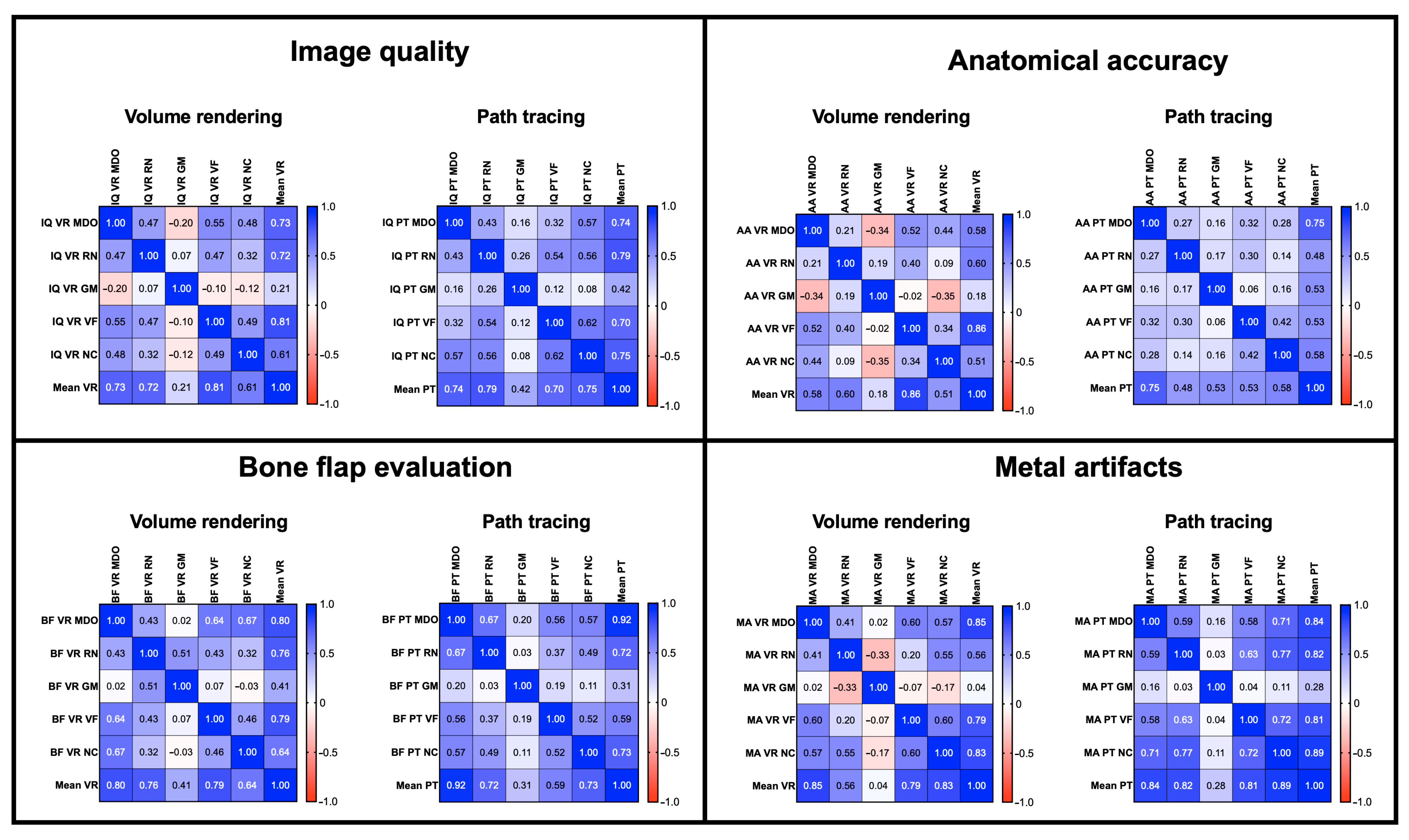
3.3. Image Analysis—Inter-Reader Agreement

The inter-reader agreement was moderate to strong across three readers with a maximum agreement for image quality of ρ = 0.55 (VR) and 0.62 (PT), for anatomical accuracy of ρ = 0.518 (VR) and 0.42 (PT), for bone flap evaluation of ρ = 0.67 (VR) and 0.67 (PT), for metal artefacts of ρ = 0.60 (VR) and ρ = 0.77 (PT) (Figure 5). As shown in Table 3, despite the fourth reader (GM) finding PT significantly better than VR images, he generally showed no significant agreement with the other observers.
However, there is a tendency for a better agreement in path tracing than volume rendering images among all readers.

4. Discussion

In this paper, we quantitatively investigated the possible superiority of PT compared to VR reconstructions in complex head and neck surgery followed by free bone flap reconstructions. The superiority of PT images resulted statistically significant across the different readers. Moreover, PT reconstructions are rated to be less prone to metal artefacts by three out of five readers (Figure 3), which may help further the evaluations of the bone flap in case of a non-removable dental prosthesis or metal fixation materials used in the surgery. These advantages may lead to a better post-surgical evaluation of bone flap reconstruction and immediate surgical revision if something is not correctly positioned. However, in our series, none of the patients required surgical revision even if evaluated only with VR reconstructions, so the clinical impact of these images remains unclear.
Despite the readers finding that almost all parameters were better defined in PT than in VR images, the interrater agreement was low to moderate and non-significant for one reader (GM), who still judged PT images significantly better than VR images. Looking at the single scores assigned by readers, this may be due to a slightly different evaluation among the same patients. However, as shown in Figure 3, the interrater agreement in PT images is generally higher than in VR rendering images, probably because of consistently higher scores of PT reconstructions.
Traditional VR 3D reconstructions have already proved helpful in evaluating complex anatomy in pre-and post-surgical settings in otolaryngology [9], craniofacial surgery planning [10,11], in trauma patients [12,13] and vascular pathology [14].
On behalf of volumetric visualisation, PT rendering is different from VR to generate 3D images. The newest technique allows elaborating photorealistic representation of volumetric data with a more natural shape, shadows and depth of field. PT, also called Cinematic Rendering (CR), has already been demonstrated to be feasible in multiple clinical situations, such as gastro-intestinal [15,16,17,18,19], genito-urinary [20,21,22], cardiovascular [23,24,25] and musculoskeletal imaging [26]. However, most papers on PT or CR usually showcase the technique without evaluating the benefits of these kinds of images generated.
In literature, few papers quantitatively investigate this aspect, as conducted in this paper. Binder et al. evaluated the role of CR compared to conventional computed tomography in anatomy teaching. They found an overall time reduction of image interpretation by medical students of about 65.56%. Moreover, musculoskeletal and vascular anatomy rendering was significantly rated higher than conventional CT visualisation [27]. Vascular anatomy for pre-surgical planning was also significantly better evaluated and with faster comprehension with CR [28]. However, CR was found inferior to axial contrast-enhanced angiography in predicting vascular invasion by deep soft tissue sarcomas, so these images should be used in association with traditional 2D visualisation [29].
Wollschlaeger et al. evaluated the impact of CR compared to VR for pre-surgical lower limb fracture. They conclude that CR images are significantly superior to VR in anatomical accuracy and image quality [30].
This technique’s “photorealistic” nature of the images generated may better comprehend complex situations. The different and more complex lighting models used and, consequently, the realistic shadows of the images highlight depth perception. This is especially evident in overlapping anatomical structures, like maxillo-facial anatomy. Steffen et al. recently found this in applying Cinematic Rendering to facial skeleton [31]. Moreover Elshafei et al. demonstrated that CR leads to a significantly faster and easier comprehension of surgical hepatopancreatobiliary tumors anatomy. More interestingly, the results are independent of surgeon experience level [28].
One drawback of these reconstructions is that they require more computational power than traditional VR acquisition. For example, in our workstation, while interactive VR was rendered in real-time above 60 frames per second, a single PT image required about 5 s to generate, so it is impossible to visualise a PT volume in real-time like VR reconstructions to date.
The study has several limitations. First of all, it is a retrospective study. A prospective clinical study will be more appropriate to evaluate the impact of the PT images compared to VR ones, and to evaluate if PT images lead to different post-surgical management of the patients. As mentioned previously, even with only the VR reconstructions, our head and neck surgeons did not perform corrective surgery on this series of patients. The second limitation is the relatively low number of cases. A higher number of patients may highlight the differences between the two techniques and modify the inter-reader agreement. Third, since generating PT images required elaboration time, the readers evaluated the patients based on a few VR and PT reconstructions pictures. This approach lacks interactivity granted by traditional volumetric visualization, hence may influence observers’ evaluation. However, all the observers did not require more images. Moreover, the paper focused on the differences in 3D imaging visualization technique, highlighted in the same images from the same point of view. The availability of a full 3D model may modify the score and need to be investigated once the PT rendering performance will be similar of VR to experience the same level of interactivity. The lower performance and the interface lag of PT rendering may be frustrating for some users.

5. Future Perspectives

Once established in a prospective study the impact of these images elaborated with the PT algorithm, a future perspective may be the application of virtual reality and high-performing computing. The combination may allow real-time visualisation of all acquired volume with enhanced depth of field perception and more visualising freedom.

6. Conclusions

Thanks to its enhanced lighting model with realistic shadows and better depth perceptions, PT images are superior to traditional VR images in general image quality, anatomical accuracy and bone flap evaluation. Moreover, three out of four readers rated PT images less prone to metal artefacts. Consequently, PT images may be more helpful than traditional VR images in comprehending post-surgery head and neck bone flap reconstructions.

Author Contributions

Conceptualization, N.C.; methodology, N.C. and R.N.; writing—original draft preparation, N.C.; writing—review and editing, N.C. and R.N.; cases randomization, A.F.; visualization, G.M. and V.F.; supervision, M.D., D.M. and S.M. All authors have read and agreed to the published version of the manuscript.

Funding

This research received no external funding.

Institutional Review Board Statement

The ethic approval was waived due to the following reasons: it is a retrospective study performed on already anonymized patients’ database. The paper is about a post-elaboration technique, no additional scans were necessary. This new kind of reconstruction did not modify the clinical outcome of the patients.

Informed Consent Statement

Informed consent to perform CT scan was obtained from all subjects involved in the study.

Data Availability Statement

Data is not available due to privacy restrictions.

Conflicts of Interest

The authors declare no conflict of interest.

References

  1. Flohr, T.; Schaller, S.; Stierstorfer, K.; Bruder, H.; Ohnesorge, B.; Schoepf, J. Multi-detector row CT systems and image-reconstruction techniques. Radiology 2005, 235, 756–773. [Google Scholar] [CrossRef] [PubMed]
  2. Fuchs, T.; Kachelrieß, M.; Kalender, W.A. Technical advances in multi–slice spiral CT. Eur. J. Radiol. 2000, 36, 69–73. [Google Scholar] [CrossRef] [PubMed]
  3. Prokop, M. General principles of MDCT. Eur. J. Radiol. 2003, 45, S4–S10. [Google Scholar] [CrossRef] [PubMed]
  4. Luccichenti, G.; Cademartiri, F.; Pezzella, F.R.; Runza, G.; Belgrano, M.; Midiri, M.; Sabatini, U.; Bastianello, S.; Krestin, G.P. 3D reconstruction techniques made easy: Know-how and pictures. Eur. Radiol. 2005, 15, 2146–2156. [Google Scholar] [CrossRef]
  5. El-Seoud, S.A.; Mady, A.S.; Rashed, E. An Interactive Mixed Reality Ray Tracing Rendering Mobile Application of Medical Data in Minimally Invasive Surgeries. Int. J. Online Biomed. Eng. 2019, 15, 4–14. [Google Scholar] [CrossRef] [Green Version]
  6. Comaniciu, D.; Engel, K.; Georgescu, B.; Mansi, T. Shaping the Future through Innovations: From Medical Imaging to Precision Medicine 1. Cinematic Rendering: Photorealistic Visualisation of Medical Images. Med. Image Anal. 2016, 33, 19–26. [Google Scholar] [CrossRef]
  7. Debevec, P. Rendering synthetic objects into real scenes: Bridging traditional and image-based graphics with global illumination and high dynamic range photography. In Proceedings of the 25th Annual Conference on Computer Graphics and Interactive Techniques, Orlando, FL, USA, 19–24 July 1998. [Google Scholar]
  8. Dappa, E.; Higashigaito, K.; Fornaro, J.; Leschka, S.; Wildermuth, S.; Alkadhi, H. Cinematic rendering—An alternative to volume rendering for 3D computed tomography imaging. Insights Imaging 2016, 7, 849–856. [Google Scholar] [CrossRef] [Green Version]
  9. Guo, Y.; Liu, Y.; Lu, Q.-H.; Zheng, K.-H.; Shi, L.-J.; Wang, Q.-J. CT two-dimensional reformation versus three-dimensional volume rendering with regard to surgical findings in the preoperative assessment of the ossicular chain in chronic suppurative otitis media. Eur. J. Radiol. 2013, 82, 1519–1524. [Google Scholar] [CrossRef]
  10. Vannier, M.W.; Marsh, J.L.; Warren, J.O. Three dimensional CT reconstruction images for craniofacial surgical planning and evaluation. Radiology 1984, 150, 179–184. [Google Scholar] [CrossRef]
  11. Song, S.W.; Jun, B.C.; Chae, S.R.; Kim, B.G. Clinical Utility of Three-Dimensional Facial Computed Tomography in the Treatment of Nasal Bone Fractures: A New Modality Involving an Air-Bone View with a Volume Rendering Technique. Indian J. Otolaryngol. Head Neck Surg. 2013, 65, 210–215. [Google Scholar] [CrossRef]
  12. Schmidle, G.; Rieger, M.; Klauser, A.; Thauerer, M.; Hoermann, R.; Gabl, M. Intraosseous rotation of the scaphoid: Assessment by using a 3D CT model—An anatomic study. Eur. Radiol. 2014, 24, 1357–1365. [Google Scholar] [CrossRef] [PubMed]
  13. Falchi, M.; Rollandi, G.A. CT of pelvic fractures. Eur. J. Radiol. 2004, 50, 96–105. [Google Scholar] [CrossRef] [PubMed]
  14. Lawler, L.P.; Fishman, E.K. Multi–Detector Row CT of Thoracic Disease with Emphasis on 3D Volume Rendering and CT Angiography. RadioGraphics 2001, 21, 1257–1273. [Google Scholar] [CrossRef] [PubMed] [Green Version]
  15. Rowe, S.P.; Chu, L.C.; Fishman, E.K. Cinematic rendering of small bowel pathology: Preliminary observations from this novel 3D CT visualisation method. Abdom. Radiol. 2018, 43, 2928–2937. [Google Scholar] [CrossRef]
  16. Rowe, S.P.; Chu, L.C.; Fishman, E.K. Evaluation of Stomach Neoplasms with 3-Dimensional Computed Tomography: Focus on the Potential Role of Cinematic Rendering. J. Comput. Assist. Tomogr. 2018, 42, 661–666. [Google Scholar] [CrossRef]
  17. Chu, L.C.; Rowe, S.P.; Fishman, E.K. Cinematic rendering of focal liver masses. Diagn. Interv. Imaging 2019, 100, 467–476. [Google Scholar] [CrossRef]
  18. Rowe, S.P.; Chu, L.C.; Fishman, E.K. Computed Tomography Cinematic Rendering in the Evaluation of Colonic Pathology. J. Comput. Assist. Tomogr. 2019, 43, 475–484. [Google Scholar] [CrossRef]
  19. Rowe, S.P.; Chu, L.C.; Fishman, E.K. Initial experience with 3D CT cinematic rendering of acute pancreatitis and associated complications. Abdom. Radiol. 2020, 45, 1290–1298. [Google Scholar] [CrossRef]
  20. Rowe, S.P.; Chu, L.C.; Meyer, A.R.; Gorin, M.A.; Fishman, E.K. The application of cinematic rendering to CT evaluation of upper tract urothelial tumors: Principles and practice. Abdom. Radiol. 2019, 44, 3886–3892. [Google Scholar] [CrossRef]
  21. Rowe, S.P.; Meyer, A.R.; Gorin, M.A.; Chu, L.C.; Fishman, E.K. Evaluation of the urinary bladder using three-dimensional CT cinematic rendering. Diagn. Interv. Imaging 2020, 101, 771–781. [Google Scholar] [CrossRef]
  22. Jin, Y.-R.; Yang, X.-L.; Li, Q.-Q.; Yan, Z.-L.; Yang, H.-Y.; Liao, C. CT of ovarian cancer: 3D cinematic rendering for preoperative evaluation. J. Ovarian Res. 2018, 11, 86. [Google Scholar] [CrossRef] [PubMed] [Green Version]
  23. Arias, E.A.; Sandoval, J.P.; Farjat-Pasos, J.I.; Rodríguez-Zanella, H.; de la Mora-Cervantes, R. Novel Cinematic Rendering Use for Ventricular Septal Ruptures. JACC Cardiovasc. Interv. 2021, 14, 1151–1153. [Google Scholar] [CrossRef] [PubMed]
  24. Zimmerman, S.L.; Rowe, S.P.; Fishman, E.K. Cinematic rendering of CT angiography for visualization of complex vascular anatomy after hybrid endovascular aortic aneurysm repair. Emerg. Radiol. 2021, 28, 839–843. [Google Scholar] [CrossRef] [PubMed]
  25. Colli, A.; Tua, L.; Punzo, B.; Baldi, D.; Cademartiri, F.; Gerosa, G. Cinematic Rendering: An Alternative to Classical Volume Rendering for Acute Aortic Dissection. Ann. Thorac. Surg. 2019, 108, e121. [Google Scholar] [CrossRef]
  26. Dreizin, D.; LeBedis, C.A.; Nascone, J.W. Imaging Acetabular Fractures. Radiol. Clin. North Am. 2019, 57, 823–841. [Google Scholar] [CrossRef]
  27. Binder, J.S.; Scholz, M.; Ellmann, S.; Uder, M.; Grützmann, R.; Weber, G.F.; Krautz, C. Cinematic Rendering in Anatomy: A Crossover Study Comparing a Novel 3D Reconstruction Technique to Conventional Computed Tomography. Anat. Sci. Educ. 2020, 14, 22–31. [Google Scholar] [CrossRef]
  28. Elshafei, M.; Binder, J.; Baecker, J.; Brunner, M.; Uder, M.; Weber, G.F.; Grützmann, R.; Krautz, C. Comparison of Cinematic Rendering and Computed Tomography for Speed and Comprehension of Surgical Anatomy. JAMA Surg. 2019, 154, 738–744. [Google Scholar] [CrossRef] [Green Version]
  29. Li, K.; Yan, R.; Ma, H.; Zhang, D.-F.; Ding, Y.; Li, Z. Value of the Cinematic Rendering From Volumetric Computed Tomography Data in Evaluating the Relationship Between Deep Soft Tissue Sarcomas of the Extremities and Adjacent Major Vessels. J. Comput. Assist. Tomogr. 2019, 43, 386–391. [Google Scholar] [CrossRef]
  30. Wollschlaeger, L.; Boos, J.; Jungbluth, P.; Grassmann, J.-P.; Schleich, C.; Latz, D.; Kroepil, P.; Antoch, G.; Windolf, J.; Schaarschmidt, B. Is CT-based cinematic rendering superior to volume rendering technique in the preoperative evaluation of multifragmentary intraarticular lower extremity fractures? Eur. J. Radiol. 2020, 126, 108911. [Google Scholar] [CrossRef]
  31. Steffen, T.; Winklhofer, S.; Starz, F.; Wiedemeier, D.; Ahmadli, U.; Stadlinger, B. Three-dimensional perception of cinematic rendering versus conventional volume rendering using CT and CBCT data of the facial skeleton. Ann. Anat.-Anat. Anz. 2022, 241, 151905. [Google Scholar] [CrossRef]
Figure 1. Flowchart of the study.
Jimaging 09 00024 g001
Figure 2. VR (a,b) and PT (c,d) reconstruction of scapular bone flap (arrows). The major depth of field and the better handling of metal artefacts of PT reconstructions are the key factors for better understanding post-surgery imaging compared to VR reconstructions. The patient underwent previous cranial vault surgery for trauma (arrowheads).
Jimaging 09 00024 g002
Figure 3. VR (a,b) and PT (c,d) reconstruction of the scapular bone flap (arrows). In PT image (d), the mesh of the orbit floor (asterisks) is better depictable than in VR image (b).
Jimaging 09 00024 g003
Figure 4. VR (a,b) and PT (c,d) reconstruction of fibular bone flap. In PT images, there are fewer metal artefacts from non-removable dental implants (arrowheads) and metal bone flap fixation materials (arrows).
Jimaging 09 00024 g004
Figure 5. Inter-reader agreement correlation matrixes across all four parameters in VR and PT reconstructions (red negative correlation, blue positive correlation).
Jimaging 09 00024 g005
Table 1. Inclusion and exclusion criteria.
Inclusion Criteria Exclusion Criteria
-
Major head and neck surgery and microvascular bone reconstruction for oncologic disease and/or its complications
-
Absence of postoperative CT scan
-
Severe motion artefacts
-
Non-oncologic bone reconstruction
Table 2. Evaluated parameters and explanation of the 5-point Likert scale.
Likert’s Scale Scores
Parameters1 2 3 4 5
Image Quality Non-diagnostic Poor quality Good quality Very good quality Excellent quality
Anatomical Accuracy Non-diagnostic Poorly demarcated bone margins Good demarcation of
bone margins
Very good demarcation of
bone margins
Excellent demarcation of bone margins
Bone Flap Evaluation Non-diagnostic Poor quality Good quality Very good quality Excellent quality
Metal Artifact Severe Strong Moderate Mild No artefacts
Table 3. Likert’s scale evaluation results across all readers and parameters. Values are expressed as median (MDO, RN, VF, GM, NC = readers; VR = volume rendering; PT = path tracing).
Image Quality Anatomical Accuracy Bone Flap Evaluation Metal Artifacts
VR PT pVR PT pVR PT pVR PT p
ReaderMDO34<0.05 34<0.05 24<0.05 34<0.05
RN34<0.05 34<0.05 34<0.05 330.65
VF44<0.05 45<0.05 45<0.05 34<0.05
GM34<0.05 34<0.05 34<0.05 34<0.05
NC34< 0.05 44<0.05 34< 0.05 34<0.05
Table 4. Results of the Likert’s scale evaluation of ll readers considered together (VR = volume rendering; PT = path tracing; SD = standard deviation).
Image Quality Anatomical Accuracy Bone Flap Evaluation Metal Artefacts
VR PT VR PT VR PT VR PT
Median34343434
Mean±SD3.19 ± 0.47 4.01 ± 0.56 3.24 ± 0.37 4.14 ± 0.45 3.19 ± 0.55 4.16 ± 0.56 3.15 ± 0.58 3.79 ± 0.64
p<0.05 <0.05 <0.05 <0.05
Disclaimer/Publisher’s Note: The statements, opinions and data contained in all publications are solely those of the individual author(s) and contributor(s) and not of MDPI and/or the editor(s). MDPI and/or the editor(s) disclaim responsibility for any injury to people or property resulting from any ideas, methods, instructions or products referred to in the content.

