German-Wide Analysis of the Prevalence and the Propagation Factors of the Zoonotic Dermatophyte Trichophyton benhamiae

Abstract
:1. Introduction
2. Material and Methods
2.1. Study Structure
2.1.1. Private Breeding
2.1.2. Pets
2.2. Questionnaires
2.3. Diagnostics
2.3.1. Sampling of Guinea Pigs
2.3.2. Culture
2.3.3. DNA Extraction
2.4. PCR Method
- yellow variant: 5′-CGATAGGAATCAACGTTCCATC/5′-CCCCGAAAGAGGAGGT (for/rev)
- white variant: 5′-GATAGGGACCAACGTTCCG/5′-CCCGAAAGAGGGGGC (for/rev)
2.5. Statistics
3. Results
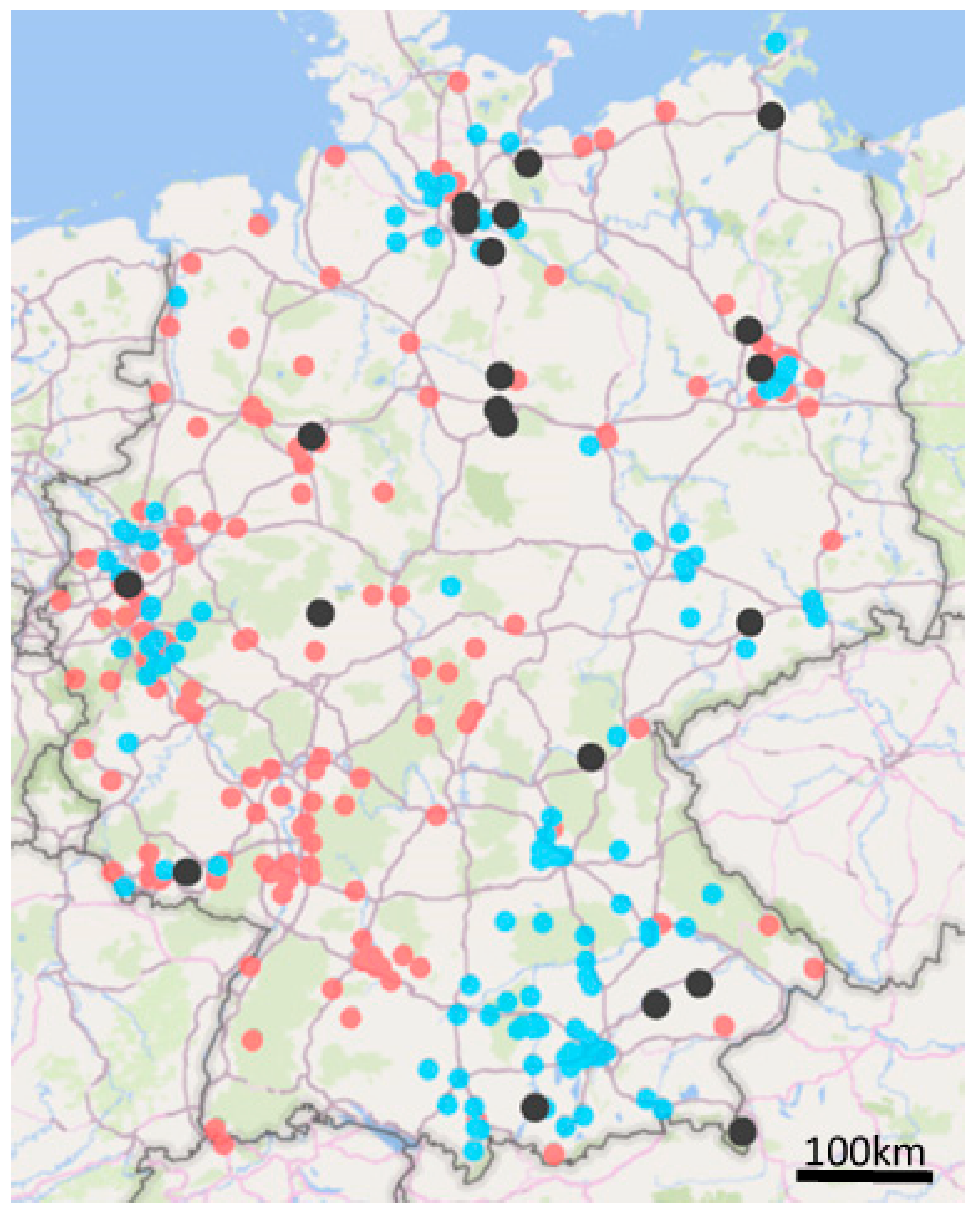
3.1. Prevalence of Farm Animals
3.2. Influence of Risk Factors
3.3. Prevalence of Symptomatic Pets
3.4. Prevalence of Human T. benhamiae Infections
4. Discussion
5. Concluding Remarks
Supplementary Materials
Author Contributions
Funding
Acknowledgments
Conflicts of Interest
References
- Kupsch, C.; Berlin, M.; Gräser, Y. Dermatophyten und Meerschweinchen Dermophytes and guinea pigs. Der Hautarzt 2017, 68, 827–830. [Google Scholar] [CrossRef]
- Kraemer, A.; Hein, J.; Heusinger, A.; Mueller, R.S. Clinical signs, therapy and zoonotic risk of pet guinea pigs with dermatophytosis. Mycoses 2012, 56, 168–172. [Google Scholar] [CrossRef] [PubMed]
- Overgaauw, P.; Avermaete, K.; Mertens, C.; Meijer, M.; Schoemaker, N. Prevalence and zoonotic risks of Trichophyton mentagrophytes and Cheyletiella spp. in guinea pigs and rabbits in Dutch pet shops. Vet. Microbiol. 2017, 205, 106–109. [Google Scholar] [CrossRef] [PubMed]
- White, T.J.; Bruns, T.; Lee, S.; Taylor, J. Amplification and Direct Sequencing of Fungal Ribosomal RNA Genes for Phylogenetics. In PCR Protocols; Elsevier: Amsterdam, The Netherlands, 1990; pp. 315–322. [Google Scholar]
- Kupsch, C.; Czaika, V.A.; Deutsch, C.; Graeser, Y. Trichophyton mentagrophytes—A new genotype of zoophilic dermatophyte causes sexually transmitted infections. J. Dtsch. Dermatol. Ges. 2019, 17, 493–501. [Google Scholar] [CrossRef] [PubMed]
- Uhrlaß, S.; Krüger, C.; Nenoff, P. Microsporum canis: Current data on the prevalence of the zoophilic dermatophyte in central Germany. Hautarzt 2015, 66, 855. [Google Scholar] [CrossRef] [PubMed]
- Bloch, M.; Cavignaux, R.; Debourgogne, A.; Dorin, J.; Machouart, M.; Contet-Audonneau, N. From guinea pig to man: Tinea outbreak due to Trichophyton mentagrophytes var. porcellae in pet shops in Nancy (France). J. Mycol. Med. 2016, 26, 227–232. [Google Scholar] [CrossRef] [PubMed]
- Tekin, H.G.; Sigsgaard, V.; Zachariae, C.; Hare, R.K.; Arendrup, M.C.; Saunte, D. Would you like to purchase a rodent with dermatophytes? Mycoses 2019, 62, 584–587. [Google Scholar] [CrossRef] [PubMed]
- Kupsch, C.; Ohst, T.; Pankewitz, F.; Nenoff, P.; Uhrlaß, S.; Winter, I.; Gräser, Y. The agony of choice in dermatophyte diagnostics—Performance of different molecular tests and culture in the detection of Trichophyton rubrum and Trichophyton interdigitale. Clin. Microbiol. Infect. 2016, 22, 735.e11–735.e17. [Google Scholar] [CrossRef] [PubMed] [Green Version]
- Hein, J. Dermatophytose bei Kaninnchen und Meerschweinchen—Ein Update einer Zoonose. Kleintierpraxis 2016, 61, 675–688. [Google Scholar]
- Drouot, S.; Mignon, B.; Fratti, M.; Roosje, P.; Monod, M. Pets as the main source of two zoonotic species of the Trichophyton mentagrophytes complex in Switzerland, Arthroderma vanbreuseghemii and Arthroderma benhamiae. Vet. Dermatol. 2009, 20, 13–18. [Google Scholar] [CrossRef] [PubMed]
- Kraemer, A.; Mueller, R.S.; Werckenthin, C.; Straubinger, R.K.; Hein, J. Dermatophytes in pet Guinea pigs and rabbits. Vet. Microbiol. 2012, 157, 208–213. [Google Scholar] [CrossRef] [PubMed]
- Vangeel, I.; Pasmans, F.; Vanrobaeys, M.; De Herdt, P.; Haesebrouck, F. Prevalence of dermatophytes in asymptomatic guinea pigs and rabbits. Vet. Rec. 2000, 146, 440–441. [Google Scholar] [CrossRef]
- Weiss, R.; Böhm, K.H.; Mumme, J.; Nicklas, W. 13 Years of veterinary mycological routine diagnostics. Isolation of dermatophytes in the years 1965–1977. Sabouraudia 1979, 17, 345–353. [Google Scholar] [PubMed]
- Smith, J.M.B.; Rush-Munro, F.M.; McCarthy, M. Animals as a Reservoir of Human Ringworm in New Zealand. Australas. J. Dermatol. 1969, 10, 169–182. [Google Scholar] [CrossRef] [PubMed]
- D’Ovidio, D.; Santoro, D. Survey of Zoonotic Dermatoses in Client-Owned Exotic Pet Mammals in Southern Italy. Zoonoses Public Health 2014, 62, 100–104. [Google Scholar] [CrossRef] [PubMed]
- de Hoog, G.S.; Guarro, J.; Gené, J.; Ahmed, S.; Al-Hatmi, A.M.S.; Figueras, M.J.; Vitale, R.G. Atlas of Clinical Fungi 2019, 3rd ed.; Reus: Utrecht, The Netherlands, 2019. [Google Scholar]
- Contet-Andonneau, N.; Leyer, C. Émergence d’un dermatophyte transmis par le cochon d’Inde et proche de Trichophyton mentagrophytes var. erinacei: T. mentagrophytes var. porcellae. J. Mycol. Med. 2010, 2, 321–325. [Google Scholar] [CrossRef]
- Sabou, M.; Denis, J.; Boulanger, N.; Forouzanfar, F.; Glatz, I.; Lipsker, D.; Poirier, P.; Candolfi, E.; Letscher-Bru, V. Molecular identification of Trichophyton benhamiae in Strasbourg, France: A 9-year retrospective study. Med. Mycol. 2017, 56, 723–734. [Google Scholar] [CrossRef] [PubMed]


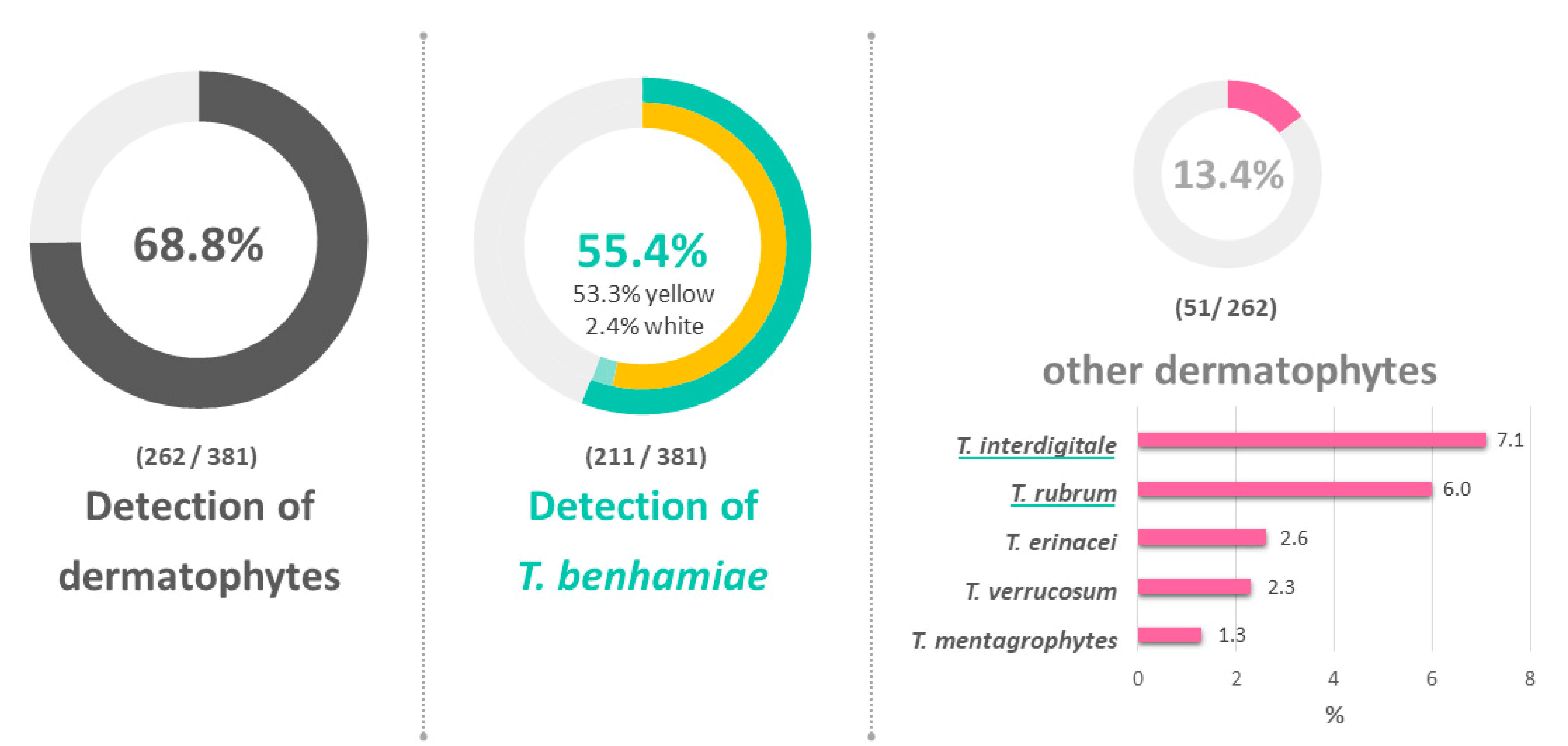
| Species | Number of Samples (Percent) |
|---|---|
| Negative | 119/381 (31.2%) |
| Dermatophytes positive | 262/381 (68.8%) |
| T. benhamiae (yellow) *1 | 203/381 (53.3%) |
| T. benhamiae (white) | 9/381 (2.4%) |
| Other Dermatophytes *2 | 51/262 (13.4%) |
| T. interdigitale | 27/262 (7.1%) |
| T. rubrum | 23/262 (6.0%) |
| T. erinacei | 10/262 (2.6%) |
| T. verrucosum/eriotrephon | 6/262 (2.3%) |
| T. mentagrophytes | 5/262 (1.3%) |
| Factor | Characteristics | Frequency (n) (%) | T. benhamiae Detection (%) | p-Value |
|---|---|---|---|---|
| Sex | Female | 259 (20.2) | 55.2 | 0.074 |
| Male | 77 (68.0) | 49.4 | ||
| Castrated | 45 (11.8) | 66.7 | ||
| Male + castrated | 122 (32.0) | 55.7 | 0.506 | |
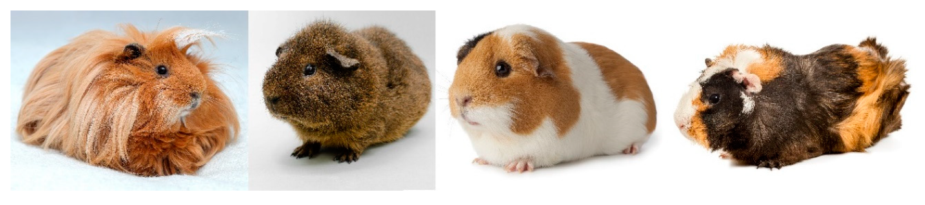
| Animal race/Pheno type | Long hair + curls | 50 (13.1) | 74.0 | 0.00002 * |
| Long hair − curls | 39 (10.2) | 30.8 | ||
| Teddy | 90 (23.6) | 51.1 | ||
| Rex | 69 (18.1) | 68.1 | ||
| Rosette (SH + curls) | 69 (18.1) | 62.3 | ||
| Smooth hair | 64 (16.8) | 40.6 | ||
| Husbandry condition | Outdoor | 114 (29.9) | 51.8 | 0.011 |
| Cold barn | 133 (34.9) | 46.6 | ||
| Indoor | 134 (35.2) | 67.2 | ||
| Stock Dynamics | Static (autarkic) | 202 (53.0) | 37.1 | 1.19 × 10⁻¹⁴ |
| Dynamic | 179 (47.0) | 76.0 | ||
| Coat length | short | 300 (78.7) | 56.3 | 0.276 |
| long | 81 (21.3) | 51.9 | ||
| Curls | Ye | 118 (31.0) | 68.6 | 0.000328 |
| No | 263 (69.0) | 49.4 |
| Age | Average (Months) | Median (Months) |
|---|---|---|
| of all animals (breedings) | 15.8 | 12 |
| of animals positive for T. benhamiae (breedings) * | 15.7 | 12 |
| of symptomatic pets (laboratory data) | 17.5 | 6 |
| Samples Analyzed | Number |
|---|---|
| Total | 9636 |
| with suspected mycosis | 1035 |
| Tested positive for dermatophytes | 382 |
| T. benhamiae | 375 |
| T. benhamiae (yellow) | 357 |
| T. benhamiae (white) | 18 |
| T. rubrum | 3 |
| Microsporum (M.) canis | 1 |
| T. mentagrophytes | 1 |
| T. spp. | 1 |
| T. interdigitale | 1 |
| Patients Recorded | Number |
|---|---|
| Total | 51,238 |
| Number of T. benhamiae infections | 163 |
| of which culture diagnosed | 126 |
| thereof diagnosed by PCR | 145 |
| thereof contact with guinea pigs known | 32/163 |
© 2020 by the authors. Licensee MDPI, Basel, Switzerland. This article is an open access article distributed under the terms and conditions of the Creative Commons Attribution (CC BY) license (http://creativecommons.org/licenses/by/4.0/).
Share and Cite
Berlin, M.; Kupsch, C.; Ritter, L.; Stoelcker, B.; Heusinger, A.; Gräser, Y. German-Wide Analysis of the Prevalence and the Propagation Factors of the Zoonotic Dermatophyte Trichophyton benhamiae. J. Fungi 2020, 6, 161. https://0-doi-org.brum.beds.ac.uk/10.3390/jof6030161
Berlin M, Kupsch C, Ritter L, Stoelcker B, Heusinger A, Gräser Y. German-Wide Analysis of the Prevalence and the Propagation Factors of the Zoonotic Dermatophyte Trichophyton benhamiae. Journal of Fungi. 2020; 6(3):161. https://0-doi-org.brum.beds.ac.uk/10.3390/jof6030161
Chicago/Turabian StyleBerlin, Max, Christiane Kupsch, Lea Ritter, Benjamin Stoelcker, Anton Heusinger, and Yvonne Gräser. 2020. "German-Wide Analysis of the Prevalence and the Propagation Factors of the Zoonotic Dermatophyte Trichophyton benhamiae" Journal of Fungi 6, no. 3: 161. https://0-doi-org.brum.beds.ac.uk/10.3390/jof6030161