Review

Sensing and Responding to Hypersaline Conditions and the HOG Signal Transduction Pathway in Fungi Isolated from Hypersaline Environments: Hortaea werneckii and Wallemia ichthyophaga

Faculty of Medicine, Institute of Biochemistry and Molecular Biology, University of Ljubljana, Vrazov trg 2, 1000 Ljubljana, Slovenia
Submission received: 30 September 2021 / Revised: 11 November 2021 / Accepted: 15 November 2021 / Published: 19 November 2021
(This article belongs to the Special Issue Signal Transductions in Fungi)

Abstract

:
Sensing and responding to changes in NaCl concentration in hypersaline environments is vital for cell survival. In this paper, we identified and characterized key components of the high-osmolarity glycerol (HOG) signal transduction pathway, which is crucial in sensing hypersaline conditions in the extremely halotolerant black yeast Hortaea werneckii and in the obligate halophilic fungus Wallemia ichthyophaga. Both organisms were isolated from solar salterns, their predominating ecological niche. The identified components included homologous proteins of both branches involved in sensing high osmolarity (SHO1 and SLN1) and the homologues of mitogen-activated protein kinase module (MAPKKK Ste11, MAPKK Pbs2, and MAPK Hog1). Functional complementation of the identified gene products in S. cerevisiae mutant strains revealed some of their functions. Structural protein analysis demonstrated important structural differences in the HOG pathway components between halotolerant/halophilic fungi isolated from solar salterns, salt-sensitive S. cerevisiae, the extremely salt-tolerant H. werneckii, and halophilic W. ichthyophaga. Known and novel gene targets of MAP kinase Hog1 were uncovered particularly in halotolerant H. werneckii. Molecular studies of many salt-responsive proteins confirm unique and novel mechanisms of adaptation to changes in salt concentration.

1. Introduction

Solar salterns and other hypersaline environments are extreme habitats that prevent the growth of most organisms except those that are well adapted to extremely high salt concentrations. Our laboratory has long been involved in studies of the molecular mechanisms of adaptations to extremely high NaCl concentrations. Focus is predominantly on two fungal model organisms isolated from solar salterns: the extremely halotolerant Hortaea werneckii and the obligate halophilic fungus Wallemia ichthyophaga [1,2,3,4,5,6]. While H. werneckii is unique in its adaptability to fluctuations in salt concentration and grows without NaCl as well as in the presence of up to 5 M NaCl, W. ichthyophaga is a true halophile that requires the presence of at least 10% of NaCl and also grows in the presence of up to 32% NaCl. Whole genome sequencing of both fungi, ion measurements, and studies of the adaptations of numerous proteins and membrane properties to high NaCl concentrations have revealed many new molecular mechanisms of adaptation. Some of these mechanisms are common to both fungi, but many are unique to halotolerant H. werneckii and halophilic W. ichthyophaga. Sensing changes in salt concentrations, which leads to appropriate responses regarding the expression of osmoresponsive genes, is vital for survival in environments with increased NaCl concentrations.
We were therefore particularly interested in the high-osmolarity glycerol (HOG) signal transduction pathway, which is involved in sensing hyperosmolar conditions. Identifying components of the HOG pathway in H. werneckii and W. ichthyophaga and investigating their roles in the adaptation to hypersaline conditions demonstrated that both fungi are well adapted to such conditions. Furthermore, both fungi sense and respond to increased environmental salinity differently. In this review article, we summarize the key molecular adaptations of H. werneckii and W. ichthyophaga, focusing on the identification and structural properties of HOG pathway components.

2. Molecular Adaptations of Halotolerant H. werneckii

The extremely halotolerant black yeast H. werneckii (Capnodiales, Dothideomycetidae, Dothideomycetes, Pezizomycotina) is described as an opportunistic pathogen and the primary etiological agent of tinea nigra, which can occur on the salty hands and feet of humans. It has been isolated from different hypersaline environments, with brine in solar salterns representing its primary ecological niche [2,7]. H. werneckii is an example of a very adaptable fungal species, as it can grow without salt as well as in environments with extremely high NaCl concentrations (up to 30%).
Molecular studies have identified general and also unique molecular mechanisms of adaptation that contribute to the extreme halotolerance of H. werneckii [1,4,5]. The most striking result came from the whole genome sequencing of H. werneckii, which was also the first published whole genome sequence of a fungal species isolated from an environment with an extremely high NaCl concentration. With almost 50 Mb, the H. werneckii genome is larger than the known genomes of other Capnodiales representatives, and, unlike in other species with large genomes, the large genome of H. werneckii is not due to large amounts of repeated DNA content. Analysis has demonstrated recent duplication of the H. werneckii genome, with multiple copies of halotolerance genes, in particular, genes that code for metal cation transporters [8,9]. The H. werneckii genome contains almost 16,000 genes of which 90% are duplicates with an average protein sequence divergence of 5%. While post-duplication events normally include large-scale gene deletions and reductions, the whole genome duplication appears stable in H. werneckii, as the genome was not reduced even after long-term growth in the presence of salt stress [9,10].
These whole genome data fit well with the results of molecular studies on individual genes. Most of the studied genes at the protein level are duplicated and code for two functionally identical proteins, often with a salt-dependent expression pattern. Such proteins are involved in general responses, as exemplified by the salt-dependent expression of two forms of glycerol phosphate dehydrogenase, which are involved in the synthesis of glycerol (the main compatible solute in H. werneckii) [11], fatty acid desaturases and elongases, which are involved in salt-dependent changes of membrane phospholipids [12,13,14], and especially metal cation transporters that are not only duplicated but exist as multiple copies in the H. werneckii genome [8]. The abundance of cation transporters in the H. werneckii genome indicates the importance of regulating potassium and sodium transport across the plasma membrane. Enrichment was not only observed for the transporters that maintain high intracellular K+/Na+ ratios and participate in Na+ efflux and K+ influx (e.g., ENA, NHA, and TRK) at high NaCl concentrations, but also for those involved in K+ efflux (TOK) and Na+ influx (PHO) at low NaCl concentrations [8,15,16,17]. This fits well with the lifestyle of H. werneckii, which thrives in environments with fluctuations in NaCl concentration, as maintenance of appropriate K+/Na+ ratios is crucial not only at high NaCl concentrations but also at low NaCl concentrations.
Two proteins with a specific biochemical signature of halophily were also identified in H. werneckii: hydroxymethylglutaryl CoA (HMG-CoA) reductase), the main regulatory enzyme in the mevalonate pathway [18,19], and 3′-phosphoadenosine-5′-phosphatase encoded by the HAL2 gene, a known target of salt toxicity in yeast [20]. NaCl-dependent regulation of HMG-CoA reductase in H. werneckii also appears to be linked to its halotolerant character. Both enzyme activity and protein levels in H. werneckii depend on environmental salinity, with high levels under both hypo- and hyper-saline conditions. Interestingly, we found that in H. werneckii, the biological consequence of HMG-CoA regulation relates to posttranslational protein modification by prenylation and not to sterol synthesis, as shown, for example, in S. cerevisiae. Thus, regulated expression and activity of HMG-CoA reductase can be considered an important biochemical signature of halophilism. In evolutionary terms, the maintenance of high HMG-CoA reductase levels in hypo- and hyper-saline environments may also reflect the physiological adaptation of halophilic fungi to metabolic demands under extreme conditions [19].
Another example of a protein with specific responses to changes in salinity is 3′-phosphoadenosine-5′-phosphatase Hal2. Two isoforms of the Hal2 protein appear to significantly contribute to the adaptation of H. werneckii to fluctuations in environmental salinity. With genetic and biochemical validation, the specific sequence motif of Hal2, which we call the META sequence, was identified to exert an effect on salt tolerance in the HwHal2B isoform as well as when introduced into the SAL1 gene in Arabidopsis thaliana [20,21].

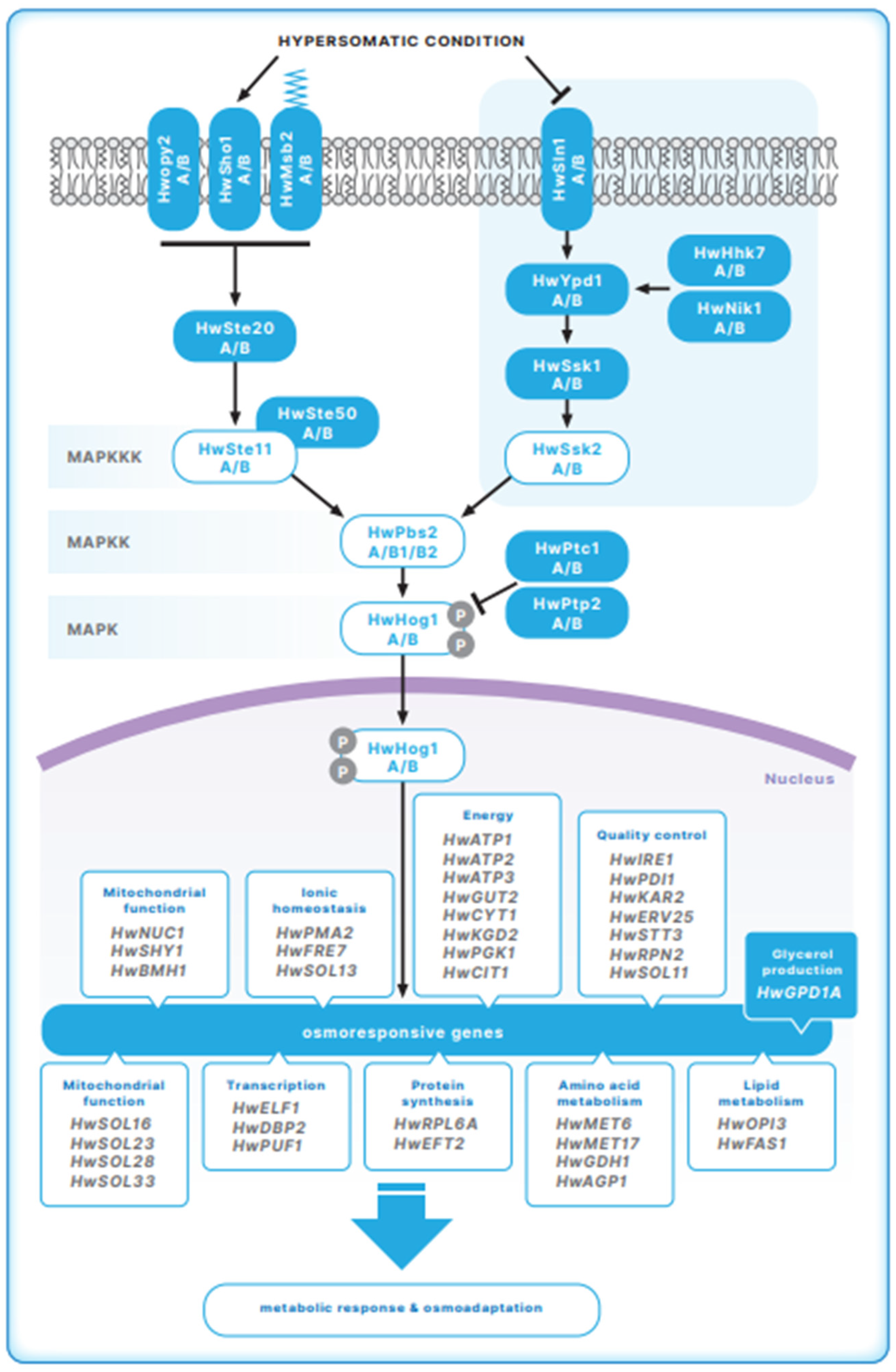
3. The HOG Signal Transduction Pathway in Halotolerant H. werneckii

The HOG signal transduction pathway is an example of a MAP kinase (mitogen-activated protein kinase) signal transduction pathway that senses and responds to hyperosmolar conditions and thus plays an important role in the adaptation to hypersaline conditions. It consists of the proteins of the sensory apparatus and the module of three kinases: MAPKKK (mitogen-activated protein kinase kinase kinase), MAPKK (mitogen-activated protein kinase kinase), and MAPK (mitogen-activated protein kinase); these are activated by sequential phosphorylation. This mechanism is best understood in the yeast S. cerevisiae. Hyperosmolar signals are sensed and transduced by two branches in S. cerevisiae: SHO1 and SLN1 that converge in the activation of MAPKK ScPbs2. In the SHO1 branch, the transmembrane ScSho1, membrane anchor ScOpy2, mucines ScMsb2 and ScHkr1, guanine exchange factor ScCdc24, and GTPase ScCdc42 are involved in the SHO1 sensory apparatus, which leads to the activation of ScSte20. Consequently, MAPKKK ScSte11 is activated by phosphorylation by the ScSte20 kinase. In the SLN1 branch, the proteins Sln1, Ypd1, and Ssk1 form the two-component phosphorelay system in which the sensor histidine kinase ScSln1 (located in the plasma membrane) is inactivated under hyperosmolar conditions. The ScYpd1 intermediate protein and the ScSsk1 response regulator then transduce the signal to the MAPKKKs ScSsk2/ScSsk22. The MAPKKKs ScSte11 and ScSsk2/ScSsk22 activate the MAPKK ScPbs2, which serves as a scaffold protein of the two upstream pathway branches and upon phosphorylation activates ScHog1 MAPK [22,23,24,25]. Under hyperosmotic conditions, ScHog1 phosphorylation is accompanied by an import of ScHog1 into the nucleus, which leads to the regulation of osmoresponsive genes. In addition, activated ScHog1 has targets in the cytosol and plasma membrane (18) that also play a role in osmoadaptation. The phosphorylation state of the MAPK Hog1 is transient and is controlled by various phosphoprotein phosphatases, such as Ser/ Thr phosphatase Ptc1 [26] and phosphotyrosine phosphatases Ptp2 and Ptp3 [27].
Homologous components to the S. cerevisiae HOG pathway have also been identified in other yeasts and fungi and are involved in responses to hyperosmolar conditions, virulence regulation, gut colonization, and animal and plant infection with pathogenic fungi. According to published data, it seems that two-component phosphorelay systems play important roles in sensing and subsequent HOG pathway activation in different fungi. For example, in the filamentous fungi Aspergillus fumigatus and Aspergillus nidulans, the HOG pathway is involved in osmotic stress responses [28,29], and the Sho1 protein is not involved in osmosensing in A. nidulans [28]. Likewise, the HOG pathway in the pathogenic fungus Cryptococcus neoformans is activated exclusively via the two-component phosphorelay system [30]. Histidine kinases were also identified as the main regulators of the HOG signal transduction pathway in Magnaporthe oryzae [31] and Candida albicans [32,33]
Since the HOG pathway is highly conserved among eukaryotes, similar components to those in S. cerevisiae were expected in H. werneckii. Indeed, the genome of H. werneckii contains all the key components of the HOG signal transduction pathway. In line with the genome duplication in H. werneckii, we identified two copies of each gene that code for the components of the HOG pathway [8,9] (Figure 1). As there are no genetic tools available to allow manipulation of the H. werneckii genome, the putative roles of HOG pathway components identified in H. werneckii were studied by functional complementation in S. cerevisiae mutant strains [34,35,36].

3.1. The Sensory Apparatus of the HOG Pathway in H. werneckii

Although important differences were found in the sensory apparatus when compared to S. cerevisiae, it appears that, in contrast to other studied fungi, both the SLN1 and SHO1 branches play a role in sensing hypersaline conditions in H. werneckii, as we were able to identify components of both branches in H. werneckii. Interestingly, besides the SLN1 gene, which codes for membrane-bound histidine kinase Sln1, the genome of H. werneckii contains two additional histidine kinase genes, HwNIK1 and HwHHK7, which code for cytosolic histidine kinases Nik1 and Hhk7 [8,38]. While the role of membrane-bound Sln1 in sensing hyperosmolar conditions is well documented in S. cerevisiae, the role of the cytosolic histidine kinases, Nik1 and Hhk7, is less understood. Both were identified and proposed to be involved in sensing hyperosmolar conditions in pathogenic fungi [28,39].
We identified two isoforms of histidine kinase HwHHk7 in H. werneckii: HwHHk7A and B (Figure 1) [38]. Phylogenetic comparisons with other fungal histidine kinases (HK) and analyses of structural motifs confirmed that they belong to group 7 of fungal HKs. According to published data [40], histidine kinases Sln1 and HK 7 groups are positioned close together, and thus late separation from a common ancestor was proposed. However, these proteins differ in their intracellular localization: while histidine kinases from the Sln1 group are membrane-bound, histidine kinases from the HK7 group are soluble cytosolic proteins. It has been proposed that some of the histidine kinases other than yeast orthologs might transmit the signal through Ypd1; for example, in A. nidulans, the HOG pathway was reconstructed and revealed that the Sho1 branch is not involved in the osmoresponsive activation of the MAPK Hog1. It was speculated that this role could well be occupied by the HK 7 protein AnM7 [28,41].
To obtain insight into the role of the HwHHk7 isoforms in response to changes in salinity in halotolerant H. werneckii, we assessed the expression of both isoforms in response to changes in NaCl concentration. We found that the expression of the HwHH7A gene did not change significantly with changes in NaCl concentration. By contrast, transcription of the HwHHK7B gene was very salt-responsive. Moreover, we observed two types of responses: an early response with high induction of gene expression under hyposaline conditions and a late increased expression of the HwHH7B gene under hypersaline stress [38]. Based on these results, we proposed that in H. werneckii, the high induction of HwHHK7B gene expression as an early response to hyposaline stress could be the result of the specialized role of this HK in response to conditions of modest osmolarity, as has already been demonstrated for the Sln1 HK in S. cerevisiae [42]. H. werneckii is extremely adaptable to changes in NaCl concentration. Salt-dependent expression of the HwHH7B gene suggests that HwHhk7B plays an important role in sensing and adapting to the sudden changes that are common in solar salterns, natural habitats of H. werneckii.
Analysis of the H. werneckii genome also confirmed the presence of genes that code for proteins of the sensory apparatus of the SHO1 branch, membrane-bound HwSho1 A and B, HwOpy2 A and B, and HwMsb2 A and B. Structural and functional studies were performed on HwSho1 [34]. Comparison of amino-acid sequences of HwSho1 A and B in extremely halotolerant H. werneckii with those of salt-sensitive S. cerevisiae, C. albicans, and A. fumigatus and moderately halotolerant Debaryomyces hansenii showed highly conserved transmembrane and SH3 domains and a poorly conserved linker domain containing the Ste11-binding site. Despite differences in the Ste11-binding domain between H. werneckii and S. cerevisiae Sho1 proteins, both HwSho1 isoforms fully complemented the function of the native S. cerevisiae Sho1 protein when expressed in the S. cerevisiae strain with the deleted SHO1 gene. Furthermore, they both activated the HOG pathway under conditions of osmotic stress. Moreover, we demonstrated that in S. cerevisiae, both HwSho1 proteins have characteristic subcellular localizations similar to the Sho1 protein [34] These data suggest that HwSho1 is involved in sensing hypersaline stress conditions in H. werneckii.
It appears that the sensory apparatus in the H. werneckii HOG pathway is much more complex when compared to S. cerevisiae and other plant and human pathogenic fungi. Based on our studies, we propose that the signaling to HwPbs2 in H. werneckii is conveyed through both branches: [43] the SHO1 branch, which is similar to that in S. cerevisiae, and [30] the SLN1 branch, which is similar to that in pathogenic fungi, such as A. nidulans and C. albicans, in which histidine kinases were proposed to be responsible for transmitting the signal to the MAP kinase module. We can speculate that the combination of both sensory systems in sensing changes in environmental salinity enables a more fine-tuned response to osmolarity in H. werneckii.

3.2. The MAP Kinase Module of the HOG Pathway in H. werneckii

Besides the genes of the sensory apparatus in the H. werneckii genome, gene duplication was also confirmed for downstream HOG pathway key components, including HwSTE20, HwSTE50, and genes that code for the three kinases of the MAP kinase module (HwSTE11 A and B, HwPBS2 A and B, and HwHOG1 A and B). Further structural and functional studies of HwPbs2 and HwHog1 uncovered unique features of these kinases. Interestingly, in the case of the MAPKK HwPbs2, two gene copies of HWPBS2 A and B are transcribed and translated into three different isoforms: while HwPBS2A is translated into one protein product (HwPbs2A), HwPBS2B is translated into two protein products (HwPbs2B1 and HwPbs2B2). Multiple isoforms were confirmed by Western blot analysis with specific antibodies raised against HwPbs2. It was found that they respond differently to changes in salt concentration. Their salt-responsive expression profiles suggest that HwPbs2a and HwPbs2b2 play roles in quick adaptation to severe hypersaline shock and that HwPbs2b1 plays a role in adaptation to moderate salt stress (Lenassi, Doctoral thesis, unpublished data).
The H. werneckii HOG signal transduction pathway includes two functionally redundant MAPK homologues, HwHog1A and HwHog1B. To better understand the function of the HOG pathway in H. werneckii, we observed the osmotic stress responsive phosphorylation pattern of HwHog1 and the effect of the Hog1 kinase inhibitor on cell viability and survival under hypersaline conditions. Activation of the HOG pathway in H. werneckii in response to increased osmolarity revealed that HwHog1A and HwHog1B are fully activated by constitutive phosphorylation with the upstream kinase HwPbs2 only at concentrations above 17% NaCl [44]. Thus, while phosphorylation is transient for most fungal species, this does not appear to be the case in extremely halotolerant H. werneckii. Likewise, the effect of HwHog1 kinase activity inhibition on restricting H. werneckii colony growth was only observed at osmolyte concentrations of ≥17%.
Studies on the H. werneckii transcriptome revealed many novel osmoresponsive genes, most of which were not shown before for S. cerevisiae or other fungi. Direct interaction with the MAP kinase HwHog 1 was demonstrated for more than one third of salt-responsive genes (Figure 1), with the majority of them upregulated in high-salinity-adapted H. werneckii cells [37] Genes associated with energy supply were highly represented among the upregulated genes in cells growing at 30% NaCl. An increase in energy production appears to be one of the fundamental adaptations that maintain ion homeostasis and osmotic equilibrium in a hyperosmotic environment. During long-term adaptive growth under extreme salinity, H. werneckii must maintain a high production of ATP that powers various transmembrane transporters [4,37,45]. In contrast to constitutive phosphorylation of HwHog1 in response to NaCl, the activation of osmoresponsive gene transcription was transient, which suggests that other factors besides the HwHog1 phosphorylation signal are involved in the effects of HwHog1 in H. werneckii [44].
We also showed that H. werneckii can discriminate between different osmolytes. NaCl induces continuous phosphorylation of HwHog1, whereas this phosphorylation is transient in response to KCl or sorbitol. Discrimination between osmolyte types was also confirmed at the level of osmoresponsive gene transcription. In H. werneckii cells, genes such as HwSTL1 and HwGPD1, which are involved in glycerol transport and synthesis, showed early induction of transcription in response to NaCl stress, early or late induction in response to sorbitol stress, and no effects in response to KCl stress [44]. This osmolyte-specific response was also demonstrated in the expression of genes related to mitochondrial function. In hypersaline medium, an increased expression of genes involved in energy production and oxidative damage protection was observed, whereas adaptation to a non-ionic osmolyte resulted in a decrease in ATP synthesis and lipid peroxidation levels in mitochondria. This was also confirmed with a proteomic study of mitochondrial proteins, revealing a preferential accumulation of energy metabolism enzymes in hypersaline medium and an accumulation of protein chaperones in the non-ionic osmolyte [46].
In summary, the extremely halotolerant yeast-like fungus H. werneckii, isolated from hypersaline saltern water, can grow in both environments without salt as well as with extremely high salt concentrations (even up to saturated NaCl solutions). We assume that this extremely adaptable fungus has an efficient sensory system and molecular machinery that responds to changing conditions in the environment. As a response to severe changes in salinity in the environment, H. werneckii activates the HOG signaling pathway, resulting in the expression of many salt-responsive genes. The HOG signal transduction pathway is vital for the extreme osmotolerance of H. werneckii and regulates common and osmolyte-specific processes. Particularly, the HOG pathway seems to be specifically responsive to Na+, an ion typically present at high concentrations in the natural habitat of H. werneckii, solar eutrophic salterns.

4. Molecular Adaptations of the Halophilic Fungus W. ichthyophaga

W. ichthyophaga (Wallemiales, Wallemiomycetes) is a xerophilic filamentous fungi. Although xerotolerance is rare in Basidiomycota, W. ichthyophaga is the most halophilic fungus known. It requires a minimum of 10% (w/v) NaCl, with a growth optimum at 15–20% NaCl, but is metabolically active even at 32% NaCl [6] Strains of W. ichthyophaga have been isolated from hypersaline waters of solar salterns, bitterns, and salted meat [47,48]. Morphological studies of W. ichthyophaga have shown that changes in the size of cell aggregates and the thickness of the cell wall are crucial. The thickness of the cell wall can increase up to three-fold, while the size of cell clumps can increase even more [6].
In contrast to the large and duplicated genome of H. werneckii, the genome of W. ichthyophaga is very compact, with 9.6 Mb and only 4884 predicted protein-coding genes [49]. Genome analysis showed that except for the enriched protein family of P-type ATPases, cation transporters are sparse and exhibit low expression levels. Additionally, the expression of most of them is independent of salt concentration. Conversely, a significant expansion of hydrophobins, small amphipathic proteins that reside in the cell wall of filamentous fungi and are involved in a range of processes of cellular growth and development, was found. This significant expansion is specific for W. ichthyophaga, as from the estimated 15 hydrophobin genes found in the genome of a common ancestor of W. ichthyophaga, the gene number increased to 26 in halophilic W. ichthyophaga [49] Compared to those of other fungi, hydrophobins in Wallemia contain a higher proportion of acidic amino acids on their surface. As this is similar to archaeal halophilic proteins [50] it is reasonable to suggest that hydrophobins play a role as halophilic proteins. With their acidic amino acids exposed at the surface, they can bind water and salt and facilitate adaptation to salt exposure. Transcriptome analysis supported this idea, as >50% of hydrophobins are differentially expressed at different salinities [49].
W. ichthyophaga use the strategy of compatible solutes and, as in halotolerant H. werneckii, glycerol is the main compatible solute in W. ichthyophaga. The W. ichthyophaga genome contains genes for the enzymes that are involved in glycerol synthesis. The homologue of GPD1, WiGPD1, exhibited lower expression levels and a slower response to hyperosmotic shock in comparison to that of halotolerant H. werneckii. As in halotolerant H. werneckii, the expression of this gene is regulated by MAP kinase WiHog1 [51]. Genes such as SLT1 for the plasma membrane glycerol/H+ symporter and FPS1, which codes for aquaglyceroporin channel Fps1, were found in multiple copies in W. ichthyophaga, suggesting their involvement in regulating glycerol levels. Among other proteins, the main regulatory enzyme in sterol synthesis, HMG-CoA reductase, also belongs to salt-responsive proteins in W. ichthyophaga and responds with a characteristic salt-dependent U-shape activity pattern [19]. The high activity and protein levels of HMG-CoA reductase in W. ichthyophaga under hypo- and hyper-saline conditions suggest that, as in extremely halotolerant H. werneckii, HMG-CoA reductase (with NaCl-dependent responses) plays an important role in the physiological adaptation of W. ichthyophaga to both hypo- and hyper-saline conditions.

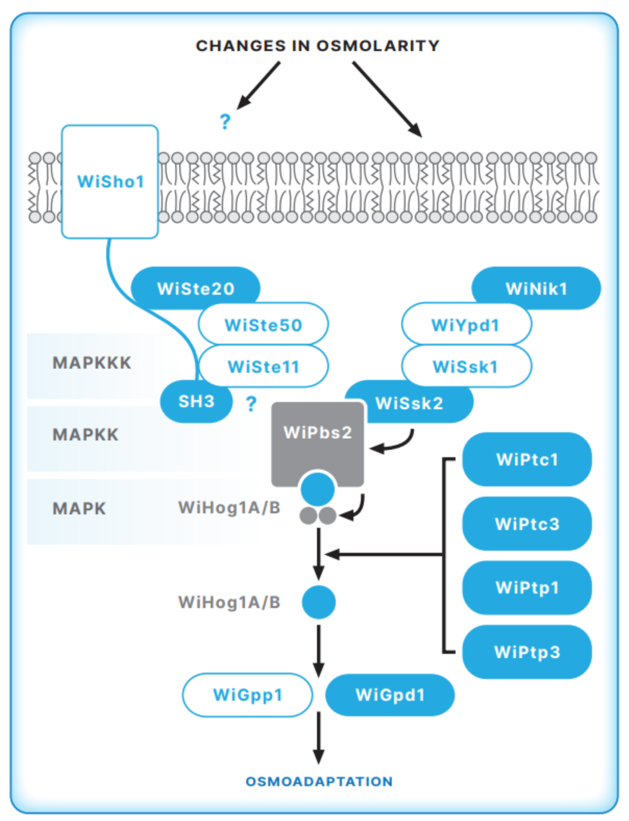
5. The HOG Signal Transduction Pathway in the Halophilic Fungus W. ichthyophaga

As demonstrated by whole genome analysis [49], W. ichthyophaga contains all key genes of the HOG signal transduction pathway (Figure 2). Further molecular studies revealed similarities as well as important differences in the molecular machinery involved in sensing and responding to changes in NaCl concentrations between halophilic W. ichthyophaga and halotolerant H. werneckii and other fungi. Although many key proteins of the HOG pathway are conserved in halophilic W. ichthyophaga, the architecture and activation of the HOG pathway are specific. While all genes of the HOG components are present in two copies in H. werneckii, in W. ichthyophaga, all HOG pathway components, except the final MAP kinase Hog 1 (WiHog1 A and B), are present in only one isoform [51,52].

5.1. The Sensory Apparatus of the HOG Pathway in W. ichthyophaga

Compared to S. cerevisiae and halotolerant H. werneckii, W. ichthyophaga lacks a considerable number of orthologs that are involved in the osmosensing apparatus of the SHO1 branch of the HOG pathway, e.g., the mucins Msb2 and Hkr1 and the membrane anchor Opy2. Conversely, other orthologs of this branch, e.g., WiCdc24, WiCdc42, WiSte20, WiSte50, and WiSte11, are present [52]. Despite the presence of proteins of the SHO1 sensory apparatus in halophilic W. ichthyophaga, further studies did not confirm activation of the HOG pathway through this branch. Structural analysis of WiSho1 domains, important for interactions with downstream protein partners, showed that crucial motifs for the interactions with MAPKKK Ste11 MAKK Pbs2 are not conserved in W. ichthyophaga orthologs. Moreover, WiSho1 expressed in S. cerevisiae mutant cells did not efficiently complement the function of ScSho1 [52]
The core phosphorelay system of the SLN1-like branch is well conserved in W. ichthyophaga [52]. This system contains WiYpd1, WiSsk1, the kinase WiSsk2, and the response regulator WiSkn7. Conversely, no membrane-bound ScSln1 ortholog was found in W. ichthyophaga. Instead, as in the genome of halotolerant H. werneckii, we confirmed the Group III cytosolic histidine kinase WiNik1. The W. ichthyophaga genome contains four hybrid histidine kinases that belong to groups I or II, III, and VIII. It was demonstrated for several fungi that group III histidine kinases are involved in two-component HOG pathway signaling [30]. We proposed that the HAMP domain repeats (present in histidine kinases, adenyl cyclases, methyl-accepting proteins, and phosphatases) [53] in WiNik1 revealed by structural studies [52] might be involved in osmosensing in W. ichthyophaga, as has been demonstrated for DhNik1 in moderately halotolerant D. hansenii, CaNik1 in C. albicans [54] and ClNik1 in Candida lusitaniae [39] It has been shown that group III histidine kinases are sensitive to the fungicide fludioxonil, and thus this fungicide represents a potent tool to investigate group III histidine kinase signaling [55]. Our investigation of the sensitivity of W. ichthyophaga cells to fludioxonil demonstrated that the presence of fludioxonil completely inhibited the growth of W. ichthyophaga cells at all NaCl concentrations used. This result strongly implies that WiNik1 plays a role in osmosensing machinery.

5.2. The MAP Kinase Module of the HOG Pathway in W. ichthyophaga

As MAPKK Pbs2 is the central element of the HOG pathway that can in principle receive hyperosmolarity signals through both the SHO1 and SLN1 branches [56], we further analyzed WiPbS2, which was identified in the W. ichthyophaga genome. Structural domains, important for interactions with upstream proteins involved in WiPbs2 signal transmission, were compared with ScPbs2 and the orthologs of some other fungi (27) It was demonstrated that the proline-rich motif that is crucial for Sho1 binding and signaling through the SHO1 branch in S. cerevisiae [57] is not conserved in W. ichthyophaga [52]. Another important feature of Pbs2 from S. cerevisiae is the docking site for MAPKKK Ste11 [25,58] Again, in WiPbs2, the Ste11 docking site is only poorly conserved [52]. Poor interactions of WiPbs2 with WiSho1 and WiSte11 indicate that WiPbs2 is not activated via the SHO1 branch, which is in line with the observation that WiSho1 could not complement the function of ScSho1 in the S. cerevisiae mutant strain. Conversely, we found that the ScSsk2/ScSsk22 activation of WiPbs2 is fully conserved [52] This finding and the sensitivity of W. ichthyophaga cells to fludioxonil highlight the importance of two-component HOG pathway signaling in W. ichthyophaga.
The W. ichthyophaga genome has two gene copies of MAP kinase Hog1, WiHOG A, and WiHOG B. Although protein sequence alignment revealed high conservation of the Hog1 kinase domains and motifs in both of the WiHog1A and WiHog1B isoforms, WiHog1A is not fully functional when compared with WiHog1B and ScHog1 in the S. cerevisiae mutant strain [51,52] Supported also by the observed lower phosphorylation level of WiHog1 A upon hyperosmotic stress, it was proposed that WiHog1A cannot optimally interact with the activating MAPKK Pbs2 or its targets. This might be explained by small differences in the ATP-binding site, activation loop, common docking domain, and/or PBD-2 region. Conversely, it was found that WiHog1B was fully phosphorylated; in the S. cerevisiae hog1Δ background, WiHog1B serves as a fully functional kinase. Moreover, WiHog1B also improves the tolerance of the yeast strain to high salinity, which was not observed for HwHog1 expression from the halotolerant H. werneckii in S. cerevisiae [13] Additionally, MAPK WiHog1 upregulated GPD1 transcription, which is important for the synthesis of the main compatible solute, glycerol, in W. ichthyophaga. Again, efficient interaction of the GPD1 promoter was demonstrated with WiHog1B but not WiHog1A [26,30].
As in halotolerant H. werneckii, WiHog1 transcript levels are salt-dependent [26]. Interestingly, W. ichthyophaga shows the opposite to the usual phosphorylation pattern found in S. cerevisiae, H. werneckii, and most other studied fungi. In W. ichthyophaga, MAP kinase WiHog1 is constitutively phosphorylated under optimal osmotic conditions (at concentrations of 15–20% NaCl) and is dephosphorylated under both hypo- and hyper-saline conditions. A similar phosphorylation model has so far been reported for pathogenic C. neoformans, serotype A, and it was assumed that a unique mechanism of Hog1 phosphorylation in C. neoformans is responsible for its higher stress resistance and the virulence of its serotype A [1]. Thus, it is reasonable to speculate that, in a similar manner, such HOG pathway regulation might cause the high salt resistance of W. ichthyophaga. This phosphorylation pattern also implies that phosphatases play an important role in HOG pathway regulation in W. ichthyophaga. The HOG pathway in S. cerevisiae is regulated by negative feedback through the action of phosphothreonine and phosphotyrosine phosphatases (17,18). In the W. ichthyophaga genome, we identified the phosphothreonine phosphatase WiPtc1, which is most similar to ScPtc1 and WiPtc3, which is related to ScPtc3 and its paralog ScPtc2 [20,54]. Furthermore, the W. ichthyophaga genome contains two phosphotyrosine phosphatases: WiPtp1 (an ortholog of ScPtp1) and WiPtp3, the latter being more related to ScPtp3, a broad-range phosphatase in S. cerevisiae [56] The presence of these phosphatases in the W. ichthyophaga genome support the importance of WiHog1 dephosphorylation in regulating the HOG signal transduction pathway.
Together, our investigation of the architecture of the W. ichthyophaga HOG pathway shows that the interactions of WiPbs2 kinase and the SHO1-branch orthologs are not conserved, which in turn suggests that these orthologs are not involved in WiPbs2 activation. Conversely, the ScSsk2/ScSsk22 activation of WiPbs2 is fully conserved, and W. ichthyophaga cells are sensitive to fludioxonil, which highlights the importance of the two-component HOG pathway signaling in W. ichthyophaga [39]. A unique WiHog1 phosphorylation pattern was demonstrated to be necessary for the high (er) stress-resistance of C. neoformans [1] and could also help W. ichthyophaga to survive in its naturally hypersaline environments.
In this review, the importance of the HOG pathway in adapting to higher NaCl concentrations in halotolerant H. werneckii and the obligate halophile W. ichthyophaga is summarized. Although there are many similarities between halotolerant and halophilic fungi, molecular analyses of whole genome sequences of both fungi performed in our laboratory during the past years have revealed important differences in sensing and responding to severe changes in NaCl concentrations, typical for the natural ecological niche of H. werneckii and W. ichthyophaga. We confirmed that the HOG pathway plays an important role in adaptation in both fungi. Studies revealed that the MAP kinase module is conserved in both fungi; however, the mechanisms of activation differ between halotolerant H. werneckii and halophilic W. ichthyophaga. In H. werneckii, the HOG pathway is activated via both SHO and SLN branches, whereas in W. ichthyophaga, only the SLN branch appears to be involved. Secondly, HwHog1 is phosphorylated in halotolerant H. werneckii and dephosphorylated in halophilic W. ichthyophaga under hypersaline conditions. Molecular investigations of extremely halotolerant H. werneckii and halophilic W. ichthyophaga, isolated from the same hypersaline environments, demonstrate that different fungi can employ diverse mechanisms to combat the same harsh conditions. We can also speculate that these differences make an important contribution to the halotolerant and halophilic characteristics of H. werneckii and W.ichthyophaga.

Funding

This study was funded by Research Grant P1-0170, and in part by a Young Researcher Fellowships from the Slovenian Research Agency.

Institutional Review Board Statement

Not applicable.

Informed Consent Statement

Not applicable.

Conflicts of Interest

The author declares that the research was conducted in the absence of any commercial or financial relationships that could be construed as a potential conflict of interest.

References

  1. Gunde-Cimerman, N.; Ramos, J.; Plemenitaš, A. Halotolerant and halophilic fungi. Mycol. Res. 2009, 113, 1231–1241. [Google Scholar] [CrossRef]
  2. Gunde-Cimerman, N.; Zalar, P. Extremely halotolerant and halophilic fungi inhabit brine in solar salterns around the globe. Food Technol. Biotechnol. 2014, 52, 170–179. [Google Scholar]
  3. Gunde-Cimerman, N.; Plemenitaš, A.; Oren, A. Strategies of adaptation of microorganisms of the three domains of life to high salt concentrations. FEMS Microbiol. Rev. 2018, 42, 353–375. [Google Scholar] [CrossRef] [PubMed]
  4. Petrovicˇ, U.; Gunde-Cimerman, N.; Plemenitaš, A. Cellular responses to environmental salinity in the halophilic black yeast Hortaea werneckii. Mol. Microbiol. 2002, 45, 665–672. [Google Scholar] [CrossRef] [PubMed] [Green Version]
  5. Plemenitaš, A.; Lenassi, M.; Konte, T.; Kejžar, A.; Zajc, J.; Gostinčar, C.; Gunde-Cimerman, N. Adaptation to high salt concentrations in halotolerant/halophilic fungi: A molecular perspective. Front. Microbiol. 2014, 5, 199. [Google Scholar] [CrossRef] [PubMed] [Green Version]
  6. Zajc, J.; Kogej, T.; Galinski, E.A.; Ramos, J.; Gunde-Cimerman, N. Osmoadaptation Strategy of the Most Halophilic Fungus, Wallemia ichthyophaga, Growing Optimally at Salinities above 15% NaCl. Appl. Environ. Microbiol. 2014, 80, 247–256. [Google Scholar] [CrossRef] [PubMed] [Green Version]
  7. Butinar, L.; Sonjak, S.; Zalar, P.; Plemenitaš, A.; Gunde-Cimerman, N. Melanized halophilic fungi are eukaryotic members of microbial communities in hypersaline waters of solar salterns. Bot. Mar. 2005, 48, 73–79. [Google Scholar] [CrossRef]
  8. Lenassi, M.; Gostinčar, C.; Jackman, S.; Turk, M.; Sadowski, I.; Nislow, C.; Jones, S.; Birol, I.; Cimerman, N.G.; Plemenitaš, A. Whole Genome Duplication and Enrichment of Metal Cation Transporters Revealed by De Novo Genome Sequencing of Extremely Halotolerant Black Yeast Hortaea werneckii. PLoS ONE 2013, 8, e71328. [Google Scholar] [CrossRef] [Green Version]
  9. Sinha, S.; Flibotte, S.; Neira, M.; Formby, S.; Plemenitaš, A.; Cimerman, N.G.; Lenassi, M.; Gostinčar, C.; Stajich, J.E.; Nislow, C. Insight into the Recent Genome Duplication of the Halophilic Yeast Hortaea werneckii: Combining an Improved Genome with Gene Expression and Chromatin Structure. G3: Genes|Genomes|Genet. 2017, 7, 2015–2022. [Google Scholar] [CrossRef] [Green Version]
  10. Gostinčar, C.; Stajich, J.E.; Kejžar, A.; Sinha, S.; Nislow, C.; Lenassi, M.; Gunde-Cimerman, N. Seven Years at High Salinity—Experimental Evolution of the Extremely Halotolerant Black Yeast Hortaea werneckii. J. Fungi 2021, 7, 723. [Google Scholar] [CrossRef]
  11. Lenassi, M.; Zajc, J.; Gostinčar, C.; Gorjan, A.; Gunde-Cimerman, N.; Plemenitaš, A. Adaptation of the glycerol-3-phosphate dehydrogenase Gpd1 to high salinities in the extremely halotolerant Hortaea werneckii and halophilic Wallemia ichthyophaga. Fungal Biol. 2011, 115, 959–970. [Google Scholar] [CrossRef] [PubMed]
  12. Gostinčar, C.; Turk, M.; Plemenitaš, A.; Gunde-Cimerman, N. The expressions of [delta]9-,[delta]12-desaturases and an elongase by the extremely halotolerant Hortaea werneckii are salt dependent. FEMS Yeast Res. 2009, 9, 247–256. [Google Scholar] [CrossRef] [PubMed] [Green Version]
  13. Turk, M.; Méjanelle, L.; Šentjurc, M.; Grimalt, J.O.; Gunde-Cimerman, N.; Plemenitaš, A. Salt-induced changes in lipid composition and membrane fluidity of halophilic yeast-like melanized fungi. Extremophiles 2003, 8, 53–61. [Google Scholar] [CrossRef] [PubMed]
  14. Turk, M.; Plemenitaš, A.; Gunde-Cimerman, N. Extremophilic yeasts: Plasma-membrane fluidity as determinant of stress tolerance. Fungal Biol. 2011, 115, 950–958. [Google Scholar] [CrossRef] [PubMed]
  15. Gorjan, A.; Plemenitaš, A. Identification and characterization of ENA ATPases HwENA1 and HwENA2 from the halophilic black yeast Hortaea werneckii. FEMS Microbiol. Lett. 2006, 265, 41–50. [Google Scholar] [CrossRef] [PubMed] [Green Version]
  16. Kogej, T.; Ramos, J.; Plemenitaš, A.; Gunde-Cimerman, N. The Halophilic Fungus Hortaea werneckii and the Halotolerant Fungus Aureobasidium pullulans Maintain Low Intracellular Cation Concentrations in Hypersaline Environments. Appl. Environ. Microbiol. 2005, 71, 6600–6605. [Google Scholar] [CrossRef] [Green Version]
  17. Plemenitaš, A.; Konte, T.; Gostinčar, C.; Cimerman, N.G. Transport systems in halophilic fungi. In Yeast Membrane Transport—Advances in Experimental Medicine and Biology; Springer: Cham, Switzerland, 2016; Volume 892, pp. 307–325. [Google Scholar]
  18. Vaupotic, T.; Plemenitaš, A. Osmoadaptation-dependent activity of microsomal HMG-CoA reductase in the extremely halotolerant black yeastHortaea werneckiiis regulated by ubiquitination. FEBS Lett. 2007, 581, 3391–3395. [Google Scholar] [CrossRef] [Green Version]
  19. Vaupotič, T.; Veranic, P.; Petrovič, U.; Gunde-Cimerman, N.; Plemenitaš, A. HMG-CoA reductase is regulated by environmental salinity and its activity is essential for halotolerance in halophilic fungi. Stud. Mycol. 2008, 61, 61–66. [Google Scholar] [CrossRef]
  20. Vaupotič, T.; Gunde-Cimerman, N.; Plemenitaš, A. Novel 3′-phosphoadenosine-5′-phosphatases from extremely halotolerant Hortaea werneckii reveal insight into molecular determinants of salt tolerance of black yeasts. Fungal Genet. Biol. 2007, 44, 1109–1122. [Google Scholar] [CrossRef] [PubMed]
  21. Gašparič, M.B.; Lenassi, M.; Gostinčar, C.; Rotter, A.; Plemenitaš, A.; Gunde-Cimerman, N.; Gruden, K.; Žel, J. Insertion of a Specific Fungal 3′-phosphoadenosine-5′-phosphatase Motif into a Plant Homologue Improves Halotolerance and Drought Tolerance of Plants. PLOS ONE 2013, 8, e81872. [Google Scholar] [CrossRef] [PubMed]
  22. Hohmann, S. Osmotic Stress Signaling and Osmoadaptation in Yeasts. Microbiol. Mol. Biol. Rev. 2002, 66, 300–372. [Google Scholar] [CrossRef] [PubMed] [Green Version]
  23. Hohmann, S. Control of high osmolarity signalling in the yeastSaccharomyces cerevisiae. FEBS Lett. 2009, 583, 4025–4029. [Google Scholar] [CrossRef] [PubMed] [Green Version]
  24. Saito, H.; Posas, F. Response to Hyperosmotic Stress. Genetics 2012, 192, 289–318. [Google Scholar] [CrossRef] [Green Version]
  25. Tatebayashi, K.; Yamamoto, K.; Nagoya, M.; Takayama, T.; Nishimura, A.; Sakurai, M.; Momma, T.; Saito, H. Osmosensing and scaffolding functions of the oligomeric four-transmembrane domain osmosensor Sho1. Nat. Commun. 2015, 6, 6975. [Google Scholar] [CrossRef] [Green Version]
  26. Warmka, J.; Hanneman, J.; Lee, J.; Amin, D.; Ota, I. Ptc1, a Type 2C Ser/Thr Phosphatase, Inactivates the HOG Pathway by Dephosphorylating the Mitogen-Activated Protein Kinase Hog1. Mol. Cell. Biol. 2001, 21, 51–60. [Google Scholar] [CrossRef] [PubMed] [Green Version]
  27. Jacoby, T.; Flanagan, H.; Faykin, A.; Seto, A.G.; Mattison, C.; Ota, I. Two Protein-tyrosine Phosphatases Inactivate the Osmotic Stress Response Pathway in Yeast by Targeting the Mitogen-activated Protein Kinase, Hog1. J. Biol. Chem. 1997, 272, 17749–17755. [Google Scholar] [CrossRef] [Green Version]
  28. Furukawa, K.; Hoshi, Y.; Maeda, T.; Nakajima, T.; Abe, K. Aspergillus nidulans HOG pathway is activated only by two-component signalling pathway in response to osmotic stress. Mol. Microbiol. 2005, 56, 1246–1261. [Google Scholar] [CrossRef]
  29. Ma, D.; Li, R. Current Understanding of HOG-MAPK Pathway in Aspergillus fumigatus. Mycopathologia 2012, 175, 13–23. [Google Scholar] [CrossRef]
  30. Bahn, Y.-S. Master and Commander in Fungal Pathogens: The Two-Component System and the HOG Signaling Pathway. Eukaryot. Cell 2008, 7, 2017–2036. [Google Scholar] [CrossRef] [Green Version]
  31. Jacob, S.; Foster, A.J.; Yemelin, A.; Thines, E. High osmolarity glycerol (HOG) signalling in Magnaporthe oryzae: Identification of MoYPD1 and its role in osmoregulation, fungicide action, and pathogenicity. Fungal Biol. 2015, 119, 580–594. [Google Scholar] [CrossRef] [PubMed] [Green Version]
  32. Román, E.; Correia, I.; Prieto, A.D.; Alonso, R.; Pla, J. The HOG MAPK pathway in Candida albicans: More than an osmosensing pathway. Int. Microbiol. 2019, 23, 23–29. [Google Scholar] [CrossRef] [PubMed]
  33. Yamada-Okabe, T.; Mio, T.; Ono, N.; Kashima, Y.; Matsui, M.; Arisawa, M.; Yamada-Okabe, H. Roles of Three Histidine Kinase Genes in Hyphal Development and Virulence of the Pathogenic Fungus Candida albicans. J. Bacteriol. 1999, 181, 7243–7247. [Google Scholar] [CrossRef] [Green Version]
  34. Fettich, M.; Lenassi, M.; Veranič, P.; Gunde-Cimerman, N.; Plemenitaš, A. Identification and characterization of putative osmosensors, HwSho1A and HwSho1B, from the extremely halotolerant black yeast Hortaea werneckii. Fungal Genet. Biol. 2011, 48, 475–484. [Google Scholar] [CrossRef] [PubMed]
  35. Kejžar, A.; Grötli, M.; Tamás, M.J.; Plemenitaš, A.; Lenassi, M. HwHog1 kinase activity is crucial for survival of Hortaea werneckii in extremely hyperosmolar environments. Fungal Genet. Biol. 2015, 74, 45–58. [Google Scholar] [CrossRef] [PubMed]
  36. Turk, M.; Plemenitaš, A. The HOG pathway in the halophilic black yeastHortaea werneckii: Isolation of theHOG1homolog gene and activation of HwHog1p. FEMS Microbiol. Lett. 2002, 216, 193–199. [Google Scholar] [CrossRef] [PubMed] [Green Version]
  37. Vaupotič, T.; Plemenitaš, A. Differential gene expression and Hog1 interaction with osmoresponsive genes in the extremely halotolerant black yeast Hortaea werneckii. BMC Genom. 2007, 8, 280. [Google Scholar] [CrossRef] [PubMed] [Green Version]
  38. Lenassi, M.; Plemenitaš, A. Novel group VII histidine kinase HwHhk7B from the halophilic fungi Hortaea werneckii has a putative role in osmosensing. Curr. Genet. 2007, 51, 393–405. [Google Scholar] [CrossRef] [PubMed]
  39. Randhawa, A.; Chawla, S.; Mondal, A.K. Functional dissection of HAMP domains in NIK1 ortholog from pathogenic yeast Candida lusitaniae. Gene 2016, 577, 251–257. [Google Scholar] [CrossRef] [PubMed]
  40. Catlett, N.L.; Yoder, O.C.; Turgeon, B.G. Whole-Genome Analysis of Two-Component Signal Transduction Genes in Fungal Pathogens. Eukaryot. Cell 2003, 2, 1151–1161. [Google Scholar] [CrossRef] [PubMed] [Green Version]
  41. Han, K.-H.; Prade, R.A. Osmotic stress-coupled maintenance of polar growth in Aspergillus nidulans. Mol. Microbiol. 2002, 43, 1065–1078. [Google Scholar] [CrossRef] [PubMed]
  42. O’Rourke, S.M.; Herskowitz, I. Unique and Redundant Roles for HOG MAPK Pathway Components as Revealed by Whole-Genome Expression Analysis. Mol. Biol. Cell 2004, 15, 532–542. [Google Scholar] [CrossRef] [PubMed]
  43. Bahn, Y.-S.; Geunes-Boyer, S.; Heitman, J. Ssk2 Mitogen-Activated Protein Kinase Kinase Kinase Governs Divergent Patterns of the Stress-Activated Hog1 Signaling Pathway in Cryptococcus neoformans. Eukaryot. Cell 2007, 6, 2278–2289. [Google Scholar] [CrossRef] [PubMed] [Green Version]
  44. Kejžar, A.; Cibic, M.; Grøtli, M.; Plemenitaš, A.; Lenassi, M. The unique characteristics of HOG pathway MAPKs in the extremely halotolerant Hortaea werneckii. FEMS Microbiol. Lett. 2015, 362, 1–19. [Google Scholar] [CrossRef] [Green Version]
  45. Gostinčar, C.; Lenassi, M.; Gunde-Cimerman, N.; Plemenitaš, A. Fungal Adaptation to Extremely High Salt Concentrations. Int. Rev. Cytol. 2011, 77, 71–96. [Google Scholar] [CrossRef]
  46. Vaupotic, T.; Veranic, P.; Jenoe, P.; Plemenitaš, A. Mitochondrial mediation of environmental osmolytes discrimination during osmoadaptation in the extremely halotolerant black yeast Hortaea werneckii. Fungal Genet. Biol. 2008, 45, 994–1007. [Google Scholar] [CrossRef]
  47. Jančič, S.; Nguyen, H.D.T.; Frisvad, J.; Zalar, P.; Schroers, H.-J.; Seifert, K.A.; Gunde-Cimerman, N. A Taxonomic Revision of the Wallemia sebi Species Complex. PLOS ONE 2015, 10, e0125933. [Google Scholar] [CrossRef] [Green Version]
  48. Jančič, S.; Zalar, P.; Kocev, D.; Schroers, H.-J.; Džeroski, S.; Gunde-Cimerman, N. Halophily reloaded: New insights into the extremophilic life-style of Wallemia with the description of Wallemia hederae sp. nov. Fungal Divers. 2016, 76, 97–118. [Google Scholar] [CrossRef] [Green Version]
  49. Zajc, J.; Liu, Y.; Dai, W.; Yang, Z.; Hu, J.; Gostinčar, C.; Gunde-Cimerman, N. Genome and transcriptome sequencing of the halophilic fungus Wallemia ichthyophaga: Haloadaptations present and absent. BMC Genom. 2013, 14, 617. [Google Scholar] [CrossRef] [PubMed] [Green Version]
  50. Madern, D.; Ebel, C.; Zaccai, G. Halophilic adaptation of enzymes. Extremophiles 2000, 4, 91–98. [Google Scholar] [CrossRef]
  51. Konte, T.; Plemenitaš, A. The HOG signal transduction pathway in the halophilic fungus Wallemia ichthyophaga: Identification and characterisation of MAP kinases WiHog1A and WiHog1B. Extremophiles 2013, 17, 623–636. [Google Scholar] [CrossRef] [PubMed]
  52. Konte, T.; Terpitz, U.; Plemenitaš, A. Reconstruction of the High-Osmolarity Glycerol (HOG) Signaling Pathway from the Halophilic Fungus Wallemia ichthyophaga in Saccharomyces cerevisiae. Front. Microbiol. 2016, 7, 901. [Google Scholar] [CrossRef]
  53. Meena, N.; Kaur, H.; Mondal, A.K. Interactions among HAMP Domain Repeats Act as an Osmosensing Molecular Switch in Group III Hybrid Histidine Kinases from Fungi. J. Biol. Chem. 2010, 285, 12121–12132. [Google Scholar] [CrossRef] [PubMed] [Green Version]
  54. Srikantha, T.; Tsai, L.; Daniels, K.; Enger, L.; Highley, K.; Soll, D.R. The two-component hybrid kinase regulator CaNIKl of Candida albicans. Microbiology 1998, 144, 2715–2729. [Google Scholar] [CrossRef] [PubMed] [Green Version]
  55. Furukawa, K.; Randhawa, A.; Kaur, H.; Mondal, A.K.; Hohmann, S. Fungal fludioxonil sensitivity is diminished by a constitutively active form of the group III histidine kinase. FEBS Lett. 2012, 586, 2417–2422. [Google Scholar] [CrossRef] [PubMed] [Green Version]
  56. Krantz, M.; Becit, E.; Hohmann, S. Comparative analysis of HOG pathway proteins to generate hypotheses for functional analysis. Curr. Genet. 2006, 49, 152–165. [Google Scholar] [CrossRef] [PubMed]
  57. A Marles, J.; Dahesh, S.; Haynes, J.; Andrews, B.J.; Davidson, A.R. Protein-Protein Interaction Affinity Plays a Crucial Role in Controlling the Sho1p-Mediated Signal Transduction Pathway in Yeast. Mol. Cell 2004, 14, 813–823. [Google Scholar] [CrossRef] [PubMed]
  58. Zarrinpar, A.; Bhattacharyya, R.P.; Nittler, M.; A Lim, W. Sho1 and Pbs2 Act as Coscaffolds Linking Components in the Yeast High Osmolarity MAP Kinase Pathway. Mol. Cell 2004, 14, 825–832. [Google Scholar] [CrossRef]
Figure 1. Model of the HOG pathway architecture in H. werneckii. Putative HOG-pathway components identified in the genome of H. werneckii are shown. HwSho1 and kinases Hw HhK, HwSte11, HwPbs2 and HwHog1 proteins were further characterized as summarized in the text. Target osmoresponsive genes of HwHog1 identified in H. werneckii in our studies [37], are also presented (Illustration made by Matej Kocjan).
Jof 07 00988 g001
Figure 2. Model of the HOG pathway architecture in W. ichthyophaga. Putative HOG-pathway components identified in the genome of W. ichthyophaga are shown. WiSho1, WiSte11, WiPbs2, and WiHog1 proteins were characterized in detail as summarized in the text. Identified target genes of WiHog1, WiGpp1, and WiGpd1are shown. (Illustration made by Matej Kocjan).
Jof 07 00988 g002
Publisher’s Note: MDPI stays neutral with regard to jurisdictional claims in published maps and institutional affiliations.

