Article

FFGA1 Protein Is Essential for Regulating Vegetative Growth, Cell Wall Integrity, and Protection against Stress in Flammunina filiformis

1
Mycological Research Center, College of Life Science, Fujian Agriculture and Forestry University, Fuzhou 350002, China
2
Institute of Soil and Fertilizer, Guizhou Academy of Agricultural Sciences, Guiyang 550006, China
3
Institute of Oceanography, Minjiang University, Fuzhou 350108, China
*
Authors to whom correspondence should be addressed.
Submission received: 26 March 2022 / Revised: 9 April 2022 / Accepted: 12 April 2022 / Published: 14 April 2022
(This article belongs to the Special Issue Genetics and Breeding of Basidiomycetes for Biotechnology)

Abstract

:
Flammulina filiformis is a popular mushroom which has been regarded as a potential model fungus for mycelium growth, fruiting body development, and stress response studies. Based on a genome-wide search, four genes encoding heterotrimeric G protein α subunits were identified in F. filiformis. The data of conserved domain analysis showed that these genes contain only one subgroup I of Gα subunit (Gαi), similar to many other fungi. To explore the function of Gαi, FfGa1 over-expression (OE) and RNA interference (RNAi) strains were generated using the Agrobacterium tumefaciens-mediated transformation (ATMT) approach. RNAi transformant strains showed remarkably reduced growth on PDA medium and added sensitivity to cell wall-enforcing agents with maximum growth inhibition, but showed better growth in response to hypertonic stress-causing agents, while OE strains exhibited more resistance to thermal stress and mycoparasite Trichoderma as compared to the wild-type and RNAi strains. Taken together, our results indicated that FfGa1 positively regulates hyphal extension, and is crucial for the maintenance of cell wall integrity and protection against biotic and abiotic (hypertonic and thermal) stress.

1. Introduction

Flammulina filiformis is one of the most widely cultivated mushrooms with nutritional and medical values [1,2]. To date, extensive research has shown that mushroom growth is mainly affected by temperature, moisture, light, carbon dioxide concentration, pathogenic microorganisms, and other environment factors [3,4]. However, F. filiformis is more sensitive to high temperatures, resulting in strong negative effects on hyphal extension, primordium formation, and fruiting body yield when compared with Pleurotus ostreatus, Lentinula edodes, and Volvariella volvacea [3,5]. Owing to the important progress in multiple omics research and RNA interference and overexpression systems construction [5,6,7], together with the characteristics that obvious phenotypic traits between vegetive growth and fruiting body development, and sensitivity to environmental stimuli, F. filiformis has been regarded as a potentially excellent model for macro-fungal genetics, development, and environmental stress response studies [2,6].
Heterotrimeric G protein is critically important in transduction extracellular stimuli into intracellular signaling pathways in almost all eukaryotes [8,9]. It is composed of Gα, Gß, and Gγ subunits conserved in fungi; its classification is mainly based on types of Gα subunits [10]. Gα subunits were classified into three distinct groups in most fungi, which obtained the group I subfamily (related to the mammalian Gαi superfamily), group II subfamily, and group III subfamily (related to the mammalian Gαs superfamily) [11]. Different Gα subunits show diverse or overlapping functions in the same species [11,12,13]. For example, there are three Gα encoding genes, gna-1, gna-2, and gna-3, in Neurospora crassa [11]; deletion of gna-1 or gna-3 causes a defect in sexual life cycles [14,15], but the absence of gna-2 has no obvious effect on growth and development [16]. What’s more, three Gα subunits induce hyphal growth, but have various effects on gliotoxin production in Aspergillus fumigatus [17].
Gαi subunit is important for mycelium growth, development, and the stress response in fungi. Gαi has a positive effect on conidial germination, thermal tolerance, and cell wall integrity in Metarhizium robertsii [18], but has the opposite effect on thermal stress in N. crassa [19]. In addition, Gαi is involved in antagonism against Sclerotium rolfsii in Trichoderma virens [20]. However, the function of Gαi subunits in F. filiformis remains unknown. In the present study, four genes (FfGa1-4) encoding Gα subunits were identified in F. filiformis, which were classified into three distinct groups. Among them, we found that only the FFGA1 mRNA level was significantly increased in mycelium stage when compared with all the fruiting body development stages. Therefore, FfGa1 was selected to explore its roles in vegetative stage of F. filiformis. The data of this study are beneficial for investigating the influence and mechanism of G proteins on vegetative growth and stress response in macro-fungi.

2. Materials and Methods

2.1. Fungal Strains and Culture Conditions

The wild-type F. filiformis dikaryotic strains FL19 and mononuclear strain L11 (mononucleate protoplast from FL19) and Trichoderma sp.0018 (mycopathogen) were obtained from the Fujian Edible Fungi Germplasm Resource Collection Center of China. The strain L11 and transformants were maintained on potato dextrose agar (PDA) or complete yeast medium (CYM); plasmid propagation of Escherichia coli (DH5α) and Agrobacterium tumefaciens (GV3101) were incubated in lysogeny broth (LB) containing ampicillin (100 μg/mL) or kanamycin (50 μg/mL); co-cultured wild-type L11 and A. tumefaciens were inoculated on induction medium (IM) which included 10 mM glucose, 10 mM K2HPO4, 10 mM KH2PO4, 0.7 mM CaCl2, 2 mM MgSO4·7H2O, 9 μM FeSO4·7H2O, 2.5 mM NaCl, 4 mM (NH4)2SO4, 0.5%(w/v) glycerol, 200 μM acetosyringone(AS), and 40 mM 2-(N-Morpholino)ethanesulfonic acid(MES)(pH5.3), as previously described [21,22]. The fruiting bodies of dikaryotic FL19 strains were cultivated on the medium as Wang et al. described [23].

2.2. Phylogenetic Analysis of Gα Sequences

To identify and compare the Gα subunit in F. filiformis, the Gα protein sequences of S. commune were used as queries for the BLASTP search against the predicted protein sequence database of the L11 genome (not published). A phylogenetic tree was constructed with the MEGA6 program using the neighbor joining method with a 1000 bootstrap value [24]; the motif of Gα proteins was predicted using the MEME website (http://meme-suite.org/tools/meme, accessed on 16 March 2021).
In order to analyze sequence conservative structural characteristics, the amino acid sequences were aligned in Clustal W, domain functions were searched in the National Center of Biotechnology Information (NCBI), the phyre2 (http://www.sbg.-bio.ic.ac.uk/phyre2/html/page.cgi?id=index, accessed on 16 March 2021) was used to predict the protein structure, and the final result was visualized with GeneDoc (Version 2.7) after manually editing.

2.3. Generation of Overexpression and Knockdown Strains

A specialized pBHg-BCA1 transformation vector [1] with the hygromycin B phosphotransferase (Hpt) gene as a selectable marker and glyceraldehyde-3-phosphate dehydrogenase (Gpd) gene promoter (found in the Mycological Research Center of Fujian Agriculture and Forestry University, Fuzhou, China) was used for FfGa1 gene knockdown (RNAi) and overexpression (OE) vector construction by replacing the SpeI-ApaI fragment BAC1 with the target fragment.
For the construction of overexpression vector FfGa1-OE, the full-length open reading frame of the FfGa1 gene was amplified from L11 gDNA using primer pairs (VFfGa1-OE-F/R) with added SpeI and ApaI sites. The PCR product was digested with SpeI and ApaI, and then ligated into the pBHg-BCA1 plasmid, while the FfGa1-RNAi vector fragment consisted of two parts to form the hairpin; the left fragment contained the 4th exon and 3rd intron, and the right fragment contained the 4th exon in an opposite orientation; two parts were amplified by PCR individually with SpeI/ApaI restriction sites using the primers (VFfGa1-RNAiL-F/R and VFfGa1-RNAiW-F/R), and then were introduced into pBHg-BCA1 vector driven by the endogenous Gpd promotor to generate the FfGa1-RNAi vector.

2.4. Fungal Transformation and Screening of Positive Transgenic Strains

F. filiformis L11 strain was transformed by plasmids FfGa1-RNAi and FfGa1-OE using the Agrobacterium tumefaciens-mediated transformation approach (ATMT) [25,26] with a slight modification in the method, as mycelial plugs from the edge of the L11 colony were transferred into centrifuge tubes together with A. tumefaciens in liquid IM for 6 h; after inoculation, co-cultures were maintained on solid IM medium covered with sterile cellophane at 25 °C for 4 days; in order to remove the A. tumefaciens as cleanly as possible, co-cultures were rinsed in a 50 mL sterile tube, which contained 40 mL of sterile water added to 200 ug/mL cefotaxime; finally, mycelial plugs were dried with sterile filter paper and then cultured on CYM medium supplemented with 30 µg/mL hygromycin and 100 µg/mL cefotaxime at 25 °C.
All putative transformants were first selected on PDA plates containing hygromycin B (50 μg/mL) five times to stabilize the genotype for further use. For molecular confirmation, total genomic DNA was extracted using a modified cetyltrimethylammonium bromide (CTAB) method from the 7 day-old mycelium of each putative transformant, grown separately on cellophane overlaying PDA at 25 °C without hygromycin. Isolated DNA was used as a template for PCR amplification. Positive transformants of FfGa1-OE and FfGa1-RNAi were verified using the forward primer GBT-F (from the Gpd promoter) and the reverse primers VFfGa1-OE-R and VFfGa1-Ri-R (for the FfGa1 gene silencing fragment: a part of the Gpd promoter, 4th exon, and 3rd intron) to amplify the fragment in separate PCR amplifications. Whole-genome sequencing of transformants was conducted to analyze vector insert sequence and estimate the copy number of insert fragment. Resequencing reads mapped on OE/RNAi vector and genome were extracted to analyze vector insert sequences via Burrows–Wheeler Aligner (BWA version 0.7.17) as previously described [27]; the sequence depth of vector and genome were calculated by SAMtools Version 1.9, and then the copy number of insert was counted by the ratio of average sequence depth of genome and insert fragments [28].Total RNA was also isolated from the mycelium of putative transformant strains to measure the expression levels of FfGa1 by quantitative real-time polymerase chain reaction (qPCR).

2.5. Phenotypic Characterization of OE and RNAi Transformants

To investigate the growth rate of the OE and RNAi strains, hyphal plugs (4 mm) were obtained from the growing periphery of the wild-type (WT) and transformants colonies, inoculated separately onto PDA medium. Growth-initiation lines were drawn based on a cross line with the hyphal plug as the center on the 3rd day, and growth-termination lines were drawn on the 6th day. Apical extension rate = distance (growth-initiation line to growth-termination line)/3 d.
To measure mycelial dry weight biomass, the WT and the transformants were cultured into liquid PDB medium, which was PDA medium without agar, incubated in a dark chamber at 25 °C. After 10 days, mycelium was collected by sterilized sieves, then washed 3 times using sterilized distilled water, pressed between the filter papers to remove water, and dried at 85 °C for at least 12 h.
For the sensitivity test, the WT and transformants were inoculated on PDA medium supplemented with hypertonic stress agents 0.3 M NaCl and 0.5 M KCl, and the cell wall-perturbing agents were supplemented with 0.01% sodium dodecyl sulfate (SDS), 200 ug/mL Congo red, or 200 ug/mL Calcofluor white (CFW), in the dark at 25 °C for 6 days. For thermal stress, strains were maintained on PDA medium for 3 days in the dark at 25 °C and then transferred to 30 °C for 3 to 7 days in the incubators for the thermal stress assay. Inhibition ratio = 100% − (the growth rate of untreated strain − the growth rate of treated train) × 100%.

2.6. Resistance of F. filiformis Transformants against Trichoderma

To assess the resistance of F. filiformis transformants against pathogens, the Trichoderma sp.0018 strain was employed for a pathogenicity assay using the dual culture technique. A mycelial plug (4 mm) of Trichoderma sp.0018 was placed on medium 3 cm apart from the margin of the plate. However, a WT/OE/RNAi mycelial plug was inoculated on the same medium plate 4 days prior to Trichoderma sp.0018 inoculation, due to the slow growth of the WT/OE/RNAi strain when compared to Trichoderma sp.0018. The effect of the co-cultivation of fungal strains was determined for 3 days. In another assay, Trichoderma culture filtrate was used for the resistance of F. filiformis transformants against Trichoderma [29]. For the preparation of culture filtrate, mycelium plugs of Trichoderma sp.0018 were inoculated into 100 mL liquid PDB medium in a flask and placed into an incubator shaker (150 rpm) at 25 °C for 3 days. After incubation, PDA medium was prepared with culture filtrate, and different concentrations of culture filtrate were used to assess the effect on the growth of F. filiformi (WT/OE/RNAi) strains in the dark at 25 °C for 6 days.

2.7. RNA Isolation, Complementary DNA (cDNA) Synthesis, and Expression Analysis of FFGA, Hydrophobin, and Chitin Synthetase

Total RNA from the transformants was isolated using an E.Z.N.A.™ Plant RNA Kit (Omega, Stamford, CT, USA) according to the manufacturer’s protocol and quantified using a NanoND-1000 spectrophotometer (NanoDrop Technologies, Wilmington, DE, USA). For each sample, 1000 ng of RNA sample was reverse transcribed into a total volume of 20 μL of cDNA using TransScript®® One-Step gDNA Removal and cDNA Synthesis SuperMix (TransGen Biotech, Beijing, China), random primers, and an oligo dT primer base.
To measure the expression levels of FFGA, as well as hydrophobin and chitin synthetase, quantitative PCR was performed on a CFX96 Real-Time PCR Detection System (Bio-Rad, Hercules, CA, USA) using the Transcript®® Tip qPCR SuperMix (TransGen Biotech, Beijing, China) according to the manufacturer’s instructions. Three amplifications with three technical replicates were conducted for each sample. All amplifications included a denaturation step for 10 s at 95 °C, followed by 40 cycles of 5 s at 95 °C and 30 s at 58 °C. RAS and GAPDH were used as reference genes for the normalization of the qPCR data [30]. All primers in the study (Supplementary Table S1) satisfied the requirement that Sean Taylor mentioned in RT-qPCR MIQE [31]. The expression of the genes was analyzed using the 2−ΔΔCt method [32].

2.8. Statistical Analysis

Three biological replications with technical replicates were employed for each experiment. Statistical analysis was performed using SPSS version 25.0. The statistical significance was evaluated by one-way ANOVA in combination with LSD’s multiple-comparison test, using the different lower case to express significant differences among samples (p < 0.05) [33]. The results were visualized into heat maps by TBtools version 1.055 and histograms of the means ± standard deviations by GraphPad Prism version 7.02.

3. Results

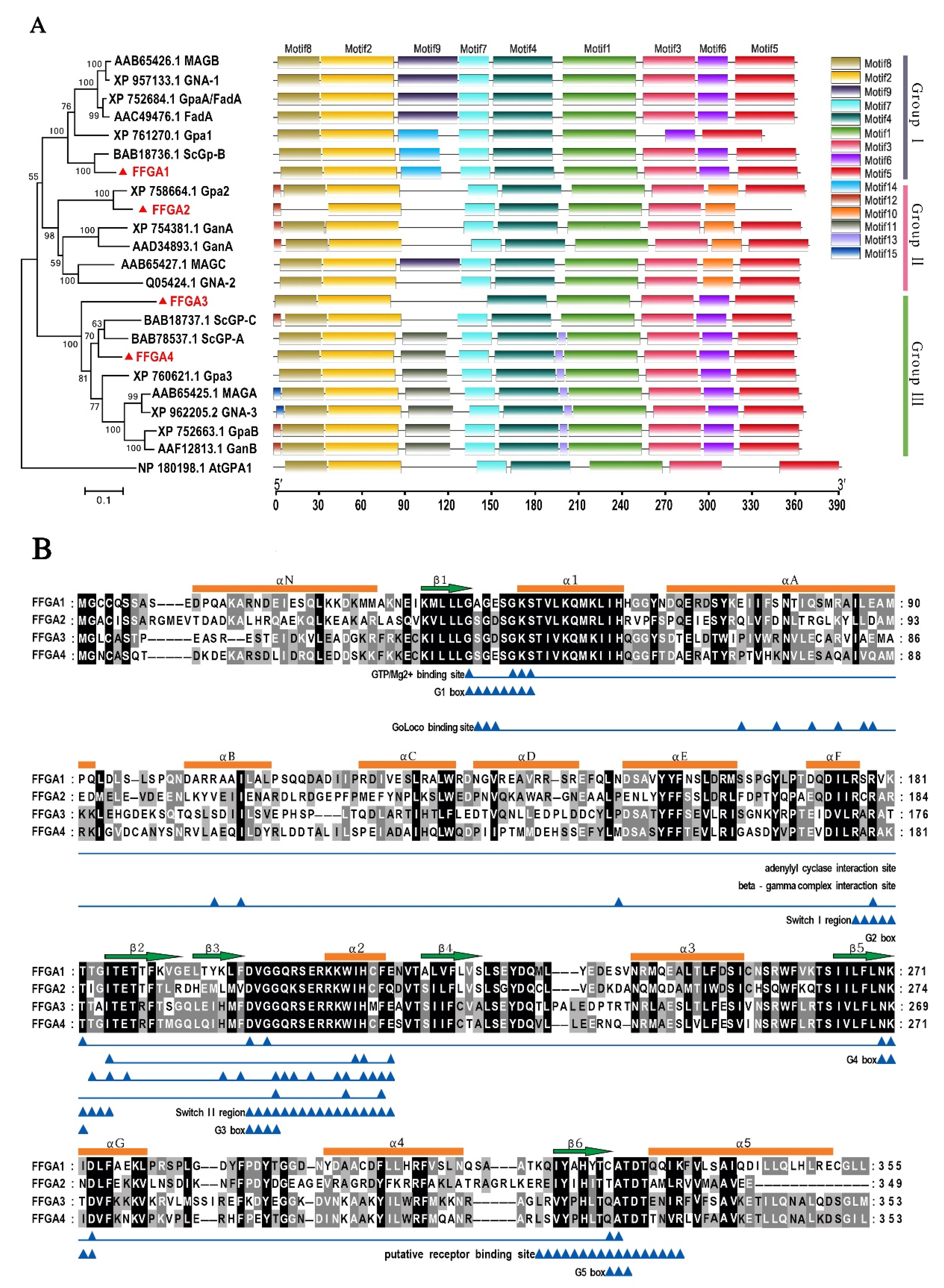
3.1. Identification and Characterization of Gα Subunit

We identified four putative genes, FfGa1 (OK156002), FfGa2 (OK156003), FfGa3 (OK156004), and FfGa4 (OK156005), encoding Gα subunits in the F. filiformis genome, which encode the polypeptide ranging from 339 to 355 amino acids in length. The products of the four genes shared 33.6 to 55.56% identity, along with 40.28 to 86.22% identity to ScGP-B from S. commune.
In order to determine the evolutionary relationship of the identified FFGA1-4 in F. filiformis with Gα ortholog sequences reported in other fungal species (i.e., Aspergillus nidulans (AAD34893.1, BAB78537.1 AAF12813.1), Aspergillus fumigatus (XP_752684.1, XP_754381.1 and XP_752663.1), N. crassa (XP_957133.1, Q05424.1 and XP_962205.2), Magnaporthe grisea (AAB65426.1, AAC49476.1, AAF12813.1), Ustilago maydis (XP_761270.1, XP_758664.1, XP_760621.1), and S. commune (BAB18737.1, BAB78537.1, BAB18736.1)), and a detailed phylogenetic analysis and conserved motif comparison were performed. The phylogenetic tree showed three prominent clades, which correspond to the three major groups described in a previous study [34]; all proteins had the Motif 1–5 presenting the conserved G1-5 box of G protein, and displayed strong variations in the motif of 90aa–130aa (Figure 1A). FFGA1 of F. filiformis showed a high degree of identity and motif similarity with BAB18726.1 of S. commune, which belongs to the Gα group I subfamily (related to the mammalian Gαi superfamily), and FFGA2 exhibited a closer association with Gα subunits in group II, while both FFGA3 and FFGA4 were clustered into group III.
Pfam-based domain prediction (http://pfam.janelia.org, 18 March 2021) revealed the presence of highly conserved guanine nucleotide-binding sites in four FFGA proteins, which is a key domain for G-protein subunit identification. Furthermore, there was a conserved protein fold among Gα proteins consisting of the Ras-like domain (RasD) and Helix domain (HD) [10], as shown in Figure 1B. RasD is a six β-sheet (β1–β6) surrounded by five α-helices (α1–α5), which is a highly conserved sequence in G1-G5 box, as reported by Yao [35], and is involved in the distinctive characteristics of G protein, including the GTP binding motif “GXGESGKS” (located in G1) of Motif 2, GTPase domain “DXXGQ” (located in switch II) of Motif 4, and myristoylation (MGXXXS) [10,32]. Therefore, the protein sequence analysis indicated that four Gα subunits are classical Gα proteins, whereas FFGA1 showed the two conserved functional motifs of the Gαi subunit, canonical ADP-ribosylation sequences for cholera toxin “RSRVK” (located in switch I), and ADP-ribosylation site for pertussis toxin “CXXX” (located at its C terminus), thus further confirming that FFGA1 belongs to the Gαi subfamily (Figure 1B).

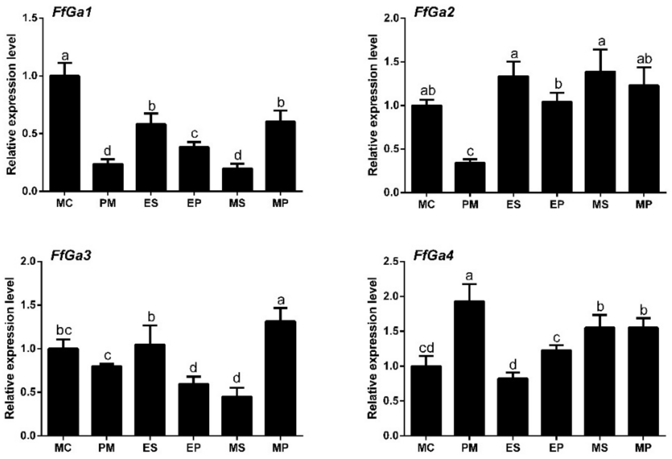
3.2. Expression of Gα Subunit at Different Stages of F. filiformis

To explore the possible relationship between the expression level of FfGa1-4 and morphological growth, FfGa1-4 transcript accumulation on different tissues, including mycelium (MY), elongation-stage stipes (ES), mature-stage stipes (MS), elongation-stage pileus (EP), and mature-stage pileus (MP), was assessed. The expression at the mycelial stage was used as a control, and it was assigned an expression value of one for normalization. Our results showed that the mRNA abundance of Gα genes was low in PM, except for FfGa4 (Figure 2). When compared with MC, the FfGa1 mRNA level was significantly reduced at all the stages tested with fold decreases of −0.7, −0.4, −0.6, −0.8, and −0.4 at PM, ES. EP, MS, and MP stages, respectively; the FfGa2 transcript level was slightly increased in stipes and pileus, a similar result to that shown in FfGa4, while the expression level of FfGa3 showed a weak reduction in PM, EP, and MS, and exhibited a distinct difference between elongation pileus and mature pileus. The result showed that the expression of FfGa1 in all parts of fruiting bodies was lower than that of mycelium, whereas the expression of FfGa2-4 presented no regularity between mycelium and several fruiting body tissues. Therefore, we could generate the OE and RNAi transformants and analyzed their respective phenotypes to explore the exact roles of the FfGa1 on the vegetative stage.

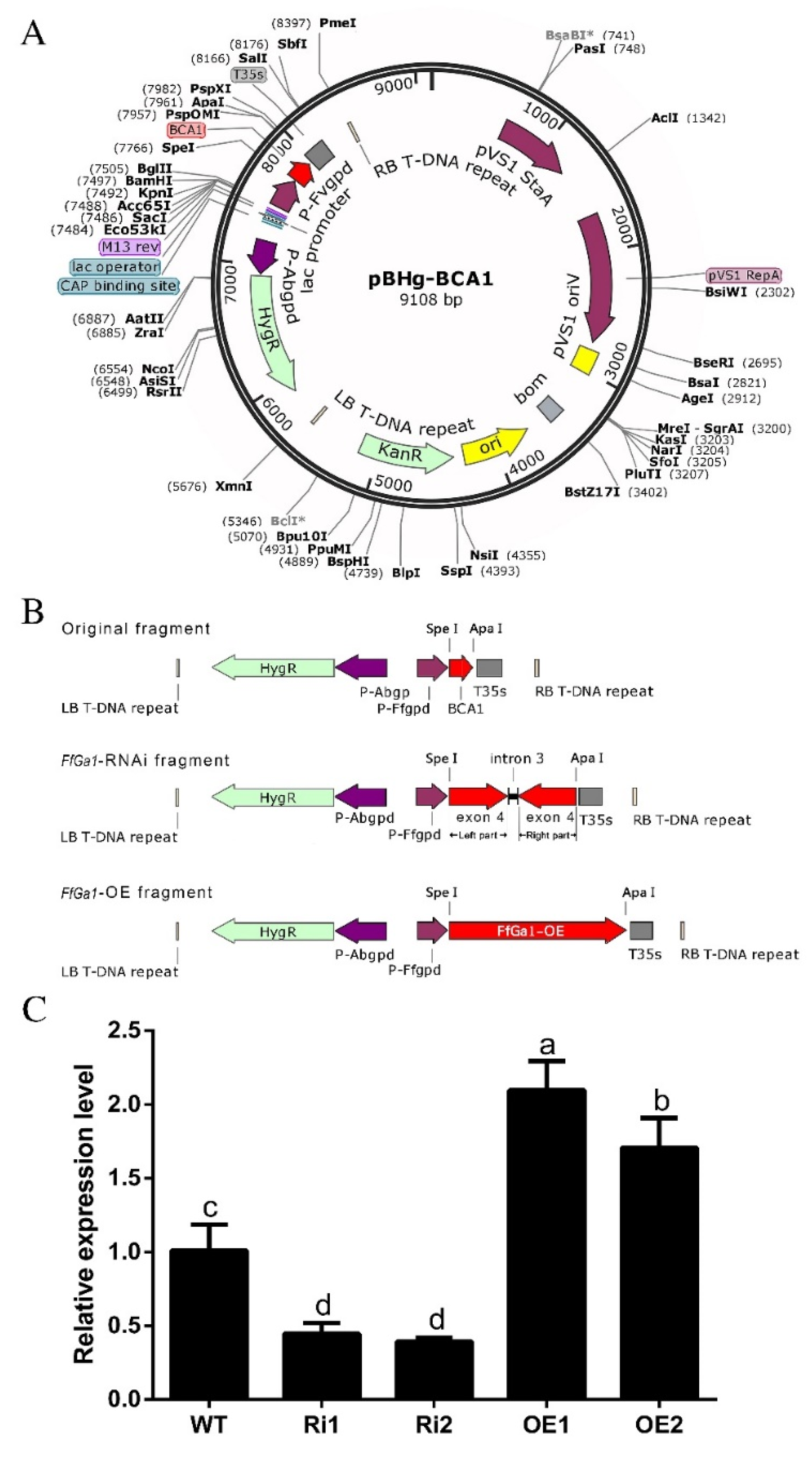
3.3. Generation of Overexpression and Knockdown Transformants

FfGa1-OE and RNAi vectors were constructed based on the binary vector pBHg-BCA1 mediated by A. tumefaciens (Figure 3A,B). Putative FfGa1-OE and RNAi transformants were screened by PCR with primer pairs (Supplementary Table S1) for FfGa1-OE and RNAi transformants. The results from PCR confirmation assays showed that the target gene fragments were stably inserted in the WT strain (Supplementary Figure S1). To access the efficiency of FfGa1 gene silencing and overexpression, we performed qPCR analysis to check the expression of FfGa1 in the transformants. Our qPCR results showed that the transcript levels of FfGa1 in the OE1 and OE2 transformants were up-regulated with an approximate fold increase of 2- and 1.6-fold higher, respectively, than in the WT strain, whereas FfGa1 transcription in RNAi transformants was decreased by more than 50% (Figure 3C). In addition, the almost complete LB-RB sequence with one copy was identified in four transformants by the whole-genome sequencing approach (Supplementary Table S2). Therefore, these transformants were chosen for further study.

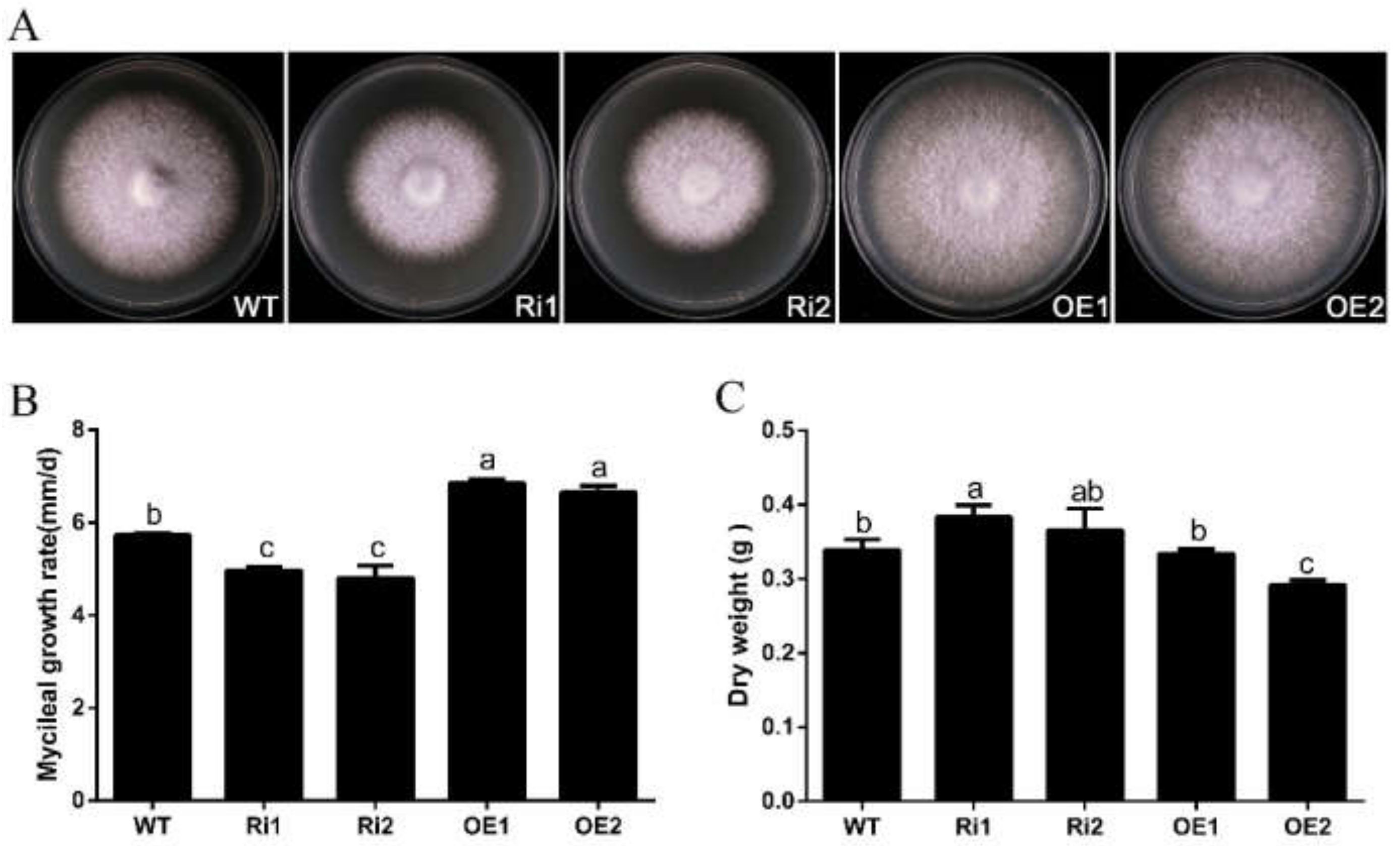
3.4. FfGa1 Positively Regulates Vegetative Growth in F. filiformis

The Gα subunit was reported to positively regulate vegetative growth in the fungus Penicillium camemberti [12]. To understand the physiological function of FfGa1 in the growth of F. filiformis. The WT, RNAi, and OE strains cultured on PDA medium for 6 days and hyphal growth rate and morphology were monitored. When compared to the WT strain, the OE strains exhibited a significantly increased growth, while the RNAi transformants showed a reduction in growth rate with compact, fluffy, more dense aerial hyphae (Figure 4A,B). We further measured the mycelial dry weight of WT, RNAi, and the OE strains maintained on PDB medium. This showed that mycelial dry weight accumulation was slightly higher for the RNAi strains when compared to the WT strain (Figure 4C). Based on this result, we conclude that FfGa1 positively regulates hyphal extension, but the knockdown of FfGa1 could slightly increase the mycelial weight of F. filiformis.

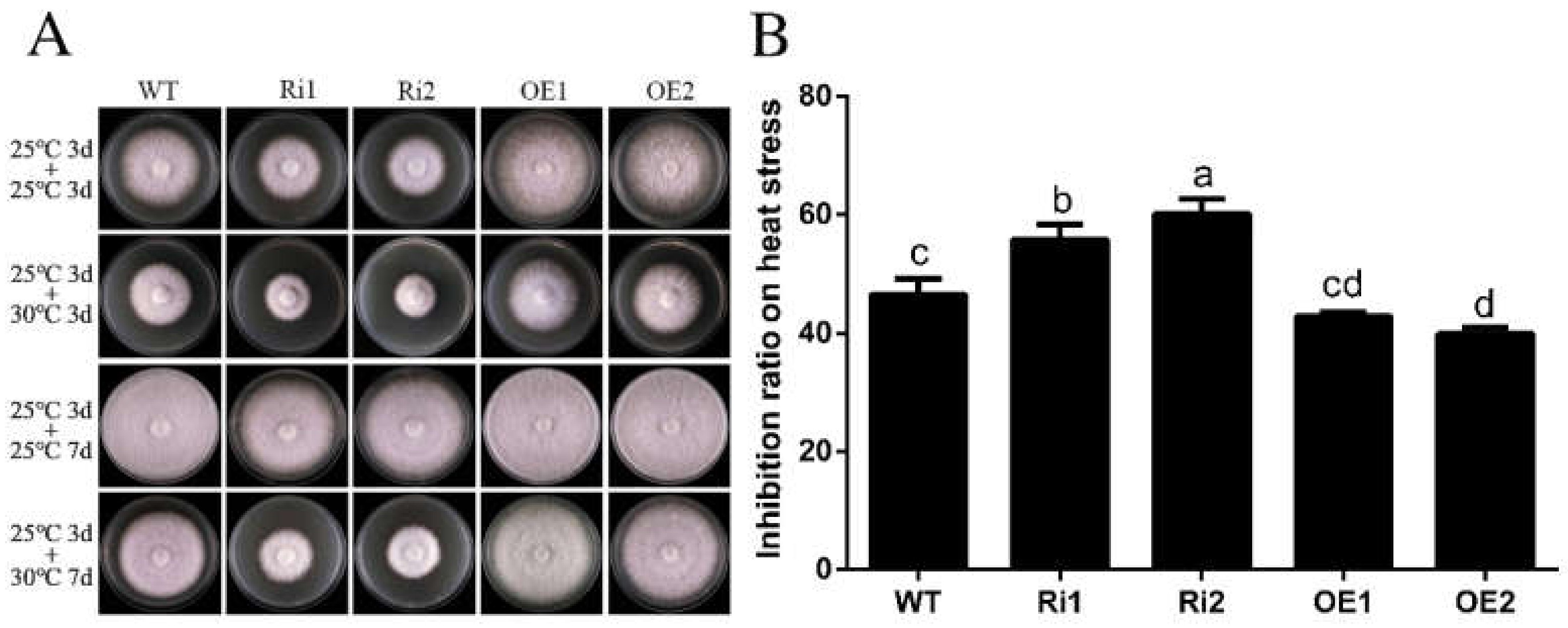
3.5. FfGa1 Facilitates Thermoresistance

A previous study showed that the PGa1 was involved in the regulation of heat stress response in the fungus Penicillium chrysogenum [36]. To explore the potential role of the FFGA1 subunit in the thermal stress response in F. filiformis, PDA plates inoculated with WT, RNAi, and OE strains were firstly incubated at a normal temperature of 25 °C; three days later, some of the WT, RNAi, and OE strain plates were transferred into a different incubator with a high temperature of 30 °C for 3 days, and the growth rate was measured. In order to obtain a more visible phenotypical difference, the treatment time was prolonged for an additional 4 days to take another photo. Results showed that the RNAi strains were more sensitive to chronical heat stress in comparison to WT, while the OE strains showed high tolerance to thermal stress with maximum colonies in the lower inhibition ratio (Figure 5). Therefore, these results indicate that FfGa1 has a positive impact on thermal resistance in F. filiformis.

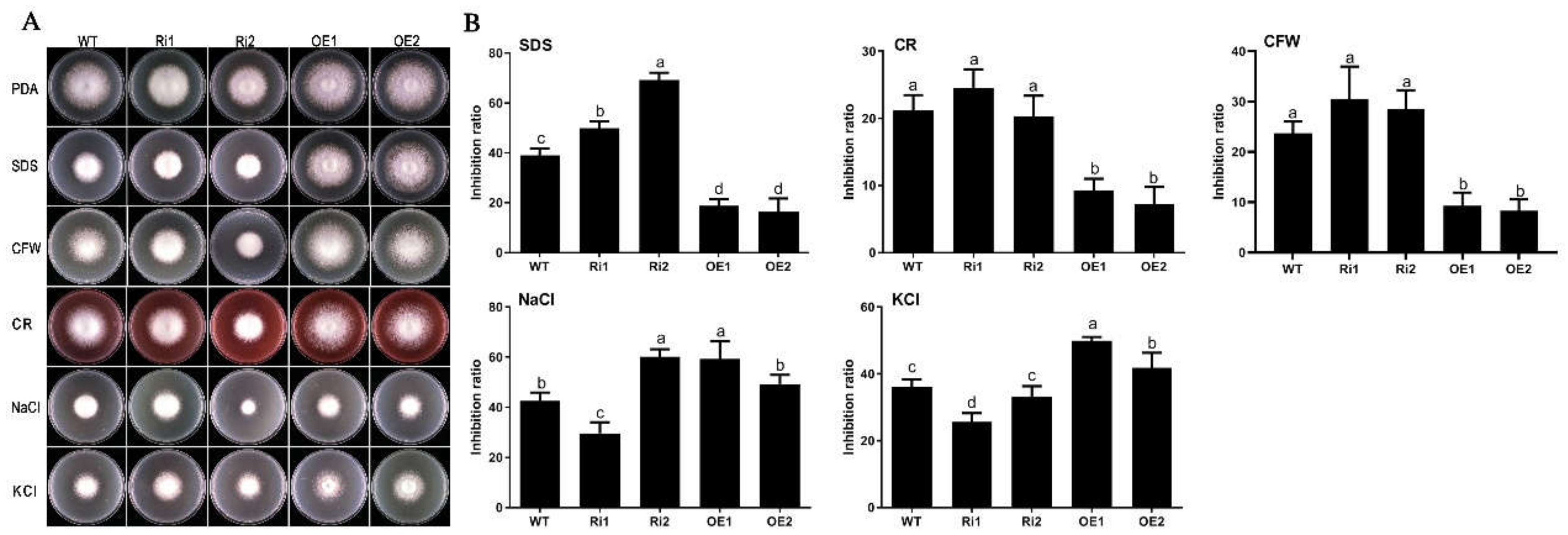
3.6. FfGa1 Is Required for Maintenance of Cell Wall Integrity and Hypertonic Stress Response in F. filiformis

The resistance to SDS, Congo red (CR), and CFW is considered as a barometer, reflecting cell wall integrity in fungi [37]. To establish the role of FfGa1 in cell wall integrity in F. filiformis, we compared the growth rates of the WT, RNAi, and the OE strains on PDA medium supplemented with cell wall-perturbing agents. The data showed that the growth rates of all strains were decreased on treated PDA, and F. filiformis was more sensitive to SDS than CR and CFW. On the other hand, OE transformants were more tolerant to SDS at a lower inhibition ratio with respect to WT; on the contrary, the growth rate of RNAi transformants was reduced severely upon SDS treatment when compared to wild-type transformants, and a similar pattern was observed when strains were treated with Congo red or CFW.
We further tested whether FfGa1 could be involved in response to hyperosmolarity in F. filiformis by culturing the WT, RNAi, and the OE strains on PDA medium supplemented with hypertonic agents 0.3 M NaCl and 0.5 M KCl. The results showed that OE transformants were more significantly inhibited at 0.5 M KCl than WT, and the opposite phenotypes of all RNAi transformants (Ri1 and Ri2) had a lower inhibition ratio than OE transformants (Figure 6). Under NaCl stress, OE transformants exhibited a higher inhibition effect than WT; Ri1 transformant had a lower inhibition ratio than OE transformants, while a higher inhibition phenotype was observed in Ri2 transformants, yet the mechanism of different response between Ri1 and Ri2 was unknown. In conclusion, these results suggest that FfGa1 is favorable to the maintenance of cell wall integrity and involved in the regulation of hypertonic stress responses in F. filiformis.

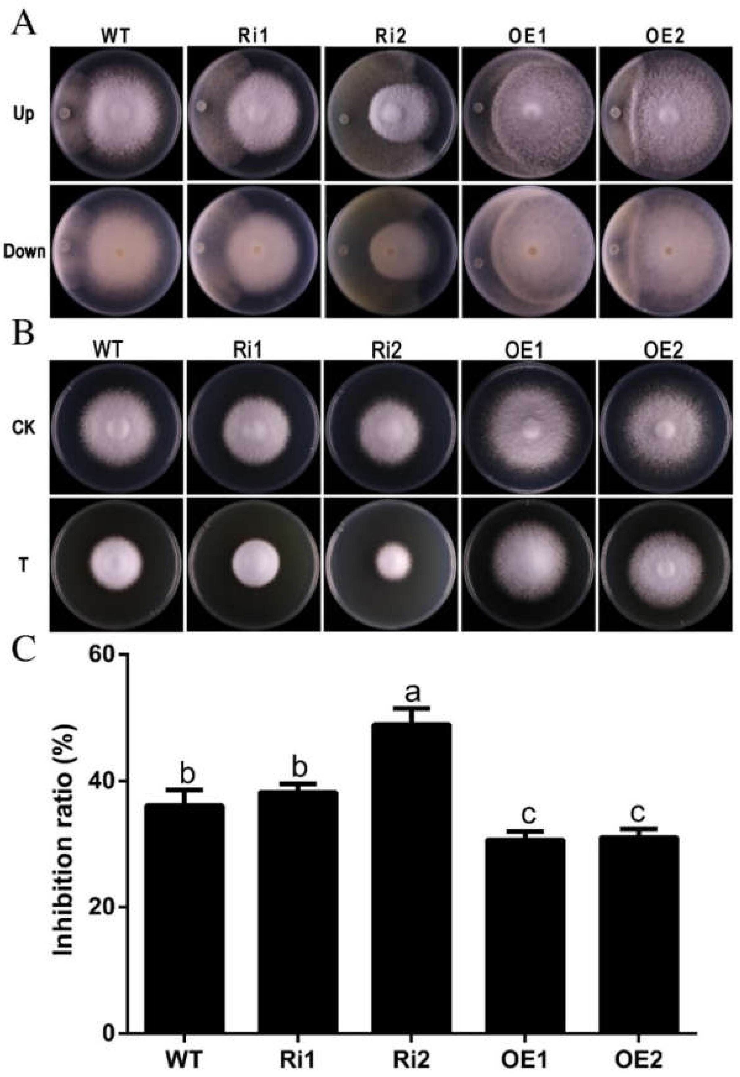
3.7. FfGa1 Involved in Resistance to Trichoderma sp.0018

Trichoderma is a well-known mycopathogen that can reduce mushroom production seriously [38]. To evaluate the role of FfGa1 in F. filiformis against Trichoderma sp.0018, the dual culture method was applied. Plate antagonistic assay results showed that OE1 and OE2 transformants exhibited obvious resistance against Trichoderma by forming a prominent zone of interaction, while no distinct zone of interaction was observed as a result of the co-cultivation of WT or RNAi strains with Trichoderma sp.0018 (Figure 7A).
We have also assessed the resistance of F. filiformis transformants against Trichoderma sp.0018 through culture filtrate assy. PDA medium supplemented with 50% v/v Trichoderma sp.0018 culture broth was used as treatment based on the preliminary experiment (Supplementary Figure S2). Results obtained showed the growth of RNAi strain was highly inhibited on Trichoderma sp.0018 culture broth medium, while the OE transformants were more resistant and grew faster when compared with WT and RNAi transformants (Figure 7B,C). These results suggest FfGa1 plays an important role in the defense mechanism of F. filiformis against Trichoderma sp.0018.

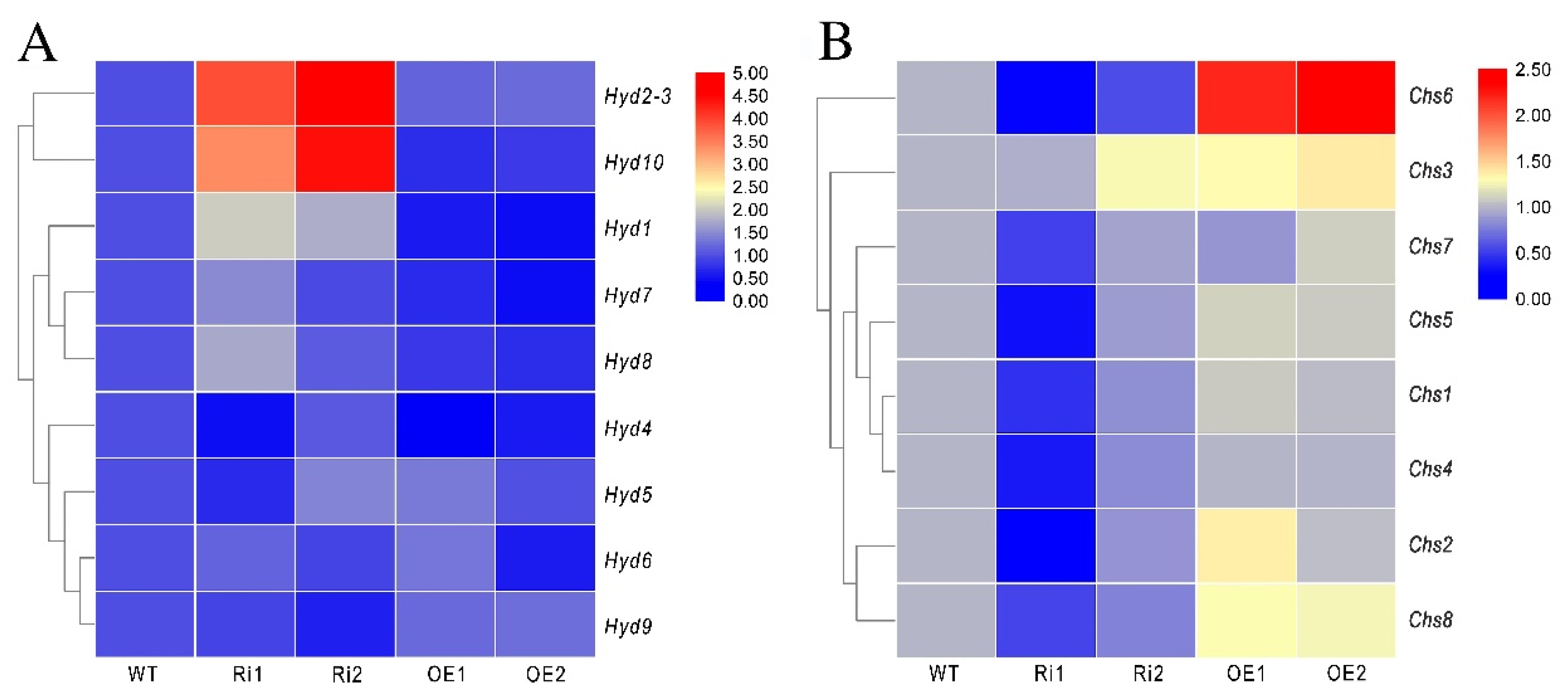
3.8. FfGa1 Regulates the Expression of the Hydrophobin and Chitin Synthase Genes

Since the Gα subunit is involved in the regulation of hydrophobin expression in previous studies [39,40], we performed quantitative PCR (qPCR) to test the effects of FfGa1 on the transcription of the hydrophobin genes Hyd1, Hyd2-3, Hyd4, Hyd5, Hyd6, Hyd7, Hyd8, Hyd9, and Hyd10. Our results showed that the majority of these genes, especially Hyd2-3, Hyd10, and Hyd1, were up-regulated in the RNAi transformants, while in OE transformants, Hyd10 and Hyd1 were down-regulated significantly (Figure 8A).
According to the literature, chitin synthase genes are related to cell wall integrity stress, and Chs5 or Chs7 deletion of Fusarium verticillioides increases susceptibility to SDS stress and exhibits reduction in growth [41]. Therefore, we also performed quantitative PCR (qPCR) to examine the expression of eight chitin synthetase genes. Almost all of these genes were down-regulated more or less in the RNAi strains, and the mRNA level of CHS6 was increased obviously in OE transformants. This indicates that FfGa1 favors the mRNA accumulation of most chitin synthetase genes.

4. Discussion

Gα protein, as a crucial signal transduction component, has been shown to be indispensable in many biological functions [42]. A variety of Gα subunits usually imply distinct or overlapping function in some organisms, such as Aspergillus flavus [43], Saccharomyces cerevisiae [44,45], and Fusarium oxysporum [46]. In contrast to the majority of filamentous fungi studied to date, which contain three genes that encode Gα proteins [47], four genes encoding Gα proteins were identified based on a genome-wide search of F. filiformis; corresponding protein sequences were clustered into three distinct well-characterized groups, and only FFGA1 had a higher transcript level in mycelium than fruiting body tissues.
In order to explore the function of FfGa1, RNAi and overexpression techniques were used to generate transformants for phenotypic assays. The results showed that the FfGa1 positively regulated hyphal extension, since compared to the WT, OE transformants exhibited an increase in growth rate with a larger colony diameter, and reverse phenotypic characteristics were observed in RNAi transformants. This result is consistent with previous studies [11]. In addition, we found that the knockdown FfGa1 led to a reduction in the growth rate of PDA but showed a minor increase in mycelium accumulation in PDB. In agreement with the role of Gαi of F. filiformis in hyphal growth, the deletion of cpg-1 resulted in a greatly reduced radial growth rate with scarce aerial hyphae, and a higher dry weight when compared with wild-type Cryphonectria parasitica [39]. Similar studies carried out in P. chrysogenum showed different results, in which Pga1 presented a strong negative effect on apical extension, but had no influence on biomass [48]. However, in this regard, there is a rough correlation between aerial hyphae and dry weight in N. crassa, as constitutively active Gαi strains exhibited a higher growth rate, more aerial hyphae, and a greater mass [19]. In light of these evidences, one plausible explanation is that the Gα may play different roles in hyphal growth due to culture conditions and specific species.
A close negative relationship between the Gα subunit expression level and the adaptability to environmental stimuli was verified in P. chrysogenum [36]. However, diverse results are found in our experiments showing that the overexpression FfGa1 increase the susceptibility to hypertonic stress, but is beneficial in enhancing hyphal tolerance to chronic high temperature and maintaining cell wall integrity. Similarly, TrGpa1 positively regulates the adaptability to thermal stress in Basidiomycota Tremella fuciformis [49]. In contrast to the negative influence of Gαi on hyperosmolarity in P. chrysogenum, the disruption of Gαi strains was less tolerant to hypertonic stress in N.crassa [50] and Cochliobolus heterostrophus [51]. Therefore, these findings, together with the previous research, highlight the important role of the Gαi subunit in regulating stress response in fungi.
Trichoderma, well known as a biocontrol agent against plant pathogens [52], is an harmful pathogen for mushroom cultivation as it suppresses mycelial growth and fruiting body production [38,53]. When confronted with Trichoderma sp., there is no distinguishable phenotypical trait in the contact zones for Agaricus bisporus, which could result in almost 100% crop loss [54]. Similar results regarding the response to Trichoderma sp.0018 attack were obtained in WT and RNAi transformants of F. filiformis. Interestingly, there are physiological responses to form mycelium assemblage barriers against the invasion of Trichoderma in OE transformants, with a lower inhibition ratio on the PDA medium amended with Trichoderma sp.0018 fermentation broth. Similarity, during co-cultivation of Lentinula edodes with Trichoderma, mycelium assemblage is also observed, which has been verified that the barrages characterized by brown antagonism lines are beneficial for resisting Trichoderma invasion and for defending territories in the contact zones [55]. Similar morphological changes were also reported in P. ostreatus [56] and S. commune [57]. Furthermore, the Gα has been verified to increase the ability to defend against pathogens in plants [58]. Hence, the results showed that the antagonism line regulated by FfGa1 might have positive effects on enhancing the resistance of F. filiformis mycelia to biotic stress.
Generally, the cell wall is regarded as the first defensive barrier to biotic and abiotic stress in fungi [59]. Chitin is a core component of the inner cell wall, hydrophobins are a key class of cell wall proteins, and both of them are affected by Gα subunits [39,60]. As downstream genes related to the signaling pathway, the deletion of Chs5 resulted in the abnormal morphology of hyphae and decreased the tolerance to heat stress, cell wall-perturbing compounds, and the deletion of chs7 in Metarhizium acridum [61]; in contrast, the disruption of Hyd1 increased the adaptability to thermal stress in Beauveria bassiana [62], and showed a high ability to maintain cell wall integrity in Aspergillus fumigatus [63]. In this study, most Chs gene expression levels were increased in OE transformants, though unlike Chs genes, the expression patterns of Hyds did not exhibit apparent regularity. Hyd1 mRNA levels were reduced in OE transformants. As mentioned above, the data from the comparison analysis of phenotype traits and RNA quantification showed that FfGa1 enhanced the cell wall integrity and biotic stress resistance of F. filiformis mycelia.

5. Conclusions

In summary, our results have shown that FfGa1 plays a crucial positive role in cell wall integrity maintenance and protection against mycoparasite Trichoderma, as well as thermal stress in F. filiformis. In addition, we established that FfGa1 increased hyphal extension and regulated the expression of the hydrophobin and chitin synthetase genes. This study is beneficial for exploring the role of Gα signaling pathways in macro-fungi and provides a suitable target point for the improved production of edible fungi.

Supplementary Materials

The following are available online at https://0-www-mdpi-com.brum.beds.ac.uk/article/10.3390/jof8040401/s1, Figure S1: PCR confirmation of the OE and RNAi transformants, Figure S2: Effect of fermentation broth of Trichoderma sp. 0018 on mycelium growth rate in WT, Table S1: All primers used in study, Table S2: Insert fragment information of four Flammulina filiformis transformants.

Author Contributions

M.D., B.X. and F.L. designed this study; M.D., Y.X., M.W., H.Y. and B.H. performed the experiments; M.D., Y.L., H.Y. and Y.T. analyzed all data; M.D. and I.M. wrote the initial manuscript, and all authors contributed to writing and editing of the final manuscript. All authors have read and agreed to the published version of the manuscript.

Funding

The research was supported by funds from the Fujian Edible Fungi Engineering Technology Research Center and the National Fungi Breeding Center (Fujian Branch) for providing the experimental facilities. This research was supported by “Industrialization Project of Wood Rot Edible Fungi (fjzycxny2017010)”to B.X., the “Major Science and Technology Project of Guizhou Province ([2019]3007-4)” to M.D., and the “National Natural Science Foundation of China (31902088)”to Y.T. and the “National Natural Science Foundation of China (32002108)” to F.L.

Institutional Review Board Statement

Not applicable.

Informed Consent Statement

Not applicable.

Data Availability Statement

Not applicable.

Acknowledgments

We are grateful to Youjin Deng, Chuanzheng Wei and Qianhui Huang for advice and the Fujian Edible Fungi Engineering Technology Research Center for providing the experimental facilities.

Conflicts of Interest

The authors declare no conflict of interest.

References

  1. Tao, Y.; Chen, R.; Yan, J.; Long, Y.; Tong, Z.; Song, H.; Xie, B. A hydrophobin gene, Hyd9, plays an important role in the formation of aerial hyphae and primordia in Flammulina filiformis. Gene 2019, 706, 84–90. [Google Scholar] [CrossRef] [PubMed]
  2. Park, Y.-J.; Baek, J.H.; Lee, S.; Kim, C.; Rhee, H.; Kim, H.; Seo, J.-S.; Park, H.-R.; Yoon, D.-E.; Nam, J.-Y. Whole genome and global gene expression analyses of the model mushroom Flammulina velutipes reveal a high capacity for lignocellulose degradation. PLoS ONE 2014, 9, e93560. [Google Scholar] [CrossRef] [PubMed]
  3. Dowom, S.A.; Rezaeian, S.; Pourianfar, H.R. Agronomic and environmental factors affecting cultivation of the winter mushroom or Enokitake: Achievements and prospects. Appl. Microbiol. Biotechnol. 2019, 103, 2469–2481. [Google Scholar] [CrossRef] [PubMed]
  4. Kertesz, M.A.; Thai, M. Compost bacteria and fungi that influence growth and development of Agaricus bisporus and other commercial mushrooms. Appl. Microbiol. Biotechnol. 2018, 102, 1639–1650. [Google Scholar] [CrossRef]
  5. Liu, X.-B.; Xia, E.-H.; Li, M.; Cui, Y.-Y.; Wang, P.-M.; Zhang, J.-X.; Xie, B.-G.; Xu, J.-P.; Yan, J.-J.; Li, J. Transcriptome data reveal conserved patterns of fruiting body development and response to heat stress in the mushroom-forming fungus Flammulina filiformis. PLoS ONE 2020, 15, e0239890. [Google Scholar] [CrossRef]
  6. Liu, J.-Y.; Chang, M.-C.; Meng, J.-L.; Feng, C.-P.; Wang, Y. A comparative proteome approach reveals metabolic changes associated with Flammulina velutipes mycelia in response to cold and light stress. J. Agric. Food Chem. 2018, 66, 3716–3725. [Google Scholar] [CrossRef]
  7. Yamada, M.; Kurano, M.; Inatomi, S.; Taguchi, G.; Okazaki, M.; Shimosaka, M. Isolation and characterization of a gene coding for chitin deacetylase specifically expressed during fruiting body development in the basidiomycete Flammulina velutipes and its expression in the yeast Pichia pastoris. FEMS Microbiol. Lett. 2008, 289, 130–137. [Google Scholar] [CrossRef]
  8. Urano, D.; Jones, A.M. Heterotrimeric G protein–coupled signaling in plants. Annu. Rev. Plant Biol. 2014, 65, 365–384. [Google Scholar] [CrossRef]
  9. Pandey, S.; Vijayakumar, A. Emerging themes in heterotrimeric G-protein signaling in plants. Plant Sci. 2018, 270, 292–300. [Google Scholar] [CrossRef]
  10. Oldham, W.M.; Hamm, H.E. Structural basis of function in heterotrimeric G proteins. Q. Rev. Biophys. 2006, 39, 117–166. [Google Scholar] [CrossRef]
  11. Li, L.; Wright, S.J.; Krystofova, S.; Park, G.; Borkovich, K.A. Heterotrimeric G protein signaling in filamentous fungi. Annu. Rev. Microbiol. 2007, 61, 423–452. [Google Scholar] [CrossRef]
  12. Garcia-Rico, R.O.; Gil-Duran, C.; Rojas-Aedo, J.F.; Vaca, I.; Figueroa, L.; Levican, G.; Chavez, R. Heterotrimeric G protein alpha subunit controls growth, stress response, extracellular protease activity, and cyclopiazonic acid production in Penicillium camemberti. Fungal Biol. 2017, 121, 754–762. [Google Scholar] [CrossRef]
  13. Yamagishi, K.; Kimura, T.; Suzuki, M.; Shinmoto, H. Suppression of fruit-body formation by constitutively active G-protein α-subunits ScGP-A and ScGP-C in the homobasidiomycete Schizophyllum commune. Microbiology 2002, 148, 2797–2809. [Google Scholar] [CrossRef]
  14. Baasiri, R.A.; Lu, X.; Rowley, P.S.; Turner, G.E.; Borkovich, K.A. Overlapping functions for two G protein α subunits in Neurospora crassa. Genetics 1997, 147, 137–145. [Google Scholar] [CrossRef]
  15. Kays, A.M.; Rowley, P.S.; Baasiri, R.A.; Borkovich, K.A. Regulation of conidiation and adenylyl cyclase levels by the Gα protein GNA-3 in Neurospora crassa. Mol. Cell. Biol. 2000, 20, 7693–7705. [Google Scholar] [CrossRef]
  16. Kays, A.M.; Borkovich, K.A. Severe impairment of growth and differentiation in a Neurospora crassa mutant lacking all heterotrimeric Gα proteins. Genetics 2004, 166, 1229–1240. [Google Scholar] [CrossRef]
  17. Park, H.S.; Kim, M.J.; Yu, J.H.; Shin, K.S. Heterotrimeric G-protein signalers and RGSs in Aspergillus fumigatus. Pathogens 2020, 9, 902. [Google Scholar] [CrossRef]
  18. Tong, Y.; Wu, H.; Liu, Z.; Wang, Z.; Huang, B. G-Protein Subunit Galphai in Mitochondria, MrGPA1, Affects Conidiation, Stress Resistance, and Virulence of Entomopathogenic Fungus Metarhizium robertsii. Front. Microbiol. 2020, 11, 1251. [Google Scholar] [CrossRef]
  19. Yang, Q.; Borkovich, K.A. Mutational activation of a Gαi causes uncontrolled proliferation of aerial hyphae and increased sensitivity to heat and oxidative stress in Neurospora crassa. Genetics 1999, 151, 107–117. [Google Scholar] [CrossRef]
  20. Mukherjee, P.K.; Latha, J.; Hadar, R.; Horwitz, B.A. Role of two G-protein alpha subunits, TgaA and TgaB, in the antagonism of plant pathogens by Trichoderma virens. Appl. Environ. Microbiol. 2004, 70, 542–549. [Google Scholar] [CrossRef]
  21. Nielsen, P.; Sørensen, J. Multi-target and medium-independent fungal antagonism by hydrolytic enzymes in Paenibacillus polymyxa and Bacillus pumilus strains from barley rhizosphere. FEMS Microbiol. Ecol. 1997, 22, 183–192. [Google Scholar] [CrossRef]
  22. Shi, L.; Fang, X.; Li, M.; Mu, D.; Ren, A.; Tan, Q.; Zhao, M. Development of a simple and efficient transformation system for the basidiomycetous medicinal fungus Ganoderma lucidum. World J. Microbiol. Biotechnol. 2012, 28, 283–291. [Google Scholar] [CrossRef] [PubMed]
  23. Wang, W.; Liu, F.; Jiang, Y.; Wu, G.; Guo, L.; Chen, R.; Chen, B.; Lu, Y.; Dai, Y.; Xie, B. The multigene family of fungal laccases and their expression in the white rot basidiomycete Flammulina velutipes. Gene 2015, 563, 142–149. [Google Scholar] [CrossRef] [PubMed]
  24. Tamura, K.; Stecher, G.; Peterson, D.; Filipski, A.; Kumar, S. MEGA6: Molecular evolutionary genetics analysis version 6.0. Mol. Biol. Evol. 2013, 30, 2725–2729. [Google Scholar] [CrossRef]
  25. Chen, X.; Stone, M.; Schlagnhaufer, C.; Romaine, C.P. A fruiting body tissue method for efficient Agrobacterium-mediated transformation of Agaricus bisporus. Appl. Environ. Microbiol. 2000, 66, 4510–4513. [Google Scholar] [CrossRef]
  26. Hou, L.; Wang, L.; Wu, X.; Gao, W.; Zhang, J.; Huang, C. Expression patterns of two pal genes of Pleurotus ostreatus across developmental stages and under heat stress. BMC Microbiol. 2019, 19, 231. [Google Scholar] [CrossRef]
  27. Schouten, H.J.; vande Geest, H.; Papadimitriou, S.; Bemer, M.; Schaart, J.G.; Smulders, M.J.; Perez, G.S.; Schijlen, E. Re-sequencing transgenic plants revealed rearrangements at T-DNA inserts, and integration of a short T-DNA fragment, but no increase of small mutations elsewhere. Plant Cell Rep. 2017, 36, 493–504. [Google Scholar] [CrossRef]
  28. Thomy, J.; Sanchez, F.; Gut, M.; Cruz, F.; Alioto, T.; Piganeau, G.; Grimsley, N.; Yau, S. Combining Nanopore and Illumina sequencing permits detailed analysis of insertion mutations and structural variations produced by PEG-mediated transformation in Ostreococcus tauri. Cells 2020, 10, 664. [Google Scholar] [CrossRef]
  29. Wang, L.X.; Ren, L.L.; Liu, X.B.; Shi, J.; Wang, J.Z.; Luo, Y.Q. Effects of endophytic fungi in Mongolian pine on the selection behavior of woodwasp (Sirex noctilio) and the growth of its fungal symbiont. Pest Manag. Sci. 2019, 75, 492–505. [Google Scholar] [CrossRef]
  30. Tao, Y.; van Peer, A.F.; Huang, Q.; Shao, Y.; Zhang, L.; Xie, B.; Jiang, Y.; Zhu, J.; Xie, B. Identification of novel and robust internal control genes from Volvariella volvacea that are suitable for RT-qPCR in filamentous fungi. Sci. Rep. 2016, 6, 29236. [Google Scholar] [CrossRef]
  31. Taylor, S.; Wakem, M.; Dijkman, G.; Alsarraj, M.; Nguyen, M. A practical approach to RT-qPCR—Publishing data that conform to the MIQE guidelines. Methods 2010, 50, S1–S5. [Google Scholar] [CrossRef]
  32. Livak, K.J.; Schmittgen, T.D. Analysis of relative gene expression data using real-time quantitative PCR and the 2−ΔΔCT method. Methods 2001, 25, 402–408. [Google Scholar] [CrossRef]
  33. Hu, X.; Li, Y.; Li, C.; Fu, Y.; Cai, F.; Chen, Q.; Li, D. Combination of fucoxanthin and conjugated linoleic acid attenuates body weight gain and improves lipid metabolism in high-fat diet-induced obese rats. Arch. Biochem. Biophys. 2012, 519, 59–65. [Google Scholar] [CrossRef]
  34. Bölker, M. Sex and crime: Heterotrimeric G proteins in fungal mating and pathogenesis. Fungal Genet. Biol. 1998, 25, 143–156. [Google Scholar] [CrossRef]
  35. Yao, X.-Q.; Malik, R.U.; Griggs, N.W.; Skjærven, L.; Traynor, J.R.; Sivaramakrishnan, S.; Grant, B.J. Dynamic coupling and allosteric networks in the α subunit of heterotrimeric G proteins. J. Biol. Chem. 2016, 291, 4742–4753. [Google Scholar] [CrossRef]
  36. Garcia-Rico, R.O.; Martin, J.F.; Fierro, F. Heterotrimeric Ga protein Pga1 from Penicillium chrysogenum triggers germination in response to carbon sources and affects negatively resistance to different stress conditions. Fungal Genet. Biol. 2011, 48, 641–649. [Google Scholar] [CrossRef]
  37. Hong, Z.; Mann, P.; Brown, N.H.; Tran, L.E.; Shaw, K.J.; Hare, R.S.; DiDomenico, B. Cloning and characterization of KNR4, a yeast gene involved in (1, 3)-beta-glucan synthesis. Mol. Cell. Biol. 1994, 14, 1017–1025. [Google Scholar]
  38. Williams, J.; Clarkson, J.M.; Mills, P.R.; Cooper, R.M. Saprotrophic and mycoparasitic components of aggressiveness of Trichoderma harzianum groups toward the commercial mushroom Agaricus bisporus. Appl. Environ. Microbiol. 2003, 69, 4192–4199. [Google Scholar] [CrossRef]
  39. Segers, G.C.; Nuss, D.L. Constitutively activated Gα negatively regulates virulence, reproduction and hydrophobin gene expression in the chestnut blight fungus Cryphonectria parasitica. Fungal Genet. Biol. 2003, 38, 198–208. [Google Scholar] [CrossRef]
  40. Seibel, C.; Gremel, G.; do Nascimento Silva, R.; Schuster, A.; Kubicek, C.P.; Schmoll, M. Light-dependent roles of the G-protein α subunit GNA1 of Hypocrea jecorina (anamorph Trichoderma reesei). BMC Biol. 2009, 7, 58. [Google Scholar] [CrossRef]
  41. Gandía, M.; Harries, E.; Marcos, J.F. The myosin motor domain-containing chitin synthase PdChsVII is required for development, cell wall integrity and virulence in the citrus postharvest pathogen Penicillium digitatum. Fungal Genet. Biol. 2014, 67, 58–70. [Google Scholar] [CrossRef] [PubMed]
  42. Ivey, F.; Hodge, P.; Turner, G.; Borkovich, K. The G alpha i homologue gna-1 controls multiple differentiation pathways in Neurospora crassa. Mol. Biol. Cell 1996, 7, 1283–1297. [Google Scholar] [CrossRef] [PubMed]
  43. Liu, Y.; Yang, K.; Qin, Q.; Lin, G.; Hu, T.; Xu, Z.; Wang, S. G protein α subunit GpaB is required for asexual development, aflatoxin biosynthesis and pathogenicity by regulating cAMP signaling in Aspergillus flavus. Toxins 2018, 10, 117. [Google Scholar] [CrossRef] [PubMed]
  44. Harashima, T.; Heitman, J. The Gα protein Gpa2 controls yeast differentiation by interacting with kelch repeat proteins that mimic Gβ subunits. Mol. Cell 2002, 10, 163–173. [Google Scholar] [CrossRef]
  45. Guo, M.; Aston, C.; Burchett, S.A.; Dyke, C.; Fields, S.; Rajarao, S.J.R.; Uetz, P.; Wang, Y.; Young, K.; Dohlman, H.G. The yeast G protein α subunit Gpa1 transmits a signal through an RNA binding effector protein Scp160. Mol. Cell 2003, 12, 517–524. [Google Scholar] [CrossRef]
  46. Jain, S.; Akiyama, K.; Takata, R.; Ohguchi, T. Signaling via the G protein α subunit FGA2 is necessary for pathogenesis in Fusarium oxysporum. FEMS Microbiol. Lett. 2005, 243, 165–172. [Google Scholar] [CrossRef]
  47. Mah, J.-H.; Yu, J.-H. Upstream and downstream regulation of asexual development in Aspergillus fumigatus. Eukaryot. Cell 2006, 5, 1585–1595. [Google Scholar] [CrossRef]
  48. García-Rico, R.O.; Martín, J.F.; Fierro, F. The pga1 gene of Penicillium chrysogenum NRRL 1951 encodes a heterotrimeric G protein alpha subunit that controls growth and development. Res. Microbiol. 2007, 158, 437–446. [Google Scholar] [CrossRef]
  49. Zhu, H.; Liu, D.; Zheng, L.; Chen, L.; Ma, A. Characterization of a G protein α subunit encoded gene from the dimorphic fungus-Tremella fuciformis. Antonie van Leeuwenhoek 2021, 114, 1949–1960. [Google Scholar] [CrossRef]
  50. Ivey, F.D.; Kays, A.M.; Borkovich, K.A. Shared and independent roles for a Gαi protein and adenylyl cyclase in regulating development and stress responses in Neurospora crassa. Eukaryot. Cell 2002, 1, 634–642. [Google Scholar] [CrossRef]
  51. Horwitz, B.A.; Sharon, A.; Lu, S.-W.; Ritter, V.; Sandrock, T.M.; Yoder, O.; Turgeon, B.G. A G Protein Alpha Subunit from Cochliobolus heterostrophus Involved in Mating and Appressorium Formation. Fungal Genet. Biol. 1999, 26, 19–32. [Google Scholar] [CrossRef]
  52. Zin, N.A.; Badaluddin, N.A. Biological functions of Trichoderma sp. for agriculture applications. Ann. Agric. Sci. 2020, 65, 168–178. [Google Scholar] [CrossRef]
  53. Wang, G.; Cao, X.; Ma, X.; Guo, M.; Liu, C.; Yan, L.; Bian, Y. Diversity and effect of Trichoderma sp. associated with green mold disease on Lentinula edodes in China. MicrobiologyOpen 2016, 5, 709–718. [Google Scholar] [CrossRef]
  54. Kosanovic, D.; Grogan, H.; Kavanagh, K. Exposure of Agaricus bisporus to Trichoderma aggressivum f. europaeum leads to growth inhibition and induction of an oxidative stress response. Fungal Biol. 2020, 124, 814–820. [Google Scholar] [CrossRef]
  55. Savoie, J.M.; Mata, G. The antagonistic action of Trichoderma sp. hyphae to Lentinula edodes hyphae changes lignocellulotytic activities during cultivation in wheat straw. World J. Microbiol. Biotechnol. 1999, 15, 369–373. [Google Scholar] [CrossRef]
  56. Savoie, J.M.; Mata, G. Trichoderma harzianum metabolites pre-adapt mushrooms to Trichoderma aggressivum antagonism. Mycologia 2003, 95, 191–199. [Google Scholar] [CrossRef]
  57. Ujor, V.C.; Monti, M.; Peiris, D.G.; Clements, M.O.; Hedger, J.N. The mycelial response of the white-rot fungus, Schizophyllum commune to the biocontrol agent, Trichoderma viride. Fungal Biol. 2012, 116, 332–341. [Google Scholar] [CrossRef]
  58. Lee, S.; Rojas, C.M.; Ishiga, Y.; Pandey, S.; Mysore, K.S. Arabidopsis heterotrimeric G-proteins play a critical role in host and nonhost resistance against Pseudomonas syringae pathogens. PLoS ONE 2013, 8, e82445. [Google Scholar] [CrossRef]
  59. Fuchs, B.B.; Mylonakis, E. Our paths might cross: The role of the fungal cell wall integrity pathway in stress response and cross talk with other stress response pathways. Eukaryot. Cell 2009, 8, 1616–1625. [Google Scholar] [CrossRef]
  60. Coca, M.A.; Damsz, B.; Yun, D.J.; Hasegawa, P.M.; Bressan, R.A.; Narasimhan, M.L. Heterotrimeric G-proteins of a filamentous fungus regulate cell wall composition and susceptibility to a plant PR-5 protein. Plant. J. 2000, 22, 61–69. [Google Scholar] [CrossRef]
  61. Zhang, J.; Jiang, H.; Du, Y.; Keyhani, N.O.; Xia, Y.; Jin, K. Members of chitin synthase family in Metarhizium acridum differentially affect fungal growth, stress tolerances, cell wall integrity and virulence. PLoS Pathog. 2019, 15, e1007964. [Google Scholar] [CrossRef] [PubMed]
  62. Zhang, S.; Xia, Y.X.; Kim, B.; Keyhani, N.O. Two hydrophobins are involved in fungal spore coat rodlet layer assembly and each play distinct roles in surface interactions, development and pathogenesis in the entomopathogenic fungus, Beauveria bassiana. Mol. Microbiol. 2011, 80, 811–826. [Google Scholar] [CrossRef] [PubMed]
  63. Valsecchi, I.; Dupres, V.; Stephen-Victor, E.; Guijarro, J.I.; Gibbons, J.; Beau, R.; Bayry, J.; Coppee, J.-Y.; Lafont, F.; Latgé, J.-P. Role of hydrophobins in Aspergillus fumigatus. J. Fungi 2018, 4, 2. [Google Scholar] [CrossRef] [PubMed]
Figure 1. Analysis of Gα amino acid sequences. (A) Phylogenetic analysis of FFGA and the Gα protein from other fungi. The phylogenetic tree was constructed using MEGA software version 6 with a bootstrap method of 1000 replications. Conserved motifs marked in different colors and numbers were generated by MEME tool. Motif 1 contains the G3 box and Switch II region; Motif 2 contains the G1 box; Motif 3 contains the G4 box; Motif 4 contains the G2 box and the Switch I region; Motif 5 contains the G5 box. (B) Multiple sequence alignment and secondary structure features of FFGA1-4. There is a pertussis toxin CXXX (located in C terminus) and cholera toxin RSRVK (located in switch I) in FFGA1. Black background with white letters represents the high-identity region in the amino acid sequence, while secondary structure (α helices and β-sheet) ruler numbering was based on FFGA1.
Jof 08 00401 g001
Figure 2. FfGa gene expression patterns during different development tissues. The expression was monitored at MC: mycelium; PM: primordium; ES: elongation stage of stipes; EP: elongation stage of pileus; MS: mature stage of stipes; MP: mature stage of pileus. The GAPDH and RAS genes were used as the reference genes for evaluation of the expression. The expression at the mycelium stage was assigned a value of 1 for normalization. The letters “a”, “b”, “c”, and “d” indicate statistically significant difference at 0.05 level between samples. Bars with no common letters are significantly different (p < 0.05).
Jof 08 00401 g002
Figure 3. Construction strategy of OE and RNAi vectors and transformants screening. (A) Vector map representing original plasmid pBHg-BCA1 used for the generation of transformants. (B) Cloning strategy showing the RNAi and OE cassette employed in generation of transformants. (C) FfGa1 expression in transformants. The expression of FfGa1 in WT was set as a value of 1. The meaning of “a”, “b”, “c”, and “d” see Figure 2 legend.
Jof 08 00401 g003
Figure 4. FfGa1 is required for vegetative growth. (A) Colony morphology of WT and transformants cultured on PDA medium and photographed after 6 days inoculation. (B) The growth rates of WT, RNAi, and OE strains cultured on PDA. (C) Biomass of the WT and transformants cultured in PDB medium for 10 days. See Figure 2 legend for explanation of “a”, “b” and “c”.
Jof 08 00401 g004
Figure 5. FfGa1 is required for regulation of thermal stress response. (A) The difference of colony morphology and diameter of strains on PDA medium treated at temperatures of 25 and 30 °C. Photographs were taken after 3 and 7 days. (B) Statistical representation of the inhibition rates at 30 °C from WT and transformants. See Figure 2 legend for explanation of “a”, “b” “c” and “d”.
Jof 08 00401 g005
Figure 6. Growth response of WT and transformants under cell wall stress and hypertonic stress. (A) Morphology of WT and transformants cultured on PDA medium supplemented with cell wall stressors, i.e., 0.01% SDS or 200 µg/mL Congo-Red (CR), while 0.3 M NaCl or 0.5 M KCl were used as osmotic stress causing agents for six days. (B) Mycelial growth inhibition ratio of WT and transformants under cell wall stress or hypertonic stress. See Figure 2 legend for explanation of “a”, “b” “c” and “d”.
Jof 08 00401 g006
Figure 7. FfGa1 regulates the resistance to Trichoderma sp.0018 in WT, RNAi, and OE. (A) WT and transformants’ antagonism against Trichoderma sp.0018. The top represents a photo taken from the front-side plate; the bottom represents a photo taken from the reverse-side plate. (B) Colony phenotype of WT and mutant strains cultured on PDA medium prepared with or without Trichoderma sp.0018 fermentation broth. T represents PDA medium prepared with Trichoderma sp.0018 fermentation broth, CK represents PDA medium. Strains were cultured for 6 days then plates were photographed. (C) Statistical analysis of mycelial growth inhibition ratio of WT and transformants cultured on PDA medium prepared with or without Trichoderma sp.0018 fermentation broth. See Figure 2 legend for explanation of “a”, “b” and “c”.
Jof 08 00401 g007
Figure 8. Expression of hydrophobin and chitin synthase genes in transformants. (A) Transcript levels of hydrophobin genes in transformants. (B) Transcript levels of chitin synthase genes in transformants. The expression of genes in WT was set as a value of 1, the results were visualized by TBtools.
Jof 08 00401 g008
Publisher’s Note: MDPI stays neutral with regard to jurisdictional claims in published maps and institutional affiliations.

