Article

Diversity of Colletotrichum Species Causing Apple Bitter Rot and Glomerella Leaf Spot in China

1
State Key Laboratory of Crop Stress Biology in Arid Areas and College of Plant Protection, Northwest A&F University, Yangling, Xianyang 712100, China
2
College of Food & Bioengineering, Henan University of Science and Technology, Luoyang 471003, China
3
Department of Plant Pathology and Microbiology, Iowa State University, Ames, IA 50011, USA
*
Authors to whom correspondence should be addressed.
These authors contributed equally to this work.
Submission received: 28 May 2022 / Revised: 5 July 2022 / Accepted: 15 July 2022 / Published: 18 July 2022
(This article belongs to the Topic Fungal Diversity)

Abstract

:
Bitter rot and Glomerella leaf spot (GLS) of apples, caused by Colletotrichum species, are major diseases of apples around the world. A total of 98 isolates were obtained from apple fruits with bitter rot, and 53 isolates were obtained from leaves with leaf spot in the primary apple production regions in China. These isolates were characterized morphologically, and five gene regions (ITS, ACT, GAPDH, CHS-1 and TUB2) were sequenced for each isolate. A phylogenetic analysis, combined with a comparison of the morphological, cultural and pathogenic characters, sorted bitter rot isolates into six species: C. alienum, C. fructicola, C. gloeosporioides sensu stricto, C. nymphaeae, C. siamense and one new species, C. orientalis Dandan Fu & G.Y. Sun. Among these, C. siamense was the predominant pathogen associated with bitter rot. Isolates from leaf spot were identified as two species, C. aenigma and C. fructicola. This is the first report of C. orientalis as an apple bitter rot pathogen worldwide, and the results provide important insights into the diversity of Colletotrichum species in China.

1. Introduction

Apple bitter rot (ABR) is a common pre- and post-harvest disease in nearly all apple-growing areas worldwide. Because of its latent infection ability, crop losses can be severe from mid- to late-summer under prolonged warm and wet weather conditions [1]. The earliest record of a pathogen causing ABR is from 1856 when Gloeosporium fructigenum was described as the causal agent [2]. The fungus causing ABR was renamed several times until all species became synonymous to Glomerella cingulata (Stoneman) Spauld. & H. Schrenk (anamorph: Colletotrichum gloeosporioides (Penz.) Penz. & Sacc) in 1903. In 1965, C. acutatum J. H. Simmonds was distinguished from C. gloeosporioides based on physiology and morphology [3]. ABR pathogens were mainly reported to be C. gloeosporioides, G. cingulata and C. acutatum [4]. Jones et al. found that C. acutatum and C. gloeosporioides were recovered from 81% and 19%, respectively, of 165 symptomatic fruits collected from orchards in western Michigan [5]. Shi et al. reported that C. acutatum was the most predominant species (70%) associated with ABR in orchards in Arkansas, North Carolina and Virginia [6]. Restriction fragment length polymorphisms (RFLP) and random amplified polymorphic DNA (RAPD) analyses indicated high intraspecific diversity [1,7,8,9], which, however, might reflect interspecific differences in the revised Colletotrichum taxonomic system [10]. In addition to apple fruit bitter rot, Colletotrichum species also incur foliar disease, namely Glomerella leaf spot (GLS) [11]. GLS was first reported in Brazil in the 1980s and was subsequently reported in the USA and East Asia [1,7]. The disease causes severe leaf fall off on susceptible cultivars, such as Gala and Golden Delicious. The new Colletotrichum taxonomic system was established with polyphasic approaches with an emphasis on multigene phylogeny, in which ‘C. gloeosporioides’ and ‘C. acutatum’ are both monophyletic species complexes, with over 20 and 30 independent species, respectively [10,12]. Thus far, a number of ABR pathogenic species, belonging either to the C. acutatum species complex (CASC) (C. abscissum, C. acutatum, C. fioriniae, C. godetiae, C. melonis, C. nymphaeae and C. paranaense) or the C. gloeosporioides species complex (CGSC) (C. chrysophilum, C. fragariae, C. fructicola, C. gloeosporioides s. str., C. noveboracense, C. siamense, C. alienum and C. theobromicola) have been reported worldwide [13,14,15,16,17,18,19,20,21,22,23,24,25,26,27]. Compared with ABR, relatively few GLS pathogens have been recognized thus far; these include C. fructicola and C. aenigma, belonging to the CGSC; C. karstii, belonging to the C. boninense species complex (CBSC); and C. limetticola, belonging to the CASC [11,20,28,29,30,31,32].
In China, apple bitter rot occurs in almost all producing areas, and the pathogens have been identified as C. gloeosporioides and C. acutatum [33,34,35]. Unfortunately, these species may all represent species complexes. GLS is an emerging disease that was first reported in 2012, and the pathogens have been identified as C. fructicola and C. aenigma [28], yet hidden pathogen diversity may exist due to insufficient investigation. Therefore, the main objective of this study is to investigate the Colletotrichum species diversity associated with ABR and GLS in China; gaining this knowledge will provide clues towards more effective control measures against these devastating diseases.

2. Materials and Methods

2.1. Isolates

Isolates were collected from diseased apple tissues exhibiting bitter rot and leaf spot symptoms in commercial apple orchards in four provinces, including Liaoning, Shandong, Henan and Shaanxi, of China from 2009 to 2013. Small pieces of symptomatic tissue were cut from lesions, immersed in 70% alcohol for 1 min, rinsed with sterile water and then dried on sterilized filter paper before placement into Petri dishes with Potato Dextrose Agar (PDA, Becton, Dickinson and Company, Franklin Lakes, NJ, USA). Cultures were incubated for 4 days at 25 °C in darkness. A mycelial disc was taken from the actively growing edge of a mono-conidial colony, and then transferred onto new PDA plates. Monosporic isolates were obtained from the new cultures. The surfaces of the PDA plates were scraped with sterile water and collected as conidia suspensions. Monosporic isolates were deposited in the Fungal Laboratory of Northwest A&F University, Yangling, Shaanxi Province, China. After 7 days at 25 °C in darkness, the sizes and shapes of 50 conidia harvested from the cultures were measured and recorded [36]. The colony diameter, color of the conidial masses and zonation of the colony were recorded. Appressoria were induced using a slide culture technique, in which a 1 cm2 segment of PDA containing the isolate was placed in sterile water in a sterile Petri dish, covered with a sterile coverslip and incubated under high humidity at 25 °C in darkness. After 2 days, the shapes and sizes of 50 appressoria on the coverslip were recorded.

2.2. DNA Extraction and PCR Amplification

The protocol from Barnes et al. was used to extract DNA from the mycelia by scraping the surface of the PDA after it had been cultured for 7 days at 25 °C [37]. The quantity and quality of the DNA were estimated by UV microscopic spectrophotometer (Nanodrop 2000, Thermo Fisher Scientific, Waltham, MA, USA). The partial rDNA-ITS, actin (ACT), glyceraldehyde-3-phosphate dehydrogenase (GAPDH), chitin synthase (CHS-1) and β-tubulin-2 (TUB2) genes were amplified by PCR using primer pairs of ITS1-F [38] + ITS4 [39], ACT-512F + ACT-783R [40], GDF1 + GDR1 [41], CHS-79F + CHS-354R [40] and Bt2a + Bt2b [42], respectively. The PCR protocols were performed as described by Damm et al. [43]. The sequences of the isolates described in this study were deposited in GenBank; the accession numbers are listed in Table 1.

2.3. Sequence Alignment and Phylogenetic Analysis

Preliminary alignments of the multi-locus sequences were conducted using Clustal X [44] with a manual adjustment and BioEdit for visual improvement wherever necessary. The concatenation of the five-gene sequences was completed in PhyloSuite [45]. A maximum likelihood (ML) analysis was performed by RAxML version 8 [46] under the GTR model [47], and a non-parametric bootstrap analysis with 1000 repetitions [48] was used to determine the statistical support of the phylogeny. Bayesian inference (BI) phylogeny construction was performed with MrBayes version 3.2.1 [49], with the GTR + G + I nucleotide substitution model. The analysis included two separate runs for 1 × 10⁷ generations; each run was sampled every 1000 generations, and the convergence of all the parameters was checked using internal diagnostics. To construct the 50% majority-rule consensus tree, the first 25% generations were discarded as burn-in. The phylogenetic tree (Figure 1) was visualized using FigTree v 1.4.4. A potential recombination event between C. fioriniae and C. orientalis was detected based on a pairwise homoplasy index (PHI) analysis of the Genealogical Concordance Phylogenetic Species Recognition concept in SplitsTree version 4.11.3 using the multi-locus alignment dataset [50,51].

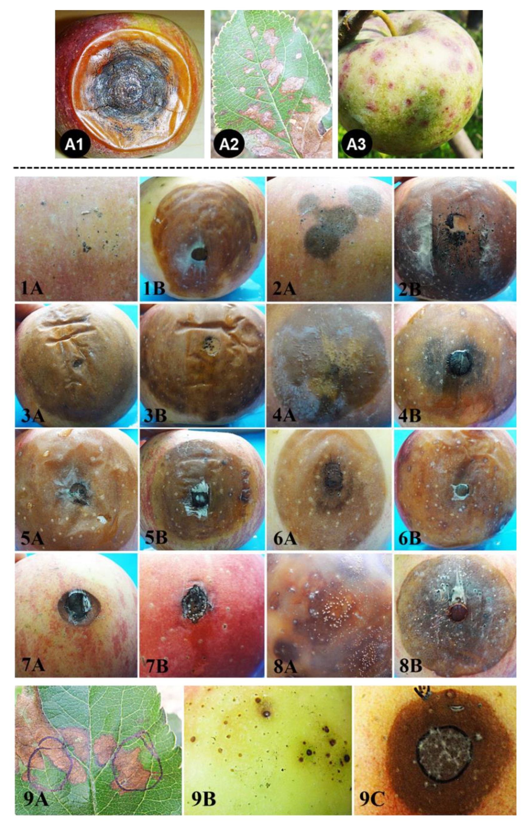
2.4. Pathogenicity Tests

Twelve representative isolates of Colletotrichum were chosen based on species identity and locations. Healthy apple fruits and leaves were selected, washed with tap water, blown dry in the hood and surface-sterilized with 70% ethanol prior to inoculation. Leaves and fruits were drop-inoculated with the conidia suspension (approximately 106/mL in concentration) or mycelial plugs. The fruits’ wounds were made by sterile insect needles with about 10 holes within a circular area of 5 mm in diameter. After inoculation, the fruits were incubated at 25 °C in plastic bags. The disease incidence of each fungal isolate was recorded 3 days after inoculation. For each isolate, at least five fruit/leaf inoculation replicates were performed in each experiment, and the inoculation experiment was repeated two times.
Table 1. Fungal isolates and sequences used in the phylogenetic analysis of this study.
SpeciesType StrainHostCountyGenBank No.
ITSACTGAPDHCHS-1TUB2
C. acerbumCBS 128530 1Malus domesticaNew ZealandJQ948459JQ949780JQ948790JQ949120JQ950110
C. acutatumCBS 112996 1Carica papayaAustraliaJQ005776JQ005839JQ948677JQ005797JQ005860
CBS 126521 2AnemonehybrideNetherlandsJQ948366JQ949687JQ948697JQ949027JQ950017
C. aenigmaICMP 18608 1Persea americanaIsraelJX010244JX009443JX010044JX009774JX010389
ICMP 18686 2Pyrus pyrifoliaJapanJX010243JX009519JX009913JX009789JX010390
F12PGXY03Malus domesticaChinaKF772117KF772027KF772087KF772057KF772147
F12PGXY04Malus domesticaChinaKF772118KF772028KF772088KF772058KF772148
W12PGYXY15Malus domesticaChinaKF791590KF791569KF791583KF791576KF791597
C. aeschynomenesICMP 17673 1Aeschynomene sp.USAJX010176JX009483JX009930JX009799JX010392
C. alienumICMP 12071 1Malus domesticaNew ZealandJX010251JX009572JX010028JX009882JX010411
ICMP 18621 3Persea americanaNew ZealandJX010246JX009552JX009959JX009755JX010386
F11PGZH02Malus domesticaChinaKF772119KF772029KF772089KF772059KF772149
C. asianumICMP 18580 1Coffea arabicaThailandFJ972612JX009584JX010053JX009867JX010406
ICMP 18696 3Mangifera indicaAustraliaJX010192JX009576JX009915JX009753JX010384
C. boninenseCBS 123755 1Crinum asiaticumJapanJQ005153JQ005501JQ005240JQ005327JQ005588
C. cuscutaeIMI 304802 1Cuscuta sp.DominicaJQ948195JQ949516JQ948525JQ948856JQ949846
C. fioriniaeCBS 128517 1Fiorinia externaUSAJQ948292JQ949613JQ948622JQ948953JQ949943
CBS 125396 2Malus domesticaUSAJQ948299JQ949620JQ948629JQ948960JQ949950
CBS 128517 1Fiorinia externaUSAJQ948292JQ949613JQ948622JQ948953JQ949943
CBS 125396 2Malus domesticaUSAJQ948299JQ949620JQ948629JQ948960JQ949950
CBS 363003 2Camellia reticulataChinaJQ948339JQ949660JQ948669JQ949000JQ949990
ATCC 28992 2Malus domesticaUSAJQ948297JQ949618JQ948627JQ948958JQ949948
CBS 129938 2Malus domesticaUSAJQ948296JQ949617JQ948626JQ948957JQ949947
CBS 129948 2Tulipa sp.UKJQ948344JQ949665JQ948674JQ949005JQ949995
IMI 324996 2Malus pumilaUSAJQ948301JQ949622JQ948631JQ948962JQ949952
ATCC 12097 2Rhododendron sp.USAJQ948307JQ949628JQ948637JQ948968JQ949958
CBS 200.35 2Rubus sp.USAJQ948293JQ949614JQ948623JQ948954JQ949944
CBS 490.92 2Solanum lycopersicumNew ZealandJQ948326JQ949647JQ948656JQ948987JQ949977
CBS 119293 2Vaccinium corymbosum (blueberry)New ZealandJQ948314JQ949635JQ948644JQ948975JQ949965
C. fructicolaCBS 130416 1Coffea arabicaThailandJX010165FJ907426JX010033JX009866JX010405
F12PGSQ01Malus domesticaChinaKF772124KF772034KF772094KF772064KF772154
F12PGSQ05Malus domesticaChinaKF772125KF772035KF772095KF772065KF772155
F12PGXY01Malus ×domesticaChinaKF772126KF772036KF772096KF772066KF772156
W12PGYSQ06M. ×domesticaChinaKF791591KF791570KF791584KF791577KF791598
F10PGCJJ1M. ×domesticaChinaKF772128KF772038KF772098KF772068KF772158
F10PGCJJ3M. ×domesticaChinaKF772129KF772039KF772099KF772069KF772159
F10PGHLD1M. ×domesticaChinaKF772130KF772040KF772100KF772070KF772160
F11PGYT02M. ×domesticaChinaKF772131KF772041KF772101KF772071KF772161
F11PGYT04M. ×domesticaChinaKF772132KF772042KF772102KF772072KF772162
C. gloeosporioidesCBS 112999 1Citrus sinensisItalyJX010152JX009531JX010056JX009818JX010445
CBS 119204 3Pueraria lobataUSAJX010150JX009502JX010013JX009790GQ849434
F11PGQX17M. ×domesticaChinaKF772111KF772021KF772081KF772051KF772141
F12PGDL01M. ×domesticaChinaKF772112KF772022KF772082KF772052KF772142
F12PGLQ30M. ××domesticaChinaKF772113KF772023KF772083KF772053KF772143
F12PGLQ33M. ×domesticaChinaKF772114KF772024KF772084KF772054KF772144
F12PGLQ34M. ×domesticaChinaKF772115KF772025KF772085KF772055KF772145
F11PGZH23M. ×domesticaChinaKF772116KF772026KF772086KF772056KF772146
C. godetiaeCBS 133.44 1Clarkia hybridaDenmarkJQ948402JQ949723JQ948733JQ949063JQ950053
CBS 198.53 2M. sylvestrisNetherlandsJQ948432JQ949753JQ948763JQ949093JQ950083
C. horiiICPM 10492 1Diospyros kakiJapanGQ329690JX009438GQ329681JX009752JX010450
C. hymenocallidisCBS 125378 1HymenocallisChinaJX010278GQ856775JX010019GQ856730JX010410
C. kahawae subsp. kahawaeIMI 319418 1Coffea arabicaKenyaJX010231JX009452JX010012JX009813JX010444
C. karstiiCBS 132134 4Malus sp.USAJQ005181JQ005529JQ005268JQ005355JQ005615
C. lupiniCBS 109225 1Lupinus albusUkraineJQ948155JQ949476JQ948485JQ948816JQ949806
C. musaeCBS 116870 1Musa sp.USAJX010146JX009433JX010050JX009896HQ596280
ICMP 17817 3Musa sapientumKenyaJX010142JX009432JX010015JX009815JX010395
C. nymphaeaeCBS 515.78 1Nymphaea albaNetherlandsJQ948197JQ949518JQ948527JQ948858JQ949848
IMI 370491 2M. pumilaBrazilJQ948204JQ949525JQ948534JQ948865JQ949855
F10PGBYS12M. ×domesticaChinaKF772133KF772043KF772103KF772073KF772163
C. orchidophilumCBS 632.80 1Dendrobium sp.USAJQ948151JQ949472JQ948481JQ948812JQ949802
C. orientalisF10PGBYS1M. ×domestica China KF772134KF772044KF772104KF772074KF772164
F10PGBYS2M. ×domestica ChinaKF772135KF772045KF772105KF772075KF772165
F10PGBYS3M. ×domestica ChinaKF772136KF772046KF772106KF772076KF772166
F10PGBYS4M. ×domestica ChinaKF772137KF772047KF772107KF772077KF772167
F10PGBYS7M. ×domestica ChinaKF772138KF772048KF772108KF772078KF772168
F10PGBYS8M. ×domestica ChinaKF772139KF772049KF772109KF772079KF772169
F10PGBYS10M. ×domestica ChinaKF772140KF772050KF772110KF772080KF772170
CBS 128555 2Malus domesticaNew ZealandJQ948305JQ949626JQ948635JQ948966JQ949956
C. queenslandicumICMP 1778 1Carica papayaAustraliaJX010276JX009447JX009934JX009899JX010414
ICMP 18705 3Coffea sp.FijiJX010185JX009490JX010036JX009890JX010412
C. salicisCBS 113.14 2M. ×domesticaGermanyJQ948478JQ949799JQ948809JQ949139JQ950129
IMI 385055 2M. ×domesticaNew ZealandJQ948472JQ949793JQ948803JQ949133JQ950123
C. salsolaeICMP 19051 1Salsola tragusHungaryJX010242JX009562JX009916JX009863JX010403
C. siamenseCBS 130417 1Coffea arabicaThailandJX010171FJ907423JX009924JX009865JX010404
ICMP 17795 3M. ×domesticaUSAJX010162JX009506JX010051JX009805JX010393
F12PGSQ02M. ×domesticaChinaKF772127KF772037KF772097KF772067KF772157
F10PGWFT2M. ×domesticaChinaKF772120KF772030KF772090KF772060KF772150
F11PGQX26M. ×domesticaChinaKF772121KF772031KF772091KF772061KF772151
F11PGLQ22M. ×domesticaChinaKF772122KF772032KF772092KF772062KF772152
F12PGMJ01M. ×domesticaChinaKF772123KF772033KF772093KF772063KF772153
C. simmondsiiCBS 126524 2Cyclamen sp.NetherlandsJQ948281JQ949602JQ948611JQ948942JQ949932
CBS 122122 1Carica papayaAustraliaJQ948276JQ949597JQ948606JQ948937
C. tropicaleCBS 124949 1Theobroma cacaoPanamaJX010264JX009489JX010007JX009870
ICMP 18672 3Litchi chinensisJapanJX010275JX009480JX010020JX009826
1 Cannon et al. (2012) [10]; 2 Damm et al. (2012a) [12]; 3 Weir et al. (2012) [52]; 4 Damm et al. (2012b) [53].

3. Results

3.1. Isolate Isolation

In total, 151 isolates were isolated from symptomatic leaf and fruit lesions in four apple-growing provinces. Among these, 98 were from bitter rot lesions, and 53 were from GLS lesions. Based on the conidial morphology and ITS sequence, 17 isolates were typical for the C. acutatum complex, while 134 isolates were typical for the C. gloeosporioides complex.

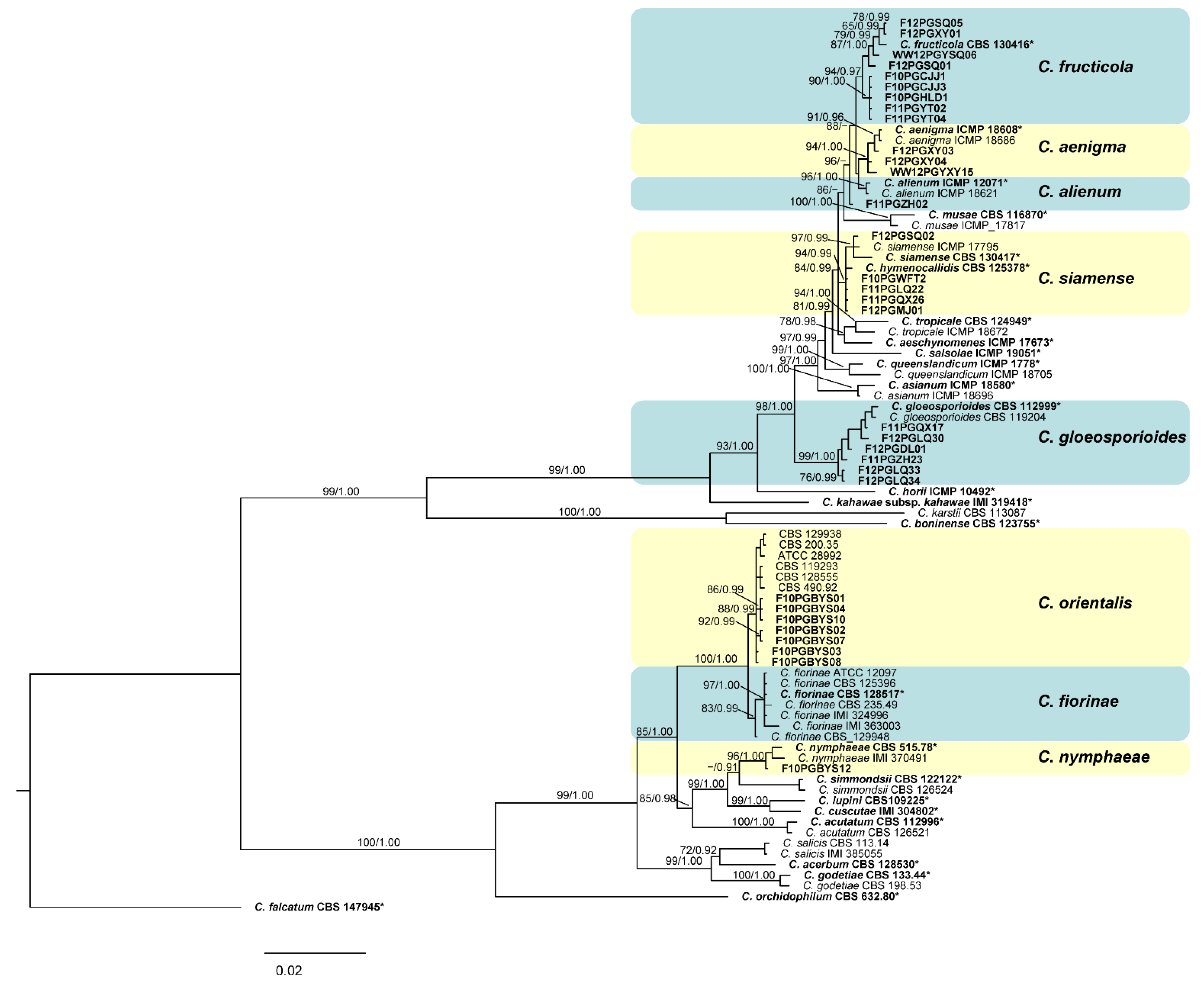
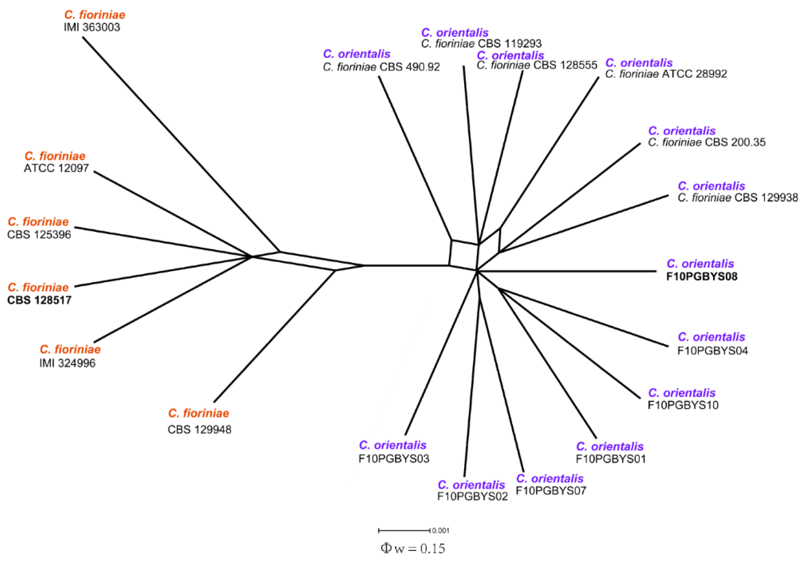
3.2. Phylogenetic Analysis
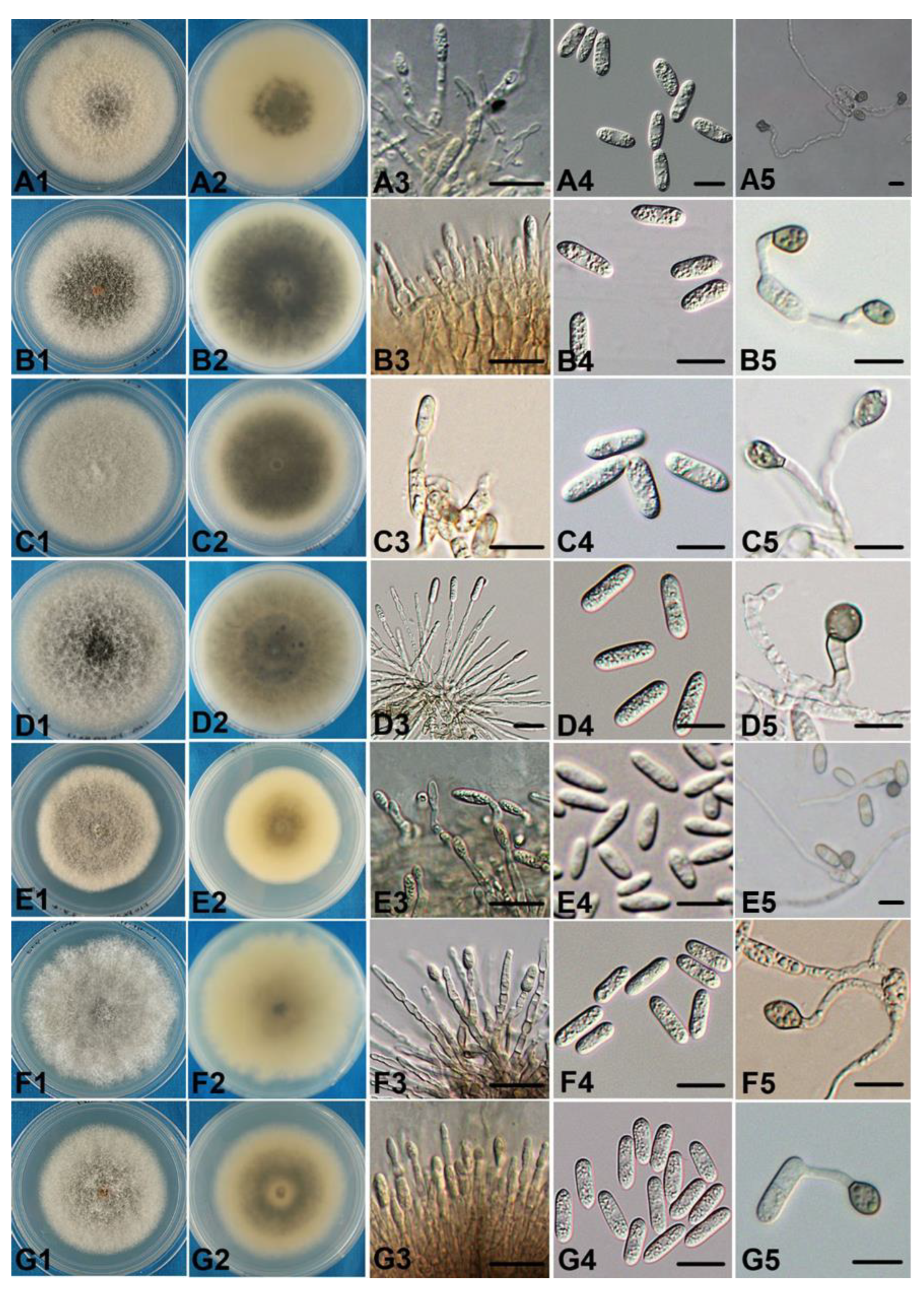
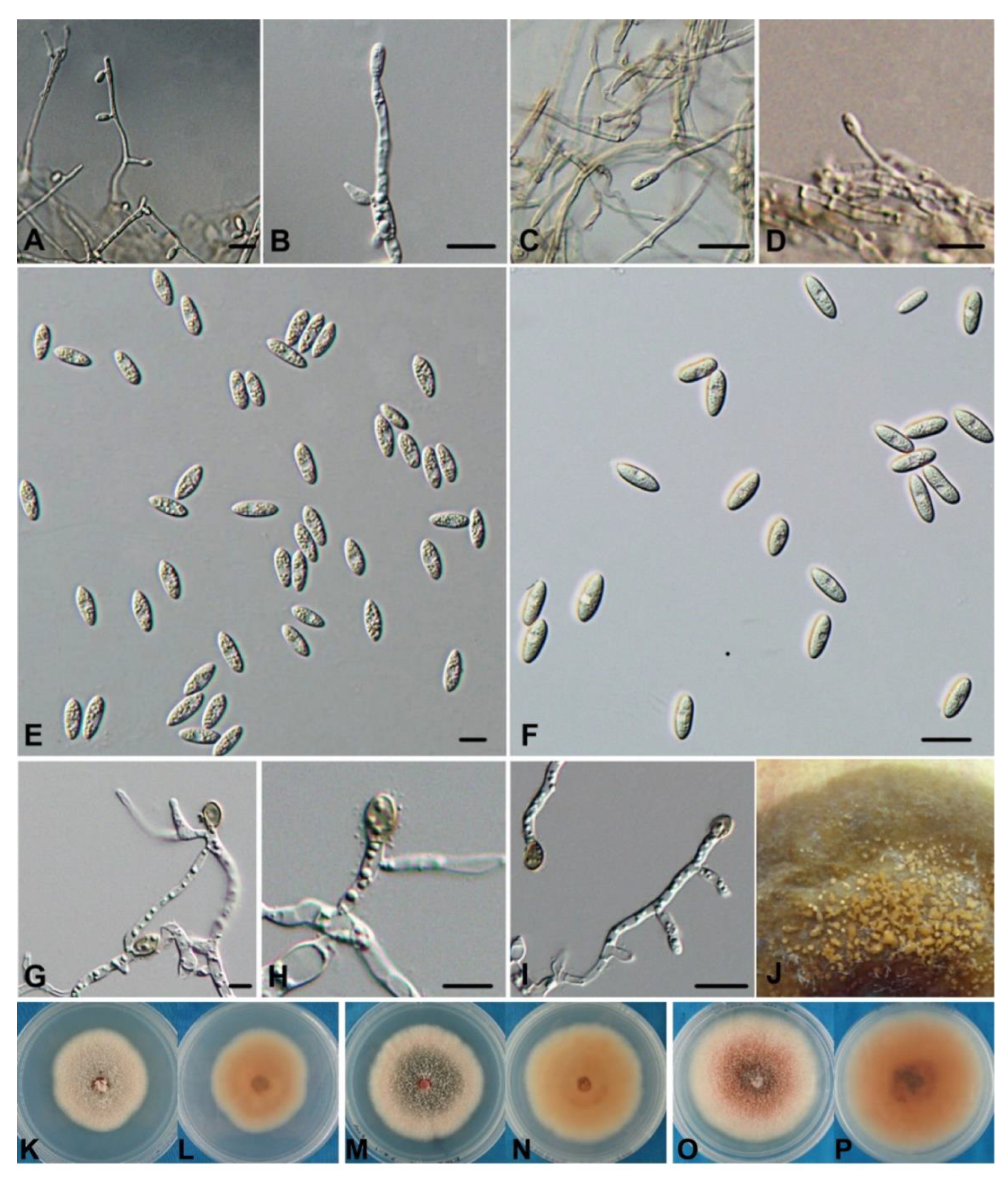
Based on ITS sequences and cultural characters, 32 representative isolates were chosen for further phylogenic analysis. The five-locus (ITS, ACT, GAPDH, CHS-1 and TUB2) phylogenetic analysis included 51 reference isolates [10,12,27]. Concatenated sequence alignment contained a total of 1916 characters, among which 551 were parsimony informative (28.8%). The BI tree, along with both the Bayesian posterior probability values and maximum likelihood bootstrap support values, are shown in Figure 1. The Bayesian tree was identical to the maximum likelihood tree in topology.
The phylogram supported eight defined clades, representing C. aenigma, C. alienum, C. fructicola, C. gloeosporioides, C. nymphaeae, C. siamense and a candidate for a new species, respectively. Four isolates clustered with C. hymenocallidis (CBS 125378), and one isolate grouped with C. siamense sensu stricto (CBS 130417 and ICMP 17795), which both belong to C. siamense sensu lato [52]. The clades of C. fructicola (CBS 130416), C. aenigma (ICMP 18608 and, ICMP 18686), C. alienum (ICMP 12071 and ICMP 18621) and C. gloeosporioides (CBS 112999 and CBS 119204) each included nine, three, one and six apple isolates, respectively.
The remaining eight isolates from the diseased apple fruits belonged to the C. acutatum complex. One isolate clustered together with C. nymphaeae (CBS 515.78 and IMI 370491), while the other seven formed a separate clade together with CBS 128555 (Figure 1). As revealed by the previous multi-locus molecular phylogenetic analysis [12], C. fiorinae contains two well-separated clades; one clade contains CBS 128555, and the other clade contains the type strain CBS 128517. Here, we propose that the CBS 12855 clade should better be defined as an independent taxon unit, which we have named C. orientals. The new species delimitation was also supported by the PHI analysis in which no obvious evidence of recombination was detected between the two clades (Figure 2).

3.3. Taxonomy

Based on the result of multigene phylogeny, the 32 Colletotrichum isolates characterized in this study were grouped into seven species: C. aenigma (three isolates), C. alienum (one isolate), C. fructicola (nine isolates), C. gloeosporioides (six isolates), C. nymphaeae (one isolate), C. siamense (five isolates) and C. orientalis (seven isolates).
  • Colletotrichum aenigmaB. Weir & P.R. Johnst. Studies in Mycology 73: 135. 2012. [52] Figure 3(A1–A5).
Description: Vegetative hyphae are 1–5.5 μm diam, hyaline, smooth-walled, septate and branched. Conidiophores are formed directly on hyphae. Conidiophores are hyaline and smooth-walled; they are sometimes septate and branched. Conidiogenous cells are hyaline, smooth-walled, cylindrical and not clearly separated from the hyphae by a septum. Conidia are straight, cylindric or clavate with rounded ends; sometimes they taper slightly to one end, (11.8–)15.5–17.5(–18.8) × (3.8–)4.5–5.5(–6) μm, mean ± SD = 16.46 ± 1.30 × 5.06 ± 0.56 μm (n = 50), L/W ratio = 3.3. Appressoria are elliptical or ovoid; some have broad, irregular lobes, (7.5–)8.5–9.5(–10.3) × (5.5–)6–7(–8.2) μm, mean ± SD = 9.07 ± 0.68 × 6.63 ± 0.50 μm (n = 50), L/W ratio = 1.4. Colonies on the PDA are flat with an entire margin; the aerial mycelium is sparse, cottony, and white-to-pale gray; in reverse, it is a pale honey and olivaceous gray towards the center with a growth rate of 68–80 mm in 7 d at 25 °C.
Specimen examined: China, Henan Province: Xiayi County, on the fruit surface of an apple (Malus ×domestica Borkh.), 7 September 2012, Dandan Fu, F12PGXY03; China, Henan Province: Xiayi County, on a leaf spot of an apple (M. ×domestica Borkh.), 7 September 2012, Wei Wang, W12PGYXY15.
Notes: C. aenigma could not be separated from C. alienum by ITS sequence analysis, nor from C. tropicale by ACT. The conidia of the holotype (ICMP 18608) of C. aenigma were (12–)14–15(–16.5) × (5–)6–6.5(–7.5) μm, and the appressoria were subglobose [52], whereas the isolate F12PGXY03 had longer and thinner conidia, and the appressoria were generally oval-shaped and longer than those of ICMP 18608. Additionally, the cultural characters of our isolates were different from those of ICMP 18608.
  • ColletotrichumalienumB. Weir & P.R. Johnst. Studies in Mycology 73: 139. 2012. [52] Figure 3(B1–B5).
Description: Vegetative hyphae are 1–9 μm diam, hyaline, smooth-walled, septate and branched. Conidiophores are hyaline, smooth-walled, septate and branched. Conidiogenous cells are hyaline, smooth, ovoid-elliptical or short-cylindrical and often clearly have a septum. Conidia are straight, mostly cylindrical with broadly rounded ends; a few taper towards the basal end, (12.9–)15–17(–19.7) × (3.4–)4–5(–6.1) μm, mean ± SD = 16.27 ± 1.37 × 4.73 ± 0.56 μm (n = 50), L/W ratio = 3.4. Appressoria are mostly simple and subglobose or elliptical; a few have broad, irregular lobes, (6.8–)8–10(–10.8) × (5.1–)6–7(–7.6) μm, mean ± SD = 8.91 ± 0.98 × 6.46 ± 0.57 μm (n = 50), L/W ratio = 1.4. Colonies grown on the PDA (Difco) are 85 mm after 7 d at 25 °C; the aerial mycelium is dense, cottony and gray with an orange conidial ooze visible towards the center; in reverse, it is dark gray towards the center with sporadic black flecks and pale gray towards the edge.
Specimen examined: China, Henan Province: Zhengzhou City, on the fruit surface of an apple (Malus ×domestica Borkh.), 28 September 2011, Dandan Fu F11PGZH02.
  • Colletotrichum fructicolaPrihastuti, L. Cai & K.D. Hyde. Fungal Diversity 39: 96, 2009. [54] Figure 3(C1–C5).
Description: Vegetative hyphae are 1–11 µm diam, hyaline to pale brown, smooth-walled, septate and branched. Conidiophores are hyaline and smooth-walled; a few are septate and branched. Conidiogenous cells are hyaline, smooth, cylindrical and not clearly separated from the hyphae by a septum. Conidia are hyaline, aseptate, straight and cylindrical with both ends rounded or one end slightly acute, (13.1–)14.5–16(–18.5) × (4.5–)5–5.5(–6.2) µm, mean ± SD = 15.38 ± 1.16 × 5.29 ± 0.40 μm (n = 50), L/W ratio = 2.9. Appressoria are single or in loose groups, pale to dark brown, ovoid, cylindrical or fusoid and sometimes slightly irregular, (6–)8.5–11(–13) × (4.4–)5.5–6.5(–8.4) µm, mean ± SD = 9.66 ± 1.74 × 5.94 ± 0.81 μm (n = 50), L/W ratio = 1.6. Colonies on the PDA are 7880 mm after 7 d. The aerial mycelium is white to pale gray, dense and cottony; in reverse, it is dark gray towards the center and pale gray at the edge.
Specimen examined: China, Henan Province: Shangqiu City, on the fruit surface of an apple (Malus ×domestica Borkh.), 6 September 2012, Dandan Fu F12PGSQ01, F12PGSQ05; from the leaf of an apple, Wei Wang, WW12PGYSQ06; Xiayi County, 7 September 2012, F12PGXY01. Liaoning Province: Xingcheng City, F10PGCJJ1, F10PGCJJ3; Huludao City, F10PGHLD1; Shandong Province: Yantai City, 29 September 2011, Dandan Fu, F11PGYT02, F11PGYT04.
Notes: The ITS sequence analysis did not separate C. fructicola from C. aeschynomenes, and the ACT sequence analysis could not separate it from C. alienum, C. dianesei, C. queenslandicum and C. siamense. Similarly, neither GAPDH nor TUB2 separated this species from C. alienum. The CHS-1 sequence analysis did not separate it from some of the isolates of C. siamense. Nevertheless, these taxa were well-distinguished using multi-gene analysis.
  • Colletotrichum gloeosporioides (Penz.) Penz. & Sacc. Atti Reale Ist. Veneto Sci. Lett. Arti., Series 6, 2: 670. 1884. [55] Figure 3(D1–D5).
Description: Vegetative hyaline are 1–8 μm diam, hyaline to medium brown, smooth, septate and branched. Conidiophores are hyaline, smooth-walled, one to three celled and sometimes branched. Conidiogenous cells are hyaline, smooth, cylindrical and often clearly have a septum. Conidia are straight and mostly cylindrical with broadly rounded ends; they are sometimes slightly acute, tapering gradually to the ends, (13.1–)14–15(–16.1) × (3.8–)4.5–5.5(–5.8) μm, mean ± SD = 14.48 ± 0.70 × 5.12 ± 0.41 μm (n = 50), L/W ratio = 1.4. Appressoria are simple or in small groups and subglobose or elliptical; a few are irregular, (6.6–)7.5–9(–13.8) × (4.6–)5.5–6(–7.2) μm, mean ± SD = 8.32 ± 1.24 × 6.02± 0.72 μm (n = 50), L/W ratio = 1.4. Colonies grown on the PDA(Difco) are 75–80 mm after 7 d at 25 °C; the aerial mycelium is dense, cottony and pale gray to medium gray towards the center, in reverse, olivaceous gray, with sporadic dark gray flecks. Colonies on the OA are flat with an entire margin; the aerial mycelium is sparse, panniform and pale gray. An orange conidial ooze is visible in the mycelium.
Specimen examined: China, Shaanxi Province: Qian County, on the fruit surface of an apple (Malus ×domestica Borkh.), 24 September 2011, Dandan Fu F11PGQX17, 20 September 2010, F12PGLQ30, F12PGLQ33, F12PGLQ34; Dali County, 16 August 2012, F12PGDL01; Henan Province: Zhengzhou City, 28 September 2011, F11PGZH23.
  • Colletotrichum nymphaeae (Pass.) Aa. Netherlands J. PI. Pathol., Supplement 1 84: 110. 1978. [56] Figure 3(E1–E5).
Description: Vegetative hyphae are 1–5 μm diam, hyaline, smooth, septate and branched. Conidiophores are formed directly on hyphae. Conidiophores are hyaline and smooth; a few are septate and branched. Conidiogenous cells are hyaline, smooth, cylindrical or fusiform and not clearly separated from the hyphae by a septum. Conidia are straight and cylindrical to clavate, with one end rounded and the other end or two ends acute, (6.8–)9–13(–15.9) × (3.4–)4–4.5(–5.3) μm, mean ± SD = 11.24 ± 2.19 × 4.24 ± 0.44 μm (n = 50), L/W ratio = 2.7. Appressoria are simple or in a small group and mostly subglobose or elliptical; a few have an irregular outline, (4.8–)6.5–7.5(–9) × (4.3–)5–6(–7.9) μm, mean ± SD = 6.97 ± 0.85 × 5.64 ± 0.60 μm (n = 50), L/W ratio = 1.2. Colonies on the PDA are flat with an entire margin. The aerial mycelium is sparse, grayish-yellow or cinnamon towards the center and white at the edge; in reverse, it is dark olivaceous gray. It has a growth rate of 54–60 mm after 7 d.
Specimen examined: China, Liaoning Province: Zhuanghe City, on the fruit surface of an apple (Malus ×domestica Borkh.), 20 September 2010, Jieli Zhuang F10PGBYS12.
  • Colletotrichum siamense Prihastuti, L. Cai & K. D. Hyde. Fungal Diversity 39: 98. 2009. [54] Figure 3(F1–F5,G1–G5).
Description: Vegetative hyphae are 1–8 µm diam, hyaline to pale brown, smooth-walled, septate and branched. Conidiophores are hyaline to pale brown, smooth-walled and one or two celled; a few are branched. Conidiogenous cells are hyaline, smooth, cylindrical and clearly separated from the hyphae by a septum. Conidia are hyaline, aseptate, straight and cylindrical with both ends rounded, (11.9–)14.5–15.5(–18.9) × (4–)4.5–5(–5.5) µm, mean ± SD = 15.08 ± 1.14 × 4.87± 0.31 µm (n = 50), L/W ratio = 3.1. Appressoria are single or in loose groups, pale to dark brown, ovoid and subglobose or short, mean ± SD = 9.79 ± 1.60 × 5.94 ± 0.77 µm (n = 50), L/W ratio = 1.6. Colonies on the PDA have an entire margin. The aerial mycelium is white, and the colonies are dense, cottony, white to pale gray or dark gray. Occasionally, orange conidial ooze is visible in the center; in reverse, it is buff with sporadic dark gray spots or grayish dark towards the center and pale gray at the edge. Colonies on the PDA are 6875 mm after 7 d.
Specimen examined: China, Henan Province: Shangqiu City, 6 September 2012, Dandan Fu F12PGSQ02; Mengjin County, 12 August 2012, Dandan Fu, F12PGMJ01; Shaanxi Province: Liquan County, on the fruit surface of an apple, 24 September 2011, Dandan Fu, F11PGLQ22; Qian County, F11PGQX26; Liaoning Province: Suizhong County, 20 September 2010, Jieli Zhuang F10PGWFT2.
Notes: After Prihastuti et al. separated Colletotrichum siamense from the C. gloeosporioides complex, more isolates from multiple hosts were identified as this species [54]. ITS sequences separated C. siamense well from other taxa, but the ACT sequence did not separate it from C. alienum, C. hymenocallidis, C. queenslandicum or C. fructicola. Similarly, GAPDH and TUB2 do not separate this species from C. hymenocallidis. C. hymenocallidis was first introduced by Yang et al. from Hymenocallis americana [57] but was recently synonymized with C. siamense [52]. However, Sharma et al. considered C. siamense to be a species complex based on an ApMat sequence analysis because C. siamense showed high sequence variability [58]. Moreover, Liu et al. indicated that more isolates need to be included to support further splitting of C. siamense, which possibly resurrects C. hymenocallidis [59].
  • Colletotrichum orientalisDandan Fu & G.Y. Sun, sp. nov. Figure 4.
Mycobank: MB 808171.
Etymology: Referring to the isolates collected from the eastern region of China.
Description: Vegetative hyphae are 1–6 μm, hyaline, smooth-walled, septate and branched. Conidiophores are formed directly on the hyphae. Conidiophores are hyaline, smooth-walled, simple or septate and branched. Conidia are hyaline, smooth-walled, aseptate, straight and fusiform or cylindrical with both ends acute, (12.8–)14–16(–18.5) ×(3.9–)4–5(–5.5) μm, mean ± SD = 15.07 ± 1.23 × 4.51 ± 0.38 μm (n = 50), L/W ratio = 3.3 μm. Appressoria are single or in loose groups, pale-to-medium brown, smooth, oval-shaped and ellipsoidal or irregularly outlined, (7–)8–9.5(–11.5) × (4.4–)5.5–6(–7.2) μm, mean ± SD = 8.74 ± 0.99 × 5.84 ± 0.53 μm (n = 50), L/W ratio = 1.5. Colonies on the PDA have an entire margin and are compacted cottony to felty. They are orangish red towards the center and pale gray towards the edge. The aerial mycelium is white to pale gray, and the conidiomata are sparse with masses of orange conidia. In reverse pale brownish pink. Colonies on the OA have an entire margin. The aerial mycelium is sparse, white to pale gray, and on the surface with visible masses of orange conidia scattered in circles; in reverse, it is pale buff. Colonies on the PDA are 4551 mm after 7 d (6775 mm in 10 d).
Holotype: China, Liaoning Province: Zhuanghe City, on the fruit surface of an apple (Malus ×domestica Borkh.), 20 September 2010, Coll. Jieli Zhuang, F10PGBYS08 (CGMCC3.17216; isotype in HMAS244986 as dry culture).
Additional specimen examined: China, Liaoning Province: Zhuanghe City, on the fruit surface of an apple (Malus ×domestica Borkh.), 20 September 2010, Jieli Zhuang, F10PGBYS01 (CGMCC 3.17217), F10PGBYS02–04, F10PGBYS07–08, F10PGBYS10.
Notes: Freeman and Shabi studied isolates from fruit rot of apples and peaches (based on the ITS sequence, probably identifiable as C. fioriniae), which produced lesions on many different fruits, indicating that isolates of this group have the ability to cross-infect fruit from multiple hosts [60]. In this paper, we isolated seven isolates from apple bitter rot in Liaoning Province. A phylogenetic analysis (Figure 1) showed that they constituted a monophyletic clade together with the six C. fioriniae isolates (CBS 129938, CBS200.35, ATCC 28992, CBS 119293, CBS 128555 and CBS 490.92). In Damm et al. [12], the clade was well-separated from the clade containing the C. fioriniae holotype CBS 128517. Separation between the two clades was also evident in Damm et al. [12], which was treated as intraspecific heterogeneity. In this study, the PHI analysis detected no significant evidence of recombination between the two clades (Figure 2). Therefore, we denominate the clade containing the ABR isolates a new species.

3.4. Pathogenicity Tests

In the fruit infection assays, the isolates isolated from apples with bitter rot symptoms were pathogenic to the apple fruits in both the unwounded and wounded inoculations (Table 2). Dark brown rot lesions, similar in appearance, were produced in all cases (Figure 5). Of the non-wounded inoculations, Colletotrichum alienum (F11PGZH02) had the highest infection incidence (100%), whereas the isolates of C. gloeosporioides (F11PGQX17) and C. orientalis (F10PGBYS08) had the lowest incidences (33%); the others were in the middle. Of the wounded inoculations, all isolates had a 100% infection incidence. Lesions incurred by different isolates were similar in size, except that the lesions incurred by C. nymphaeae (F10PGBYS12, belonging to the C. acutatum complex) were apparently smaller (Figure 5).
Isolates isolated from the GLS lesions caused GLS lesions on both the apple fruits and leaves (Table 3). The isolates of C. aenigma (F12PGXY03, W12PGYXY15) and C. fructicola (F12PGSQ0503, W12PGYSQ06) were pathogenic on the leaves and fruits of Gala apples, but non-pathogenic on Fuji apple leaves or fruits in the non-wounded inoculation (Figure 5), which is in accordance with the observation that Fuji apples are resistant to GLS disease [61].

4. Discussion

China is the largest apple-producing country in the world. Bitter rot has been a common disease in almost all apple production areas and can cause large economic losses under disease-favorable temperature and humidity conditions. Glomerella leaf spot (GLS) has been a severe foliar disease on cvs. Gala, Jonagold and Golden Delicious in the USA and Brazil. It was found first in Henan, China, in 2010 [28]. Now, it has become prevalent in all major apple-producing areas in China. Thus far, however, the species diversity of apple Colletotrichum pathogens in China is largely unclear. In this study, we collected and characterized 151 isolates from four apple-producing provinces and identified six known species, as well as one new species, demonstrating that diverse Colletotrichum species can infect apples. Moreover, C. orientalis was shown for the first time to be an apple Colletotrichum pathogen.
Among the identified species, C. siamense, C. fructicola, C. aenigma, C. alienum and C. gloeosporioides belong to the CGSC, while C. orientalis and C. nymphaeae belong to the CASC. Overall, the CGSC species appear to be more prevalent compared with the CASC species. Moreover, fruit isolates and leaf isolates differ significantly in their genetic makeups. Seven species were recognized as isolates from apple fruits, whereas only two (C. fructicola and C. aenigma) were recognized as leaf spot isolates. In a pathogenicity test, ABR isolates fail to incur GLS symptoms, and the GLS isolates fail to incur ABR symptoms, indicating a pathogenic differentiation among the two groups of pathogens. Such results are in accordance with a previous study that demonstrated the intraspecific differentiation in the pathogenicity of GLS and ABR for C. fructicola [62].
C. siamense is a species that includes members from diverse hosts and that has a worldwide distribution. The diversity is so high that there has been controversy regarding whether it should be treated as a single species or a species complex. In a recent study carried out by Liu and others [63], six independent species very close to C. siamense s. str. (C. communis, C. hymenocallidis, C. dianesei, C. endomangiferae, C. jasmini-sambac and C. murrayae) were renamed as C. siamense. In this study, the four characterized isolates clustered together with C. hymenocallidis, and one isolate clustered with C. siamense s. str. Based on broad species criteria, these isolates should all be regarded as C. siamense sensus lato. C. fructicola represents another important pathogen species identified in this study. C. fructicola has a very broad host range, having been isolated from over eight plant families as endophytes and as plant pathogens. In this study, C. fructicola was isolated from both bitter rot and Glomerella leaf spot lesions. C. fructicola causes Glomerella leaf spot in Brazil but has been more commonly identified as a bitter rot pathogen in central USA, Brazil and Uruguay. In Uruguay in particular, most isolates from apple bitter rot were identified as C. fructicola. Interestingly, despite the prevalence of C. fructicola in Uruguay, Glomerella leaf spot disease does not occur in the field [62]. In the future, it would be interesting to determine whether there are distinctive C. fructicola populations for isolates from leaf lesions and fruit lesions in China.
In a previous study [12], C. fiorinae has been defined as a species with two well-separated clades. We propose here that the two clades should be regarded as different species due to the fact that the pairwise homoplasy index (PHI) analysis in SplitsTree did not detect evidence of recombination between them. Therefore, we have named the new lineage C. orientalis. C. alienum and C. gloeosporioides are two other species common in fruits. Interestingly, C. alienum has only been reported in New Zealand and Australia, and C. gloeosporioides has never been reported on apples in China. The identification of these species on apples in China highlights the importance of a diversity survey.
Compared with apple bitter rot, GLS is a relatively new disease. Velho and others have suggested that GLS pathogens originate from apple bitter rot pathogens [62]. In this study, all isolates caused fruit rot upon wound inoculation, whereas only the isolates from the leaf spot lesions incurred leaf spot symptoms. Importantly, such pathogenic differentiation could occur in the same species (e.g., F12PGSQ01 and W12PGYSQ06). Such a pathogenicity differentiation pattern is in line with the report by Velho and others [62]. Isolates showing an intraspecific pathogenic variation would be valuable resources for comparative studies aiming to dissect the mechanisms that underlie the adaptive evolution of apple Colletotrichum pathogens.
In summary, based on a systemic survey of Colletotrichum isolates, in this study, we identified seven species associated with the GLS and ABR diseases in China, highlighting the rich species diversity of Colletotrichum spp. on apples. There are tens of apple-producing provinces in China that are variable in terms of climate and soil conditions; further survey efforts into the hidden species diversity and the structural variations among the different regions is critical for effective control of these two important diseases.

Author Contributions

Formal analysis, Y.C., D.F. and X.L.; investigation, Y.C., D.F., W.W. and X.L.; resources, D.F., W.W. and G.S.; data curation, Y.C., D.F. and X.L.; writing—original draft preparation, Y.C., D.F. and X.L.; writing—review and editing, M.L.G., R.Z. and G.S.; supervision, X.L., R.Z. and G.S.; funding acquisition, X.L., R.Z. and G.S. All authors have read and agreed to the published version of the manuscript.

Funding

This research was funded by the National Key Research and Development Program of China (2019YFD1002000), the National Natural Science Foundation of China (32072374, 31972220), and the China Agriculture Research System of MOF and MARA (CARS-27).

Institutional Review Board Statement

Not applicable.

Informed Consent Statement

Not applicable.

Data Availability Statement

Not applicable.

Acknowledgments

We thank Zengqiang Zhou (Zhengzhou Fruit Research Institute, Chinese Academy of Agriculture Sciences), Yingzi Wang (Yantai Agricultural Science & Technology Institute) and Gongming Sun (Shangqiu Agriculture & Forestry Science Institute) for their help with sampling.

Conflicts of Interest

The authors declare no conflict of interest. The funders had no role in the design of the study; in the collection, analyses or interpretation of data; in the writing of the manuscript; or in the decision to publish the results.

References

  1. González, E.; Sutton, T.B. Population diversity within isolates of Colletotrichum spp. causing Glomerella leaf spot and bitter rot of apple in three orchards in North Carolina. Plant Dis. 2004, 88, 1335–1340. [Google Scholar] [CrossRef] [PubMed] [Green Version]
  2. Schrenk, H.V.; Spaulding, P. The bitter rot of apples. USDA Bur. Plant Ind. Bull. 1903, 44, 1–54. [Google Scholar]
  3. Simmond, J.H. A study of the species of Colletotrichum causing ripe fruit rots in Queensland. Qld. J. Agric. Anim. Sci. 1966, 22, 437–459. [Google Scholar]
  4. Sutton, T.B. Bitter rot. In Compendium of Apple and Pear Disease; Jones, A.L., Aldwinckle, H.S., Eds.; The American Phytopathological Society: St. Paul, MN, USA, 1990; pp. 15–26. [Google Scholar]
  5. Jones, A.L.; Ehret, G.R.; Meyer, M.P.; Shane, W.W. Occurrence of bitter rot on apple in Michigan. Plant Dis. 1996, 80, 1294–1297. [Google Scholar] [CrossRef]
  6. Shi, Y.; Correll, J.C.; Guerber, J.C.; Rom, C.R. Frequency of Colletotrichum species causing bitter rot of apple in the southeastern United States. Plant Dis. 1996, 80, 692–696. [Google Scholar] [CrossRef]
  7. González, E.; Sutton, T.B.; Correll, J.C. Clarification of the etiology of Glomerella leaf spot and bitter rot of apple caused by Colletotrichum spp. based on morphology and genetic, molecular, and pathogenicity tests. Phytopathology 2006, 96, 982–992. [Google Scholar] [CrossRef] [Green Version]
  8. Lee, D.H.; Kim, D.H.; Jeon, Y.A.; Uhm, J.Y.; Hong, S.B. Molecular and cultural characterization of Colletotrichum spp. causing bitter rot of apples in Korea. Plant Pathol. 2007, 23, 37–44. [Google Scholar] [CrossRef] [Green Version]
  9. Giaretta, D.R.; Bogo, A.; Coelho, C.; Guidolin, A.F.; de Mesquita-Dantas, A.C.; Gomes, E.A. ITS-rDNA phylogeny of Colletotrichum spp. causal agent of apple Glomerella leaf spot. Cienc. Rural 2010, 40, 806–812. [Google Scholar] [CrossRef]
  10. Cannon, P.F.; Damm, U.; Johnston, P.R.; Weir, B.S. Colletotrichum: Current status and future directions. Stud. Mycol. 2012, 73, 181–213. [Google Scholar] [CrossRef] [Green Version]
  11. Velho, A.C.; Stadnik, M.J.; Wallhead, M. Unraveling Colletotrichum species associated with Glomerella leaf spot of apple. Trop. Plant Pathol. 2019, 44, 197–204. [Google Scholar] [CrossRef]
  12. Damm, U.; Cannon, P.F.; Woudenberg, J.H.; Crous, P.W. The Colletotrichum acutatum species complex. Stud. Mycol. 2012, 73, 37–113. [Google Scholar] [CrossRef] [PubMed] [Green Version]
  13. Mari, M.; Guidarelli, M.; Martini, C.; Spadoni, A. First report of Colletotrichum acutatum causing bitter rot on apple in Italy. Plant Dis. 2011, 96, 144. [Google Scholar] [CrossRef] [PubMed]
  14. Cheon, W.; Kim, Y.S.; Jeon, Y.H. First report of anthracnose caused by Colletotrichum gloeosporioides on Malus prunifolia in Korea. Plant Dis. 2012, 96, 766. [Google Scholar] [CrossRef]
  15. Kou, L.P.; Gaskins, V.; Luo, Y.G.; Jurick, W.M. First report of Colletotrichum fioriniae causing postharvest decay on ‘Nittany’ apple fruit in the United States. Plant Dis. 2013, 98, 993. [Google Scholar] [CrossRef] [PubMed]
  16. Baroncelli, R.; Sreenivasaprasad, S.; Thon, M.R.; Sukno, S.A. First report of apple bitter rot caused by Colletotrichum godetiae in the United Kingdom. Plant Dis. 2014, 98, 1000–1001. [Google Scholar] [CrossRef]
  17. Velho, A.C.; Stadnik, M.J.; Casanova, L.; Mondino, P.; Alaniz, S. First report of Colletotrichum karstii causing Glomerella leaf spot on apple in Santa Catarina State, Brazil. Plant Dis. 2014, 98, 157–158. [Google Scholar] [CrossRef]
  18. Wallhead, M.; Broders, G.; Beaudoin, E.; Peralta, C.; Broders, K. Phylogenetic assessment of Colletotrichum species associated with bitter rot and Glomerella leaf spot in the northeastern US. Phytopathology 2014, 104, 123–124. [Google Scholar]
  19. Munir, M.; Amsden, B.; Dixon, E.; Vaillancourt, L.; Gauthier, N.A. Characterization of Colletotrichum species causing bitter rot of apples in Kentucky orchards. Plant Dis. 2016, 100, 2194–2203. [Google Scholar] [CrossRef] [Green Version]
  20. Velho, A.C.; Alaniz, S.; Casanova, L.; Mondino, P.; Stadnik, M.J. New insights into the characterization of Colletotrichum species associated with apple diseases in southern Brazil and Uruguay. Fungal Biol. 2015, 119, 229–244. [Google Scholar] [CrossRef]
  21. Wenneker, M.; Pham, K.; Kerkhof, E.; Harteveld, D.O. First report of preharvest fruit rot of ’Pink Lady’ apples caused by Colletotrichum fructicola in Italy. Plant Dis. 2021, 105, 1561. [Google Scholar] [CrossRef]
  22. Bragança, C.A.; Damm, U.; Baroncelli, R.; Massola Júnior, N.S.; Crous, P.W. Species of the Colletotrichum acutatum complex associated with anthracnose diseases of fruit in Brazil. Fungal Biol. 2016, 120, 547–561. [Google Scholar] [CrossRef] [PubMed]
  23. Nodet, P.; Baroncelli, R.; Faugère, D.; Le Floch, G. First report of apple bitter rot caused by Colletotrichum fioriniae in Brittany, France. Plant Dis. 2016, 100, 1497. [Google Scholar] [CrossRef]
  24. Khodadadi, F.; González, J.B.; Martin, P.L.; Giroux, E.; Bilodeau, G.J.; Peter, K.A.; Doyle, V.P.; Aćimović, S.G. Identification and characterization of Colletotrichum species causing apple bitter rot in New York and description of C. noveboracense sp. nov. Sci. Rep. 2020, 10, 11043. [Google Scholar] [CrossRef] [PubMed]
  25. Cabrefiga, J.; Pizà, D.; Vilardell, P.; Luque, J. First report of Colletotrichum chrysophilum causing apple bitter rot in Spain. Plant Dis. 2022, PDIS07211578PDN. [Google Scholar] [CrossRef] [PubMed]
  26. Wenneker, M.; Pham, K.; Lemmers, M.; de Boer, F.A.; van der Lans, A.M.; van Leeuwen, P.J.; Hollinger, T.C. First report of Colletotrichum godetiae causing bitter rot on ‘Golden Delicious’ apples in the Netherlands. Plant Dis. 2015, 100, 218. [Google Scholar] [CrossRef]
  27. Taylor, J.W.; Jacobson, D.J.; Kroken, S.; Kasuga, T.; Geiser, D.M.; Hibbett, D.S.; Fisher, M.C. Phylogenetic species recognition and species concepts in fungi. Fungal Genet. Biol. 2000, 31, 21–32. [Google Scholar] [CrossRef] [PubMed] [Green Version]
  28. Wang, C.X.; Zhang, Z.F.; Li, B.H.; Wang, H.Y.; Dong, X.L. First report of Glomerella leaf spot of apple caused by Glomerella cingulata in China. Plant Dis. 2012, 96, 912–913. [Google Scholar] [CrossRef]
  29. Velho, A.C.; Stadnik, M.J.; Casanova, L.; Mondino, P.; Alaniz, S. First report of Colletotrichum nymphaeae causing apple bitter rot in Southern Brazil. Plant Dis. 2014, 98, 567–568. [Google Scholar] [CrossRef]
  30. Moreira, R.R.; Peres, N.A.; May De Mio, L.L. Colletotrichum acutatum and C. gloeosporioides species complexes associated with apple in Brazil. Plant Dis. 2019, 103, 268–275. [Google Scholar] [CrossRef] [Green Version]
  31. Alaniz, S.; Cuozzo, V.; Martínez, V.; Stadnik, M.J.; Mondino, P. Ascospore infection and Colletotrichum species causing Glomerella leaf spot of apple in Uruguay. Plant Pathol. J. 2019, 35, 100–111. [Google Scholar] [CrossRef]
  32. Zhang, Z.; Yan, M.; Li, W.; Guo, Y.; Liang, X. First report of Colletotrichum aenigma causing apple Glomerella leaf spot on the Granny Smith cultivar in China. Plant Dis. 2021, 105, 1563. [Google Scholar] [CrossRef] [PubMed]
  33. Wang, X.M.; Li, J.Y. Studies on species of Colletotrichum in Province Shaanxi, China. Acta Mycol. Sin. 1987, 6, 211–218. [Google Scholar]
  34. Zhang, R.; Wang, S.F.; Cui, J.Q.; Sun, G.Y. First report of bitter rot caused by Colletotrichum acutatum on apple in China. Plant Dis. 2008, 92, 1471. [Google Scholar] [CrossRef] [Green Version]
  35. Zhang, R.; Wang, S.F.; Cui, J.Q.; Sun, G.Y. Identification of pathogens causing apple fruit bitter rot in Shaanxi and Henan Provinces. Sci. Agric. Sin. 2009, 42, 3224–3229. [Google Scholar]
  36. Than, P.P.; Shivas, R.G.; Jeewon, R.; Pongsupasamit, S.; Marney, T.S.; Taylor, P.W.; Hyde, K.D. Epitypification and phylogeny of Colletotrichum acutatum J.H. Simmonds. Fungal Divers. 2008, 28, 97–108. [Google Scholar]
  37. Barnes, I.; Roux, J.; Wingfield, M.J.; Coetzee, M.P.; Brenda, D.; Wingfield, B.D. Characterization of Seiridium spp. associated with cypress canker based on β-tubulin and histone sequences. Plant Dis. 2001, 85, 317–321. [Google Scholar] [CrossRef] [PubMed] [Green Version]
  38. Gardes, M.; Bruns, T.D. ITS primers with enhanced specificity for basidiomycetes -- application to the identification of mycorrhizae and rusts. Mol. Ecol. 1993, 2, 113–118. [Google Scholar] [CrossRef]
  39. White, T.J.; Bruns, T.; Lee, S.; Taylor, J. Amplification and direct sequencing of fungal ribosomal RNA genes for phylogenetics. In PCR Protocols: A Guide to Methods and Applications; Innis, M.A., Gelfand, D.H., Sninsky, J.J., White, T.J., Eds.; Academic Press: San Diego, CA, USA, 1990; pp. 315–322. [Google Scholar]
  40. Carbone, I.; Kohn, L.M. A method for designing primer sets for speciation studies in filamentous ascomycetes. Mycologia 1999, 91, 553–556. [Google Scholar] [CrossRef]
  41. Guerber, J.C.; Liu, B.; Correll, J.C.; Johnston, P.R. Characterization of diversity in Colletotrichum acutatum sensu lato by sequence analysis of two introns, mtDNA and intron RFLPs, and mating compatibility. Mycologia 2003, 95, 872–895. [Google Scholar] [CrossRef]
  42. Glass, N.L.; Donaldson, G.C. Development of primer sets designed for use with the PCR to amplify conserved genes from filamentous ascomycetes. Appl. Environ. Microbiol. 1995, 61, 1323–1330. [Google Scholar] [CrossRef] [Green Version]
  43. Damm, U.; Woudenberg, J.H.; Cannon, P.F.; Crous, P.W. Colletotrichum species with curved conidia from herbaceous hosts. Fungal Divers. 2009, 39, 45–87. [Google Scholar]
  44. Thompson, J.D.; Gibson, T.J.; Plewniak, F.; Jeanmougin, F.; Higgins, D.G. The Clustal_X windows interface: Flexible strategies for multiple sequence alignment aided by quality analysis tools. Nucleic Acids Res. 1997, 25, 4876–4882. [Google Scholar] [CrossRef] [PubMed] [Green Version]
  45. Zhang, D.; Gao, F.; Jakovlić, I.; Zou, H.; Zhang, J.; Li, W.X.; Wang, G.T. PhyloSuite: An integrated and scalable desktop platform for streamlined molecular sequence data management and evolutionary phylogenetics studies. Mol. Ecol. Resour. 2020, 20, 348–355. [Google Scholar] [CrossRef] [PubMed]
  46. Stamatakis, A. RAxML version 8: A tool for phylogenetic analysis and post-analysis of large phylogenies. Bioinformatics 2014, 30, 1312–1313. [Google Scholar] [CrossRef]
  47. Stamatakis, A. RAxML-VI-HPC: Maximum likelihood-based phylogenetic analyses with thousands of taxa and mixed models. Bioinformatics 2006, 22, 2688–2690. [Google Scholar] [CrossRef]
  48. Felsenstein, J. Confidence limits on phylogenies: An approach using the bootstrap. Evolution 1985, 39, 783–791. [Google Scholar] [CrossRef]
  49. Ronquist, F.; Teslenko, M.; van der Mark, P.; Ayres, D.L.; Darling, A.; Höhna, S.; Larget, B.; Liu, L.; Suchard, M.A.; Huelsenbeck, J.P. MrBayes 3.2: Efficient Bayesian phylogenetic inference and model choice across a large model space. Syst. Biol. 2012, 61, 539–542. [Google Scholar] [CrossRef] [Green Version]
  50. Huson, D.H. SplitsTree: Analyzing and visualizing evolutionary data. Bioinformatics 1998, 14, 68–73. [Google Scholar] [CrossRef]
  51. Huson, D.H.; Bryant, D. Application of phylogenetic networks in evolutionary studies. Mol. Biol. Evol. 2006, 23, 254–267. [Google Scholar] [CrossRef]
  52. Weir, B.S.; Johnston, P.R.; Damm, U. The Colletotrichum gloeosporioides species complex. Stud. Mycol. 2012, 73, 115–180. [Google Scholar] [CrossRef] [Green Version]
  53. Damm, U.; Cannon, P.F.; Woudenberg, J.H.; Johnston, P.R.; Weir, B.S.; Tan, Y.P.; Shivas, R.G.; Crous, P.W. The Colletotrichum boninense species complex. Stud. Mycol. 2012, 73, 1–36. [Google Scholar] [CrossRef] [PubMed] [Green Version]
  54. Prihastuti, H.; Cai, L.; Chen, H.; McKenzie, E.H.; Hyde, K.D. Characterization of Colletotrichum species associated with coffee berries in northern Thailand. Fungal Divers. 2009, 39, 89–109. [Google Scholar]
  55. Penzig, A.G.O. Fungi agrumicoli. Contribuzione allo studio dei funghi parassiti degli agrumi. In Michelia; Patavii: Berlin, Germany, 1882; pp. 385–508, Sumptibus auctoris, Typis seminarii. [Google Scholar]
  56. van der Aa, H.A. A leaf spot of Nymphaea alba in the Netherlands. Netherlands J Plant Pathol. 1978, 84, 109–115. [Google Scholar]
  57. Yang, Y.L.; Liu, Z.Y.; Cai, L.; Hyde, K.D.; Yu, Z.N.; McKenzie, E.H. Colletotrichum anthracnose of Amaryllidaceae. Fungal Divers. 2009, 39, 123–146. [Google Scholar]
  58. Sharma, G.; Kumar, N.; Weir, B.S.; Hyde, K.D.; Shenoy, B.D. The ApMat marker can resolve Colletotrichum species: A case study with Mangifera indica. Fungal Divers. 2013, 61, 117–138. [Google Scholar] [CrossRef]
  59. Liu, F.; Damm, U.; Cai, L.; Crous, P.W. Species of the Colletotrichum gloeosporioides complex associated with anthracnose diseases of Proteaceae. Fungal Divers. 2013, 61, 89–105. [Google Scholar] [CrossRef]
  60. Freeman, S.; Shabi, E. Cross-infection of subtropical and temperate fruits by Colletotrichum species from various hosts. Physiol. Mol. Plant Pathol. 1996, 49, 395–404. [Google Scholar] [CrossRef]
  61. Liu, Y.; Li, B.; Wang, C.; Liu, C.; Kong, X.; Zhu, J.; Dai, H. Genetics and molecular marker identification of a resistance to Glomerella leaf spot in apple. Hortic. Plant J. 2016, 2, 121–125. [Google Scholar] [CrossRef] [Green Version]
  62. Rockenbach, M.F.; Velho, A.C.; Gonçalves, A.E.; Mondino, P.E.; Alaniz, S.M.; Stadnik, M.J. Genetic structure of Colletotrichum fructicola associated to apple bitter rot and Glomerella leaf spot in southern Brazil and Uruguay. Phytopathology 2016, 106, 774–781. [Google Scholar] [CrossRef] [Green Version]
  63. Liu, F.; Wang, M.; Damm, U.; Crous, P.W.; Cai, L. Species boundaries in plant pathogenic fungi: A Colletotrichum case study. BMC Evol. Biol. 2016, 16, 1–14. [Google Scholar] [CrossRef] [Green Version]
Figure 1. Phylogram of the Colletotrichum species resulting from maximum likelihood and Bayesian analyses based on the combined alignment dataset of ITS, ACT, GAPDH, CHS-1 and TUB-2 sequences. Bootstrap support values above 60% and Bayesian posterior probability values above 0.9 were given at the nodes. Isolates isolated in this study and type strains are shown in bold. * Ex-holotype, ex-neotype, ex-epitype strains.
Jof 08 00740 g001
Figure 2. Pairwise homoplasy index (PHI) test of C. fioriniae and C. orientalis using both LogDet transformed and splits decomposition. PHI test results (ΦW) > 0.05 indicate the lack of recombination within the dataset.
Jof 08 00740 g002
Figure 3. Morphological and cultural characters of Colletotrichum isolates: (A) C. aenigma; (B) C. alienum; (C) C. fructicola; (D) C. gloeosporioides; (E) C. nymphaeae; (F) C. siamense; (G) C. hymenocallidis. Upper (1) and reverse (2) of cultures on PDA; (3) conidiophores; (4) conidia; (5) appressoria. Bars = 10 μm.
Jof 08 00740 g003
Figure 4. Colletotrichum orientalis (F10PGBYS08): (AD) conidiophores; (E,F) conidia; (GI) appressoria; (J) apple fruit lesion symptom with non-wounded conidial inoculation. Scale bars = 10 μm. (K,L) Colony on PDA (F10PGBYS08); (M,N) colony on PDA (F10PGBYS04); (O,P) colony on PDA (F10PGBYS05).
Jof 08 00740 g004
Figure 5. Typical field symptoms of ABR and GLS diseases (top) and artificial inoculation results (bottom). Top, field symptoms, (A1A3) represent fruit ABR, GLS on leaves and fruits, respectively. Bottom, (1A8B) represent typical symptoms on Fuji apples under unwounded or wounded inoculation conditions. A: Non-wounded; B: wounded; 1: C. aenigma (F12PGXY03); 2: C. alienum (F11PGZH02); 3: C. fructicola (F12PGSQ01); 4: C. gloeosporioides (F11PGQX17); 5: C. siamense (F12PGSQ02); 6: C. siamense (F11PGLQ22); 7: C. nymphaeae (F10PGBYS12); 8: C. orientalis (F10PGBYS08). (9A9C) Symptoms on cv. Gala apple leaves and fruits inoculated with conidial suspension of isolate C. fructicola W12PGYSQ06 from GLS. (9A) Leaf inoculation; (9B) fruit from unwounded inoculation; (9C) fruit from wounded inoculation.
Jof 08 00740 g005
Table 2. Pathogenicity test of representative Colletotrichum isolates on Fuji apple fruits.
SpeciesIsolateNon-WoundedWounded
C. alienumF11PGZH02++++++
C. fructicolaF12PGSQ01+++++
C. gloeosporioidesF11PGQX17++++
C. nymphaeaeF10PGBYS12+++++
C. siamenseF12PGSQ02+++++
C. orientalisF10PGBYS08++++
+: Infection incidence < 50%; ++: 50% < Infection incidence < 100%; +++: Infection incidence = 100%.
Table 3. Pathogenicity test of selected isolates on apple leaves.
SpeciesIsolateOriginInoculation CultivarInoculation Outcome
C. aenigmaF12PGXY03GLS lesionFuji
Gala+
W12PGYXY15GLS lesionFuji
Gala+
C. fructicolaF12PGSQ05GLS lesionFuji
Gala+
W12PGYSQ06GLS lesionFuji
Gala+
C. alienumF11PGZH02ABR lesionFuji
Gala
C. fructicolaF12PGSQ01ABR lesionFuji
Gala
C. gloeosporioidesF11PGQX17ABR lesionFuji
Gala
C. nymphaeaeF10PGBYS12ABR lesionFuji
Gala
C. siamenseF12PGSQ02ABR lesionFuji
Gala
C. orientalisF10PGBYS08ABR lesionFuji
Gala
+: Pathogenic; −: Non-pathogenic.
Publisher’s Note: MDPI stays neutral with regard to jurisdictional claims in published maps and institutional affiliations.

