
Novel Approaches in the Drug Development and Delivery Systems for Age-Related Macular Degeneration
 ,
,  , , and
Abstract
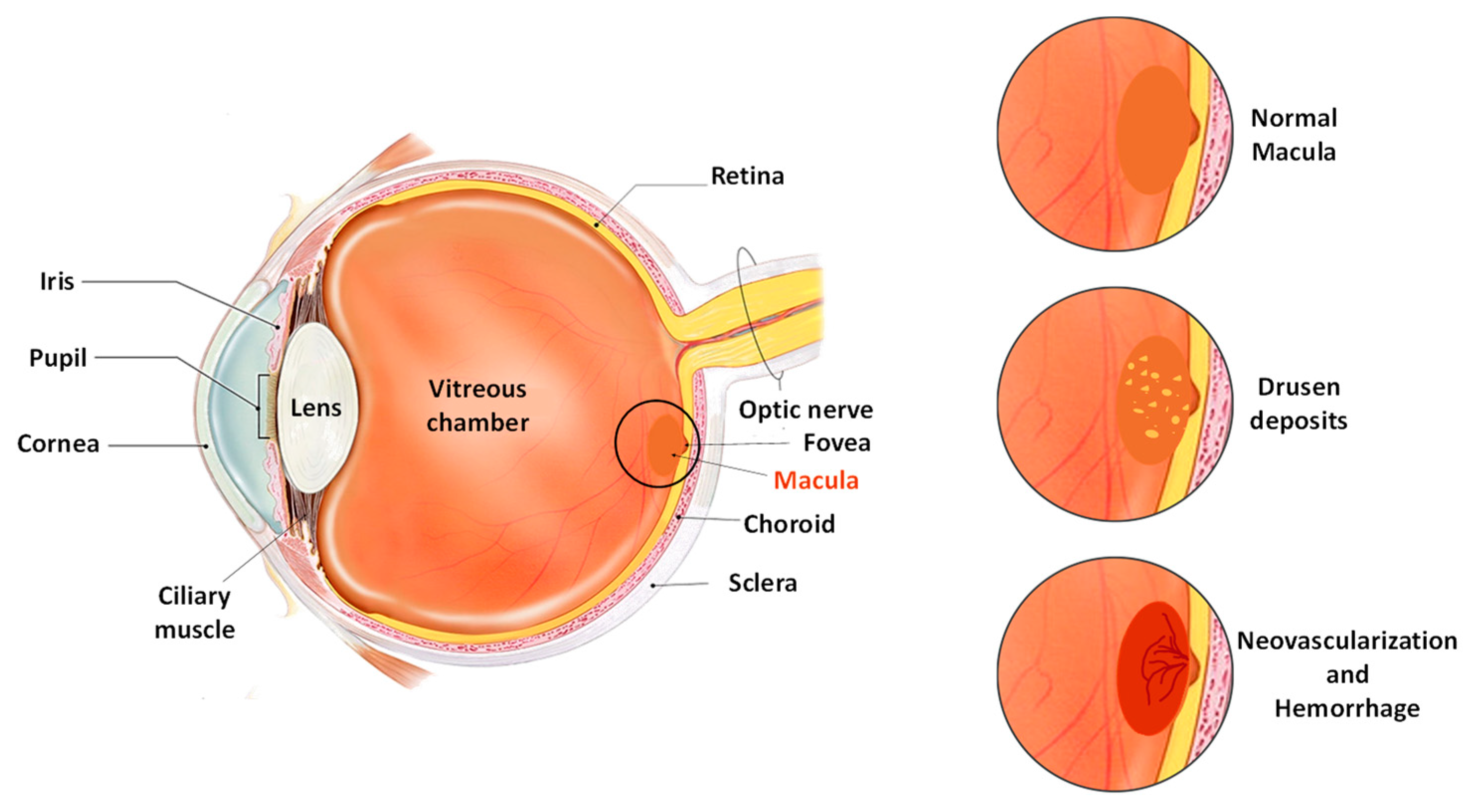
:1. Introduction
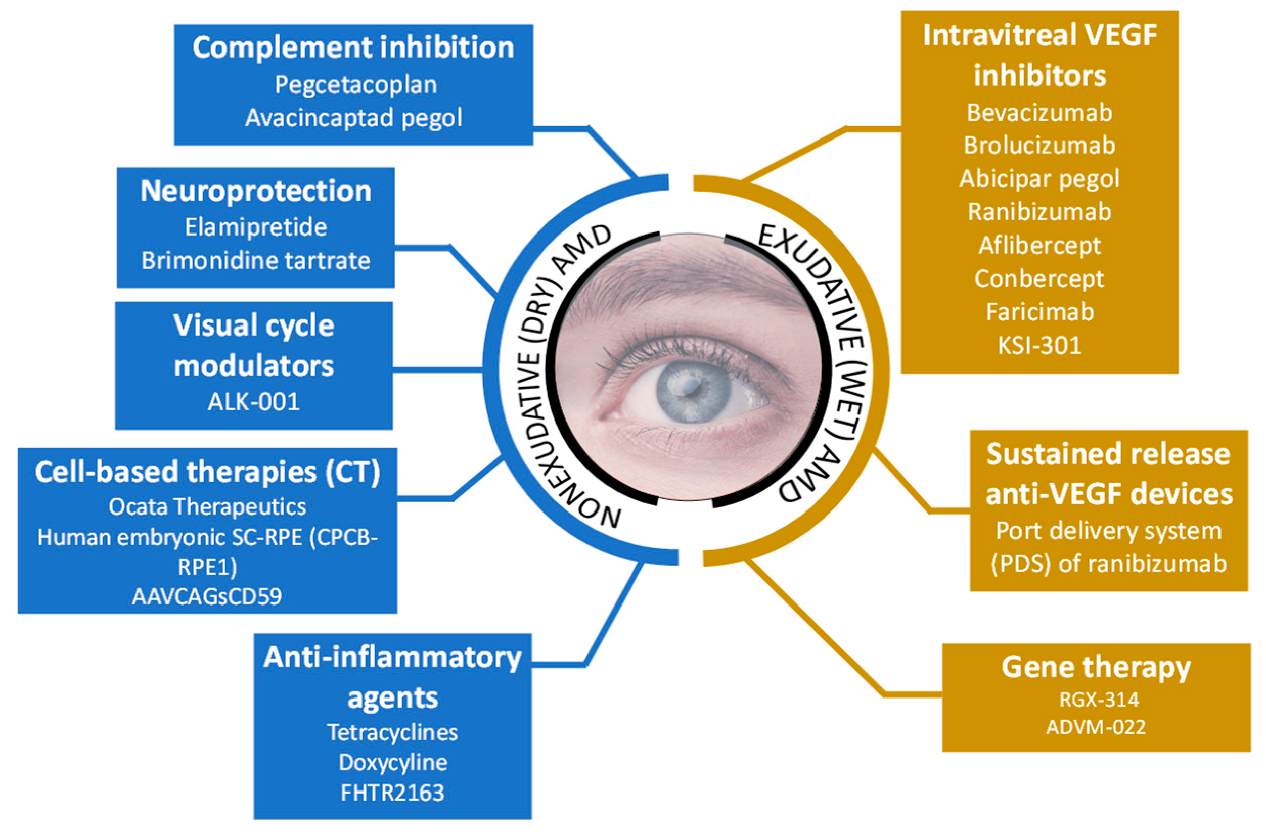
2. Current Approaches for the Treatment of AMD
2.1. Inhibition of Complement System
2.2. Modulation of the Visual Cycle
2.3. Cell-Based Therapies
2.4. Reduction of Inflammation
2.5. Neuroprotective Strategies
2.6. Inhibition of Vascular Endothelial Growth Factor (VEGF)
2.7. Sustained Releasing Anti-VEGF Devices

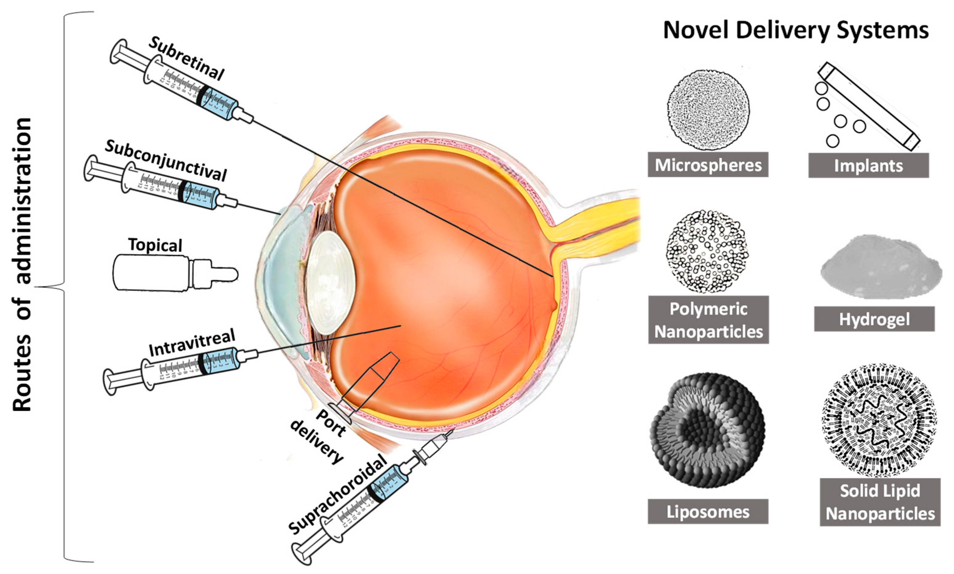
3. Novel Drug Delivery Systems for AMD
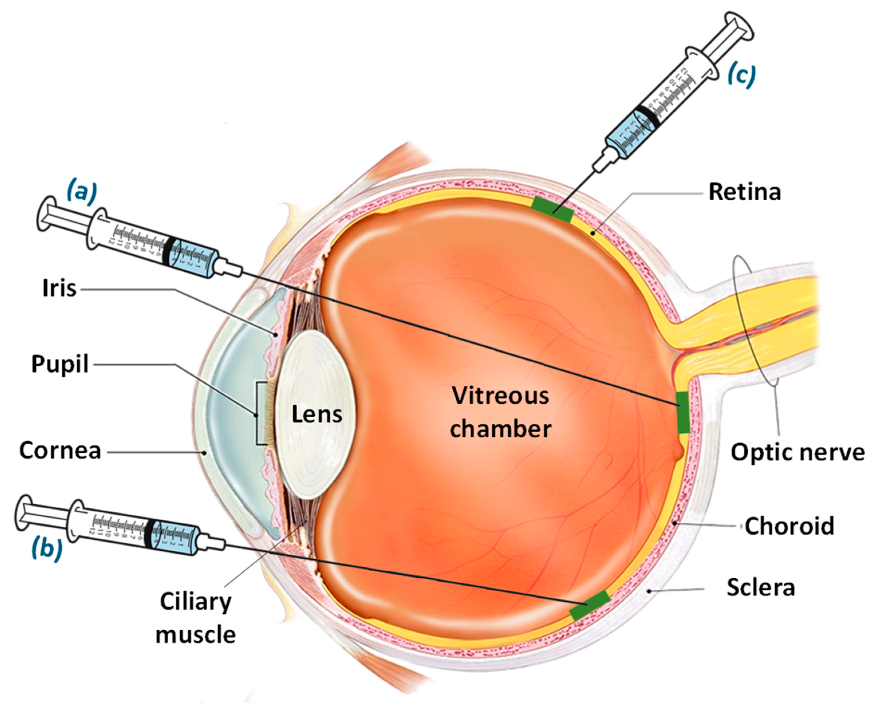
3.1. Intravitreal Route of Delivery

| Study Objective | Study Design | Type of Therapy Involved | Outcome with Safety Reports | References |
|---|---|---|---|---|
| Assessment of safety and efficacy of verteporfin therapy in AMD patients from Japan | Fifty-year-old patients with best-corrected visual acuity, classic-counting choroidal neovascularization secondary to AMD, and a lesion of greater linear dimension ≤5400 µm. | Treated with verteporfin intravenously followed with the administration of light for 15 min | Verteporfin therapy for choroidal neovascularization due to AMD demonstrated similar safety and an effective angiographic and vision effect as that reported in Caucasian patients. | [102] |
| Safety and efficacy assessment of verteporfin photodynamic therapy in sub-foveal choroidal neovascularization (CNV) related to AMD | Forty-eight patients with sub-foveal CNV secondary to AMD were enrolled with follow-up for at least one year | Treated with verteporfin photodynamic therapy | The study indicated an 83.3% improvement in patients with CNV secondary to AMD with a reduction in visual acuity deterioration due to sub-foveal CNV. | [103] |
| Efficacy and safety of photodynamic therapy with verteporfin combined with intravitreal triamcinolone in choroidal neovascularization to AMD | One hundred eighty-four patients with sub-foveal choroidal neovascularization with follow-up intervals of 3 months. | A solution containing 25 mg of triamcinolone was injected intravitreally after photodynamic therapy | Pooled verteporfin photodynamic therapy with intravitreal triamcinolone showed significant improvement in efficacy compared to standard verteporfin photodynamic therapy | [104] |
| Efficacy and safety of combined ranibizumab with verteporfin photodynamic compared with monotherapy of ranibizumab in patients with sub-foveal choroidal neovascularization AMD | Double-masked randomized phase lllb clinical trial on 321 patients | Combination of ranibizumab with verteporfin photodynamic compared with monotherapy of ranibizumab | A well-tolerated result after 12 months of applications was demonstrated in the clinical trial result | [105] |
| Safety and efficacy after twenty-four-month angiographic outcomes from clinical trials studying photodynamic therapy with verteporfin | Double-masked, placebo-controlled, randomized clinical trial | Patients visiting 28 ophthalmology practices in Europe and North America were treated with verteporfin and placebo-controlled | Verteporfin therapy for sub-foveal choroidal neovascularization caused by pathologic myopia with AMD demonstrated maintenance of safety throughout treatment for 24 months with improved visual acuity. | [106] |
| Effect of lesion size, visual acuity, and lesion composition on visual acuity change with or without verteporfin therapy | Three placebo-controlled, randomized clinical trials | Treatment of patient (minimally classic lesion with AMD and with no classic choroidal neovascularization with AMD) with verteporfin in photodynamic therapy | Lesion size after treatment of AMD with photodynamic therapy followed with AMD indicated improvement in vision with significant safety and efficacy | [107] |
| Safety and efficacy of photodynamic therapy combined with verteporfin and bevacizumab | Randomized controlled pilot clinical trial on one hundred and sixty-five patients with classic or occult CNV owing to AMD in at least one eye that had been never been treated previously | Treatment with either single photodynamic therapy with verteporfin or single administration of intravitreal bevacizumab, or in combination | Significant improvement in safety in visual acuity after treatment of one month followed with maintenance over three months was observed | [108] |
| Comparison of safety and efficacy of anecortave acetate with verteporfin and photodynamic therapy | Five hundred thirty patients with classic sub-foveal choroidal neovascularization with AMD tested following masked, randomized, multicenter, parallel-group, active control, noninferiority clinical trial | Tenon’s capsule-based anecortave acetate periocular posterior juxta scleral depot was administered for six months in the test group, compared with verteporfin photodynamic therapy | The results demonstrated safety and efficacy with the benefits of anecortave acetae for the treatment of choroidal neovascularization risk associated with either the drug or photodynamic therapy | [109] |
| Visual outcome after the intravitreal injection of triamcinolone acetonide for exudative AMD | Comparative non-randomized clinical trial | Twenty patients with bilateral exudative AMD were treated with a unilateral intravitreal injection of triamcinolone acetonide | Intravitreal injection of triamcinolone acetonide improved the visual acuity of the eye with preconceived safety | [110] |
| Anti-vascular endothelial growth factor antibody for the treatment of predominantly classic neovascularization in AMD patients | Randomized, double-masked, active-treatment-controlled clinical trial | Treatment of AMD patients with ranibizumab and compared with verteporfin photodynamic therapy | Ranibizumab indicated significant improvement in the verteporfin photodynamic therapy after two years of clinical trials on patients with AMD | [111] |
| Safety and efficacy assessment of verteporfin photodynamic therapy in reduction of vision loss for the patient with sub-foveal occult associated with AMD | A total of three hundred sixty-four eligible patients with ≥50 years were included in the study | The patients were treated with verteporfin photodynamic therapy and compared with placebo considering the baseline of visual acuity | Verteporfin photodynamic therapy in the management of occult associated with AMD demonstrated safety and efficacy | [112] |
| Formulation | Bioactive Agent | Composition | Preparation Method | Significant Outcomes | References |
|---|---|---|---|---|---|
| Microspheres | Bevacizumab | PLGA, PCADK, dextran, PEG | Solid-in-oil-in-water emulsification method |
| [86] |
| Polymeric nanoparticles | Bevacizumab | PLGA and PVA | Modified solvent emulsification-evaporation method |
| [87] |
| Polymeric nanoparticles | Bevacizumab | PLGA, Tween 80, and PVA | Modified double-emulsion solvent evaporation procedure |
| [88] |
| Mesoporous silica nanoparticles (MSNs) | Bevacizumab | Tetraethyl orthosilicate (TEOS), cetyltrimethylammonium chloride (CTAC), triethanolamine (TEA), APTES, and mPEG-NHS | Soft template method and Nanocasting strategy |
| [89] |
| Polymeric nanoparticles | Bevacizumab | Chitosan and Sodium tripolyphosphate | Ionic gelation method |
| [90] |
| Modified polymeric nanocarriers | Bevacizumab | Chitosan, Poly(ethylene glycol) methacrylate, Tween 80, Span 80, Sodium tripolyphosphate, and Sodium sulfate | Michael addition reaction and double crosslinking (ionic and covalent) reaction in reverse emulsion |
| [91] |
| Multivesicular liposomes | Bevacizumab | 1,2-dioleoyl-sn-glycero-3-phosphocholine, 2-dipalmitoyl-sn-glycero-3-phosphoglycerol, cholesterol, and triolein | Double emulsification technique |
| [92] |
| Solid lipid implants | Ranibizumab | Triglycerides H12, Triglycerides Dynasan D118, and Hydroxypropyl-β-cyclodextrine (HP-β-CD) | Twin-screw extrusion method |
| [93] |
| PLGA microparticles | Ranibizumab | PLGA and PVA | Water-in-oil-in-water (W/O/W) double emulsion |
| [94] |
| Microspheres | Ranibizumab | PLGA, bovine serum albumin, PEG, Mg(OH)2, and PVA | Double emulsion, solvent evaporation technique |
| [95] |
| Microsphere-hydrogel | Ranibizumab | PLGA, PVA, Bovine serum albumin (BSA), PEG, poly(ethylene glycol)-co-(L-lactic acid) diacrylate (PEG-PLLA-DA) and N-isopropylacrylamide (NIPAAm) | Modified double-emulsion, solvent evaporation technique, and free radical polymerization method |
| [96] |
| Microsphere-hydrogel | Aflibercept | PLGA, PVA, BSA, PEG, PEG-PLLA-DA, and NIPAAm | Modified double-emulsion, solvent evaporation technique and free radical polymerization method |
| [97,98] |
| Polymeric nanoparticles | Aflibercept | PLGA and PVA | Double-emulsion diffusion method |
| {100} |
| Microparticles | PolyActive™ | PEG, Polybutylphthalate | Water-in-oil-in-water emulsion (w/o/w) process |
| [100] |
| Intravitreal implants | Dexamethasone | PLGA and PCL | Extrusion method |
| [101] |
3.2. Delivery through Subretinal Space
3.3. Delivery through Suprachoroidal Space
3.4. Port Delivery System
3.5. Delivery through Other Routes
4. Overview of Patent Situation
5. Conclusions
Author Contributions
Funding
Institutional Review Board Statement
Informed Consent Statement
Data Availability Statement
Conflicts of Interest
References
- Stahl, A. The Diagnosis and Treatment of Age-Related Macular Degeneration. Dtsch. Arztebl. Int. 2020, 117, 513–520. [Google Scholar] [CrossRef] [PubMed]
- Pennington, K.L.; DeAngelis, M.M. Epidemiology of age-related macular degeneration (AMD): Associations with cardiovascular disease phenotypes and lipid factors. Eye Vis. 2016, 3, 34. [Google Scholar] [CrossRef] [PubMed] [Green Version]
- Wong, W.L.; Su, X.; Li, X.; Cheung, C.M.; Klein, R.; Cheng, C.Y.; Wong, T.Y. Global prevalence of age-related macular degeneration and disease burden projection for 2020 and 2040: A systematic review and meta-analysis. Lancet Glob. Health 2014, 2, e106–e116. [Google Scholar] [CrossRef] [PubMed] [Green Version]
- World Health Organization. World Report on Vision. World Health Organization. License: CC BY-NC-SA 3.0 IGO. 2019. Available online: https://apps.who.int/iris/handle/10665/328717 (accessed on 10 December 2022).
- Ruan, Y.; Jiang, S.; Gericke, A. Age-Related Macular Degeneration: Role of Oxidative Stress and Blood Vessels. Int. J. Mol. Sci. 2021, 22, 1296. [Google Scholar] [CrossRef] [PubMed]
- Heesterbeek, T.J.; Lorés-Motta, L.; Hoyng, C.B.; Lechanteur, Y.T.E.; den Hollander, A.I. Risk factors for progression of age-related macular degeneration. Ophthalmic Physiol. Opt. 2020, 40, 140–170. [Google Scholar] [CrossRef] [Green Version]
- Chen, M.; Xu, H. Parainflammation, chronic inflammation, and age-related macular degeneration. J. Leukoc. Biol. 2015, 98, 713–725. [Google Scholar] [CrossRef] [Green Version]
- Carneiro, Â.; Andrade, J.P. Nutritional and Lifestyle Interventions for Age-Related Macular Degeneration: A Review. Oxidative Med. Cell. Longev. 2017, 2017, 6469138. [Google Scholar] [CrossRef]
- De Koning-Backus, A.P.M.; Buitendijk, G.H.S.; Kiefte-de Jong, J.C.; Colijn, J.M.; Hofman, A.; Vingerling, J.R.; Haverkort, E.B.; Franco, O.H.; Klaver, C.C.W. Intake of Vegetables, Fruit, and Fish is Beneficial for Age-Related Macular Degeneration. Am. J. Ophthalmol. 2019, 198, 70–79. [Google Scholar] [CrossRef]
- Velilla, S.; García-Medina, J.J.; García-Layana, A.; Dolz-Marco, R.; Pons-Vázquez, S.; Pinazo-Durán, M.D.; Gómez-Ulla, F.; Arévalo, J.F.; Díaz-Llopis, M.; Gallego-Pinazo, R. Smoking and Age-Related Macular Degeneration: Review and Update. J. Ophthalmol. 2013, 2013, 895147. [Google Scholar] [CrossRef]
- Maugeri, A.; Barchitta, M.; Mazzone, M.G.; Giuliano, F.; Agodi, A. Complement System and Age-Related Macular Degeneration: Implications of Gene-Environment Interaction for Preventive and Personalized Medicine. BioMed Res. Int. 2018, 2018, 7532507. [Google Scholar] [CrossRef] [Green Version]
- Merle, H.; Béral, L.; Rocher, M.; Pierre, M.; Jean-Charles, A.; Béra, O.; Rosamont, L.A.; Robert, P.Y.; Lézin, A. Class II Human Leukocyte Antigen (HLA) and Susceptibility to Polypoidal Choroidal Vasculopathy in Afro-Caribbean Descent. Clin. Ophthalmol. 2022, 16, 1047–1053. [Google Scholar] [CrossRef]
- Korb, C.A.; Elbaz, H.; Schuster, A.K.; Nickels, S.; Ponto, K.A.; Schulz, A.; Wild, P.S.; Münzel, T.; Beutel, M.E.; Schmidtmann, I.; et al. Five-year cumulative incidence and progression of age-related macular degeneration: Results from the German population-based Gutenberg Health Study (GHS). Graefe’s Arch. Clin. Exp. Ophthalmol. 2022, 260, 55–64. [Google Scholar] [CrossRef] [PubMed]
- Flores, R.; Carneiro, Â.; Vieira, M.; Tenreiro, S.; Seabra, M.C. Age-Related Macular Degeneration: Pathophysiology, Management, and Future Perspectives. Ophthalmologica 2021, 244, 495–511. [Google Scholar] [CrossRef] [PubMed]
- Fernandes, A.R.; Zielińska, A.; Sanchez-Lopez, E.; dos Santos, T.; Garcia, M.L.; Silva, A.M.; Karczewski, J.; Souto, E.B. Exudative versus Nonexudative Age-Related Macular Degeneration: Physiopathology and Treatment Options. Int. J. Mol. Sci. 2022, 23, 2592. [Google Scholar] [CrossRef] [PubMed]
- Kwon, Y.H.; Kim, Y.A.; Yoo, Y.H. Chapter 3—Loss of Pigment Epithelial Cells Is Prevented by Autophagy. In Autophagy: Cancer, Other Pathologies, Inflammation, Immunity, Infection, and Aging; Hayat, M.A., Ed.; Academic Press: Cambridge, MA, USA, 2017; pp. 105–117. [Google Scholar]
- Yonekawa, Y.; Kim, I.K. Clinical Characteristics and Current Treatment of Age-Related Macular Degeneration. Cold Spring Harb. Perspect. Med. 2015, 5, a017178. [Google Scholar] [CrossRef] [Green Version]
- Zhang, X.; Sivaprasad, S. Drusen and pachydrusen: The definition, pathogenesis, and clinical significance. Eye 2021, 35, 121–133. [Google Scholar] [CrossRef]
- Ahmed, D.; Stattin, M.; Haas, A.-M.; Graf, A.; Krepler, K.; Ansari-Shahrezaei, S. Drusen characteristics of type 2 macular neovascularization in age-related macular degeneration. BMC Ophthalmol. 2020, 20, 381. [Google Scholar] [CrossRef]
- Notomi, S.; Shiose, S.; Ishikawa, K.; Fukuda, Y.; Kano, K.; Mori, K.; Wada, I.; Kaizu, Y.; Matsumoto, H.; Akiyama, M.; et al. Drusen and pigment abnormality predict the development of neovascular age-related macular degeneration in Japanese patients. PLoS ONE 2021, 16, e0255213. [Google Scholar] [CrossRef] [PubMed]
- Curcio, C.A. Soft Drusen in Age-Related Macular Degeneration: Biology and Targeting Via the Oil Spill Strategies. Investig. Ophthalmol. Vis. Sci. 2018, 59, AMD160–AMD181. [Google Scholar] [CrossRef] [PubMed] [Green Version]
- Domalpally, A.; Danis, R.P.; Trane, R.; Blodi, B.A.; Clemons, T.E.; Chew, E.Y. Atrophy in Neovascular Age-Related Macular Degeneration: Age-Related Eye Disease Study 2 Report Number 15. Ophthalmol. Retin. 2018, 2, 1021–1027. [Google Scholar] [CrossRef]
- Yeo, N.J.Y.; Chan, E.J.J.; Cheung, C. Choroidal Neovascularization: Mechanisms of Endothelial Dysfunction. Front. Pharmacol. 2019, 10, 1363. [Google Scholar] [CrossRef] [PubMed] [Green Version]
- Somasundaran, S.; Constable, I.J.; Mellough, C.B.; Carvalho, L.S. Retinal pigment epithelium and age-related macular degeneration: A review of major disease mechanisms. Clin. Exp. Ophthalmol. 2020, 48, 1043–1056. [Google Scholar] [CrossRef] [PubMed]
- Wong, K.-H.; Nam, H.-Y.; Lew, S.-Y.; Naidu, M.; David, P.; Kamalden, T.A.; Hadie, S.N.H.; Lim, L.-W. Discovering the Potential of Natural Antioxidants in Age-Related Macular Degeneration: A Review. Pharmaceuticals 2022, 15, 101. [Google Scholar] [CrossRef] [PubMed]
- Ghorbani, S.; Yong, V.W. The extracellular matrix as modifier of neuroinflammation and remyelination in multiple sclerosis. Brain 2021, 144, 1958–1973. [Google Scholar] [CrossRef] [PubMed]
- Hadziahmetovic, M.; Malek, G. Age-Related Macular Degeneration Revisited: From Pathology and Cellular Stress to Potential Therapies. Front. Cell Dev. Biol. 2021, 8, 612812. [Google Scholar] [CrossRef] [PubMed]
- Ishikawa, M.; Jin, D.; Sawada, Y.; Abe, S.; Yoshitomi, T. Future Therapies of Wet Age-Related Macular Degeneration. J. Ophthalmol. 2015, 2015, 138070. [Google Scholar] [CrossRef] [Green Version]
- Varela-Fernández, R.; Díaz-Tomé, V.; Luaces-Rodríguez, A.; Conde-Penedo, A.; García-Otero, X.; Luzardo-Álvarez, A.; Fernández-Ferreiro, A.; Otero-Espinar, F.J. Drug Delivery to the Posterior Segment of the Eye: Biopharmaceutic and Pharmacokinetic Considerations. Pharmaceutics 2020, 12, 269. [Google Scholar] [CrossRef] [Green Version]
- Burhan, A.M.; Klahan, B.; Cummins, W.; Andrés-Guerrero, V.; Byrne, M.E.; O’Reilly, N.J.; Chauhan, A.; Fitzhenry, L.; Hughes, H. Posterior Segment Ophthalmic Drug Delivery: Role of Muco-Adhesion with a Special Focus on Chitosan. Pharmaceutics 2021, 13, 1685. [Google Scholar] [CrossRef]
- Cabral de Guimaraes, T.A.; Daich Varela, M.; Georgiou, M.; Michaelides, M. Treatments for dry age-related macular degeneration: Therapeutic avenues, clinical trials and future directions. Br. J. Ophthalmol. 2022, 106, 297–304. [Google Scholar] [CrossRef]
- Moisseiev, E.; Loewenstein, A. Novel Long-acting Pharmacotherapy for Exudative Age Related Macular Degeneration. Curr. Pharm. Des. 2018, 24, 4860–4863. [Google Scholar] [CrossRef]
- Iglicki, M.; González, D.P.; Loewenstein, A.; Zur, D. Longer-acting treatments for neovascular age-related macular degeneration—Present and future. Eye 2021, 35, 1111–1116. [Google Scholar] [CrossRef] [PubMed]
- Hernández-Zimbrón, L.F.; Zamora-Alvarado, R.; Ochoa-De la Paz, L.; Velez-Montoya, R.; Zenteno, E.; Gulias-Cañizo, R.; Quiroz-Mercado, H.; Gonzalez-Salinas, R. Age-Related Macular Degeneration: New Paradigms for Treatment and Management of AMD. Oxidative Med. Cell. Longev. 2018, 2018, 8374647. [Google Scholar] [CrossRef] [PubMed]
- Wu, J.; Sun, X. Complement system and age-related macular degeneration: Drugs and challenges. Drug Des. Dev. Ther. 2019, 13, 2413–2425. [Google Scholar] [CrossRef] [PubMed]
- Merle, N.S.; Noe, R.; Halbwachs-Mecarelli, L.; Fremeaux-Bacchi, V.; Roumenina, L.T. Complement System Part II: Role in Immunity. Front. Immunol. 2015, 6, 257. [Google Scholar] [CrossRef] [PubMed] [Green Version]
- Kim, B.J.; Mastellos, D.C.; Li, Y.; Dunaief, J.L.; Lambris, J.D. Targeting complement components C3 and C5 for the retina: Key concepts and lingering questions. Prog. Retin. Eye Res. 2021, 83, 100936. [Google Scholar] [CrossRef] [PubMed]
- Dobó, J.; Kocsis, A.; Gál, P. Be on Target: Strategies of Targeting Alternative and Lectin Pathway Components in Complement-Mediated Diseases. Front. Immunol. 2018, 9, 1851. [Google Scholar] [CrossRef] [PubMed] [Green Version]
- Morgan, B.P.; Walters, D.; Serna, M.; Bubeck, D. Terminal complexes of the complement system: New structural insights and their relevance to function. Immunol. Rev. 2016, 274, 141–151. [Google Scholar] [CrossRef]
- Liao, D.S.; Grossi, F.V.; El Mehdi, D.; Gerber, M.R.; Brown, D.M.; Heier, J.S.; Wykoff, C.C.; Singerman, L.J.; Abraham, P.; Grassmann, F.; et al. Complement C3 Inhibitor Pegcetacoplan for Geographic Atrophy Secondary to Age-Related Macular Degeneration: A Randomized Phase 2 Trial. Ophthalmology 2020, 127, 186–195. [Google Scholar] [CrossRef] [Green Version]
- Jaffe, G.J.; Westby, K.; Csaky, K.G.; Monés, J.; Pearlman, J.A.; Patel, S.S.; Joondeph, B.C.; Randolph, J.; Masonson, H.; Rezaei, K.A. C5 Inhibitor Avacincaptad Pegol for Geographic Atrophy Due to Age-Related Macular Degeneration: A Randomized Pivotal Phase 2/3 Trial. Ophthalmology 2021, 128, 576–586. [Google Scholar] [CrossRef]
- Jaffe, G.J.; Sahni, J.; Fauser, S.; Geary, R.S.; Schneider, E.; McCaleb, M.J.I.O.; Science, V. Development of IONIS-FB-LRx to treat geographic atrophy associated with AMD. Investig. Ophthalmol. Vis. Sci. 2020, 61, 4305. [Google Scholar]
- Merle, N.S.; Church, S.E.; Fremeaux-Bacchi, V.; Roumenina, L.T. Complement System Part I—Molecular Mechanisms of Activation and Regulation. Sec. Mol. Innate Immun. 2015, 6, 262. [Google Scholar] [CrossRef] [PubMed] [Green Version]
- Yeong, J.L.; Loveman, E.; Colquitt, J.L.; Royle, P.; Waugh, N.; Lois, N. Visual cycle modulators versus placebo or observation for the prevention and treatment of geographic atrophy due to age-related macular degeneration. Cochrane Database Syst. Rev. 2018, 12, CD013154. [Google Scholar] [CrossRef] [Green Version]
- Thirunavukarasu, A.J.; Ross, A.C.; Gilbert, R.M. Vitamin A, systemic T-cells, and the eye: Focus on degenerative retinal disease. Front. Nutr. 2022, 9, 914457. [Google Scholar] [CrossRef] [PubMed]
- Alkeus Pharmaceuticals, Inc. A Phase 2/3 Multicenter, Randomized, Double-Masked, Parallel-Group, Placebo-Controlled Study to Investigate the Safety, Pharmacokinetics, Tolerability, and Efficacy of ALK-001 in Geographic Atrophy Secondary to Age-related Macular Degeneration; NCT03845582. Available online: clinicaltrials.gov (accessed on 12 February 2022).
- Yang, S.; Zhou, J.; Li, D. Functions and Diseases of the Retinal Pigment Epithelium. Front. Pharmacol. 2021, 12, 727870. [Google Scholar] [CrossRef] [PubMed]
- Surendran, H.; Nandakumar, S.; Reddy, K.V.B.; Stoddard, J.; Mohan, K.V.; Upadhyay, P.K.; McGill, T.J.; Pal, R. Transplantation of retinal pigment epithelium and photoreceptors generated concomitantly via small molecule-mediated differentiation rescues visual function in rodent models of retinal degeneration. Stem Cell Res. Ther. 2021, 12, 70. [Google Scholar] [CrossRef]
- Ribeiro, J.; Procyk, C.A.; West, E.L.; O’Hara-Wright, M.; Martins, M.F.; Khorasani, M.M.; Hare, A.; Basche, M.; Fernando, M.; Goh, D.; et al. Restoration of visual function in advanced disease after transplantation of purified human pluripotent stem cell-derived cone photoreceptors. Cell Rep. 2021, 35, 109022. [Google Scholar] [CrossRef]
- Zhao, C.; Wang, Q.; Temple, S. Stem cell therapies for retinal diseases: Recapitulating development to replace degenerated cells. Development 2017, 144, 1368–1381. [Google Scholar] [CrossRef] [Green Version]
- Sharma, A.; Jaganathan, B.G. Stem Cell Therapy for Retinal Degeneration: The Evidence to Date. Biologics: Targets & therapy. Biologics 2021, 15, 299–306. [Google Scholar] [CrossRef]
- Bracha, P.; Moore, N.A.; Ciulla, T.A. Induced pluripotent stem cell-based therapy for age-related macular degeneration. Expert Opin. Biol. Ther. 2017, 17, 1113–1126. [Google Scholar] [CrossRef]
- Forest, D.L.; Johnson, L.V.; Clegg, D.O. Cellular models and therapies for age-related macular degeneration. Dis. Model. Mech. 2015, 8, 421–427. [Google Scholar] [CrossRef] [Green Version]
- NCT. A Study of an Encapsulated Cell Technology (ECT) Implant for Patients with Atrophic Macular Degeneration. Available online: https://clinicaltrials.gov/show/NCT004479542007 (accessed on 12 February 2022).
- Kashani, A.H.; Lebkowski, J.S.; Rahhal, F.M.; Avery, R.L.; Salehi-Had, H.; Dang, W.; Lin, C.-M.; Mitra, D.; Zhu, D.; Thomas, B.B.; et al. A bioengineered retinal pigment epithelial monolayer for advanced, dry age-related macular degeneration. Sci. Transl. Med. 2018, 10, eaao4097. [Google Scholar] [CrossRef] [PubMed] [Green Version]
- Telander, D.G. Inflammation and Age-Related Macular Degeneration (AMD). Semin. Ophthalmol. 2011, 26, 192–197. [Google Scholar] [CrossRef]
- Kauppinen, A.; Paterno, J.J.; Blasiak, J.; Salminen, A.; Kaarniranta, K. Inflammation and its role in age-related macular degeneration. Cell. Mol. Life Sci. 2016, 73, 1765–1786. [Google Scholar] [CrossRef] [PubMed] [Green Version]
- Olivares-González, L.; Velasco, S.; Campillo, I.; Rodrigo, R. Retinal Inflammation, Cell Death and Inherited Retinal Dystrophies. Int. J. Mol. Sci. 2021, 22, 2096. [Google Scholar] [CrossRef] [PubMed]
- Mirshahi, A.; Azimi, P.; Abdolahi, A.; Mirshahi, R.; Abdollahian, M. Oral Doxycycline Reduces the Total Number of Intraocular Bevacizumab Injections Needed to Control Neovascular Age-related Macular Degeneration. Med. Hypothesis Discov. Innov. Ophthalmol. J. 2017, 6, 23–29. [Google Scholar]
- Khanani, A.M.; Hershberger, V.S.; Pieramici, D.J.; Khurana, R.N.; Brunstein, F.; Ma, L.; Maass, K.F.; Honigberg, L.A.; Tom, I.; Chen, H.; et al. Phase 1 Study of the Anti-HtrA1 Antibody-binding Fragment FHTR2163 in Geographic Atrophy Secondary to Age-related Macular Degeneration. Am. J. Ophthalmol. 2021, 232, 49–57. [Google Scholar] [CrossRef]
- Williams, B.L.; Seager, N.A.; Gardiner, J.D.; Pappas, C.M.; Cronin, M.C.; Amat di San Filippo, C.; Anstadt, R.A.; Liu, J.; Toso, M.A.; Nichols, L.; et al. Chromosome 10q26-driven age-related macular degeneration is associated with reduced levels of HTRA1 in human retinal pigment epithelium. Proc. Natl. Acad. Sci. USA 2021, 118, e2103617118. [Google Scholar] [CrossRef]
- Ou, K.; Li, Y.; Liu, L.; Li, H.; Cox, K.; Wu, J.; Liu, J.; Dick, A.D. Recent developments of neuroprotective agents for degenerative retinal disorders. Neural Regen. Res. 2022, 17, 1919–1928. [Google Scholar]
- Lin, J.B.; Murakami, Y.; Miller, J.W.; Vavvas, D.G. Neuroprotection for Age-Related Macular Degeneration. Ophthalmol. Sci. 2022, 2, 100192. [Google Scholar] [CrossRef]
- Nhu, N.T.; Xiao, S.-Y.; Liu, Y.; Kumar, V.B.; Cui, Z.-Y.; Lee, S.-D. Neuroprotective Effects of a Small Mitochondrially-Targeted Tetrapeptide Elamipretide in Neurodegeneration. Front. Integr. Neurosci. 2022, 15, 747901. [Google Scholar] [CrossRef]
- Kuppermann, B.D.; Patel, S.S.; Boyer, D.S.; Augustin, A.J.; Freeman, W.R.; Kerr, K.J.; Guo, Q.; Schneider, S.; López, F.J. Phase 2 study of the safety and efficacy of brimonidine drug delivery system (Brimo DDS) generation 1 in patients with geographic atrophy secondary to age-related macular degeneration. Retina 2021, 41, 144–155. [Google Scholar] [CrossRef] [PubMed]
- Farnoodian, M.; Wang, S.; Dietz, J.; Nickells Robert, W.; Sorenson Christine, M.; Sheibani, N. Negative regulators of angiogenesis: Important targets for treatment of exudative AMD. Clin. Sci. 2017, 131, 1763–1780. [Google Scholar] [CrossRef] [PubMed] [Green Version]
- Bressler, S.B. Introduction: Understanding the Role of Angiogenesis and Antiangiogenic Agents in Age-Related Macular Degeneration. Ophthalmology 2009, 116, S1–S7. [Google Scholar] [CrossRef] [PubMed]
- Khanani, A.M.; Zarbin, M.A.; Barakat, M.R.; Albini, T.A.; Kaiser, P.K.; Guruprasad, B.; Agashivala, N.; Yu, J.S.; Wykoff, C.C.; MacCumber, M.W. Safety Outcomes of Brolucizumab in Neovascular Age-Related Macular Degeneration: Results from the IRIS Registry and Komodo Healthcare Map. JAMA Ophthalmol. 2022, 140, 20–28. [Google Scholar] [CrossRef]
- Hussain, R.M.; Weng, C.Y.; Wykoff, C.C.; Gandhi, R.A.; Hariprasad, S.M. Abicipar pegol for neovascular age-related macular degeneration. Expert Opin. Biol. Ther. 2020, 20, 999–1008. [Google Scholar] [CrossRef] [PubMed]
- Zhou, P.; Zheng, S.; Wang, E.; Men, P.; Zhai, S. Conbercept for Treatment of Neovascular Age-Related Macular Degeneration and Visual Impairment due to Diabetic Macular Edema or Pathologic Myopia Choroidal Neovascularization: A Systematic Review and Meta-Analysis. Front. Pharmacol. 2021, 12, 696201. [Google Scholar] [CrossRef]
- Nair, A.A.; Finn, A.P.; Sternberg, P., Jr. Spotlight on Faricimab in the Treatment of Wet Age-Related Macular Degeneration: Design, Development and Place in Therapy. Drug Des. Dev. Ther. 2022, 16, 3395–3400. [Google Scholar] [CrossRef]
- Spini, A.; Giometto, S.; Donnini, S.; Posarelli, M.; Dotta, F.; Ziche, M.; Tosi, G.M.; Girardi, A.; Lucenteforte, E.; Gini, R.; et al. Risk of intraocular pressure increase with intravitreal injections of Vascular Endothelial Growth Factor Inhibitors: A cohort study. Am. J. Ophthalmol. 2022, 248, 45–50. [Google Scholar] [CrossRef]
- Koponen, S.; Kokki, E.; Kinnunen, K.; Ylä-Herttuala, S. Viral-Vector-Delivered Anti-Angiogenic Therapies to the Eye. Pharmaceutics 2021, 13, 219. [Google Scholar] [CrossRef]
- Kiss, S.; Grishanin, R.; Nguyen, A.; Rosario, R.; Greengard, J.S.; Nieves, J.; Gelfman, C.M.; Gasmi, M. Analysis of Aflibercept Expression in NHPs following Intravitreal Administration of ADVM-022, a Potential Gene Therapy for nAMD. Mol. Ther. Methods Clin. Dev. 2020, 18, 345–353. [Google Scholar] [CrossRef]
- Adams, B.S.; Sorhaitz, W.; Stringham, J. Aflibercept. In StatPearls; StatPearls Publishing: Treasure Island, FL, USA, 2022. [Google Scholar]
- Campochiaro, P.A.; Marcus, D.M.; Awh, C.C.; Regillo, C.; Adamis, A.P.; Bantseev, V.; Chiang, Y.; Ehrlich, J.S.; Erickson, S.; Hanley, W.D.; et al. The Port Delivery System with Ranibizumab for Neovascular Age-Related Macular Degeneration: Results from the Randomized Phase 2 Ladder Clinical Trial. Ophthalmology 2019, 126, 1141–1154. [Google Scholar] [CrossRef] [PubMed] [Green Version]
- Kertes, P.J.; Galic, I.J.; Greve, M.; Williams, G.; Baker, J.; Lahaie, M.; Sheidow, T. Efficacy of a Treat-and-Extend Regimen with Ranibizumab in Patients With Neovascular Age-Related Macular Disease: A Randomized Clinical Trial. JAMA Ophthalmol. 2020, 138, 244–250. [Google Scholar] [CrossRef] [PubMed] [Green Version]
- Ammar, M.J.; Hsu, J.; Chiang, A.; Ho, A.C.; Regillo, C.D. Age-related macular degeneration therapy: A review. Curr. Opin. Ophthalmol. 2020, 31, 215–221. [Google Scholar] [CrossRef] [PubMed]
- Thomas, C.N.; Sim, D.A.; Lee, W.H.; Alfahad, N.; Dick, A.D.; Denniston, A.K.; Hill, L.J. Emerging therapies and their delivery for treating age-related macular degeneration. Br. J. Pharmacol. 2022, 179, 1908–1937. [Google Scholar] [CrossRef]
- Gote, V.; Sikder, S.; Sicotte, J.; Pal, D. Ocular Drug Delivery: Present Innovations and Future Challenges. J. Pharmacol. Exp. Ther. 2019, 370, 602. [Google Scholar] [CrossRef]
- Yang, B.; Li, G.; Liu, J.; Li, X.; Zhang, S.; Sun, F.; Liu, W. Nanotechnology for Age-Related Macular Degeneration. Pharmaceutics 2021, 13, 2035. [Google Scholar] [CrossRef]
- Li, H.; Zhu, X.; Wang, M.; Zhao, D.; Li, H.; Yan, J. Drug sustained release from degradable drug-loaded in-situ hydrogels in the posterior eye: A mechanistic model and analytical method. J. Biomech. 2022, 136, 111052. [Google Scholar] [CrossRef]
- Thareja, A.; Hughes, H.; Alvarez-Lorenzo, C.; Hakkarainen, J.J.; Ahmed, Z. Penetration Enhancers for Topical Drug Delivery to the Ocular Posterior Segment—A Systematic Review. Pharmaceutics 2021, 13, 276. [Google Scholar] [CrossRef]
- Fogli, S.; Del Re, M.; Rofi, E.; Posarelli, C.; Figus, M.; Danesi, R. Clinical pharmacology of intravitreal anti-VEGF drugs. Eye 2018, 32, 1010–1020. [Google Scholar] [CrossRef] [Green Version]
- Volkmann, I.; Knoll, K.; Wiezorrek, M.; Greb, O.; Framme, C. Individualized treat-and-extend regime for optimization of real-world vision outcome and improved patients’ persistence. BMC Ophthalmol. 2020, 20, 122. [Google Scholar] [CrossRef] [Green Version]
- Liu, J.; Li, S.; Li, G.; Li, X.; Yu, C.; Fu, Z.; Li, X.; Teng, L.; Li, Y.; Sun, F. Highly bioactive, bevacizumab-loaded, sustained-release PLGA/PCADK microspheres for intravitreal therapy in ocular diseases. Int. J. Pharm. 2019, 563, 228–236. [Google Scholar] [CrossRef] [PubMed]
- Sousa, F.; Cruz, A.; Fonte, P.; Pinto, I.M.; Neves-Petersen, M.T.; Sarmento, B. A new paradigm for antiangiogenic therapy through controlled release of bevacizumab from PLGA nanoparticles. Sci. Rep. 2017, 7, 3736. [Google Scholar] [CrossRef] [PubMed] [Green Version]
- Zhang, X.-P.; Sun, J.-G.; Yao, J.; Shan, K.; Liu, B.-H.; Yao, M.-D.; Ge, H.-M.; Jiang, Q.; Zhao, C.; Yan, B. Effect of nanoencapsulation using poly (lactide-co-glycolide) (PLGA) on anti-angiogenic activity of bevacizumab for ocular angiogenesis therapy. Biomed. Pharmacother. 2018, 107, 1056–1063. [Google Scholar] [CrossRef] [PubMed]
- Sun, J.G.; Jiang, Q.; Zhang, X.P.; Shan, K.; Liu, B.H.; Zhao, C.; Yan, B. Mesoporous silica nanoparticles as a delivery system for improving antiangiogenic therapy. Int. J. Nanomed. 2019, 14, 1489–1501. [Google Scholar] [CrossRef] [Green Version]
- Badiee, P.; Varshochian, R.; Rafiee-Tehrani, M.; Abedin Dorkoosh, F.; Khoshayand, M.R.; Dinarvand, R. Ocular implant containing bevacizumab-loaded chitosan nanoparticles intended for choroidal neovascularization treatment. J. Biomed. Mater. Res. A 2018, 106, 2261–2271. [Google Scholar] [CrossRef]
- Savin, C.-L.; Popa, M.; Delaite, C.; Costuleanu, M.; Costin, D.; Peptu, C.A. Chitosan grafted-poly(ethylene glycol) methacrylate nanoparticles as carrier for controlled release of bevacizumab. Mater. Sci. Eng. C 2019, 98, 843–860. [Google Scholar] [CrossRef]
- Mu, H.; Wang, Y.; Chu, Y.; Jiang, Y.; Hua, H.; Chu, L.; Wang, K.; Wang, A.; Liu, W.; Li, Y.; et al. Multivesicular liposomes for sustained release of bevacizumab in treating laser-induced choroidal neovascularization. Drug Deliv. 2018, 25, 1372–1383. [Google Scholar] [CrossRef] [Green Version]
- Vollrath, M.; Engert, J.; Winter, G. Long-term release and stability of pharmaceutical proteins delivered from solid lipid implants. Eur. J. Pharm. Biopharm. 2017, 117, 244–255. [Google Scholar] [CrossRef]
- Tanetsugu, Y.; Tagami, T.; Terukina, T.; Ogawa, T.; Ohta, M.; Ozeki, T. Development of a Sustainable Release System for a Ranibizumab Biosimilar Using Poly(lactic-co-glycolic acid) Biodegradable Polymer-Based Microparticles as a Platform. Biol. Pharm. Bull. 2017, 40, 145–150. [Google Scholar] [CrossRef] [Green Version]
- Osswald, C.R.; Guthrie, M.J.; Avila, A.; Valio, J.A.; Mieler, W.F.; Kang-Mieler, J.J. In Vivo Efficacy of an Injectable Microsphere-Hydrogel Ocular Drug Delivery System. Curr. Eye Res. 2017, 42, 1293–1301. [Google Scholar] [CrossRef]
- Liu, W.; Borrell, M.A.; Venerus, D.C.; Mieler, W.F.; Kang-Mieler, J.J. Characterization of Biodegradable Microsphere-Hydrogel Ocular Drug Delivery System for Controlled and Extended Release of Ranibizumab. Transl. Vis. Sci. Technol. 2019, 8, 12. [Google Scholar] [CrossRef] [PubMed]
- Liu, W.; Lee, B.-S.; Mieler, W.F.; Kang-Mieler, J.J. Biodegradable Microsphere-Hydrogel Ocular Drug Delivery System for Controlled and Extended Release of Bioactive Aflibercept In Vitro. Curr. Eye Res. 2019, 44, 264–274. [Google Scholar] [CrossRef] [PubMed]
- Kim, S.; Kang-Mieler, J.J.; Liu, W.; Wang, Z.; Yiu, G.; Teixeira, L.B.C.; Mieler, W.F.; Thomasy, S.M. Safety and Biocompatibility of Aflibercept-Loaded Microsphere Thermo-Responsive Hydrogel Drug Delivery System in a Nonhuman Primate Model. Transl. Vis. Sci. Technol. 2020, 9, 30. [Google Scholar] [CrossRef] [PubMed] [Green Version]
- Kelly, S.J.; Hirani, A.; Shahidadpury, V.; Solanki, A.; Halasz, K.; Varghese Gupta, S.; Madow, B.; Sutariya, V. Aflibercept Nanoformulation Inhibits VEGF Expression in Ocular In Vitro Model: A Preliminary Report. Biomedicines 2018, 6, 92. [Google Scholar] [CrossRef] [PubMed] [Green Version]
- Adamson, P.; Wilde, T.; Dobrzynski, E.; Sychterz, C.; Polsky, R.; Kurali, E.; Haworth, R.; Tang, C.-M.; Korczynska, J.; Cook, F.; et al. Single ocular injection of a sustained-release anti-VEGF delivers 6months pharmacokinetics and efficacy in a primate laser CNV model. J. Control. Release 2016, 244, 1–13. [Google Scholar] [CrossRef] [Green Version]
- Stein, S.; Auel, T.; Kempin, W.; Bogdahn, M.; Weitschies, W.; Seidlitz, A. Influence of the test method on in vitro drug release from intravitreal model implants containing dexamethasone or fluorescein sodium in poly (d,l-lactide-co-glycolide) or polycaprolactone. Eur. J. Pharm. Biopharm. 2018, 127, 270–278. [Google Scholar] [CrossRef]
- Japanese Age-Related Macular Degeneration Trial (JAT) Study Group. Japanese age-related macular degeneration trial: 1-year results of photodynamic therapy with verteporfin in Japanese patients with subfoveal choroidal neovascularization secondary to age-related macular degeneration. Am. J. Ophthalmol. 2003, 136, 1049–1061. [Google Scholar] [CrossRef]
- Shyong, M.-P.; Lee, F.-L.; Chen, S.-J.; Tung, T.-H.; Tsai, D.-C.; Hsu, W.-M. Photodynamic Therapy for Subfoveal Choroidal Neovascularization Secondary to Age-related Macular Degeneration. J. Chin. Med. Assoc. 2005, 68, 419–424. [Google Scholar] [CrossRef] [Green Version]
- Augustin, A.J.; Schmidt-Erfurth, U. Verteporfin therapy combined with intravitreal triamcinolone in all types of choroidal neovascularization due to age-related macular degeneration. Ophthalmology 2006, 113, 14–22. [Google Scholar] [CrossRef]
- Kaiser, P.K.; Boyer, D.S.; Cruess, A.F.; Slakter, J.S.; Pilz, S.; Weisberger, A. Verteporfin plus Ranibizumab for Choroidal Neovascularization in Age-Related Macular Degeneration: Twelve-Month Results of the DENALI Study. Ophthalmology 2012, 119, 1001. [Google Scholar] [CrossRef]
- Blinder, K.J.; Blumenkranz, M.S.; Bressler, N.M.; Bressler, S.B.; Donato, G.; Lewis, H.; Williams, G.A. Verteporfin therapy of subfoveal choroidal neovascularization in pathologic myopia: 2-year results of a randomized clinical trial—VIP report no. 3. Ophthalmology 2003, 110, 667–673. [Google Scholar] [CrossRef] [PubMed]
- Blinder, K.J.; Bradley, S.; Bressler, N.M.; Bressler, S.B.; Donati, G.; Hao, Y.; Virgili, G. Effect of lesion size, visual acuity, and lesion composition on visual acuity change with and without verteporfin therapy for choroidal neovascularization secondary to age-related macular degeneration: TAP and VIP report no. 1. Am. J. Ophthalmol. 2003, 136, 407–418. [Google Scholar] [CrossRef] [PubMed]
- Lazic, R.; Gabric, N. Verteporfin therapy and intravitreal bevacizumab combined and alone in choroidal neovascularization due to age-related macular degeneration. Ophthalmology 2007, 114, 1179–1185. [Google Scholar] [CrossRef] [PubMed]
- Slakter, J.S.; Bochow, T.W.; D’Amico, D.J.; Marks, B.; Jerdan, J.; Sullivan, E.K.; Robertson, S.M.; Slakter, J.S.; Sullins, G.; Zilliox, P. Anecortave acetate (15 milligrams) versus photodynamic therapy for treatment of subfoveal neovascularization in age-related macular degeneration. Ophthalmology 2006, 113, 3–13. [Google Scholar] [CrossRef] [PubMed]
- Jonas, J.B.; Spandau, U.H.; Harder, B.; Vossmerbaeumer, U.; Kamppeter, B.A. Intereye difference in exudative age-related macular degeneration with minimally classic or occult subfoveal neovascularization after unilateral intravitreal injection of triamcinolone acetonide. Am. J. Ophthalmol. 2005, 139, 1073–1079. [Google Scholar] [CrossRef]
- Brown, D.M.; Michels, M.; Kaiser, P.K.; Heier, J.S.; Sy, J.P.; Ianchulev, T. Ranibizumab versus verteporfin photodynamic therapy for neovascular age-related macular degeneration: Two-year results of the ANCHOR study. Ophthalmology 2009, 116, 57–65.e55. [Google Scholar] [CrossRef]
- Kaiser, P.K. Verteporfin PDT for subfoveal occult CNV in AMD: Two-year results of a randomized trial. Curr. Med. Res. Opin. 2009, 25, 1853–1860. [Google Scholar] [CrossRef]
- Himawan, E.; Ekström, P.; Buzgo, M.; Gaillard, P.; Stefánsson, E.; Marigo, V.; Loftsson, T.; Paquet-Durand, F. Drug delivery to retinal photoreceptors. Drug Discov. Today 2019, 24, 1637–1643. [Google Scholar] [CrossRef]
- Irigoyen, C.; Amenabar Alonso, A.; Sanchez-Molina, J.; Rodríguez-Hidalgo, M.; Lara-López, A.; Ruiz-Ederra, J. Subretinal Injection Techniques for Retinal Disease: A Review. J. Clean. Med. 2022, 11, 4717. [Google Scholar] [CrossRef]
- Peng, Y.; Tang, L.; Zhou, Y. Subretinal Injection: A Review on the Novel Route of Therapeutic Delivery for Vitreoretinal Diseases. Ophthalmic Res. 2017, 58, 217–226. [Google Scholar] [CrossRef]
- Ho, A.C.; Chang, T.S.; Samuel, M.; Williamson, P.; Willenbucher, R.F.; Malone, T. Experience with a Subretinal Cell-based Therapy in Patients with Geographic Atrophy Secondary to Age-related Macular Degeneration. Am. J. Ophthalmol. 2017, 179, 67–80. [Google Scholar] [CrossRef] [PubMed] [Green Version]
- Heier, J.S.; Ho, A.C.; Samuel, M.A.; Chang, T.; Riemann, C.D.; Kitchens, J.W.; Slakter, J.S.; Leiderman, Y.I.; Spencer, R.; Williams, G.A.; et al. Safety and Efficacy of Subretinally Administered Palucorcel for Geographic Atrophy of Age-Related Macular Degeneration: Phase 2b Study. Ophthalmol. Retin. 2020, 4, 384–393. [Google Scholar] [CrossRef] [PubMed]
- Moore, N.A.; Bracha, P.; Hussain, R.M.; Morral, N.; Ciulla, T.A. Gene therapy for age-related macular degeneration. Expert Opin. Biol. Ther. 2017, 17, 1235–1244. [Google Scholar] [CrossRef] [PubMed]
- Holmgaard, A.B.; Askou, A.L.; Jensen, E.G.; Alsing, S.; Bak, R.O.; Mikkelsen, J.G.; Corydon, T.J. Targeted Knockout of the Vegfa Gene in the Retina by Subretinal Injection of RNP Complexes Containing Cas9 Protein and Modified sgRNAs. Mol. Ther. 2021, 29, 191–207. [Google Scholar] [CrossRef]
- Ling, S.; Yang, S.; Hu, X.; Yin, D.; Dai, Y.; Qian, X.; Wang, D.; Pan, X.; Hong, J.; Sun, X.; et al. Lentiviral delivery of co-packaged Cas9 mRNA and a Vegfa-targeting guide RNA prevents wet age-related macular degeneration in mice. Nat. Biomed. Eng. 2021, 5, 144–156. [Google Scholar] [CrossRef]
- Campochiaro, P.A.; Lauer, A.K.; Sohn, E.H.; Mir, T.A.; Naylor, S.; Anderton, M.C.; Kelleher, M.; Harrop, R.; Ellis, S.; Mitrophanous, K.A. Lentiviral Vector Gene Transfer of Endostatin/Angiostatin for Macular Degeneration (GEM) Study. Hum. Gene Ther. 2017, 28, 99–111. [Google Scholar] [CrossRef]
- Binley, K.; Widdowson, P.S.; Kelleher, M.; de Belin, J.; Loader, J.; Ferrige, G.; Carlucci, M.; Esapa, M.; Chipchase, D.; Angell-Manning, D.; et al. Safety and Biodistribution of an Equine Infectious Anemia Virus-Based Gene Therapy, RetinoStat®, for Age-Related Macular Degeneration. Hum. Gene Ther. 2012, 23, 980–991. [Google Scholar] [CrossRef]
- Constable, I.J.; Pierce, C.M.; Lai, C.M.; Magno, A.L.; Degli-Esposti, M.A.; French, M.A.; McAllister, I.L.; Butler, S.; Barone, S.B.; Schwartz, S.D.; et al. Phase 2a Randomized Clinical Trial: Safety and Post Hoc Analysis of Subretinal rAAV.sFLT-1 for Wet Age-related Macular Degeneration. EBioMedicine 2016, 14, 168–175. [Google Scholar] [CrossRef] [Green Version]
- Rakoczy, E.P.; Lai, C.-M.; Magno, A.L.; Wikstrom, M.E.; French, M.A.; Pierce, C.M.; Schwartz, S.D.; Blumenkranz, M.S.; Chalberg, T.W.; Degli-Esposti, M.A.; et al. Gene therapy with recombinant adeno-associated vectors for neovascular age-related macular degeneration: 1 year follow-up of a phase 1 randomised clinical trial. Lancet 2015, 386, 2395–2403. [Google Scholar] [CrossRef] [Green Version]
- Lambert, N.G.; Zhang, X.; Rai, R.R.; Uehara, H.; Choi, S.; Carroll, L.S.; Das, S.K.; Cahoon, J.M.; Kirk, B.H.; Bentley, B.M.; et al. Subretinal AAV2.COMP-Ang1 suppresses choroidal neovascularization and vascular endothelial growth factor in a murine model of age-related macular degeneration. Exp. Eye Res. 2016, 145, 248–257. [Google Scholar] [CrossRef] [Green Version]
- Naftali Ben Haim, L.; Moisseiev, E. Drug Delivery via the Suprachoroidal Space for the Treatment of Retinal Diseases. Pharmaceutics 2021, 13, 967. [Google Scholar] [CrossRef] [PubMed]
- Giddabasappa, A.; Lalwani, K.; Norberg, R.; Gukasyan, H.J.; Paterson, D.; Schachar, R.A.; Rittenhouse, K.; Klamerus, K.; Mosyak, L.; Eswaraka, J. Axitinib inhibits retinal and choroidal neovascularization in in vitro and in vivo models. Exp. Eye Res. 2016, 145, 373–379. [Google Scholar] [CrossRef] [PubMed] [Green Version]
- Kaiser, P.K.; Ciulla, T.; Kansara, V. Suprachoroidal CLS-AX (axitinib injectable suspension), as a Potential Long-Acting Therapy for Neovascular Age-Related Macular Degeneration (nAMD). Investig. Ophthalmol. Vis. Sci. 2020, 61, 3977. [Google Scholar]
- Hancock, S.E.; Phadke, A.; Kansara, V.; Boyer, D.; Rivera, J.; Marlor, C.; Podos, S.; Wiles, J.; McElheny, R.; Ciulla, T.A.; et al. Ocular Pharmacokinetics and Safety of Suprachoroidal A01017, Small Molecule Complement Inhibitor, Injectable Suspension in Rabbits. Investig. Ophthalmol. Vis. Sci. 2020, 61, 3694. [Google Scholar]
- Limoli, P.G.; Vingolo, E.M.; Limoli, C.; Scalinci, S.Z.; Nebbioso, M. Regenerative Therapy by Suprachoroidal Cell Autograft in Dry Age-related Macular Degeneration: Preliminary In Vivo Report. JoVE J. Vis. Exp. 2018, 132, e56469. [Google Scholar] [CrossRef]
- Limoli, P.G.; Limoli, C.; Vingolo, E.M.; Scalinci, S.Z.; Nebbioso, M. Cell surgery and growth factors in dry age-related macular degeneration: Visual prognosis and morphological study. Oncotarget 2016, 7, 46913–46923. [Google Scholar] [CrossRef] [Green Version]
- Oner, A.; Gonen, Z.B.; Sevim, D.G.; Smim Kahraman, N.; Unlu, M. Suprachoroidal Adipose Tissue-Derived Mesenchymal Stem Cell Implantation in Patients with Dry-Type Age-Related Macular Degeneration and Stargardt’s Macular Dystrophy: 6-Month Follow-Up Results of a Phase 2 Study. Cell. Reprogram. 2018, 20, 329–336. [Google Scholar] [CrossRef] [Green Version]
- Zhang, K.; Hopkins, J.J.; Heier, J.S.; Birch, D.G.; Halperin, L.S.; Albini, T.A.; Brown, D.M.; Jaffe, G.J.; Tao, W.; Williams, G.A. Ciliary neurotrophic factor delivered by encapsulated cell intraocular implants for treatment of geographic atrophy in age-related macular degeneration. Proc. Natl. Acad. Sci. USA 2011, 108, 6241–6245. [Google Scholar] [CrossRef] [Green Version]
- Shen, J.; Kim, J.; Tzeng, S.Y.; Ding, K.; Hafiz, Z.; Long, D.; Wang, J.; Green, J.J.; Campochiaro, P.A. Suprachoroidal gene transfer with nonviral nanoparticles. Sci. Adv. 2020, 6, eaba1606. [Google Scholar] [CrossRef]
- Patel, S.R.; Kissner, J.; Farjo, R.; Zarnitsyn, V.; Noronha, G. Efficacy of Suprachoroidal Aflibercept in a Laser Induced Choroidal Neovascularization Model. Investig. Ophthalmol. Vis. Sci. 2016, 57, 286. [Google Scholar]
- Eichenbaum, D.A.; Ahmed, A.; Hiya, F. Ranibizumab port delivery system: A clinical perspective. BMJ Open Ophthalmol. 2022, 7, e001104. [Google Scholar] [CrossRef] [PubMed]
- Wykoff, C.C.; Campochiaro, P.A.; Pieramici, D.J.; Khanani, A.M.; Gune, S.; Maia, M.; Kågedal, M.; Ding, H.T.; Maass, K.F. Pharmacokinetics of the Port Delivery System with Ranibizumab in the Ladder Phase 2 Trial for Neovascular Age-Related Macular Degeneration. Ophthalmol. Ther. 2022, 11, 1705–1717. [Google Scholar] [CrossRef] [PubMed]
- Al-khersan, H.; Patel, N.A.; Yannuzzi, N.A.; Lin, J.; Smiddy, W.E. Cost Analysis: Port Delivery System versus Monthly Ranibizumab for Wet Age-Related Macular Degeneration Treatment. Ophthalmol. Retin. 2022, 6, 1105–1106. [Google Scholar] [CrossRef] [PubMed]
- Holekamp, N.M.; Campochiaro, P.A.; Chang, M.A.; Miller, D.; Pieramici, D.; Adamis, A.P.; Brittain, C.; Evans, E.; Kaufman, D.; Maass, K.F.; et al. Archway Randomized Phase 3 Trial of the Port Delivery System with Ranibizumab for Neovascular Age-Related Macular Degeneration. Ophthalmology 2022, 129, 295–307. [Google Scholar] [CrossRef] [PubMed]
- Rafiei, F.; Tabesh, H.; Farzad, F. Sustained subconjunctival drug delivery systems: Current trends and future perspectives. Int. Ophthalmol. 2020, 40, 2385–2401. [Google Scholar] [CrossRef]
- Wong, W.T.; Dresner, S.; Forooghian, F.; Glaser, T.; Doss, L.; Zhou, M.; Cunningham, D.; Shimel, K.; Harrington, M.; Hammel, K.; et al. Treatment of Geographic Atrophy with Subconjunctival Sirolimus: Results of a Phase I/II Clinical Trial. Investig. Ophthalmol. Vis. Sci. 2013, 54, 2941–2950. [Google Scholar] [CrossRef] [Green Version]
- Chaw, S.Y.; Novera, W.; Chacko, A.-M.; Wong, T.T.L.; Venkatraman, S. In vivo fate of liposomes after subconjunctival ocular delivery. J. Control. Release 2021, 329, 162–174. [Google Scholar] [CrossRef]
- Wels, M.; Roels, D.; Raemdonck, K.; De Smedt, S.C.; Sauvage, F. Challenges and strategies for the delivery of biologics to the cornea. J. Control. Release 2021, 333, 560–578. [Google Scholar] [CrossRef]
- Chaney, P. PAN-90806: Once-Daily Topical Anti-VEGF Eye Drop for Wet AMD and Other Neovascular Eye Disease; Ophthalmology Innovation Summit: San Francisco, CA, USA, 2019. [Google Scholar]
- Danis, R.; McLaughlin, M.M.; Tolentino, M.; Staurenghi, G.; Ye, L.; Xu, C.-F.; Kim, R.Y.; Johnson, M.W. Pazopanib eye drops: A randomised trial in neovascular age-related macular degeneration. Br. J. Ophthalmol. 2014, 98, 172–178. [Google Scholar] [CrossRef]
- Csaky, K.G.; Dugel, P.U.; Pierce, A.J.; Fries, M.A.; Kelly, D.S.; Danis, R.P.; Wurzelmann, J.I.; Xu, C.F.; Hossain, M.; Trivedi, T. Clinical evaluation of pazopanib eye drops versus ranibizumab intravitreal injections in subjects with neovascular age-related macular degeneration. Ophthalmology 2015, 122, 579–588. [Google Scholar] [CrossRef]
- de Cogan, F.; Hill, L.J.; Lynch, A.; Morgan-Warren, P.J.; Lechner, J.; Berwick, M.R.; Peacock, A.F.A.; Chen, M.; Scott, R.A.H.; Xu, H.; et al. Topical Delivery of Anti-VEGF Drugs to the Ocular Posterior Segment Using Cell-Penetrating Peptides. Investig. Ophthalmol. Vis. Sci. 2017, 58, 2578–2590. [Google Scholar] [CrossRef] [PubMed] [Green Version]
- Brown, E.E.; Ball, J.D.; Chen, Z.; Khurshid, G.S.; Prosperi, M.; Ash, J.D. The Common Antidiabetic Drug Metformin Reduces Odds of Developing Age-Related Macular Degeneration. Investig. Ophthalmol. Vis. Sci. 2019, 60, 1470–1477. [Google Scholar] [CrossRef] [PubMed] [Green Version]
- Stewart, M.W.; Garg, S.; Newman, E.M.; Jeffords, E.; Konopińska, J.; Jackson, S.; Sikorski, B.L.; Rawner, E.S. Safety and therapeutic effects of orally administered akst4290 in newly diagnosed neovascular age-related macular degeneration. Retina 2022, 42, 1038–1046. [Google Scholar] [CrossRef] [PubMed]
- Mitchell, P.; Liew, G.; Gopinath, B.; Wong, T.Y. Age-related macular degeneration. Lancet 2018, 392, 1147–1159. [Google Scholar] [CrossRef]
- Howard, A.N.; Landrum, J.T.; Bone, R.A. Mesozeaxanthin Formulations for Treatment of Retinal disorders. U.S. Patent 6329432B22001, 11 December 2001. [Google Scholar]
- Marcus, D.M.; Chu, C.K. Methods and Compositions for Treatment of Macular and Retinal Disease. U.S. Patent 9326999B2, 3 May 2007. [Google Scholar]
- Inana, G.; McLaren, M. Methods and Compositions for Detecting and Treating Retinal Diseases. U.S. Patent 20050176662, 25 August 2005. [Google Scholar]
- Ambati, J. Methods and Compositions for the Treatment of Ocular Neovascularization. U.S. Patent 4198988B2, 1 November 2008. [Google Scholar]
- Capasso, C.; Winum, J.-Y. Novel method of treating macular degeneration: A patent evaluation (WO2018/107005). Expert Opin. Ther. Pat. 2019, 29, 749–752. [Google Scholar] [CrossRef]
- Ildefonso Cristhian, J.; Lewin Alfred, S.; Li, Q. Delivery of Card Protein as Therapy for Occular Inflammation. Patent WO 2014/164703 A1, 11 March 2014. [Google Scholar]
- Ildefonso Cristhian, J.; Lewin Alfred, S.; Li, Q. Delivery of Card Protein as Therapy for Ocular Inflammation. Patent CA 2940513 A1, 11 March 2014. [Google Scholar]
- Ildefonso Cristhian, J.; Lewin Alfred, S.; Li, Q. Delivery of Card Protein as Therapy for Occular Inflammation. U.S. Patent 10981961 B2, 11 March 2014. [Google Scholar]
- Perlee, L.; Hamon, S. Methods of Associating Genetic Variants with a Clinical Outcome in Patients Suffering from Age-Related Macular Degeneration Treated with Anti-VEGF. Japan Patent WO 2017/096031 A1, 1 December 2016. [Google Scholar]
- Shibagaki, K.; Toshimori, M.; Oka, K.; Maeda, S.; Inoue, H. Method of Predicting Responsiveness of Wet AMD Patient to Anti-Vegf Therapy. Patent CA 3044385 A1, 30 November 2017. [Google Scholar]
- Shibagaki, K.; Toshimori, M.; Oka, K.; Maeda, S.; Inoue, H. Method for Predicting Efficacy of Anti-Vegf Drug Treatment for Exudative Age-Related Macular Degeneration. Canada Patent 110023762 A, 30 November 2017. [Google Scholar]
- Shibagaki, K.; Toshimori, M.; Oka, K.; Maeda, S.; Inoue, H. Method for Predicting Efficacy of Anti-Vegf Drug Treatment for Exudative Age-Related Macular Degeneration. Patent WO 2018/101427 A1, 30 November 2017. [Google Scholar]
- Yoo, S.; Reinhardt Rickey, R.; Van Everen, S.; Kozarsky Karen, F.; Simpson Curran, M.; Wu, Z.; Campochiaro Peter, A.; Shen, J.; Ding, K.U.N. Treatment of Ocular Diseases with Fully-Human Post-Translationally Modified Anti-Vegf Fab. Patent CA 3076905 A1, 26 September 2018. [Google Scholar]
- Simpson Curran, M.; Yoo, S.; Kozarsky Karen, F.; Reinhardt Rickey, R.; Coruzzi Laura, A. Treatment of Ocular Diseases with Fully-Human Post-Translationally Modified Anti-Vegf Fab. Australia Patent 2017/250797 A1, 14 April 2017. [Google Scholar]
- Simpson Curran, M.; Yoo, S.; Kozarsky Karen, F.; Reinhardt Rickey, R.; Coruzzi Laura, A. Treatment of Ocular Diseases with Fully-Human Post-Translationally Modified Anti-Vegf Fab. Patent CA 3019665 A1, 14 April 2017. [Google Scholar]
- Smith Henry, J. Treatment of Age-Related Macular Degeneration. U.S. Patent 2020/0262903 A1, 15 February 2019. [Google Scholar]
- Lee In, H. Novel Peptide Inhibiting Bet Protein and Composition Comprising same for Prophylaxis and Treatment of Eye Disease. Patent WO 2022/131717 A1, 13 December 2021. [Google Scholar]
- Yang, Y.; Wei, Y. Fusion protein, virus vector and medicine for treating age-related macular degeneration. Canada Patent 110423281 A, 31 July 2019. [Google Scholar]
- Lee, Y.-I.; Lee Steven Hyun, S.; Park Tae, K. Pharmaceutical Composition Containing mTOR Inhibitor for Treating Macular Degeneration. Australia Patent 2017/323898 B2, 17 March 2017. [Google Scholar]
- Lee, Y.-I.; Lee Steven Hyun, S.; Park Tae, K. Pharmaceutical Composition Containing mTOR Inhibitor for Treating Macular Degeneration. U.S. Patent 10583150 B2, 17 March 2017. [Google Scholar]
- Lee, Y.-I.; Lee Steven Hyun, S.; Park Tae, K. Pharmaceutical Composition Containing mTor Inhibitor for Treating Macular Degeneration. Patent EP 3517133 A2, 17 March 2017. [Google Scholar]
- Borodic Gary, E. Novel Method of Treating Macular Degeneration Using Botulinum Toxin-Based Pharmaceuticals. U.S. Patent 2020/0368331 A1, 7 August 2020. [Google Scholar]
- Meyer Kathrin, C.; Likhite, S. Improved Delivery of Gene Therapy Vectors to Retinal Cells Using a Glycoside Hydrolase Enzyme. U.S. Patent 2022/0233655 A1, 15 April 2020. [Google Scholar]
- Dor Philippe, J.M.; Mudumba, S.; Nivaggioli, T.; Weber David, A.; Farooq, S.; Takhar Sudeep, K. Rapamycin Formulations for Treatment of Age-Related Macular Degeneration. Patent EP 2187743 B1, 18 August 2008. [Google Scholar]
- Albrecht, V.; Spaniol, S.; Scheglmann, D. Pegylated Compounds for Age-Related Macular Degeneration. U.S. Patent 9040079 B2, 19 November 2008. [Google Scholar]
- Lee Young, I.L.L.; Lee Steven Hyun, S.; Khim, Y.; Shin Hye, J. VEGFR-1 cDNA rAAV Pharmaceutical Composition for Treating Macular Degeneration Containing AAV Including cDNA of Soluble VEGFR Variant. Patent KR 20190046628 A, 20 September 2018. [Google Scholar]


| Patent Number (Publication Year) | Jurisdiction | Applicant | Title | Detail of Work | Ref. |
|---|---|---|---|---|---|
| WO0023262W (2014) CA2940513A (2015) US10981961B2 (2021) | WIPO, Canada, & United States | University of Florida | Delivery of card protein as therapy for ocular inflammation | Methods of administration of an expression vector that delivers a secretable and cell penetrating CARD to a subject in need of treatment or the prevention of AMD or related conditions | [156,157,158] |
| US0064403W (2017) | United States | Regeneron Pharmaceuticals, Inc. | Methods of associating genetic variants with a clinical outcome in patients suffering from AMD treated with anti-VEGF | Associating a genetic variant with intraretinal fluid as a marker for response to therapy with anti-VEGF in AMD with regard to visual acuity, anatomic outcomes or treatment frequency | [159] |
| CA 3044385A (2018) CN80074187A (2019) JP043133W (2018) | Canada, China, & Japan | Santen Pharma Co Ltd. | Method of predicting responsiveness of wet AMD patient to anti-VEGF therapy | Predicting the efficacy of an anti-VEGF drug treatment for exudative AMD by measuring the concentration of at least one marker protein and correlating the concentration measured with the efficacy of the anti-VEGF drug | [160,161,162] |
| CA3076905A (2019) AU250797A (2018) US16645877A (2020) | Canada, Australia, & United States | Regeneron Pharmaceuticals, Inc. | Treatment of ocular diseases with fully-human post-translationally modified Anti-VEGF Fab | Compositions and approaches for the delivery of a fully human post-translationally modified (HuPTM) monoclonal antibody (mAb) or the antigen-binding fragment of a mAb against human vascular endothelial growth factor (hVEGF) to the retina/vitreal humour in the eye(s) of human subjects diagnosed with ocular diseases | [163,164,165] |
| US16501058A A1(2020) | United States | Henry J. Smith | Treatment of age-related macular degeneration | Novel strategy for the treatment of vascular and inflammatory aspects of the AMD using a nanocarrier coated with an anti-VEGFR targeting agent. | [166] |
| KR018859W (2022) | South Korea | BenoBio Co., Ltd. | Novel peptide inhibiting BET protein and composition comprising same for prophylaxis and treatment of eye disease | Novel peptide contained in a specialized formulation to ameliorate inflammation caused by retinal damage resulting from epigenetic changes, and effects of the peptide on the prophylaxis and amelioration of various eye diseases caused by retinal degeneration. | [167] |
| CN10705207A (2019) | China | Chengdu Genevector Biotechnology Co., Ltd. | Fusion protein, virus vector and medicine for treating age-related macular degeneration | Fusion protein, virus vector and medicine for treating age-related macular degeneration and relationship to the technical field of gene therapy. The fusion protein can treat the age-related macular degeneration, and has the characteristics of being low in dosage, continuous in action, and low in immunization risk | [168] |
| KR20180028890A (2017) EP17848931A (2019) AU323898A (2021) US16327850A (2020) | South Korea, Europe, Australia, and United States | Soonchunhyang University Industry Academy Cooperation Foundation; CuroGene Life Sciences Co., Ltd. | Pharmaceutical composition containing mTOR inhibitor for treating macular degeneration | Formulation designed for the management of AMD using a recombinant vector into which an shRNA having mTOR inhibition was incorporated. The pharmaceutical composition according to the present invention can effectively treat age-related macular degeneration, a representative retinal disease that causes blindness in adults. | [169,170,171] |
| US16987831A (2020) | United States | Gary E. Borodic | Novel method of treating macular degeneration using botulinum toxin-based pharmaceuticals | Methods of treatment for prevention and/or treatment of visual loss from AMD by employing formulations including botulinum neurotoxin. The formulations may be applied to an intraocular or extraocular region of a patient for altering VEGF production and is associated with the suppression of chorio-retinal leakage from macular pathologies. | [172] |
| US 202017611972A (2022) | United States | The Research Institute at Nationwide Children’s Hospital | Improved delivery of gene therapy vectors to retinal cells using a glycoside hydrolase enzyme | Methods of targeting specific cell types within the retina using optimized gene therapy vectors in combination with a glycoside hydrolase enzyme, such as neuraminidase for treating visual impairment, retinal degeneration, and vision-related disorders | [173] |
| EP08827362A (2014) | Europe | Santen Pharmaceutical Co., Ltd. | Rapamycin formulations for treatment of age-related macular degeneration | Liquid formulations for the treatment and prevention of AMD progression by delivery of rapamycin (sirolimus) to the eye of the human subject | [174] |
| US74348908A (2015) | United States | CeramOptec Industries Inc. | PEGylated compounds for age-related macular degeneration | PEGylated liposome containing a hydrophobic photosensitizer and the photodynamic therapy method for treating CNV associated with AMD | [175] |
| KR20180112734A (2019) | South Korea | CuroGene Life Sciences Co., Ltd. | Pharmaceutical composition for treating macular degeneration containing AAV including cDNA of the soluble VEGFR variant | Pharmaceutical product for treating or preventing AMD to a recombinant vector carrying a soluble VEGF receptor variant cDNA | [176] |
Disclaimer/Publisher’s Note: The statements, opinions and data contained in all publications are solely those of the individual author(s) and contributor(s) and not of MDPI and/or the editor(s). MDPI and/or the editor(s) disclaim responsibility for any injury to people or property resulting from any ideas, methods, instructions or products referred to in the content. |
© 2023 by the authors. Licensee MDPI, Basel, Switzerland. This article is an open access article distributed under the terms and conditions of the Creative Commons Attribution (CC BY) license (https://creativecommons.org/licenses/by/4.0/).
Share and Cite
Paliwal, H.; Prajapati, B.G.; Srichana, T.; Singh, S.; Patel, R.J. Novel Approaches in the Drug Development and Delivery Systems for Age-Related Macular Degeneration. Life 2023, 13, 568. https://0-doi-org.brum.beds.ac.uk/10.3390/life13020568
Paliwal H, Prajapati BG, Srichana T, Singh S, Patel RJ. Novel Approaches in the Drug Development and Delivery Systems for Age-Related Macular Degeneration. Life. 2023; 13(2):568. https://0-doi-org.brum.beds.ac.uk/10.3390/life13020568
Chicago/Turabian StylePaliwal, Himanshu, Bhupendra Gopalbhai Prajapati, Teerapol Srichana, Sudarshan Singh, and Ravish J. Patel. 2023. "Novel Approaches in the Drug Development and Delivery Systems for Age-Related Macular Degeneration" Life 13, no. 2: 568. https://0-doi-org.brum.beds.ac.uk/10.3390/life13020568