Comparative Cytocompatibility and Mineralization Potential of Bio-C Sealer and TotalFill BC Sealer

 ,
,  and
and 
Abstract
:1. Introduction
2. Material and Methods
2.1. Material Extracts
2.2. Cell Cultures
2.3. Cell Viability Assay
2.4. Cell Migration
2.5. Cell Morphology
2.6. Cell Attachment and Surface Morphology
2.7. Alizarin Red (AR) Assay
2.8. Statistical Analysis
3. Results
3.1. MTT Assay
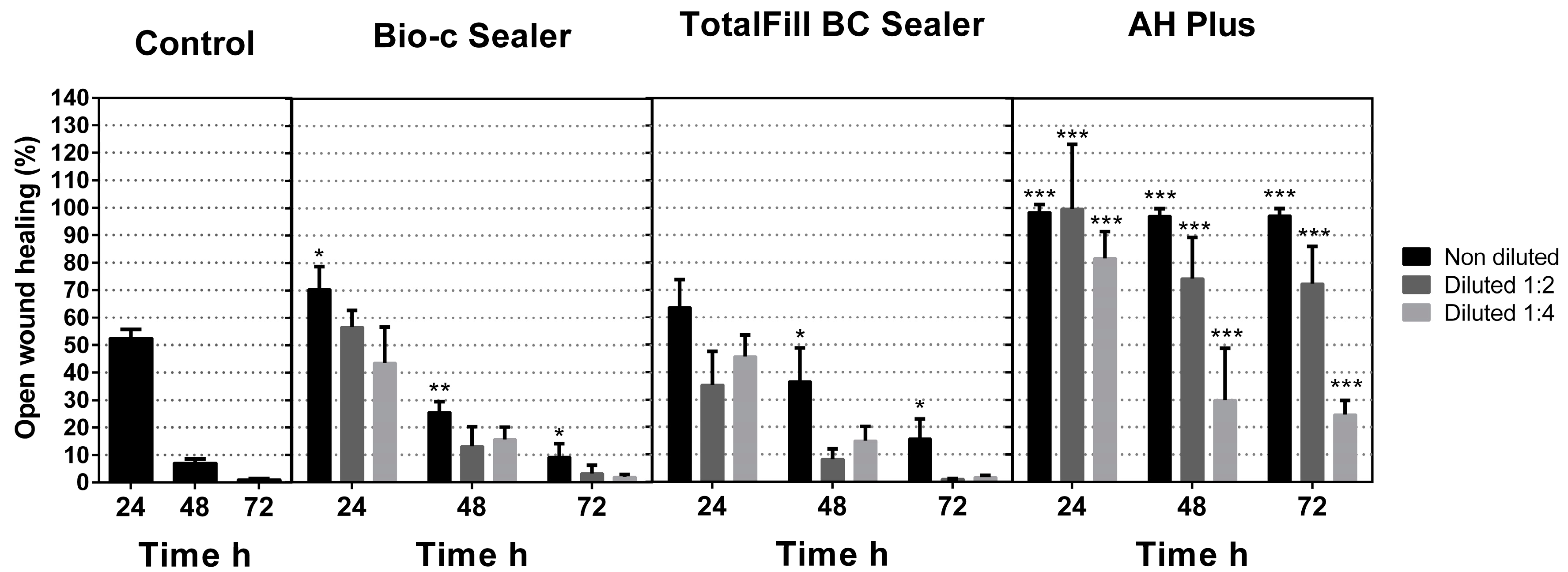
3.2. Cell Migration
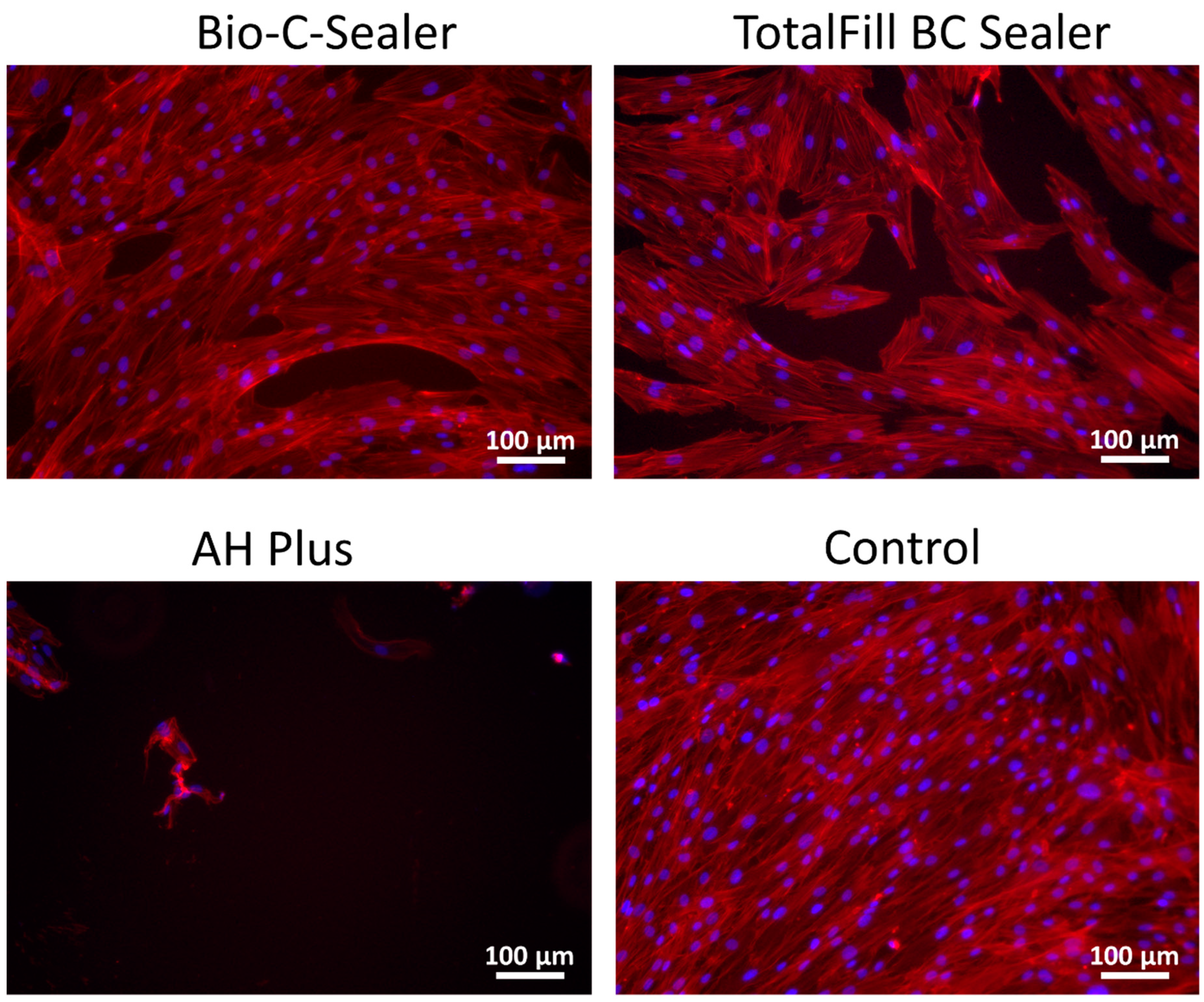
3.3. Cell Morphology
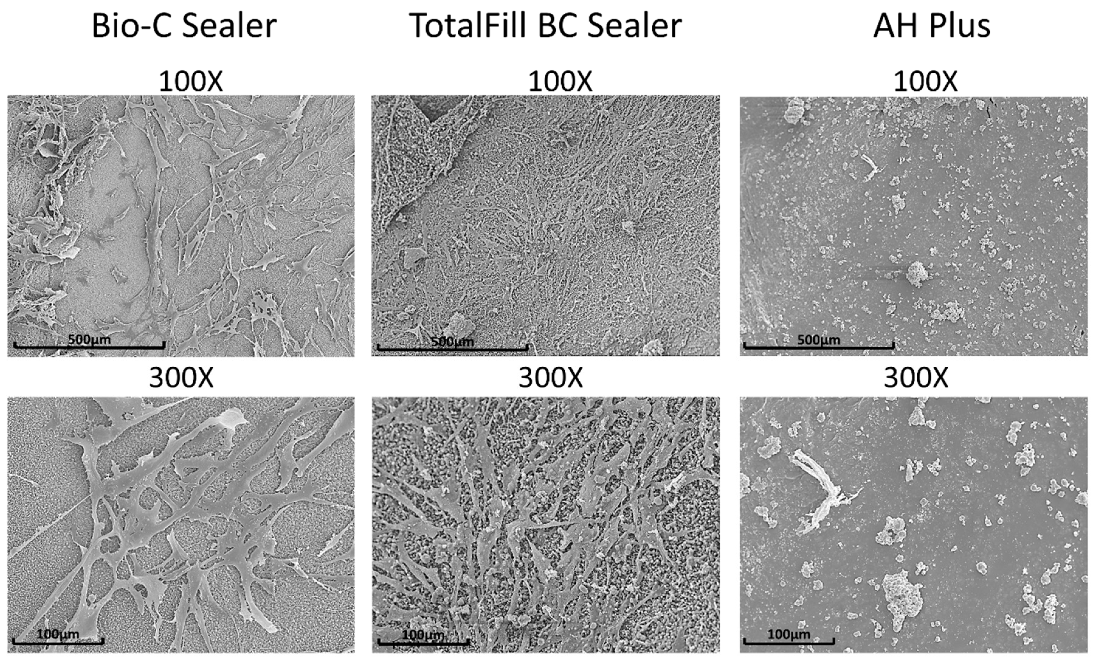
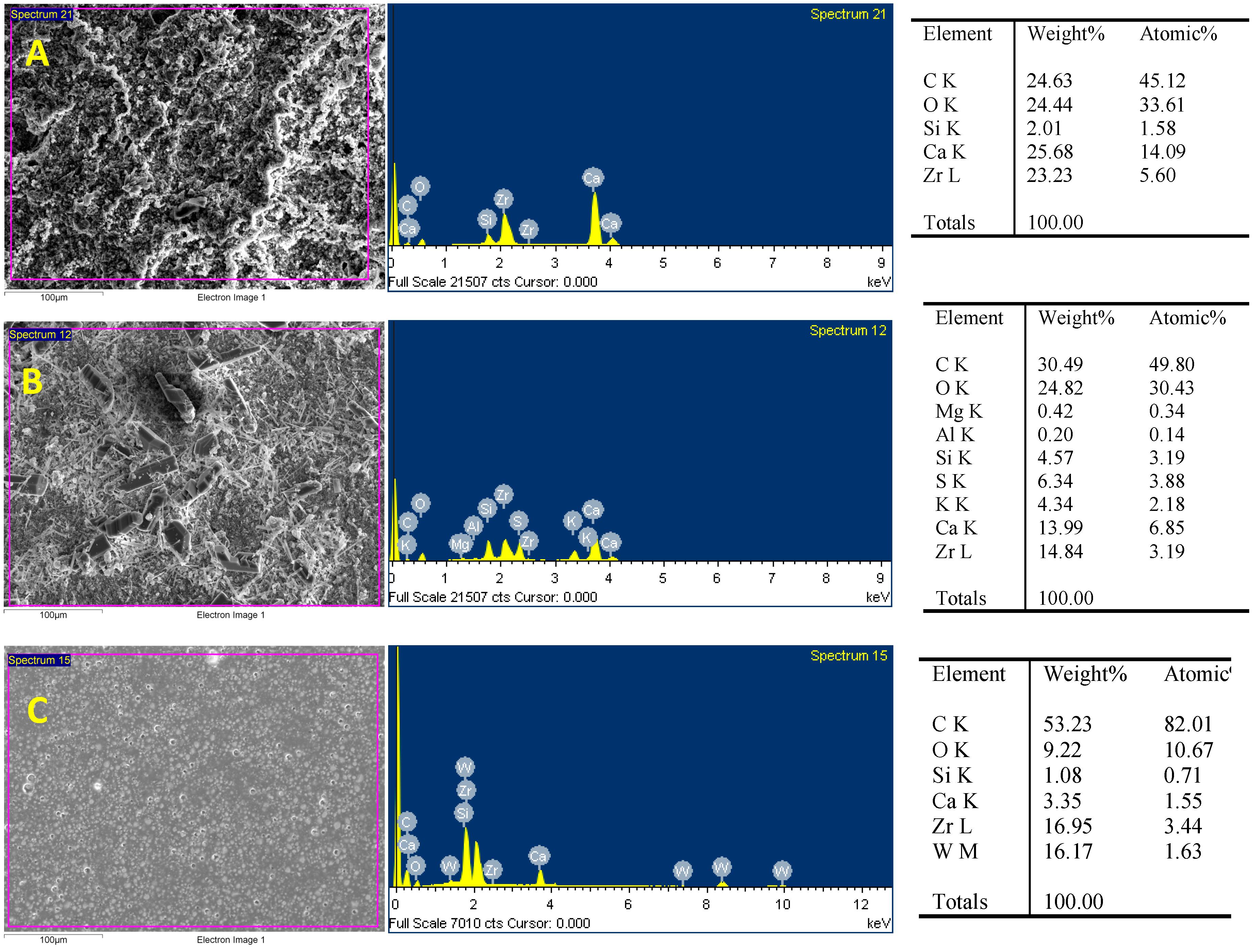
3.4. Cell Attachment on Materials and Characterization of Set Materials
3.5. Alizarin Red Assay
4. Discussion
5. Conclusions
Author Contributions
Funding
Conflicts of Interest
References
- Ferreira, C.M.A.; Sassone, L.M.; Goncalves, A.S.; de Carvalho, J.J.; Tomas-Catala, C.J.; Garcia-Bernal, D.; Onate-Sanchez, R.E.; Rodriguez-Lozano, F.J.; Silva, E. Physicochemical, cytotoxicity and in vivo biocompatibility of a high-plasticity calcium-silicate based material. Sci. Rep. 2019, 9, 3933. [Google Scholar] [CrossRef] [PubMed]
- Braga, J.M.; Oliveira, R.R.; de Castro Martins, R.; Vieira, L.Q.; Sobrinho, A.P. Assessment of the cytotoxicity of a mineral trioxide aggregate-based sealer with respect to macrophage activity. Dent Traumatol 2015, 31, 390–395. [Google Scholar] [CrossRef] [PubMed]
- Cintra, L.T.A.; Benetti, F.; de Azevedo Queiroz, I.O.; de Araujo Lopes, J.M.; Penha de Oliveira, S.H.; Sivieri Araujo, G.; Gomes-Filho, J.E. Cytotoxicity, Biocompatibility, and Biomineralization of the New High-plasticity MTA Material. J. Endod. 2017, 43, 774–778. [Google Scholar] [CrossRef] [PubMed] [Green Version]
- Benetti, F.; Queiroz, I.O.A.; Cosme-Silva, L.; Conti, L.C.; Oliveira, S.H.P.; Cintra, L.T.A. Cytotoxicity, Biocompatibility and Biomineralization of a New Ready-for-Use Bioceramic Repair Material. Braz. Dent. J. 2019, 30, 325–332. [Google Scholar] [CrossRef] [PubMed] [Green Version]
- Cosme-Silva, L.; Gomes-Filho, J.E.; Benetti, F.; Dal-Fabbro, R.; Sakai, V.T.; Cintra, L.T.A.; Ervolino, E.; Viola, N.V. Biocompatibility and immunohistochemical evaluation of a new calcium silicate-based cement, Bio-C Pulpo. Int. Endod. J. 2019, 52, 689–700. [Google Scholar] [CrossRef]
- Silva Almeida, L.H.; Moraes, R.R.; Morgental, R.D.; Pappen, F.G. Are premixed calcium silicate-based endodontic sealers comparable to conventional materials? A systematic review of in vitro studies. J. Endod. 2017, 43, 527–535. [Google Scholar] [CrossRef] [PubMed]
- Jimenez-Sanchez, M.D.C.; Segura-Egea, J.J.; Diaz-Cuenca, A. Higher hydration performance and bioactive response of the new endodontic bioactive cement MTA HP repair compared with ProRoot MTA white and NeoMTA plus. J. Biomed. Mater. Res. B Appl. Biomater. 2019, 107, 2109–2120. [Google Scholar] [CrossRef]
- Camps, J.; Jeanneau, C.; Laurent, P.; About, I. Bioactivity of a calcium silicate-based endodontic cement (bioroot rcs): interactions with human periodontal ligament cells in vitro. J. Endod. 2015, 41, 1469–1473. [Google Scholar] [CrossRef]
- Taraslia, V.; Anastasiadou, E.; Lignou, C.; Keratiotis, G.; Agrafioti, A.; Kontakiotis, E.G. Assessment of cell viability in four novel endodontic sealers. Eur. J. Dent. 2018, 12, 287–291. [Google Scholar] [CrossRef]
- Jeanneau, C.; Giraud, T.; Laurent, P.; About, I. Bioroot RCS extracts modulate the early mechanisms of periodontal inflammation and regeneration. J. Endod. 2019, 45, 1016–1023. [Google Scholar] [CrossRef]
- Lee, J.K.; Kim, S.; Lee, S.; Kim, H.C.; Kim, E. In vitro comparison of biocompatibility of calcium silicate-based root canal sealers. Materials 2019, 12, 2411. [Google Scholar] [CrossRef] [PubMed]
- Kebudi Benezra, M.; Schembri Wismayer, P.; Camilleri, J. Interfacial characteristics and cytocompatibility of hydraulic sealer cements. J. Endod. 2018, 44, 1007–1017. [Google Scholar] [CrossRef] [PubMed]
- Sultana, N.; Singh, M.; Nawal, R.R.; Chaudhry, S.; Yadav, S.; Mohanty, S.; Talwar, S. Evaluation of biocompatibility and osteogenic potential of tricalcium silicate-based cements using human bone marrow-derived mesenchymal stem cells. J. Endod. 2018, 44, 446–451. [Google Scholar] [CrossRef]
- Candeiro, G.T.M.; Moura-Netto, C.; D’Almeida-Couto, R.S.; Azambuja-Junior, N.; Marques, M.M.; Cai, S.; Gavini, G. Cytotoxicity, genotoxicity and antibacterial effectiveness of a bioceramic endodontic sealer. Int. Endod. J. 2016, 49, 858–864. [Google Scholar] [CrossRef] [PubMed]
- Rodriguez-Lozano, F.J.; Collado-Gonzalez, M.; Tomas-Catala, C.J.; Garcia-Bernal, D.; Lopez, S.; Onate-Sanchez, R.E.; Moraleda, J.M.; Murcia, L. GuttaFlow bioseal promotes spontaneous differentiation of human periodontal ligament stem cells into cementoblast-like cells. Dent. Mater. 2019, 35, 114–124. [Google Scholar] [CrossRef] [PubMed]
- Rodriguez-Lozano, F.J.; Collado-Gonzalez, M.; Lopez-Garcia, S.; Garcia-Bernal, D.; Moraleda, J.M.; Lozano, A.; Forner, L.; Murcia, L.; Onate-Sanchez, R.E. Evaluation of changes in ion release and biological properties of NeoMTA-Plus and Endocem-MTA exposed to an acidic environment. Int. Endod. J. 2019, 52, 1196–1209. [Google Scholar] [CrossRef] [PubMed]
- Collado-Gonzalez, M.; Lopez-Garcia, S.; Garcia-Bernal, D.; Onate-Sanchez, R.E.; Tomas-Catala, C.J.; Moraleda, J.M.; Lozano, A.; Forner, L.; Rodriguez-Lozano, F.J. Biological effects of acid-eroded MTA Repair HP and ProRoot MTA on human periodontal ligament stem cells. Clin. Oral Investig. 2019. [Google Scholar] [CrossRef]
- Collado-Gonzalez, M.; Tomas-Catala, C.J.; Onate-Sanchez, R.E.; Moraleda, J.M.; Rodriguez-Lozano, F.J. Cytotoxicity of guttaflow bioseal, guttaflow2, mta fillapex, and ah plus on human periodontal ligament stem cells. J. Endod. 2017, 43, 816–822. [Google Scholar] [CrossRef] [PubMed]
- Lopez-Garcia, S.; Myong-Hyun, B.; Lozano, A.; Garcia-Bernal, D.; Forner, L.; Llena, C.; Guerrero-Girones, J.; Murcia, L.; Rodriguez-Lozano, F.J. Cytocompatibility, bioactivity potential, and ion release of three premixed calcium silicate-based sealers. Clin. Oral Investig. 2019. [Google Scholar] [CrossRef]
- Schmalz, G.; Widbiller, M.; Galler, K.M. Material tissue interaction--from toxicity to tissue regeneration. Oper. Dent. 2016, 41, 117–131. [Google Scholar] [CrossRef]
- Zhou, H.M.; Du, T.F.; Shen, Y.; Wang, Z.J.; Zheng, Y.F.; Haapasalo, M. In vitro cytotoxicity of calcium silicate-containing endodontic sealers. J. Endod. 2015, 41, 56–61. [Google Scholar] [CrossRef] [PubMed]
- Konjhodzic-Prcic, A.; Jakupovic, S.; Hasic-Brankovic, L.; Vukovic, A. In vitro comparison of cytotoxicity of four root canal sealers on human gingival fibroblasts. Med. Arch. 2015, 69, 24–27. [Google Scholar] [CrossRef]
- Jung, S.; Libricht, V.; Sielker, S.; Hanisch, M.R.; Schäfer, E.; Dammaschke, T. Evaluation of the biocompatibility of root canal sealers on human periodontal ligament cells ex vivo. Odontology 2019, 107, 54–63. [Google Scholar] [CrossRef] [PubMed]
- Jung, S.; Sielker, S.; Hanisch, M.R.; Libricht, V.; Schäfer, E.; Dammaschke, T. Cytotoxic effects of four different root canal sealers on human osteoblasts. PLoS ONE 2018, 13, e0194467. [Google Scholar] [CrossRef] [PubMed]
- Rodriguez-Lozano, F.J.; Garcia-Bernal, D.; Onate-Sanchez, R.E.; Ortolani-Seltenerich, P.S.; Forner, L.; Moraleda, J.M. Evaluation of cytocompatibility of calcium silicate-based endodontic sealers and their effects on the biological responses of mesenchymal dental stem cells. Int. Endod. J. 2017, 50, 67–76. [Google Scholar] [CrossRef] [PubMed]
- Zordan-Bronzel, C.L.; Tanomaru-Filho, M.; Rodrigues, E.M.; Chavez-Andrade, G.M.; Faria, G.; Guerreiro-Tanomaru, J.M. Cytocompatibility, bioactive potential and antimicrobial activity of an experimental calcium silicate-based endodontic sealer. Int. Endod. J. 2019, 52, 979–986. [Google Scholar] [CrossRef] [PubMed]
- Wang, Y.; Zhou, Y.; Jin, L.; Pang, X.; Lu, Y.; Wang, Z.; Yu, Y.; Yu, J. Mineral trioxide aggregate enhances the osteogenic capacity of periodontal ligament stem cells via NF-κB and MAPK signaling pathways. J. Cell Physiol 2018, 233, 2386–2397. [Google Scholar] [CrossRef] [PubMed]
- Lee, B.N.; Hong, J.U.; Kim, S.M.; Jang, J.H.; Chang, H.S.; Hwang, Y.C.; Hwang, I.N.; Oh, W.M. Anti-inflammatory and osteogenic effects of calcium silicate-based root canal sealers. J. Endod. 2019, 45, 73–78. [Google Scholar] [CrossRef]
- Giacomino, C.M.; Wealleans, J.A.; Kuhn, N.; Diogenes, A. Comparative biocompatibility and osteogenic potential of two bioceramic sealers. J. Endod. 2019, 45, 51–56. [Google Scholar] [CrossRef]
- Cuppini, M.; Zatta, K.C.; Mestieri, L.B.; Grecca, F.S.; Leitune, V.C.B.; Guterres, S.S.; Collares, F.M. Antimicrobial and anti-inflammatory drug-delivery systems at endodontic reparative material: Synthesis and characterization. Dent. Mater. 2019, 35, 457–467. [Google Scholar] [CrossRef]
- Souza, G.L.; Rosatto, C.M.P.; Silva, M.J.B.; Silva, M.V.; Rocha Rodrigues, D.B.; Moura, C.C.G. Evaluation of apoptosis/necrosis and cytokine release provoked by three root canal sealers in human polymorphonuclears and monocytes. Int. Endod. J. 2019, 52, 629–638. [Google Scholar] [CrossRef] [PubMed]
- Kumari, T.V.; Usha, V.; Anil, K.; Bindu, M. Cell surface interactions in the study of biocompatibility. Trends Biomater. Artif Organs 2002, 15, 37–41. [Google Scholar]
- Accardo, C.; Himel, V.T.; Lallier, T.E. A novel GuttaFlow sealer supports cell survival and attachment. J. Endod. 2014, 40, 231–234. [Google Scholar] [CrossRef] [PubMed]
- Ahmed, H.M.A.; Luddin, N.; Kannan, T.P.; Mokhtar, K.I.; Ahmad, A. White mineral trioxide aggregate mixed with calcium chloride dihydrate: Chemical analysis and biological properties. Restor. Dent. Endod. 2017, 42, 176–187. [Google Scholar] [CrossRef] [PubMed]
- Moon, H.J.; Lee, J.H.; Kim, J.H.; Knowles, J.C.; Cho, Y.B.; Shin, D.H.; Lee, H.H.; Kim, H.W. Reformulated mineral trioxide aggregate components and the assessments for use as future dental regenerative cements. J. Tissue Eng. 2018, 9, 2041731418807396. [Google Scholar] [CrossRef]
- Reszka, P.; Nowicka, A.; Dura, W.; Marek, E.; Lipski, M. SEM and EDS study of TotalFill BC Sealer and GuttaFlow Bioseal root canal sealers. Dent. Med. Probl. 2019, 56, 167–172. [Google Scholar] [CrossRef] [Green Version]
- Viapiana, R.; Guerreiro-Tanomaru, J.M.; Hungaro-Duarte, M.A.; Tanomaru-Filho, M.; Camilleri, J. Chemical characterization and bioactivity of epoxy resin and Portland cement-based sealers with niobium and zirconium oxide radiopacifiers. Dent. Mater. 2014, 30, 1005–1020. [Google Scholar] [CrossRef]
- Candeiro, G.T.; Correia, F.C.; Duarte, M.A.; Ribeiro-Siqueira, D.C.; Gavini, G. Evaluation of radiopacity, pH, release of calcium ions, and flow of a bioceramic root canal sealer. J. Endod. 2012, 38, 842–845. [Google Scholar] [CrossRef]
- Seo, D.G.; Lee, D.; Kim, Y.M.; Song, D.; Kim, S.Y. Biocompatibility and mineralization activity of three calcium silicate-based root canal sealers compared to conventional resin-based sealer in human dental pulp stem cells. Materials 2019, 12, 2482. [Google Scholar] [CrossRef]


© 2019 by the authors. Licensee MDPI, Basel, Switzerland. This article is an open access article distributed under the terms and conditions of the Creative Commons Attribution (CC BY) license (http://creativecommons.org/licenses/by/4.0/).
Share and Cite
López-García, S.; Pecci-Lloret, M.R.; Guerrero-Gironés, J.; Pecci-Lloret, M.P.; Lozano, A.; Llena, C.; Rodríguez-Lozano, F.J.; Forner, L. Comparative Cytocompatibility and Mineralization Potential of Bio-C Sealer and TotalFill BC Sealer. Materials 2019, 12, 3087. https://0-doi-org.brum.beds.ac.uk/10.3390/ma12193087
López-García S, Pecci-Lloret MR, Guerrero-Gironés J, Pecci-Lloret MP, Lozano A, Llena C, Rodríguez-Lozano FJ, Forner L. Comparative Cytocompatibility and Mineralization Potential of Bio-C Sealer and TotalFill BC Sealer. Materials. 2019; 12(19):3087. https://0-doi-org.brum.beds.ac.uk/10.3390/ma12193087
Chicago/Turabian StyleLópez-García, Sergio, Miguel R. Pecci-Lloret, Julia Guerrero-Gironés, María P. Pecci-Lloret, Adrián Lozano, Carmen Llena, Francisco Javier Rodríguez-Lozano, and Leopoldo Forner. 2019. "Comparative Cytocompatibility and Mineralization Potential of Bio-C Sealer and TotalFill BC Sealer" Materials 12, no. 19: 3087. https://0-doi-org.brum.beds.ac.uk/10.3390/ma12193087