
Synthesis of Selected Mixed Oxide Materials with Tailored Photocatalytic Activity in the Degradation of Tetracycline

 , , , and
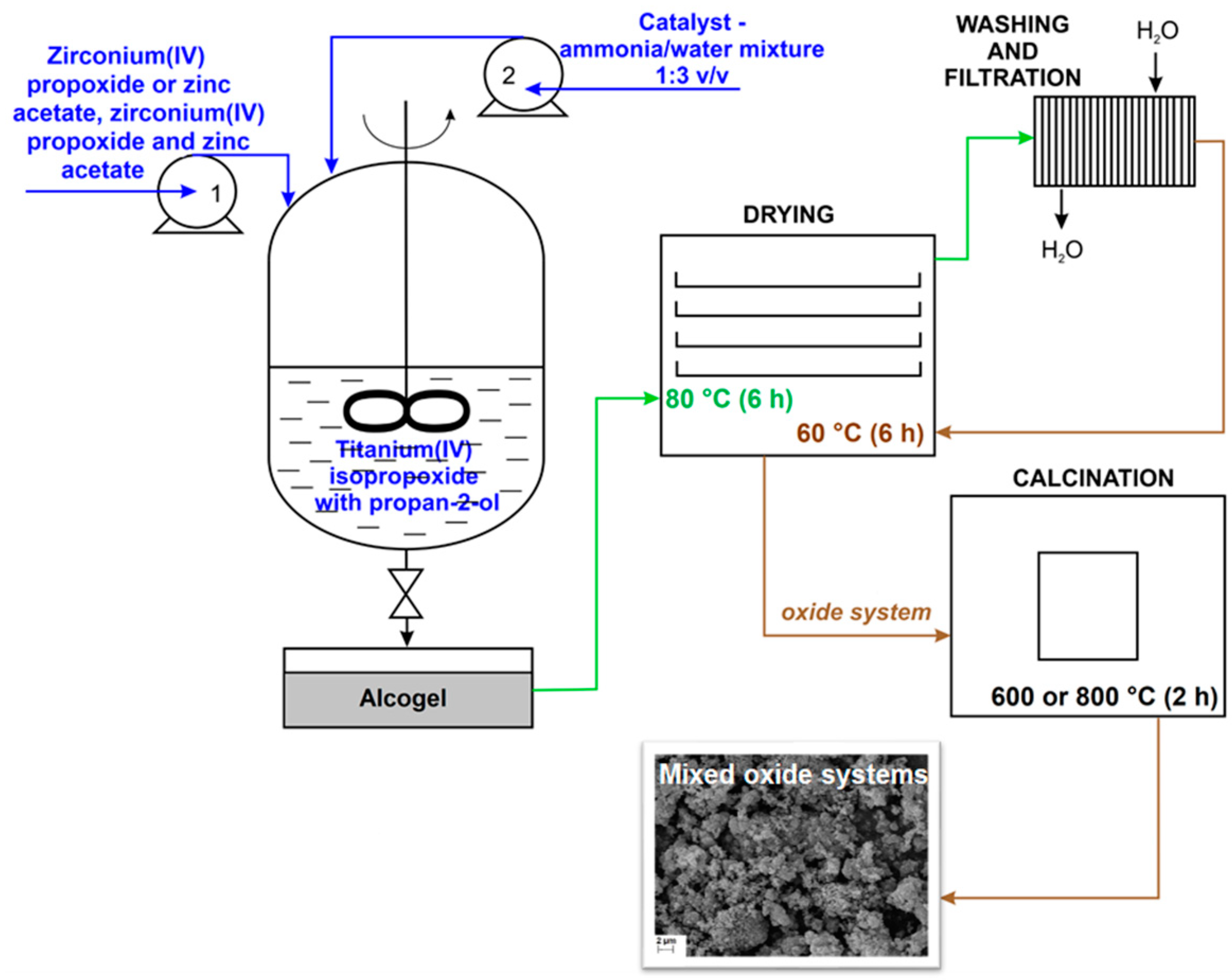
Abstract
:1. Introduction
2. Materials and Methods
2.1. Chemicals and Materials
2.2. Fabrication of Mixed Oxide Systems
2.3. Characterization of Synthesized Photocatalysts
2.4. Photo-Oxidation Activity of Synthesized Photocatalysts
2.5. Kinetic Study
2.6. LC–MS/MS Determination of Degradation Products of Tetracycline
3. Results and Discussion
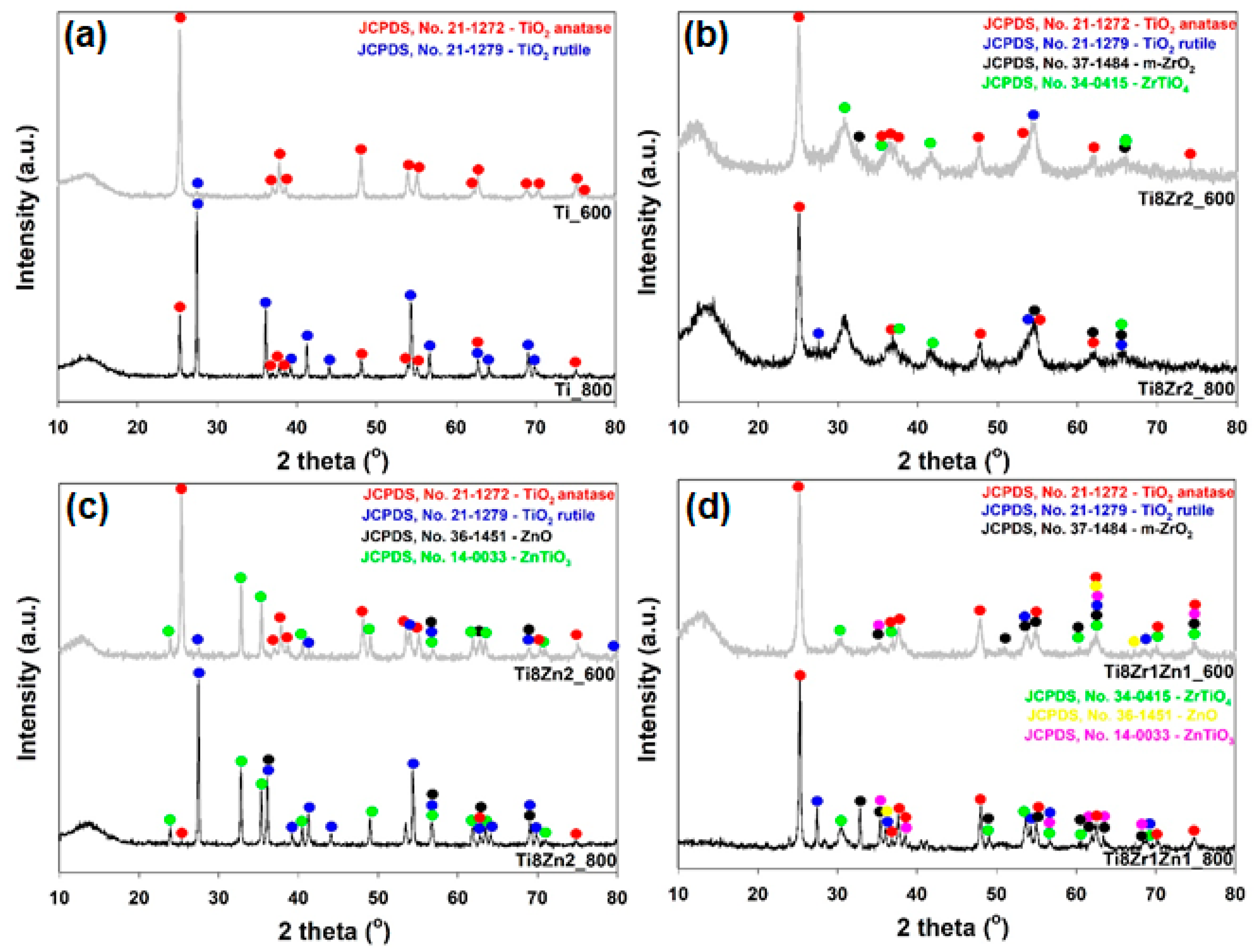
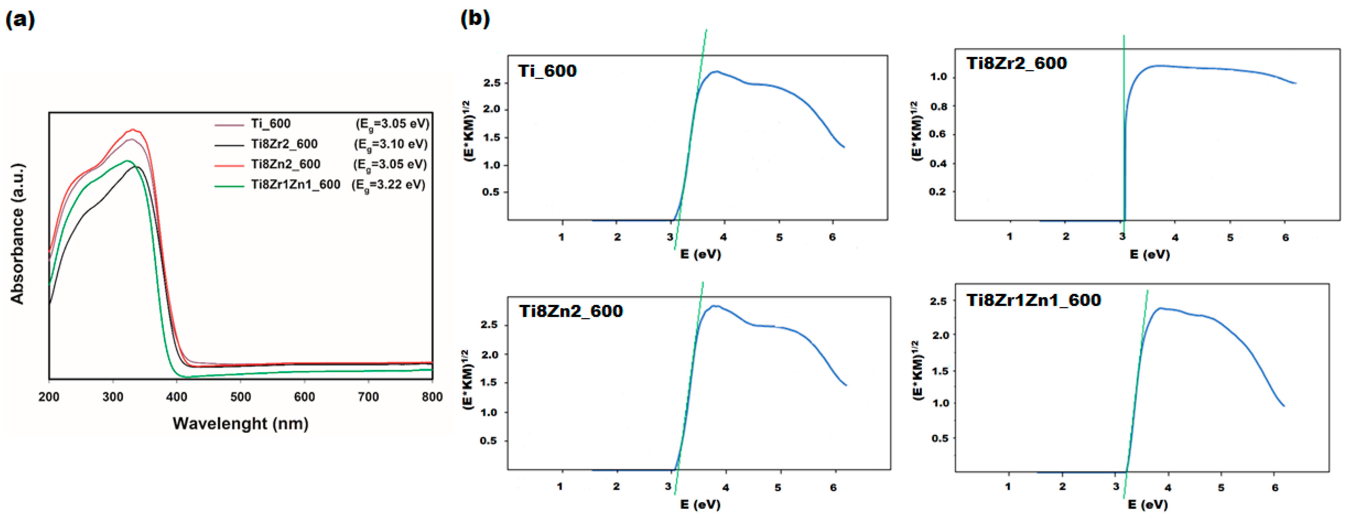
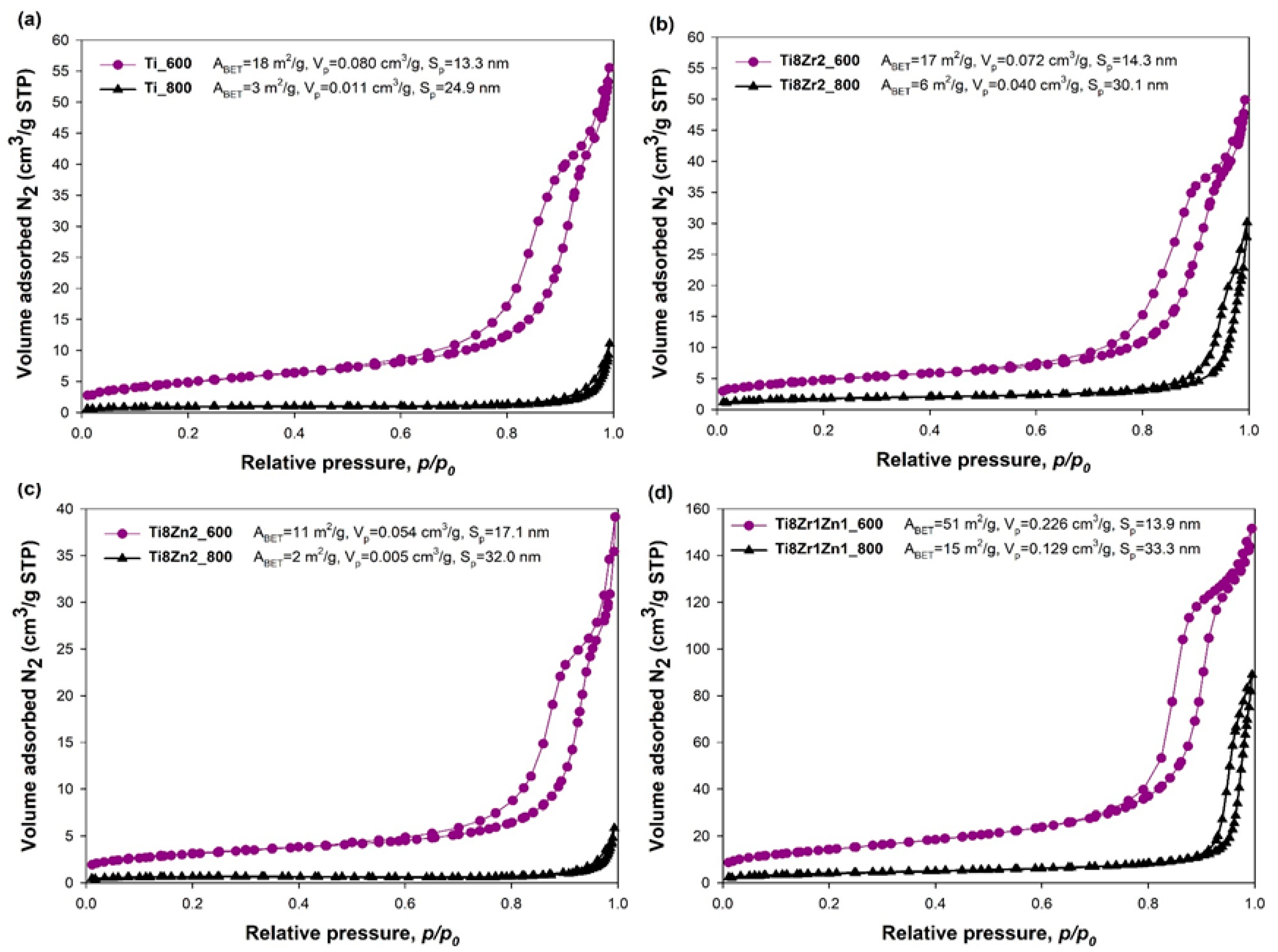
3.1. Physicochemical Characterization of Photocatalysts
3.2. Photocatalytic Degradation of Tetracycline
3.3. Identification of Photodegradation Products
4. Conclusions
Supplementary Materials
Author Contributions
Funding
Institutional Review Board Statement
Informed Consent Statement
Data Availability Statement
Acknowledgments
Conflicts of Interest
References
- Kümmerer, K. Antibiotics in the aquatic environment—A review—Part I. Chemosphere 2009, 75, 417–434. [Google Scholar] [CrossRef] [PubMed]
- Borghi, A.A.; Palma, M.S.A. Tetracycline: Production, waste treatment and environmental impact assessment. Braz. J. Pharm. Sci. 2014, 50, 25–40. [Google Scholar] [CrossRef]
- Watkinson, A.J.; Murby, E.J.; Costanzo, S.D. Removal of antibiotics in conventional and advanced wastewater treatment: Implications for environmental discharge and wastewater recycling. Water Res. 2007, 41, 4164–4176. [Google Scholar] [CrossRef]
- Rivera-Utrilla, J.; Gómez-Pacheco, C.V.; Sánchez-Polo, M.; López-Peńalver, J.J.; Ocampo-Pérez, R. Tetracycline removal from water by adsorption/bioadsorption on activated carbons and sludge-derived adsorbents. J. Environ. Manag. 2013, 131, 16–24. [Google Scholar] [CrossRef]
- Fernández-Calvińo, D.; Bermúdez-Couso, A.; Arias-Estévez, M.; Nóvoa-Muńoz, J.C.; Fernández-Sanjurjo, M.J.; Álvarez-Rodríguez, E.; Núńez-Delgado, A. Competitive adsorption/desorption of tetracycline, oxytetracycline and chlortetracycline on two acid soils: Stirred flow chamber experiments. Chemosphere 2015, 134, 361–366. [Google Scholar] [CrossRef] [PubMed]
- Pan, S.F.; Zhu, M.P.; Chen, J.P.; Yuan, Z.H.; Zhong, L.B.; Zheng, Y.M. Separation of tetracycline from wastewater using forward osmosis process with thin film composite membrane-implications for antibiotics recovery. Sep. Purif. Technol. 2015, 153, 76–83. [Google Scholar] [CrossRef]
- Ouaissa, Y.A.; Chabani, M.; Amrane, A.; Bensmaili, A. Removal of tetracycline by electrocoagulation: Kinetic and isotherm modeling through adsorption. J. Environ. Chem. Eng. 2014, 2, 177–184. [Google Scholar] [CrossRef] [Green Version]
- Miyata, M.; Ihara, I.; Yoshid, G.; Toyod, K.; Umetsu, K. Electrochemical oxidation of tetracycline antibiotics using a Ti/TiO2 anode for wastewater treatment of animal husbandry. Water Sci. Technol. 2011, 63, 456–461. [Google Scholar] [CrossRef]
- López-Peńalver, J.J.; Sánchez-Polo, M.; Gómez-Pacheco, C.V.; Rivera-Utrilla, J. Photodegradation of tetracyclines in aqueous solution by using UV and UV/H2O2 oxidation processes. J. Chem. Technol. Biotechnol. 2010, 85, 1325–1333. [Google Scholar] [CrossRef]
- Reyes, C.; Fernandez, J.; Freer, J.; Mondaca, M.A.; Zaror, C.; Malato, S.; Mansilla, H.D. Degradation and inactivation of tetracycline by TiO2 photocatalysis. J. Photochem. Photobiol. A Chem. 2006, 184, 141–146. [Google Scholar] [CrossRef]
- Palominos, R.A.; Mondaca, M.A.; Giraldo, A.; Penuela, G.; Pérez-Moya, M.; Mansilla, H.D. Photocatalytic oxidation of the antibiotic tetracycline on TiO2 and ZnO suspenssions. Catal. Today 2009, 144, 100–105. [Google Scholar] [CrossRef]
- Ibhadon, A.O.; Fitzpatrick, P. Heterogeneous photocatalysis: Recent advances and applications. Catalysts 2013, 3, 189–218. [Google Scholar] [CrossRef] [Green Version]
- Hoffmann, R.; Martin, T.; Choi, W.; Bahnemannt, W. Environmental applications of semiconductor photocatalysis. Chem. Rev. 1995, 95, 69–96. [Google Scholar] [CrossRef]
- Hashimoto, K.; Irle, H.; Fujishima, A. TiO2 photocatalysis: A historical overview and future prospects. Jpn. J. Appl. Phys. 2005, 44, 8269–8285. [Google Scholar] [CrossRef]
- Mahlambi, M.M.; Nglla, C.J.; Mamba, B.B. Recent developments in environmental photocatalytic degradation of organic pollutants: The case of titanium dioxide nanoparticeles—A review. J. Nanomater. 2015, 2015, 5. [Google Scholar] [CrossRef] [Green Version]
- Vlazan, P.; Ursu, D.H.; Irina-Moisescu, C.; Miron, I.; Sfirloaga, P.; Rusu, E. Structural and electrical properties of TiO2/ZnO core—Shell nanoparticles synthesized by hydrothermal method. Mater. Charact. 2015, 101, 153–158. [Google Scholar] [CrossRef]
- Wang, H.; Zhang, L.; Chen, Z.; Hu, J.; Li, S.; Wang, Z.; Liu, J.; Wang, X. Semiconductor heterojunction photocatalysts: Design, construction, and photocatalytic performances. Chem. Soc. Rev. 2014, 43, 5234–5244. [Google Scholar] [CrossRef]
- Low, J.; Yu, J.; Jaroniec, M.; Wageh, S.; Al-Ghamdi, A.A. Heterojunction photocatalysts. Adv. Mater. 2017, 29, 1601694. [Google Scholar] [CrossRef] [PubMed]
- Carević, M.V.; Abazović, N.D.; Savić, T.D.; Novaković, T.B.; Pjević, D.J.; Čomor, M.I. Binary oxide ceramics for enhanced phenols degradation under simulated Solar light. J. Am. Ceram. Soc. 2018, 101, 1420–1431. [Google Scholar] [CrossRef]
- Prasannalakshmi, P.; Shanmugam, N. Fabrication of TiO2/ZnO nanocomposites for solar energy driven photocatalysis. Mater. Sci. Semicond. Process. 2017, 61, 114–124. [Google Scholar] [CrossRef]
- Pasang, T.; Namratha, K.; Parvin, T.; Ranganathaiah, C.; Byrappa, K. Tuning of band gap in TiO2 and ZnO nanoparticles by selective doping for photocatalytic applications. Mater. Res. Innov. 2015, 19, 73–80. [Google Scholar] [CrossRef]
- Das, A.; Kumar, P.M.; Bhagavathiachari, M.; Nair, R.G. Hierarchical ZnO-TiO2 nanoheterojunction: A strategy driven approach to boost the photocatalytic performance through the synergy of improved surface area and interfacial charge transport. Appl. Surf. Sci. 2020, 534, 147321. [Google Scholar] [CrossRef]
- Siwińska-Stefańska, K.; Kubiak, A.; Piasecki, A.; Dobrowolska, A.; Czaczyk, K.; Motylenko, M.; Rafaja, D.; Ehrlich, H.; Jesionowski, T. Hydrothermal synthesis of multifunctional TiO2-ZnO oxide systems with desired antibacterial and photocatalytic properties. Appl. Surf. Sci. 2019, 463, 791–801. [Google Scholar] [CrossRef]
- García-Ramírez, E.; Mondragón-Chaparro, M.; Zelaya-Angel, O. Band gap coupling in photocatalytic activity in ZnO–TiO2 thin films. Appl. Phys. A 2012, 108, 291–297. [Google Scholar] [CrossRef]
- Pirzada, B.M.; Mir, N.A.; Qutub, N.; Mehraj, O.; Sabir, S.; Muneer, M. Synthesis, characterization and optimization of photocatalytic activity of TiO2/ZrO2 nanocomposite heterostructures. Mater. Sci. Eng. B 2015, 193, 137–145. [Google Scholar] [CrossRef]
- Li, M.; Li, X.; Jiang, G.; He, G. Hierarchically macro—mesoporous ZrO2–TiO2 composites with enhanced photocatalytic activity. Ceram. Int. 2015, 41, 5749–5757. [Google Scholar] [CrossRef]
- Wang, J.; Yu, Y.; Li, S.; Guo, L.; Wang, E.; Cao, Y. Doping behavior of Zr4+ ions in Zr4+-doped TiO2 nanoparticles. J. Phys. Chem. C 2013, 117, 27120–27126. [Google Scholar] [CrossRef]
- Luo, Q.; Cai, Q.-Z.; Li, X.-W.; Pan, Z.-H.; Li, Y.-J.; Chen, X.-D.; Yan, Q.-S. Preparation and characterization of ZrO2/TiO2 composite photocatalytic film by micro-arc oxidation. Trans. Nonferrous Met. Soc. China 2013, 23, 2945–2950. [Google Scholar] [CrossRef]
- Pantelides, S.T. The electronic structure of impurities and other point defects in semiconductors. Rev. Mod. Phys. 1978, 50, 797–858. [Google Scholar] [CrossRef]
- Siwińska-Ciesielczyk, K.; Świgoń, D.; Rychtowski, P.; Moszyński, D.; Zgoła-Grześkowiak, A.; Jesionowski, T. The performance of multicomponent oxide systems based on TiO2, ZrO2 and SiO2 in the photocatalytic degradation of Rhodamine B: Mechanism and kinetic studies. Colloids Surf. A Physicochem. Eng. Asp. 2020, 586, 124272. [Google Scholar] [CrossRef]
- Górska, P.; Zaleska, A.; Kowalska, E.; Klimczuk, T.; Sobczak, J.W.; Skwarek, E.; Janusz, W.; Hupka, J. TiO2 photoactivity in vis and UV light: The influence of calcination temperature and surface properties. Appl. Catal. B Environ. 2008, 84, 440–447. [Google Scholar] [CrossRef]
- Sharmila Juliet, S.; Ramalingom, S.; Ravidhas, C.; Moses Ezhil Raj, A. Effect of calcination temperature on titanium oxide nanocrystallites in the anatase phase synthesized by sol-gel route. IOSR J. Appl. Phys. 2017, 9, 32–39. [Google Scholar] [CrossRef]
- Ramamoorthy, S.; Das, S.; Balan, R.; Lekshmi, I.C. TiO2-ZrO2 nanocomposite with tetragonal zirconia phase and photocatalytic degradation of Alizarin Yellow GG azo dye under natural sunlight. Mater. Today Proc. 2021. [Google Scholar] [CrossRef]
- Lukáč, J.; Klementová, M.; Bezdička, P.; Bakardjieva, S.; Šubrt, J.; Szatmáry, L.; Bastl, Z.; Jirkovský, J. Influence of Zr as TiO2 doping ion on photocatalytic degradation of 4-chlorophenol. Appl. Catal. B Environ. 2007, 74, 83–91. [Google Scholar] [CrossRef]
- Kavan, L.; Attia, A.; Lenzmann, F.; Elder, S.H.; Grätzel, M. Lithium insertion into zirconia-stabilized mesoscopic TiO2 (anatase). J. Electrochem. Soc. 2000, 147, 2897–2902. [Google Scholar] [CrossRef]
- Yang, J.; Ferreira, J.M.F. On the titania phase transition by zirconia additive in a sol-gel-derived powder. Mater. Res. Bull. 1998, 33, 389–394. [Google Scholar] [CrossRef]
- Hanaor, D.A.H.; Sorrell, C.C. Review of the anatase to rutile phase transformation. J. Mater. Sci. 2011, 46, 855–874. [Google Scholar] [CrossRef] [Green Version]
- Pei, C.C.; Leung, W.W.-F. Photocatalytic degradation of Rhodamine B by TiO2/ZnO nanofibers under visible-light irradiation. Sep. Purif. Technol. 2013, 114, 108–116. [Google Scholar] [CrossRef]
- Arin, J.; Thongtem, S.; Thongtem, T. Single-step synthesis of ZnO/TiO2 nanocomposites by microwave radiation and their photocatalytic activities. Mater. Lett. 2013, 96, 78–81. [Google Scholar] [CrossRef]
- Hussein, A.M.; Iefanova, A.V.; Koodali, R.T.; Logue, B.A.; Shende, R.V. Interconnected ZrO2 doped ZnO/TiO2 network photoanode for dye-sensitized solar cells. Energy Rep. 2018, 4, 56–64. [Google Scholar] [CrossRef]
- Okada, K.; Yamamoto, N.; Kameshima, Y.; Yasumori, A.; MacKenzie, K.J. Effect of silica additive on the anatase-to-rutile phase transition. J. Am. Ceram. Soc. 2001, 84, 1591–1596. [Google Scholar] [CrossRef]
- Chithambararaj, A.; Bose, A.C. Investigation on structural, thermal, optical and sensing properties of meta-stable hexagonal MoO3 nanocrystals of one dimensional structure. Beilstein J. Nanotechnol. 2011, 2, 585–592. [Google Scholar] [CrossRef] [PubMed] [Green Version]
- Vione, D.; Minero, C.; Maurino, V.; Carlotti, M.E.; Picatonotto, T.; Pelizzetti, E. Degradation of phenol and benzoic acid in the presence of a TiO2-based heterogeneous photocatalyst. Appl. Catal. B Environ. 2005, 58, 79–88. [Google Scholar] [CrossRef]
- Sing, K.S.W.; Everett, D.H.; Haul, R.A.W.; Moscou, L.; Pierott, R.A.; Rouquérol, J.; Siemieniewska, T. Reporting physisorption data for gas/solid systems. Pure Appl. Chem. 1985, 57, 603–619. [Google Scholar] [CrossRef]
- Tian, G.; Pan, K.; Fu, H.; Jing, L.; Zhou, W. Enhanced photocatalytic activity of S-doped TiO2-ZrO2 nanoparticles under visible-light irradiation. J. Hazard. Mater. 2009, 166, 939–944. [Google Scholar] [CrossRef]
- Ilkhechi, N.N.; Kaleji, B.K. High temperature stability and photocatalytic activity of nanocrystalline anatase powders with Zr and Si co-dopants. J. Sol-Gel Sci. Technol. 2014, 69, 351–356. [Google Scholar] [CrossRef]
- Konyar, M.; Yatmaz, H.C.; Öztürk, K. Sintering temperature effect on photocatalyticeffciencie of ZnO/TiO2 composite platers. Appl. Surf. Sci. 2012, 258, 7440–7447. [Google Scholar] [CrossRef]
- Zhang, M.; An, T.; Liu, X.; Hu, X.; Sheng, G.; Fu, J. Preparation of a high-activity ZnO/TiO2 photocatalyst via homogeneous hydrolysis method with low temperature crystallization. Mater. Lett. 2010, 64, 1883–1886. [Google Scholar] [CrossRef]
- Wang, Z.; Zhang, B.; Li, F. A Simple and cheap method for preparation of coupled ZrO2-ZnO with high photocatalytic activities. Front. Environ. Sci. Eng. China 2007, 1, 454–458. [Google Scholar] [CrossRef]
- Han, S.; Zhao, D.; Lund, H.; Rockstroh, N.; Bartling, S.; Doronkin, D.E.; Grunwaldt, J.-D.; Gao, M.; Jiang, G.; Kondratenko, E.V. TiO2-supported catalysts with ZnO and ZrO2 for non-oxidative dehydrogenation of propane: Mechanistic analysis and application potential. Catal. Sci. Technol. 2020, 10, 7046–7055. [Google Scholar] [CrossRef]
- Aghabeygi, S.; Khademi-Shamami, M. ZnO/ZrO2 nanocomposite: Sonosynthesis, characterization and its application for wastewater treatment. Ultrason. Sonochem. 2018, 41, 458–465. [Google Scholar] [CrossRef] [PubMed]
- Simon, Q.; Barreca, D.; Gasparotto, A. CuO/ZnO nanocomposites investigated by X-ray Photoelectron and X-ray Excited Auger Electron Spectroscopies. Surf. Sci. Spectra 2010, 17, 93–101. [Google Scholar] [CrossRef]
- Dolat, D.; Moszyński, D.; Guskos, N.; Ohtani, B.; Morawski, A.W. Preparation of photoactive nitrogen-doped rutile. Appl. Surf. Sci. 2013, 266, 410–419. [Google Scholar] [CrossRef]
- Moradi, S.; Azar, P.A.; Farshid, S.R.; Khorrami, S.A.; Givianrad, M.H. Effect of additives on characterization and photocatalytic activity of TiO2/ZnO nanocomposite prepared via sol-gel process. Int. J. Chem. Eng. 2012, 2012, 215373. [Google Scholar] [CrossRef] [Green Version]
- Vaizoğullar, A.I.; Balci, A.; Uğurlu, M.; Karaoğlu, M.H. Synthesis of TiO2 and ZrO2/TiO2 composite microspheres and their photo-catalytic degradation of methylene blue. AKU J. Sci. Eng. 2016, 16, 54–60. [Google Scholar] [CrossRef] [Green Version]
- Wang, Y.; Zhang, D.; Shi, L.; Li, L.; Zhang, J. Novel transparent ternary nanocomposite films of trialkoxysilane-capped poly(methyl methacrylate)/zirconia/titania with incorporating networks. Mater. Chem. Phys. 2008, 110, 463–470. [Google Scholar] [CrossRef]
- Nurhasanah, I.; Kadarisman Gunawan, V.; Sutanto, H. Cerium oxide nanoparticles application for rapid adsorptive removal of tetracycline in water. J. Environ. Chem. Eng. 2020, 8, 103613. [Google Scholar] [CrossRef]
- Zhu, X.-D.; Wang, Y.-J.; Sun, R.-J.; Zhou, D.-M. Photocatalytic degradation of tetracycline in aqueous solution by nanosized TiO2. Chemosphere 2013, 92, 925–932. [Google Scholar] [CrossRef]
- Figueroa, R.A.; Leonard, A.; MacKay, A.A. Modeling tetracycline antibiotic sorption to clays. Environ. Sci. Technol. 2004, 38, 476–483. [Google Scholar] [CrossRef]
- Chen, W.-R.; Huang, C.-H. Adsorption and transformation of tetracycline antibiotics with aluminum oxide. Chemosphere 2010, 79, 779–785. [Google Scholar] [CrossRef]
- Gu, C.; Karthikeyan, K.G. Interaction of tetracycline with aluminum and iron hydrous oxides. Environ. Sci. Technol. 2005, 39, 2660–2667. [Google Scholar] [CrossRef] [PubMed]
- Li, S.; Hu, J. Photolytic and photocatalytic degradation of tetracycline: Effect of humic acid on degradation kinetics and mechanisms. J. Hazard. Mater. 2016, 318, 134–144. [Google Scholar] [CrossRef] [PubMed]
- Cao, J.; Lai, L.; Lai, B.; Yao, G.; Chen, X.; Song, L. Degradation of tetracycline by peroxymonosulfate activated with zero-valent iron: Performance, intermediates, toxicity and mechanism. Chem. Eng. J. 2019, 364, 45–56. [Google Scholar] [CrossRef]


Publisher’s Note: MDPI stays neutral with regard to jurisdictional claims in published maps and institutional affiliations. |
© 2021 by the authors. Licensee MDPI, Basel, Switzerland. This article is an open access article distributed under the terms and conditions of the Creative Commons Attribution (CC BY) license (https://creativecommons.org/licenses/by/4.0/).
Share and Cite
Siwińska-Ciesielczyk, K.; Andrzejczak, A.; Paukszta, D.; Piasecki, A.; Moszyński, D.; Zgoła-Grześkowiak, A.; Jesionowski, T. Synthesis of Selected Mixed Oxide Materials with Tailored Photocatalytic Activity in the Degradation of Tetracycline. Materials 2021, 14, 5361. https://0-doi-org.brum.beds.ac.uk/10.3390/ma14185361
Siwińska-Ciesielczyk K, Andrzejczak A, Paukszta D, Piasecki A, Moszyński D, Zgoła-Grześkowiak A, Jesionowski T. Synthesis of Selected Mixed Oxide Materials with Tailored Photocatalytic Activity in the Degradation of Tetracycline. Materials. 2021; 14(18):5361. https://0-doi-org.brum.beds.ac.uk/10.3390/ma14185361
Chicago/Turabian StyleSiwińska-Ciesielczyk, Katarzyna, Angelika Andrzejczak, Dominik Paukszta, Adam Piasecki, Dariusz Moszyński, Agnieszka Zgoła-Grześkowiak, and Teofil Jesionowski. 2021. "Synthesis of Selected Mixed Oxide Materials with Tailored Photocatalytic Activity in the Degradation of Tetracycline" Materials 14, no. 18: 5361. https://0-doi-org.brum.beds.ac.uk/10.3390/ma14185361