
Multifunctional Iron Oxide Magnetic Nanoparticles for Biomedical Applications: A Review

 ,
,  , , ,
, , , 
Abstract
:1. Introduction
2. Integration of Organic Dyes and Magnetic Iron Oxide Nanoparticles
3. Integration of Biomolecules and Magnetic Iron Oxide Nanoparticles
4. Integration of Quantum Dots and Magnetic Iron Oxide Nanoparticles
5. Integration of Noble Metal Nanoparticles and Magnetic Iron Oxide Nanoparticles
6. Integration of Stimuli-Responsive Polymers and Magnetic Iron Oxide Nanoparticles
6.1. Integration of Thermo-Responsive Polymers
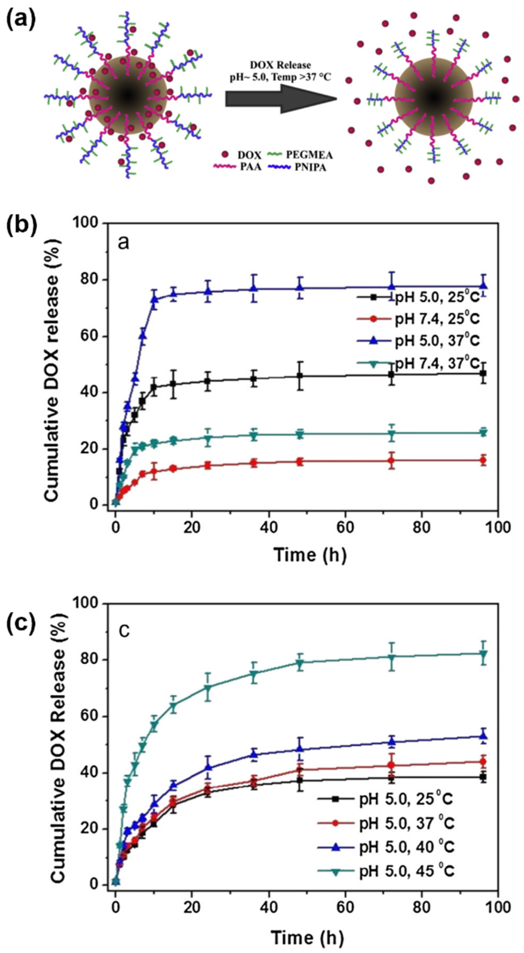
6.2. Integration of pH-Responsive Polymers
7. Integration of Multiple Conjugates with Magnetic Iron Oxide Nanoparticles
8. Summary and Perspectives
Author Contributions
Funding
Institutional Review Board Statement
Informed Consent Statement
Data Availability Statement
Conflicts of Interest
References
- Medhi, R.; Srinoi, P.; Ngo, N.; Tran, H.-V.; Lee, T.R. Nanoparticle-Based Strategies to Combat COVID-19. ACS Appl. Nano Mater. 2020, 3, 8557–8580. [Google Scholar] [CrossRef]
- Farka, Z.; Juřík, T.; Kovář, D.; Trnková, L.; Skládal, P. Nanoparticle-Based Immunochemical Biosensors and Assays: Recent Advances and Challenges. Chem. Rev. 2017, 117, 9973–10042. [Google Scholar] [CrossRef]
- Han, X.; Xu, K.; Taratula, O.; Farsad, K. Applications of Nanoparticles in Biomedical Imaging. Nanoscale 2019, 11, 799–819. [Google Scholar] [CrossRef]
- Canaparo, R.; Foglietta, F.; Giuntini, F.; Della Pepa, C.; Dosio, F.; Serpe, L. Recent Developments in Antibacterial Therapy: Focus on Stimuli-Responsive Drug-Delivery Systems and Therapeutic Nanoparticles. Molecules 2019, 24, 1991. [Google Scholar] [CrossRef] [PubMed] [Green Version]
- Nguyen, M.D.; Tran, H.-V.; Xu, S.; Lee, T.R. Fe3O4 Nanoparticles: Structures, Synthesis, Magnetic Properties, Surface Functionalization, and Emerging Applications. Appl. Sci. 2021, 11, 11301. [Google Scholar] [CrossRef]
- Jeon, M.; Halbert, M.V.; Stephen, Z.R.; Zhang, M. Iron Oxide Nanoparticles as T1 Contrast Agents for Magnetic Resonance Imaging: Fundamentals, Challenges, Applications, and Prospectives. Adv. Mater. 2021, 33, 1906539. [Google Scholar] [CrossRef] [PubMed]
- Alphandéry, E. Light-Interacting Iron-Based Nanomaterials for Localized Cancer Detection and Treatment. Acta Biomater. 2021, 124, 50–71. [Google Scholar] [CrossRef] [PubMed]
- Liu, S.; Yu, B.; Wang, S.; Shen, Y.; Cong, H. Preparation, Surface Functionalization and Application of Fe3O4 Magnetic Nanoparticles. Adv. Colloid Interface Sci. 2020, 281, 102165. [Google Scholar] [CrossRef] [PubMed]
- Lee, N.; Yoo, D.; Ling, D.; Cho, M.H.; Hyeon, T.; Cheon, J. Iron Oxide Based Nanoparticles for Multimodal Imaging and Magnetoresponsive Therapy. Chem. Rev. 2015, 115, 10637–10689. [Google Scholar] [CrossRef] [PubMed]
- Siregar, J.; Septiani, N.L.W.; Abrori, S.A.; Sebayang, K.; Irzaman; Fahmi, M.Z.; Humaidi, S.; Sembiring, T.; Sembiring, K.; Yuliarto, B. Review—A Pollutant Gas Sensor Based on Fe3O4 Nanostructures: A Review. J. Electrochem. Soc. 2021, 168, 027510. [Google Scholar] [CrossRef]
- Wang, F.; Tan, W.B.; Zhang, Y.; Fan, X.; Wang, M. Luminescent Nanomaterials for Biological Labelling. Nanotechnology 2006, 17, R1–R13. [Google Scholar] [CrossRef]
- Cheng, W.; Chen, H.; Liu, C.; Ji, C.; Ma, G.; Yin, M. Functional Organic Dyes for Health-Related Applications. VIEW 2020, 1, 20200055. [Google Scholar] [CrossRef]
- Jenkins, R.; Burdette, M.K.; Foulger, S.H. Mini-Review: Fluorescence Imaging in Cancer Cells Using Dye-Doped Nanoparticles. RSC Adv. 2016, 6, 65459–65474. [Google Scholar] [CrossRef]
- Frangioni, J.V. In Vivo Near-Infrared Fluorescence Imaging. Curr. Opin. Chem. Biol. 2003, 7, 626–634. [Google Scholar] [CrossRef] [PubMed]
- Zhang, L.; Tong, S.; Zhang, Q.; Bao, G. Lipid-Encapsulated Fe3O4 Nanoparticles for Multimodal Magnetic Resonance/Fluorescence Imaging. ACS Appl. Nano Mater. 2020, 3, 6785–6797. [Google Scholar] [CrossRef]
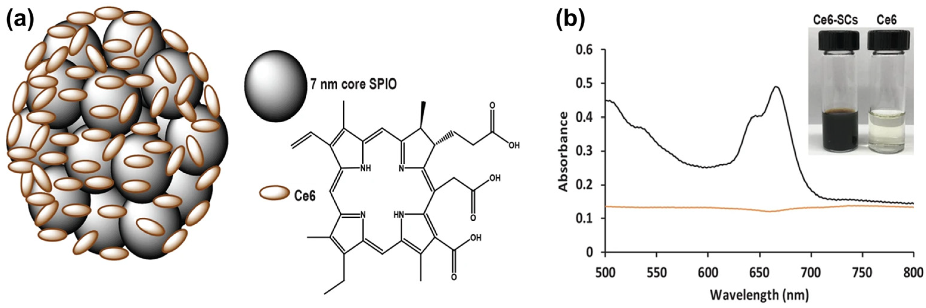
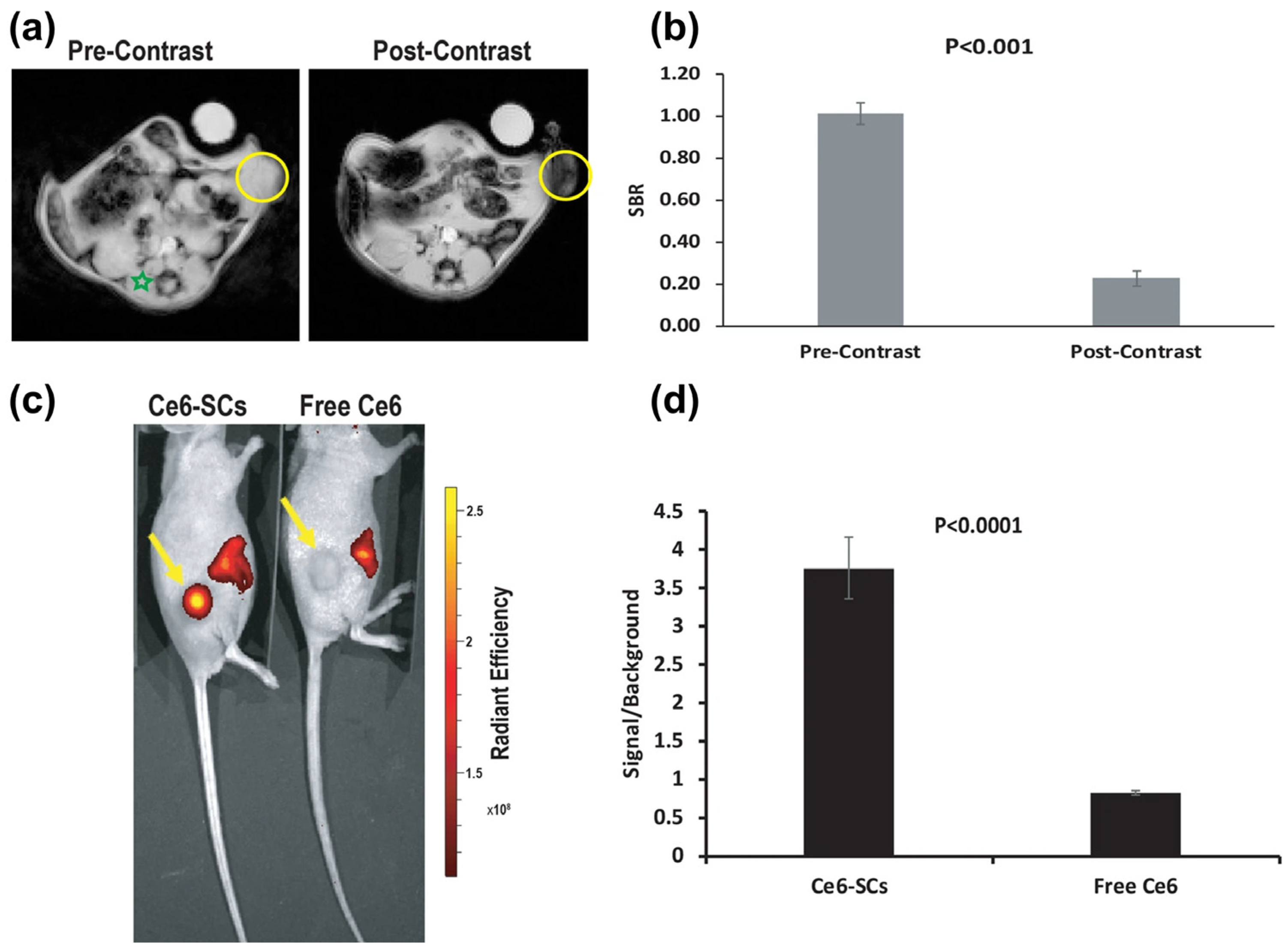
- Amirshaghaghi, A.; Yan, L.; Miller, J.; Daniel, Y.; Stein, J.M.; Busch, T.M.; Cheng, Z.; Tsourkas, A. Chlorin E6-Coated Superparamagnetic Iron Oxide Nanoparticle (SPION) Nanoclusters as a Theranostic Agent for Dual-Mode Imaging and Photodynamic Therapy. Sci. Rep. 2019, 9, 2613. [Google Scholar] [CrossRef] [Green Version]
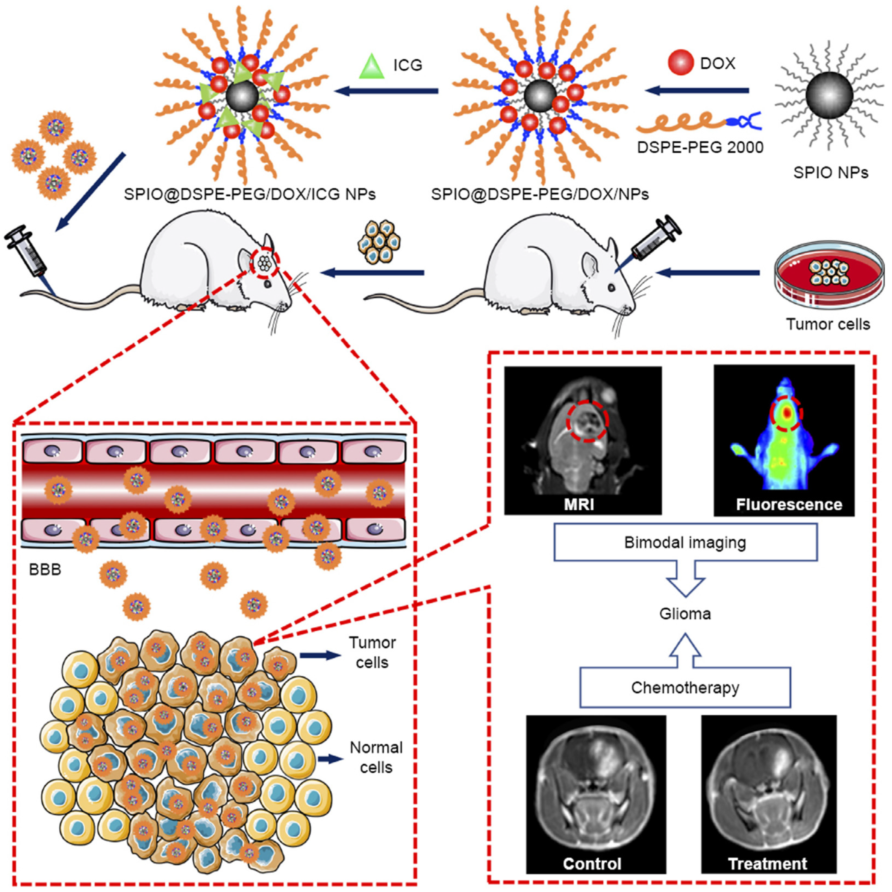
- Shen, C.; Wang, X.; Zheng, Z.; Gao, C.; Chen, X.; Zhao, S.; Dai, Z. Doxorubicin and Indocyanine Green Loaded Superparamagnetic Iron Oxide Nanoparticles with PEGylated Phospholipid Coating for Magnetic Resonance with Fluorescence Imaging and Chemotherapy of Glioma. Int. J. Nanomed. 2019, 14, 101–117. [Google Scholar] [CrossRef] [Green Version]
- Lu, Y.; He, B.; Shen, J.; Li, J.; Yang, W.; Yin, M. Multifunctional Magnetic and Fluorescent Core–Shell Nanoparticles for Bioimaging. Nanoscale 2015, 7, 1606–1609. [Google Scholar] [CrossRef]
- Yan, K.; Li, H.; Li, P.; Zhu, H.; Shen, J.; Yi, C.; Wu, S.; Yeung, K.W.K.; Xu, Z.; Xu, H.; et al. Self-Assembled Magnetic Fluorescent Polymeric Micelles for Magnetic Resonance and Optical Imaging. Biomaterials 2014, 35, 344–355. [Google Scholar] [CrossRef]
- Wate, P.S.; Banerjee, S.S.; Jalota-Badhwar, A.; Mascarenhas, R.R.; Zope, K.R.; Khandare, J.; Misra, R.D.K. Cellular Imaging Using Biocompatible Dendrimer-Functionalized Graphene Oxide-Based Fluorescent Probe Anchored with Magnetic Nanoparticles. Nanotechnology 2012, 23, 415101. [Google Scholar] [CrossRef]
- Lu, X.; Jiang, R.; Fan, Q.; Zhang, L.; Zhang, H.; Yang, M.; Ma, Y.; Wang, L.; Huang, W. Fluorescent-Magnetic Poly(Poly(Ethyleneglycol)Monomethacrylate)-Grafted Fe3O4 Nanoparticles from Post-Atom-Transfer-Radical-Polymerization Modification: Synthesis, Characterization, Cellular Uptake and Imaging. J. Mater. Chem. 2012, 22, 6965–6973. [Google Scholar] [CrossRef]
- Huang, P.; Li, Z.; Lin, J.; Yang, D.; Gao, G.; Xu, C.; Bao, L.; Zhang, C.; Wang, K.; Song, H.; et al. Photosensitizer-Conjugated Magnetic Nanoparticles for in Vivo Simultaneous Magnetofluorescent Imaging and Targeting Therapy. Biomaterials 2011, 32, 3447–3458. [Google Scholar] [CrossRef]
- Lee, J.E.; Lee, N.; Kim, H.; Kim, J.; Choi, S.H.; Kim, J.H.; Kim, T.; Song, I.C.; Park, S.P.; Moon, W.K.; et al. Uniform Mesoporous Dye-Doped Silica Nanoparticles Decorated with Multiple Magnetite Nanocrystals for Simultaneous Enhanced Magnetic Resonance Imaging, Fluorescence Imaging, and Drug Delivery. J. Am. Chem. Soc. 2010, 132, 552–557. [Google Scholar] [CrossRef] [PubMed]
- Kim, J.; Kim, H.S.; Lee, N.; Kim, T.; Kim, H.; Yu, T.; Song, I.C.; Moon, W.K.; Hyeon, T. Multifunctional Uniform Nanoparticles Composed of a Magnetite Nanocrystal Core and a Mesoporous Silica Shell for Magnetic Resonance and Fluorescence Imaging and for Drug Delivery. Angew. Chem. Int. Ed. 2008, 47, 8438–8441. [Google Scholar] [CrossRef] [PubMed]
- Gallagher, J.J.; Tekoriute, R.; O’Reilly, J.-A.; Kerskens, C.; Gun’ko, Y.K.; Lynch, M. Bimodal Magnetic-Fluorescent Nanostructures for Biomedical Applications. J. Mater. Chem. 2009, 19, 4081–4084. [Google Scholar] [CrossRef] [Green Version]
- Ge, Y.; Zhang, Y.; He, S.; Nie, F.; Teng, G.; Gu, N. Fluorescence Modified Chitosan-Coated Magnetic Nanoparticles for High-Efficient Cellular Imaging. Nanoscale Res. Lett. 2009, 4, 287–295. [Google Scholar] [CrossRef] [Green Version]
- Lee, H.; Yu, M.K.; Park, S.; Moon, S.; Min, J.J.; Jeong, Y.Y.; Kang, H.-W.; Jon, S. Thermally Cross-Linked Superparamagnetic Iron Oxide Nanoparticles: Synthesis and Application as a Dual Imaging Probe for Cancer in Vivo. J. Am. Chem. Soc. 2007, 129, 12739–12745. [Google Scholar] [CrossRef]
- Lu, C.-W.; Hung, Y.; Hsiao, J.-K.; Yao, M.; Chung, T.-H.; Lin, Y.-S.; Wu, S.-H.; Hsu, S.-C.; Liu, H.-M.; Mou, C.-Y.; et al. Bifunctional Magnetic Silica Nanoparticles for Highly Efficient Human Stem Cell Labeling. Nano Lett. 2007, 7, 149–154. [Google Scholar] [CrossRef]
- Bertorelle, F.; Wilhelm, C.; Roger, J.; Gazeau, F.; Ménager, C.; Cabuil, V. Fluorescence-Modified Superparamagnetic Nanoparticles: Intracellular Uptake and Use in Cellular Imaging. Langmuir 2006, 22, 5385–5391. [Google Scholar] [CrossRef]
- Kircher, M.F.; Mahmood, U.; King, R.S.; Weissleder, R.; Josephson, L. A Multimodal Nanoparticle for Preoperative Magnetic Resonance Imaging and Intraoperative Optical Brain Tumor Delineation. Cancer Res. 2003, 63, 8122–8125. [Google Scholar]
- Skaat, H.; Margel, S. Synthesis of Fluorescent-Maghemite Nanoparticles as Multimodal Imaging Agents for Amyloid-β Fibrils Detection and Removal by a Magnetic Field. Biochem. Biophys. Res. Commun. 2009, 386, 645–649. [Google Scholar] [CrossRef]
- Lucky, S.S.; Soo, K.C.; Zhang, Y. Nanoparticles in Photodynamic Therapy. Chem. Rev. 2015, 115, 1990–2042. [Google Scholar] [CrossRef]
- Wang, R.; Li, X.; Yoon, J. Organelle-Targeted Photosensitizers for Precision Photodynamic Therapy. ACS Appl. Mater. Interfaces 2021, 13, 19543–19571. [Google Scholar] [CrossRef] [PubMed]
- Fayazi, R.; Habibi-Rezaei, M.; Heiat, M.; Javadi-Zarnaghi, F.; Taheri, R.A. Glycated Albumin Precipitation Using Aptamer Conjugated Magnetic Nanoparticles. Sci. Rep. 2020, 10, 10716. [Google Scholar] [CrossRef] [PubMed]
- Blumenfeld, C.M.; Schulz, M.D.; Aboian, M.S.; Wilson, M.W.; Moore, T.; Hetts, S.W.; Grubbs, R.H. Drug Capture Materials Based on Genomic DNA-Functionalized Magnetic Nanoparticles. Nat. Commun. 2018, 9, 2870. [Google Scholar] [CrossRef] [PubMed]
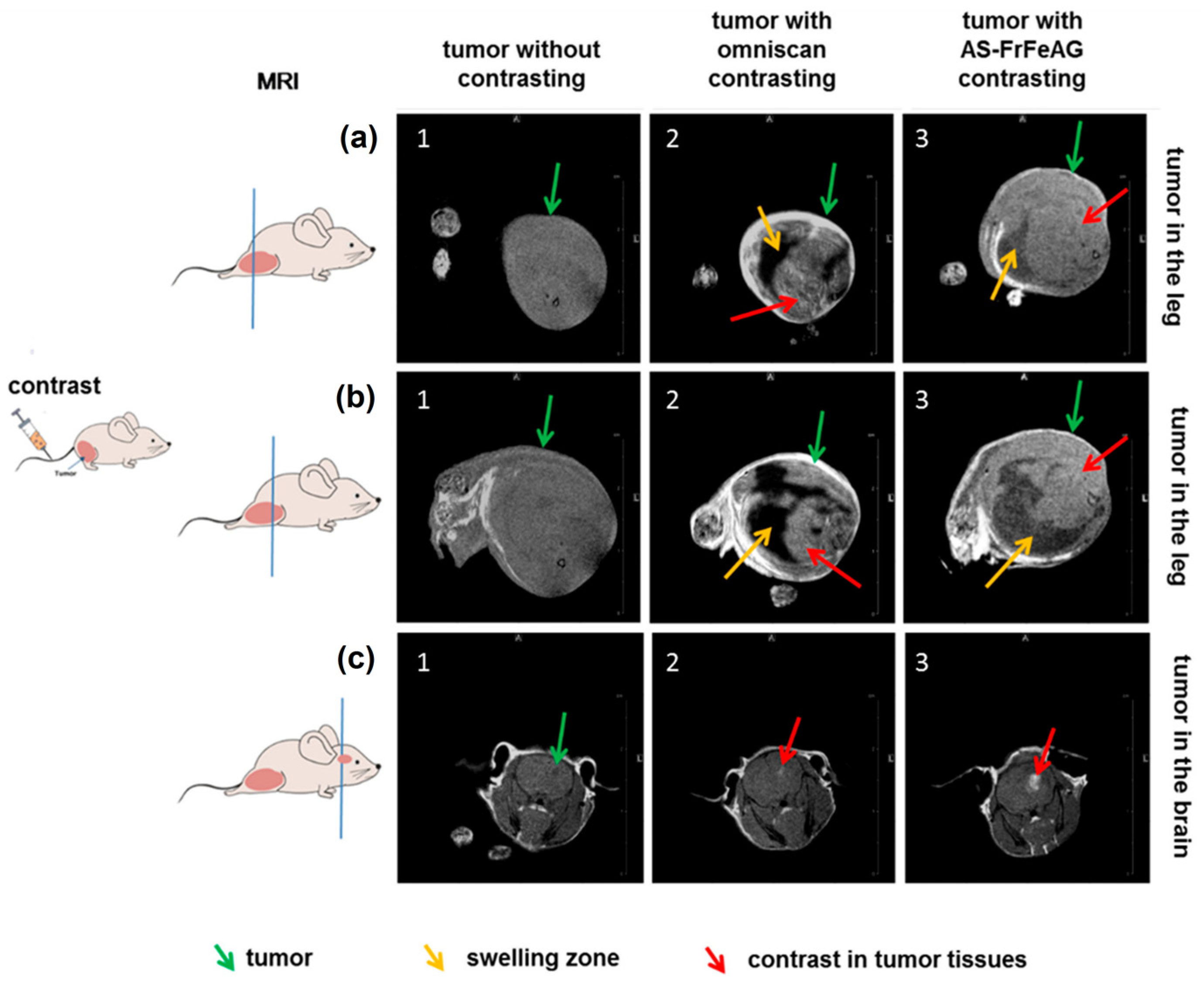
- Kolovskaya, O.S.; Zamay, T.N.; Zamay, G.S.; Babkin, V.A.; Medvedeva, E.N.; Neverova, N.A.; Kirichenko, A.K.; Zamay, S.S.; Lapin, I.N.; Morozov, E.V.; et al. Aptamer-Conjugated Superparamagnetic Ferroarabinogalactan Nanoparticles for Targeted Magnetodynamic Therapy of Cancer. Cancers 2020, 12, 216. [Google Scholar] [CrossRef] [PubMed] [Green Version]
- Bakshi, S.; Zakharchenko, A.; Minko, S.; Kolpashchikov, D.M.; Katz, E. Towards Nanomaterials for Cancer Theranostics: A System of DNA-Modified Magnetic Nanoparticles for Detection and Suppression of RNA Marker in Cancer Cells. Magnetochemistry 2019, 5, 24. [Google Scholar] [CrossRef] [Green Version]
- Su, Y.; Xue, T.; Wu, L.; Hu, Y.; Wang, J.; Xu, Q.; Chen, Y.; Lin, Z. Label-Free Detection of Biomarker Alpha Fetoprotein in Serum by SsDNA Aptamer Functionalized Magnetic Nanoparticles. Nanotechnology 2019, 31, 095104. [Google Scholar] [CrossRef]
- Wen, C.-Y.; Bi, J.-H.; Wu, L.-L.; Zeng, J.-B. Aptamer-Functionalized Magnetic and Fluorescent Nanospheres for One-Step Sensitive Detection of Thrombin. Microchim. Acta 2017, 185, 77. [Google Scholar] [CrossRef]
- Sanli, S.; Ghorbani-Zamani, F.; Moulahoum, H.; Gumus, Z.P.; Coskunol, H.; Odaci Demirkol, D.; Timur, S. Application of Biofunctionalized Magnetic Nanoparticles Based-Sensing in Abused Drugs Diagnostics. Anal. Chem. 2020, 92, 1033–1040. [Google Scholar] [CrossRef]
- Oltolina, F.; Colangelo, D.; Miletto, I.; Clemente, N.; Miola, M.; Verné, E.; Prat, M.; Follenzi, A. Tumor Targeting by Monoclonal Antibody Functionalized Magnetic Nanoparticles. Nanomaterials 2019, 9, 1575. [Google Scholar] [CrossRef] [Green Version]
- Fernández, T.; Martínez-Serrano, A.; Cussó, L.; Desco, M.; Ramos-Gómez, M. Functionalization and Characterization of Magnetic Nanoparticles for the Detection of Ferritin Accumulation in Alzheimer’s Disease. ACS Chem. Neurosci. 2018, 9, 912–924. [Google Scholar] [CrossRef]
- Wong, C.-H.; Chen, C.-P.; Chang, C.-C.; Chen, C.-Y. Bio-Functionalized Magnetic Nanoparticles for the Immunoassay of Fetal Fibronectin: A Feasibility Study for the Prediction of Preterm Birth. Sci. Rep. 2017, 7, 42461. [Google Scholar] [CrossRef] [Green Version]
- Zhu, X.; Lu, N.; Zhou, Y.; Xuan, S.; Zhang, J.; Giampieri, F.; Zhang, Y.; Yang, F.; Yu, R.; Battino, M.; et al. Targeting Pancreatic Cancer Cells with Peptide-Functionalized Polymeric Magnetic Nanoparticles. Int. J. Mol. Sci. 2019, 20, 2988. [Google Scholar] [CrossRef] [Green Version]
- Ilyas, S.; Ullah, N.K.; Ilyas, M.; Wennhold, K.; Iqbal, M.; Schlößer, H.A.; Hussain, M.S.; Mathur, S. Mediating the Fate of Cancer Cell Uptake: Dual-Targeted Magnetic Nanovectors with Biotin and Folate Surface Ligands. ACS Biomater. Sci. Eng. 2020, 6, 6138–6147. [Google Scholar] [CrossRef]
- Clauson, R.M.; Chen, M.; Scheetz, L.M.; Berg, B.; Chertok, B. Size-Controlled Iron Oxide Nanoplatforms with Lipidoid-Stabilized Shells for Efficient Magnetic Resonance Imaging-Trackable Lymph Node Targeting and High-Capacity Biomolecule Display. ACS Appl. Mater. Interfaces 2018, 10, 20281–20295. [Google Scholar] [CrossRef] [PubMed]
- Kuo, F.-Y.; Lin, W.-L.; Chen, Y.-C. Affinity Capture Using Peptide-Functionalized Magnetic Nanoparticles to Target Staphylococcus Aureus. Nanoscale 2016, 8, 9217–9225. [Google Scholar] [CrossRef] [PubMed]
- Huang, J.; Chen, X.; Fu, X.; Li, Z.; Huang, Y.; Liang, C. Advances in Aptamer-Based Biomarker Discovery. Front. Cell Dev. Biol. 2021, 9, 571. [Google Scholar] [CrossRef] [PubMed]
- Pearce, A.; Haas, M.; Viney, R.; Pearson, S.-A.; Haywood, P.; Brown, C.; Ward, R. Incidence and Severity of Self-Reported Chemotherapy Side Effects in Routine Care: A Prospective Cohort Study. PLoS ONE 2017, 12, 0184360. [Google Scholar] [CrossRef]
- Farahavar, G.; Abolmaali, S.S.; Gholijani, N.; Nejatollahi, F. Antibody-Guided Nanomedicines as Novel Breakthrough Therapeutic, Diagnostic and Theranostic Tools. Biomater. Sci. 2019, 7, 4000–4016. [Google Scholar] [CrossRef]
- Michalet, X.; Pinaud, F.F.; Bentolila, L.A.; Tsay, J.M.; Doose, S.; Li, J.J.; Sundaresan, G.; Wu, A.M.; Gambhir, S.S.; Weiss, S. Quantum Dots for Live Cells, in Vivo Imaging, and Diagnostics. Science 2005, 307, 538–544. [Google Scholar] [CrossRef] [Green Version]
- Hild, W.A.; Breunig, M.; Goepferich, A. Quantum Dots—Nano-Sized Probes for the Exploration of Cellular and Intracellular Targeting. Eur. J. Pharm. Biopharm. 2008, 68, 153–168. [Google Scholar] [CrossRef]
- Klostranec, J.M.; Chan, W.C.W. Quantum Dots in Biological and Biomedical Research: Recent Progress and Present Challenges. Adv. Mater. 2006, 18, 1953–1964. [Google Scholar] [CrossRef]
- Medintz, I.L.; Uyeda, H.T.; Goldman, E.R.; Mattoussi, H. Quantum Dot Bioconjugates for Imaging, Labelling and Sensing. Nat. Mater. 2005, 4, 435–446. [Google Scholar] [CrossRef] [PubMed]
- Maeda, Y.; Yoshino, T.; Matsunaga, T. Novel Nanocomposites Consisting of in Vivo-Biotinylated Bacterial Magnetic Particles and Quantum Dots for Magnetic Separation and Fluorescent Labeling of Cancer Cells. J. Mater. Chem. 2009, 19, 6361–6366. [Google Scholar] [CrossRef]
- Ang, C.Y.; Giam, L.; Chan, Z.M.; Lin, A.W.H.; Gu, H.; Devlin, E.; Papaefthymiou, G.C.; Selvan, S.T.; Ying, J.Y. Facile Synthesis of Fe2O3 Nanocrystals without Fe(CO)5 Precursor and One-Pot Synthesis of Highly Fluorescent Fe2O3–CdSe Nanocomposites. Adv. Mater. 2009, 21, 869–873. [Google Scholar] [CrossRef]
- Fernández, B.; Gálvez, N.; Cuesta, R.; Hungría, A.B.; Calvino, J.J.; Domínguez-Vera, J.M. Quantum Dots Decorated with Magnetic Bionanoparticles. Adv. Funct. Mater. 2008, 18, 3931–3935. [Google Scholar] [CrossRef]
- Gao, J.; Zhang, W.; Huang, P.; Zhang, B.; Zhang, X.; Xu, B. Intracellular Spatial Control of Fluorescent Magnetic Nanoparticles. J. Am. Chem. Soc. 2008, 130, 3710–3711. [Google Scholar] [CrossRef]
- Xie, M.; Hu, J.; Long, Y.-M.; Zhang, Z.-L.; Xie, H.-Y.; Pang, D.-W. Lectin-Modified Trifunctional Nanobiosensors for Mapping Cell Surface Glycoconjugates. Biosens. Bioelectron. 2009, 24, 1311–1317. [Google Scholar] [CrossRef]
- Wang, H.; Shen, J.; Li, Y.; Wei, Z.; Cao, G.; Gai, Z.; Hong, K.; Banerjee, P.; Zhou, S. Magnetic Iron Oxide–Fluorescent Carbon Dots Integrated Nanoparticles for Dual-Modal Imaging, Near-Infrared Light-Responsive Drug Carrier and Photothermal Therapy. Biomater. Sci. 2014, 2, 915–923. [Google Scholar] [CrossRef]
- Ou, J.; Wang, F.; Huang, Y.; Li, D.; Jiang, Y.; Qin, Q.-H.; Stachurski, Z.H.; Tricoli, A.; Zhang, T. Fabrication and Cyto-Compatibility of Fe3O4/SiO2/Graphene–CdTe QDs/CS Nanocomposites for Drug Delivery. Colloids Surf. B 2014, 117, 466–472. [Google Scholar] [CrossRef]
- Kyeong, S.; Jeong, C.; Kim, H.Y.; Hwang, D.W.; Kang, H.; Yang, J.-K.; Lee, D.S.; Jun, B.-H.; Lee, Y.-S. Fabrication of Mono-Dispersed Silica-Coated Quantum Dot-Assembled Magnetic Nanoparticles. RSC Adv. 2015, 5, 32072–32077. [Google Scholar] [CrossRef]
- Lee, J.; Kim, H.; Sim, T.; Song, R. A New Quantum Dot–Platinum Conjugate for Self-Assembled Nanoconjugates by Coordination Bonding Mediated Recognition. Chem. Commun. 2013, 49, 6182–6184. [Google Scholar] [CrossRef]
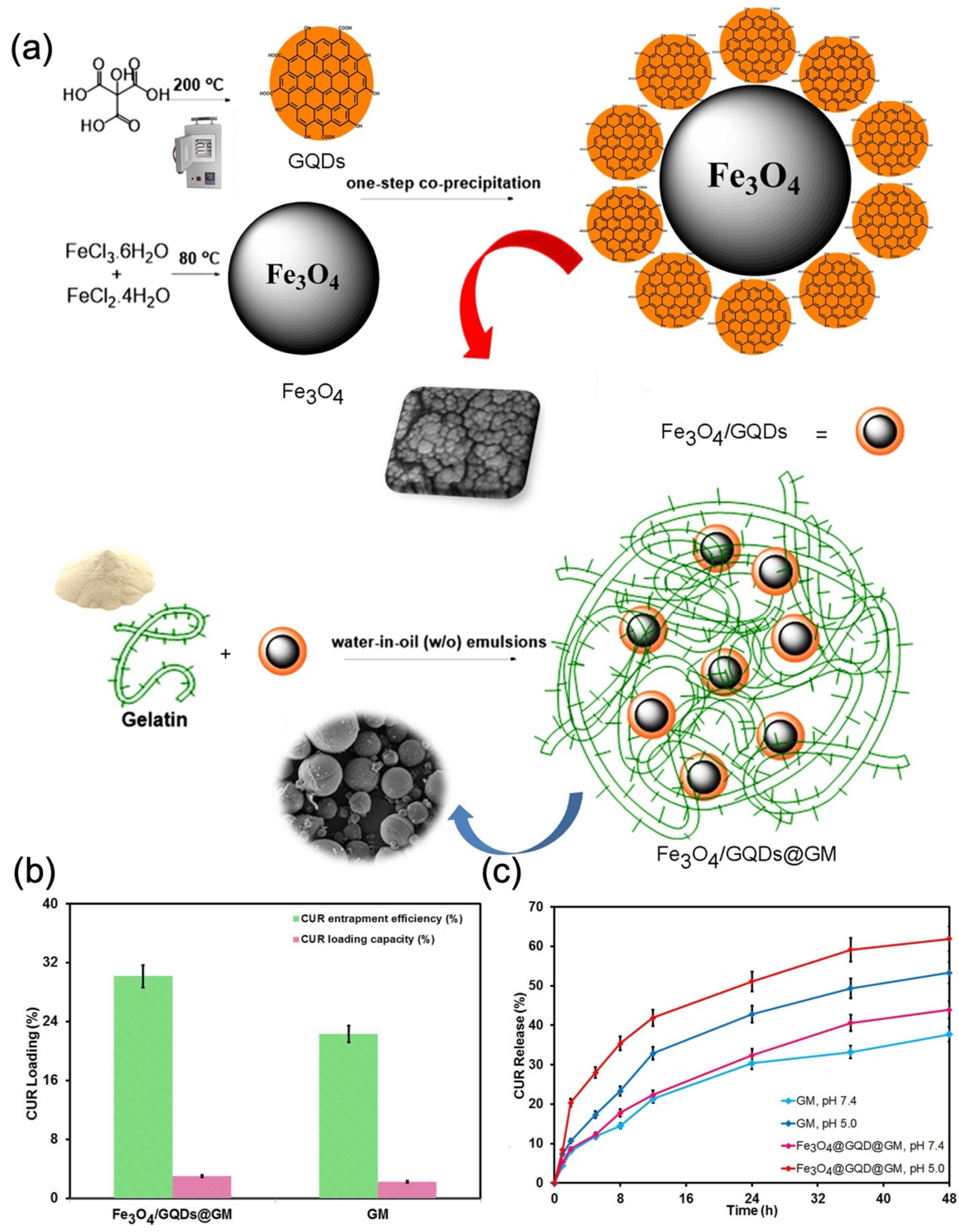
- Pooresmaeil, M.; Namazi, H. PH-Sensitive Ternary Fe3O4/GQDs@G Hybrid Microspheres; Synthesis, Characterization and Drug Delivery Application. J. Alloys Compd. 2020, 846, 156419. [Google Scholar] [CrossRef]
- Derfus, A.M.; Chan, W.C.W.; Bhatia, S.N. Probing the Cytotoxicity of Semiconductor Quantum Dots. Nano Lett. 2004, 4, 11–18. [Google Scholar] [CrossRef] [Green Version]
- Kirchner, C.; Liedl, T.; Kudera, S.; Pellegrino, T.; Muñoz Javier, A.; Gaub, H.E.; Stölzle, S.; Fertig, N.; Parak, W.J. Cytotoxicity of Colloidal CdSe and CdSe/ZnS Nanoparticles. Nano Lett. 2005, 5, 331–338. [Google Scholar] [CrossRef] [PubMed]
- Jain, P.K.; Huang, X.; El-Sayed, I.H.; El-Sayed, M.A. Noble Metals on the Nanoscale: Optical and Photothermal Properties and Some Applications in Imaging, Sensing, Biology, and Medicine. Acc. Chem. Res. 2008, 41, 1578–1586. [Google Scholar] [CrossRef] [PubMed]
- Li, J.-F.; Huang, Y.-F.; Duan, S.; Pang, R.; Wu, D.-Y.; Ren, B.; Xu, X.; Tian, Z.-Q. SERS and DFT Study of Water on Metal Cathodes of Silver, Gold and Platinum Nanoparticles. Phys. Chem. Chem. Phys. 2010, 12, 2493–2502. [Google Scholar] [CrossRef] [Green Version]
- Huang, C.-C.; Liao, H.-Y.; Shiang, Y.-C.; Lin, Z.-H.; Yang, Z.; Chang, H.-T. Synthesis of Wavelength-Tunable Luminescent Gold and Gold/Silver Nanodots. J. Mater. Chem. 2009, 19, 755–759. [Google Scholar] [CrossRef]
- Kim, M.; Lee, J.-H.; Nam, J.-M. Plasmonic Photothermal Nanoparticles for Biomedical Applications. Adv. Sci. 2019, 6, 1900471. [Google Scholar] [CrossRef] [Green Version]
- Wu, S.; Butt, H.-J. Near-Infrared Photochemistry at Interfaces Based on Upconverting Nanoparticles. Phys. Chem. Chem. Phys. 2017, 19, 23585–23596. [Google Scholar] [CrossRef] [Green Version]
- Siddique, S.; Chow, J.C.L. Application of Nanomaterials in Biomedical Imaging and Cancer Therapy. Nanomaterials 2020, 10, 1700. [Google Scholar] [CrossRef]
- Manthe, R.L.; Foy, S.P.; Krishnamurthy, N.; Sharma, B.; Labhasetwar, V. Tumor Ablation and Nanotechnology. Mol. Pharm. 2010, 7, 1880–1898. [Google Scholar] [CrossRef] [PubMed] [Green Version]
- Shams, S.F.; Ghazanfari, M.R.; Schmitz-Antoniak, C. Magnetic-Plasmonic Heterodimer Nanoparticles: Designing Contemporarily Features for Emerging Biomedical Diagnosis and Treatments. Nanomaterials 2019, 9, 97. [Google Scholar] [CrossRef] [PubMed] [Green Version]
- Multari, C.; Miola, M.; Laviano, F.; Gerbaldo, R.; Pezzotti, G.; Debellis, D.; Verné, E. Magnetoplasmonic Nanoparticles for Photothermal Therapy. Nanotechnology 2019, 30, 255705. [Google Scholar] [CrossRef] [PubMed]
- Zhou, H.; Oh, S.; Kim, J.E.; Zou, F.; Hwang, D.Y.; Lee, J. In Vivo Study of Spiky Fe3O4@Au Nanoparticles with Different Branch Lengths: Biodistribution, Clearance, and Biocompatibility in Mice. ACS Appl. Bio Mater. 2019, 2, 163–170. [Google Scholar] [CrossRef]
- Li, J.; Zheng, L.; Cai, H.; Sun, W.; Shen, M.; Zhang, G.; Shi, X. Facile One-Pot Synthesis of Fe3O4@Au Composite Nanoparticles for Dual-Mode MR/CT Imaging Applications. ACS Appl. Mater. Interfaces 2013, 5, 10357–10366. [Google Scholar] [CrossRef]
- Hou, X.; Wang, X.; Liu, R.; Zhang, H.; Liu, X.; Zhang, Y. Facile Synthesis of Multifunctional Fe3O4@SiO2@Au Magneto-Plasmonic Nanoparticles for MR/CT Dual Imaging and Photothermal Therapy. RSC Adv. 2017, 7, 18844–18850. [Google Scholar] [CrossRef] [Green Version]
- Ding, Q.; Liu, D.; Guo, D.; Yang, F.; Pang, X.; Che, R.; Zhou, N.; Xie, J.; Sun, J.; Huang, Z.; et al. Shape-Controlled Fabrication of Magnetite Silver Hybrid Nanoparticles with High Performance Magnetic Hyperthermia. Biomaterials 2017, 124, 35–46. [Google Scholar] [CrossRef]
- Mahmoudi, M.; Serpooshan, V. Silver-Coated Engineered Magnetic Nanoparticles Are Promising for the Success in the Fight against Antibacterial Resistance Threat. ACS Nano 2012, 6, 2656–2664. [Google Scholar] [CrossRef]
- Nguyen, T.T.; Mammeri, F.; Ammar, S. Iron Oxide and Gold Based Magneto-Plasmonic Nanostructures for Medical Applications: A Review. Nanomaterials 2018, 8, 149. [Google Scholar] [CrossRef] [Green Version]
- Silva, S.M.; Tavallaie, R.; Sandiford, L.; Tilley, R.D.; Gooding, J.J. Gold Coated Magnetic Nanoparticles: From Preparation to Surface Modification for Analytical and Biomedical Applications. Chem. Commun. 2016, 52, 7528–7540. [Google Scholar] [CrossRef] [Green Version]
- Xu, L.; Wang, Y.-Y.; Huang, J.; Chen, C.-Y.; Wang, Z.-X.; Xie, H. Silver Nanoparticles: Synthesis, Medical Applications and Biosafety. Theranostics 2020, 10, 8996–9031. [Google Scholar] [CrossRef] [PubMed]
- Ruggiero, M.R.; Crich, S.G.; Sieni, E.; Sgarbossa, P.; Forzan, M.; Cavallari, E.; Stefania, R.; Dughiero, F.; Aime, S. Magnetic Hyperthermia Efficiency and 1H-NMR Relaxation Properties of Iron Oxide/Paclitaxel-Loaded PLGA Nanoparticles. Nanotechnology 2016, 27, 285104. [Google Scholar] [CrossRef] [Green Version]
- Bryan, W.W.; Medhi, R.; Marquez, M.D.; Rittikulsittichai, S.; Tran, M.; Lee, T.R. Porous Silver-Coated PNIPAM-Co-AAc Hydrogel Nanocapsules. Beilstein J. Nanotechnol. 2019, 10, 1973–1982. [Google Scholar] [CrossRef]
- Wei, M.; Gao, Y.; Li, X.; Serpe, M.J. Stimuli-Responsive Polymers and Their Applications. Polym. Chem. 2017, 8, 127–143. [Google Scholar] [CrossRef] [Green Version]
- Chen, L.; Li, L.; Zhang, H.; Liu, W.; Yang, Y.; Liu, X.; Xu, B. Magnetic Thermosensitive Core/Shell Microspheres: Synthesis, Characterization and Performance in Hyperthermia and Drug Delivery. RSC Adv. 2014, 4, 46806–46812. [Google Scholar] [CrossRef]
- Purushotham, S.; Chang, P.E.J.; Rumpel, H.; Kee, I.H.C.; Ng, R.T.H.; Chow, P.K.H.; Tan, C.K.; Ramanujan, R.V. Thermoresponsive Core–Shell Magnetic Nanoparticles for Combined Modalities of Cancer Therapy. Nanotechnology 2009, 20, 305101. [Google Scholar] [CrossRef]
- Purushotham, S.; Ramanujan, R.V. Thermoresponsive Magnetic Composite Nanomaterials for Multimodal Cancer Therapy. Acta Biomater. 2010, 6, 502–510. [Google Scholar] [CrossRef] [PubMed]
- Shen, S.; Ding, B.; Zhang, S.; Qi, X.; Wang, K.; Tian, J.; Yan, Y.; Ge, Y.; Wu, L. Near-Infrared Light-Responsive Nanoparticles with Thermosensitive Yolk-Shell Structure for Multimodal Imaging and Chemo-Photothermal Therapy of Tumor. Nanomedicine 2017, 13, 1607–1616. [Google Scholar] [CrossRef] [PubMed]
- Kim, Y.-J.; Ebara, M.; Aoyagi, T. A Smart Hyperthermia Nanofiber with Switchable Drug Release for Inducing Cancer Apoptosis. Adv. Funct. Mater. 2013, 23, 5753–5761. [Google Scholar] [CrossRef]
- Kakwere, H.; Leal, M.P.; Materia, M.E.; Curcio, A.; Guardia, P.; Niculaes, D.; Marotta, R.; Falqui, A.; Pellegrino, T. Functionalization of Strongly Interacting Magnetic Nanocubes with (Thermo)Responsive Coating and Their Application in Hyperthermia and Heat-Triggered Drug Delivery. ACS Appl. Mater. Interfaces 2015, 7, 10132–10145. [Google Scholar] [CrossRef] [PubMed] [Green Version]
- Zhang, Z.-Q.; Song, S.-C. Thermosensitive/Superparamagnetic Iron Oxide Nanoparticle-Loaded Nanocapsule Hydrogels for Multiple Cancer Hyperthermia. Biomaterials 2016, 106, 13–23. [Google Scholar] [CrossRef] [PubMed]
- Jaiswal, M.K.; Pradhan, L.; Vasavada, S.; De, M.; Sarma, H.D.; Prakash, A.; Bahadur, D.; Dravid, V.P. Magneto-Thermally Responsive Hydrogels for Bladder Cancer Treatment: Therapeutic Efficacy and in Vivo Biodistribution. Colloids Surf. B 2015, 136, 625–633. [Google Scholar] [CrossRef] [PubMed]
- Liu, T.-Y.; Liu, K.-H.; Liu, D.-M.; Chen, S.-Y.; Chen, I.-W. Temperature-Sensitive Nanocapsules for Controlled Drug Release Caused by Magnetically Triggered Structural Disruption. Adv. Funct. Mater. 2009, 19, 616–623. [Google Scholar] [CrossRef]
- Sun, Q.; Cheng, D.; Yu, X.; Zhang, Z.; Dai, J.; Li, H.; Liang, B.; Shuai, X. A pH-Sensitive Polymeric Nanovesicle Based on Biodegradable Poly(ethylene glycol)-b-Poly(2-(diisopropylamino)ethyl aspartate) as a MRI-Visible Drug Delivery System. J. Mater. Chem. 2011, 21, 15316–15326. [Google Scholar] [CrossRef]
- Dutta, S.; Parida, S.; Maiti, C.; Banerjee, R.; Mandal, M.; Dhara, D. Polymer Grafted Magnetic Nanoparticles for Delivery of Anticancer Drug at Lower pH and Elevated Temperature. J. Colloid Interface Sci. 2016, 467, 70–80. [Google Scholar] [CrossRef]
- Guo, J.; Yang, W.; Deng, Y.; Wang, C.; Fu, S. Organic-Dye-Coupled Magnetic Nanoparticles Encaged Inside Thermoresponsive PNIPAM Microcapsules. Small 2005, 1, 737–743. [Google Scholar] [CrossRef]
- Chen, L.-B.; Zhang, F.; Wang, C.-C. Rational Synthesis of Magnetic Thermosensitive Microcontainers as Targeting Drug Carriers. Small 2009, 5, 621–628. [Google Scholar] [CrossRef]
- Liu, T.-Y.; Hu, S.-H.; Liu, K.-H.; Shaiu, R.-S.; Liu, D.-M.; Chen, S.-Y. Instantaneous Drug Delivery of Magnetic/Thermally Sensitive Nanospheres by a High-Frequency Magnetic Field. Langmuir 2008, 24, 13306–13311. [Google Scholar] [CrossRef] [Green Version]
- Liu, C.; Guo, J.; Yang, W.; Hu, J.; Wang, C.; Fu, S. Magnetic Mesoporous Silica Microspheres with Thermo-Sensitive Polymer Shell for Controlled Drug Release. J. Mater. Chem. 2009, 19, 4764–4770. [Google Scholar] [CrossRef]
- Aqil, A.; Vasseur, S.; Duguet, E.; Passirani, C.; Benoît, J.P.; Jérôme, R.; Jérôme, C. Magnetic Nanoparticles Coated by Temperature Responsive Copolymers for Hyperthermia. J. Mater. Chem. 2008, 18, 3352–3360. [Google Scholar] [CrossRef]
- Satarkar, N.S.; Hilt, J.Z. Magnetic Hydrogel Nanocomposites for Remote Controlled Pulsatile Drug Release. J. Control. Release 2008, 130, 246–251. [Google Scholar] [CrossRef]
- Regmi, R.; Bhattarai, S.R.; Sudakar, C.; Wani, A.S.; Cunningham, R.; Vaishnava, P.P.; Naik, R.; Oupicky, D.; Lawes, G. Hyperthermia Controlled Rapid Drug Release from Thermosensitive Magnetic Microgels. J. Mater. Chem. 2010, 20, 6158–6163. [Google Scholar] [CrossRef]
- Mornet, S.; Vasseur, S.; Grasset, F.; Duguet, E. Magnetic Nanoparticle Design for Medical Diagnosis and Therapy. J. Mater. Chem. 2004, 14, 2161–2175. [Google Scholar] [CrossRef]
- Fortin, J.-P.; Wilhelm, C.; Servais, J.; Ménager, C.; Bacri, J.-C.; Gazeau, F. Size-Sorted Anionic Iron Oxide Nanomagnets as Colloidal Mediators for Magnetic Hyperthermia. J. Am. Chem. Soc. 2007, 129, 2628–2635. [Google Scholar] [CrossRef] [PubMed]
- Morris, G.E.; Vincent, B.; Snowden, M.J. Adsorption of Lead Ions onto N-Isopropylacrylamide and Acrylic Acid Copolymer Microgels. J. Colloid Interface Sci. 1997, 190, 198–205. [Google Scholar] [CrossRef]
- Schild, H.G.; Tirrell, D.A. Microcalorimetric Detection of Lower Critical Solution Temperatures in Aqueous Polymer Solutions. J. Phys. Chem. 1990, 94, 4352–4356. [Google Scholar] [CrossRef]
- Winnik, F.M. Phase Transition of Aqueous Poly-(N-isopropylacrylamide) Solutions: A Study by Non-Radiative Energy Transfer. Polymer 1990, 31, 2125–2134. [Google Scholar] [CrossRef]
- Saunders, B.R.; Vincent, B. Thermal and Osmotic Deswelling of Poly(NIPAM) Microgel Particles. J. Chem. Soc. Faraday Trans. 1996, 92, 3385–3389. [Google Scholar] [CrossRef]
- Zhou, S.; Chu, B. Synthesis and Volume Phase Transition of Poly(methacrylic acid-co-N-isopropylacrylamide) Microgel Particles in Water. J. Phys. Chem. B 1998, 102, 1364–1371. [Google Scholar] [CrossRef]
- Nun, N.; Hinrichs, S.; Schroer, M.A.; Sheyfer, D.; Grübel, G.; Fischer, B. Tuning the Size of Thermoresponsive Poly(N-isopropyl acrylamide) Grafted Silica Microgels. Gels 2017, 3, 34. [Google Scholar] [CrossRef] [PubMed] [Green Version]
- Singh, R.; Deshmukh, S.A.; Kamath, G.; Sankaranarayanan, S.K.R.S.; Balasubramanian, G. Controlling the Aqueous Solubility of PNIPAM with Hydrophobic Molecular Units. Comput. Mater. Sci. 2017, 126, 191–203. [Google Scholar] [CrossRef] [Green Version]
- Jaber, J.; Mohsen, E. Synthesis of Fe3O4@silica/Poly(N-isopropylacrylamide) as a Novel Thermo-Responsive System for Controlled Release of H3PMo12O40 Nano Drug in AC Magnetic Field. Colloids Surf. B 2013, 102, 265–272. [Google Scholar] [CrossRef]
- Snowden, M.J.; Chowdhry, B.Z.; Vincent, B.; Morris, G.E. Colloidal Copolymer Microgels of N-Isopropylacrylamide and Acrylic Acid: pH, Ionic Strength and Temperature Effects. J. Chem. Soc. Faraday Trans. 1996, 92, 5013–5016. [Google Scholar] [CrossRef]
- Hayashi, K.; Nakamura, M.; Miki, H.; Ozaki, S.; Abe, M.; Matsumoto, T.; Sakamoto, W.; Yogo, T.; Ishimura, K. Magnetically Responsive Smart Nanoparticles for Cancer Treatment with a Combination of Magnetic Hyperthermia and Remote-Control Drug Release. Theranostics 2014, 4, 834–844. [Google Scholar] [CrossRef]
- Serpe, M.J.; Yarmey, K.A.; Nolan, C.M.; Lyon, L.A. Doxorubicin Uptake and Release from Microgel Thin Films. Biomacromolecules 2005, 6, 408–413. [Google Scholar] [CrossRef] [PubMed]
- Perillo, E.; Hervé-Aubert, K.; Allard-Vannier, E.; Falanga, A.; Galdiero, S.; Chourpa, I. Synthesis and in Vitro Evaluation of Fluorescent and Magnetic Nanoparticles Functionalized with a Cell Penetrating Peptide for Cancer Theranosis. J. Colloid Interface Sci. 2017, 499, 209–217. [Google Scholar] [CrossRef] [PubMed]
- Badruddoza, A.Z.M.; Rahman, M.T.; Ghosh, S.; Hossain, M.Z.; Shi, J.; Hidajat, K.; Uddin, M.S. β-Cyclodextrin Conjugated Magnetic, Fluorescent Silica Core–Shell Nanoparticles for Biomedical Applications. Carbohydr. Polym. 2013, 95, 449–457. [Google Scholar] [CrossRef] [PubMed]
- Wang, F.; Chen, X.; Zhao, Z.; Tang, S.; Huang, X.; Lin, C.; Cai, C.; Zheng, N. Synthesis of Magnetic, Fluorescent and Mesoporous Core-Shell-Structured Nanoparticles for Imaging, Targeting and Photodynamic Therapy. J. Mater. Chem. 2011, 21, 11244–11252. [Google Scholar] [CrossRef] [Green Version]
- Wang, C.; Chen, J.; Talavage, T.; Irudayaraj, J. Gold Nanorod/Fe3O4 Nanoparticle “Nano-Pearl-Necklaces” for Simultaneous Targeting, Dual-Mode Imaging, and Photothermal Ablation of Cancer Cells. Angew. Chem. Int. Ed. 2009, 48, 2759–2763. [Google Scholar] [CrossRef] [PubMed]
- Sun, X.; Du, R.; Zhang, L.; Zhang, G.; Zheng, X.; Qian, J.; Tian, X.; Zhou, J.; He, J.; Wang, Y.; et al. A pH-Responsive Yolk-Like Nanoplatform for Tumor Targeted Dual-Mode Magnetic Resonance Imaging and Chemotherapy. ACS Nano 2017, 11, 7049–7059. [Google Scholar] [CrossRef] [PubMed]
- Levy, I.; Sher, I.; Corem-Salkmon, E.; Ziv-Polat, O.; Meir, A.; Treves, A.J.; Nagler, A.; Kalter-Leibovici, O.; Margel, S.; Rotenstreich, Y. Bioactive Magnetic near Infra-Red Fluorescent Core-Shell Iron Oxide/Human Serum Albumin Nanoparticles for Controlled Release of Growth Factors for Augmentation of Human Mesenchymal Stem Cell Growth and Differentiation. J. Nanobiotechnol. 2015, 13, 34. [Google Scholar] [CrossRef] [PubMed] [Green Version]
- Torkpur-Biglarianzadeh, M.; Salami-Kalajahi, M. Multilayer Fluorescent Magnetic Nanoparticles with Dual Thermoresponsive and pH-Sensitive Polymeric Nanolayers as Anti-Cancer Drug Carriers. RSC Adv. 2015, 5, 29653–29662. [Google Scholar] [CrossRef]
- Zhou, X.; Chen, L.; Wang, A.; Ma, Y.; Zhang, H.; Zhu, Y. Multifunctional Fluorescent Magnetic Nanoparticles for Lung Cancer Stem Cells Research. Colloids Surf. B 2015, 134, 431–439. [Google Scholar] [CrossRef] [PubMed]
- Zhang, L.; Wang, T.; Li, L.; Wang, C.; Su, Z.; Li, J. Multifunctional Fluorescent-Magnetic Polyethyleneimine Functionalized Fe3O4–Mesoporous Silica Yolk–Shell Nanocapsules for SiRNA Delivery. Chem. Commun. 2012, 48, 8706–8708. [Google Scholar] [CrossRef] [PubMed]
- McCarthy, J.E.; Prina-Mello, A.; Rakovich, T.; Volkov, Y.; Gun’ko, Y.K. Fabrication and Characterization of Multimodal Magnetic—Fluorescent Polystyrene Nanowires as Selective Cell Imaging Probes. J. Mater. Chem. 2011, 21, 14219–14225. [Google Scholar] [CrossRef]
- Ke, J.-H.; Lin, J.-J.; Carey, J.R.; Chen, J.-S.; Chen, C.-Y.; Wang, L.-F. A Specific Tumor-Targeting Magnetofluorescent Nanoprobe for Dual-Modality Molecular Imaging. Biomaterials 2010, 31, 1707–1715. [Google Scholar] [CrossRef]
- Cho, Y.-S.; Yoon, T.-J.; Jang, E.-S.; Soo Hong, K.; Young Lee, S.; Ran Kim, O.; Park, C.; Kim, Y.-J.; Yi, G.-C.; Chang, K. Cetuximab-Conjugated Magneto-Fluorescent Silica Nanoparticles for in Vivo Colon Cancer Targeting and Imaging. Cancer Lett. 2010, 299, 63–71. [Google Scholar] [CrossRef]
- Fu, A.; Wilson, R.J.; Smith, B.R.; Mullenix, J.; Earhart, C.; Akin, D.; Guccione, S.; Wang, S.X.; Gambhir, S.S. Fluorescent Magnetic Nanoparticles for Magnetically Enhanced Cancer Imaging and Targeting in Living Subjects. ACS Nano 2012, 6, 6862–6869. [Google Scholar] [CrossRef] [Green Version]
- Lee, J.-H.; Jun, Y.; Yeon, S.-I.; Shin, J.-S.; Cheon, J. Dual-Mode Nanoparticle Probes for High-Performance Magnetic Resonance and Fluorescence Imaging of Neuroblastoma. Angew. Chem. Int. Ed. 2006, 45, 8160–8162. [Google Scholar] [CrossRef]
- Medarova, Z.; Pham, W.; Kim, Y.; Dai, G.; Moore, A. In Vivo Imaging of Tumor Response to Therapy Using a Dual-Modality Imaging Strategy. Int. J. Cancer 2006, 118, 2796–2802. [Google Scholar] [CrossRef] [PubMed]
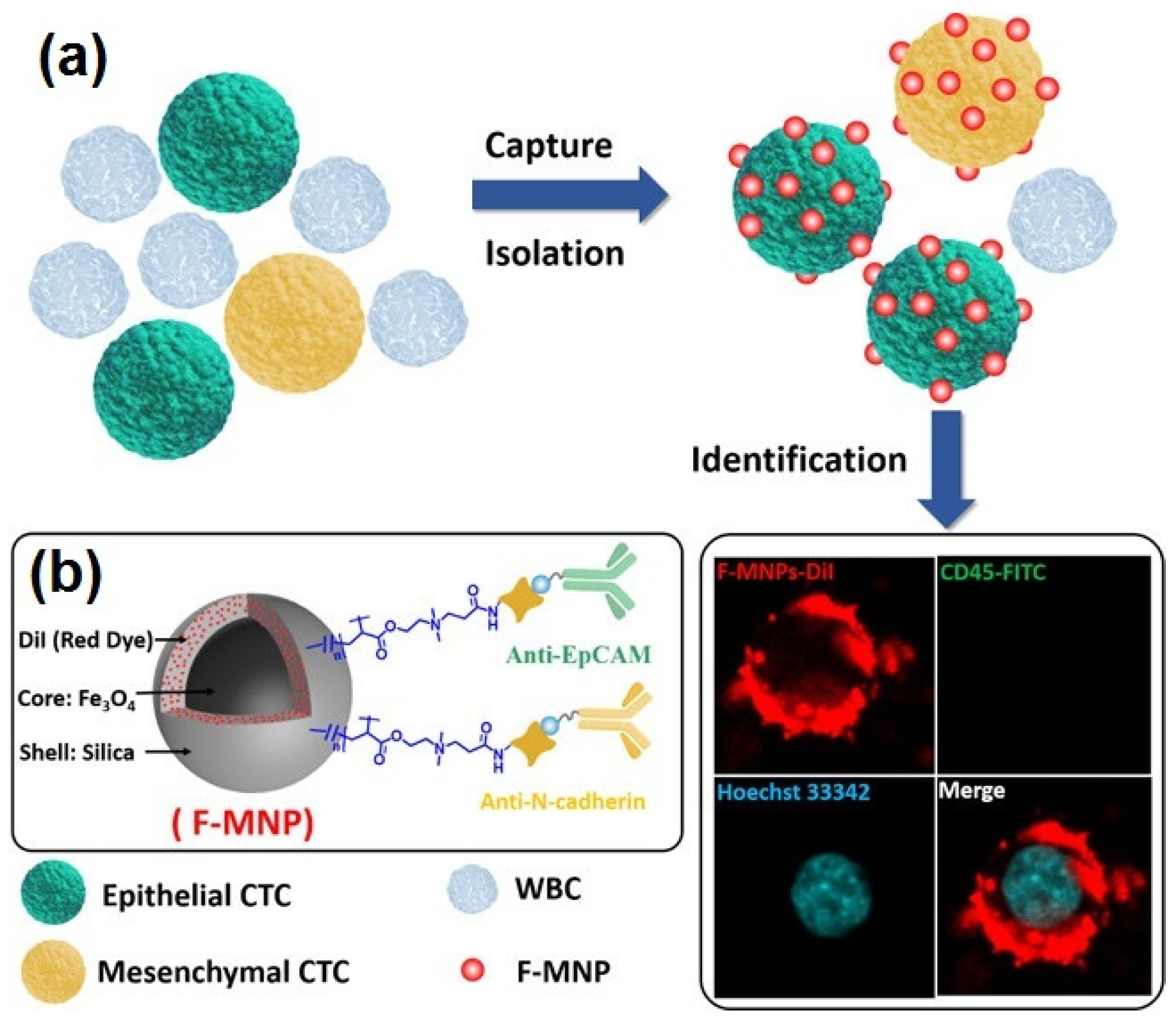
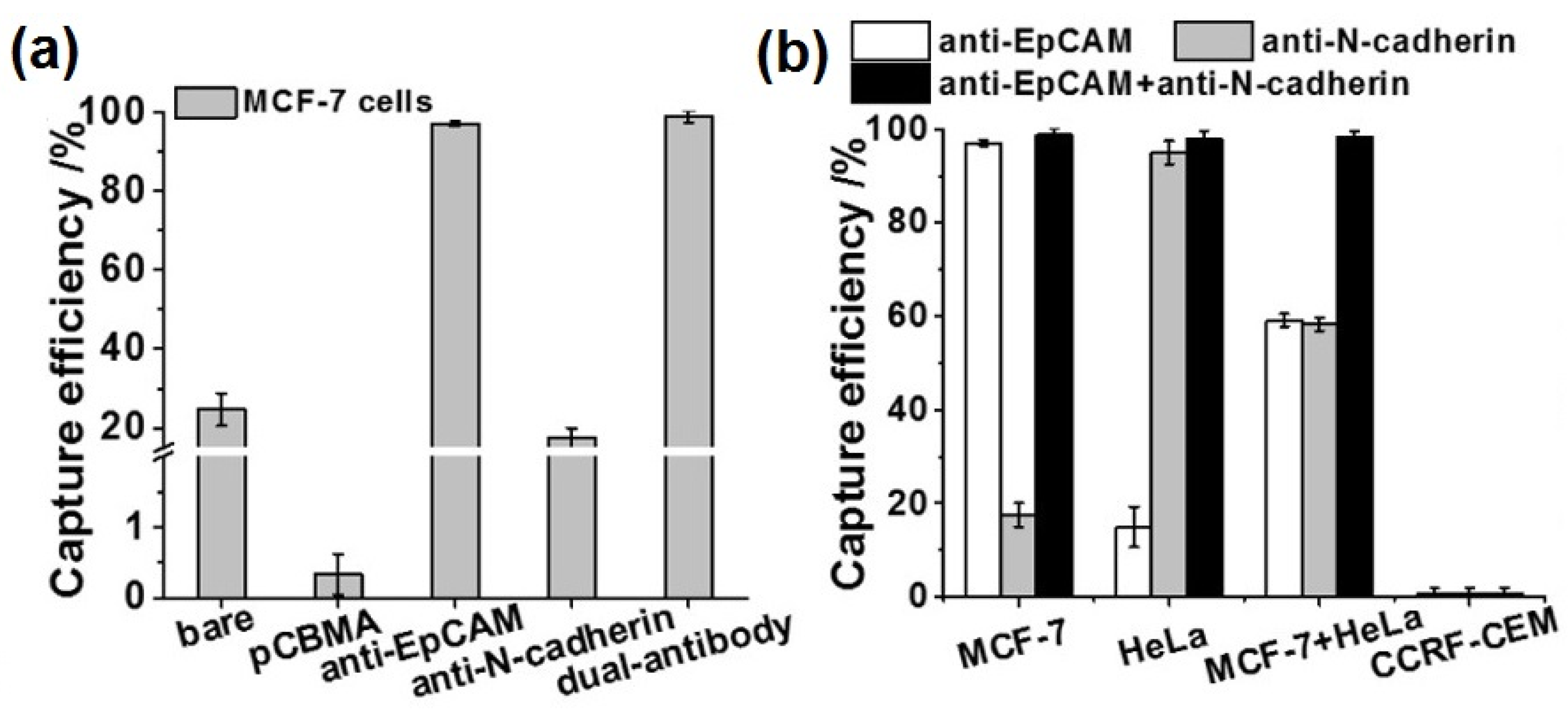
- Wang, Z.; Sun, N.; Liu, H.; Chen, C.; Ding, P.; Yue, X.; Zou, H.; Xing, C.; Pei, R. High-Efficiency Isolation and Rapid Identification of Heterogeneous Circulating Tumor Cells (CTCs) Using Dual-Antibody-Modified Fluorescent-Magnetic Nanoparticles. ACS Appl. Mater. Interfaces 2019, 11, 39586–39593. [Google Scholar] [CrossRef] [PubMed]
- White, S.B.; Kim, D.-H.; Guo, Y.; Li, W.; Yang, Y.; Chen, J.; Gogineni, V.R.; Larson, A.C. Biofunctionalized Hybrid Magnetic Gold Nanoparticles as Catalysts for Photothermal Ablation of Colorectal Liver Metastases. Radiology 2017, 285, 809–819. [Google Scholar] [CrossRef] [PubMed]
- Chang, Z.; Wang, Z.; Shao, D.; Yue, J.; Xing, H.; Li, L.; Ge, M.; Li, M.; Yan, H.; Hu, H.; et al. Shape Engineering Boosts Magnetic Mesoporous Silica Nanoparticle-Based Isolation and Detection of Circulating Tumor Cells. ACS Appl. Mater. Interfaces 2018, 10, 10656–10663. [Google Scholar] [CrossRef] [PubMed]
- Wang, C.; Shen, W.; Rong, Z.; Liu, X.; Gu, B.; Xiao, R.; Wang, S. Layer-by-Layer Assembly of Magnetic-Core Dual Quantum Dot-Shell Nanocomposites for Fluorescence Lateral Flow Detection of Bacteria. Nanoscale 2020, 12, 795–807. [Google Scholar] [CrossRef] [PubMed]
- Nie, Y.; Liu, Y.; Zhang, Q.; Zhang, F.; Ma, Q.; Su, X. Fe3O4 NP@ZIF-8/MoS2 QD-Based Electrochemiluminescence with Nanosurface Energy Transfer Strategy for Point-of-Care Determination of ATP. Anal. Chim. Acta 2020, 1127, 190–197. [Google Scholar] [CrossRef]
- Ahmed, S.R.; Dong, J.; Yui, M.; Kato, T.; Lee, J.; Park, E.Y. Quantum Dots Incorporated Magnetic Nanoparticles for Imaging Colon Carcinoma Cells. J. Nanobiotechnol. 2013, 11, 28. [Google Scholar] [CrossRef] [Green Version]
- Tran, M.V.; Susumu, K.; Medintz, I.L.; Algar, W.R. Supraparticle Assemblies of Magnetic Nanoparticles and Quantum Dots for Selective Cell Isolation and Counting on a Smartphone-Based Imaging Platform. Anal. Chem. 2019, 91, 11963–11971. [Google Scholar] [CrossRef]
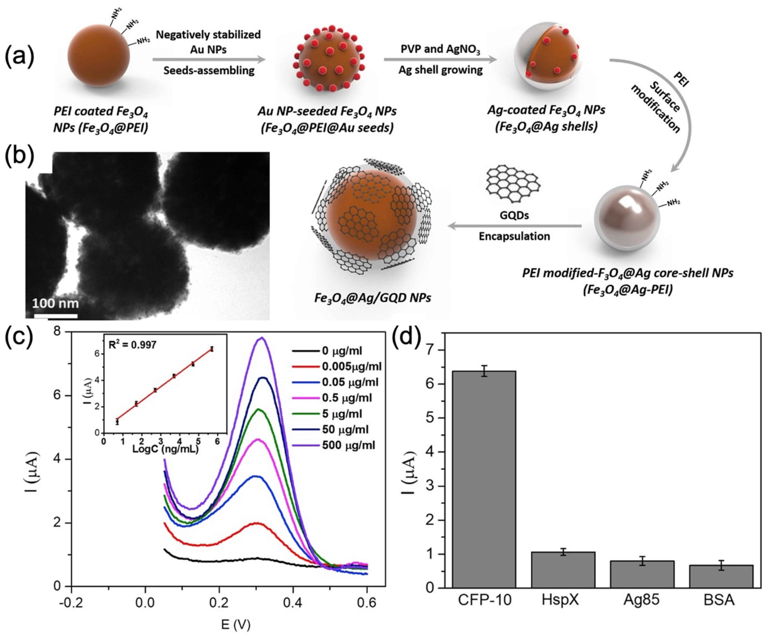
- Tufa, L.T.; Oh, S.; Tran, V.T.; Kim, J.; Jeong, K.-J.; Park, T.J.; Kim, H.-J.; Lee, J. Electrochemical Immunosensor Using Nanotriplex of Graphene Quantum Dots, Fe3O4, and Ag Nanoparticles for Tuberculosis. Electrochim. Acta 2018, 290, 369–377. [Google Scholar] [CrossRef]
- Wang, G.; Gao, W.; Zhang, X.; Mei, X. Au Nanocage Functionalized with Ultra-Small Fe3O4 Nanoparticles for Targeting T1–T2 Dual MRI and CT Imaging of Tumor. Sci. Rep. 2016, 6, 28258. [Google Scholar] [CrossRef] [Green Version]
- Bardhan, R.; Chen, W.; Perez-Torres, C.; Bartels, M.; Huschka, R.M.; Zhao, L.L.; Morosan, E.; Pautler, R.G.; Joshi, A.; Halas, N.J. Nanoshells with Targeted Simultaneous Enhancement of Magnetic and Optical Imaging and Photothermal Therapeutic Response. Adv. Funct. Mater. 2009, 19, 3901–3909. [Google Scholar] [CrossRef]
- Jun, B.-H.; Noh, M.S.; Kim, J.; Kim, G.; Kang, H.; Kim, M.-S.; Seo, Y.-T.; Baek, J.; Kim, J.-H.; Park, J.; et al. Multifunctional Silver-Embedded Magnetic Nanoparticles as SERS Nanoprobes and Their Applications. Small 2010, 6, 119–125. [Google Scholar] [CrossRef] [PubMed]
- Li, W.-S.; Wang, X.-J.; Zhang, S.; Hu, J.-B.; Du, Y.-L.; Kang, X.-Q.; Xu, X.-L.; Ying, X.-Y.; You, J.; Du, Y.-Z. Mild Microwave Activated, Chemo-Thermal Combinational Tumor Therapy Based on a Targeted, Thermal-Sensitive and Magnetic Micelle. Biomaterials 2017, 131, 36–46. [Google Scholar] [CrossRef] [PubMed]
- Yang, X.; Grailer, J.J.; Rowland, I.J.; Javadi, A.; Hurley, S.A.; Matson, V.Z.; Steeber, D.A.; Gong, S. Multifunctional Stable and pH-Responsive Polymer Vesicles Formed by Heterofunctional Triblock Copolymer for Targeted Anticancer Drug Delivery and Ultrasensitive MR Imaging. ACS Nano 2010, 4, 6805–6817. [Google Scholar] [CrossRef]
- Yang, S.; Chen, D.; Li, N.; Mei, X.; Qi, X.; Li, H.; Xu, Q.; Lu, J. A Facile Preparation of Targetable pH-Sensitive Polymeric Nanocarriers with Encapsulated Magnetic Nanoparticles for Controlled Drug Release. J. Mater. Chem. 2012, 22, 25354–25361. [Google Scholar] [CrossRef]
- Ma, H.; Liu, Y.; Shi, M.; Shao, X.; Zhong, W.; Liao, W.; Xing, M.M.Q. Theranostic, pH-Responsive, Doxorubicin-Loaded Nanoparticles Inducing Active Targeting and Apoptosis for Advanced Gastric Cancer. Biomacromolecules 2015, 16, 4022–4031. [Google Scholar] [CrossRef]
- Yang, H.Y.; Jang, M.-S.; Gao, G.H.; Lee, J.H.; Lee, D.S. pH-Responsive Biodegradable Polymeric Micelles with Anchors to Interface Magnetic Nanoparticles for MR Imaging in Detection of Cerebral Ischemic Area. Nanoscale 2016, 8, 12588–12598. [Google Scholar] [CrossRef]
- Sasikala, A.R.K.; GhavamiNejad, A.; Unnithan, A.R.; Thomas, R.G.; Moon, M.; Jeong, Y.Y.; Park, C.H.; Kim, C.S. A Smart Magnetic Nanoplatform for Synergistic Anticancer Therapy: Manoeuvring Mussel-Inspired Functional Magnetic Nanoparticles for pH Responsive Anticancer Drug Delivery and Hyperthermia. Nanoscale 2015, 7, 18119–18128. [Google Scholar] [CrossRef]


| NP | Conjugate | Morphology | Final Size (nm) | Applications | Ref. |
|---|---|---|---|---|---|
| IONPs@ DPSE-PEG | DiO, or DiI, or DiD, or DiR | Spherical | 24–46 | Multimodal MRI and FI | [15] |
| SPIONs | Ce6 | Spherical | 92 | Theranostic agent for dual-mode imaging and photodynamic therapy | [16] |
| SPIONs@ DSPE-PEG | ICG | Spherical (core-shell) | 23 | Tumor MR and fluorescence imaging and drug delivery for DOX | [17] |
| Fe3O4@SiO2-CMCS | Cy5 | Spherical (core-shell) | 51 | Bioimaging | [18] |
| Fe3O4@poly(HFMA-co-VBK)-g-PEG | VBK | Spherical (core-shell) | 146 | Magnetic resonance and optical imaging | [19] |
| GO-PAMAM-Fe3O4 | Cy5 | Irregular | - | Cellular imaging | [20] |
| Fe3O4-P(PEGMA) | FITC | Spherical | 36 | Bioimaging | [21] |
| Fe3O4 | Ce6 | Spherical | 15–25 | Dual-mode NIR fluorescence imaging and MRI of gastric cancer and photodynamic therapy (PDT) | [22] |
| mSiO2-Fe3O4-PEG | RITC or FITC | Spherical | 93 | Enhanced MRI, FI, and drug delivery for DOX | [23] |
| Fe3O4@mSiO2-PEG | FITC or RITC | Spherical (core-shell) | 45–105 | MRI and FI | [24] |
| IONPs@PSSS-PAH | RhB | - | - | Bioimaging | [25] |
| γ-Fe2O3@CS | FITC | - | 14 | Cellular imaging | [26] |
| SPIONs@poly(TMSMA-r-PEGMA-r-NAS) | Cy5.5 | Core-shell | ~26 | Dual-mode MRI and optical imaging | [27] |
| SPIONs@SiO2 | FITC | Spherical (core-shell) | 50 | MRI for human stem cell labeling | [28] |
| γ-Fe2O3 | RhB or fluorescein diacetate maleimide | ~ | ~30 | Cellular imaging | [29] |
| IONPs-cross-linked dextran | Cy5.5 | ~ | ~ | Preoperative MRI and intraoperative optical probe | [30] |
| γ-Fe2O3 | CR or RITC | Spherical | 14–15 | Multimodal imaging agents for amyloid-β fibril detection and removal | [31] |
| NP | Conjugate | Morphology | Final Size (nm) | Applications | Ref. |
|---|---|---|---|---|---|
| IONPs | GRAA33 aptamer | Spherical | 68 ± 4 | Remove prefibrillar amyloid aggregates | [34] |
| IONPs | Genomic DNA | Irregular | NA | Capture DOX, 98% | [35] |
| SPIONs | AS-14, AS-42 | Irregular | ~20 | Cancer treatment | [36] |
| IONPs | DNA | Spherical | 50 ± 5 | Detect and regulate mRNA, 99% | [37] |
| IONPs | ssDNA | Spherical | 200–400 | AFP capture for pregnancy test, 87% | [38] |
| γ-Fe2O3 | DNA | Irregular | NA | Thrombin detection, limit of 97 pM | [39] |
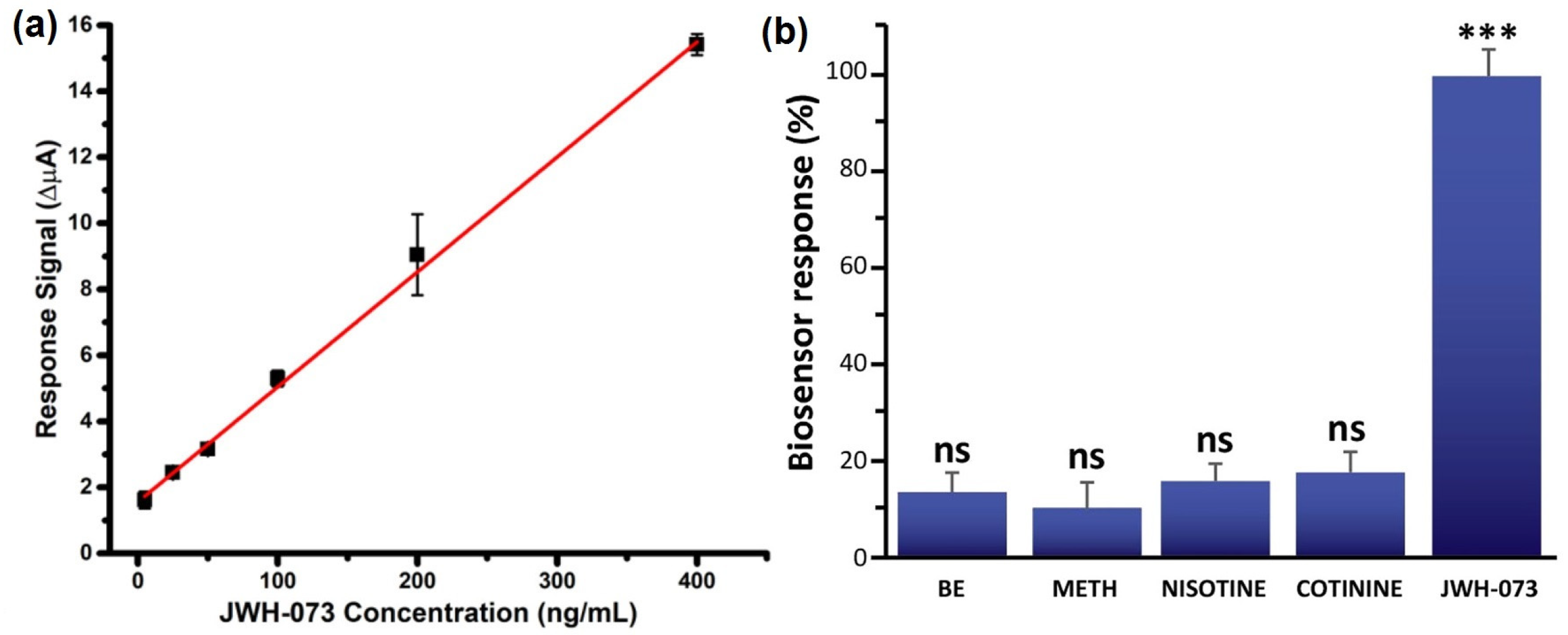
| IONPs | Anti-K2 antibody | Spheroidal | 17–25 | JWH-073 detection | [40] |
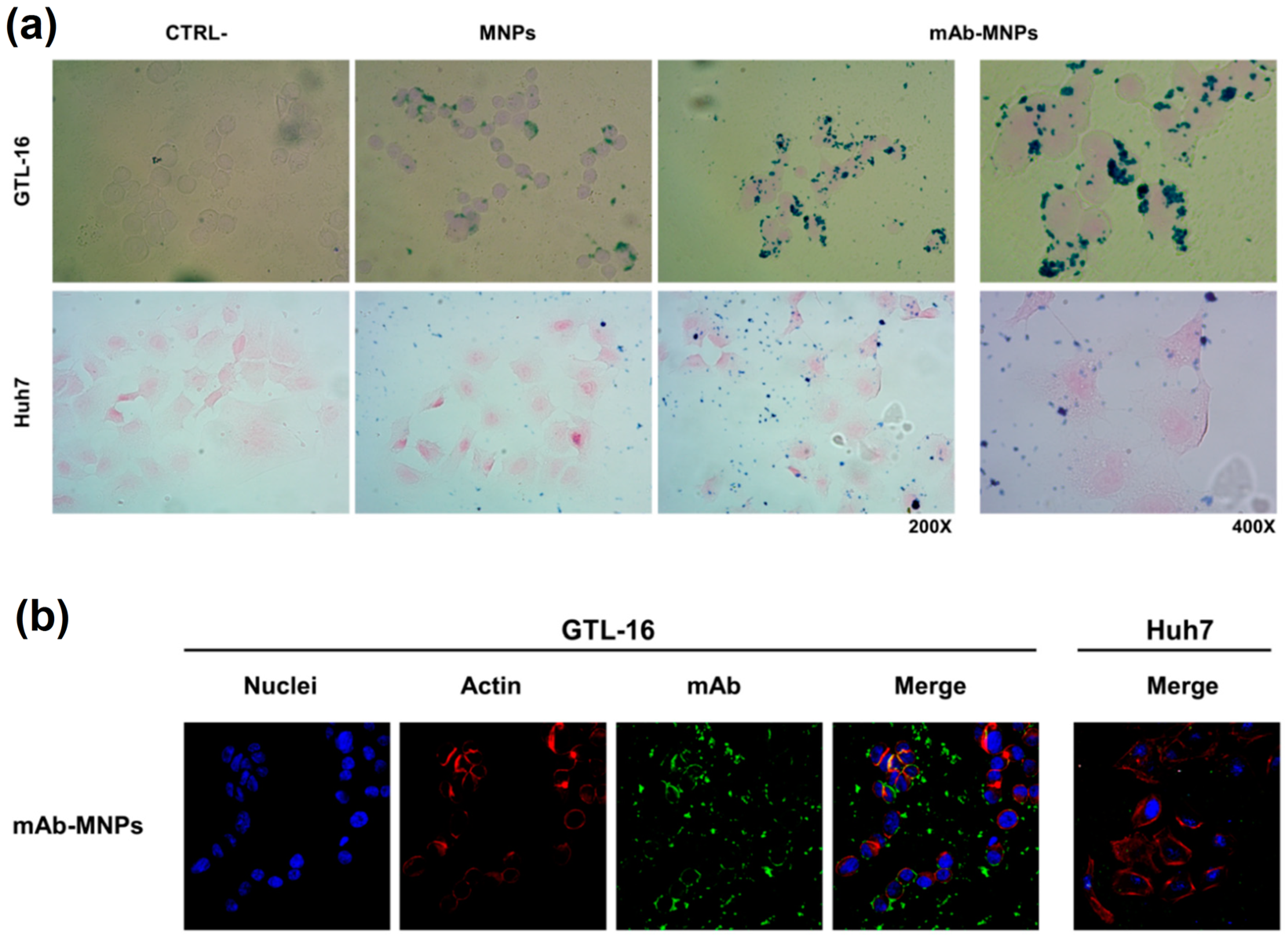
| SPIONs | mAb | NA | NA | Cancer drug delivery | [41] |
| IONPs | Anti-ferritin antibody | Spherical | ~50 | Alzheimer’s disease early diagnosis | [42] |
| IONPs | Anti-fFN antibody | Spherical | 37–63 | Preterm birth diagnosis | [43] |
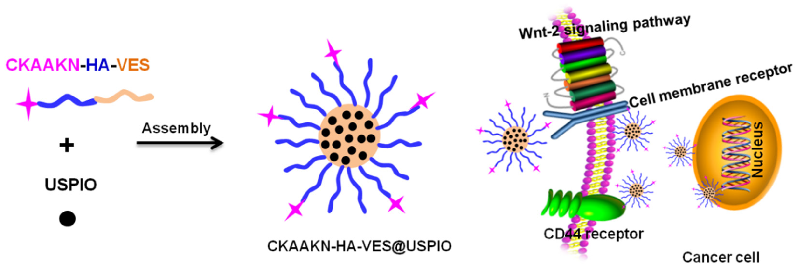
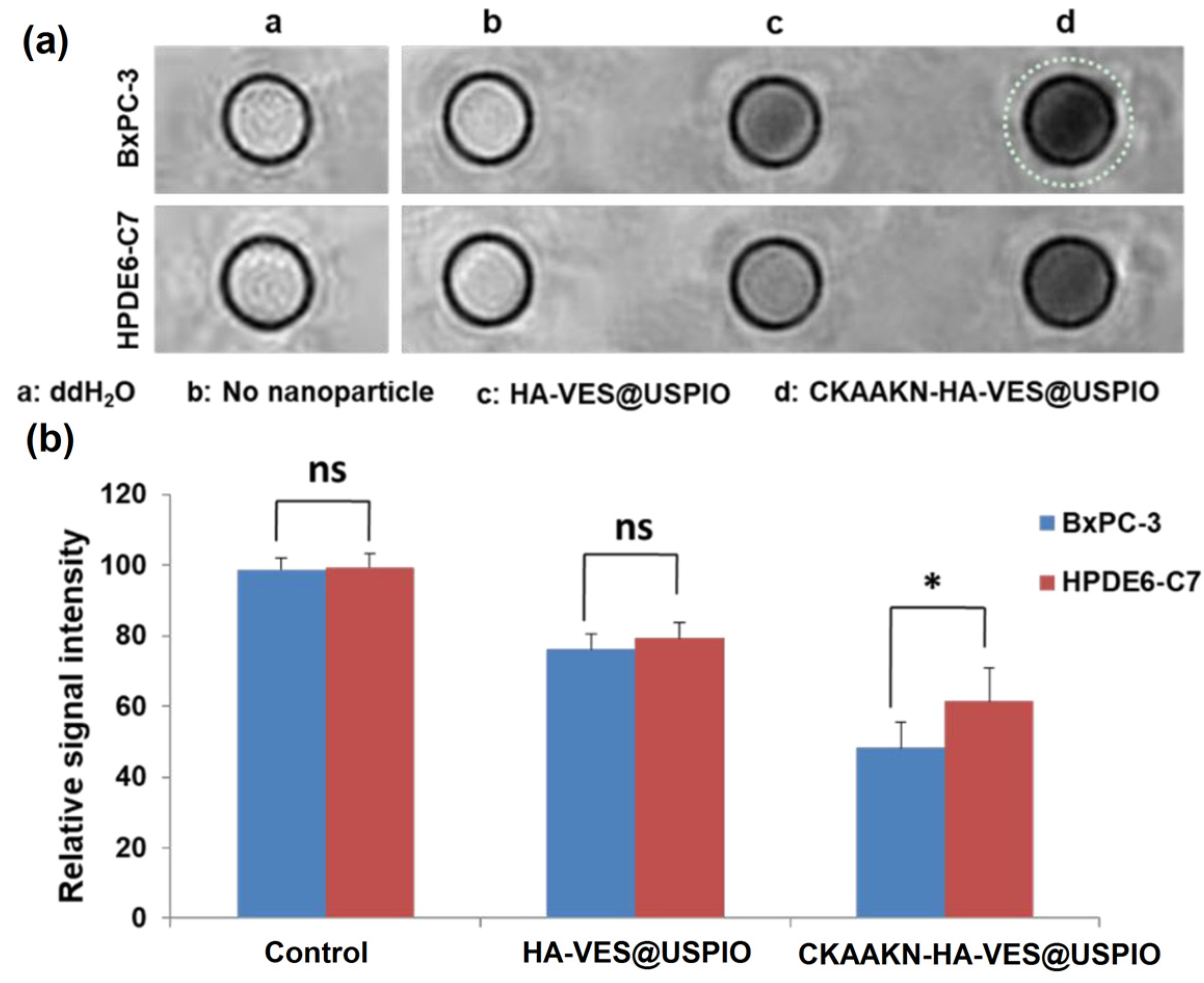
| USPIO | CKAAKN peptide | Spheroidal | ~60 | Enhanced MRI contrast agent | [44] |
| IONPs | Biotin, folic acid | Spherical | ~70 | Anticancer drug carrier | [45] |
| IONPs | Lipidoid | Spherical | 65 ± 5 | MRI-trackable immunotherapy | [46] |
| IONPs@Al2O3 | Peptide | Irregular | Staphylococcus aureus pathogen screening | [47] |
| NP | Conjugate | Morphology | Final Size (nm) | Applications | Ref. |
|---|---|---|---|---|---|
| Fe3O4@CDs | Sphere | 100 | Dual-modal imaging, near-IR light-responsive drug carrier, and photothermal therapy | [60] | |
| Fe3O4/SiO2/graphene-CdTe QDs/chitosan nanocomposites | Chitosan | Sphere | 177 | Drug delivery | [61] |
| Fe3O4@SiO2@ CdSe-CdS-ZnS QDs | Sphere | 75 | Potential use in magnetic field-assisted cell separation | [62] | |
| QD–Pt(II)–6G–IONP | (diamine)PtCl2 complexes | Sphere | 38 | Potential use in combined bioimaging systems such as FMT and MRI | [63] |
| Fe3O4/graphene–QDs@gelatin microspheres | Gelatin | Sphere | 460 | Drug delivery | [64] |
| NP | Morphology | Final Size (nm) | Applications | Ref. |
|---|---|---|---|---|
| Fe3O4@Au | Nanosphere (core–shell) | 20 | Photothermal therapy | [75] |
| Nanospikes | 48–52 | Theragnostic agent | [76] | |
| Fe3O4@Au-mPEG-PEI | Nanosphere (core–shell) | 263 | MR/CT dual-mode imaging | [77] |
| Fe3O4@SiO2@ GNSs-PEG | Nanosphere | ~100 | MR/CT dual imaging and photothermal therapy | [78] |
| Fe3O4@Ag-PAA | Nanosphere (core–shell) | ~10 | Photothermal therapy | [79] |
| Fe3O4-Ag-PAA | Nanosphere (heteromer) | ~10 | Photothermal therapy | [79] |
| SPION-Ag | Nanosphere (core–shell) | ~30 | Antibacterial | [80] |
| SPION-Au | Nanosphere (core–shell) | ~30 | Antibacterial | [80] |
| SPION-Au/Ag | Nanosphere (core–shell) | ~40 | Antibacterial | [80] |
| NP | Conjugate | Morphology | Final Size (nm) | Applications | Ref. |
|---|---|---|---|---|---|
| IONPs | PLGA | Nonspherical | 80–90 | AMF-triggered DDS (LCST) | [84] |
| SPIONs@ carbon | PNIPAm-MBAm | Spherical (core–shell) | 280 | Inductive magnetic heating-triggered DDS (LCST) for 5-Fluoruoracil | [87] |
| IONPs | PNIPAm-co-AAc | Irregular | 14–43 | AMF-triggered MHT and DDS (LCST) for DOX | [88,89] |
| IONPs@SiO2 | PNIPAm-co-PVNP | Yolk-shell | ~165 | NIR-triggered DDS (LCST) for DOX | [90] |
| Fe3O4 and γ-Fe2O3 | PNIPAm-co-HAAm | MNPs embedded in polymer nanofiber | ~350 | AMF-triggered MHT and DDS (LCST) for DOX | [91] |
| IONPs | PNIPAm-co-PEGMEA | Cubic | 19–22 | AMF-triggered DDS (LCST) for DOX | [92] |
| SPIONs | PPZ | Spherical | 153 | AMF-triggered MHT, and MRI | [93] |
| Fe3O4 | PNIPAm, PEG, dopamine | Spherical | 200 | AMF-triggered MHT and DDS (LCST) for DOX, and MRI | [94] |
| IONPs | PEO-b-PPO-b-PEO, gelatin | Spherical | 28–36 | AMF-triggered DDS (LCST) | [95] |
| SPIONs | PEG-b- PAsp(DIP) | Spherical nanovesicles | ~200 | DDS (pH) for DOX, and MRI | [96] |
| IONPs | [PNIPAm-r(PEGMEA)]-b-PAA | Irregular | 23–27 | AMF-triggered MHT and DDS (LCST + pH) for DOX, and MRI | [97] |
| NP | Conjugates | Morphology | Final Size (nm) | Applications | Ref. |
|---|---|---|---|---|---|
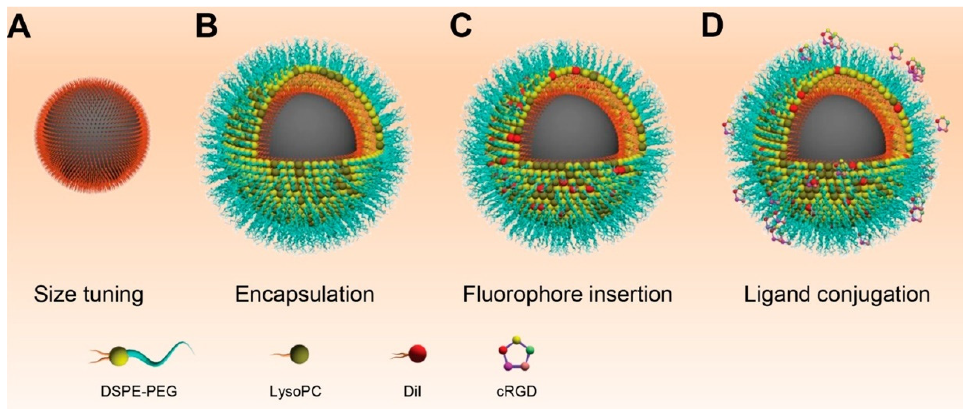
| IONPs@DPSE-mPEG | DiI and Cyclic RGD or Cyclic RAD | Spherical (core–shell) | 24–46 | Multimodal tumor-targeting MRI and FI | [15] |
| SPION@PEG | Cy5.5 and gH625 | - | 98 | Multimodal tumor-targeting MRI, FI, and hyperthermia | [118] |
| Gelatin@IONPs | Cy7 and HSA-DVS-FGF2 | Spherical (core–shell) | >103 | Controlled release of growth factors for augmentation of human mesenchymal stem growth and differentiation | [123] |
| Fe3O4@SiO2@ p(HEMA) | FITC, FA, and p(NIPAAM-co-AA) | Spherical (core–shell) | ~ | Dual thermoresponsive and pH-sensitive drug delivery for DOX | [124] |
| Fe3O4@PEI@ PEG | Cy5.5 and HCBP-1 | Spherical | ~30 | MRI and FI agent for lung cancer stem cell targeting | [125] |
| Fe3O4@SiO3-β-CD | FITC and FA | Spherical (core–shell) | 70 | Magnetic manipulation, bioimaging, cell targeting, and drug delivery | [119] |
| Fe3O4@mSiO2-PEI | RhB and siRNA | Spherical (yolk-shell) | 63 | Fluorescence tracking and magnetically guided small interfering RNA delivery | [126] |
| IONPs-PS | Coumarin-153, IgG, and ICAM-1 | Nanowires | 7440 × 270 | Potential tumor targeting MRI and FI | [127] |
| Fe3O4@mSiO2 | FITC, AlC4Pc, and FA | Spherical (yolk-shell) | ~50 | Tumor targeting MRI, FI, and photodynamic therapy | [120] |
| Fe3O4@PAA | Rh123, and FA-PEG | Spherical | ~86 | Dual-modal molecular imaging | [128] |
| IONP@SiO2-PEG | RhB and cetuximab | Spherical (core–shell) | ~60 | In vivo colon cancer targeting and imaging | [129] |
| SPION@SiO2 | Cy5.5 and RGD | Spherical (core–shell) | ~97 | Magnetically enhanced cancer imaging and targeting | [130] |
| DySiO2-Fe3O4 | RhB and HmenB1 | Spherical | ~45 | Dual-modal MRI and FI of neuroblastoma | [131] |
| SPION@dextran | Cy5.5 and EPPT | Spherical | 41 | Dual-modal in vivo imaging of tumor response to therapy | [132] |
| MNPs | DiI and anti-EpCAM, or anti-N-cadherin | Spherical | ~160 | CTC capture, 98.8% | [133] |
| USPIO/Ag NPs or Au NPs | Anti-MG1 | Spheroidal | ~47 | Photothermal ablation of liver metastases | [134] |
| IONPs | FITC and anti-EpCAM | Spherical | ~200 | CTC isolation and detection, 87% | [135] |
| Fe3O4@CdSe/ZnS QDs | S. pneumoniae antibodies | Spherical | 150 | Detection of bacteria | [136] |
| Fe3O4@ZIF–8/MoS2 QDs | cDNA | - | 100 | Detection of ATP | [137] |
| CdTe QDs-Fe3O4 | hCC49 antibodies | Spherical | 50 | Imaging colon carcinoma cells | [138] |
| IONP@QDs (QDs:575, 605, 635, 485) | anti-HER2 antibodies | - | 250 ± 81 | Selective cell isolation and counting on smartphone-based imaging platforms | [139] |
| Fe3O4@Ag/graphene–QDs | anti-CFP-10 antibodies | Spherical NPs deposited onto glassy carbon electrodes | 270 | Electrochemical immunosensor for tuberculosis | [140] |
| Aurod-(Fe3O4) | Herceptin | Nanorods | 15 | Dual-mode imaging, and photothermal ablation of cancer cells | [121] |
| Au NC@Fe3O4 | Folic acid | Nanocages | 70 | Multimodal contrast agents | [141] |
| Au NS@Fe3O4 /SiO2 | Streptavidin | Nanosphere | ~100 | Magnetic and optical imaging and photothermal therapy | [142] |
| Fe3O4@SiO2/Ag | Raman-label compounds | Nanosphere (core–shell) | 50 | Cancer cell targeting | [143] |
| Fe3O4@SiO2/Ag | Raman-label compounds, and antibody | Nanosphere (core–shell) | 50 | Cancer cell separation | [143] |
| IONPs | PPyCOOH, PEG, FA | Irregular | 47 | AMF triggered MHT and DDS (Tg) for DOX | [116] |
| MNPs | PEG-g-p(AAm-co-AN)-A54 | Irregular | 80 | AMF-triggered MHT and DDS (UCST) for DOX | [144] |
| SPIONs | PEG-(Glu-Hyd)-PEG-FA | Spherical nanovesicles | 150 | DDS (pH) for DOX, and MRI | [145] |
| mSiO2@ SPIONs | HAMA-b-DBAM-FA | Spherical | ~200 | DDS (pH) for DOX, and MRI | [146] |
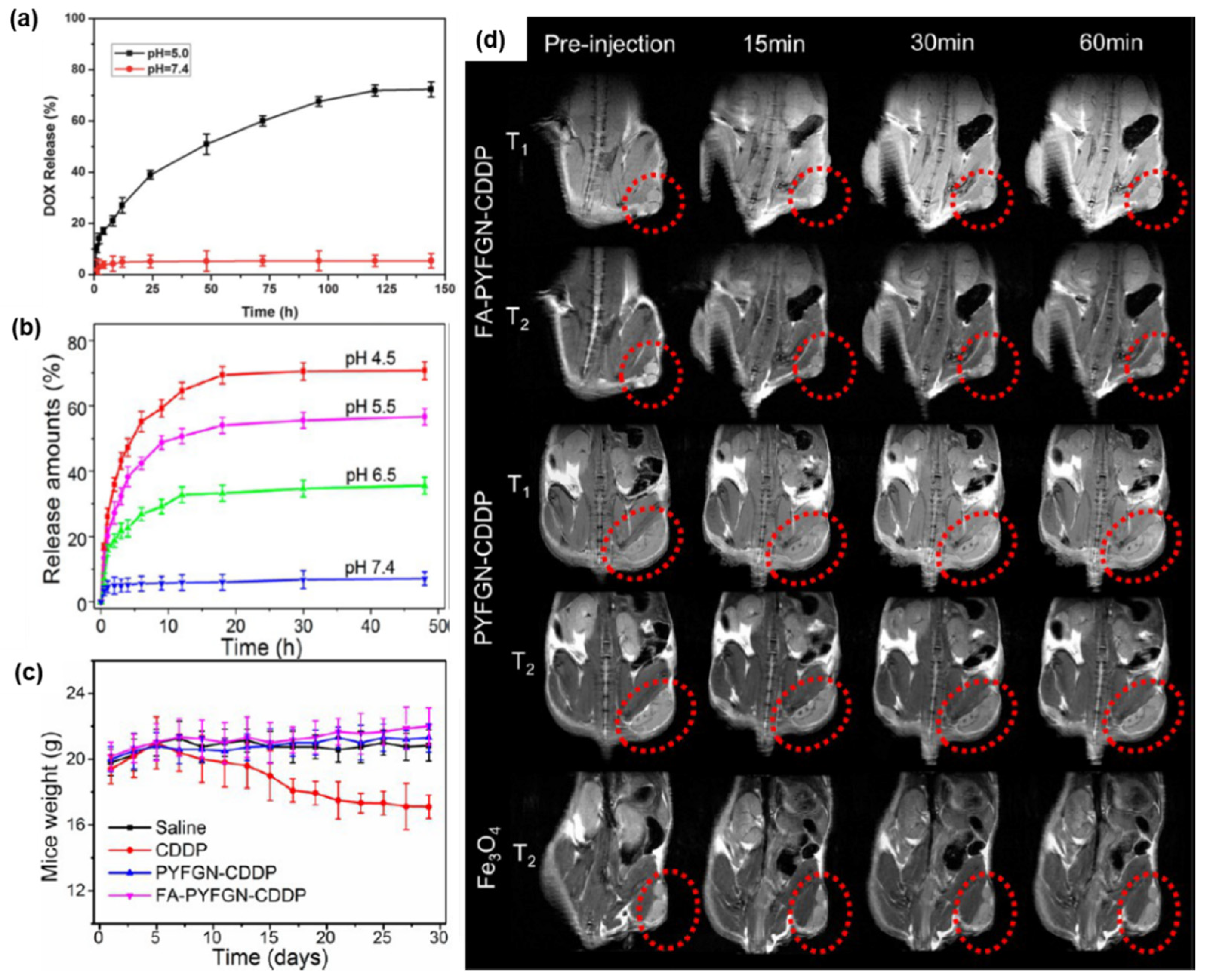
| Fe3O4@Gd2O3 | PEG-FA | Yolk-shell | 109 | DDS (pH) for cisplatin, and dual-mode MRI | [122] |
| IONPs | poly(β-aminoester)-FA | Irregular | 50–200 | DDS (pH) for DOX, and MRI | [147] |
| IONPs | mPEG-b-P(DPA-DE)LG | — | ~120 | pH-triggered MRI probes | [148] |
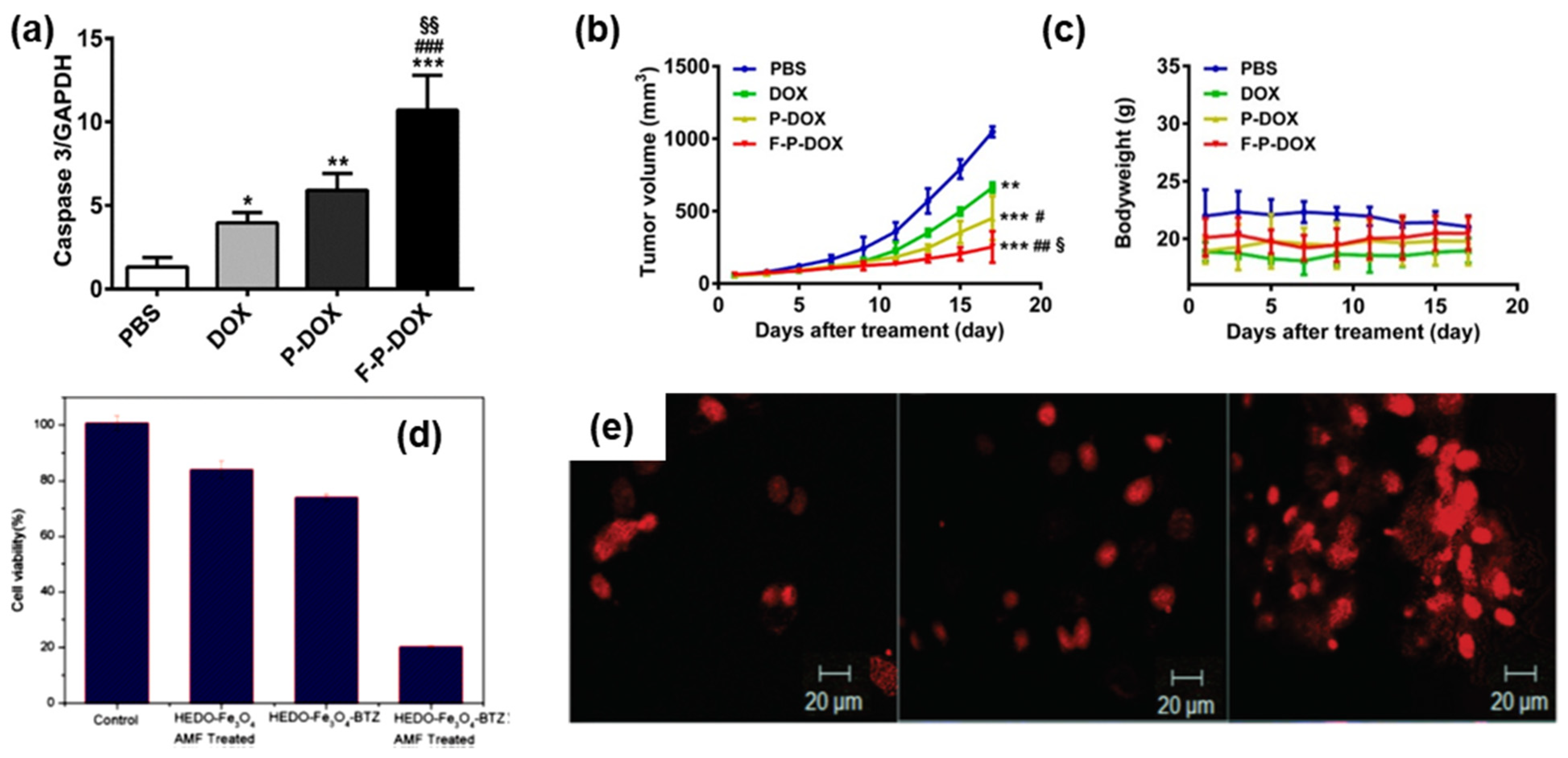
| SPIONs | p(HEMA-co-DMA) | Spherical | ~20 | DDS (pH) for BTZ, and MHT | [149] |
Publisher’s Note: MDPI stays neutral with regard to jurisdictional claims in published maps and institutional affiliations. |
© 2022 by the authors. Licensee MDPI, Basel, Switzerland. This article is an open access article distributed under the terms and conditions of the Creative Commons Attribution (CC BY) license (https://creativecommons.org/licenses/by/4.0/).
Share and Cite
Tran, H.-V.; Ngo, N.M.; Medhi, R.; Srinoi, P.; Liu, T.; Rittikulsittichai, S.; Lee, T.R. Multifunctional Iron Oxide Magnetic Nanoparticles for Biomedical Applications: A Review. Materials 2022, 15, 503. https://0-doi-org.brum.beds.ac.uk/10.3390/ma15020503
Tran H-V, Ngo NM, Medhi R, Srinoi P, Liu T, Rittikulsittichai S, Lee TR. Multifunctional Iron Oxide Magnetic Nanoparticles for Biomedical Applications: A Review. Materials. 2022; 15(2):503. https://0-doi-org.brum.beds.ac.uk/10.3390/ma15020503
Chicago/Turabian StyleTran, Hung-Vu, Nhat M. Ngo, Riddhiman Medhi, Pannaree Srinoi, Tingting Liu, Supparesk Rittikulsittichai, and T. Randall Lee. 2022. "Multifunctional Iron Oxide Magnetic Nanoparticles for Biomedical Applications: A Review" Materials 15, no. 2: 503. https://0-doi-org.brum.beds.ac.uk/10.3390/ma15020503