Magnetic Nanoparticles for Biomedical Purposes: Modern Trends and Prospects


Department of Chemistry, M.V. Lomonosov Moscow State University, Leninskie Gori build. 1/3, Moscow 119991, Russia
*
Author to whom correspondence should be addressed.
Magnetochemistry 2020, 6(3), 30; https://0-doi-org.brum.beds.ac.uk/10.3390/magnetochemistry6030030
Submission received: 16 June 2020
/
Revised: 14 July 2020
/
Accepted: 15 July 2020
/
Published: 17 July 2020
(This article belongs to the Special Issue Magnetic Nanoparticles 2020)
Abstract
:The presented paper is a review article discussing existing synthesis methods and different applications of nanosized magnetic nanoparticles. It was shown that, in addition to the spectrum of properties typical for nanomaterials (primarily a large specific surface area and a high fraction of surface atoms), magnetic nanoparticles also possess superparamagnetic properties that contribute to their formation of an important class of biomedical functional nanomaterials. This primarily concerns iron oxides magnetite and maghemite, for which in vitro and in vivo studies have shown low toxicity and high biocompatibility in comparison with other magnetic nanomaterials. Due to their exceptional chemical, biological, and physical properties, they are widely used in various areas, such as magnetic hyperthermia, targeted drug delivery, tissue engineering, magnetic separation of biological objects (cells, bacteria, viruses, DNA, and proteins), and magnetic diagnostics (they are used as agents for MRS and immunoassay). In addition to discussing the main problems and prospects of using nanoparticles of magnetic iron oxides for advanced biomedical applications, information is also reflected on their structure, production methods, and properties.
1. Introduction
It is recognized that nanoparticles are the ultrafine objects that combine atoms of chemical elements or molecules of organic and inorganic compounds with sizes of several nanometers (nm; 1 nm = 10−9 m) [1]. Nanoparticles exist in nature and are also the result of human activity. Because of their submicron size, they have unique properties, primarily a huge surface area per volume unit, a high value of fraction of surface atoms and near-surface layers, and the ability to exhibit quantum effects. Their unusual properties cannot be predicted from the properties of bulk materials. They are extensively used in a wide range of scientific and technical fields, including ecology, catalysis, healthcare, and engineering.
The International Organization for Standardization (ISO) gives the definition of the nanoparticle as a “nano-object with all three external dimensions in the nanoscale” where nanoscale means “length range approximately from 1 nm to 100 nm” [2]. Similarly, the ISO standard defines two-dimensional (nanoplates and nanodisks) and one-dimensional (nanotubes and nanofibres) nano-objects. According to the ISO recommendations (2010), a nanomaterial is defined as a “material with any external nanoscale dimension or having internal nanoscale surface structure”. However, in 2011, the Commission of the European Union approved a broader nanomaterial definition: “a natural, incidental, or manufactured material containing particles, in an unbound state or as an aggregate or as an agglomerate and where, for 50% or more of the particles in the number size distribution, one or more external dimensions is in the size range 1–100 nm. In specific cases and where warranted by concerns for the environment, health, safety, or competitiveness the number size distribution threshold of 50% may be replaced by a threshold between 1 and 50%.”
Magnetic nanoparticles contain magnetic elements such as iron, chromium, gadolinium, cobalt, manganese, nickel, and their chemical compounds, for example, oxides, ferrites, and alloys, and they can be directed using magnetic fields.
The physical and chemical properties of magnetic nanoparticles are largely dependent on their shape, crystalline structures, size, and their chemical components. In addition to the properties common to nanomaterials, magnetic nanoparticles also have specific magnetic properties, such as low Curie temperature, high magnetic susceptibility, and superparamagnetism [3].
Superparamagnetism is a form of magnetism that manifests itself in small ferrimagnetic and ferromagnetic particles. If such particles are small enough (150 nm or less, the exact size value depends on the chemical composition of the particle), they become single-domain, meaning the uniform orientation of individual spins over entire particle volume and formation of uniformly magnetized state. As a thermal fluctuation the magnetic moment of such particles can flip direction randomly, resulting in zero average magnetization of superparamagnetic particles. However, when an external magnetic field is applied, such particles behave like paramagnets even at temperatures below the Curie point or Néel point. The magnetic susceptibility of such superparamagnetic systems significantly exceeds than that of paramagnetic ones.
2. Categories of Magnetic Nanoparticles
Magnetic nanoparticles used or considered for biomedical applications can be divided into the following categories [4].
- Oxides (γ-Fe2O3, Fe3O4, NiO [11]);
Magnetic properties of cobalt nanoparticles, FeCo alloys, and ferrites, like maximum saturation magnetization at room temperature, coercive force, and anisotropy, allow one to obtain magnetic resonance spectoscopy (MRS) data with good resolution when these particles are used as contrast agents. Moreover, these properties give such nanoparticulate systems a great potential for heating in variable magnetic fields and drug delivery applications. However, cobalt and nickel nanoparticles are prone to oxidation. In addition, these nanoparticles, as well as their compounds, have rather high toxicity; therefore, their use for biomedical purposes is limited [4,5]. To reduce the toxic effect, cobalt ferrite nanoparticles are coated with polymer coatings (polyvinyl alcohol, polyvinylpyrrolidone, and r polyethylene glycol) [13,14,15]. Such particles show negligible cytotoxicity at concentrations up to 150 μg/mL. ZnFe2O4 nanoparticles are preferred for biomedical use among all ferrites due to the low toxicity of zinc ions (they are non-toxic and biocompatible at ZnFe2O4 concentrations below 125 μg/mL). These particles are considered promising T2 MRS agents as magnetic vectors for targeted drug delivery [18,19].
The toxicity of iron and its oxides is lower. The iron nanoparticles tendency to oxidize also limits their biomedical application [20,21]. To solve the problem, it is proposed to use a polymer coating, like biopolymer nanoskin [6], which prevents the undesirable process of oxidation or interaction with external media without impairment to the iron nanoparticles magnetic interaction. Superparamagnetic iron oxide nanoparticles (SPIONs), such as maghemite (γ-Fe2O3) or magnetite (Fe3O4), are the primarily used magnetic nanoparticles for biomedical purposes due to their low cost, simple preparation approach, low toxicity, superparamagnetism, and biocompatibility [3].
Magnetite (Fe3O4) possesses ferromagnetic properties. It reveals a reversed spinel structure with a face-centered cubic unit cell (eight molecules per unit cell) and a lattice parameter a = 0.839 nm. The cell is based on 32 oxygen (O2−) ions located along the crystallographic direction (111), which form octahedral and mixed tetrahedral/octahedral layers. Magnetite contains iron ions Fe2+ and Fe3+. Its structure could be written as (Fe3+)A(Fe2+Fe3+)BO4, where half of the Fe3+ ions occupy tetrahedral voids (A) and are surrounded by four oxygen atoms, while the mixture of Fe2+/Fe3+ ions occupies octahedral voids (B) and is surrounded by six oxygen atoms. The Fe3+ ions of positions A and B are arranged in antiferromagnetic order, while the Fe2+ ions located at the B position and determined macroscopic ferromagnetic properties. Magnetite is a semiconductor. It has a relatively high electrical conductivity (it is considered a half-metal [22]). The conductivity of Fe3O4 arises due to the rapid jump-like movement of electrons between Fe2+ and Fe3+ ions located in the octahedral voids (B) [23].
Maghemite, γ-Fe2O3, is a ferrimagnetic oxide with a spinel structure similar to that of magnetite. The unit cell of maghemite consists of 32 O2− forming a cubic close-packed structure, while Fe3+ is unevenly distributed between tetrahedral and octahedral voids. In contrast to the Fe3O4 structure, the structure of maghemite has a vacancy of divalent iron ions. The structure of maghemite (γ-Fe2O3) can be written as 0.75(Fe3+)A(Fe3+5/3V1/3)BO4, where V represents the Fe2+ vacancy located in octahedral positions. The unit cell of maghemite (a = 0.8347 nm) is slightly smaller than the magnetite cell due to the formation of cationic vacancies and the smaller size of Fe3+ ions compared to Fe2+ ions. Maghemite should be considered as completely oxidized magnetite and as an n-type semiconductor (band gap 2.0 eV) [24].
3. SPIONs Synthesis
3.1. Chemical Methods
One of the most common methods for the SPIONs nanoparticles synthesis is the coprecipitation method. The essence of this method is to obtain magnetic nanoparticles from aqueous solutions of ferric and ferrous salts in a molar ratio of 2:1 by slowly adding a precipitating agent (aqueous solution of ammonia, hydroxide, or sodium carbonate) to the solution at room temperature or by heating (Scheme 1).
A concomitant reaction is the oxidation of magnetite to maghemite with atmospheric oxygen. Therefore, this method produces magnetic nanoparticles that are intermediate in composition between magnetite and maghemite [25,26]. To obtain predominantly magnetite nanoparticles, this reaction is eliminated by passing inert gases argon or nitrogen through reagent solutions. Depending on the type of iron salts, the nature of the precipitating agent, solvents, temperature and pH control, magnetite nanoparticles 1–40 nm in size have been prepared by co-precipitation method.
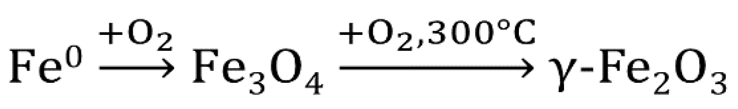
Oxidation. Magnetite nanoparticles can be obtained by oxidation of iron nanoparticles [21,27]. Maghemite nanoparticles are obtained by oxidation of iron and magnetite nanoparticles with atmospheric oxygen at 300 °C (Scheme 2).
It is not advisable to use higher temperatures. Higher temperatures can lead not only to a noticeable sintering of nanoparticles to submicron sizes, but also to transition of a thermodynamically unstable polymorphic modification of iron (III) oxide-maghemite (γ-Fe2O3) to hematite (α-Fe2O3) in the temperature range from 300 to 500 °C.
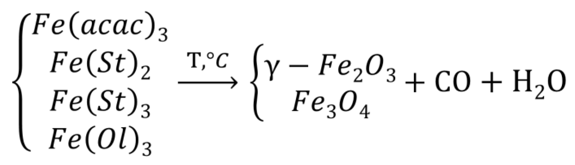
Thermal decomposition is one of the most efficient methods for producing maghemite nanoparticles with a narrow size distribution (Scheme 3). Two methods, “heating up” and “hot injection”, are used to achieve thermal decomposition. The “heating up” involves the continuous heating of a solution consisting of a solvent, surfactants, and the precursor compounds to a predefined temperature, at which the nanoparticles begin cluster formation and grow. Conversely, the “hot injection” approach provokes rapid and homogeneous nucleation by introducing reagents into a hot solution of a surfactant and is finalized by a controlled growth phase. Both methods use the same principle of heating a non-magnetic organometallic precursor compound in the presence of surfactants and organic solvents [27]. The most common precursor compounds are iron (III) acetylacetonate (Fe(acac)3) and iron (II, III) stearate (Fe(St)2, Fe(St)3), iron (III) oleate (Fe(Ol)3) fatty acids are used as surfactants.
Argon is used to maintain an inert atmosphere. The optimal temperature required for this reaction is in the range from 100 to 350 °C, thermal degradation of precursors leads to the formation of crystalline magnetic nanoparticles with a size of 4 to 30 nm in diameter with a narrow size distribution [28]. Reaction time and temperature are crucial factors for the particle size control. The thermal decomposition of iron stearates makes it possible to control the shape of the resulting nanoparticles (spheres, cubes, and disks), which depends on the nature and concentration of the reagents [29].
Hydrothermal and solvothermal methods. The hydrothermal synthesis method is based on the high solubility of a large amount of inorganic substances in water at elevated temperature and pressure and the possibility of subsequent crystallization of the dissolved material from the liquid phase [30,31,32]. High temperatures contribute to rapid nucleation under the influence of precipitants. The solvothermal method differs from the hydrothermal method in that the solvent used is not water, but organic solvents. These methods involve the use of autoclaves in order to create and maintain elevated temperature (130–250 °C) and pressure (0.3–4 MPa). Precursors are salts of ferrous and ferric iron. As precipitants, solutions of sodium hydroxide, tetramethylammonium hydroxide, and hydrazine are used. Moreover, microwave radiation can be applied to heat the mixture, and in this case, we talk about the hydrothermal-microwave or solvothermal-microwave methods. Typically, the shape, particle size, and size distribution depend on the concentration of the precursor, and the time and the temperature of the process fulfilling. The sizes of the resulting nanoparticles are usually in the range from 10 to 50 nm. Short autoclaving times make it possible to obtain monodisperse particles.
Polyol synthesis is a method in which a polyhydric alcohol or a mixture of alcohols is used as a solvent or as a solvent and a reducing agent. In the polyol synthesis of SPIONs, polyethylene glycol interacts with a ferric salt, which is not only a solvent in this case, but also a reducing agent, precipitant, and stabilizer. Another polyol synthesis of maghemite nanoparticles with an average size of 11 nm is based on oxidative alkaline hydrolysis of Fe2+ and Fe3+ salts in a mixture of polyethylene glycol/diethylene glycol or N-methyldiethanolamine) [33]. The synthesis of hydrophilic magnetite (Fe3O4) nanoparticles can be carried out by thermolysis of iron (III) acetylacetonate in four different liquid polyols: diethylene glycol, triethylene glycol, tetraethylene glycol, and polyethylene glycol. The average particle size, depending on the polyol used, varies from 7 to 15 nm [34]. Roughly spherical, highly crystallized, and almost uniform in size, maghemite nanoparticles of average size 11 nm are synthesized by forced hydrolysis of iron acetates in a diethylene glycol [35,36].
The microemulsion method consists of mixing two microemulsions (water/surfactant/inorganic solvent) containing an aqueous solution of iron salts and a precipitating reagent. When two microemulsions are mixed with reagents dissolved in iron oxides, magnetite nanoparticles (partially oxidized to maghemite) are formed, the size of the particles (1–50 nm) can be controlled by the size of micelles. The advantage of the method is a narrow size distribution of the resulting nanoparticles. The disadvantages of the method include the difficulty of cleaning from surfactants and relatively small amounts of the resulting nanoparticles [37].
3.2. Physical Methods
There are two procedures of physical methods for producing magnetic nanoparticles: top-down and bottom-up. The top-down methods are based on milling to reduce the size of macroscopic magnetic materials to the nanometer range. The main disadvantages of these methods include the difficulty in regulating the size and shape of the particles. In addition, the grinding procedure leads to the formation of crystal lattice defects, which cause deviations in magnetic properties compared with particles of the same size [38]. Nanoparticles of iron oxides from the bottom-up method can be obtained by laser evaporation of micron particle-sized metal oxide powders. As a result of the sharp temperature gradient outside the evaporation zone, very rapid condensation and nucleation from the gas phase occur, and nanoparticles with sizes from 20 to 50 nm are formed [39,40].
3.3. Biological Methods
Through biomineralization, some living organisms can produce magnetic particles [41,42]. Magnetotactic bacteria orient themselves and migrate along the lines of the geomagnetic field. This ability is based on intracellular magnetic structures, magnetosomes, which contain nanometer membrane magnetite crystals coated with protein. The formation of magnetosomes is based on a mineralization process with biological control of the accumulation of iron and the deposition of a mineral particle with a certain size and orientation in the membrane vesicle in certain places in the cell. Under conditions of anaerobic synthesis in a laboratory similar to the living conditions of magnetotactic bacteria, homogeneous particles with a core diameter of 20 to 45 nm can be obtained. Even though the magnetosomes exhibit excellent magnetic properties for medical use (especially for hyperthermia), they still have not been used in medicine because of their bacterial protein coating [43].
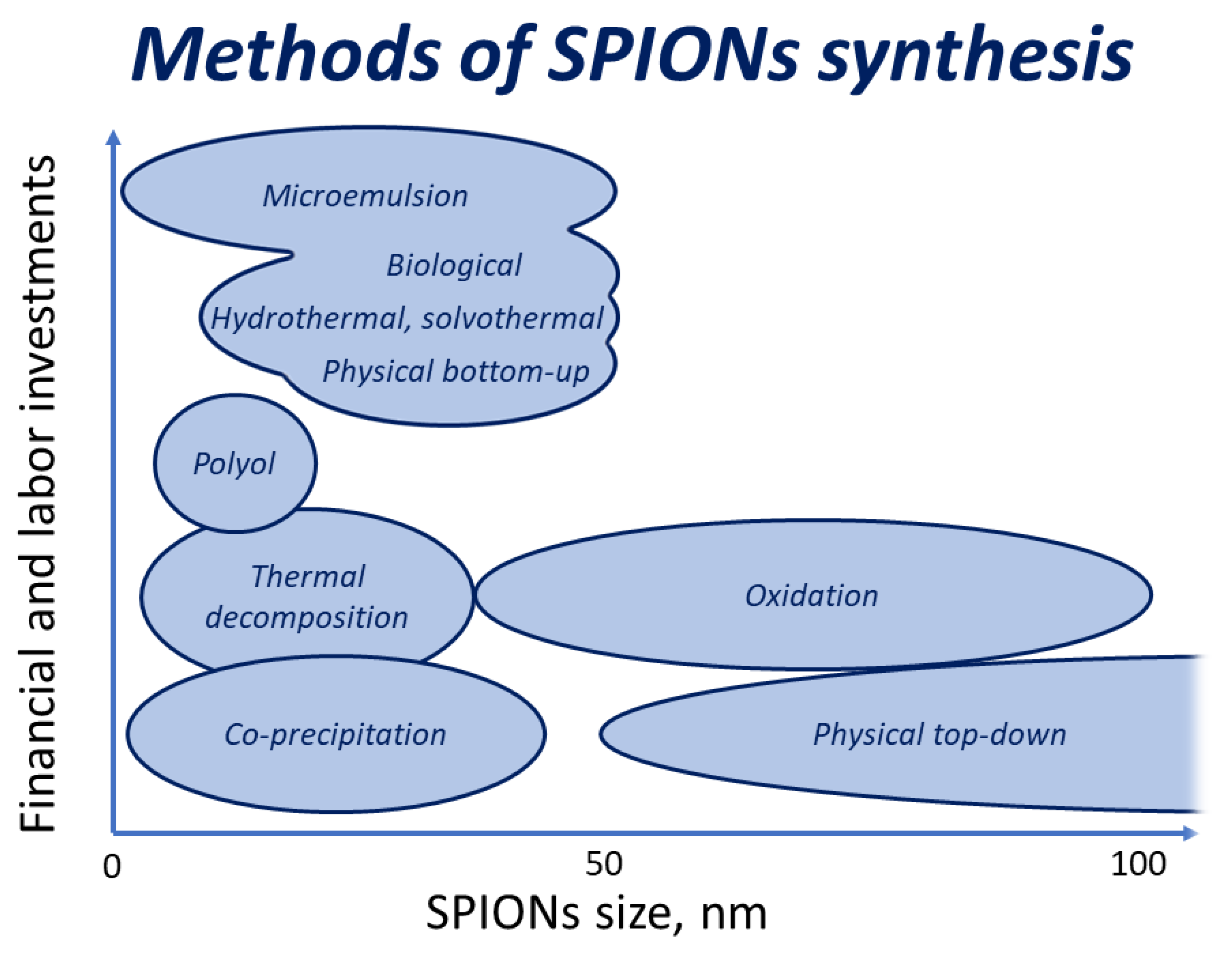
Thus, there is a wide range of methods for the synthesis of nanoparticles of superparamagnetic iron oxides. These particles of size 1–40 nm can be obtained by coprecipitation of iron (II, III) salts. This method is characterized by the low cost of reagents and equipment. It is easy to carry out and it does not allow obtaining monodisperse particles of a certain shape. The morphology of SPIONs obtained by oxidation of iron and its oxides is determined by the morphology of the particles of the precursors and the temperature of the process. Sintering does not allow obtaining particles of several nanometers in size. In addition, a side process of the formation of hematite, the nanoparticles of which do not possess superparamagnetic properties, is possible. Hydrothermal and solvothermal methods allow controlling the shape and size distribution of SPIONs but require expensive equipment. The advantage of the microemulsion method is a narrow size distribution of SPIONs, but the biomedical use of the obtained particles requires complex and impractical process of purification. The polyol method allows obtaining spherical, highly crystallized, and almost uniform in size (~11 nm) SPIONs [44], but this method also requires complex purification. By means of physical top-down methods one can obtain particles in a wide distribution of shapes and sizes (often exceeding the nanometer range). Physical bottom-up methods require expensive equipment, and biological methods are difficult to implement. Thus, the previously listed methods, depending on the needs, make it possible to either obtain particles of the desired shape, size, and range of particle size distribution, or to obtain particles of various shapes and sizes, but to carry out the process quickly, without significant costs for reagents and equipment (Figure 1).
4. Toxicity of SPIONs
SPIONs attract great attention both due to their superparamagnetic properties and their low toxicity to the human body [45]. A study comparing the toxicity of nanoparticles of oxides of various metals (CuO, TiO2, ZnO, CuZnFe2O4, Fe3O4, and Fe2O3) in vitro showed that iron oxides magnetite and maghemite had no or very low toxicity at concentrations of 20–100 μg/mL [46]. Other in vitro studies, depending on the type of test cells and the presence of a shell on iron oxide nanoparticles (surfactant or polymer), show that the toxicity limit for iron nanoparticles lies in the concentration range of 10 to 1000 μg/mL [47,48,49].
When creating and using preparations based on the superparamagnetic nanoparticles of iron oxides, it is necessary to take into account possible processes underlying their toxicity, as nanoparticles of iron oxides have a large surface area containing activators (metal ions of variable valence), leading to the formation of reactive oxygen species (ROS): hydrogen peroxide H2O2, superoxide anion of the radical O2·, hydroxyl radical OH·. Subsequently, ROS are absorbed in cells, where they activate anti-inflammatory mediators, potentially causing oxidative stress. In addition, ROS can damage cells by lipid peroxidation, protein changes, damage to genetic materials, impaired signal transduction, and modulation of gene transcription as a result of reaction with macromolecules, which ultimately leads to cell death as a result of necrosis or apoptosis [50].
In addition, it should be taken into account that although the dose of iron oxides administered, for example, intravenously, is 1.25–5% of the total iron supply in the body, magnetic targeting of a specific organ of iron oxides to maximize the benefits of treatment or diagnosis leads to their high concentrations in target organs. Therefore, this local iron overload can lead to high levels of free iron ion concentration in the tissue and cause aberrant cellular reactions such as cytotoxicity, oxidative stress, and inflammatory processes. This effect leads to DNA damage, which can initiate carcinogenesis or have a significant impact on the genetics of next generations, if the genome fidelity in germ cells is not supported. Indeed, according to published data, the excess of iron after intramuscular injections of the iron–dextrin complex leads to pleomorphic sarcoma and spindle-shaped cell sarcoma in rats [51]. These neoplasms are assumed to be the result of a solid-state carcinogenesis, when the formation of a tumor is provoked by the implantation of a foreign body (particles of iron oxides, in particular) [52].
SPIONs possess low in vivo toxicity. Their toxicity depends on their localization in various tissues. According to studies in rats, a single intravenous administration of iron oxides (0.8 mg/kg) can stimulate toxic effects in lungs, liver, and kidneys, without an effect on the brain and heart. This correlates well with another in vivo study that demonstrated how intravenous injection of iron oxides (10 mg/kg) in rats enhances oxidative stress in the kidneys, spleen, and liver, that reaches a maximal value after about 3 days, and then slowly fades. The liver returned to normal state within a week; the spleen and kidneys were recovering for 3 weeks. No histopathological changes were observed in the cellular structures of the kidneys, spleen, and liver 1 and 7 days after the administration of magnetic particles. In [53], the toxicity of SPIONs is described in detail.
In vivo studies performed in humans are significantly rarer. Ferumoxtran-10 (magnetic nanoparticles coated with dextran) caused only short-lasting and mild side effects, such as nausea, diarrhea, and urticaria [37]. It is believed that this occurs mainly because iron oxides can be destroyed and excreted through the metabolism of endogenous iron. The iron released from the drug is metabolized in the liver and subsequently used to form red blood cells or excreted through the kidneys. There are currently several MRS contrast agents (Endorem™, Feridex®, GastroMARK®, Lumirem®, Resovist®, and Sinerem®) and magnetic drug delivery (FluidMAG®, MagNaGel®32, and TargetMAG®) that are approved in United States of America by the Food and Drug Administration (US FDA) and meet the current standards regarding patient use [54].
The introduction of SPIONs into the human body could cause not only toxic effects, but also provides health benefits. Superparamagnetic iron oxides can possibly serve as an iron ion source for the body. The authors of [55] showed biotransformation of SPIONs into poorly-magnetic iron species. These species can be stored into ferritin proteins over a period at least of three months. The paper also discusses the mechanism of magnetic nanoparticles biotransformation. If the concentration of iron introduced into the body in form of oxides is not excessive, then iron ions can be absorbed in the body and can be used to perform one of a wide range of functions of iron ions in the human body:
- Iron is involved in the formation of hemoglobin, which is part of red blood cells and is responsible for the transfer of oxygen by blood cells to body tissues and the removal of carbon dioxide from tissues.
- Iron is necessary for the appearance of lymphocytes, and the formation and functioning of the immune system.
- Iron ions are necessary for proper conduction of nerve impulses along nerve fibers.
- Iron ions take part in the metabolism. They are involved in the creation of various enzymes responsible for the many processes taking place in the body.
- Iron ions are required for the formation of thyroid hormones.
- Iron ions involved in the processes of toxins neutralization in the liver.
The fact that SPIONs in various experiments exhibit both immunosuppressive and immunostimulating properties is shown in the paper [56]. However, determination of the relationship between the route of administration and the dose of SPIONs for various types of immune cells and other functions of iron ions in the human body is currently a very important and urgent task.
5. Biomedical Applications of Magnetic Nanoparticles
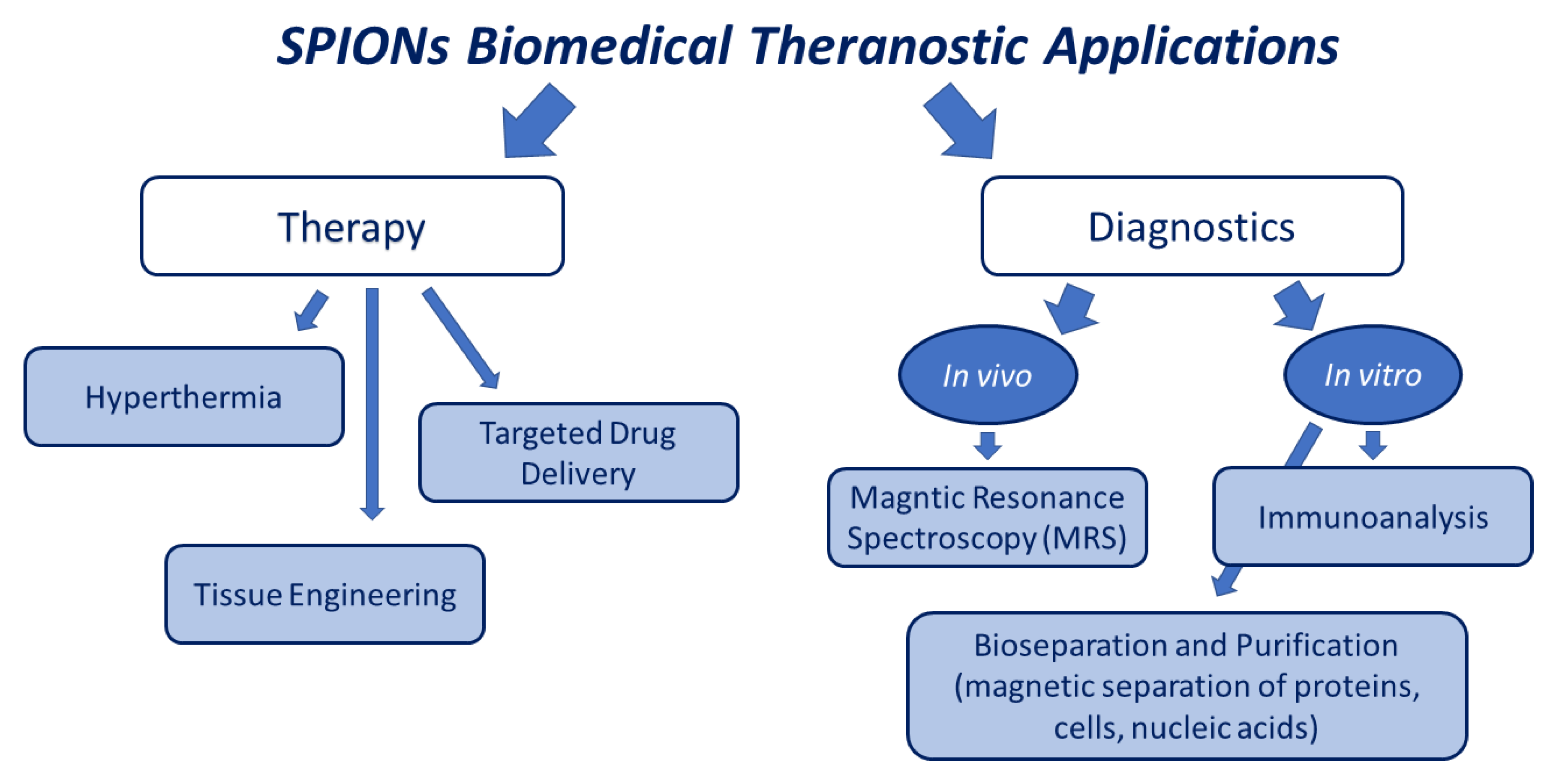
In recent years, SPIONs have shown great potential in a wide range of medical and near-medical fields (Figure 2). Below we will review particular applications of SPIONs.
5.1. Methods of Magnetic Bioseparation
The magnetic bioseparation method involves extraction of biological objects attached to the surface of nanomaterials with magnetic susceptibility from their biological medium by means of external magnetic field. Currently, superparamagnetic nanoparticles of iron oxides are used for bioseparation, as an external magnetic field easily magnetizes them in a magnetic separator. After the external magnetic field is removed, bioelements captured using superparamagnetic nanoparticles again diffuse into the solutions. The method of magnetic bioseparation is commonly used in the purification and separation of biological objects of various types, such as nucleic acids, proteins, bacteria, and cells.
Magnetic separation of proteins. Traditional methods of proteins separation, including precipitation in organic solvents, membrane separation, chromatography, and salting out regularly require changes in certain parameters, such as temperature, pH, ionic strength, dielectric constant, etc. These manipulations are labor-intensive and can damage target proteins. Among the most important methods for isolating proteins we should also mention the affinity chromatography. However, the inability of standard column liquid systems to handle samples containing solid particles is the disadvantage of all chromatography procedures of the kind. Recently, many methods combine magnetic nanoparticles with traditional separation and purification methods to separate different types of protein. Magnetic separation methods have noticeable advantages over standard separation procedures. These processes are usually quite non-sophisticated. One test tube can be used to perform all the stages of the cleaning procedure. Expensive filters, centrifuges, liquid chromatographic systems, or other equipment are not needed. Untreated samples containing suspended solid material can be used for the separation process. Some cases (for example, intracellular proteins isolation), even allow reducing the total separation time by combining the stages of separation and disintegration [57]. Target peptides or proteins are usually resistant to damage during magnetic separation. Even extensive protein complexes, which are destroyed by traditional methods, can remain unchanged after this procedure.
The magnetic affinity adsorption method involves the usage of particles with immobilized affinity ligands. Isolation of peptides and protein mostly often uses antibodies, streptavidin, protein A, and protein G. The above-mentioned immobilized ligands included into the magnetic particles can also serve as general solid phases allowing to immobilize modified affinity ligands (for example, biotinylated molecules, secondary antibodies, in the case of immobilized streptavidin or antibodies in the case of immobilized proteins G and A).
In order to achieve ligand immobilization, standard procedures in affine chromatography are used: modifying the inorganic particles surface to produce accessible functional groups that provide easy immobilization of affine ligands, such as -COOH,- OH, or -NH2. In this case, the surface of magnetite (or comparable magnetic materials including ferrites or maghemite) is modified by the method of silanization. In addition, encapsulated particles with an outer layer of a biocompatible polymer are used. Biopolymers such as alginate, chitosan, and agarose are used to prepare these particles. The simplest way to prepare magnetic polymer particles is combining the magnetic particles with a biopolymer solution, and the resulting bulk magnetic gel is mechanically divided into small particles. As an alternative, spherical particles are prepared using the water-in-oil suspension method.
Magnetic cell separation. Magnetic cell separation is considered very promising in bioengineering, clinical diagnostics, and food engineering. This method implies the use of superparamagnetic nanoparticles coated with antibodies to penetrate the cell surface antigens. In comparison with the traditional techniques of cells separation, their magnetic separation has obvious advantages [58,59]:
- target cells are easily separated from blood samples, bone marrow, ground tissues, or culture media;
- separation is not hindered by impurity or solvent ions; and
- this method does not lead to the destruction of detectable cells.
This method is suitable for detecting prokaryotic organisms including bacteria. Isolation of the specific bacteria associated with magnetic nanoparticles is done through the antigen–antibody reaction, and, as a rule, is carried out by inoculation of sample granules in a culture medium. Bacteria immunologically bound to magnetic particles, as a rule, preserve viability and can continue to multiply provided their nutritional needs are met. Subsequently, the isolated fraction can be washed to exclude non-specifically bound organisms before placing into a suitable growth medium. Target bacteria could be isolated from the media and accumulated to a suitable grade. The limitations of this method include the presence of antibodies applied to the surface of the target organism and the need for a significant concentration of free antigen to the target cell [60].
The method of immunomagnetic separation was used to separate cancer cells, blood lymphocytes, Salmonella typhimurium [61], Streptococcus mutans, Streptococcus sobrinus [62], and E. coli O157: H7 [63].
Magnetic separation of nucleic acids. Nucleic acid separation is becoming an important technique in molecular biology. Isolation of RNA and DNA is a stage preceding multiple diagnostic and biochemical processes, including cloning, detection, amplification, hybridization, sequencing, and DNA synthesis. Many subsequent manipulations are impeded in the presence of large amounts of contaminating materials, for instance, carbohydrates or proteins. Above that, DNA and RNA can be contaminated by each other. With that being said, many modern techniques that include DNA or RNA identification, for example, forensics, diagnosis of infections, typing of tissues and blood, and identification of genetic variations, require tools for reliable, efficient, and reproducible isolation of nucleic acids from complex compounds.
Prior to the use of modern technologies, nucleic acid separation was a laborious, multi-stage process of extraction and centrifugation, heavily restrained by low purity and small yields of the separating samples. Magnetic separation of nucleic acids demonstrated multiple advantages over the competing methods, allowing isolating nucleic acids directly from non-prepared samples, including culture media, water, physiological liquids, including blood, etc. Sample volumes produce practically no restrictions on this method. Samples can be taken relatively selectively and easily even from viscous compounds using the ability to adjust the magnetic properties of solid particles.
The isolation process uses magnetic carriers with immobilized affinity ligands or obtained by coating SPIONs with a biopolymer exhibiting affinity for the target nucleic acid. Methods for the synthesis of modified magnetic particles, as well as available commercial particles used for the separation of nucleic acids, are reviewed in [64].
5.2. Diagnostic Application of Magnetic Nanoparticles
MRS is a visualization method for clinical use, which originated from the measurement of nuclear magnetic resonance (NMR) signals. The effectiveness of MRS can be significantly improved by influencing the properties of the magnetic resonance signal of the examined tissues with contrast agents. Contrast agents are injected in 40–50% of all MRS examinations. MRS contrast agents are divided into positive (T1 agents) and negative (T2 agents) contrast agents. Positive contrasting agents can shorten the longitudinal relaxation time (T1) of protons, resulting in a brighter image. Negative contrasting agents can shorten the transverse relaxation time (T2) of protons.
Currently, the majority of available MRS contrasting agents are compounds based on gadolinium chelate complexes (T1 agents), as a brighter image is clinically preferable for easy detection and better resolution. Unfortunately, gadolinium-based compounds introduce a risk to the health of patients with liver or kidney diseases since they cannot be effectively removed from the body of such patients. In addition, these complexes induce brain damage because of unwanted accumulation. A general warning was issued by the US FDA for all contrast agents containing gadolinium with a recommendation not to use them in all acute renal failure cases.
Compared to gadolinium-based contrasting agents, the compositions based on magnetite and maghemite (T2) show higher biocompatibility and safety. However, a darker image using T2 contrast agent is characterized by low resolution and background noise. The clinical use of iron oxide nanoparticles in imaging the lymph nodes, spleen, and liver is limited as the particles accumulate passively at the mentioned sites. With that said, iron oxide nanoparticle-based contracts agents previously approved by the US FDA were banned in the United States.
However, according to an experimental study conducted over the past few years, extremely small magnetite and maghemite nanoparticles below 5 nm in size are potentially promising T1 contrast agents [65,66].
The use of magnetic nanoparticles in immunoassay. To obtain magnetic immunoanalytical agents, nanoparticles of magnetic iron oxides are associated with a wide range of compounds (antibodies, oligonucleatides, streptavidin, biotine, etc.). Magnetic nanoparticles are used in the analysis of a wide range of bacteria (Listeria monocytogenes, Escherichia coli, Chlamydia trachomatis, Vibrio cholerae, Salmonella, Shigella dysenteriae, Staphylococcus aureus, Vibno parahaemolyticus, Chlamydia trachomatis, etc.), West Nile virus, enteroviruses, and parasites such as Plasmodium falciparum (causing severe malaria) or Schistosoma mansoni [67,68,69].
There are several areas of application of iron oxides in immunoassay. Magnetic nanoparticles with immobilized immune-active reagents are used as a solid phase of immunoassay. Specific antibodies and antigens are immobilized on the surface of magnetic nanoparticles, and detection is carried out using enzymes, radioisotopes, fluorescent markers or chemiluminescent labels. This method is characterized by high expressivity, as the analysis is carried out in a pseudo-homogeneous mode (magnetic agents are dispersed in volume).
5.3. Tissue Engineering
Recently, magnetic nanoparticles are increasingly used in tissue engineering in the development of functional substitutes for damaged tissues. This new technology is a promising approach for overcoming the organ transplant stress caused by a shortage of donor organs. Tissue engineering includes the following processes,
- isolation of autologous cells from healthy tissues or stem cells, expansion of the number of cells to the required;
- transferring genes of interest to cells to enhance or modify cellular functions;
- construction of three-dimensional (3D) tissue-like structures; and
- transplantation of received designs to patients.
Even though the general technology of these processes in tissue engineering has already been established, there are still many problems that need to be addressed at each stage. The ability to manipulate and control cells remotely at each stage can provide a powerful tool for tissue engineering. Magnetic nanoparticles are such an instrument [71,72]. They can be used to isolate the necessary cells by magnetic separation methods for gene delivery when modifying cell functions. Magnetic nanoparticles attached to functional areas, both on the cell membrane and on the internal cellular components, can act as transducers of the applied magnetic fields and provide noninvasive control of various cellular functions. These particles can also be used to determine the location of cell adhesion, plating cells on a scaffold or in a predetermined shape. In 2006, a technique was developed for constructing a framework using magnetic particles [73]. Magnetic fields were used to position magnetic nanoparticles coated with thrombin in two-dimensional (2D) hexagonal arrays. Particles were used as nucleation centers for ordered fibrin growth, creating an ordered fibrin gel framework for endothelial cells.
In addition, magnetic particles have obvious advantages for in vivo use. It has been demonstrated that endothelial progenitor cells, which can promote angiogenesis and revascularization in ischemic regions, can be remotely guided both in vitro and in vivo using a magnetic field [74].
5.4. Magnetic Hyperthermia
Hyperthermia, a moderate increase in temperature to 40–43 °C, can cause the death of cancer cells and enhance the effects of radiation and chemotherapy. However, the achievement of its full potential as a clinically significant treatment method was limited due to its inability to heat malignant cells efficiently and locally. This problem can be circumvented by the intravenous administration of magnetic nanoparticles aimed at cancer cells that accumulate in the tumor, followed by the use of an alternating magnetic field to increase the temperature of the nanoparticles located in the tumor tissue. This targeted approach allows locally heating cancer cells, at the same time, without damaging surrounding normal tissues, which potentially increases the effectiveness and safety of hyperthermia. The most used materials for magnetic hyperthermia are magnetite or maghemite nanoparticles.
Magnetic nanoparticles can be delivered to the tumor via intratumoral, intra-arterial, intracavitary, and intravenous administration. Their oral administration is not possible as most of the nanoparticles will be excreted from the body. Intratumoral and intracavitary administration localizes magnetic particles in the tumor and can lead to effective heating of primary tumors. Although the above methods of administration are well suited for specific cases, intravenous administration is the most versatile delivery method for a wide range of oncological diseases. When magnetic particles of iron oxides are delivered in this way, the accumulation of nanoparticles in the tumor partially depends on the effect of increased permeability and retention [75]. This effect refers to the tendency of nanoparticles to predominantly accumulate in tumors due to the permeability of their vasculature and poor lymphatic drainage. Target ligands (antibodies and their fragments, ligands of specific receptors localized on the surface of tumor cells, peptides, and aptamers) associated with magnetic particles can enhance the absorption of nanoparticles by malignant cells [76]. Their predominant accumulation in malignant neoplasms leads to targeted local heating of tumors and the preservation of neighboring normal tissues under the influence of an alternating magnetic field.
Despite the promising results of preclinical trials of magnetic hyperthermia, there are many unsolved problems in this area. These include the establishment of optimal limits of magnetic field strength and frequency, their correlation with the duration of treatment, the toxicity of magnetic nanoparticles (including the dependence of toxicity on the presence of specific ligands that improve the accumulation of magnetic particles in tumor cells), and determining their optimal concentration in the affected organ.
5.5. Targeted Drug Delivery
One of the promising and rapidly developing areas of modern pharmacology is targeted delivery of drugs. About a hundred years ago in 1906, the chemist and biologist Paul Erlich (1854–1915) introduced the term “magic bullet” into scientific jargon. He used this term for a drug that into the patient’s body itself find and kill the causative agent of the disease, without harming the patient’s health. After the 1970s, the possibility of creating such drug delivery systems was under exploration. In such systems, medicinal substances are fixed on a carrier or included in capsules and associated with molecules (vectors) possessing affinity for certain cells [77]. Carriers delay the action of the drug substance until the target organs or tissues are reached, and the vectors transport the drug substance directly to the pharmacological target. There are two strategies for targeted delivery of drugs to damaged tissues: passive targeting and active targeting. The passive strategy is carried out by systems consisting of medicinal substances and carriers. It is provided due to the increased permeability of capillaries in the lesion. Active drug delivery systems involve vector molecules. Hormones, enzymes, peptides, antibodies, glycoproteins, glycolipids, and viruses can be considered as vectors for drug delivery. Carbon nanotubes, liposomes, micelles, polymers, dendrimers, fullerenes, nanodispersed silica, erythrocytes, leukocytes, etc. are currently the most actively studied as drug carriers.
For those drug delivery systems available at the present time, carriers and capsules for targeted drug delivery systems have several disadvantages that need to be excluded. The disadvantages of organic nanoparticles (polymer nanoparticles, liposomes, and micelles) include limited chemical and mechanical stability, swelling, susceptibility to microbiological attacks, lack of control over the rate of release of drugs, and high cost. Modern synthesis methods make it possible to obtain polymer particles with a wide distribution in size, which can lead to heterogeneous pharmacological properties. An alternative is the use of dendrimers which are monodisperse in nature and globular in morphology. However, the main disadvantage of dendrimers is their high cost. There are also problems with the removal of dendrimers from the body [54]. Inorganic carriers also have several disadvantages. The use of fullerenes, carbon nanotubes, and graphene and its oxide as carriers raises doubts about their toxicity. The use of SiO2 also raises a question in connection with the fact that silanol groups on the surface of silica interact with the surface of phospholipids of erythrocyte membranes, and this leads to hemolysis. In addition, silica can cause metabolic changes leading to the development of melanoma [77]. Thus, the question of choosing an optimal carrier for directed delivery systems remains unresolved.
Potential carriers and vectors in directional delivery systems are magnetic nanoparticles (Figure 3). Their main advantages are that they can be
- visualized (superparamagnetic nanoparticles are used as contrast agents for MRS),
- be guided or held in place by a magnetic field, and
- can be heated in a variable magnetic field to cause the release of the drug.
For use in drug delivery, these magnetic nanocarriers must be biocompatible and nontoxic. The size of magnetic nanoparticles affects not only the magnetic properties, charge, and surface chemistry, but it greatly affects the bioavailability of particles in the body. On the one hand, large particles with a diameter of more than 200 nm are excreted by the liver and spleen. Particles larger than 5 microns cause capillary blockade. Particles less than 10 nm in size will be quickly removed by renal clearance. Taking into account the critical size of the appearance of superparamagnetic properties in iron oxide nanoparticles, their optimal size (providing a long circulation time in the blood and the effectiveness of magnetic targeting) is 10–100 nm.
Methods for loading drugs onto magnetic particles are divided into two main types:
- chemical binding (compounding a drug substance with a carrier using chemical bonds) and
- physical binding (compounding a drug substance with a carrier via physical interactions).
Chemical bonding is usually formed by covalent bonds, which are formed due to amino, carboxyl, and thiol groups located on the surface of the magnetic particle and in the drug. As a rule, these functional groups are added to the surface of magnetic particles through their polymer coating (chitosan, polyetherimide, dextran, or polyethylene glycol). The same chemical groups can also be used to anchor peptides, antibodies, and viruses to provide improved targeting. The polymer coating not only allows us to fix the drug or vector on the surface of a magnetic particle, it also improves their hydrophobicity and stability. After magnetic induction, the release of the drug can be initiated not only by external stimulus factors, such as a limited increase in temperature, but also by internal factors, such as, for example, a change in pH value.
Physical binding. Physical interactions include electrostatic, hydrophilic/hydrophobic, and affinity interactions. This interaction has several advantages, including fast binding rate and high efficiency, it does not have intermediate stages of modification. However, physical interactions are often unable to avoid early drug release.
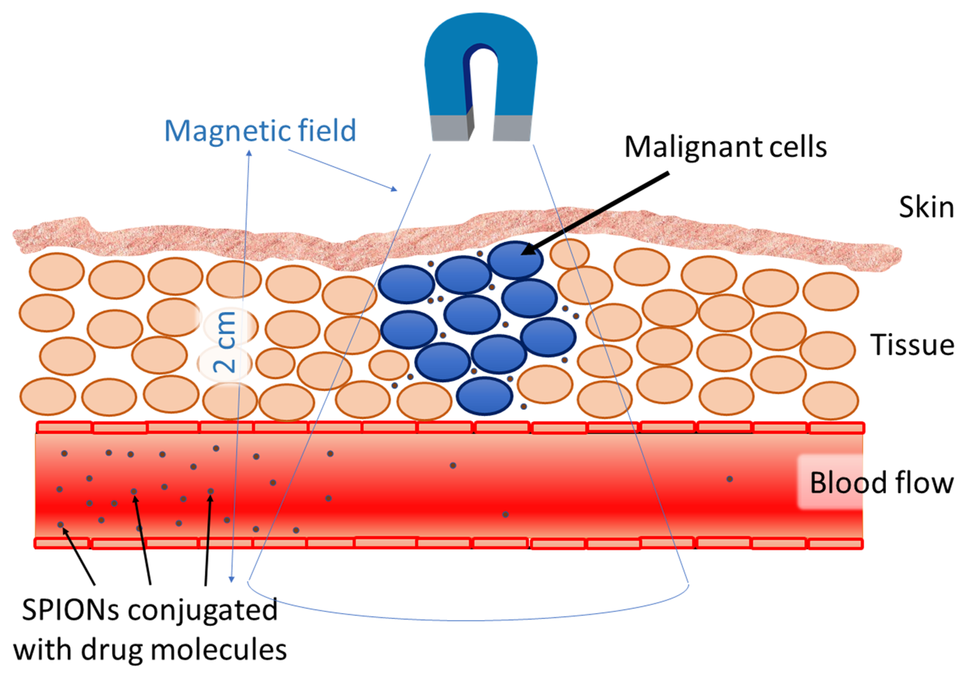
Magnetic fields are well suited for medical purposes because they do not hinder most biological processes. However, there are several problems associated with the use of magnetic carriers and vectors in targeted drug delivery systems [78]. One of them is the effect of blood flow on the target area of accumulation of targeted delivery systems. To retain magnetic particles in large arteries, strong magnetic fields will be required. The linear velocity of blood in large arteries is 50–100 times higher than the velocity of blood flow in the capillaries (±0.5 cm/s). Another problem associated with magnetic vectors is related to the depth of penetration of the magnetic field into the target object: it is difficult to send magnetic particles to targets located inside the body more than 2 cm from the skin, as the magnetic field decreases with distance. However, the work in [79] describes the development of a new drug delivery system using magnetic fields (using superconducting magnets) which can target magnetic particles in blood vessels located deep inside the body.
Magnetic targeting has been used to treat inflammatory processes [80]. The study was conducted in vivo. Magnetoliposomes better accumulate in the focus of inflammation under the influence of a magnetic field than in its absence. It was shown that the presence of the magnetic field led to lower amounts of iron in the liver, spleen, and plasma than was found in mice in which a magnet had not been applied.
Because of the technical difficulties described above, there have been few studies of magnetic drugs delivery systems in humans. They did not receive active distribution and wide development. After successful results in animal experiments [81], studies were conducted on 14 patients with advanced tumors (colon adenocarcinoma or hypernephroma). Intravenous administration of epirubicin-loaded ferrofluids (particle size 100 nm) was accompanied by magnetic targeting (0.8 T). The distance between the surface of the tumor and the magnets was less than 0.5 cm. According to the results obtained by MRS and histology, magnetic particles were successfully localized using 60–120 min of exposure. In another clinical study [82], 11 patients, after a failed traditional treatment of malignant tumors, were treated with magnetic drug delivery systems (0.8 T) and examined using MRS. Tumors of all 11 patients differed by location, but all of them were close to the skin. During the observation period, three patients showed positive dynamics, five patients were stable, and in three patients the disease progressed.
6. Conclusions and Prospects
Magnetic nanoparticles are becoming an attractive and increasingly important tool in the diagnostics and therapeutic treatment (theranostics) of diseases. They are actively used for a wide range of analyses and in the separation of genes, proteins, and cells. There are still many problems with finding the ideal agents for MRS, which increase the sensitivity of the method and at the same time have low toxicity.
However, nanosized particles of magnetic iron oxides have been shown to be of high potential in this area. Drug delivery using magnetic nanoparticles is still in the initial stages of the development. However, already conducted experiments open great opportunities for modern nanoscience and practical nanomedicine.
The development of artificial tissue engineering methods based on magnetic nanoparticles are also of great interest for many researchers and also opens broad prospects for transplantology and will help to solve the problems of deficiency of donor organs.
Author Contributions
Conceptualization, M.Y.M.; writing—original draft preparation, O.I.V. and V.P.S.; writing—review and editing, T.I.S. All authors have read and agreed to the published version of the manuscript.
Funding
The authors thank the Russian Science Foundation, grant number 16-13-10365 for the financial support.
Conflicts of Interest
The authors declare no conflict of interest.
References
- Jeevanandam, J.; Barhoum, A.; Chan, Y.S.; Dufresne, A.; Danquah, M.K. Review on nanoparticles and nanostructured materials: History, sources, toxicity and regulations. Beilstein J. Nanotechnol. 2018, 9, 1050–1074. [Google Scholar] [CrossRef] [Green Version]
- Boverhof, D.R.; Bramante, C.M.; Butala, J.H.; Clancy, S.F.; Lafranconi, M.; West, J.; Gordon, S.C. Comparative assessment of nanomaterial definitions and safety evaluation considerations. Regul. Toxicol. Pharm. 2015, 73, 137–150. [Google Scholar] [CrossRef] [PubMed] [Green Version]
- Wu, K.; Su, D.; Liu, J.; Saha, R.; Wang, J.P. Magnetic nanoparticles in nanomedicine: A review of recent advances. Nanotechnology 2019, 30, 502003. [Google Scholar] [CrossRef] [PubMed] [Green Version]
- Ziarani, M.G.; Malmir, M.; Lashgari, N.; Badiei, A. The role of hollow magnetic nanoparticles in drug delivery. RSC Adv. 2019, 9, 25094–25106. [Google Scholar] [CrossRef] [Green Version]
- Kaur, M.; Chopra, D.S. Green synthesis of iron nanoparticles for biomedical applications. Glob. J. Nanomed. 2018, 4, 68–77. [Google Scholar]
- Ding, X. Magnetic properties of nano zerovalent iron particles coated with biopolymers. Adv. Mater. Res. 2014, 912–914, 32–35. [Google Scholar] [CrossRef]
- Zhang, D.; Kong, Y.Y.; Sun, J.H.; Huo, S.J.; Zhou, M.; Gui, Y.L.; Mu, X.; Chen, H.Y.S.Q.; Xu, Q. Co-delivery nanoparticles with characteristics of intracellular precision release drugs for overcoming multidrug resistance. Int. J. Nanomed. 2017, 16, 2081–2108. [Google Scholar] [CrossRef] [Green Version]
- Li, Y.Q.; Xu, M.; Dhawan, U.; Liu, W.C.; Wu, K.T.; Liu, X.R.; Lin, C.; Zhao, G.; Wu, Y.C.; Chung, R.J. Iron-gold alloy nanoparticles serve as a cornerstone in hyperthermia-mediated controlled drug release for cancer therapy. Int. J. Nanomed. 2018, 13, 5499–5509. [Google Scholar] [CrossRef] [Green Version]
- Shokuhfar, A.; Seyyed, A.; Seyyed, S. Size controlled synthesis of FeCo alloy nanoparticles and study of the particle size and distribution effects on magnetic properties. Adv. Mater. Sci. Eng. 2014, 3, 1–10. [Google Scholar] [CrossRef] [Green Version]
- Jimenez, L.F.J.; Barros, A.H.; Caamaño De Ávila, Z.I. Synthesis, and characterization of Fe55Co45 magnetic nanoparticles by polyol and green chemistry method. Results in Phys. 2018, 15, 1–6. [Google Scholar] [CrossRef]
- Jaydeep, A.; Prateeti, C.; Balaram, D.; Arnab, D.; Sandeep, D.; Somenath, R.; Chen, J.; Tanmay, C. Preparation and characterization of ferromagnetic nickel oxide nanoparticles from three different precursors: Application in drug delivery. RSC Adv. 2015, 5, 35917–35928. [Google Scholar]
- Chaitali, D.; Arup, G.; Manisha, A.; Ajay, G.; Madhuri, M. Improvement of anticancer drug release by cobalt ferrite magnetic nanoparticles through combined pH and temperature responsive technique. ChemPhysChem 2018, 19, 1–18. [Google Scholar]
- Covaliu, C.I.; Jitaru, I.; Paraschiv, G.; Vasile, E.; Biriş, S.-Ş.; Diamandescu, L.; Ionita, V.; Iovu, H. Core–shell hybrid nanomaterials based on CoFe2O4 particles coated with PVP or PEG biopolymers for applications in biomedicine. Powder Technol. 2013, 237, 415–426. [Google Scholar] [CrossRef]
- Mirzaee, S.; Shayesteh, S.F. Ultrasound induced strain in ultrasmall CoFe2O4@polyvinyl alcohol nanocomposites. Ultrason. Sonochem. 2018, 40, 583–586. [Google Scholar] [CrossRef] [PubMed]
- Wang, G.; Ma, Y.; Mu, J.; Zhang, Z.; Zhang, X.; Zhang, L.; Zhang, H.; Che, Y.; Bai, J.H.; Xie, H. Monodisperse polyvinylpyrrolidone-coated CoFe2O4 nanoparticles: Synthesis, characterization and cytotoxicity study. Appl. Surf. Sci. 2016, 365, 114–119. [Google Scholar] [CrossRef]
- Phadatare, M.R.; Khot, V.M.; Salunkhe, A.B.; Thorat, N.D.; Pawar, S.H. Studies on polyethylene glycol coating on NiFe2O4 nanoparticles for biomedical applications. J. Magn. Magn. Mater. 2012, 324, 770–772. [Google Scholar] [CrossRef]
- Dong-Hyun, K.; Nikles, D.; Brazel, C. Synthesis and characterization of multifunctional chitosan- MnFe2O4 nanoparticles for magnetic hyperthermia and drug delivery. Materials 2010, 3, 4051–4065. [Google Scholar]
- Kanagesan, S.; Hashim, M.; AB Aziz, S.; Ismail, I.; Tamilselvan, S.; Alitheen, N.; Purna Chandra Rao, B. Evaluation of Antioxidant and Cytotoxicity Activities of Copper Ferrite (CuFe2O4) and Zinc Ferrite (ZnFe2O4) Nanoparticles Synthesized by Sol-Gel Self-Combustion Method. Appl. Sci. 2016, 6, 184. [Google Scholar] [CrossRef]
- Hoque, S.M.; Hossain, M.S.; Choudhury, S.; Akhter, S.; Hyder, F. Synthesis and characterization of ZnFe2O4 nanoparticles and its biomedical applications. Mater. Lett. 2016, 162, 60–63. [Google Scholar] [CrossRef] [Green Version]
- Srinivasan, S.Y.; Paknikar, K.M.; Bodas, D.; Gajbhiye, V. Applications of cobalt ferrite nanoparticles in biomedical nanotechnology. Nanomedicine 2018, 13, 1221–1238. [Google Scholar] [CrossRef]
- McBain, S.C.; You, H.H.P.; Dobson, J. Magnetic nanoparticles for gene and drug delivery. Int. J. Nanomed. 2008, 3, 169–180. [Google Scholar]
- Chang, M.-T.; Chou, L.-J.; Hsieh, C.-H.; Chueh, Y.-L.; Wang, Z.L.; Murakami, Y.; Shindo, D. Magnetic and electrical characterizations of half-metallic Fe3O4 nanowires. Adv. Mater. 2007, 19, 2290–2294. [Google Scholar] [CrossRef] [Green Version]
- Mantovan, R.; Lamperti, A.; Georgieva, M.; Tallarida, G.; Fanciulli, M. CVD synthesis of polycrystalline magnetite thin films: Structural, magnetic and magnetotransport properties. J. Phys. D Appl. Phys. 2010, 43, 065002. [Google Scholar] [CrossRef] [Green Version]
- Noqta, O.A.; Aziz, A.A.; Usman, I.A.; Bououdina, M. Recent advances in iron oxide nanoparticles: Synthesis and surface modification for biomedical applications. J. Supercond. Nov. Magn. 2019, 32, 779–797. [Google Scholar] [CrossRef]
- Ansari, S.; Ficiarà, E.; Ruffinatti, F.A.; Stura, I.; Argenziano, M.; Abollino, O.; Cavalli, R.; Guiot, C.; D’Agata, F. Magnetic iron oxide nanoparticles: Synthesis, characterization and functionalization for biomedical applications in the central nervous system. Materials 2019, 12, 465. [Google Scholar] [CrossRef] [Green Version]
- Al-Alawy, A.; Al-Abodi, E.; Kadhim, R. Synthesis and characterization of magnetic iron oxide nanoparticles by co-precipitation method at different conditions. J. Eng. 2018, 24, 60–72. [Google Scholar] [CrossRef] [Green Version]
- Wu1, W.; Wu, Z.; Yu, T.; Jiang, C.; Kim, W.S. Recent progress on magnetic iron oxide nanoparticles: Synthesis, surface functional strategies and biomedical applications. Sci. Technol. Adv. Mater. 2015, 16, 1–43. [Google Scholar]
- Belaïd, S.; Stanicki, D.; Elst, L.V.; Muller, R.N.; Laurent, S. Influence of experimental parameters on iron oxide nanoparticle properties synthesized by thermal decomposition: Size and nuclear magnetic resonance studies. Nanotechnology 2018, 29, 165603. [Google Scholar] [CrossRef]
- Cotin, G.; Kiefer, C.; Perton, F.; Ihiawakrim, D.; Blanco-Andujar, C.; Moldovan, S.; Lefevre, C.; Ersen, O.; Pichon, B.; Mertz, D.; et al. Unravelling the thermal decomposition parameters for the synthesis of anisotropic iron oxide nanoparticles. Nanomaterials 2018, 8, 881. [Google Scholar] [CrossRef] [Green Version]
- Daou, T.J.; Pourroy, G.; Bgin-Colin, S.; Grenche, J.; Ulhaq-Bouillet, C.; Legar, P.; Bernhardt, P.; Leuvrey, C.; Rogez, G.; Begin-Colin, S.; et al. Hydrothermal synthesis of monodisperse magnetite nanoparticles hydrothermal synthesis of monodisperse magnetite nanoparticles. Chem. Mater. 2006, 18, 4399–4404. [Google Scholar] [CrossRef]
- Yian, H.C.; Faizal, M.; Chin-Hua, C.; Shahidan, R.; Sarani, Z.; Huang, M.; Hn, L. Hydrothermal synthesis of magnetite nanoparticles as MRI contrast agents. Ceram. Int. 2010, 36, 1417–1422. [Google Scholar]
- Torres-Gómez, N.; Nava, O.; Argueta-Figueroa, L.; García-Contreras, R.; Baeza-Barrera, A.; Vilchis-Nestor, A.R. Shape tuning of magnetite nanoparticles obtained by hydrothermal synthesis: Effect of temperature. J. Nanomater. 2019, 1–15. [Google Scholar]
- Hugounenq, P.; Levy, M.; Alloyeau, D.; Lartigue, L.; Dubois, E.; Cabuil, V.; Ricolleau, C.; Roux, S.; Wilhelm, C.; Gazeau, F.; et al. Iron oxide monocrystalline nanoflowers for highly efficient magnetic hyperthermia. J. Phys. Chem. C 2012, 116, 15702–15712. [Google Scholar]
- Maity, D.; Chandrasekharan, P.; Si-Shen, F.; Xue, J.-M.; Ding, J. Polyol-based synthesis of hydrophilic magnetite nanoparticles. J. Appl. Phys. 2010, 107, 09B310. [Google Scholar] [CrossRef]
- Galindo-Gonzalez, C.; Gantz, S.; Ourry, L.; Mammeri, F.; Ammar-Merah, S.; Ponton, A. Elaboration and Rheological Investigation of Magnetic Sensitive Nanocomposite Biopolymer Networks. Macromolecules 2014, 47, 3136–3144. [Google Scholar] [CrossRef]
- Feijoo, A.V.; Lopez, M.T.; Galindo-Gonzalez, C.; Stange, S.; Nguyen, T.T.; Mammeri, F.; Ammar-Merah, S.; Ponton, A. Rheological investigation of magnetic sensitive biopolymer composites: Effect of the ligand grafting of magnetic nanoparticles. Rheol. Acta 2020, 59, 165–176. [Google Scholar] [CrossRef]
- Anzai, Y.; Piccoli, C.W.; Outwater, E.K.; Stanford, W.; Bluemke, D.A.; Nurenberg, P.; Saini, S.; Maravilla, K.R.; Feldman, D.E.; Schmiedl, U.P.; et al. Evaluation of neck and body metastases to nodes with ferumoxtran 10-enhanced MR imaging: Phase III safety and efficacy study. Radiology 2003, 228, 777–788. [Google Scholar] [CrossRef]
- Biehl, P.; Von der Lühe, M.; Dutz, S.; Schacher, F. Synthesis, characterization, and applications of magnetic nanoparticles featuring polyzwitterionic coatings. Polymers 2018, 10, 91. [Google Scholar] [CrossRef] [Green Version]
- Kurland, H.-D.; Grabow, J.; Staupendahl, G.; Müller, F.A.; Müller, E.; Dutz, S.; Bellemann, M.E. Magneticiron oxide nanopowders produced by CO2 laser evaporation ‘In situ’ coating and particle embedding ina ceramic matrix. J. Magn. Magn. Mater. 2009, 321, 1381–1385. [Google Scholar] [CrossRef]
- Kurland, H.-D.; Grabow, J.; Dutz, S.; Müller, E.; Sierka, M.; Müller, F.A. control of the crystalphase composition of FexOy nanopowders prepared by Co2 laser vaporization. Cryst. Growth Des. 2013, 13, 275–280. [Google Scholar]
- Timko, M.; Molcan, M.; Hashim, A.; Skumiel, A.; Müller, M.; Gojzewski, H.; Jozefczak, A.; Kovac, J.; Rajnak, M.; Makowski, M.; et al. Hyperthermic effect in suspension of magnetosomes prepared by variousmethods. IEEE Trans. Magn. 2013, 49, 250–254. [Google Scholar] [CrossRef]
- Koziaeva, V.; Grouzdev, D.; Dziuba, M.; Kolganova, T.; Kuznetsov, B. Diversity of magnetotactic bacteria of the Moskva River. Microbiology 2017, 86, 106–112. [Google Scholar] [CrossRef]
- Hergt, R.; Hiergeist, R.; Zeisberger, M.; Schuler, D.; Heyen, U.; Hilger, I.; Kaiser, W.A. Magnetic properties of bacterial magnetosomes as potential diagnostic and therapeutic tools. J. Magn. Magn. Mater. 2005, 293, 80–86. [Google Scholar] [CrossRef]
- Bibani, M.; Breitwieser, R.; Aubert, A.; Loyau, V.; Mercone, S.; Ammar, S.; Mammeri, F. Tailoring the magnetic properties of cobalt ferrite nanoparticles using the polyol process. Beilstein J. Nanotechnol. 2019, 10, 1166–1176. [Google Scholar] [CrossRef] [PubMed] [Green Version]
- Singh, N.; Jenkins, G.J.S.; Asadi, R.; Doak, S.H. Potential toxicity of superparamagnetic iron oxide nanoparticles. Nano Rev. 2010, 1, 5358. [Google Scholar] [CrossRef] [PubMed] [Green Version]
- Karlsson, H.L.; Cronholm, P.; Gustafsson, J.; Möller, L. Copper oxide nanoparticles are highly toxic: A comparison between metal oxide nanoparticles and carbon nanotubes. Chem. Res. Toxicol. 2008, 21, 1726–1732. [Google Scholar] [CrossRef]
- Zhu, X.; Tian, S.; Cai, Z. Toxicity assessment of iron oxide nanoparticles in zebrafish (Danio rerio) early life stages. PLoS ONE 2012, 7, e46286. [Google Scholar] [CrossRef] [Green Version]
- Bahadar, H.; Maqbool, F.; Niaz, K.; Abdollahi, M. Toxicity of nanoparticles and an overview of current experimental models. Iran. Biomed. J. 2016, 20, 1–11. [Google Scholar]
- García, A.; Espinosa, R.; Delgado, L.; Casals, E.; González, E.; Puntes, V.; Sánchez, A. Acute toxicity of cerium oxide, titanium oxide and iron oxide nanoparticles using standardized tests. Desalination 2011, 269, 136–141. [Google Scholar] [CrossRef] [Green Version]
- Yarjanli, Z.; Ghaedi, K.; Esmaeili, A.; Rahgozar, S.; Zarrabi, A. Iron oxide nanoparticles may damage to the neural tissue through iron accumulation, oxidative stress, and protein aggregation. BMC Neurosci. 2017, 18, 1–12. [Google Scholar] [CrossRef] [Green Version]
- Bhasin, G.; Kauser, H.; Athar, M. Iron augments stage-I and stage-II tumor promotion in murine skin. Cancer Lett. 2002, 183, 113–122. [Google Scholar] [CrossRef]
- Singh, N.; Manshian, B.; Jenkins, G.J.; Griffiths, S.M.; Williams, P.M.; Maffeis, T.G.; Wright, C.J.; Doak, S.H. NanoGenotoxicology: The DNA damaging potential of engineered nanomaterials. Biomaterials 2009, 30, 3891–3914. [Google Scholar]
- Vakili-Ghartavol, R.; Momtazi-Borojeni, A.A.; Vakili-Ghartavol, Z.; Aiyelabegan, H.T.; Jaafari, M.R.; Rezayat, S.M.; Bidgoli, S.A. Toxicity assessment of superparamagnetic iron oxide nanoparticles in different tissues. Artif. Cells Nanomed. Biotechnol. 2020, 48, 443–451. [Google Scholar] [CrossRef] [PubMed]
- Arruebo, M.; Fernández-Pacheco, R.; Ibarra, M.R.; Santamaría, J. Magnetic nanoparticles for drug delivery. Nano Today 2007, 2, 22–32. [Google Scholar] [CrossRef]
- Levy, M.; Luciani, N.; Alloyeau, D.; Elgrabli, D.; Deveaux, V.; Pechoux, C.; Chate, S.; Wang, G.; Vatsa, N.; Gendronf, F.; et al. Long term in vivo biotransformation of iron oxide nanoparticles. Biomaterials 2011, 32, 3988–3999. [Google Scholar] [CrossRef]
- Shah, A.; Dobrovolskaia, M.A. Immunological effects of iron oxide nanoparticles and iron-based complex drug formulations: Therapeutic benefits, toxicity, mechanistic insights, and translational considerations. Nanomed. Nanotechnol. Biol. Med. 2018, 14, 977–990. [Google Scholar] [CrossRef]
- Safarik, I.; Safarikova, M. Magnetic techniques for the isolation and purification of proteins and peptides. Biomagn. Res. Technol. 2004, 2, 7. [Google Scholar] [CrossRef] [Green Version]
- Hira, F.; Kyo-Seon, K. Magnetic nanoparticles for bioseparation. Korean J. Chem. Eng. 2017, 34, 589–599. [Google Scholar]
- Wu, A.; Ou, P.; Zeng, L. Biomedical applications of magnetic nanoparticles. Nano 2010, 5, 245–270. [Google Scholar] [CrossRef]
- Olsvik, O.; Popovic, T.; Skjerve, E.; Cudjoe, K.; Hornes, E.; Ugelstad, J.; Uhlen, M. Magnetic separation techniques in diagnostic microbiology. Clin. Microbiol. Rev. 1994, 7, 43–54. [Google Scholar] [CrossRef]
- Cai, G.; Wang, S.; Zheng, L.; Lin, J. A fluidic device for immunomagnetic separation of foodborne bacteria using self-assembled magnetic nanoparticle chains. Micromachines 2018, 9, 624. [Google Scholar] [CrossRef] [Green Version]
- Thanyasrisung, P.; Vittayaprasit, A.; Matangkasombut, O.; Sugai, M.; Na Nongkai, P.; Saipia, S.; Hoven, V.P. Separation and detection of mutans streptococci by using magnetic nanoparticles stabilized with a cell wall binding domain-conjugated polymer. Anal. Methods 2018, 10, 3332–3339. [Google Scholar] [CrossRef]
- Lin, J.; Li, M.; Li, Y.; Chen, Q. A high gradient and strength bioseparator with nano-sized immunomagnetic particles for specific separation and efficient concentration of E. coli O157:H7. J. Magn. Magn. Mater. 2015, 378, 206–213. [Google Scholar] [CrossRef]
- Berensmeier, S. Magnetic particles for the separation and purification of nucleic acids. Appl. Microbiol. Biotechnol. 2006, 73, 495–504. [Google Scholar] [CrossRef] [PubMed]
- Zeng, L.; Ren, W.; Zheng, J.; Cui, P.; Wu, A. Ultrasmall water-soluble metal-iron oxide nanoparticles as T-1-weighted contrast agents for magnetic resonance imaging. Phys. Chem. Chem. Phys. 2012, 14, 2631–2636. [Google Scholar] [CrossRef] [PubMed]
- Bao, Y.; Sherwood, J.A.; Sun, Z. Magnetic iron oxide nanoparticles as T1 contrast agents for magnetic resonance imaging. J. Mater. Chem. C 2018, 6, 1280–1290. [Google Scholar] [CrossRef]
- Zhang, L.; Tong, S.; Zhou, J.; Bao, G. Accurate quantification of disease markers in human serum using iron oxide nanoparticle-linked immunosorbent assay. Theranostics 2016, 6, 1353–1361. [Google Scholar] [CrossRef]
- Gao, L.; Fan, K.; Yan, X. Iron oxide nanozyme: A multifunctional enzyme mimetic for biomedical applications. Theranostics 2017, 7, 3207–3227. [Google Scholar] [CrossRef]
- Postnov, V.N.; Naumysheva, E.B.; Korolev, D.V.; Galagudza, M.M. Nanoscale drug delivery vehicles. Biotechnology 2013, 6, 16–42. (In Russian) [Google Scholar]
- Zhang, Z.; Xie, J.; Yu, J.; Lu, Z.; Yingshuai, L. A novel colorimetric immunoassay strategy using iron (III) oxide magnetic nanoparticle as a label for signal generation and amplification. J. Mater. Chem. B 2017, 5, 1454–1460. [Google Scholar] [CrossRef]
- Ito, A.; Kamihira, M. Tissue engineering using magnetite nanoparticles. Nanopart. Transl. Sci. Med. 2011, 355–395. [Google Scholar]
- Cartmell, S.H.; Dobson, J. The use of magnetic particles in tissue engineering. Nanotechnol. Life Sci. 2011. [Google Scholar] [CrossRef]
- Alsberg, E.; Feinstein, E.; Joy, M.P.; Prentiss, M.; Ingber, D.E. Magnetically—Guided self—Assembly of fi brin matrices with ordered nano—scale structure for tissue engineering. Tissue Eng. 2006, 12, 3247–3256. [Google Scholar] [CrossRef] [PubMed] [Green Version]
- Wilhelm, C.; Bal, L.; Smirnov, P.; Galy—Fauroux, I.; Clement, O.; Gazeau, F.; Emmerich, J. Magnetic control of vascular network formation with magnetically labeled endothelial progenitor cells. Biomaterials 2007, 28, 3797–3806. [Google Scholar] [CrossRef] [PubMed]
- Iyer, A.K.; Khaled, G.; Fang, J.; Maeda, H. Exploiting the enhanced permeability and retention effect for tumor targeting. Drug Discov. Today 2006, 11, 812–818. [Google Scholar] [CrossRef]
- DeNardo, S.J.; Denardo, G.L.; Natarajan, A.; Miers, L.A.; Foreman, A.R.; Gruettner, C.; Adamson, G.N.; Ivkov, R. Thermal dosimetry predictive of efficacy of 111In-ChL6 nanoparticle AMF–induced thermoablative therapy for human breast cancer in mice. J. Nucl. Med. 2007, 48, 437–444. [Google Scholar]
- Bharti, C.; Upendra, N.; Pal, A.; Gulati, N. Mesoporous silica nanoparticles in target drug delivery system: A review. Int. J. Pharm. Investig. 2015, 5, 124–133. [Google Scholar] [CrossRef] [Green Version]
- Bárcena, C.; Sra, A.K.; Gao, J. Applications of magnetic nanoparticles in biomedicine. Nanoscale Magn. Mater. Appl. 2009, 591–626. [Google Scholar]
- Takeda, S.; Mishima, F.; Fujimoto, S.; Izumi, Y.; Nishijima, S. Development of magnetically targeted drug delivery system using superconducting magnet. J. Magn. Magn. Mater. 2007, 311, 367–371. [Google Scholar] [CrossRef]
- Garcia-Jimeno, S.; Escribano, E.; Queralt, J.; Estelrich, J. External magnetic field-induced selective biodistribution of magnetoliposomes in mice. Nanoscale Res. Lett. 2012, 7, 452–459. [Google Scholar] [CrossRef] [Green Version]
- Lübbe, A.S.; Alexiou, C.; Bergemann, C. Clinical applications of magnetic drug targeting. J. Surg. Res. 2001, 95, 200–206. [Google Scholar] [CrossRef]
- Lemke, A.-J.; Senfft von Pilsach, M.I.; Lübbe, A.; Bergemann, C.; Riess, H.; Felix, R. MRI after magnetic drug targeting in patients with advanced solidmalignant tumors. Eur. Radiol. 2004, 14, 1949–1955. [Google Scholar] [CrossRef] [PubMed]
Scheme 1.
Coprecipitation method of superparamagnetic iron oxide nanoparticles (SPIONs) synthesys.

Scheme 2.
Oxidation method of SPIONs synthesys.

Scheme 3.
Thermal decomposition method of SPIONs synthesys.

Figure 1.
Methods of SPIONs synthesys.

Figure 2.
SPIONs biomedical theranostic applications.

Figure 3.
Directed by magnetic field delivery of SPIONs conjugated with drug molecules.

© 2020 by the authors. Licensee MDPI, Basel, Switzerland. This article is an open access article distributed under the terms and conditions of the Creative Commons Attribution (CC BY) license (http://creativecommons.org/licenses/by/4.0/).
Share and Cite
MDPI and ACS Style
Shabatina, T.I.; Vernaya, O.I.; Shabatin, V.P.; Melnikov, M.Y. Magnetic Nanoparticles for Biomedical Purposes: Modern Trends and Prospects. Magnetochemistry 2020, 6, 30. https://0-doi-org.brum.beds.ac.uk/10.3390/magnetochemistry6030030
AMA Style
Shabatina TI, Vernaya OI, Shabatin VP, Melnikov MY. Magnetic Nanoparticles for Biomedical Purposes: Modern Trends and Prospects. Magnetochemistry. 2020; 6(3):30. https://0-doi-org.brum.beds.ac.uk/10.3390/magnetochemistry6030030
Chicago/Turabian StyleShabatina, Tatyana I., Olga I. Vernaya, Vladimir P. Shabatin, and Mikhail Ya. Melnikov. 2020. "Magnetic Nanoparticles for Biomedical Purposes: Modern Trends and Prospects" Magnetochemistry 6, no. 3: 30. https://0-doi-org.brum.beds.ac.uk/10.3390/magnetochemistry6030030
Note that from the first issue of 2016, this journal uses article numbers instead of page numbers. See further details here.