Magnetite (Fe3O4) Nanoparticles in Biomedical Application: From Synthesis to Surface Functionalisation

 , and
, and
Abstract
:1. Introduction
2. Properties of Magnetite (Fe3O4) Nanoparticles
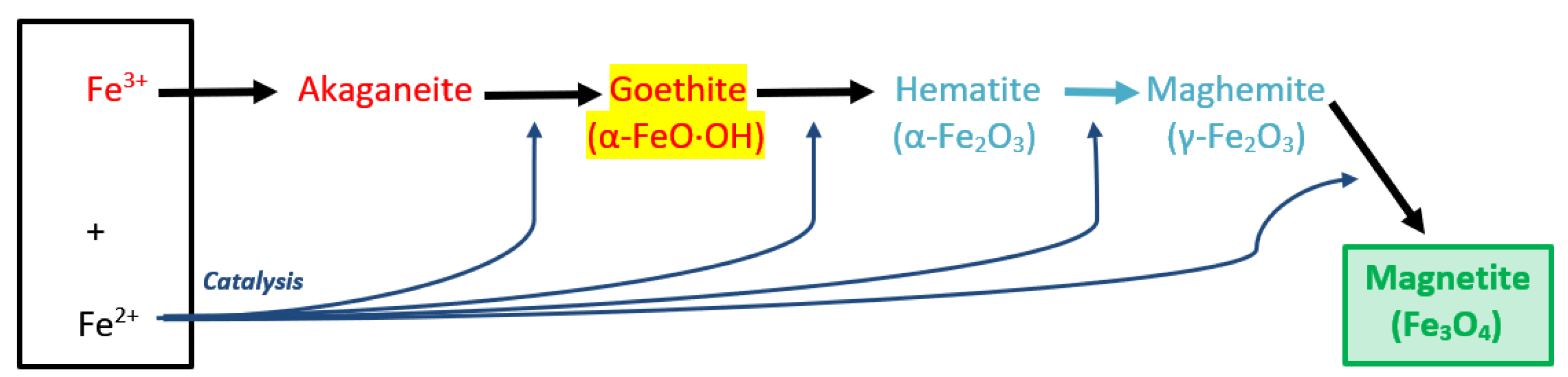
| FeO | + | Fe2O3 | → | Fe3O4 |
| (Ferrous Oxide) | (Ferric Oxide) | Magnetite |
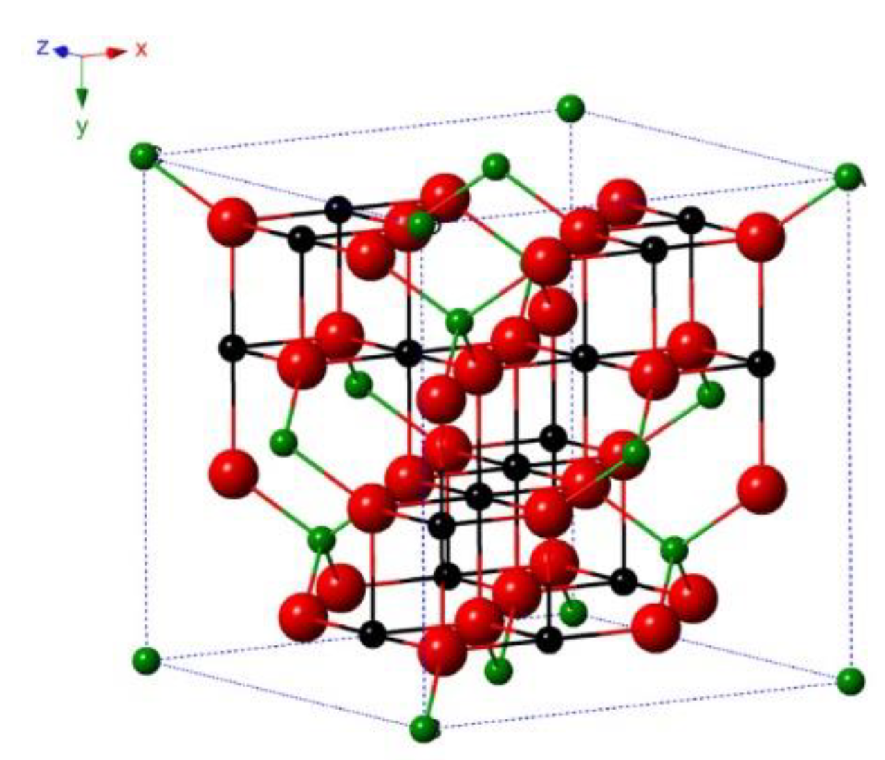
2.1. Structural and Physical
2.2. Thermal
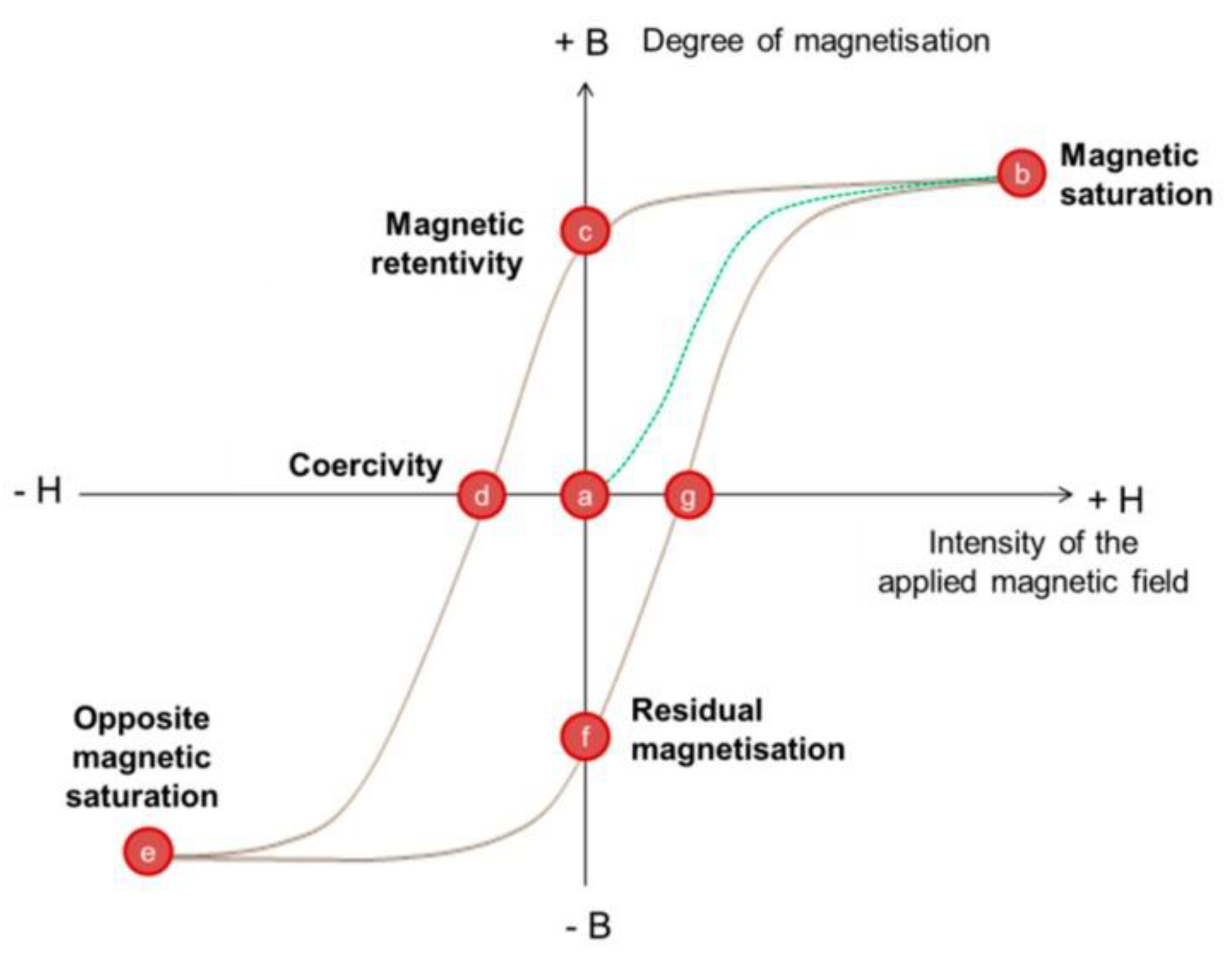
2.3. Magnetic
2.4. Optical
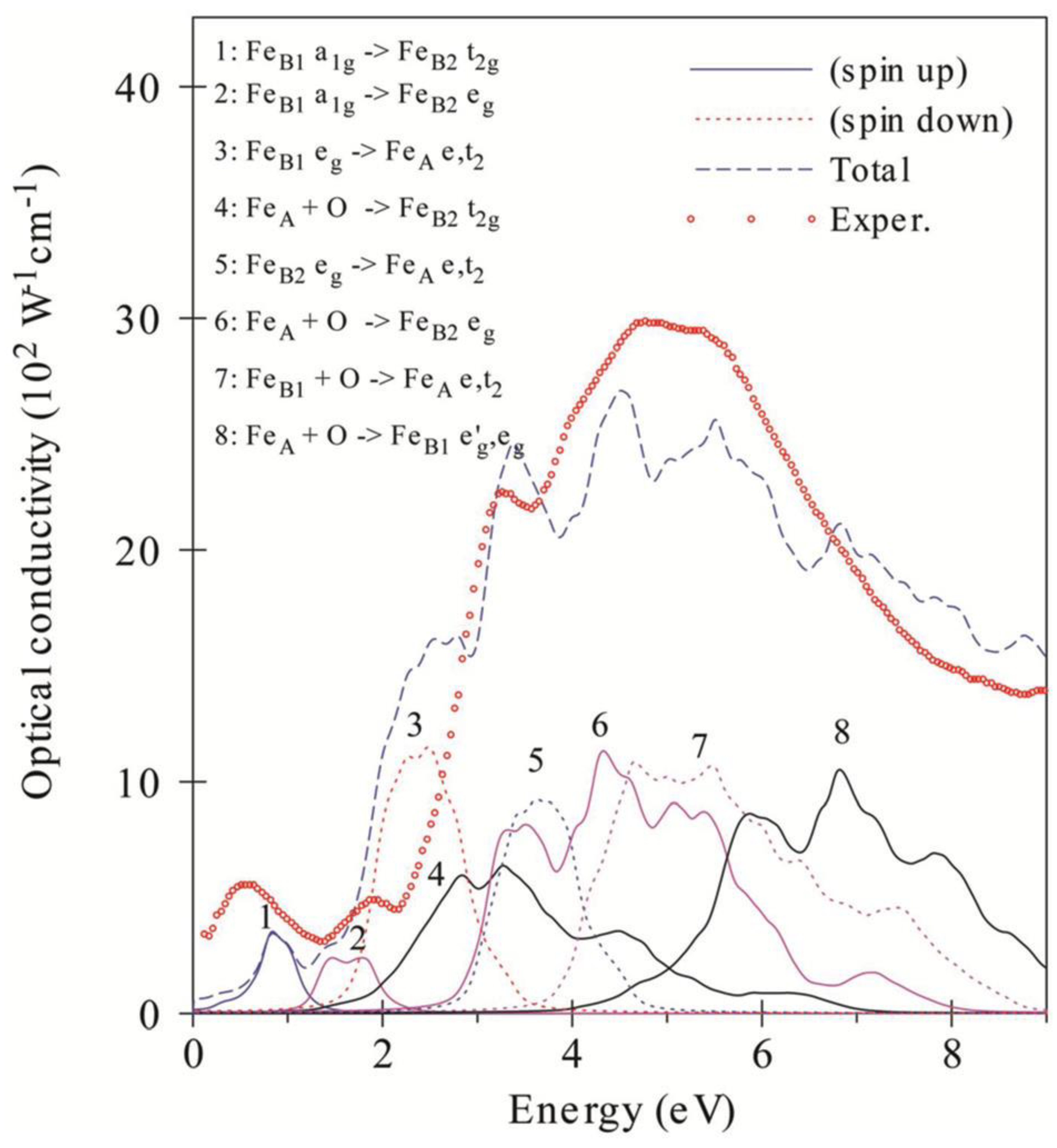
2.4.1. Electronic Band Structure
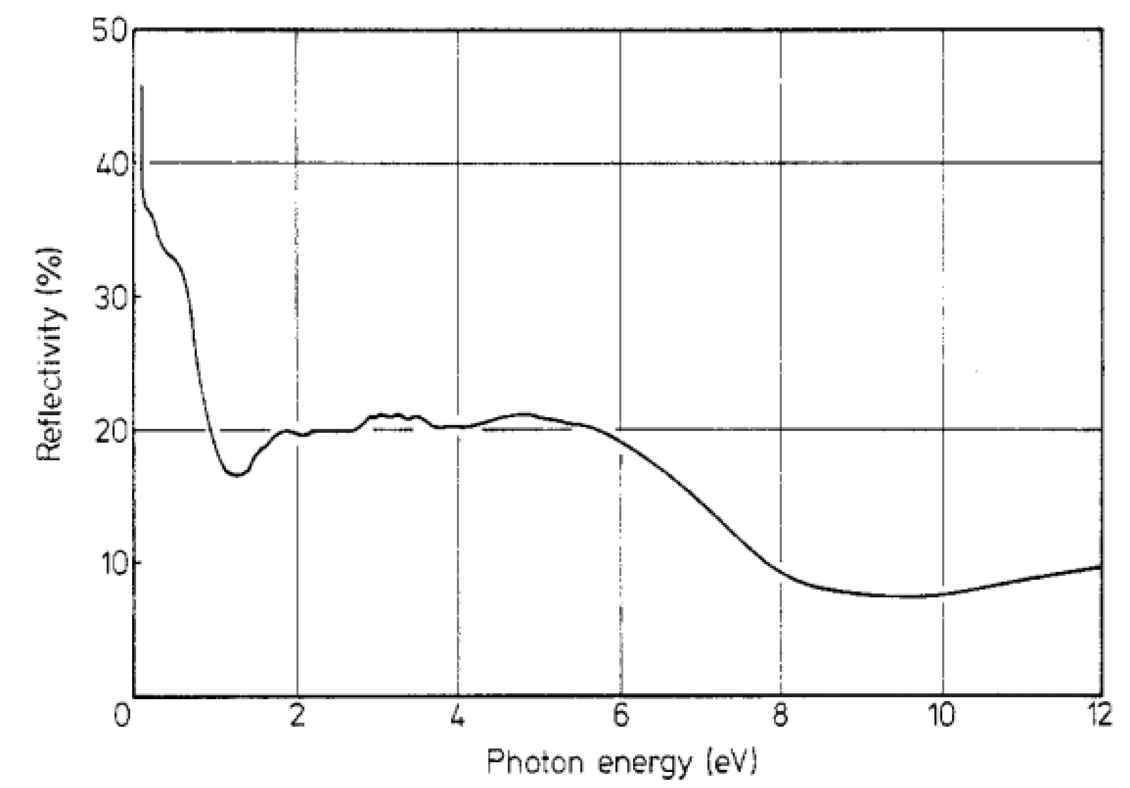
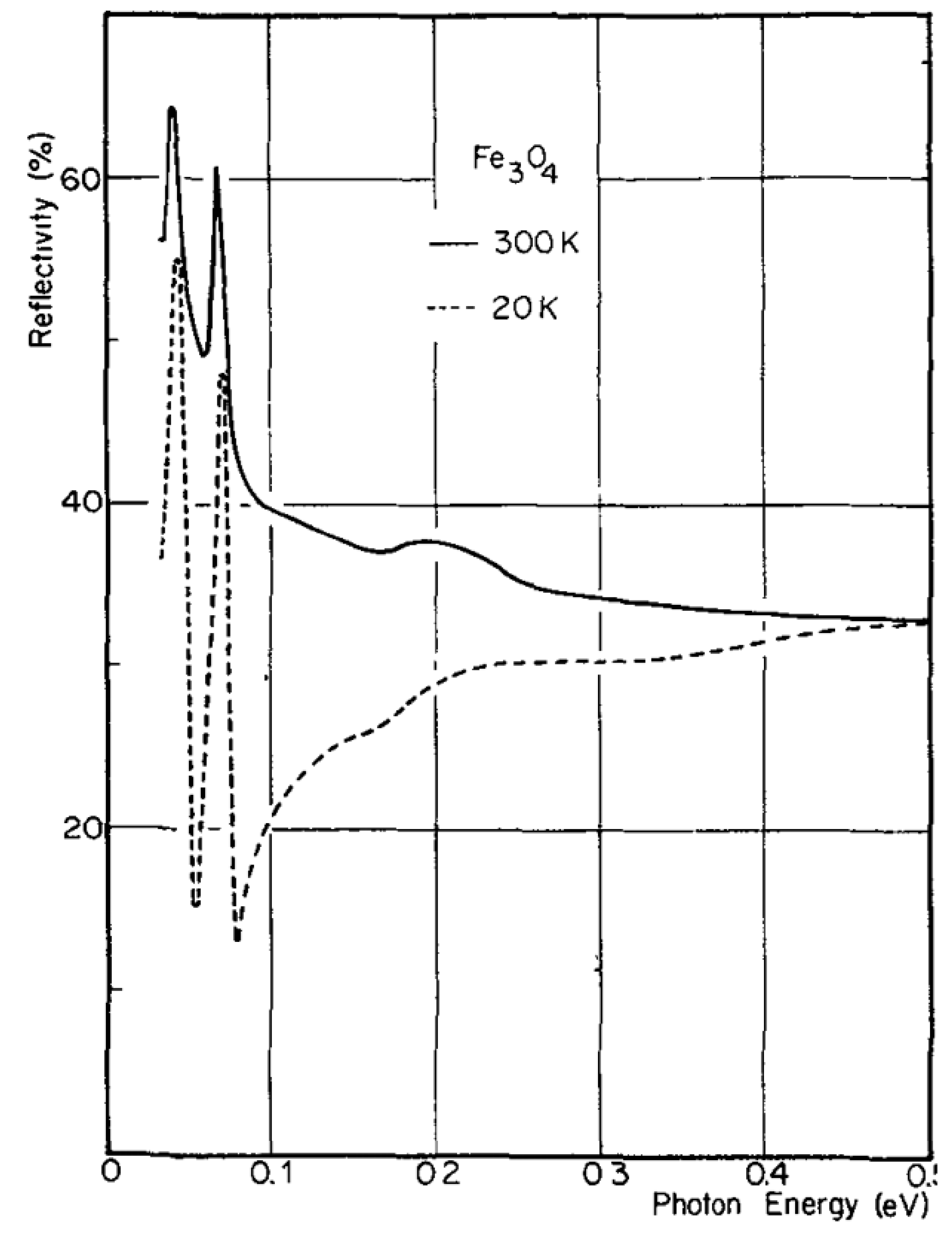
2.4.2. Reflectivity of Magnetite
2.5. Summary
3. Synthesis of Magnetite (Fe3O4) Nanoparticles
3.1. Co-precipitation Method
3.2. Solvothermal Method
3.3. Wet-chemical Reduction Method
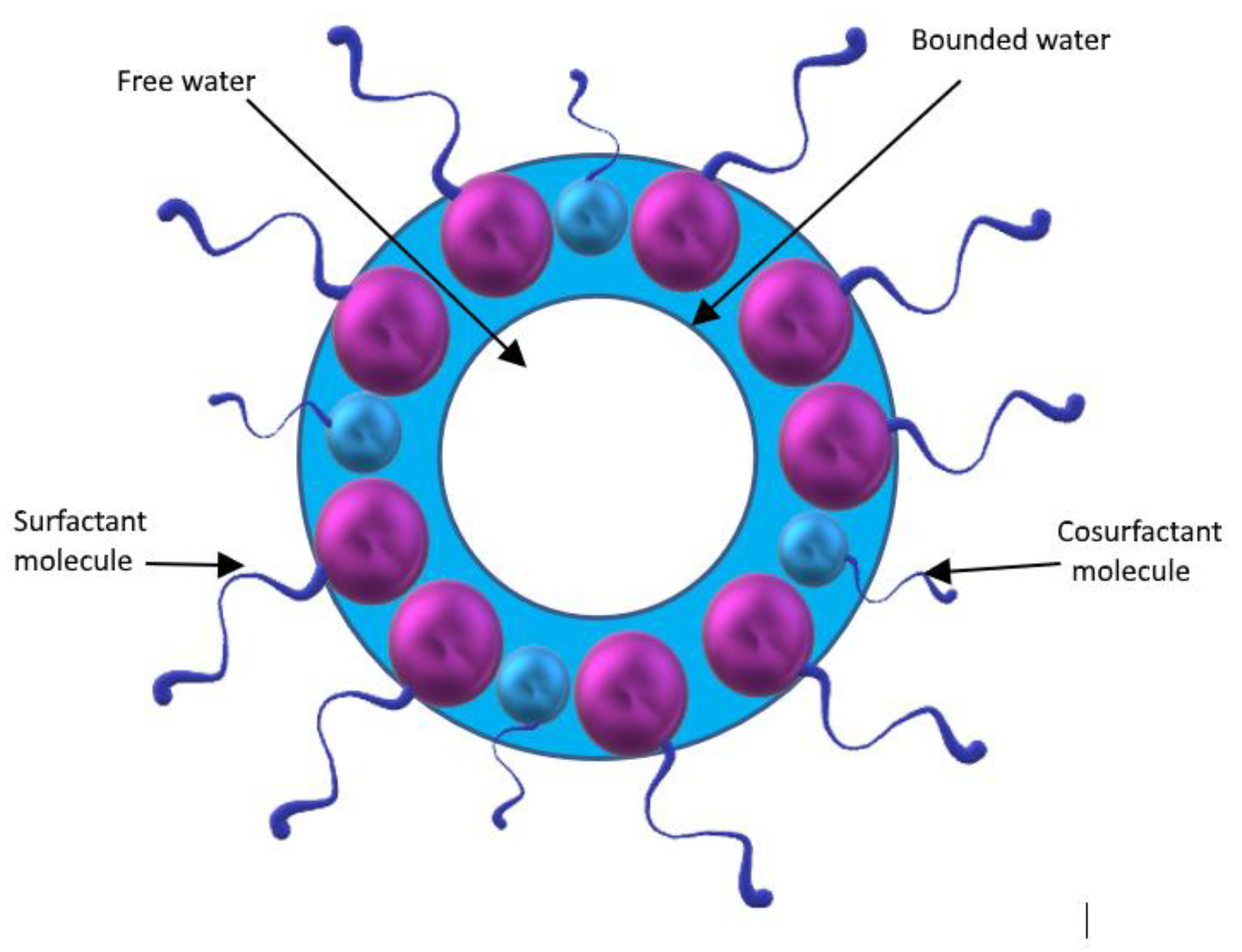
3.4. Micro-/nano-emulsion Method
3.5. Sonochemical or Sonolysis Method
3.6. Green Method (Biosynthesis)
3.7. Summary
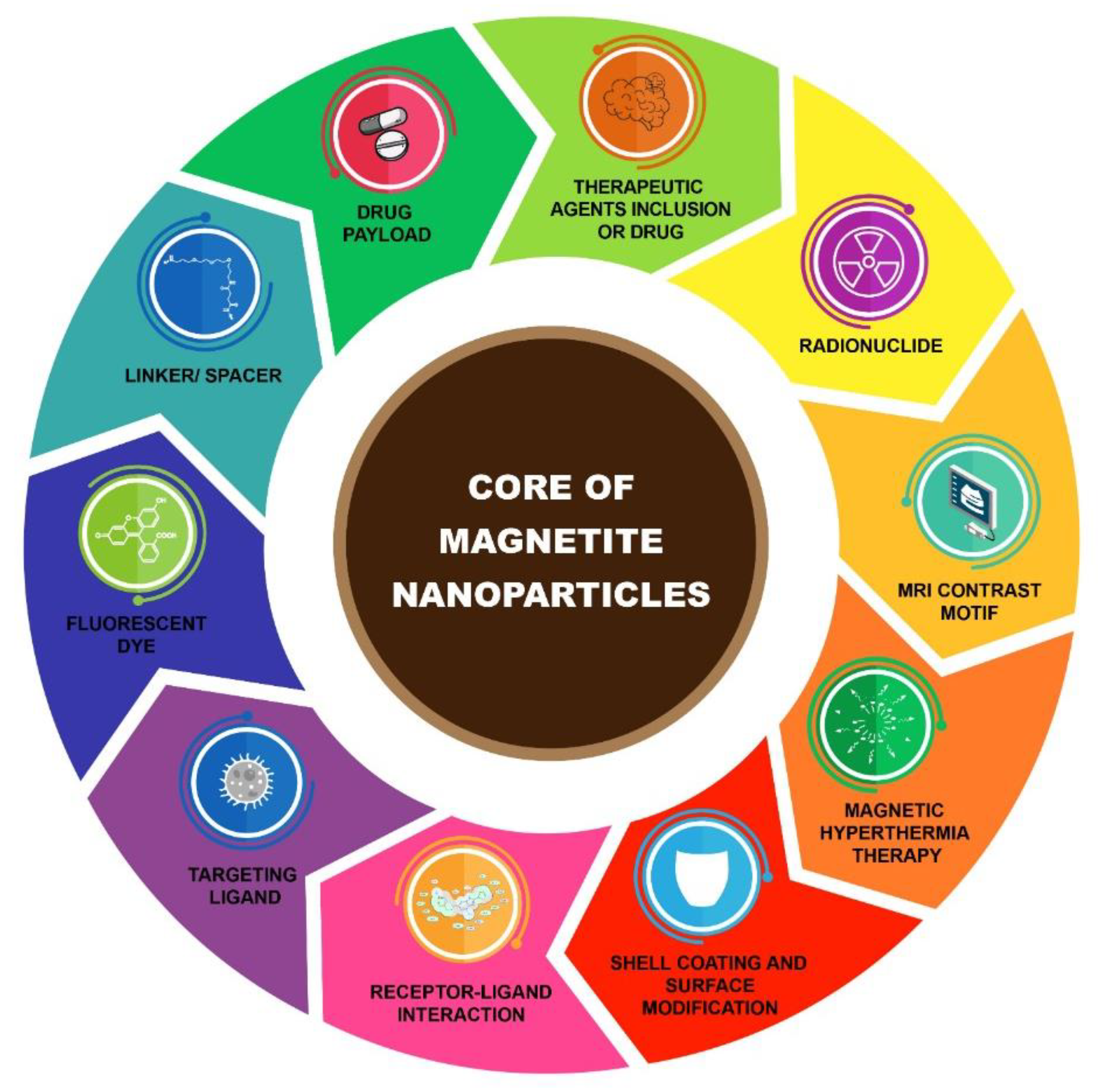
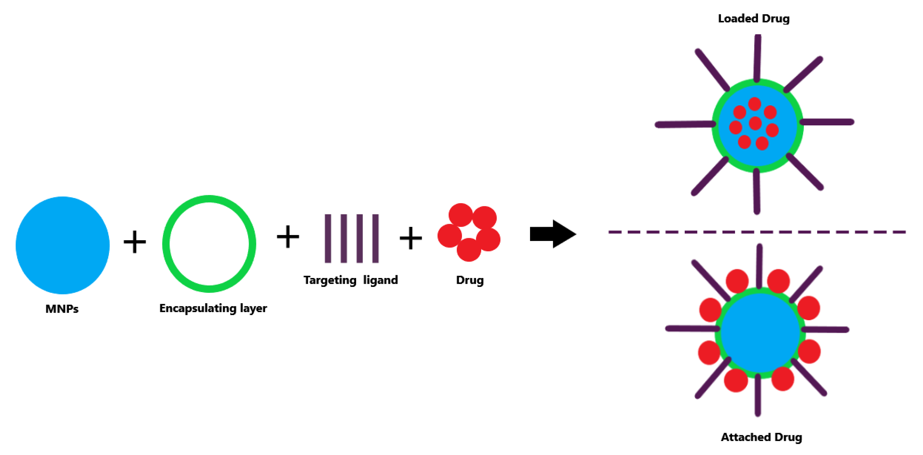
4. Surface Functionalisation of Magnetite (Fe3O4) Nanoparticles
4.1. Organic Functionalisation
4.1.1. Surfactants and Small Molecule
4.1.2. Polymers
4.1.3. Biomolecules
4.2. Inorganic Functionalisation
4.2.1. Silica (SiO2)
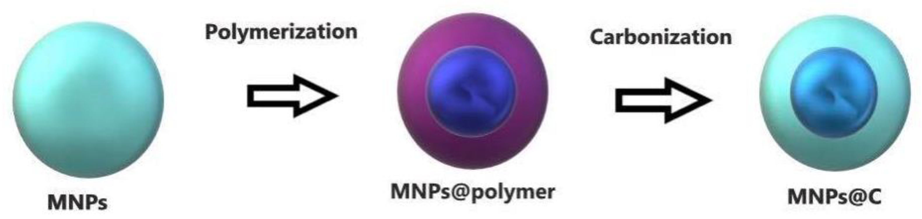
4.2.2. Carbon
5. Biomedical Applications of Magnetite (Fe3O4) Nanoparticles
5.1. In Vivo Applications
5.1.1. TDD
5.1.2. MRI
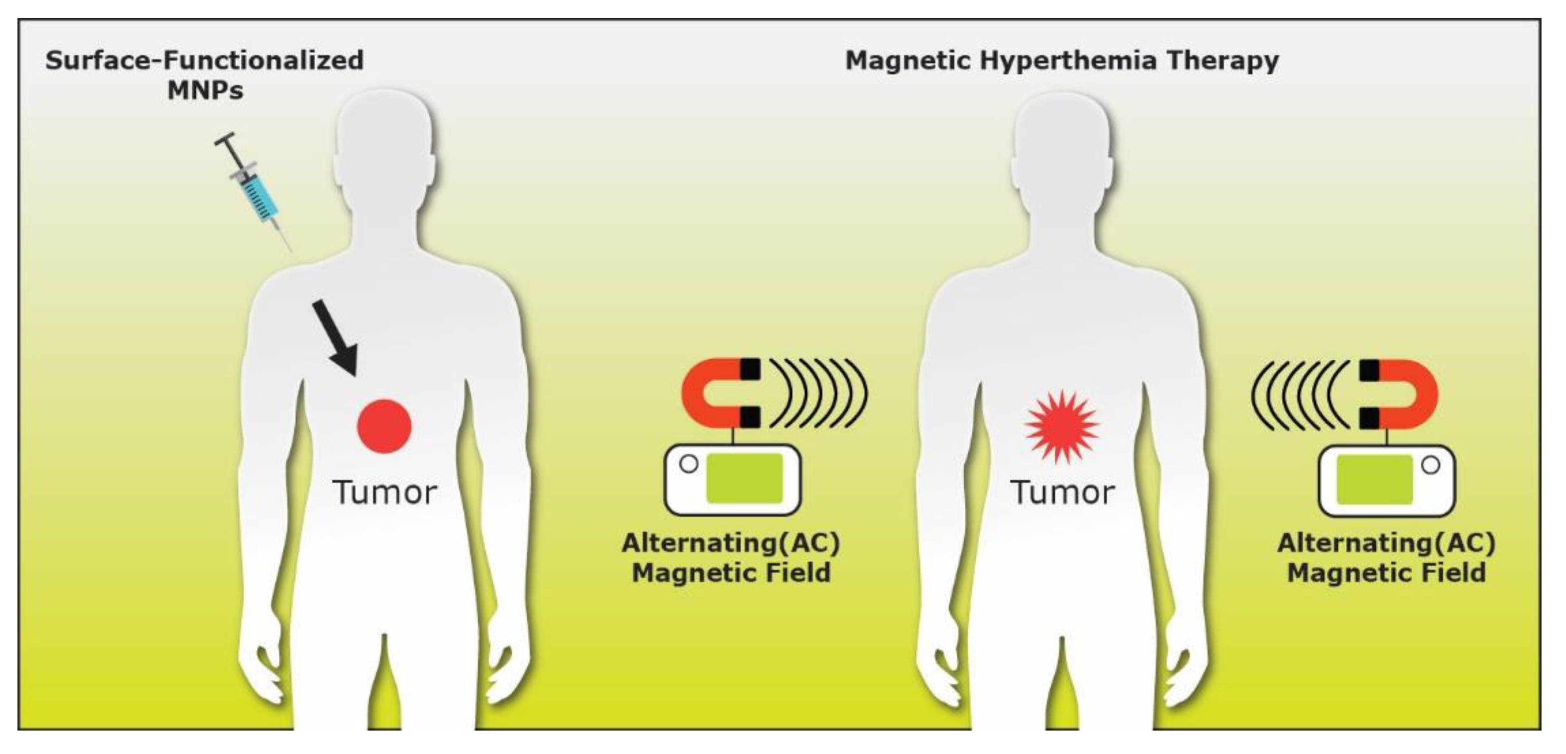
5.1.3. MHT
5.2. In Vitro Applications
5.2.1. Bioseparation
5.2.2. Biosensors
6. Conclusions and Perspectives
Author Contributions
Funding
Acknowledgments
Conflicts of Interest
References
- Sukumaran, S.; Neelakandan, M.S.; Shaji, N.; Prasad, P.; Yadunath, V.K. Magnetic Nanoparticles: Synthesis and Potential Biological Applications. JSM Nanotechnol. Nanomed. 2018, 6, 1068. [Google Scholar]
- Kandasamy, G.; Maity, D. Recent advances in superparamagnetic iron oxide nanoparticles (SPIONs) for in vitro and in vivo cancer nanotheranostics. Int. J. Pharm. 2015, 496, 191–218. [Google Scholar] [CrossRef] [PubMed]
- McNamara, K.; Tofail, S.A.M. Nanoparticles in biomedical applications. Adv. Phys. 2017, 2, 54–88. [Google Scholar] [CrossRef]
- Ghazanfari, M.R.; Kashefi, M.; Shams, S.F.; Jaafari, M.R. Perspective of Fe3O4 Nanoparticles Role in Biomedical Applications. Biochem. Res. Int. 2016, 2016, 1–32. [Google Scholar] [CrossRef] [Green Version]
- Bhuiyan, M.T.H.; Chowdhury, M.N.; Parvin, M.S. Potential Nanomaterials and their Applications in Modern Medicine: An Overview. ARC J. Cancer Sci. 2016, 2, 25–33. [Google Scholar]
- Chen, Y.-T.; Kolhatkar, A.G.; Zenasni, O.; Xu, S.; Lee, T.R. Biosensing Using Magnetic Particle Detection Techniques. Sensors 2017, 17, 2300. [Google Scholar] [CrossRef] [PubMed]
- Chen, Y.; Ding, X.; Zhang, Y.; Natalia, A.; Sun, X.; Wang, Z.; Shao, H. Design and synthesis of magnetic nanoparticles for biomedical diagnostics. Quant. Imaging Med. Surg. 2018, 8, 957–970. [Google Scholar] [CrossRef] [PubMed]
- McNamara, K.; Tofail, S.A. Nanosystems: The use of nanoalloys, metallic, bimetallic, and magnetic nanoparticles in biomedical applications. Phys. Chem. Chem. Phys. 2015, 17, 27981–27995. [Google Scholar] [CrossRef] [PubMed]
- Liu, Y.; Zhu, W.; Wu, D.; Wei, Q. Electrochemical determination of dopamine in the presence of uric acid using palladium-loaded mesoporous Fe3O4 nanoparticles. Measurement 2015, 60, 1–5. [Google Scholar] [CrossRef]
- Sheng-Nan, S.; Chao, W.; Zan-Zan, Z.; Yang-Long, H.; Venkatraman, S.S.; Zhi-Chuan, X. Magnetic iron oxide nanoparticles: Synthesis and surface coating techniques for biomedical applications. Chin. Phys. B 2014, 23, 037503. [Google Scholar]
- Xu, J.-K.; Zhang, F.-F.; Sun, J.-J.; Sheng, J.; Wang, F.; Sun, M. Bio and Nanomaterials Based on Fe3O4. Molecules 2014, 19, 21506–21528. [Google Scholar] [CrossRef] [PubMed]
- Blaney, L. Magnetite (Fe3O4): Properties, Synthesis, and Applications. Lehigh Univ. Lehigh Preserv. 2007, 15, 5. [Google Scholar]
- Wu, W.; Wu, Z.; Yu, T.; Jiang, C.Z.; Kim, W.-S. Recent progress on magnetic iron oxide nanoparticles: Synthesis, surface functional strategies and biomedical applications. Sci. Technol. Adv. Mater. 2015, 16, 023501. [Google Scholar] [CrossRef] [PubMed]
- Schlegel, A.; Alvarado, S.F.; Wachter, P. Optical properties of magnetite (Fe3O4). J. Phys. C Solid State Phys. 1979, 12, 1157–1164. [Google Scholar] [CrossRef]
- Cornell, R.M.; Schwertmann, U. The Iron Oxides: Structure, Properties, Reactions, Occurrenc and Uses; VCH: Weinheim, Germany, 1996; pp. 6, 32, 119. [Google Scholar]
- Ju, S.; Cai, T.; Lu, H.-S.; Gong, C.-D. Pressure-Induced Crystal Structure and Spin-State Transitions in Magnetite (Fe3O4). J. Am. Chem. Soc. 2012, 134, 13780–13786. [Google Scholar] [CrossRef] [PubMed]
- Sun, S.; Zeng, H. Size-Controlled Synthesis of Magnetite Nanoparticles. J. Am. Chem. Soc. 2002, 124, 8204–8205. [Google Scholar] [CrossRef] [PubMed]
- Martinez, G.; Malumbres, A.; Mallada, R.; Hueso, J.L.; Irusta, S.; Bomatí-Miguel, O.; Santamaría, J. Use of a polyol liquid collection medium to obtain ultrasmall magnetic nanoparticles by laser pyrolysis. Nanotechnology 2012, 23, 425605. [Google Scholar] [CrossRef]
- Bohra, M.; Agarwal, N.; Singh, V. A Short Review on Verwey Transition in Nanostructured Fe3O4 Materials. J. Nanomater. 2019, 2019, 1–18. [Google Scholar] [CrossRef] [Green Version]
- Liu, H.; Di Valentin, C. Band Gap in Magnetite above Verwey Temperature Induced by Symmetry Breaking. J. Phys. Chem. C 2017, 121, 25736–25742. [Google Scholar] [CrossRef]
- Wright, J.P.; Attfield, J.P.; Radaelli, P.G. Charge ordered structure of magnetite Fe3O4 below the Verwey transition. Phys. Rev. B 2002, 66, 214422. [Google Scholar] [CrossRef]
- Tanwar, S.; Awana, V.P.S.; Singh, S.P.; Pasricha, R. Magnetic Field Dependence of Blocking Temperature in Oleic Acid Functionalized Iron Oxide Nanoparticles. J. Supercond. Nov. Magn. 2012, 25, 2041–2045. [Google Scholar] [CrossRef]
- Saragi, T.; Sinaga, H.D.; Rahmi, F.; Pramesti, G.A.; Sugiarto, A.; Therigan, A.; Syakir, N.; Hidayat, S. Risdiana Blocking Temperature of Magnetite Nanoparticles Fe3O4 Encapsulated Silicon Dioxide SiO2. Key Eng. Mater. 2020, 855, 172–176. [Google Scholar] [CrossRef]
- Rumpf, K.; Granitzer, P.; Morales, M.D.P.; Poelt, P.; Reissner, M. Variable blocking temperature of a porous silicon/Fe3O4 composite due to different interactions of the magnetic nanoparticles. Nanoscale Res. Lett. 2012, 7, 445. [Google Scholar] [CrossRef] [PubMed] [Green Version]
- Majewski, P.; Thierry, B. Functionalized Magnetite Nanoparticles—Synthesis, Properties, and Bio-Applications. Crit. Rev. Solid State Mater. Sci. 2007, 32, 203–215. [Google Scholar] [CrossRef]
- Moaca, E.-A.; Coricovac, D.E.; Şoica, C.; Pinzaru, I.A.; Păcurariu, C.; Dehelean, C. Preclinical Aspects on Magnetic Iron Oxide Nanoparticles and Their Interventions as Anticancer Agents: Enucleation, Apoptosis and Other Mechanism. In Iron Ores and Iron Oxide Materials; IntechOpen: London, UK, 2018. [Google Scholar]
- Wallyn, J.; Anton, N.; Vandamme, T.F. Synthesis, Principles, and Properties of Magnetite Nanoparticles for In Vivo Imaging Applications—A Review. Pharmaceutics 2019, 11, 601. [Google Scholar] [CrossRef] [Green Version]
- Thapa, D.; Palkar, V.; Kurup, M.; Malik, S. Properties of magnetite nanoparticles synthesized through a novel chemical route. Mater. Lett. 2004, 58, 2692–2694. [Google Scholar] [CrossRef] [Green Version]
- Unni, M.; Uhl, A.M.; Savliwala, S.; Savitzky, B.H.; Dhavalikar, R.; Garraud, N.; Arnold, D.P.; Kourkoutis, L.F.; Andrew, J.S.; Rinaldi, C. Thermal Decomposition Synthesis of Iron Oxide Nanoparticles with Diminished Magnetic Dead Layer by Controlled Addition of Oxygen. ACS Nano 2017, 11, 2284–2303. [Google Scholar] [CrossRef]
- Muscas, G.; Singh, G.; Glomm, W.R.; Mathieu, R.; Kumar, P.A.; Concas, G.; Agostinelli, E.; Peddis, D. Tuning the Size and Shape of Oxide Nanoparticles by Controlling Oxygen Content in the Reaction Environment: Morphological Analysis by Aspect Maps. Chem. Mater. 2015, 27, 1982–1990. [Google Scholar] [CrossRef]
- Daoush, W.M. Co-Precipitation and Magnetic Properties of Magnetite Nanoparticles for Potential Biomedical Applications. J. Nanomed. Res. 2017, 5, 1–6. [Google Scholar] [CrossRef]
- Alvarado, S.; Erbudak, M.; Munz, P. Final state effects in the 3d-photoelectron spectrum of Fe3O4 and comparison with FexO. Phys. B C 1977, 86, 1188–1190. [Google Scholar] [CrossRef]
- Zhang, X.X.; Schoenes, J.; Reim, W.; Wachter, P. Evidence for 3dn to 3dn-14s transitions in magnetite and in lithium and magnesium ferrites. J. Phys. C Solid State Phys. 1983, 16, 6055. [Google Scholar] [CrossRef]
- Fontijn, W.W.; Van Der Zaag, P.J.; Devillers, M.A.C.; Brabers, V.A.M.; Metselaar, R.R. Optical and magneto-optical polar Kerr spectra of Fe3O4 and Mg2+- or Al3+-substituted Fe3O4. Phys. Rev. B 1997, 56, 5432–5442. [Google Scholar] [CrossRef] [Green Version]
- Schlegel, A.; Wächter, P. Optical properties of magnetite (Fe3O4) in the infrared. J. Phys. Colloq. 1980, 41, C5–C19. [Google Scholar] [CrossRef]
- Shi, D.; Sadat, M.E.; Dunn, A.W.; Mast, D.B. Photo-fluorescent and magnetic properties of iron oxide nanoparticles for biomedical applications. Nanoscale 2015, 7, 8209–8232. [Google Scholar] [CrossRef] [PubMed]
- Boxall, C.; Kelsall, G.; Zhang, Z. Photoelectrophoresis of colloidal iron oxides. Part 2.—Magnetite (Fe3O4). J. Chem. Soc. Faraday Trans. 1996, 92, 791–802. [Google Scholar] [CrossRef]
- Fontijn, W.W.; Van Der Zaag, P.J.; Feiner, L.F.; Metselaar, R.R.; Devillers, M.A.C. A consistent interpretation of the magneto-optical spectra of spinel type ferrites (invited). J. Appl. Phys. 1999, 85, 5100–5105. [Google Scholar] [CrossRef] [Green Version]
- Antonov, V.N.; Harmon, B.N.; Antropov, V.P.; Perlov, A.Y.; Yaresko, A.N. Electronic structure and magneto-optical Kerr effect of Fe3O4 and Mg2+- or Al3+-substituted Fe3O4. Phys. Rev. B 2001, 64, 134410. [Google Scholar] [CrossRef]
- Park, S.K.; Ishikawa, T.; Tokura, Y. Charge-gap formation upon the Verwey transition in Fe3O4. Phys. Rev. B 1998, 58, 3717–3720. [Google Scholar] [CrossRef]
- Pan, P.; Lin, Y.; Gan, Z.X.; Luo, X.; Zhou, W.; Zhang, N. Magnetic field enhanced photothermal effect of Fe3O4 nanoparticles. J. Appl. Phys. 2018, 123, 115115. [Google Scholar] [CrossRef]
- Cornell, R.M.; Schwertmann, U. Chapter 2–Crystal structure. In The Iron Oxides: Structure, Properties, Reactions, Occurrences and Uses, 2nd ed.; Wiley-VCH Verlag GmbH & Co. KGaA: Weinheim, Germany, 2003; 9p. [Google Scholar]
- Chicot, D.; Mendoza, J.; Zaoui, A.; Louis, G.; Lepingle, V.; Roudet, F.; Lesage, J. Mechanical properties of magnetite (Fe3O4), hematite (alpha-Fe2O3) and goethite (alpha-FeO center dot OH) by instrumented indentation and molecular dynamics analysis. Mater. Chem. Phys. 2011, 129, 862–870. [Google Scholar] [CrossRef]
- Oolman, K.B.; Kattel, S.; Rice, K.P.; Rice, W.D. Magneto-Optical Properties of Iron Oxide Nanoparticles for Use in Medical Imaging. PhD Thesis, University of Wyoming, Laramie, Wyoming, 2017. [Google Scholar]
- Yusoff, A.H.; Salimi, M.N.; Jamlos, M.F. A review: Synthetic strategy control of magnetite nanoparticles production. Adv. Nano Res. 2018, 6, 1. [Google Scholar]
- Massart, R. Preparation of aqueous magnetic liquids in alkaline and acidic media. IEEE Trans. Magn. 1981, 17, 1247–1248. [Google Scholar] [CrossRef]
- Baumgartner, J.; Dey, A.A.; Bomans, P.H.H.; Le Coadou, C.; Fratzl, P.; Sommerdijk, N.A.J.M.; Faivre, D. Nucleation and growth of magnetite from solution. Nat. Mater. 2013, 12, 310–314. [Google Scholar] [CrossRef] [PubMed]
- Thanh, N.T.K.; MacLean, N.; Mahiddine, S. Mechanisms of Nucleation and Growth of Nanoparticles in Solution. Chem. Rev. 2014, 114, 7610–7630. [Google Scholar] [CrossRef] [PubMed]
- Campbell, J. Magnetite/Silica Core-Shell Nanoparticles for HER-2 Targeted Magnetic Resonance Imaging of Breast Tumours. Ph.D. Thesis, Applied Sciences, RMIT University, Melbourne, Australia, 2013. [Google Scholar]
- Davoudi, Z.; Akbarzadeh, A.; Rahmatiyamchi, M.; Movassaghpour, A.A.; Alipour, M.; Nejati-Koshki, K.; Sadeghi, Z.; Dariushnejad, H.; Zarghami, N. Molecular Target Therapy of AKT and NF-kB Signaling Pathways and Multidrug Resistance by Specific Cell Penetrating Inhibitor Peptides in HL-60 Cells. Asian Pac. J. Cancer Prev. 2014, 15, 4353–4358. [Google Scholar] [CrossRef] [PubMed] [Green Version]
- Majidi, S.; Sehrig, F.Z.; Farkhani, S.M.; Goloujeh, M.S.; Saghati, S. Current methods for synthesis of magnetic nanoparticles. Artif. Cells Nanomed. Biotechnol. 2014, 44, 722–734. [Google Scholar] [CrossRef] [PubMed]
- Govan, J.; Gun’Ko, Y.K. Recent Advances in the Application of Magnetic Nanoparticles as a Support for Homogeneous Catalysts. Nanomaterials 2014, 4, 222–241. [Google Scholar] [CrossRef]
- Blanco-Andujar, C.; Ortega, D.; Pankhurst, Q.A.; Thanh, N.T.K. Elucidating the morphological and structural evolution of iron oxide nanoparticles formed by sodium carbonate in aqueous medium. J. Mater. Chem. 2012, 22, 12498–12506. [Google Scholar] [CrossRef] [Green Version]
- Wu, S.; Sun, A.; Zhai, F.; Wang, J.; Xu, W.; Zhang, Q.; Volinsky, A.A. Fe3O4 magnetic nanoparticles synthesis from tailings by ultrasonic chemical co-precipitation. Mater. Lett. 2011, 65, 1882–1884. [Google Scholar] [CrossRef]
- Pereira, C.; Pereira, A.M.; Fernandes, C.; Rocha, M.; Mendes, R.; Fernández-García, M.P.; Guedes, A.; Tavares, P.B.; Grenèche, J.-M.; Araújo, J.P.; et al. Superparamagnetic MFe2O4 (M = Fe, Co, Mn) Nanoparticles: Tuning the Particle Size and Magnetic Properties through a Novel One-Step Coprecipitation Route. Chem. Mater. 2012, 24, 1496–1504. [Google Scholar] [CrossRef]
- Torrisi, V.; Graillot, A.; Vitorazi, L.; Crouzet, Q.; Marletta, G.; Loubat, C.; Berret, J.-F. Preventing Corona Effects: Multiphosphonic Acid Poly(ethylene glycol) Copolymers for Stable Stealth Iron Oxide Nanoparticles. Biomacromolecules 2014, 15, 3171–3179. [Google Scholar] [CrossRef] [PubMed] [Green Version]
- Wroblewski, C.; Volford, T.; Martos, B.; Samoluk, J.; Martos, P. High Yield Synthesis and Application of Magnetite Nanoparticles (Fe3O4). Magnetochemistry 2020, 6, 22. [Google Scholar] [CrossRef]
- Faraji, M.; Yamini, Y.; Rezaee, M. Magnetic Nanoparticles: Synthesis, Stabilization, Functionalization, Characterization, and Applications. J. Iran. Chem. Soc. 2010, 7, 1–37. [Google Scholar] [CrossRef]
- Sunny, V.; Kumar, D.S.; Yoshida, Y.; Makarewicz, M.; Tabis, W.; Anantharaman, M. Synthesis and properties of highly stable nickel/carbon core/shell nanostructures. Carbon 2010, 48, 1643–1651. [Google Scholar] [CrossRef]
- Lin, X.; Ji, G.; Liu, Y.; Huang, Q.; Yang, Z.; Du, Y. Formation mechanism and magnetic properties of hollow Fe3O4 nanospheres synthesized without any surfactant. CrystEngComm 2012, 14, 8658–8663. [Google Scholar] [CrossRef]
- Tian, Y.; Yu, B.; Li, X.; Li, K. Facile solvothermal synthesis of monodisperse Fe3O4 nanocrystals with precise size control of one nanometre as potential MRI contrast agents. J. Mater. Chem. 2011, 21, 2476–2481. [Google Scholar] [CrossRef]
- Stoia, M.; Păcurariu, C.; Istratie, R.; Nižňanský, D. Solvothermal synthesis of magnetic Fe x O y/C nanocomposites used as adsorbents for the removal of methylene blue from wastewater. J. Therm. Anal. Calorim. 2015, 121, 989–1001. [Google Scholar] [CrossRef]
- Ahmadi, S.; Chia, C.-H.; Zakaria, S.; Saeedfar, K.; Asim, N. Synthesis of Fe3O4 nanocrystals using hydrothermal approach. J. Magn. Magn. Mater. 2012, 324, 4147–4150. [Google Scholar] [CrossRef]
- Wu, J.; Wang, L.; Lv, B.; Chen, J. Facile Fabrication of BCN Nanosheet-Encapsulated Nano-Iron as Highly Stable Fischer–Tropsch Synthesis Catalyst. ACS Appl. Mater. Interfaces 2017, 9, 14319–14327. [Google Scholar] [CrossRef]
- Iacovita, C.; Stiufiuc, R.; Radu, T.; Florea, A.; Stiufiuc, G.; Dutu, A.G.; Mican, S.; Tetean, R.; Lucaciu, C.M. Polyethylene Glycol-Mediated Synthesis of Cubic Iron Oxide Nanoparticles with High Heating Power. Nanoscale Res. Lett. 2015, 10, 1–16. [Google Scholar] [CrossRef] [PubMed] [Green Version]
- Aubery, C.; Solans, C. New Trends on the Synthesis of Inorganic Nanoparticles Using Microemulsions as Confined Reaction Media; IntechOpen: London, UK, 2012; pp. 195–220. [Google Scholar]
- Lu, T.; Wang, J.; Yin, J.; Wang, A.; Wang, X.; Zhang, T. Surfactant effects on the microstructures of Fe3O4 nanoparticles synthesized by microemulsion method. Colloids Surf. A Physicochem. Eng. Asp. 2013, 436, 675–683. [Google Scholar] [CrossRef]
- Hasany, S.F.; Ahmed, I.; Rajan, J.; Rehman, A. Systematic Review of the Preparation Techniques of Iron Oxide Magnetic Nanoparticles. Nanosci. Nanotechnol. 2013, 2, 148–158. [Google Scholar] [CrossRef] [Green Version]
- Xu, H.; Zeiger, B.W.; Suslick, K.S. Sonochemical synthesis of nanomaterials. Chem. Soc. Rev. 2013, 42, 2555–2567. [Google Scholar] [CrossRef] [PubMed] [Green Version]
- Dolores, R.; Raquel, S.; Adianez, G.-L. Sonochemical synthesis of iron oxide nanoparticles loaded with folate and cisplatin: Effect of ultrasonic frequency. Ultrason. Sonochem. 2015, 23, 391–398. [Google Scholar] [CrossRef] [PubMed]
- Theerdhala, S.; Bahadur, D.; Vitta, S.; Perkas, N.; Zhong, Z.; Gedanken, A. Sonochemical stabilization of ultrafine colloidal biocompatible magnetite nanoparticles using amino acid, l-arginine, for possible bio applications. Ultrason. Sonochem. 2010, 17, 730–737. [Google Scholar] [CrossRef] [PubMed]
- Lu, T.; Guo, J.; Dong, J.; Cui, Z.; Lu, T.; Zhu, C.; Zhang, D.; Ma, J. Sonochemical fabrication of Fe3O4 nanoparticles on reduced graphene oxide for biosensors. Ultrason. Sonochem. 2013, 20, 872–880. [Google Scholar]
- Teo, B.M.; Chen, F.; Hatton, T.A.; Grieser, F.; AshokKumar, M. Novel One-Pot Synthesis of Magnetite Latex Nanoparticles by Ultrasound Irradiation. Langmuir 2009, 25, 2593–2595. [Google Scholar] [CrossRef] [PubMed]
- Yew, Y.P.; Shameli, K.; Miyake, M.; Khairudin, N.B.B.A.; Mohamad, S.E.B.; Naiki, T.; Lee, K.X. Green biosynthesis of superparamagnetic magnetite Fe3O4 nanoparticles and biomedical applications in targeted anticancer drug delivery system: A review. Arab. J. Chem. 2020, 13, 2287–2308. [Google Scholar] [CrossRef]
- Lukman, A.I.; Gong, B.; Marjo, C.E.; Roessner, U.; Harris, A.T. Facile synthesis, stabilization, and anti-bacterial performance of discrete Ag nanoparticles using Medicago sativa seed exudates. J. Colloid Interface Sci. 2011, 353, 433–444. [Google Scholar] [CrossRef]
- Shameli, K.; Ahmad, B.M.; Shabanzadeh, P.; Zamanian, A.; Sangpour, P.; Abdollahi, Y.; Mohsen, Z. Green biosynthesis of silver nanoparticles using Curcuma longa tuber powder. Int. J. Nanomed. 2012, 7, 5603–5610. [Google Scholar] [CrossRef] [PubMed] [Green Version]
- Černík, M.; Padil, V.V.T. Green synthesis of copper oxide nanoparticles using gum karaya as a biotemplate and their antibacterial application. Int. J. Nanomed. 2013, 8, 889–898. [Google Scholar] [CrossRef] [PubMed] [Green Version]
- Pourhassan-Moghaddam, M.; Zarghami, N.; Mohsenifar, A.; Rahmati-Yamchi, M.; Gholizadeh, D.; Akbarzadeh, A.; Nejati-Koshki, K. Watercress-based gold nanoparticles: Biosynthesis, mechanism of formation and study of their biocompatibility in vitro. Micro Nano Lett. 2014, 9, 345–350. [Google Scholar] [CrossRef]
- Abbasi, E.; Milani, M.; Fekri Aval, S.; Kouhi, M.; Akbarzadeh, A.; Tayefi Nasrabadi, H. Silver nanoparticles: Synthesis, properties, bio-applications and limitations. Crit. Rev. Microbiol. 2014, 1, 1. [Google Scholar] [CrossRef] [PubMed]
- Mukherjee, A. Biomimetics: Learning from Nature; BoD–Books on Demand: Norderstedt, Germany, 2010. [Google Scholar]
- Pandey, S.; Oza, G.; Mewada, A.; Sharon, M. Green synthesis of highly stable gold nanoparticles using Momordica charantia as nano fabricator. Arch. Appl. Sci. Res. 2012, 4, 1135–1141. [Google Scholar]
- Awwad, A.M.; Salem, N.M. A Green and Facile Approach for Synthesis of Magnetite Nanoparticles. Nanosci. Nanotechnol. 2013, 2, 208–213. [Google Scholar] [CrossRef] [Green Version]
- Sundaram, P.A.; Augustine, R.; Kannan, M. Extracellular biosynthesis of iron oxide nanoparticles by Bacillus subtilis strains isolated from rhizosphere soil. Biotechnol. Bioprocess Eng. 2012, 17, 835–840. [Google Scholar] [CrossRef]
- Eatemadi, A.; Daraee, H.; Zarghami, N.; Yar, H.M.; Akbarzadeh, A. Nanofiber: Synthesis and biomedical applications. Artif. Cells Nanomed. Biotechnol. 2014, 44, 111–121. [Google Scholar] [CrossRef]
- Hosseininasab, S.; Pashaei-Asl, R.; Khandaghi, A.A.; Nasrabadi, H.T.; Nejati-Koshki, K.; Akbarzadeh, A.; Joo, S.W.; Hanifehpour, Y.; Davaran, S. Retracted: Synthesis, Characterization, andIn vitroStudies ofPLGA–PEGNanoparticles for Oral Insulin Delivery. Chem. Biol. Drug Des. 2014, 84, 307–315. [Google Scholar] [CrossRef]
- Nurbas, M.; Ghorbanpoor, H.; Avci, H. An eco-friendly approach to synthesis and characterization of magnetite (Fe3O4) nanoparticles using Platanus orientalis L. leaf extract. Dig. J. Nanomater. Biostructures (DJNB) 2017, 12, 993–1000. [Google Scholar]
- Manteghi, F.; Omidvari, A.; Sohrabi, B.; Afra, Y. A Herbal Extract for the Synthesis of Magnetite Nanoparticles. In Proceedings of the 18th International Electronic Conference on Synthetic Organic Chemistry, 1–11 November 2014; Available online: https://sciforum.net/conference/ecsoc-18 (accessed on 24 November 2020).
- Yew, Y.P.; Shameli, K.; Miyake, M.; Kuwano, N.; Khairudin, N.B.B.A.; Mohamad, S.E.B.; Lee, K.X. Green Synthesis of Magnetite (Fe3O4) Nanoparticles Using Seaweed (Kappaphycus alvarezii) Extract. Nanoscale Res. Lett. 2016, 11, 1–7. [Google Scholar] [CrossRef] [Green Version]
- Rasouli, E.; Basirun, W.J.; Rezayi, M.; Shameli, K.; Nourmohammadi, E.; Khandanlou, R.; Izadiyan, Z.; Sarkarizi, H.K. Ultrasmall superparamagnetic Fe3O4 nanoparticles: Honey-based green and facile synthesis and in vitro viability assay. Int. J. Nanomed. 2018, 13, 6903–6911. [Google Scholar] [CrossRef] [PubMed] [Green Version]
- Temelie, M.; Popescu, R.C.; Cocioaba, D.; Vasile, B.S.; Savu, D. Biocompatibility study of magnetite nanoparticle synthesized using a green method. Rom. J. Phys. 2018, 63, 703–716. [Google Scholar]
- Mosayebi, J.; Kiyasatfar, M.; Laurent, S. Synthesis, Functionalization, and Design of Magnetic Nanoparticles for Theranostic Applications. Adv. Health Mater. 2017, 6, 1700306. [Google Scholar] [CrossRef] [PubMed]
- Bohara, R.A.; Thorat, N.D.; Pawar, S.H. Role of functionalization: Strategies to explore potential nano-bio applications of magnetic nanoparticles. RSC Adv. 2016, 6, 43989–44012. [Google Scholar] [CrossRef]
- Sundaresan, V.; Menon, J.U.; Rahimi, M.; Nguyen, K.T.; Wadajkar, A.S. Dual-responsive polymer-coated iron oxide nanoparticles for drug delivery and imaging applications. Int. J. Pharm. 2014, 466, 1–7. [Google Scholar] [CrossRef] [PubMed] [Green Version]
- Wadajkar, A.S.; Menon, J.U.; Tsai, Y.-S.; Gore, C.; Dobin, T.; Gandee, L.; Kangasniemi, K.; Takahashi, M.; Manandhar, B.; Ahn, J.-M.; et al. Prostate cancer-specific thermo-responsive polymer-coated iron oxide nanoparticles. Biomaterials 2013, 34, 3618–3625. [Google Scholar] [CrossRef] [PubMed]
- Chen, L. Surface Functionalization and Bioconjugation of Nanoparticles for Biomedical Applications. Ph.D. Thesis, The University of Western Ontario, London, ON, Canada, 2014. [Google Scholar]
- Vera, N.P.M.; Schmidt, R.; Langer, K.; Zlatev, I.; Wronski, R.; Auer, E.; Havas, D.; Windisch, M.; Von Briesen, H.; Wagner, S.; et al. Tracking of Magnetite Labeled Nanoparticles in the Rat Brain Using MRI. PLoS ONE 2014, 9, e92068. [Google Scholar]
- Kloust, H.; Schmidtke, C.; Feld, A.; Schotten, T.; Eggers, R.; Fittschen, U.E.A.; Schulz, F.; Pöselt, E.; Ostermann, J.; Bastús, N.G.; et al. In Situ Functionalization and PEO Coating of Iron Oxide Nanocrystals Using Seeded Emulsion Polymerization. Langmuir 2013, 29, 4915–4921. [Google Scholar] [CrossRef]
- Li, L.; Jiang, W.; Luo, K.; Song, H.; Lan, F.; Wu, Y.; Gu, Z. Superparamagnetic Iron Oxide Nanoparticles as MRI contrast agents for Non-invasive Stem Cell Labeling and Tracking. Theranostics 2013, 3, 595–615. [Google Scholar] [CrossRef]
- Shen, M.; Cai, H.; Wang, X.; Cao, X.; Li, K.; Wang, S.H.; Guo, R.; Zheng, L.; Zhang, G.; Shi, X. Facile one-pot preparation, surface functionalization, and toxicity assay of APTS-coated iron oxide nanoparticles. Nanotechnology 2012, 23, 105601. [Google Scholar] [CrossRef] [PubMed]
- Dheyab, M.A.; Aziz, A.A.; Jameel, M.S.; Abu Noqta, O.; Khaniabadi, P.M.; Mehrdel, B. Simple rapid stabilization method through citric acid modification for magnetite nanoparticles. Sci. Rep. 2020, 10, 1–8. [Google Scholar] [CrossRef] [PubMed]
- Smolensky, E.D.; Park, H.-Y.E.; Zhou, Y.; Rolla, G.A.; Marjańska, M.; Botta, M.; Pierre, V.C. Scaling laws at the nanosize: The effect of particle size and shape on the magnetism and relaxivity of iron oxide nanoparticle contrast agents. J. Mater. Chem. B 2013, 1, 2818–2828. [Google Scholar] [CrossRef] [PubMed] [Green Version]
- Bae, J.; Lawrence, J.; Miesch, C.; Ribbe, A.; Li, W.; Emrick, T.; Zhu, J.; Hayward, R.C. Multifunctional Nanoparticle-Loaded Spherical and Wormlike Micelles Formed by Interfacial Instabilities. Adv. Mater. 2012, 24, 2735–2741. [Google Scholar] [CrossRef] [PubMed]
- Gao, J.; Ran, X.; Shi, C.; Cheng, H.; Cheng, T.; Su, Y. One-step solvothermal synthesis of highly water-soluble, negatively charged superparamagnetic Fe3O4 colloidal nanocrystal clusters. Nanoscale 2013, 5, 7026–7033. [Google Scholar] [CrossRef] [PubMed]
- Ali, N.; Zhang, B.; Zhang, H.; Zaman, W.; Baoliang, W.; Zhang, Q. Key synthesis of magnetic Janus nanoparticles using a modified facile method. Particuology 2014, 17, 59–65. [Google Scholar] [CrossRef]
- Cao, M.; Li, Z.; Wang, J.; Ge, W.; Yue, T.; Li, R.; Colvin, V.L.; Yu, W.W. Food related applications of magnetic iron oxide nanoparticles: Enzyme immobilization, protein purification, and food analysis. Trends Food Sci. Technol. 2012, 27, 47–56. [Google Scholar] [CrossRef]
- Iwaki, Y.; Kawasaki, H.; Arakawa, R. Human serum albumin-modified Fe3O4 magnetic nanoparticles for affinity-SALDI-MS of small-molecule drugs in biological liquids. Anal. Sci. 2012, 28, 893–900. [Google Scholar] [CrossRef] [Green Version]
- Okuda, M.; Eloi, J.-C.; Jones, S.E.W.; Sarua, A.; Richardson, R.M.; Schwarzacher, W. Fe3O4 nanoparticles: Protein-mediated crystalline magnetic superstructures. Nanotechnology 2012, 23, 415601. [Google Scholar] [CrossRef]
- Marcelo, G.; Munozbonilla, A.; Rodriguezhernandez, J.; Fernandezgarcia, M. Hybrid materials achieved by polypeptide grafted magnetite nanoparticles through a dopamine biomimetic surface anchored initiator. Polym. Chem. 2013, 4, 558–567. [Google Scholar] [CrossRef]
- Xie, J.; Wang, J.; Niu, G.; Huang, J.; Chen, K.; Li, X.; Chen, X. Human serum albumin coated iron oxide nanoparticles for efficient celllabeling. Chem. Commun. 2010, 46, 433–435. [Google Scholar] [CrossRef] [PubMed]
- Bhattacharya, D.; Chakraborty, S.P.; Pramanik, A.; Baksi, A.; Roy, S.S.; Maiti, T.K.; Ghosh, S.K.; Pramanik, P. Detection of total count of Staphylococcus aureus using anti-toxin antibody labelled gold magnetite nanocomposites: A novel tool for capture, detection and bacterial separation. J. Mater. Chem. 2011, 21, 17273. [Google Scholar] [CrossRef]
- Ye, Y.; Kuai, L.; Geng, B. A template-free route to a Fe3O4–Co3O4 yolk–shell nanostructure as a noble-metal free electrocatalyst for ORR in alkaline media. J. Mater. Chem. 2012, 22, 19132–19138. [Google Scholar] [CrossRef]
- Zhao, Y.; Zhang, W.; Lin, Y.; Du, D. 2013 The vital function of Fe3O4 @Au nanocomposites for hydrolase biosensor design and its application in detection of methyl parathion. Nanoscale 2013, 5, 1121–1126. [Google Scholar] [CrossRef] [PubMed]
- He, X.; Tana, L.; Chen, N.; Wu, X.; Ren, X.; Zhang, Y.; Meng, X.; Tang, F. Fe3O4–Au@mesoporous SiO2 microspheres: An ideal artificial enzymatic cascade system. Chem. Commun. 2013, 49, 4643. [Google Scholar] [CrossRef] [PubMed]
- Wang, D.-W.; Zhu, X.-M.; Lee, S.-F.; Chan, G.H.; Li, H.-W.; Kong, S.K.; Yu, J.C.; Cheng, C.H.K.; Wang, Y.-X.J.; Leung, K.C.-F. Folate-conjugated Fe3O4@SiO2@gold nanorods@mesoporous SiO2 hybrid nanomaterial: A theranostic agent for magnetic resonance imaging and photothermal therapy. J. Mater. Chem. B 2013, 1, 2934–2942. [Google Scholar] [CrossRef]
- Zhu, N.; Ji, H.; Yu, P.; Niu, J.; Farooq, M.U.; Akram, M.W.; Udego, I.O.; Li, H.; Niu, X. Surface Modification of Magnetic Iron Oxide Nanoparticles. Nanomaterials 2018, 8, 810. [Google Scholar] [CrossRef] [Green Version]
- Zhu, Y.; Ikoma, T.; Hanagata, N.; Kaskel, S. Rattle-Type Fe3O4 @SiO2 Hollow Mesoporous Spheres as Carriers for Drug Delivery. Small 2010, 6, 471–478. [Google Scholar] [CrossRef]
- Shao, J.; Xie, X.; Xi, Y.; Liu, X.; Yang, Y. Characterization of Fe3O4/SiO2 composite core–shell nanoparticles synthesized in isopropanol medium. Glass Phys. Chem. 2013, 39, 329–335. [Google Scholar] [CrossRef]
- Cao, Z.; Yang, L.; Ye, Q.; Cui, Q.; Qi, D.; Ziener, U. Transition-Metal Salt-Containing Silica Nanocapsules Elaborated via Salt-Induced Interfacial Deposition in Inverse Miniemulsions as Precursor to Functional Hollow Silica Particles. Langmuir 2013, 29, 6509–6518. [Google Scholar] [CrossRef]
- Stöber, W.; Fink, A.; Bohn, E. Controlled growth of monodisperse silica spheres in the micron size range. J. Colloid Interface Sci. 1968, 26, 62–69. [Google Scholar] [CrossRef]
- Wu, W.; Xiao, X.H.; Zhang, S.F.; Fan, L.X.; Peng, T.C.; Ren, F.; Jiang, C.Z. Facile fabrication of ultrafine hollow silica and magnetic hollow silica nanoparticles by a dualtemplating approach. Nanoscale Res. Lett. 2010, 5, 116. [Google Scholar] [CrossRef] [PubMed] [Green Version]
- Pinho, S.L.C.; Laurent, S.; Rocha, J.; Roch, A.; Delville, M.-H.; Mornet, S.; Carlos, L.D.; Elst, L.V.; Muller, R.N.; Geraldes, C.F.G.C. Relaxometric Studies of γ- Fe2O3@SiO2Core Shell Nanoparticles: When the Coating Matters. J. Phys. Chem. C 2012, 116, 2285–2291. [Google Scholar] [CrossRef]
- Park, J.-N.; Zhang, P.; Hu, Y.-S.; McFarland, E.W. Synthesis and characterization of sintering-resistant silica-encapsulated Fe3O4 magnetic nanoparticles active for oxidation and chemical looping combustion. Nanotechnology 2010, 21, 225708. [Google Scholar] [CrossRef] [PubMed]
- Ding, H.L.; Zhang, Y.; Wang, S.; Xu, J.M.; Xu, S.C.; Li, G.H. Fe3O4 @ SiO2 Core/Shell Nanoparticles: The Silica Coating Regulations with a Single Core for Different Core Sizes and Shell Thicknesses. Chem. Mater. 2012, 24, 4572–4580. [Google Scholar] [CrossRef]
- Bae, H.; Ahmad, T.; Rhee, I.; Chang, Y.; Jin, S.-U.; Hong, S. Carbon-coated iron oxide nanoparticles as contrast agents in magnetic resonance imaging. Nanoscale Res. Lett. 2012, 7, 44. [Google Scholar] [CrossRef] [PubMed] [Green Version]
- Li, L.; Wang, T.; Zhang, L.; Su, Z.-M.; Wang, C.; Wang, R. Selected-Control Synthesis of Monodisperse Fe3O4 @C Core-Shell Spheres, Chains, and Rings as High-Performance Anode Materials for Lithium-Ion Batteries. Chem. A Eur. J. 2012, 18, 11417–11422. [Google Scholar] [CrossRef]
- Fan, X.; Jiao, G.; Zhao, W.; Jin, P.; Li, X. Magnetic Fe3O4—Graphene composites as targeted drug nanocarriers for pH-activated release. Nanoscale 2013, 5, 1143–1152. [Google Scholar] [CrossRef]
- Li, X.; Huang, X.; Liu, D.; Wang, X.; Song, S.; Zhou, L.; Zhang, H. Synthesis of 3D hierarchical Fe3O4/graphene composites with high lithium storage capacity and for controlled drug delivery. J. Phys. Chem. C 2011, 115, 21567–21573. [Google Scholar] [CrossRef]
- Cong, H.-P.; He, J.-J.; Lu, Y.; Yu, S.-H. Water-Soluble Magnetic-Functionalized Reduced Graphene Oxide Sheets: In situ Synthesis and Magnetic Resonance Imaging Applications. Small 2010, 6, 169–173. [Google Scholar] [CrossRef]
- Chen, W.; Yi, P.; Zhang, Y.; Zhang, L.; Deng, Z.; Zhang, Z. Composites of Aminodextran-Coated Fe3O4 Nanoparticles and Graphene Oxide for Cellular Magnetic Resonance Imaging. ACS Appl. Mater. Interfaces 2011, 3, 4085–4091. [Google Scholar] [CrossRef] [PubMed]
- Zhu, Z.; Rezhdo, O.; Perrone, M.; Bao, Z.; Munir, A.; Wang, J.; Zhou, H.S.; Shao, J. Magnetite nanoparticles doped photoresist derived carbon as a suitable substratum for nerve cell culture. Colloids Surf. B Biointerfaces 2013, 102, 428–434. [Google Scholar] [CrossRef] [PubMed]
- Mendes, R.G.; Koch, B.; Bachmatiuk, A.; El-Gendy, A.A.; Krupskaya, Y.; Springer, A.; Klingeler, R.; Schmidt, O.G.; Büchner, B.; Sanchez, S.; et al. Synthesis and toxicity characterization of carbon coated iron oxide nanoparticles with highly defined size distributions. Biochim. Biophys. Acta (BBA) Gen. Subj. 2014, 1840, 160–169. [Google Scholar] [CrossRef] [PubMed]
- Bahari, A. Characteristics of Fe3O4, α-Fe2O3, and γ-Fe2O3 Nanoparticles as Suitable Candidates in the Field of Nanomedicine. J. Supercond. Nov. Magn. 2017, 30, 2165–2174. [Google Scholar] [CrossRef]
- Khan, K.; Rehman, S.; Rahman, H.U.; Khan, Q. Synthesis and application of magnetic nanoparticles. Nanomagnetism 2014, 6, 135–159. [Google Scholar]
- Abdullah, N. Surface Functionalization of Magnetic Nanoparticles Towards Biomedical Applications. Master’s Thesis, School of Mechanical, Materials, Mechatronic and Biomedical Engineering, University of Wollongong, Wollongong, Australia, 2018. [Google Scholar]
- Sandler, S.E.; Fellows, B.D.; Mefford, O.T. Best Practices for Characterization of Magnetic Nanoparticles for Biomedical Applications. Anal. Chem. 2019, 91, 14159–14169. [Google Scholar] [CrossRef] [Green Version]
- Nochehdehi, A.R.; Thomas, S.; Sadri, M.; Afghahi, S.S.S.; Hadavi, S.M. Iron Oxide Biomagnetic Nanoparticles (IO-BMNPs); Synthesis, Characterization and Biomedical Application–A Review. J. Nanomed. Nanotechnol. 2017, 8, 1–9. [Google Scholar]
- Saifullah, S.; Ali, I.; Kawish, M.; El-Shabasy, R.M.; Chen, L.; El-Seedi, H.R. Surface functionalized magnetic nanoparticles for targeted cancer therapy and diagnosis. In Metal Nanoparticles for Drug Delivery and Diagnostic Applications; Elsevier: Amsterdam, The Netherlands, 2020; pp. 215–236. [Google Scholar]
- Oltolina, F.; Peigneux, A.; Colangelo, D.; Clemente, N.; D’Urso, A.; Valente, G.; Iglesias, G.R.; Jiménez-Lopez, C.; Prat, M. Biomimetic Magnetite Nanoparticles as Targeted Drug Nanocarriers and Mediators of Hyperthermia in an Experimental Cancer Model. Cancers 2020, 12, 2564. [Google Scholar] [CrossRef]
- Vuongaf, T.K.; Le, T.T.; Do, H.D.; Nguyen, X.T.; Vu, T.T.; Le, T.L.; Tran, L.D. PMAO-assisted thermal decomposition synthesis of high-stability ferrofluid based on magnetite nanoparticles for hyperthermia and MRI applications. Mater. Chem. Phys. 2020, 245, 122762. [Google Scholar] [CrossRef]
- Estelrich, J.; Escribano, E.; Queralt, J.; Busquets, M.A. Iron Oxide Nanoparticles for Magnetically-Guided and Magnetically-Responsive Drug Delivery. Int. J. Mol. Sci. 2015, 16, 8070–8101. [Google Scholar] [CrossRef] [Green Version]
- Malumbres, A.; Martínez, G.; Mallada, R.; Hueso, J.L.; Bomati-Miguel, O.; Santamaría, J. Continuous production of iron-based nanocrystals by laser pyrolysis. Effect of operating variables on size, composition and magnetic response. Nanotechnology 2013, 24, 325603. [Google Scholar] [CrossRef] [PubMed]
- Eremin, A.V.; Gurentsov, E.V.; Mikheyeva, E.Y.; Musikhin, S.A. Binary iron–carbon nanoparticle synthesis in photolysis of Fe(CO)5 with methane and acetylene. J. Phys. Conf. Ser. 2016, 774, 012127. [Google Scholar] [CrossRef]
- Kang, J.; Kim, Y.; Kim, H.-M.; Hu, X.; Saito, N.; Choi, J.-H.; Lee, M.-H. In-situ one-step synthesis of carbon-encapsulated naked magnetic metal nanoparticles conducted without additional reductants and agents. Sci. Rep. 2016, 6, 38652. [Google Scholar] [CrossRef] [PubMed] [Green Version]
- Vieira, A.P.M.; Arias, L.S.; Neto, F.N.D.S.; Kubo, A.M.; Lima, B.H.R.; De Camargo, E.R.; Pessan, J.P.; Delbem, A.C.B.; Monteiro, D.R. Antibiofilm effect of chlorhexidine-carrier nanosystem based on iron oxide magnetic nanoparticles and chitosan. Colloids Surf. B Biointerfaces 2019, 174, 224–231. [Google Scholar] [CrossRef]
- Ficai, D.; Ficai, A.; Andronescu, E. Advances in Cancer Treatment: Role of Nanoparticles. In Nanomaterials—Toxicity and Risk Assessment; IntechOpen: London, UK, 2015; pp. 1–22. [Google Scholar]
- Chen, F.-H.; Zhang, L.-M.; Chen, Q.-T.; Zhang, Y.; Zhang, Z.-J. Synthesis of a novel magnetic drug delivery system composed of doxorubicin-conjugated Fe3O4 nanoparticle cores and a PEG-functionalized porous silica shell. Chem. Commun. 2010, 46, 8633–8635. [Google Scholar] [CrossRef] [PubMed] [Green Version]
- Deng, H.; Lei, Z. Preparation and characterization of hollow Fe3O4/SiO2@ PEG–PLA nanoparticles for drug delivery. Compos. Part B Eng. 2013, 54, 194–199. [Google Scholar] [CrossRef]
- Joshi, H.M.; De, I.; Richter, F.; He, J.; Prasad, P.V.; Dravid, V.P. Effect of silica shell thickness of Fe3O4–SiOx core–shell nanostructures on MRI contrast. J. Nanoparticle Res. 2013, 15, 460. [Google Scholar] [CrossRef]
- Mahmoud, W.E.; Bronstein, L.M.; Al-Hazmi, F.; Al-Noaiser, F.; Al-Ghamdi, A.A. Development of Fe/Fe3O4 Core–Shell Nanocubes as a Promising Magnetic Resonance Imaging Contrast Agent. Langmuir 2013, 29, 13095–13101. [Google Scholar] [CrossRef]
- Zeng, L.; Ren, W.; Xiang, L.; Zheng, J.; Chen, B.; Wu, A. Multifunctional Fe3O4–TiO2 nanocomposites for magnetic resonance imaging and potential photodynamic therapy. Nanoscale 2013, 5, 2107. [Google Scholar] [CrossRef]
- Smolensky, E.D.; Neary, M.C.; Zhou, Y.; Berquo, T.S.; Pierre, V.C. Fe3O4@organic@Au: Core–shell nanocomposites with high saturation magnetisation as magnetoplasmonic MRI contrast agents. Chem. Commun. 2011, 47, 2149–2151. [Google Scholar] [CrossRef] [Green Version]
- Zhou, Y.-M.; Wang, H.-B.; Gong, M.; Sun, Z.; Cheng, K.; Kong, X.-K.; Guo, Z.; Chen, Q. Yolk-type Au@ Fe3O4@C nanospheres for drug delivery, MRI and two-photon fluorescence imaging. Dalton Trans. 2013, 42, 9906–9913. [Google Scholar] [CrossRef] [PubMed]
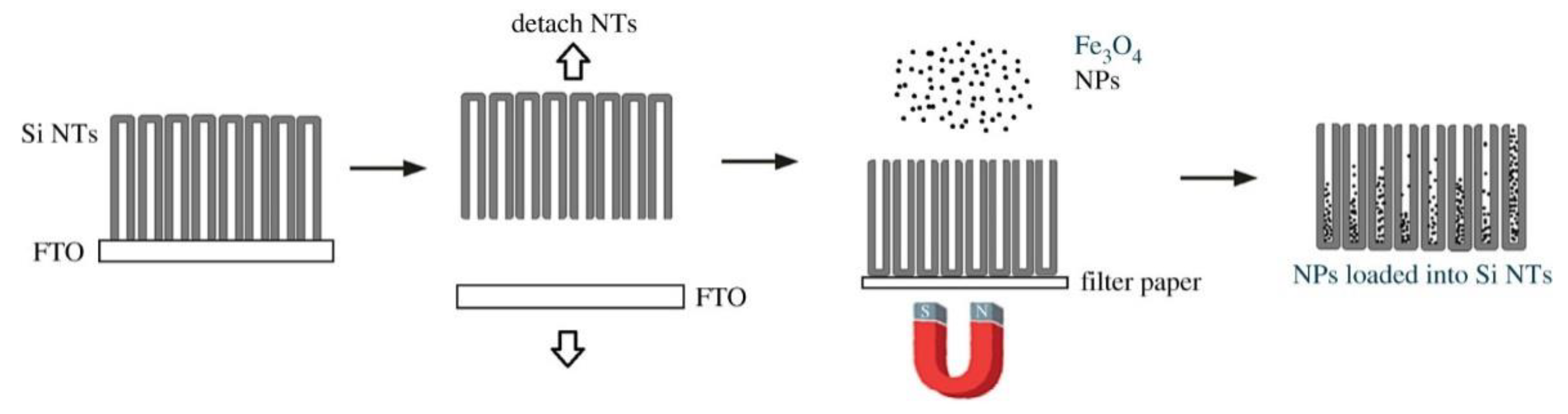
- Gonzalez-Rodriguez, R.; Granitzer, P.; Rumpf, K.; Coffer, J.L. New MRI contrast agents based on silicon nanotubes loaded with superparamagnetic iron oxide nanoparticles. R. Soc. Open Sci. 2018, 5, 180697. [Google Scholar] [CrossRef] [PubMed] [Green Version]
- Ardelean, I.L.; Stoencea, L.B.N.; Ficai, D.; Ficai, A.; Trusca, R.; Vasile, B.S.; Nechifor, G.; Andronescu, E. Development of Stabilized Magnetite Nanoparticles for Medical Applications. J. Nanomater. 2017, 2017, 1–9. [Google Scholar] [CrossRef] [Green Version]
- Moradiya, M.A.; Ladani, A.; Ladani, J.; Raiyani, C.; Markna, J.H. New Way to Treat Cancer: Magnetic Nanoparticle based Hyperthermia. J. Chem. Sci. Eng. 2019, 2, 58–60. [Google Scholar]
- Estelrich, J.; Busquets, M.A. Iron Oxide Nanoparticles in Photothermal Therapy. Molecules 2018, 23, 1567. [Google Scholar] [CrossRef] [Green Version]
- Nemala, H.B. Investigation of Temperature Dependent Magnetic Hyperthermia in Fe3O4 Ferrofluids. Ph.D. Thesis, Wayne State University, Detroit, MI, USA, 2015. [Google Scholar]
- Perigo, E.A.; Hemery, G.; Sandre, O.; Ortega, D.; Garaio, E.; Plazaola, F.; Teran, F.J. Fundamentals and advances in magnetic hyperthermia. Appl. Phys. Rev. 2015, 2, 041302. [Google Scholar] [CrossRef] [Green Version]
- Martínez, F.P.; Simeonidis, K.; Makridis, A.; Angelakeris, M.; Iglesias, O.; Guardia, P.; Cabot, A.; Yedra, L.; Estradé, S.; Peiró, F.; et al. Learning from Nature to Improve the Heat Generation of Iron-Oxide Nanoparticles for Magnetic Hyperthermia Applications. Sci. Rep. 2013, 3, 1652. [Google Scholar] [CrossRef] [Green Version]
- Sadat, M.; Patel, R.; Bud׳ko, S.L.; Ewing, R.C.; Zhang, J.; Xu, H.; Mast, D.B.; Shi, D. Dipole-interaction mediated hyperthermia heating mechanism of nanostructured Fe3O4 composites. Mater. Lett. 2014, 129, 57–60. [Google Scholar] [CrossRef]
- Sadat, M.; Patel, R.; Sookoor, J.; Bud’Ko, S.L.; Ewing, R.C.; Zhang, J.; Xu, H.; Wang, Y.; Pauletti, G.M.; Mast, D.B.; et al. Effect of spatial confinement on magnetic hyperthermia via dipolar interactions in Fe3O4 nanoparticles for biomedical applications. Mater. Sci. Eng. C 2014, 42, 52–63. [Google Scholar] [CrossRef]
- Surowiec, Z.; Miaskowski, A.; Budzyński, M. Investigation of magnetite Fe3O4 nanoparticles for magnetic hyperthermia. Nukleonika 2017, 62, 183–186. [Google Scholar] [CrossRef] [Green Version]
- Macías-Martínez, B.; Cortés, D.A.; Zugasti-Cruz, A.; Cruz-Ortíz, B.; Múzquiz-Ramos, E.M. Heating ability and hemolysis test of magnetite nanoparticles obtained by a simple co-precipitation method. J. Appl. Res. Technol. 2016, 14, 239–244. [Google Scholar] [CrossRef] [Green Version]
- Hayashi, K.; Nakamura, M.; Miki, H.; Ozaki, S.; Abe, M.; Matsumoto, T.; Sakamoto, W.; Yogo, T.; Ishimura, K. Magnetically Responsive Smart Nanoparticles for Cancer Treatment with a Combination of Magnetic Hyperthermia and Remote-Control Drug Release. Theranostics 2014, 4, 834–844. [Google Scholar] [CrossRef] [PubMed]
- Mues, B.; Buhl, E.M.; Baumann, M.; Schmitz-Rode, T.; Slabu, I. Agglomeration of magnetic nanoparticles and its effects on magnetic hyperthermia. Curr. Dir. Biomed. Eng. 2017, 3, 457–460. [Google Scholar]
- Iacovita, C.; Florea, A.; Dudric, R.; Páll, E.; Moldovan, A.; Tetean, R.; Stiufiuc, R.; Lucaciu, C.M. Small versus Large Iron Oxide Magnetic Nanoparticles: Hyperthermia and Cell Uptake Properties. Molecules 2016, 21, 1357. [Google Scholar] [CrossRef] [PubMed] [Green Version]
- Das, R.; Alonso, J.; Porshokouh, Z.N.; Kalappattil, V.; Torres, D.; Phan, M.-H.; Garaio, E.; García, J. Ángel; Llamazares, J.L.S.; Srikanth, H. Tunable High Aspect Ratio Iron Oxide Nanorods for Enhanced Hyperthermia. J. Phys. Chem. C 2016, 120, 10086–10093. [Google Scholar] [CrossRef]
- Fu, R.; Yan, Y.; Roberts, C.; Liu, Z.; Chen, Y. The role of dipole interactions in hyperthermia heating colloidal clusters of densely-packed superparamagnetic nanoparticles. Sci. Rep. 2018, 8, 1–10. [Google Scholar] [CrossRef] [PubMed]
- Fatima, H.; Kim, K.-S. Magnetic nanoparticles for bioseparation. Korean J. Chem. Eng. 2017, 34, 589–599. [Google Scholar] [CrossRef]
- Sehrig, F.Z.; Majidi, S.; Nikzamir, N.; Nikzamir, N.; Nikzamir, M.; Akbarzadeh, A. Magnetic nanoparticles as potential candidates for biomedical and biological applications. Artif. Cells Nanomed. Biotechnol. 2015, 44, 1–10. [Google Scholar] [CrossRef]
- Denison, M.I.J.; Raman, S.; Duraisamy, N.; Thangavelu, R.M.; Riyaz, S.U.M.; Gunasekaran, D.; Krishnan, K. Preparation, characterization and application of antibody-conjugated magnetic nanoparticles in the purification of begomovirus. RSC Adv. 2015, 5, 99820–99831. [Google Scholar] [CrossRef]
- Chang, J.H.; Lee, J.; Jeong, Y.; Lee, J.H.; Kim, I.J.; Park, S.E. Hydrophobic partitioning approach to efficient protein separation with magnetic nanoparticles. Anal. Biochem. 2010, 405, 135–137. [Google Scholar] [CrossRef]
- Wang, C.; Irudayaraj, J. Multifunctional Magnetic-Optical Nanoparticle Probes for Simultaneous Detection, Separation, and Thermal Ablation of Multiple Pathogens. Small 2010, 6, 283–289. [Google Scholar] [CrossRef] [PubMed]
- Shao, M.; Ning, F.; Zhao, J.; Wei, M.; Evans, D.G.; Duan, X. Preparation of Fe3O4@SiO2@Layered Double Hydroxide Core–Shell Microspheres for Magnetic Separation of Proteins. J. Am. Chem. Soc. 2012, 134, 1071–1077. [Google Scholar] [CrossRef]
- Reza, R.T.; Pérez, C.A.M.; Martínez, A.M.; Baques, D.B.; García-Casillas, P.E. Study of the Particle Size Effect on the Magnetic Separation of Bovine Serum Albumin (BSA). Sens. Lett. 2010, 8, 476–481. [Google Scholar] [CrossRef]
- Hosu, O.; Tertiș, M.; Cristea, C. Hosu Implication of Magnetic Nanoparticles in Cancer Detection, Screening and Treatment. Magnetochemistry 2019, 5, 55. [Google Scholar] [CrossRef] [Green Version]
- Saylan, Y.; Erdem, Ö.; Ünal, S.; Denizli, A. An Alternative Medical Diagnosis Method: Biosensors for Virus Detection. Biosensors 2019, 9, 65. [Google Scholar] [CrossRef] [PubMed] [Green Version]
- Vidic, J.; Manzano, M.; Chang, C.-M.; Jaffrezic-Renault, N. Advanced biosensors for detection of pathogens related to livestock and poultry. Veter Res. 2017, 48, 1–22. [Google Scholar] [CrossRef] [Green Version]
- Mehrotra, P. Biosensors and their applications—A review. J. Oral Biol. Craniofacial Res. 2016, 6, 153–159. [Google Scholar] [CrossRef] [Green Version]
- Sun, X.; Li, Q.; Wang, X.; Du, S. Amperometric Immunosensor Based on Gold Nanoparticles/Fe3O4-FCNTs-CS Composite Film Functionalized Interface for Carbofuran Detection. Anal. Lett. 2012, 45, 1604–1616. [Google Scholar] [CrossRef]


| Cation | Octahedral Site | Tetrahedral Site | Net Magnetic Moment |
|---|---|---|---|
| Ferric ions | ↑↑↑↑ ↑↑↑↑ | ↓↓↓↓ ↓↓↓↓ | Complete annihilation |
| Ferrous ions | ↑↑↑↑ ↑↑↑↑ | - | ↑↑↑↑ |
| Properties | Magnetite Nanoparticles |
|---|---|
| Molecular formula | Fe3O4 |
| Colour | Jet black |
| Density (g/cm3) | 5.18 |
| Melting point (°C) | 1583–1597 |
| Type of magnetism | Ferrimagnetic |
| Curie temperature (K) | 858 |
| Saturation magnetisation (Ms) at 300K [emug−1] | 92–100 |
| Standard Gibbs free energy of formation (ΔGf0) [kJ/mol] | −1012.6 |
| Crystallographic system | Cubic |
| Structure type | Inverse spinel |
| Lattice parameter (nm) | α = b = c = 0.8396 |
| Lattice angles | α = β = γ = 90° |
| Band gap energy (Eg) [eV] | 2.6 |
| Methods | Reaction | Condition | Reaction Temp. (°C) | Reaction Period | Size Distribution | Shape Control | Yield |
|---|---|---|---|---|---|---|---|
| Co-precipitation | Very simple | Ambient | 20–150 | Minutes | Relatively narrow | Not good | High/scalable |
| Solvothermal | Simple | High pressure | 150–220 | Hours–days | Very narrow | Very good | High/scalable |
| Wet-chemical Reduction | Very simple | Ambient | 20–150 | Minutes | Relatively narrow | Not good | High/scalable |
| Micro-/nano-emulsion | Complicated | Ambient | 20–80 | Hours | Narrow | Good | Low |
| Sonochemical/Sonolysis | Very simple | Ambient | 20–50 | Minutes | Narrow | Bad | Medium |
| Green (Biosynthesis) | Complicated | Ambient | Room temp. | Hours–days | Broad | Bad | Low |
Publisher’s Note: MDPI stays neutral with regard to jurisdictional claims in published maps and institutional affiliations. |
© 2020 by the authors. Licensee MDPI, Basel, Switzerland. This article is an open access article distributed under the terms and conditions of the Creative Commons Attribution (CC BY) license (http://creativecommons.org/licenses/by/4.0/).
Share and Cite
Ganapathe, L.S.; Mohamed, M.A.; Mohamad Yunus, R.; Berhanuddin, D.D. Magnetite (Fe3O4) Nanoparticles in Biomedical Application: From Synthesis to Surface Functionalisation. Magnetochemistry 2020, 6, 68. https://0-doi-org.brum.beds.ac.uk/10.3390/magnetochemistry6040068
Ganapathe LS, Mohamed MA, Mohamad Yunus R, Berhanuddin DD. Magnetite (Fe3O4) Nanoparticles in Biomedical Application: From Synthesis to Surface Functionalisation. Magnetochemistry. 2020; 6(4):68. https://0-doi-org.brum.beds.ac.uk/10.3390/magnetochemistry6040068
Chicago/Turabian StyleGanapathe, Lokesh Srinath, Mohd Ambri Mohamed, Rozan Mohamad Yunus, and Dilla Duryha Berhanuddin. 2020. "Magnetite (Fe3O4) Nanoparticles in Biomedical Application: From Synthesis to Surface Functionalisation" Magnetochemistry 6, no. 4: 68. https://0-doi-org.brum.beds.ac.uk/10.3390/magnetochemistry6040068