Article

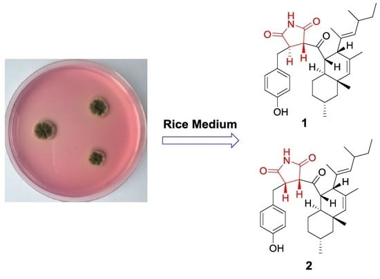
Two New Succinimide Derivatives Cladosporitins A and B from the Mangrove-derived Fungus Cladosporium sp. HNWSW-1

1
Key Laboratory of Biology and Genetic Resources of Tropical Crops, Ministry of Agriculture, Institute of Tropical Bioscience and Biotechnology, Chinese Academy of Tropical Agricultural Sciences, Haikou 571101, China
2
Hainan Key Laboratory for Research and Development of Natural Product from Li Folk Medicine, Haikou 571101, China
3
Key Laboratory of Tropical Medicinal Plant Chemistry of Ministry of Education and Hainan Normal University, Haikou 571101, China
*
Authors to whom correspondence should be addressed.
These authors contributed equally to this work.
Submission received: 29 November 2018 / Revised: 14 December 2018 / Accepted: 17 December 2018 / Published: 20 December 2018
(This article belongs to the Special Issue Bioactive Compounds from Mangroves and Their-Associated Microbes)

Abstract

:
Two new succinimide-containing derivatives, cladosporitins A (1) and B (2), were isolated from the fermentation cultures of the mangrove-derived fungus Cladosporium sp. HNWSW-1, along with a new pyrone, clapone (3), as well as the previously reported talaroconvolutin A (4) and anthraquinone (5). The structures of the isolated compounds were elucidated by 1D, 2D NMR, and HRMS spectral analysis. Compound 2 showed cytotoxicity against BEL-7042, K562 and SGC-7901 cell lines with IC50 values of 29.4 ± 0.35 μM, 25.6 ± 0.47 μM, and 41.7 ± 0.71 μM, respectively, whereas compound 4 exhibited cytotoxicity against Hela and BEL-7042 cell lines with IC50 values of 14.9 ± 0.21 μM and 26.7 ± 1.1 μM, respectively. In addition, compounds 4 and 5 displayed inhibitory activity against α-glycosidase, with IC50 values of 78.2 ± 2.1 μM and 49.3 ± 10.6 μM, respectively.

Graphical Abstract

1. Introduction

Fungi are an important resource of structurally and biologically diverse substances for drug and pesticide discovery [1,2,3,4,5,6,7]. A set of rare natural compounds from fungi, such as talaroconvolutins A-D, ZG-1494R, oteromycin, and codinaeopsin, which contain a modified tetramic acid unit linked to phenol, benzene, or indole and decalin fragments, exhibit activities against fungi, platelet-activating factors, acetyltransferase, endothelins receptors, or Plasmodium falciparum [8,9,10,11]. During our ongoing search for new bioactive metabolites from marine fungi, Cladosporium sp. HNWSW-1 was isolated from the healthy tree root of Ceriops tagal collected from the Dong Zhai Gang Mangrove Reserve in Hainan. The secondary metabolites of the genus Cladosporium have been mainly reported as polyketides derivatives, such as fatty acids [12], macrolides [13,14,15], pyrones [16,17,18], binaphthyl derivatives [19,20], α-pyridone [21] and tetramic acid derivatives [22,23]. Subsequent chemical investigations on the EtOAc extract of its fermentation cultures led to the isolation of cladosporitins A (1) and B (2), which contain the succinimide unit linked to phenol and decalin fragments. In addition, a new pyrone, clapone (3), was also isolated along with the previously reported talaroconvolutin A (4) [8] and 1,3,6-trihydroxy-7-(1-hydroxyethyl) anthracene-9,10-dione (5) [24]. Herein, we describe the isolation, structural determination, and biological activities of compounds 15.

2. Results

2.1. Structural Elucidation

Compound 1 was isolated as yellow oil. Its molecular formula was determined as C32H43NO4 by HRESIMS m/z 528.3103 [M + Na]+ (calcd. for C32H43NO4Na 528.3084) (Figure S18 in Supplementary Materials). The 1H, DEPTQ and HSQC NMR spectra (Figures S1, S2, and S6–S8 in Supplementary Materials) of compound 1 showed signals of one ketone carbonyl at δC 203.9, two amide carbonyls at δC 172.7 and δC 179.2, a para-substituted benzene ring (δC 131.5/ δH 7.10, δC 116.3/δH 6.77, δC 157.5 and δC 128.1), seven sp3 methines (δC 61.7, δC 52.9, δC 51.8, δC 45.0, δC 40.8, δC 34.8, and δC 28.1), two sp2 methines (δC 137.1 and δC 136.7), five methylenes (δC 49.1, δC 36.5, δC 35.8, δC 31.1, and δC 24.1), and six methyls (δC 23.1, δC 22.5, δC 21.1, δC 20.5, δC 15.2, and δC 12.4) (Table 1), except in the above data there were also three quaternary carbons (δC 130.7, δC 135.7, and δC 36.2). The comparison of 1H and 13C NMR data (Table 1) of the previously reported talaroconvolutin A (4) [8] revealed many similarities. Compound 1 had the same decalin moiety with the substituent that is the 4-methylhex-2-en-2-yl substituent linked to C-15 (δC 51.8) as compound 4. This is evidenced by the sequential COSY correlations (Figures S3–S5 in Supplementary Materials) from H-15 (δH 3.10) through H3-33 (δH 0.82), and H2-18 (δH 1.48/0.87), from H3-28 (δH 0.86) through H-25 (δH 5.05) and H3-29 (δH 0.92), as well as by HMBC correlations (Figures S9–S14 in Supplementary Materials) from H-14 (δH 3.62) to C-23 (δC 36.2), and C-24 (δC 135.7), from H-15 to C-17 (δC 137.1), C-22 (δC 40.8), C-24, C-30 (δC 15.2), and C-31 (δC 22.5), from H-17 (δH 5.38) to C-15, C-18 (δC 49.1), C-31, and C-32 (δC 20.5), from H2-18 to C-20 (δC 36.5), C-22, and C-33 (δC 23.1), from H-22 (δH 1.78) to C-20, C-21 (δC 24.1), and C-23, from H3-31 (δH 1.54) to C-15, C-16 (δC 130.7), and C-17, from H3-33 to C-18 and C-20, from H3-32 (δH 0.91) to C-17, C-18, C-22, and C-23, from H-25 to C-15, C-26 (δC 34.8), C-27 (δC 31.1), C-29 (δC 21.1), and C-30 (δC 15.2), from H-26 (δH 2.25) to C-27, C-28 (δC 12.4), and C-29, as well as from H3-29 to C-25 (δC 136.7), C-26, and C-27. The differences between compounds 1 and 4 are that the modified tetramic acid unit, 1,5-dihydro-2H-pyrrol-2-one unit, and C-6/C -7 double bond in compound 4 were replaced by the succinimide unit and CH2-6/CH-7 unit in compound 1, respectively, as deduced by the sequential COSY correlations (Figures S3–S5 in Supplementary Materials) from H-3 (δH 4.06) to H2-6 (δH 3.11/2.87) through H-4 (δH 3.40) and the HMBC correlations (Figures S9–S14 in Supplementary Materials) from H-3 to C-2 (δC 172.7)/C-4 (δC 45.0)/C-5 (δC 179.2)/C-6 (δC 35.8), from H-4 to C-2/C-3 (δC 61.7)/C-5/C-6/C-7 (δC 128.1), and from H2-6 to C-3/C-4/C-5/C-7/C-8 (δC 131.5). In addition, the COSY and HMBC correlations suggested that the para-disubstituted benzene ring was linked to C-4 in compound 1 via C-6. Finally, the key HMBC correlations from H-3 and H-4 to C-13 (δC 203.9) and from H-14 to C-13 confirmed that the decalin and the succinimide fragments were linked through a ketone carbonyl (C-13). According to the molecular formula and the chemical shift of C-10 (δC 157.5) in compound 1, a hydroxyl group was present on C-10. ROESY correlations (Figures S15–S17 in Supplementary Materials) from H-14 and H-19 to H3-32 suggested the relative configurations of C-14, C-19, and C-23 as shown in Figure 1. The large coupling constant (J = 12.0 Hz) of H-14/H-22 indicated their trans-diaxial orientation, while a coupling constant of 7.0 Hz between H-14 and H-15 placed these two protons in an axial-equatorial orientation [9,11]. Moreover, the ROESY correlation from H-3 to H2-6 (δH 2.87) hinted that H-3 and H-4 were on opposite sides of the succinimide ring. ROESY correlations (Figures S15–S17 in Supplementary Materials) from H3-30 to H-26 and from H-25 to H-15 proved the E-configuration of Δ24 double bond. Thus, the structure of compound 1 was established as shown in Figure 1 and was named cladosporitin A. However, the relative relationship between the chiral carbons in the succinimide fragment and those in the decalin fragment cannot be determined due to the free rotation of the C-3/C-13/C-14 linkage.
Compound 2 was also isolated as a yellow oil with the same molecular formula as compound 1, as determined by the HRESIMS peak at m/z 506.3279 [M + H]+ (calcd. for C32H44NO4 506.3265) (Figure S39 in Supplementary Materials). A detailed comparison of NMR data of compound 2 (Figures S19–S36 in the Supplementary Materials and Table 1) with those of compound 1 indicated that compound 2 had the same planar structure as compound 1. ROESY correlations (Figures S37 and S38 in Supplementary Materials and Figure 2) from H-14 and H-19 to H3-32 along with the coupling constant of H-14/H-22 (J = 12.1 Hz) and H-14/H-15 (J = 6.9 Hz) suggested that the relative configurations of C-14, C-15, C-19, C-22, and C-23 in the decalin fragment of compound 2 were the same as those of compound 1. However, the relatively large coupling constant between H-3 and H-4 of compound 2 (J = 4.1 Hz) compared to compound 1 (J = 2.4 Hz) combined with the absence of the ROESY correlation of H-3/H2-6 (δH 3.10/2.79) in compound 2 suggested the cis orientation of H-3 and H-4, which is different than that of compound 1. Thus, compound 2 was elucidated as shown in Figure 1 and named cladosporitin B.
Compound 3, a yellow oil, exhibited a prominent sodium adduct ion peak at m/z 239.0676 [M + Na]+ in the HRESIMS spectrum (Figure S50 in Supplementary Materials), suggesting a molecular formula of C13H12O3. Analysis of 1H NMR, 13C NMR, and HSQC spectra (Figures S40–S42 and S44–46 in Supplementary Materials) displayed five aromatic or olefinic methines (δC 137.3/δH 6.86, δC 124.8/δH 6.26, δC 118.3/δH 6.64, δC 109.9/δH 6.01, and δC 101.8/δH 6.70), two methyl siglets (δC 23.1/δH 2.73 and δC 18.6/δH 1.99), three sp2 oxyquaternary carbons (δC 164.0, δC 162.0, and δC 161.0), two sp2 quaternary carbons (δC 143.6 and δC 115.8), and one carbonyl (δC 182.3) (Table 2). The 1H NMR and 13C NMR data of compound 3 (Table 2) were very similar to those of 7-hydroxy-5-methyl-2- (2-oxobutyl)-4H-chromen-4-one [25]. The only major difference between them was the substituent linked to the 7-hydroxy-5-methyl-4H-chromen-4-one nucleus on C-2. The COSY correlations (Figure S43 in Supplementary Materials) of H-1′ (δH 6.26)/H-2′ (δH 6.86)/ H-3′ (δH 1.99), along with the HMBC correlations (Figures S47–S49 in Supplementary Materials and Figure 3) from H-1′ to C-2 (δC 162.0) and C-3′ (δC 18.6), from H-2′ to C-2 and C-3′, as well as from H-3 to C-1′ (δC 124.8) proved that a propylene fragment was located at C-2 in compound 3. In addition, the large coupling constant (J = 15.6) between H-1′ and H-2′ deduced the E-configuration of Δ1′ double bond. Hence, compound 3 was identified and named clapone.

2.2. The Bioactivities of Compounds 15 from Cladosporium sp. HNWSW-1

Compounds 15 were tested for their cytotoxicity against Hela, BEL-7042, K562 and SGC-7901 cell lines (Table 3). Compound 2 showed cytotoxicity against the BEL-7042, K562 and SGC-7901 cell lines with IC50 values of 29.4 ± 0.35 μM, 25.6 ± 0.47 μM and 41.7 ± 0.71 μM, respectively. Compound 4 exhibited cytotoxicity against Hela and BEL-7042 cell lines with IC50 values of 14.9 ± 0.21 μM and 26.7 ± 1.1 μM, respectively. In addition, all of the compounds were tested for their inhibitory activity against α-glycosidase (Table 3). Only compounds 4 and 5 displayed the inhibitory activity against α-glycosidase, with IC50 values of 78.2 ± 2.1 μM and 49.3 ± 10.6 μM, respectively.

3. Materials and Methods

3.1. General Experimental Procedures

Silica gel (60–80, 200–300 mesh, Qingdao Marine Chemical Co. Ltd., Qingdao, China), ODS gel (20–45 μm, Fuji Silysia Chemical Co. Ltd., Aichi-ken, Japan,), and Sephadex LH-20 (Merck, Kenilworth, NJ, USA) were used for column chromatography. TLC was conducted on precoated silica gel G plates (Qingdao Marine Chemical Co. Ltd., Qingdao, China), and spots were detected by spraying with 5% H2SO4 in EtOH followed by heating. Optical rotations were measured on a Rudolph Autopol III polarimeter (Rudolph Research Analytical, NJ, USA). IR absorptions were obtained on a Nicolet 380 FT-IR instrument (Thermo, Waltham, MA, USA) using KBr pellets. 1D and 2D NMR spectra were recorded on Bruker AV III spectrometer (Bruker, Billerica, MA, USA) (1H NMR at 500 MHz and 13C NMR at 125 MHz for 25, 1H NMR at 600 MHz and 13C NMR at 150 MHz for compound 1) using TMS as the internal standard. HRESIMS spectra were recorded with Agilent 6200/6500 iFunnel Q-TOF. Semipreparative HPLC was carried out using an ODS column (YMC-pack ODS-A, 10 × 250 mm, 5μm, 4 mL/min). Chem3D Pro 14.0 was the software used for building these 3D models and the calculation method used for energy minimizations.

3.2. Fungal Material

The strain HNWSW-1 of Cladosporium sp. was isolated from the healthy tree root of Ceriops tagal, which was collected in the Dong Zhai Gang Mangrove Reserve in Hainan province in July 2011. A healthy root sample of Ceriops tagal was washed in running tap water to remove adhered epiphytes and soil debris. The root surface was sterilized by sequential immersion in 75% (v/v) ethanol for 3 min and 5% sodium hypochlorite solution for 5 min after drying under sterile conditions, washing the surface-treated roots three times in sterile distilled water after each sterilization. Next, air-dried sterilized roots were cut into about 0.5 cm × 0.5 cm cubes, deposited on a Potato Dextrose Agar (PDA) (200 g potato, 20 g glucose, 20 g agar per liter of seawater collected in Haikou Bay, China) plate containing chloramphenicol (100 μg/mL) as a bacterial inhibitor. A reference culture is maintained in our laboratory at −80 °C. Working stocks were prepared on PDA slants stored at 4 °C.
The fungus was identified based on the DNA sequences (GenBank access No. MH 535968, the the 18S rRNA gene sequences data in Supplementary Materials) of 18Sr DNA genes. The mycelium was ground to a fine powder in liquid N2, then genomic DNA was extracted, and the 18S rDNA region was amplified by PCR using primers NS1 (5′-GTAG TCATATGCTTGTCTC-3′) and NS6 (5′-GCATCACAGACCTGTTATTGCCT C-3′). PCR products were sequenced (Applied Biosystems 3730 XL Genetic Analyzer, Applied Biosystems Inc., Foster City, CA, USA).

3.3. Fermentation and Extraction

Cladosporium sp. HNWSW-1 was cultured in PDB (the potato liquid media consisting of 200.0 g/L potato, 20.0 g/L glucose, and 1000 mL deionized water) at 28 °C and 150 rpm for 72 h. Then 5 mL seed broth was transferred aseptically to 1000 ml Erlenmeyer flasks (60 flasks), each containing rice medium (80 g rice and 160 mL water). The flasks were incubated at room temperature under static conditions for 60 days. The cultures were extracted three times by EtOAc, and the EtOAc solutions were combined and evaporated under reduced pressure to produce a dark brown gum (7.1 g).

3.4. Purification and Identification

The obtained EtOAc crude extract (7.1 g) was fractionated into 12 fractions (Fr.1–Fr.12) on silica gel VLC and eluted with a gradient elution of CH2Cl2-petroleum ether (0–100%) and MeOH-CH2Cl2 (0–100%). Fr.6 (2.4 g) was fractionated by RP-18 column chromatography with a gradient of water-MeOH to give 25 fractions (Fr.6.1–Fr.6.25). Fr.6.9 (118.2 mg) was purified by a sephadex LH-20 column (40 g) and eluted with MeOH and acetone, respectively, then further submitted to HPLC purification on ODS column eluting with 90% MeOH to yield compounds 1 (1.0 mg) and 2 (2.1 mg). Fr.6.10 (72.6 mg) was separated by a sephadex LH-20 column (40 g) and eluted with MeOH to yield 4 (2.0 mg). Fr.8 (853.1 mg) was submitted to an RP-18 column and eluted with MeOH-Water to give 7 fractions (Fr.8.1–Fr.8.7). Fr.8.2 (25.2 mg) was separated by a sephadex LH-20 column (15 g) and eluted with MeOH and further submitted to HPLC purification on ODS column eluted with 35% MeCN to yield 3 (0.6 mg) and 5 (2.2 mg).
Cladosporitin A (1): Yellow oil; [ α ] D 20 —166.6 (c 0.01, CHCl3); IR (KBr) νmax: 3290, 2926,1711,1515 cm−1; HRESIMS: m/z 528.3103 [M + Na]+ (calcd. for C32H43NO4Na, 528.3084); 1H and 13C NMR data: See Table 1.
Cladosporitin B (2): Yellow oil; [ α ] D 20 —94.5 (c 0.11, CHCl3); IR (KBr) νmax: 3247, 2956, 1713, 1515 cm−1; HRESIMS: m/z 506.3279 [M + H]+ (calcd. for C32H44NO4, 506.3265); 1H and 13C NMR data: See Table 1.
Clapone (3): Yellow oil; IR (KBr) νmax: 3435, 2978, 1673, 1400 cm−1; HRESIMS: m/z 239.0676 [M + Na]+ (calcd. for C13H12O3Na, 239.0679); 1H and 13C NMR data: See Table 2.

3.5. Bioassays for Cytotoxic Activity

The MTT method optimized by Mosmann et al. [26] was performed in vitro to test the cytotoxic activity of compounds 1–6. Adriamycin was used as a positive control and the medium without the test compound was used as a negative control in the bioassay.

3.6. Bioassays for α-Glycosidase Inhibitory Activity

The method optimized by Jong et al. [27] was performed in vitro to test the α-glucosidase inhibitory activity of compounds 1–5. Acarbose was used as positive control.

4. Conclusions

Three new compounds (13) along with the previously reported talaroconvolutin A (4) and anthraquinone (5) were isolated from the rice medium culture of mangrove-derived fungus Cladosporium sp. HNWSW-1, isolated from the healthy root of Ceriops tagal collected in the Dong Zhai Gang Mangrove Reserve in Hainan. Their structures were determined by spectroscopic methods. Compound 2 showed cytotoxicity against BEL-7042, K562 and SGC-7901 cell lines with IC50 values of 29.4 ± 0.35 μM, 25.6 ± 0.47 μM, and 41.7 ± 0.71 μM, respectively, while compound 4 exhibited cytotoxicity against the Hela and BEL-7042 cell lines with IC50 values of 14.9 ± 0.21 μM and 26.7 ± 1.1 μM, respectively. Moreover, compounds 4 and 5 displayed inhibitory activity against α-glycosidase with IC50 values of 78.2 ± 2.1 μM and 49.3 ± 10.6 μM, respectively. The results suggested that the mangrove-derived fungi are an important source of new bioactive substances.

Supplementary Materials

The NMR and HRESIMS spectra for 13 are available online at https://0-www-mdpi-com.brum.beds.ac.uk/1660-3397/17/1/4/s1.

Author Contributions

W.M., H.D., and P.W. conceived and designed the experiments; Y.C. performed the isolation of the fungus and the compounds; Y.D. performed the fermentation of the fungus; X.S. collected the root sample of Ceriops tagal; C.C. performed the biological tests; J.Y. and P.C. contributed to the acquirement of the NMR data and IR, respectively; P.W., W.M., H.D., H.C., F.K., and P.C. analyzed the data; P.W., W.M. and H.D. wrote the paper. H.C., F.K. and W.M. reviewed the paper.

Funding

This work was financially supported by Natural Science Foundation of Hainan (No. 217254), National Natural Science Foundation of China (No. 41506096 and 41776093), Financial Fund of the Ministry of Agriculture and Rural Affairs, P. R of China (NFZX2018). Key Laboratory of Tropical Medicinal Plant Chemistry of Ministry of Education, Natural Science Foundation of Hainan Province, and Hainan Normal University (No. 201601).

Conflicts of Interest

The authors declare no conflict of interest.

References

  1. Blunt, J.W.; Copp, B.R.; Keyzers, R.A.; Munro, M.H.G.; Prinsep, M.R. Marine natural products. Nat. Prod. Rep. 2015, 32, 116–211. [Google Scholar] [CrossRef] [PubMed] [Green Version]
  2. Blunt, J.W.; Copp, B.R.; Keyzers, R.A.; Munro, M.H.G.; Prinsep, M.R. Marine natural products. Nat. Prod. Rep. 2016, 33, 382–431. [Google Scholar] [CrossRef] [PubMed] [Green Version]
  3. Blunt, J.W.; Copp, B.R.; Keyzers, R.A.; Munro, M.H.G.; Prinsep, M.R. Marine natural products. Nat. Prod. Rep. 2017, 34, 235–294. [Google Scholar] [CrossRef] [PubMed] [Green Version]
  4. Zhu, H.C.; Chen, C.M.; Tong, Q.Y.; Yang, J.; Wei, G.Z.; Xue, Y.B.; Wang, J.P.; Luo, Z.W.; Zhang, Y.H. Asperflavipine A: A cytochalasan heterotetramer uniquely defined by a highly complex tetradecacyclic ting dystem from Aspergillus flavipes QCS12. Angew. Chem. Int. Ed. 2017, 56, 5242–5246. [Google Scholar] [CrossRef] [PubMed]
  5. Wang, T.T.; Wei, Y.J.; Ge, H.M.; Jiao, R.H.; Tan, R.X. Acaulide, an osteogenic macrodiolide from Acaulium sp. H-JQSF, an isopod-associated Fungus. Org. Lett. 2018, 20, 1007–1010. [Google Scholar] [CrossRef] [PubMed]
  6. Li, H.L.; Xu, R.; Li, X.M.; Yang, S.Q.; Meng, L.H.; Wang, B.G. Simpterpenoid A, a meroterpenoid with a highly functionalized cyclohexadiene moiety featuring gem-propane-1,2-dione and methylformate groups, from the mangrove-derived Penicillium simplicissimum MA-332. Org. Lett. 2018, 20, 1465–1468. [Google Scholar] [CrossRef] [PubMed]
  7. Kong, F.D.; Ma, Q.Y.; Huang, S.Z.; Wang, P.; Wang, J.F.; Zhou, L.M.; Yuan, J.Z.; Dai, H.F.; Zhao, Y.X. Chrodrimanins K-N and related meroterpenoids from the fungus Penicillium sp. SCS-KFD09 isolated from a marine worm, Sipunculus nudus. J. Nat. Prod. 2017, 80, 1039–1047. [Google Scholar] [CrossRef]
  8. Suzuki, S.; Hosoe, T.; Nozawa, K.; Kawai, K.I.; Yaguchi, T.; Udagawa, S.I. Antifungal substances against pathogenic fungi, talaroconvolutins, from Talaromyces convolutes. J. Nat. Prod. 2000, 63, 768–772. [Google Scholar] [CrossRef]
  9. West, P.R.; van Ness, J.; Varming, A.-M.; Rassing, B.; Biggs, S.; Gasper, S.; McKernan, P.A.; Piggott, J. ZG- 1494α, a novel platelet-activating factor acetyltransferase inhibitor from Penicilium rubrum, isolation, structure elucidation and biological activity. J. Antibiot. 1996, 49, 967–973. [Google Scholar] [CrossRef]
  10. Sing, S.B.; Goetz, M.A.; Jones, T.; Bill, G.F.; Giacobbe, R.A.; Herram, L.; Mile, S.S.; Williams, D.L. Oteromycin: A novel antagonist of endothelin receptor. J.Org. Chem. 1995, 60, 7040–7042. [Google Scholar] [CrossRef]
  11. Kontik, R.; Clardy, J. Codinaeopsin, an antimalarial fungal polyketide. Org. Lett. 2008, 10, 4149–4151. [Google Scholar] [CrossRef] [PubMed]
  12. Peng, X.P.; Wang, Y.; Zhu, G.L.; Zhu, W.M. Fatty acid derivatives from the halotolerant fungus Cladosporium cladosporioides. Magn. Reson. Chem. 2018, 56, 18–24. [Google Scholar] [CrossRef] [PubMed]
  13. Jadulco, R.; Proksch, P.; Wray, V.; Albrecht Berg, S.; Grafe, U. New macrolides and furan carboxylic acid derivative from the sponge-derived fungus Cladosporium herbarum. J. Nat. Prod. 2001, 64, 527–530. [Google Scholar] [CrossRef] [PubMed]
  14. Zhang, H.; Tomoda, H.; Tabata, N.; Miura, H.; Namikoshi, M.; Yamaguchi, Y.; Masuma, R.; Omura, S. Cladospolide D, a new 12-membered macrolide antibiotic produced by Cladosporium sp. FT-0012. J. Antibiot. 2001, 54, 635–641. [Google Scholar] [CrossRef] [PubMed]
  15. Shigemori, H.; Kasai, Y.; Komatsu, K.; Tsuda, M.; Mikami, Y.; Kobayashi, J.I. Sporiolides A and B, new cytotoxic twelve-membered macrolides from a marine-derived fungus Cladosporium species. Mar. Drugs 2004, 2, 164–169. [Google Scholar] [CrossRef]
  16. Jadulco, R.; Brauers, G.; Edrada, R.A.; Ebel, R.; Wray, V.; Sudarsono, S.; Proksch, P. New metabolites from sponge-derived fungi Curvularia lunata and Cladosporium herbarum. J. Nat. Prod. 2002, 65, 730–733. [Google Scholar] [CrossRef] [PubMed]
  17. Rotinsulu, H.; Yamazaki, H.; Sugai, S.; Iwakura, N.; Wewengkang, D.S.; Sumilat, D.A.; Namikoshi, M. Cladosporamide A, a new protein tyrosine phosphatase 1B inhibitor, produced by an Indonesian marine sponge-derived Cladosporium sp. J. Nat. Med. 2018. [Google Scholar] [CrossRef]
  18. Wang, L.P.; Han, X.L.; Zhu, G.L.; Wang, Y.; Chairoungdua, A.; Piyachaturawat, P.; Zhu, W.M. Polyketides from the endophytic fungus Cladosporium sp. isolated from the mangrove plant Excoecaria agallocha. Front. Chem. 2018, 6, 344–353. [Google Scholar] [CrossRef] [PubMed]
  19. Sakagami, Y.; Sano, A.; Hara, O.; Mikawa, T.; Marumo, S. Cladosporol, β-1, 3-glucan biosynthesis inhibitor, isolated from fungus, Cladosporium cladosporioides. Tetrahedron Lett. 1995, 36, 1469–1472. [Google Scholar] [CrossRef]
  20. Yamazaki, H.; Yagi, A.; Akaishi, M.; Kirikoshi, R.; Takahashi, O.; Abe, T.; Chiba, S.; Takahashi, K.; Iwakura, N.; Namikoshi, M.; et al. Halogenated cladosporols produced by the sodium halide-supplemented fermentation of the plant-associated fungus Cladosporium sp. TMPU1621. Tetrahedron Lett. 2018, 59, 59,1913–1915. [Google Scholar] [CrossRef]
  21. Ye, Y.H.; Zhu, H.L.; Song, Y.C.; Liu, J.Y.; Tan, R.X. Structural revision of aspernigrin A, reisolated from Cladosporium herbarum IFB-E002. J. Nat. Prod. 2005, 68, 1106–1108. [Google Scholar] [CrossRef] [PubMed]
  22. Liang, X.; Huang, Z.H.; Ma, X.; Qi, S.H. Unstable tetramic acid derivatives from the deep sea-derived fungus Cladosporium sphaerospermum EIODSF 008. Mar. Drugs 2018, 16, 448. [Google Scholar] [CrossRef] [PubMed]
  23. Zhu, G.L.; Kong, F.D.; Wang, Y.; Fu, P.; Zhu, W.M. Cladodionen, a cytotoxic hybrid polyketide from the marine-derived Cladosporium sp. OUCMDZ-1635. Mar. Drugs 2018, 16, 71. [Google Scholar] [CrossRef]
  24. Zhao, D.L.; Wang, D.; Tian, X.Y.; Cao, F.; Li, Y.Q.; Zhang, C.S. Anti-phytopathogenic and cytotoxic activities of crude extracts and secondary metabolites of marine-derived fungi. Mar. Drugs 2018, 16, 36. [Google Scholar] [CrossRef] [PubMed]
  25. Matsunaga, H.; Kamisuki, S.; Kaneko, M.; Yamaguguchi, Y.; Takeuchi, T.; Watashi, K.; Sugawara, F. Isolation and structure of vanitaracin A, a novel anti-hepatitis B virus compound from Talaromyces sp. Bioorg. Med. Chem. Lett. 2015, 25, 4325–4328. [Google Scholar] [CrossRef] [PubMed]
  26. Mosmann, T. Rapid colorimetric assay for cellular growth and survival: Application to proliferation and cytotoxicity assays. J. Immunol. Methods 1983, 65, 55–63. [Google Scholar] [CrossRef]
  27. Jong, A.N.; Bhandari, M.R.; Kawabata, J. α-Glucosidase inhibitors from Devil tree. Food Chem. 2007, 103, 1319–1323. [Google Scholar] [CrossRef]
Figure 1. Chemical structures of compounds 15 from Cladosporium sp. HNWSW-1.
Marinedrugs 17 00004 g001
Figure 2. The key 2D NMR correlations for compound 1.
Marinedrugs 17 00004 g002
Figure 3. The key 2D NMR correlations for compounds 2 and 3.
Marinedrugs 17 00004 g003
Table 1. 1H and 13C NMR Data for compound 1 (600 and 150 MHz, δ in ppm) and compound 2 (500 and 125 MHz, δ in ppm).
Position1 a2 b
δC, TypeδH, Mult. (J in Hz)δC, TypeδH, mult. (J in Hz)
1----
2172.7, C-172.0, C-
361.7, CH4.06, d, (2.4)61.9, CH3.40, d, (4.1)
445.0, CH3.40, m43.6, CH3.67, m
5179.2, C 178.3, C-
635.8, CH23.11, dd, (14.7, 5.9)
2.87, dd, (14.7, 5.3)
35.0, CH23.10, dd, (13.7, 5.1)
2.79, dd, (13.7, 8.7)
7128.1, C-128.2, C-
8131.5, CH7.10, d, (8.3)130.5, CH6.99, d, (8.7)
9116.3, CH6.77, d, (8.3)115.9, CH6.75 d, (8.7)
10157.5, C-155.4, C-
11116.3, CH6.77, d (8.3)115.9, CH6.75, d, (8.7)
12131.5, CH7.10, d (8.3)130.5, CH6.99, d, (8.7)
13203.9, C-202.2, C-
1452.9, CH3.62, dd (12.0, 7.0)52.6, C3.33, dd, (12.1, 6.9)
1551.8, CH3.10 m50.9, CH2.95, d, (6.9)
16130.7, C-130.1, C-
17137.1, CH5.38, s135.8, CH5.29, s
1849.1, CH21.48, m
0.87, m
48.3, CH21.43, m
0.86, m
1928.1, CH1.66, m27.4, CH1.61, m
2036.5, CH21.64, m
0.82, m
35.6, CH21.60, m
0.77, m
2124.1, CH21.32, m
0.96, m
24.3, CH21.62, m
0.75, m
2240.8, CH1.78, ddd, (12.0, 12.0, 2.3)42.2, CH1.67, m
2336.2, C-35.2, C-
24135.7, C-135.0, C-
25136.7, CH5.05, d, (8.2)136.1, CH4.66, d, (9.3)
2634.8, CH2.25, m33.9, CH2.14, m
2731.1, CH21.35, m
1.23, m
30.4, CH21.27, m
1.10, m
2812.4, CH30.86, t, (7.6)12.1, CH30.79, t, (7.1)
2921.1, CH30.92, d, (6.2)20.9, CH30.76, d, (6.6)
3015.2, CH31.45, s14.5, CH31.47, s
3122.5, CH31.54, s22.2, CH31.46, s
3220.5, CH30.91, s20.3, CH30.85, s
3323.1, CH30.82, d, (6.4)22.8, CH30.82, d, (6.2)
a Recorded in Acetone; b Recorded in CDCl3.
Table 2. 1H and 13C NMR Data for compound 3 in CH3OD (500 and 125 MHz, δ in ppm).
Position3
δC, TypeδH, Mult. (J in Hz)
1--
2162.0, C-
3109.9, CH6.01, s
4182.3, C-
4a115.8, C-
5143.6, C-
6118.3, CH6.64, dd, (2.2, 0.8)
7164.0, C-
8101.8, CH6.70, d, (2.2)
8a161.0, C-
1′124.8, CH6.26, ddd,
(15.6, 3.4, 1.7)
2′137.3, CH6.86, ddd,
(15.6, 13.7, 6.9)
3′18.6, CH31.99, dd, (6.9,1.7)
5- CH323.1, CH32.73, s
Table 3. Cytotoxic and α-glycosidase inhibitory activities of 15.
CompoundsIC50 (μM)
HelaBEL-7042K562SGC-7901α-Glycosidase
1>50>50>50>50>500
2>5029.4 ± 0.3525.6 ± 0.4741.7 ± 0.71>500
3>50>50>50>50>500
414.9 ± 0.2126.7 ± 1.1>50>5078.2 ± 2.1
5>50>50>50>5049.3 ± 10.6
Adriamycin11.5 ± 0.1811.9 ± 0.3714.2 ± 0.666.66 ± 0.2ND a
AcarboseND aND aND aND a275.7 ± 2.7
a Not detected; Values are expressed as mean ± standard deviation (SD); n = 3.

