Article

Structural and Serological Studies of the O6-Related Antigen of Aeromonas veronii bv. sobria Strain K557 Isolated from Cyprinus carpio on a Polish Fish Farm, which Contains l-perosamine (4-amino-4,6-dideoxy-l-mannose), a Unique Sugar Characteristic for Aeromonas Serogroup O6

by
Katarzyna Dworaczek
1,
Dominika Drzewiecka
2,
Agnieszka Pękala-Safińska
3 and
Anna Turska-Szewczuk
1,*
1
Department of Genetics and Microbiology, Maria Curie-Skłodowska University in Lublin, Akademicka 19, 20-033 Lublin, Poland
2
Laboratory of General Microbiology, Department of Biology of Bacteria, Faculty of Biology and Environmental Protection, University of Łódź, Banacha 12/16, 90-237 Łódź, Poland
3
Department of Fish Diseases, National Veterinary Research Institute, Partyzantów 57, 24-100 Puławy, Poland
*
Author to whom correspondence should be addressed.
Submission received: 10 June 2019 / Revised: 27 June 2019 / Accepted: 3 July 2019 / Published: 5 July 2019
(This article belongs to the Special Issue Marine Glycoconjugates: Trends and Perspectives)

Abstract

:
Amongst Aeromonas spp. strains that are pathogenic to fish in Polish aquacultures, serogroup O6 was one of the five most commonly identified immunotypes especially among carp isolates. Here, we report immunochemical studies of the lipopolysaccharide (LPS) including the O-specific polysaccharide (O-antigen) of A. veronii bv. sobria strain K557, serogroup O6, isolated from a common carp during an outbreak of motile aeromonad septicemia (MAS) on a Polish fish farm. The O-polysaccharide was obtained by mild acid degradation of the LPS and studied by chemical analyses, mass spectrometry, and 1H and 13C NMR spectroscopy. It was revealed that the O-antigen was composed of two O-polysaccharides, both containing a unique sugar 4-amino-4,6-dideoxy-l-mannose (N-acetyl-l-perosamine, l-Rhap4NAc). The following structures of the O-polysaccharides (O-PS 1 and O-PS 2) were established: O-PS 1: →2)-α-l-Rhap4NAc-(1→; O-PS 2: →2)-α-l-Rhap4NAc-(1→3)-α-l-Rhap4NAc-(1→3)-α-l-Rhap4NAc-(1→. Western blotting and an enzyme-linked immunosorbent assay (ELISA) showed that the cross-reactivity between the LPS of A. veronii bv. sobria K557 and the A. hydrophila JCM 3968 O6 antiserum, and vice versa, is caused by the occurrence of common α-l-Rhap4NAc-(1→2)-α-l-Rhap4NAc and α-l-Rhap4NAc-(1→3)-α-l-Rhap4NAc disaccharides, whereas an additional →4)-α-d-GalpNAc-associated epitope defines the specificity of the O6 reference antiserum. Investigations of the serological and structural similarities and differences in the O-antigens provide knowledge of the immunospecificity of Aeromonas bacteria and are relevant in epidemiological studies and for the elucidation of the routes of transmission and relationships with pathogenicity.

Graphical Abstract

1. Introduction

The genus Aeromonas, which belongs to the family Aeromonadaceae along with four other genera Telumonas, Oceanimonas, Oceanisphaera and Zobellella, is composed of a large number of species classified within 17 DNA-hybridization groups (HG) or genomospecies, and 14 phenospecies [1,2,3,4,5]. These Gram-negative rods are typically found in aquatic environments, e.g., freshwater, estuarine and coastal water, seawater, drinking water supplies, polluted waters, marine, and freshwater sediment and sand. Aeromonads have also been isolated from animals, food, and various clinical samples. Most frequently, Aeromonas spp. strains are pathogenic to poikilothermic animals including amphibians, fish, and reptiles. They also can be associated with infections of birds and mammals [4,5,6]. In fish, non-motile psychrophilic species of Aeromonas salmonicida subsp. salmonicida are pathogenic to Salmonidae, provoking systemic furunculosis [7,8]. In turn, the representatives of Aeromonas hydrophila, Aeromonas bestiarum, Aeromonas salmonicida, Aeromonas jandaei, and Aeromonas veronii bv. sobria have been described as causative agents of diseases in a variety of fish species. These include motile Aeromonas septicemia (MAS), clinical conditions associated with systemic infection resulting in high mortality rates and severe economic losses in aquacultures, and a chronic type of disease called motile Aeromonas infection (MAI) causing erosion of fins, skin lesions, and ulcerations [9,10,11,12]. The mesophilic and motile A. veronii, A. hydrophila, A. caviae and A. schubertii species are potentially pathogenic to humans. Immunocompromised patients and children are especially vulnerable. Clinical presentations include gastroenteritis, wound infections, biliary tract infections, pneumonia, meningitis, septic arthritis, or septicemia without an obvious focus of infection [4,13,14,15,16].
The pathogenicity of Aeromonas is determined by their ability to produce extracellularly secreted enzymes and toxins i.e., hemolysins, cytotonic and cytotoxic enterotoxins, proteases, lipases, gelatinases, and leucocidins, which play an important role in both the initial steps and spread of the infection process. Moreover, cell-surface constituents, including outer membrane proteins, S-layers, surface polysaccharides (capsule, lipopolysaccharide, and glucan), flagella, and pili, have been identified as important compounds associated with Aeromonas virulence [4,10,17,18,19].
Lipopolysaccharide (LPS) is the dominant glycolipid in the outer leaflet of the outer membrane of Gram-negative bacteria, which mediates their virulence. The high-molecular-weight S-LPS glycoforms have a tripartite structure comprising lipid A, core oligosaccharide, which together with lipid A contributes to maintenance of the integrity of the outer membrane, and the O-specific polysaccharide (O-PS), which is connected to the core and most frequently consists of a heteropolymer composed of repeating oligosaccharide units [20,21,22,23]. The O-specific polysaccharide is a surface antigen called the somatic O-antigen, whose high degree of diversity determines the specificity of each bacterium and gives the basis for their serological classification. Serotyping to identify bacterial strains is invaluable for epidemiological investigations since many O-serotypes are associated with specific disease syndromes [21,24].
Aeromonas strains are serologically heterogeneous and they have been characterized and classified based on O-antigens into 44 serogroups using the NIH scheme (National Institute of Health, Japan) proposed by Sakazaki and Shimada [25], which can be further extended to 97 O serogroups after inclusion of provisional new serogroups [26]. The variants of the O-specific polysaccharide, which represents a specific antigenic fingerprint for bacteria, might be very useful, e.g., for identification of Aeromonas strains eliciting infections in farmed fish and for diagnosis of etiological agents of gastrointestinal infections in humans.
Despite the large antigenic diversity of Aeromonas, only several serogroups i.e., O11, O16, O18, and O34 (NIH scheme) have been reported, most frequently, as predominating especially in clinical specimens. These O-serotypes of mesophilic Aeromonas were associated with most cases of bacteremia, suggesting their role in the pathogenesis of some systemic diseases [4].
As shown in previous reports, the distribution of the serogroups of Aeromonas strains may be related to geographic location [27,28]. Such differences in the occurrence of Aeromonas species serogroups were associated with outbreaks of septicemia in fish. Strains belonging to serogroup O14 have been identified as pathogens of European eels [29]. As revealed in recently published studies by Kozińska and Pękala [28], the majority of isolates that are pathogenic to carp in Polish aquacultures represented serogroups O3, O6, O41, PGO4, and PGO6, whereas serogroups O11, O16, O18, O33, PGO1, and PGO2 dominated among both carp and trout isolates. In turn, motile aeromonas septicemia (MAS) incidences in rainbow trout have been related to the strains of serogroups O11, O16, O34, and O14. Moreover, pathogenic isolates of two species: Aeromonas veronii bv. sobria and Aeromonas sobria were mainly classified within the O6 serogroup.
The A. veronii species, originally described by Hickman-Brenner et al. [30] as a novel member of the genus, is commonly associated with diarrhea. Nevertheless, this species, which consists of two biovars, A. veronii bv. sobria, which is negative for aesculin hydrolysis and ornithine decarboxylase, and A. veronii bv. veronii, which is positive for these reactions [15,31], is commonly known as a fish pathogen associated mainly with ulcerative syndrome [32,33]. It is worth emphasizing that A. veroni bv. sobria was one of the dominant species among carp isolates collected during 5 years in Polish culture facilities, and 76% of these isolates were classified as virulent [28]. In the light of the increased Aeromonas infection incidence rate and the economic importance of these diseases in cultured fish, it is essential to characterize virulence factors associated with the pathogenesis of this species, particularly including LPS, which is the major surface glycoconjugate of Gram-negative bacteria. Recently, two new structures of O-antigens were established for the species A. veronii bv. sobria and A. sobria [34,35].
Here we report immunochemical investigation of LPS, especially the O-specific polysaccharide of A. veronii bv. sobria strain K557, which was isolated from the common carp (Cyprinus carpio L.) during an outbreak of motile aeromonad infection/motile aeromonad septicemia (MAI/MAS) on a Polish fish farm [28,36]. The structural characterization revealed that the O-antigen was composed of two O-polysaccharides, both containing a unique sugar 4-amino-4,6-dideoxy-l-mannose (N-acetyl-l-perosamine, l-Rhap4NAc). Serological studies using Western blotting and an enzyme-linked immunosorbent assay (ELISA) with intact and adsorbed O-antisera showed that the O-antigen of A. veronii bv. sobria strain K557 is related but not identical to that of A. hydrophila JCM 3968 O6, which is a reference strain for Aeromonas serogroup O6 [37].

2. Results

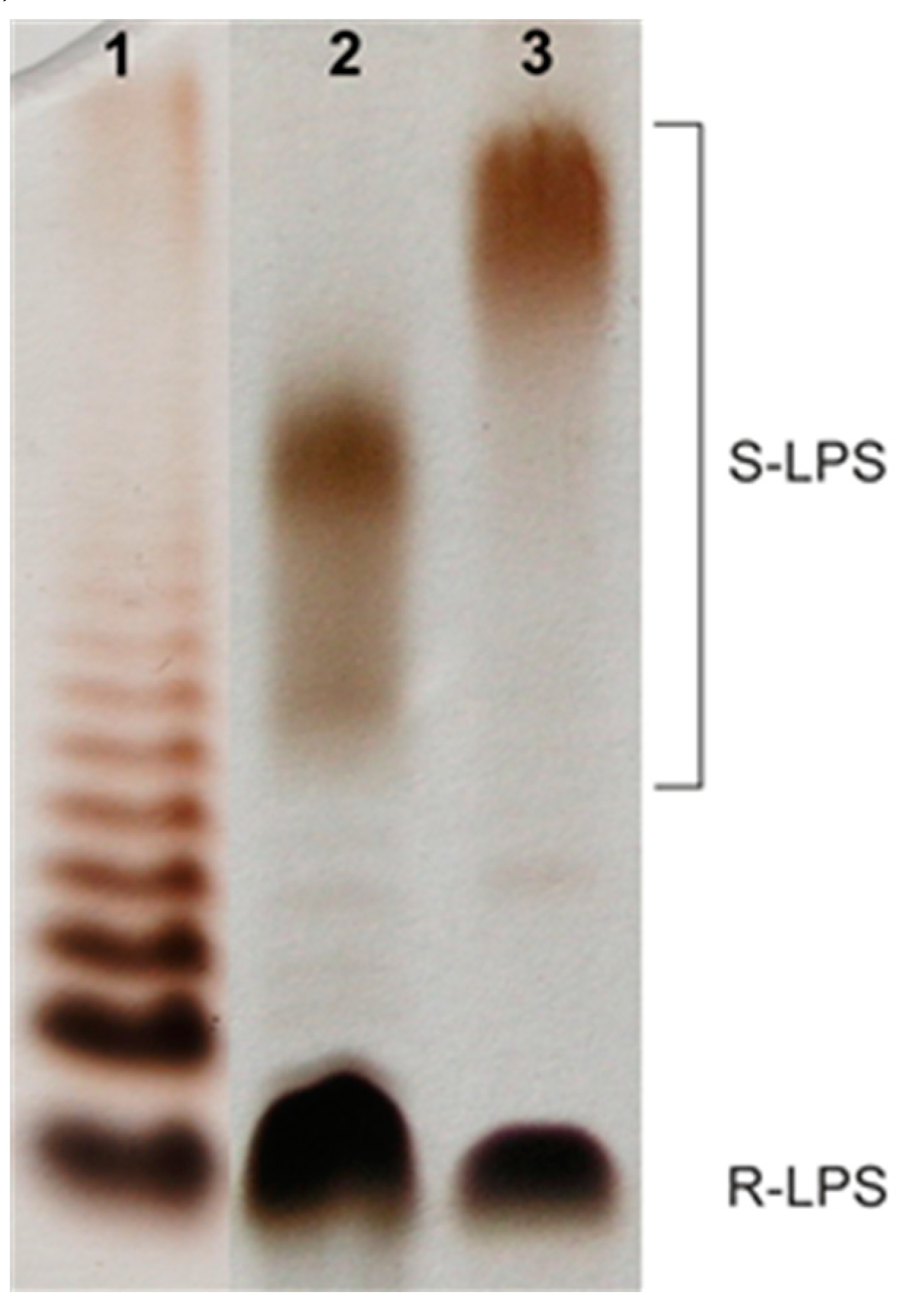
2.1. Bacterial Cultivation, Isolation of LPS, and SDS-PAGE Study

Cells of Aeromonas veronii bv. sobria strain K557 were extracted with hot aqueous 45% phenol [38], and LPS species were harvested from the phenol phase in a yield of 3.9% of the bacterial cell mass. SDS-PAGE analysis of the LPS followed by silver staining showed a pattern typical for LPS isolated from smooth bacterial cells (Figure 1) with the content of both slow-migrating high-molecular-weight (HMW) S-LPS species and fast-migrating low-molecular-weight R-LPS glycoforms. Moreover, the electrophoregram indicated that the studied S-LPS contained molecules where the core oligosaccharides were substituted with shorter O-chains in comparison to the LPS species of A. hydrophila JCM 3968, O6.

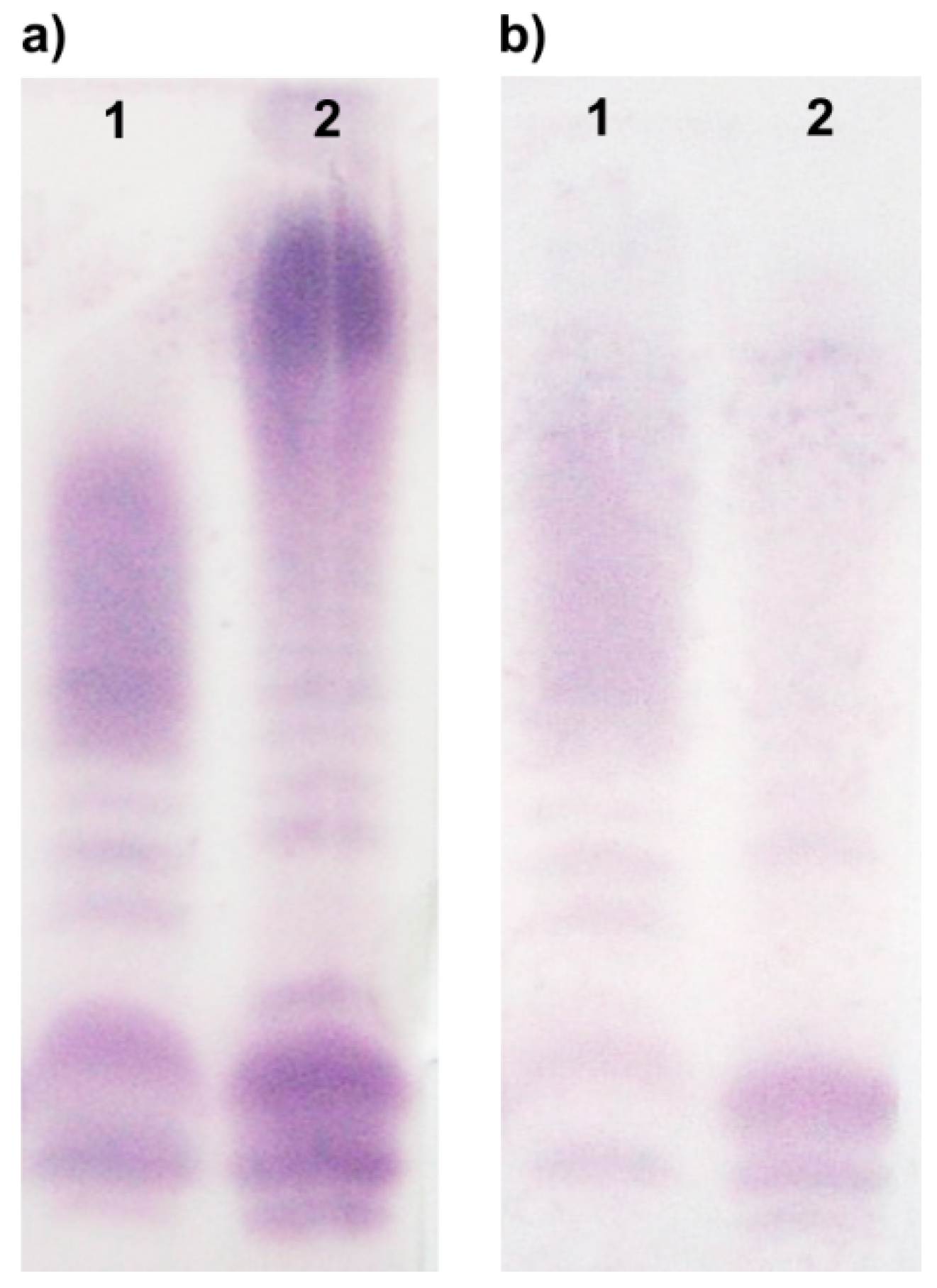
2.2. Serological Studies of the A. veronii bv. sobria K557 O-antigen

A. veronii bv. sobria strain K557 was serologically typed by agglutination tests using heat-inactivated bacteria and antisera for 44 defined Aeromonas O-serogroups (NIH system) and 20 provisional serogroups (PGO1–PGO20) for selected Polish isolates and classified as a representative of the Aeromonas serogroup O6 [28,36].
The LPS preparations from A. veronii bv. sobria strain K557 and A. hydrophila JCM 3968, which is the reference strain to serogroup O6, were studied by Western blotting and ELISA with intact and adsorbed polyclonal rabbit O-antisera.
In Western blot, the reference antiserum O6 recognized electrophoretically separated LPS molecules of both strains (Figure 2a). Strong reaction to slow-migrating bands corresponding to the O-PS containing LPS species was observed and suggested that the O-antigens shared common epitopes. Additionally, the stained bands of fast-migrating R-LPS molecules may indicate similarities in the core region of the studied strains. In turn, the other Western blot (Figure 2b) with A. veronii bv. sobria K557 O-antiserum revealed the recognition of antigenic determinants within LPS molecules of both A. veronii bv. sobria K557 and A. hydrophila JCM 3968 strains; however, the cross-reaction with the heterologous LPS was weaker than in the homologous system.
Accordingly, in ELISA (Table 1), the rabbit polyclonal O antiserum specific for A. hydrophila JCM 3968 O6 reacted strongly with the homologous LPS, and cross-reaction was observed with the LPS of A. veronii bv. sobria strain K557 to the lower titer of 1:64,000. In turn, the polyclonal O-antiserum against A. veronii bv. sobria strain K557 revealed reactions at the same level as both the homologous and heterologous LPS samples. The O6 reference antiserum revealed stronger reactivity than the K557-specific one, as demonstrated by the Western blotting results.
Adsorption of the antiserum specific for the A. veronii bv. sobria K557 O-antigen with A. hydrophila strain JCM 3968 O6 cells totally abolished the reactivity of this antiserum with both LPS preparations. The opposite reaction, i.e., adsorption of the reference O6 antiserum with the A. veronii bv. sobria K557 cells, only decreased its reactivity in the homologous system, whereas there was no reaction of the adsorbed O6 antiserum with A. veronii bv. sobria K557 LPS. The latter findings indicated that the adsorption process was complete and resulted in the removal of anti-K557 antibodies from the reference O6 antiserum. The remaining antibodies, strongly reacting with the homologous O6 LPS, were most probably specific to an additional epitope characteristic for the A. hydrophila JCM 3968 O6 antigen.
These data indicated that the reference O6 antiserum and the A. veronii bv. sobria K557 O-antiserum shared common antibodies but the reference one contained additional immunoglobulins recognizing structural determinants that were not present in the A. veronii bv. sobria K557 O-antigen.
Therefore, detailed chemical analyses were performed to establish the structure of the A. veronii bv. sobria strain K557 O-PS.

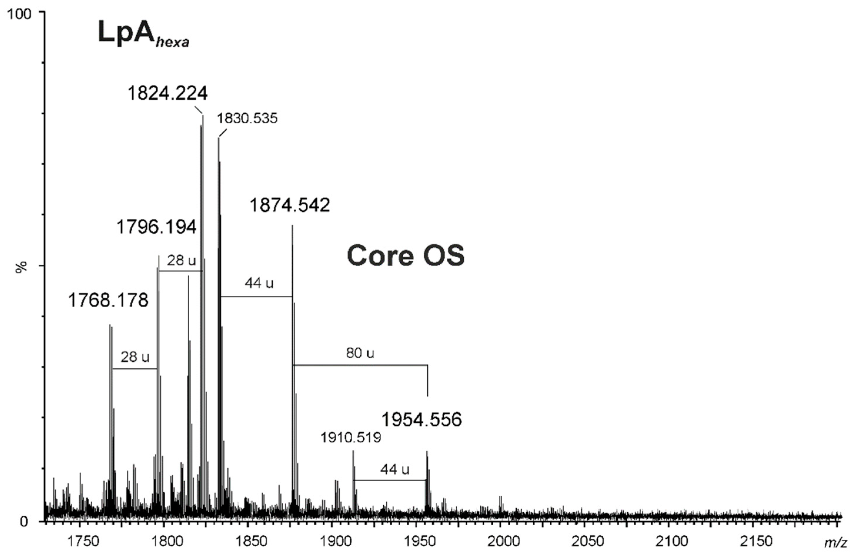
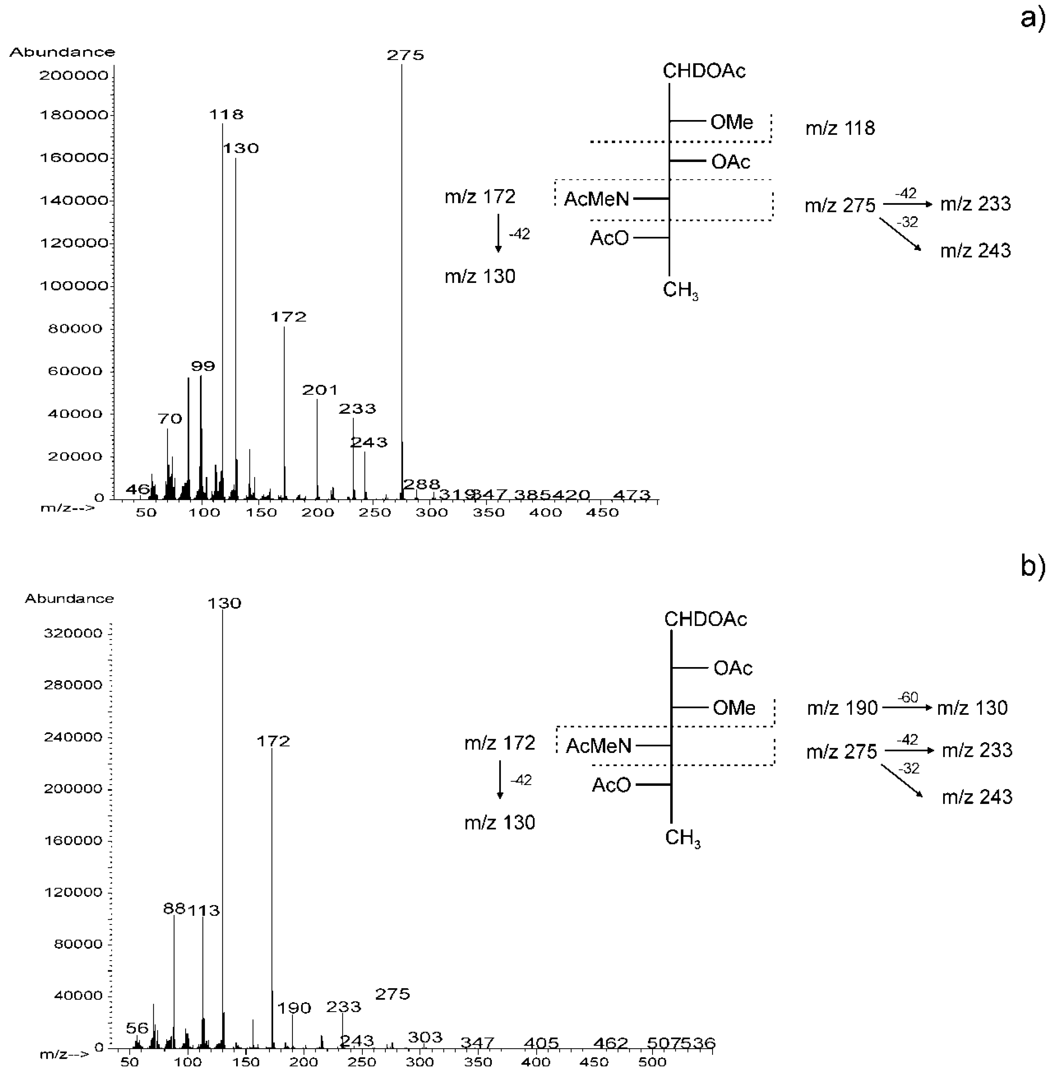
2.3. Chemical and Mass Spectrometry Analyses of LPS

Compositional analysis of the degraded polysaccharide (dgPS) liberated from the phenol-soluble LPS was performed using GLC-MS of alditol acetates. It showed the presence of d-glucose (d-Glc), d-galactose (d-Gal), 2-amino-2-deoxy-d-glucose (d-GlcN), d-glycero-d-manno-heptose (d,d-Hep), and l-glycero-d-manno-heptose (l,d-Hep) in a ratio of 1.0 : 1.2 : 1.3 : 1.5 : 3.8 as the core oligosaccharide components. The chemical analysis also revealed 6-deoxymannose (Rha), 2-amino-2,6-dideoxyglucose (QuiN), and 4-amino-4,6-dideoxyhexose (identified as Rha4N, see Section 2.4.). Rha4N was found as the main component of the O-polysaccharide part (see Section 2.4.). Kdo (3-deoxy-d-manno-2-octulosonic acid)—the only acidic sugar—was found in the LPS after treatment of the LPS with 48% aqueous HF (hydrofluoric acid), which suggested its phosphorylation [37,39,40]. The GLC-MS analysis of fatty acids as methyl esters and O-TMS derivatives revealed that 3-hydroxytetradecanoic [14:0(3-OH)], 3-hydroxyisopentadecanoic [i15:0(3-OH)], as well as dodecanoic (12:0) and tetradecanoic (14:0) acids, were the most abundant species in a ratio of 6.3 : 2.4 : 1.5 : 1.0. GlcN was identified as the sugar component of lipid A.
The negative ion matrix-assisted laser desorption/ionization time-of-flight (MALDI-TOF) mass spectrum of the A. veronii bv. sobria K557 lipopolysaccharide (Figure 3) showed the most intensive signals in the m/z range 1600–2000, which were attributable to a lipid A and a core oligosaccharide species (Y- and C-type fragment ions) arising from an in-source fragmentation at the glycosidic bond between the Kdo and the lipid A [41]. The ions at m/z 1768.17, 1796.19, and 1824.22 originated from hexaacylated lipid A species (Y-fragment ions) [37]. The ion at m/z 1824.22 represented a variant of lipid A, where the diglucosaminyl backbone bisphosphorylated at O-1 and O-4’ was substituted by four 3-hydroxytetradecanoic acids 14:0(3-OH) and two tetradecanoic acids 14:0, instead of two dodecanoic acids 12:0, compared with the ion at m/z 1768.17. In turn, the ion at m/z 1796.19 contained a sugar backbone acylated by three, instead of four, 3-hydroxytetradecanoic acids, one 3-hydroxyisopentadecanoic acid, and two dodecanoic acids. The composition of the ions is shown in Table 2.
The ions at m/z 1954.5 and 1874.5 (C-fragment ions) were assigned to the core oligosaccharide with the following composition: Hep6Hex2HexN1Kdo. The mass difference between the ions of 80 amu corresponded to species containing phosphorylated and dephosphorylated variants of the core oligosaccharide, respectively.

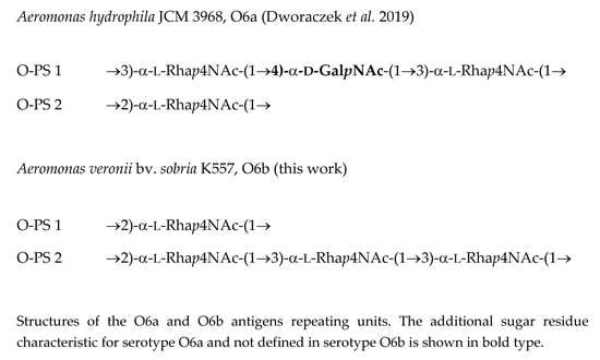
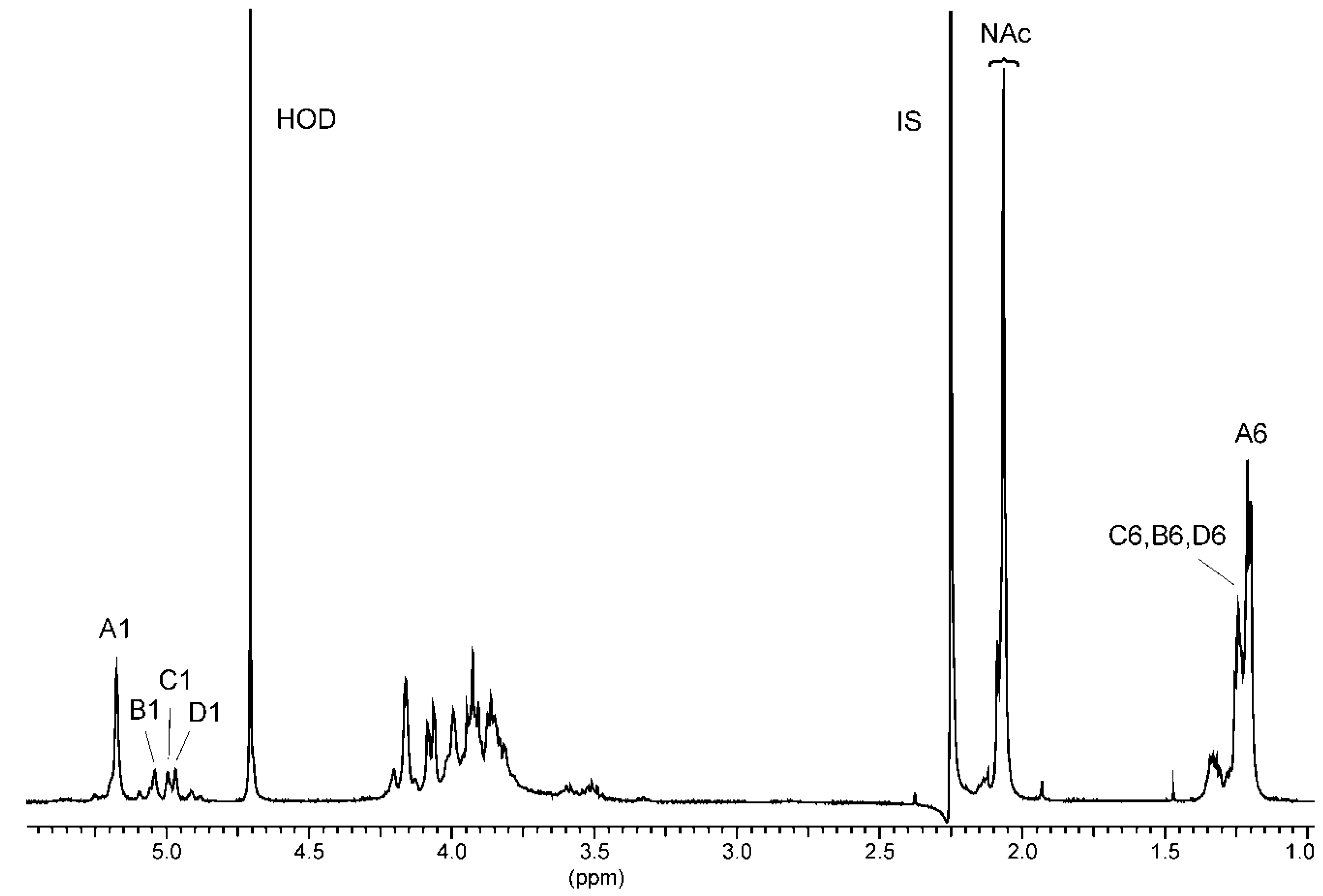
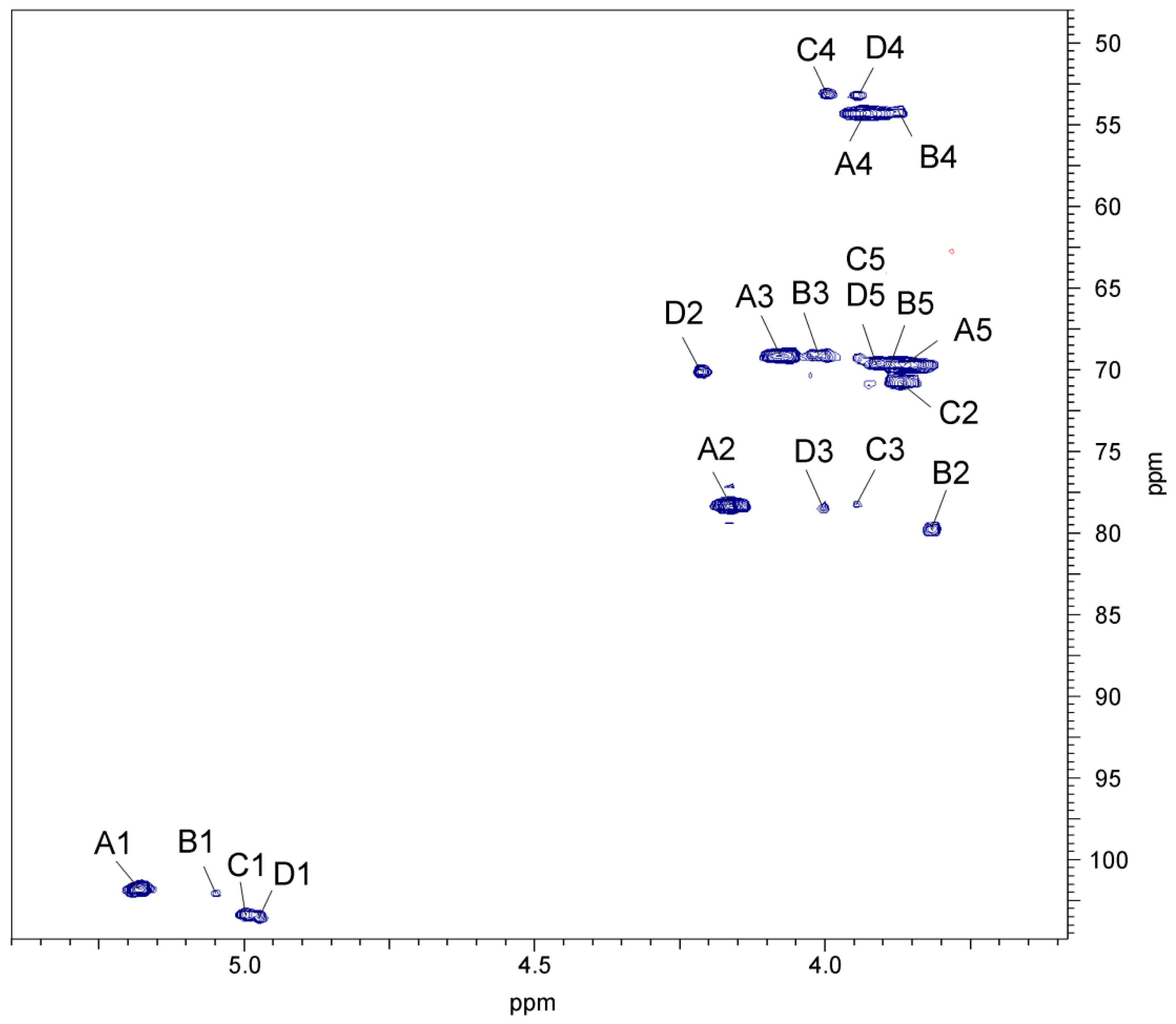
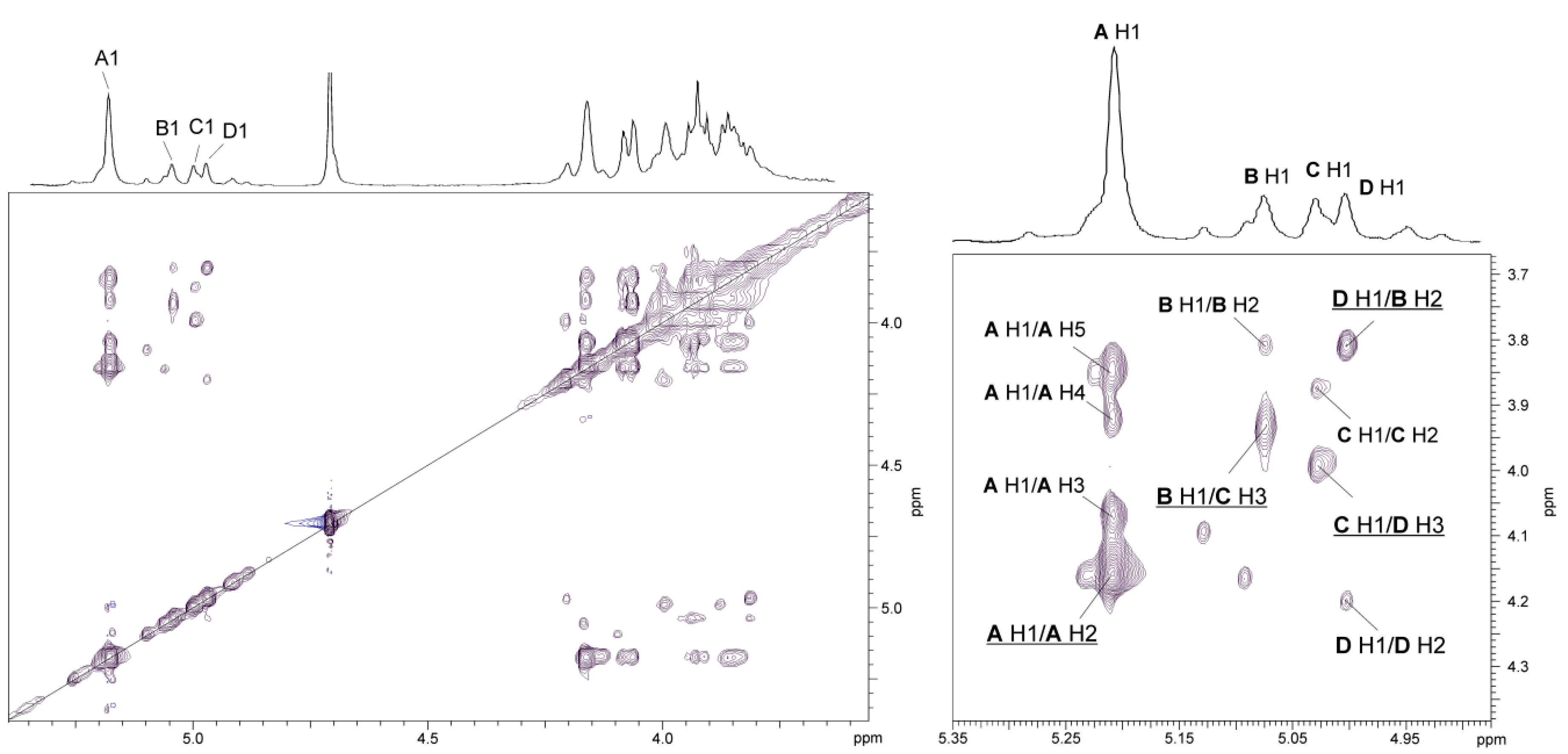
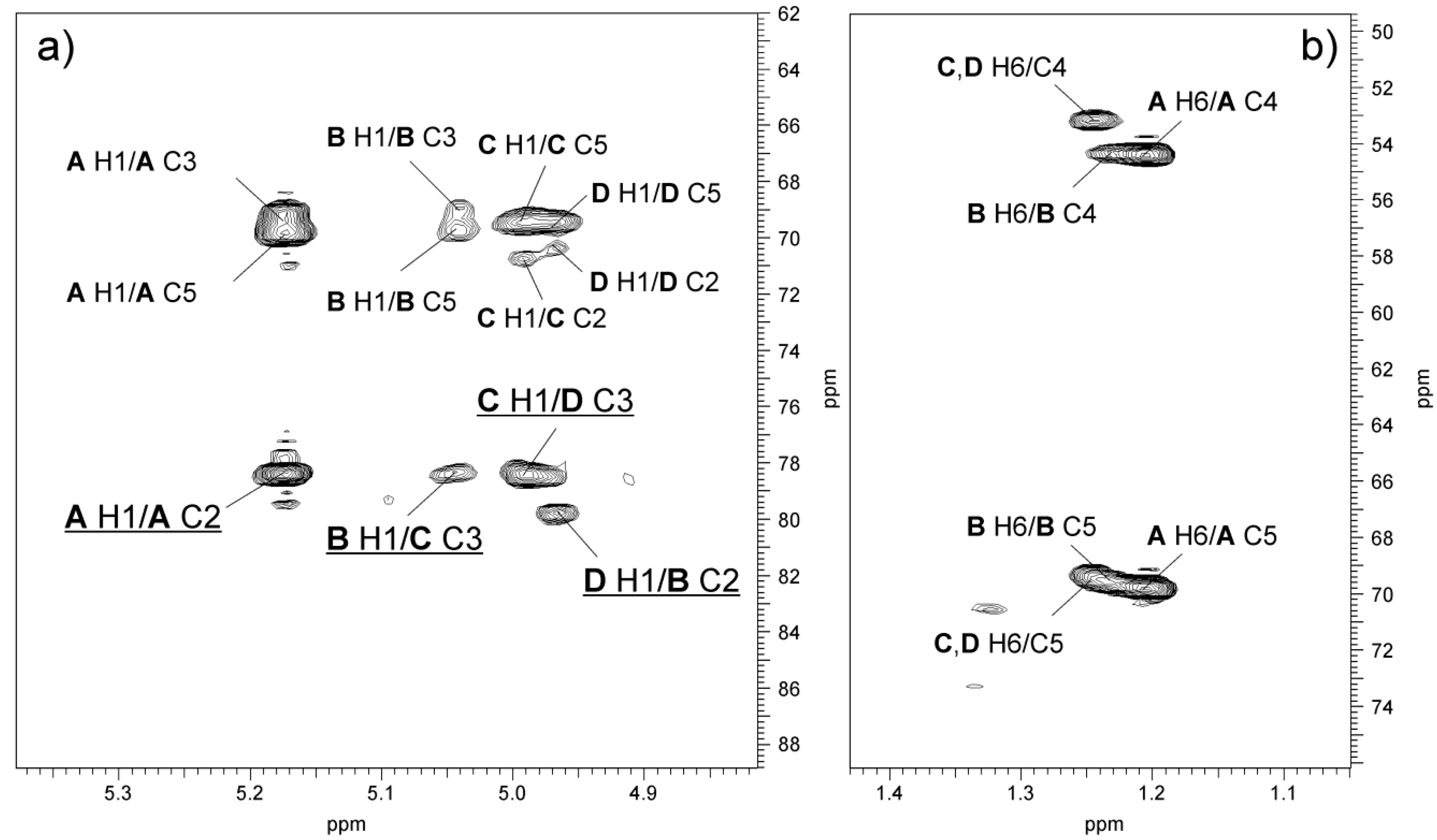
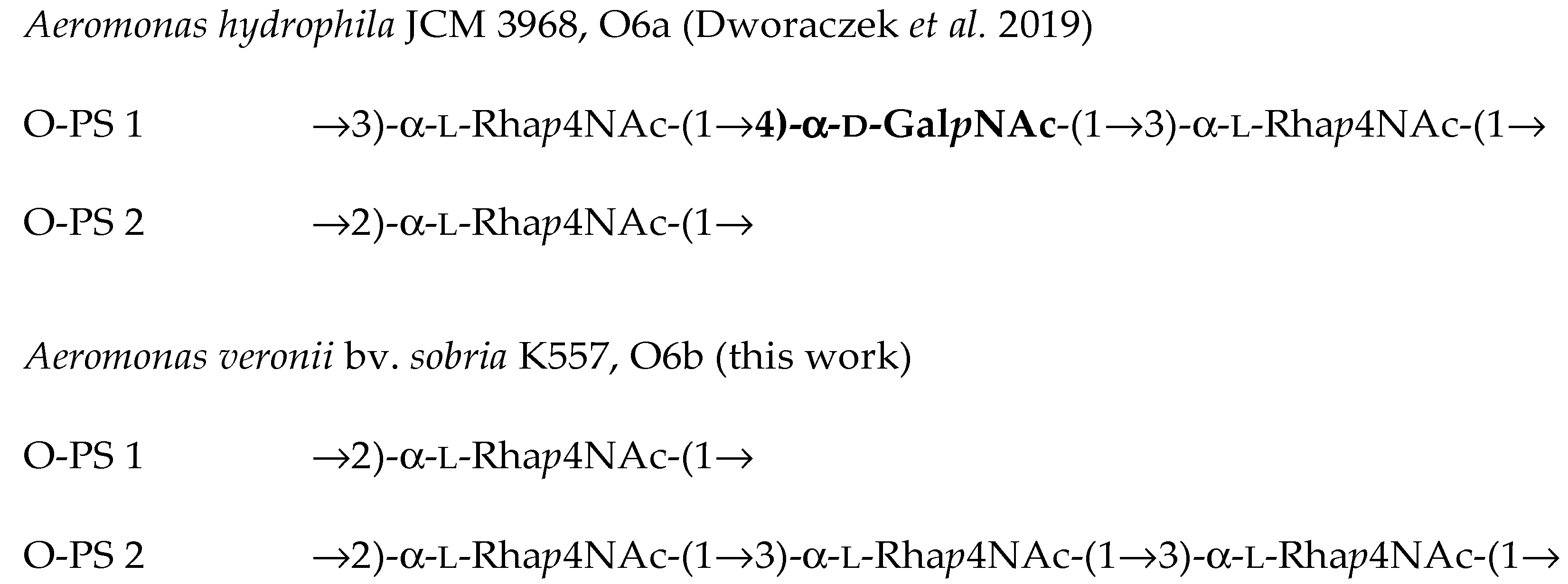
2.4. Structural Studies of O-polysaccharide (O-PS)

The O-PS was released from phenol-soluble LPS by mild-acid degradation followed by gel-permeation-chromatography (GPC) on Sephadex G-50 fine to give a high-molecular-weight O-polysaccharide with the yield of 33% of the LPS mass. GLC-MS sugar analysis of alditol acetates obtained after full acid hydrolysis of the O-PS showed the presence of rhamnose and 4-amino-4,6-dideoxymannose (Rha4N) in a peak area ratio ~1: 28.4. Other compounds detected in a small amount (below 10 %) in the GLC chromatogram of the A. veronii bv. sobria K557 O-PS, i.e., Glc, Gal and two heptose isomers (d,d-Hep and l,d-Hep), represented the core oligosaccharide sugars
Determination of the absolute configurations of the monosaccharides by GLC of the acetylated (S)- and (SR)-2-octyl glycosides [42] showed that Rha4N had the l configuration. Samples from the O-polysaccharide of Citrobacter gillenii O9a,9b [43] and the O-PS of A. hydrophila JCM 3968 [37] were used as a reference standard of d-Rha4N and l-Rha4N, respectively.
The methylation analysis of the O-PS by GLC-MS of partially methylated alditol acetates resulted in the identification of 4,6-dideoxy-3-O-methyl-4-(N-methyl)acetamidohexose (derived from 2-substituted Rha4N), and 4,6-dideoxy-2-O-methyl-4-(N-methyl)acetamidohexose (derived from 3-substituted Rha4N). The EI (electron impact) mass spectrum (Figure 4) of 4,6-dideoxy-2-O-methyl-4-(N-methyl)acetamidomannose was characterized by the presence of intense ion peaks at m/z 118 (C-1 ÷ C2 fragment), 275 (C-1 ÷ C4 fragment), and 172 (C-4 ÷ C6 fragment), and allowed distinguishing this derivative from 4,6-dideoxy-3-O-methyl-4-(N-methyl)acetamidomannose, whose EI mass spectrum contained ions at m/z 190, 275, and 172 characteristic of the C-1 ÷ C3, C-1 ÷ C4, and C-4 ÷ C6 primary fragments, respectively.
The low-field region of the 1H NMR spectrum of the O-polysaccharide (Figure 5) contained one major and three minor signals for anomeric protons at δ 5.18, 5.04, 5.00, and 4.97. The high-field region of the spectrum included signals for N-acetyl groups at δ 2.07, CH3-C groups of 6-deoxy sugars in the range of δ 1.21–1.24. The 1H and 13C resonances of the O-PS of A. veronii bv. sobria K557 were assigned using 2D homonuclear 1H,1H DQF-COSY, TOCSY, NOESY, heteronuclear 1H,13C HSQC, and HMBC experiments. The 1H and 13C NMR data are collected in Table 3.
The 1H,1H TOCSY, and DQF-COSY spectra revealed one major and three minor spin systems for monosaccharide residues, which were labelled AD in the order of the decreasing chemical shifts of their anomeric protons. The high-field positions of H-6 (δ 1.21–1.24) and C-6 (δ 18.2) resonances and the values of vicinal coupling constants 3J1,2 (~2 Hz), 3J3,4 (8.5 Hz), 3J4,5 (9 Hz), and 3J5,6 (6 Hz) characteristic for manno-pyranose allowed assigning all the A, B, C, and D spin systems (H-1/C-1 cross-peaks at δ 5.18/101.7, 5.04/102.1, 5.0/103.3, and 4.97/103.5, respectively) to Rha4NAc residues. The latter conclusion was confirmed by the correlation signals at δ 3.93/54.3 (the major one), 3.87/54.3, 3.94/53.2, and 4.00/53.2 observed in the 1H,13C HSQC spectrum (Figure 6), which indicated that the O-PS contains N-acetamido sugar.
In the TOCSY spectrum, at the H-1 coordinate, the cross-peaks with H-2-H-5 were visible for Rha4NAc A, but only one cross-peak with H-2 for Rha4NAc B, and two ones with H-2 and H-3 for Rha4NAc C and D. In turn, starting from the H-2 proton signal, cross-peaks with H-3-H-6 were visible for all spin systems; however, some signals overlapped. The 1H,1H COSY spectrum allowed unambiguous differentiation between protons within the spin system A and only partly resolved the cross-peaks for the Rha4NAc B, C and D. The difficulties in the assignment of the H-3, H-4, and H-5 of Rha4NAc B, C and D were overcome in the 1H,13C HMBC and 1H, 13C HSQC experiments.
The 13C NMR resonances of e.g., Rha4NAc C were assigned by the long-range H-6/C-4 and H-6/C-5 correlations at δ 1.24/53.2 and 1.24/69.5, and then the C-4/H-3 and C-4/H-2 correlations at δ 53.2/3.93 and 53.2/3.87, respectively, in the 1H,13C HMBC spectrum. In the 1H,13C HSQC spectrum, the cross-peak of the proton at the nitrogen-bearing carbon to the corresponding carbon at δ 4.00/53.2 was assigned to the H-4/C-4 correlation of Rha4NAc C. Moreover, in the 1H,13C HMBC spectrum, correlations of the anomeric proton with carbons C-2 and C-5 were found, and then the proton resonances were assigned from the 1H,13C HSQC spectrum. Similar long-range correlations were searched during identification of H-3, H-4 and H-5 proton signals of Rha4NAc B and D. The chemical shifts of the C-2 signal of Rha4NAc B and the C-3 signals of Rha4NAc C and D were confirmed after consideration of the methylation analysis data.
The α-configuration of all Rha4NAc residues was inferred from the relatively high-field position of the C-5 signals at δ 69.5–69.7 for the residues in the O-polysaccharides, compared with δ 68.6 and δ 72.4 for α-Rha4NAc and β-Rha4NAc, respectively [44,45].
The correlation signals between H-4 of all the Rha4NAc residues and carbonyl group signals at δC 176.0 and between the latter and methyl proton signals at δH 2.07, which were observed in the 1H,13C HMBC spectrum, confirmed that the residues building the O-PS were N-acetylated.
The positions of glycosylation were determined by downfield shifted signals of carbon atoms C-2 for residues Rha4NAc A (δ 78.3) and B (δ 79.8), and C-3 for Rha4NAc C (δ 78.3) and D (δ 78.4), compared with their positions in the spectra of the corresponding non-substituted monosaccharides [44,45,46].
In the NOESY spectrum (Figure 7), intraresidue H-1/H-2 and interresidue H-1/H-5 correlations at δ 5.18/4.16 and δ 5.18/3.85, respectively, typical of α-(1→2)-linked sugars with the manno configuration, indicated that the Rha4NAc A residues constitute the main O-polysaccharide (O-PS 1) being the homopolymer [47]. In turn, in the spectrum inter-residue NOE contacts were observed for protons of residues BC, CD, and DB. The following correlations between the anomeric protons and protons at the linkage carbons: Rhap4NAc B H-1/Rhap4NAc C H-3 at δ 5.04/3.93; Rhap4NAc C H-1/Rhap4NAc D H-3 at δ 5.00/3.99; Rhap4NAc D H-1/Rhap4NAc B H-2 at δ 4.97/3.81 demonstrated that the other O-polysaccharide (O-PS 2) is a heteropolymer with a trisaccharide repeating unit (Figure 7, Table 3).
The sequence of monosaccharides in the repeating unit of O-PS 2 was confirmed, in the 1H,13C HMBC spectrum (Figure 8), by the following inter-glycosidic cross-peaks: B H-1/C C-3 at δ 5.04/78.3; C H-1/D H-3 at δ 5.00/78.4; D H-1/B H-2 at δ 4.97/79.8.
In conclusion, the O-antigen of A. veronii bv. sobria K557, O6 consists of two structurally different O-polysaccharides, both containing only l-Rha4NAc residues. One of them (O-PS 1) is an α-(1→2)-linked homopolymer of l-Rha4NAc. The other polysaccharide, O-PS 2, is built up of trisaccharide repeating units composed of one α-(1→2)-, and two α-(1→3)-linked l-Rhap4NAc residues.
As judged by the comparison of the integral intensities of the H-1 proton signals, the content of O-PS 1 in the O-antigen of A. veronii bv. sobria K557 was predominant and was estimated at approximately 64%.
Based on the composition and methylation analyses as well as the NMR data, the following structures of the O-polysaccharides have been established:
O-PS 1    →2)-α-l-Rhap4NAc-(1→
          A
O-PS 2    →2)-α-l-Rhap4NAc-(1→3)-α-l-Rhap4NAc-(1→3)-α-l-Rhap4NAc-(1→
           B          C        D
The structure of the A. veronii bv. sobria K557 O-antigen is similar but not identical to the recently published O-PS of Aeromonas hydrophila JCM 3968, O6, which consisted of two structurally different O-polysaccharides as well (Figure 9). One of them (O-PS 1) was a heteropolymer built up of trisaccharide repeating units composed of one 4-substituted α-d-GalpNAc and two α-(1→3)-linked l-Rhap4NAc residues. The other polysaccharide, O-PS 2, was an α-(1→2)-linked homopolymer of l-Rha4NAc [37].
The serological results (Western blotting and ELISA) allowed recognition of structural determinants that are most probably responsible for antibody binding (putative epitopes). The occurrence of common α-l-Rhap4NAc-(1→2)-α-l-Rhap4NAc and α-l-Rhap4NAc-(1→3)-α-l-Rhap4NAc disaccharides is sufficient for providing cross-reactivity between the A. hydrophila JCM 3968 O6 antiserum and the LPS of A. veronii bv. sobria K557 and a similar reaction in an opposite system.
On the other hand, an additional epitope (epitopes) most probably related to a 4-substituted α-d-GalpNAc residue or a →4)-α-d-GalpNAc-(1→3)-α-l-Rhap4NAc disaccharide fragment, which has not been found in the O-antigen studied here, determines the specificity of the A. hydrophila JCM 3968 O6 serotype. This putative epitope (epitopes) seems to play an important role in the immunospecificity of the reference O6 antiserum (Figure 9).
Based on the data obtained, we suggest the division of the O6 serogroup into two subgroups: serogroup O6a for strains that share α-l-Rhap4NAc-(1→2)-α-l-Rhap4NAc and α-l-Rhap4NAc-(1→3)-α-l-Rhap4NAc disaccharides and an additional →4)-α-d-GalpNAc-associated epitope, and the other serogroup O6b deprived of the latter structural fragment in the O-antigen, with strain A. veronii bv. sobria K557 as a representative of the new subgroup.

3. Discussion

Almost every year, health disorders in freshwater fish are recorded on many farms in Poland. Although the development of a particular fish disease depends largely on climate conditions prevailing in a given zone and region, infections caused by Aeromonas spp. are the most common among bacterial fish diseases. As demonstrated recently by data collected during the last several years in Poland by Pękala-Safińska, health disorders caused by Aeromonas species were mostly observed in carp (Cyprinus carpio L.) and were usually manifested by skin lesions (MAI) in the form of ulceration as well as fish mortalities [48].
Previous studies performed on 558 isolates of mesophilic Aeromonas collected during 5 years in Polish culture facilities, among which 427 isolates were obtained from common carp and 121 from rainbow trout, revealed predominant occurrence of A. veronii bv. sobria, A. sobria and A. salmonicida species. In turn, A. veroni bv. sobria A. bestiarum and A. salmonicida were most frequently identified in both carp and trout samples; A. veroni bv. sobria was one of the dominant species only among carp isolates, and which is worth emphasizing, almost 80% of these isolates were classified as virulent [28,36]. Serological typing of all collected isolates using 44 antisera of the NIH scheme extended by 20 provisional serogroups for selected Polish isolates showed that O6 was one of the five most commonly identified serogroups, especially among carp isolates, and the other ones were O11, PGO1, O16, and O18. The report mentioned above also showed that, with the use of the antisera for serogroups from O45 to O96, there was little possibility for Aeromonas typing, since these groups occur rather rarely among fish isolates, especially in Polish aquacultures. In turn, when the 44 antisera of the NIH scheme were used, about 53% isolates were positively classified to appropriate somatic serotypes and this increased to about 88% when the NIH collection was extended by the sera for provisional serogroups of Polish origin [28,36]. Therefore, to obtain the most positive serotyping results, the postulate to include new provisional antisera against strains occurring in a given area and thus to extend the collection of antisera seems reasonable.
The inland aquaculture sector in Poland is mainly based on the culture of two species of freshwater fish - carp (49% of total production in 2015) and rainbow trout (38% of total production in 2015) [49]; thus, the lack of a commercially available vaccine dedicated to carp seems to be alarming. The most effective prevention of fish disease should involve immunoprophylaxis based on auto-vaccines chosen according to the needs of culture facilities and prepared from bacterial strains isolated from the fish or even the entire region. Auto-vaccines are confirmed to be highly effective against conditionally pathogenic microorganisms like Aeromonas sp., Yersinia ruckeri, and Pseudomonas sp. [50,51,52,53]. According to recently published studies, a vaccine containing whole cells and LPS of Aeromonas sp. seems to protect fish against MAS disease [54,55,56]. However, to avoid a failure of implemented prophylactic programs based on auto-vaccination, the serological and structural similarities and differences in O-chain polysaccharides in various serogroups and strains, which contribute to the immunospecificity of Aeromonas, should be carried out [57].
Here, the immunochemical investigation of LPS, especially the O-specific polysaccharide of A. veronii bv. sobria strain K557 were performed. SDS-PAGE and Western blotting revealed that the LPS-glycoforms of A. veronii bv. sobria K557 contained shorter O-chains than those of A. hydrophila JCM 3968; however, these molecules represented S-LPS species rather with the prevalence of the intermediate length O-antigen chains. In a recent study, Osawa et al. reported that the lengths of the E. coli O157 antigen could be modulated by wzz gene mutations and it has been shown that strains with long, intermediate, and short O-antigens vary in sensitivity to serum complement. The greater resistance of strains with intermediate and/or long length O-antigen chains to serum complement lysis than those with short O-chains is likely to have been optimized for pathogenesis during evolution [58].
Moreover, we established the structure of the O-antigen of A. veronii bv. sobria strain K557, and found that it consisted of two different O-polysaccharides. The serological studies indicated that the O-PS of A. veronii bv. sobria strain K557 is closely related but not identical to the O-antigen of A. hydrophila strain JCM 3968, which is a reference strain to Aeromonas serogroup O6. Another peculiar characteristic of both O-PSs is the presence of 4-amino-4,6-dideoxy-l-mannose (l-Rha4N, l-perosamine), quite an unusual amino sugar, for the first time identified as a compound building the O-antigen of A. hydrophila strain JCM 3968, O6 [37]. This is the second work that shows the occurrence of l-perosamine as a component of bacterial O-polysaccharides.
The chemical and mass spectrometry analyses of the phenol-soluble LPS of A. veronii bv. sobria strain K557 demonstrated the LPS glycoforms had hexaacylated lipid A species with a conserved architecture and a backbone composed of 1,4′-bisphosphorylated-β-(1→6)-linked-d-GlcN disaccharide. The residues of 3-hydroxytetradecanoate were predominant among fatty acids, similarly as previously reported for A. hydrophila [37]. However, some differences were found in the acylation profile of lipid A species, in comparison to those of A. bestiarum [59]. Amongst the ester-linked saturated fatty acids, not only dodecanoic (12:0) but also tetradecanoic (14:0) residues were found. The latter fatty acids were also detected in the lipid A of A. veronii strain Bs19, O16 [60].
The compositional analysis of the core oligosaccharide revealed two isomers of heptose (d,d-Hep and l,d-Hep), and MALDI-TOF MS confirmed that the core decasaccharide, with the following composition: Hep6Hex2HexN1Kdo1P1, has a structure shared by the LPS core regions of the A. hydrophila and A. bestiarum species [37,59]. Interestingly, the mass spectrum of the core OS of A. hydrophila JCM 3968, which was isolated from R-LPS molecules after mild acid hydrolysis and separation by gel-permeation-chromatography, showed an ion at m/z 1856.59, which corresponded to the dephosphorylated core variant with the composition Hep6Hex2HexN1Kdoanh. On the other hand, the structure of the core OS in the SR-LPS molecules of A. hydrophila JCM 3968 was slightly different: Hep6Hex1HexN1HexNAc1Kdo1P1 [37]. In these species, the O-antigen was linked to the GalNAc residue, whereas in the rough R-LPS glycoforms, the galactose appeared to be a terminal outer core sugar, similarly as has been established for the core OS of the rough mutant strain of A. hydrophila AH-3, O34 [39].
In conclusion, the immunochemical studies of LPS, which is a glycolipid characterized by high heterogeneity amongst Aeromonas sp. bacteria, may facilitate selection of vaccine strains suitable for immunoprophylaxis of MAI/MAS diseases. The O-antigen of A. veronii bv. sobria strain K557, serotype O6, studied here is composed of two O-polysaccharides, both containing a unique sugar 4-amino-4,6-dideoxy-l-mannose (N-acetyl-l-perosamine, l-Rhap4NAc). The major O-polysaccharide (O-PS 1) is built up of an α1→2 linked of l-Rhap4NAc, whereas the other one, O-PS 2, has trisaccharide repeating units composed of α1→2 and α1→3 linked l-Rhap4NAc residues. The serological studies confirmed the structural analyses and showed that the O-antigens of A. veronii bv. sobria K557, i.e., the strain isolated from the common carp during an outbreak of MAI/MAS on a Polish fish farm, and A. hydrophila JCM 3968, represent the same Aeromonas serogroup O6 and are closely related but not identical. The recently studied O-antigen of A. hydrophila JCM 3968, O6 consisted of two structurally different O-polysaccharides. One of them was a heteropolymer built up of trisaccharide repeating units composed of one α-d-GalpNAc and two α-(1→3)-linked l-Rhap4NAc residues. The other O-polysaccharide was an α-(1→2)-linked homopolymer of l-Rha4NAc. The consideration of the serological results in view of the known O-antigen structures enabled recognition of domains that could be responsible for antibody binding (putative epitopes). The occurrence of common α-l-Rhap4NAc-(1→2)-α-l-Rhap4NAc and α-l-Rhap4NAc-(1→3)-α-l-Rhap4NAc disaccharides is sufficient for providing cross-reactivity between A. hydrophila JCM 3968 O6 antiserum and the LPS of A. veronii bv. sobria 557, and vice versa. On the other hand, the additional epitope related to a 4-substituted α-d-GalpNAc residue, which has not been found in the O-antigen studied here, determines the specificity of the A. hydrophila JCM 3968 O6-serotype. This putative epitope seems to play an important role in the immunospecificity of the reference O6 antiserum.
Therefore, based on the data obtained, we suggest division of the O6 serogroup into two subgroups. The serogroup O6a includes strains that share α-l-Rhap4NAc-(1→2)-α-l-Rhap4NAc and α-l-Rhap4NAc-(1→3)-α-l-Rhap4NAc disaccharides and an additional →4)-α-d-GalpNAc-associated epitope. A. veronii bv. sobria strain K557 is a representative of the other subgroup O6b and its O-antigen is deprived of the latter structural fragment.

4. Materials and Methods

4.1. Bacterial Strain, Cultivation Conditions, and Isolation of LPS

Aeromonas veronii strain K557 was isolated from a common carp during an outbreak of motile aeromonad infection/motile aeromonad septicemia (MAI/MAS) on a Polish fish farm. The isolate was identified to the species level by restriction analysis of 16S rRNA gene amplified by polymerase chain reaction [28] and classified as Aeromonas veronii bv. sobria because of the positive reaction for arginine dihydrolase, and negative reactions for ornithine decarboxylase and aesculin hydrolysis [30,31]. Based on virulence-associated markers (hemolytic, gelatinolytic, and caseinolytic activities), strain K557 was classified as virulent for fish. For the LPS analysis, A. veronii bv. sobria K557 bacterium was cultivated with shaking (120 rpm) on tryptic soy broth (TSB) for 72 h at 28 °C. The cells were harvested by low-speed centrifugation (8000× g, 20 min). The recovered bacterial cell pellet was washed twice with 0.85% saline and once more with distilled water.
The bacterial cells (5 g dry mass) were digested with lysozyme, RNAse, and DNAse (24 h, 1 mg/g) and then with Proteinase K (36 h, 1 mg/g) in 50 mM phosphate buffer (pH 7.0) containing 5 mM MgCl2. The suspension was dialyzed against distilled water and freeze-dried. The digested cells were extracted three times with aqueous 45% phenol at 68 °C, [38] and the separated layers were dialyzed against tap and distilled water. LPS species recovered from the phenol phase were purified by ultracentrifugation at 105,000× g and freeze-dried to give a yield of 3.9% of dry bacterial cell mass.

4.2. Degradation of LPS and Isolation of O-polysaccharide

The phenol-soluble S-LPS (110 mg) was hydrolyzed with aqueous 2.5% acetic acid at 100 °C for 3 h, and lipid A precipitate was removed by centrifugation. The supernatant was concentrated and then fractionated by GPC on a column (1.8 × 80 cm) of Sephadex G-50 fine (Pharmacia, Sweden) using 1% acetic acid as the eluent and monitoring with a differential refractometer (Knauer, Berlin, Germany). The yield of the O-PS fraction was 33% of the LPS mass subjected to hydrolysis.

4.3. Chemical Analyses

For neutral and amino sugar analysis, the LPS and O-PS samples were hydrolyzed with 2 M CF3CO2H (100 °C, 4 h) or 10 M HCl for 30 min at 80 °C, respectively, and reduced with NaBD4; this was followed by acetylation with a 1:1 (v/v) mixture of acetic anhydride and pyridine (85 °C, 0.5 h).
To release acidic sugar, LPS was dephosphorylated with 48% aqueous hydrofluoric acid, HF (4 °C, 18 h) and dried under vacuum over sodium hydroxide [40]. Methanolysis was performed with 1 M MeOH/HCl (85 °C, 1 h), and the sample was extracted twice with hexane. The methanol layer was concentrated and the residue was dried and acetylated. The monosaccharides were identified as alditol and aminoalditol acetates [61] as well as acetylated methyl glycosides by GLC-MS.
For determination of the absolute configuration [42], the O-PS was subjected to 2-octanolysis (300 μL (S)-(+)-2-octanol or (SR)-(±)-2-octanol and 20 μL acetyl chloride, 100 °C, 3 h); the products were acetylated and analyzed by GLC-MS as above. A sample from the polysaccharide of Citrobacter gillenii O9a,9b [43] was used as the reference standard of d-Rha4N (D-perosamine).
Methylation of the O-PS (1.0 mg) was carried out with methyl iodide in dimethyl sulfoxide in the presence of powdered sodium hydroxide [62]. The products were recovered by extraction with chloroform/water (1:1, v/v), hydrolyzed with 10 M HCl for 30 min at 80 °C, N-acetylated, and reduced with NaBD4. The partially methylated alditol acetates derivatives were analyzed by GLC-MS.
For fatty acid analysis, a sample of the lipid A (1 mg) was subjected to methanolysis in 2 M methanolic HCl (85 °C, 12 h). The resulting fatty acid methyl esters were extracted with hexane and converted to their O-trimethylsilyl (O-TMS) derivatives, as described [63,64]. The methanol layer containing methyl glycosides was dried and acetylated with a pyridine-acetic anhydride mixture. The fatty acid derivatives and acetylated methyl glycosides were analyzed by GLC-MS as above.
All the sample derivatives were analyzed on an Agilent Technologies 7890A gas chromatograph (Agilent Technologies, Wilmington, DE, USA) connected to a 5975C MSD detector (inert XL EI/CI, Agilent Technologies, Wilmington, DE, USA). The chromatograph was equipped with an HP-5MS capillary column (Agilent Technologies, 30 m × 0.25 mm, flow rate of 1 mL/min, He as carrier gas). The temperature program for all the derivatives was as follows: 150 °C for 5 min, then 150 to 310 °C at 5 °C/min, and the final temperature was maintained for 10 min.

4.4. NMR Spectroscopy

An O-PS sample was deuterium-exchanged by freeze-drying with D2O and then examined in 99.98% D2O using acetone as an internal standard (δH 2.225, δC 31.45). 1D and 2D NMR spectra were recorded at 32 °C on a 500 MHz NMR Varian Unity Inova instrument (Varian Associates, Palo Alto, CA, USA) using Varian software Vnmrj V. 4.2 rev. (Agilent Technologies, Santa Clara, CA, USA). The following homonuclear and heteronuclear shift-correlated two-dimensional experiments were conducted for signal assignments and determination of the sugar sequence: 1H,1H DQF-COSY, 1H,1H TOCSY,1H,1H NOESY, 1H,13C HSQC, and 1H,13C HMBC. The mixing time was set to 100 and 200 ms in the TOCSY and NOESY experiments, respectively. The 1H,13C HSQC experiment with CRISIS based multiplicity editing was optimized for a coupling constant of 146 Hz. The heteronuclear multiple-bond correlation (HMBC) experiment was optimized for JH,C = 7 and 5 Hz, with 2-step low-pass filter 130 and 165 Hz to suppress one-bond correlations.

4.5. MALDI-TOF Mass Spectrometry (MS)

LPS was analyzed with matrix-assisted laser desorption/ionization time-of flight (MALDI-TOF) mass spectrometry (MS) using a Waters SYNAPT G2-Si HDMS instrument (Waters Corporation, Milford, MA, USA) equipped with a 1 kHz Nd:YAG laser system. Acquisition of the data was performed using MassLynx software version 4.1 SCN916 (Waters Corporation, Wilmslow, UK). Mass spectra were assigned with a multi-point external calibration using red phosphorous (Sigma-Aldrich, St. Louis, MO, USA) and recorded in the negative ion mode. An LPS sample (at a concentration of 10 µg/µL) was suspended in a water/methanol (1:1, v/v) solution containing 5 mM EDTA and then dissolved by ultrasonication. After desalting with some grains of cation exchange beads (Dowex 50WX8-200; Sigma-Aldrich, St. Louis, MO, USA), one microliter of the sample was transferred onto a well plate covered with a thin matrix film and allowed to dry at room temperature. The matrix solution was prepared from 2’,4’,6’-trihydroxyacetophenone (THAP) (200 mg/mL in methanol) mixed with nitrocellulose (15 mg/mL) suspended in 2-propanol/acetone (1:1, v/v) in proportion of 4:1 (v/v), according to the published method [65].

4.6. SDS-PAGE

LPS preparations were separated in 12.5% SDS-Tricine polyacrylamide electrophoresis gel and bands were visualized by silver staining after oxidation with periodate according to the published method [66].

4.7. Serological Studies

Polyclonal O-antisera against A. hydrophila JCM 3968, serogroup O6 and A. veronii bv. sobria strain K557 were obtained by immunization of New Zealand white rabbits with heat-inactivated bacteria according to the published procedure [28]. Rabbits were acclimatized in the animal facility of the National Veterinary Research Institute (Puławy, Poland), and all the experiments were performed according to the procedures approved by the local ethical committee (The Second Local Ethical Committee on Animal Testing in Lublin, the permission number 48/2012).
Western blots with rabbit antisera were performed after transferring SDS-PAGE-separated LPS profiles to Immobilon P (Millipore, St. Louis, MO, USA). The primary antibodies were detected using alkaline phosphatase-conjugated goat anti-rabbit antibodies (Sigma, St. Louis, MO, USA). Blots were developed with nitroblue tetrazolium and 5-bromo-4-chloro-3-indolylphosphate toluidine (Sigma) for 5 min, as described elsewhere [67].
The ELISA was performed as described previously [68] with some modifications, namely: 1–2 μg of the studied LPS per well were coated on flat-bottom Nunc-Immuno plates; the reaction was developed using rabbit-IgG specific peroxidase-conjugated goat antibodies (Jackson ImmunoResearch, West Grove, PA, USA); the final absorbance (A405) was read with the help of a Multiskan Go microplate reader (Thermo Fisher Scientific USA, Vantaa, Finland).
Adsorption of antisera was carried out using wet masses of bacterial cells washed in PBS (phosphate-buffered saline). Bacterial mass (100 μL) was suspended in 1 mL of serum diluted 1:50 in PBS. After 0.5 h incubation on ice, the cells were removed by centrifugation and the process was repeated two or three more times [69].

Author Contributions

A.T.-S. and K.D. conceived and designed the experiments; A.T.-S. and K.D. funding acquisition; K.D. performed Western blotting studies; D.D. carried out ELISA experiments; A.P-S.. and K.D. performed serotyping with the agglutination test; K.D. and A.T.-S. performed chemical analyses; K.D. and A.T.-S. contributed to the interpretation of mass spectra and NMR data; All authors analyzed the data; A.T.-S. and K.D. original draft preparation; All authors have reviewed the paper.

Funding

This research was financially supported by the grant from the National Science Centre (Decision No. DEC-2011/03/B/NZ1/01203) and the Polish Ministry of Science and Higher Education research funds (BS-M-11-010-18-2-05 and BS-P-11-010-18-2-04).

Acknowledgments

The authors gratefully thank Andrzej Gamian for the gift of the O-polysaccharide of Citrobacter gillenii O9 (L. Hirszfeld Institute of Immunology and Experimental Therapy, Polish Academy of Sciences, Wrocław, Poland) and Paweł Sowiński for recording the NMR spectra (Intercollegiate NMR Laboratory, Department of Chemistry, Technical University of Gdańsk, Poland). The authors wish to acknowledge the assistance and technical support of Hubert Pietras during LPS isolation and purification.

Conflicts of Interest

The authors declare no conflict of interest.

References

  1. Janda, J.M.; Duffy, P.S. Mesophilic aeromonads in human diseases: Current taxonomy, laboratory infection and infectious diseases spectrum. Rev. Infect. Dis. 1988, 10, 980–997. [Google Scholar] [CrossRef] [PubMed]
  2. Janda, J.M. Recent advances in the study of the taxonomy, pathogenicity and infectious syndromes with the genus Aeromonas. Clin. Microbiol. Rev. 1991, 4, 397–410. [Google Scholar] [CrossRef] [PubMed]
  3. Martin-Carnahan, A.; Joseph, S.W. Order XII. Aeromonadales ord. nov. In Bergey’s Manual of Systematic Bacteriology, 2nd ed.; Brenner, D.J., Krieg, N.R., Staley, J.T., Eds.; Springer: New York, NY, USA, 2005; Volume 2, pp. 556–578. [Google Scholar]
  4. Janda, J.M.; Abbott, S. The genus Aeromonas: Taxonomy, pathogenicity, and infection. Clin. Microbiol. Rev. 2010, 23, 35–73. [Google Scholar] [CrossRef] [PubMed]
  5. Huys, G. The Family Aeromonadaceae. In The Prokaryotes—Gammaproteobacteria; Rosenberg, E., DeLong, E.F., Lory, S., Stackebrandt, E., Thompson, F., Eds.; Springer: Berlin/Heidelberg, Germany, 2014; pp. 27–57. [Google Scholar] [CrossRef]
  6. Araujo, R.M.; Arribas, R.M.; Pares, R. Distribution of Aeromonas species in waters with different levels of pollution. J. Appl. Bacteriol. 1991, 71, 182–186. [Google Scholar] [CrossRef] [PubMed]
  7. Cipriano, R.C.; Bullock, G.L. Furunculosis and other diseases caused by Aeromonas salmonicida. Fish. Dis. Leafl. 2001, 66, 1–32. [Google Scholar] [CrossRef]
  8. Praveen, P.K.; Debnath, C.; Shekhar, S.; Dalai, N.; Ganguly, S. Incidence of Aeromonas spp. infection in fish and chicken meat and its related public health hazards: A review. Vet. World 2016, 9, 6–11. [Google Scholar] [CrossRef]
  9. Ibrahem, M.D.; Mostafa, M.M.; Arab, R.M.H.; Rezk, M.A.; Elghobashy, H.; Fitzsimmons, K.; Diab, A.S. (Eds.) Prevalence of Aeromonas hydrophila infection in wild and cultured tilapia nilotica (O. niloticus) in Egypt. In Proceedings of the 8th International Symposium on Tilapia in Aquaculture, Cairo, Egypt, 12–14 October 2008; Volume 2, pp. 1257–1270. [Google Scholar]
  10. Tomas, J.M. The Main Aeromonas Pathogenic Factors. ISRN Microbiol. 2012, 2012, 256261. [Google Scholar] [CrossRef]
  11. Stratev, D.; Odeyemi, O.A. An overview of motile Aeromonas septicaemia management. Aquacult. Int. 2017, 25, 1095–1105. [Google Scholar] [CrossRef]
  12. Hoel, S.; Vadstein, O.; Jakobsen, A.N. The significance of mesophilic Aeromonas spp. in minimally processed ready-to-eat seafood. Microorganisms 2019, 7, 91. [Google Scholar] [CrossRef]
  13. Holmberg, S.D.; Schell, W.L.; Fanning, G.R.; Wachsmuth, I.K.; Blake, P.A.; Brenner, D.J.; Farmer, J.J. Aeromonas intestinal infections in the United States. Ann. Int. Med. 1986, 105, 683–689. [Google Scholar] [CrossRef]
  14. Figueras, M.J. Clinical relevance of Aeromonas sM503. Rev. Clin. Microbiol. 2005, 16, 145–153. [Google Scholar] [CrossRef]
  15. Mencacci, A.; Cenci, E.; Mazzolla, R.; Farinella, S.; D’Alo, F.; Vitali, M.; Bistoni, F. Aeromonas veronii biovar veronii septicaemia and acute suppurative cholangitis in a patient with hepatitis B. J. Med. Microbiol. 2003, 52, 727–730. [Google Scholar] [CrossRef] [PubMed]
  16. Roberts, M.T.M.; Enoch, D.A.; Harris, K.A.; Karas, J.A. Aeromonas veronii biovar sobria bacteraemia with septic artritis confirmed by 16S rDNA PCR in an immunocompetent adult. J. Clin. Microbiol. 2006, 55, 241–243. [Google Scholar] [CrossRef]
  17. Garduño, R.A.; Moore, A.R.; Oliver, G.; Lizama, A.L.; Garduño, E.; Kay, W.W. Host cell invasion and intracellular resistance by Aeromonas salmonicida: Role of the S-layer. J. Clin. Microbiol. 2000, 46, 660–668. [Google Scholar] [CrossRef]
  18. Merino, S.; Rubires, X.; Aguillar, A.; Guillot, J.F.; Tomas, J.M. The role of the O-antigen lipopolysaccharide on the colonization in vivo of the germfree chicken gut by Aeromonas hydrophila serogroup O:34. Microb. Pathog. 1996, 20, 325–333. [Google Scholar] [CrossRef]
  19. Rabaan, A.A.; Gryllos, I.; Tomas, J.M.; Shaw, J.G. Motility and polar flagellum are required for Aeromonas caviae adherence to HEp-2 cells. Infect. Immun. 2001, 69, 4257–4267. [Google Scholar] [CrossRef] [PubMed]
  20. Raetz, C.R.H.; Whitfield, C. Lipopolysaccharide endotoxins. Annu. Rev. Biochem. 2002, 71, 635–700. [Google Scholar] [CrossRef]
  21. Caroff, M.; Karibian, D. Structure of bacterial lipopolysaccharides. Carbohydr. Res. 2003, 338, 2431–2447. [Google Scholar] [CrossRef]
  22. Whitfield, C.; Trent, M.S. Biosynthesis and export of bacterial lipopolysaccharides. Annu. Rev. Biochem. 2014, 83, 99–128. [Google Scholar] [CrossRef]
  23. Maldonado, R.F.; Sá-Correia, I.; Valvano, M.A. Lipopolysaccharide modification in Gram-negative bacteria during chronic infection. FEMS Microbiol. Rev. 2016, 40, 480–493. [Google Scholar] [CrossRef]
  24. Cao, H.; Wang, M.; Wang, Q.; Xu, T.; Du, Y.; Li, H. Identifying genetic diversity of O antigens in Aeromonas hydrophila for molecular serotype detection. PLoS ONE 2018, 13, e0203445. [Google Scholar] [CrossRef] [PubMed]
  25. Sakazaki, R.; Shimada, T. O-Serogrouping for mesophilic Aeromonas strains. Jpn. J. Med. Sci. Biol. 1984, 37, 247–255. [Google Scholar] [CrossRef] [PubMed]
  26. Thomas, L.V.; Gross, R.J.; Cheasty, T.; Rowe, B. Extended serogrouping scheme for motile, mesophilic Aeromonas species. J. Clin. Microbiol. 1990, 28, 980–984. [Google Scholar] [PubMed]
  27. Kokka, R.P.; Vedros, N.A.; Janda, J.M. Electrophoretic analysis of the surface components of autoagglutinating surface array protein-positive and surface array protein-negative Aeromonas hydrophila and Aeromonas sobria. J. Clin. Microbiol. 1990, 28, 2240–2247. [Google Scholar] [PubMed]
  28. Kozinska, A.; Pekala, A. Serotyping of Aeromonas species isolated from Polish fish farms in relation to species and virulence phenotype of the bacteria. Bull. Vet. Inst. Pulawy 2010, 54, 315–320. [Google Scholar]
  29. Esteve, C.; Alcaide, E.; Canals, R.; Merino, S.; Blasco, D.; Figueras, M.J.; Tomas, J.M. Pathogenic Aeromonas hydrophila serogroup O:14 and O:81 strains with S-layer. Appl. Environ. Microbiol. 2004, 70, 5898–5904. [Google Scholar] [CrossRef] [PubMed]
  30. Hickman-Brenner, F.W.; MacDonald, K.L.; Steigerwalt, A.G.; Fanning, G.R.; Brenner, D.J.; Farmer, J.J., III. Aeromonas veronii, a new ornithine decarboxylase-positive species that may cause diarrhea. J. Clin. Microbiol. 1987, 25, 900–906. [Google Scholar]
  31. Altwegg, M.; Steigerwalt, A.G.; Altwegg-Bissig, R.; Luthy-Hottenstein, J.; Brenner, D.J. Biochemical identification of Aeromonas genospecies isolated from humans. J. Clin. Microbiol. 1990, 28, 258–264. [Google Scholar]
  32. Rahman, M.; Colque-Navarro, P.; Kühn, I.; Huys, G.; Swings, J.; Möllby, R. Identification and characterization of pathogenic Aeromonas veronii bv. sobria associated with epizootic ulcerative syndrome in fish in Bangladesh. Appl. Environ. Microbiol. 2002, 68, 650–655. [Google Scholar] [CrossRef]
  33. Cai, S.-H.; Wu, Z.-H.; Jian, J.-C.; Lu, Y.-S.; Tang, J.F. Characterization of pathogenic Aeromonas veronii bv. veronii associated with ulcerative syndrome from Chinese longsnout catfish (Leiocassis longirostris Günther). Braz. J. Microbiol. 2012, 43, 382–388. [Google Scholar] [CrossRef]
  34. Turska-Szewczuk, A.; Lindner, B.; Pekala, A.; Palusinska-Szysz, M.; Choma, A.; Russa, R.; Holst, O. Structural analysis of the O-specific polysaccharide from the lipopolysaccharide of Aeromonas veronii bv. sobria strain K49. Carbohydr. Res. 2012, 353, 62–68. [Google Scholar] [CrossRef] [PubMed]
  35. Turska-Szewczuk, A.; Pietras, H.; Duda, K.A.; Kozińska, A.; Pękala, A.; Holst, O. Structure of the O-specific polysaccharide from the lipopolysaccharide of Aeromonas sobria strain Pt312. Carbohydr. Res. 2015, 403, 142–148. [Google Scholar] [CrossRef] [PubMed]
  36. Kozińska, A. Genotypic and Serological Analysis of Domestic Mesophilic Isolates Aeromonas sp. in Terms of Pathogenicity and the Type of Disease Symptoms Caused by Them in Fish. Habilitation Thesis, The National Veterinary Institute—The National Research Institute, Puławy, Poland, 2009. [Google Scholar]
  37. Dworaczek, K.; Kurzylewska, M.; Karaś, M.A.; Janczarek, M.; Pękala-Safińska, A.; Turska-Szewczuk, A. A unique sugar L-perosamine (4-amino-4,6-dideoxy-L-mannose) is a compound building two O-chain polysaccharides in the lipopolysaccharide of Aeromonas hydrophila strain JCM 3968, serogroup O6. Mar. Drugs 2019, 17, 254. [Google Scholar] [CrossRef] [PubMed]
  38. Westphal, O.; Jann, K. Bacterial lipopolysaccharide. Extraction with phenol-water and further applications of the procedure. Meth. Carbohydr. Chem. 1965, 5, 83–91. [Google Scholar]
  39. Knirel, Y.A.; Vinogradov, E.; Jimenez, N.; Merino, S.; Tomas, J.M. Structural studies on the R-type lipopolysaccharide of Aeromonas hydrophila. Carbohydr. Res. 2004, 339, 787–793. [Google Scholar] [CrossRef] [PubMed]
  40. Pieretti, G.; Corsaro, M.M.; Lanzetta, R.; Parrilli, M.; Nicolaus, B.; Gambacorta, A.; Lindner, B.; Holst, O. Structural characterization of the core region of the lipopolysaccharide from the haloalkaliphilic Halomonas pantelleriensis: Identification of the biological O-antigen repeating unit. Eur. J. Org. Chem. 2008, 2008, 721–728. [Google Scholar] [CrossRef]
  41. Domon, B.; Costello, C.E. A systamatic nomenclature for carbohydrate fragmentations in FAB MS/MS spectra of glycoconjugates. Glycoconj. J. 1988, 5, 397–409. [Google Scholar] [CrossRef]
  42. Leontein, K.; Lindberg, B.; Lönngren, J. Assignment of absolute configuration of sugars by GLC of their acetylated glycosides formed from chiral alcohols. Carbohydr. Res. 1978, 62, 359–362. [Google Scholar] [CrossRef]
  43. Lipiński, T.; Zatonsky, G.V.; Kocharova, N.A.; Jaquinod, M.; Forest, E.; Shashkov, A.S.; Gamian, A.; Knirel, Y.A. Structures of two O-chain polysaccharides of Citrobacter gillenii O9a,9b lipopolysaccharide. A new homopolymer of 4-amino-4,6-dideoxy-D-mannose (perosamine). Eur. J. Biochem. 2002, 269, 93–99. [Google Scholar] [CrossRef]
  44. Lipkind, G.M.; Shashkov, A.S.; Knirel, Y.A.; Vinogradov, E.V.; Kochetkov, N.K. A computer-assisted structural analysis of regular polysaccharides on the basis of 13C-n.m.r. data. Carbohydr. Res. 1988, 175, 59–75. [Google Scholar] [CrossRef]
  45. Ovchinnikova, O.G.; Kocharova, N.A.; Katzenellenbogen, E.; Zatonsky, G.V.; Shashkov, A.S.; Knirel, Y.A.; Lipiński, T.; Gamian, A. Structures of two O-polysaccharides of the lipopolysaccharide of Citrobacter youngae PCM 1538 (serogroup O9). Carbohydr. Res. 2004, 339, 881–884. [Google Scholar] [CrossRef] [PubMed]
  46. Jansson, P.E.; Kenne, L.; Widmalm, G. Computer-assisted structural analysis of polysaccharides with an extended version of CASPER using 1H- and 13C-NMR data. Carbohydr. Res. 1989, 188, 169–191. [Google Scholar] [CrossRef]
  47. Knirel, Y.A.; Ovod, V.V.; Paramonov, N.A.; Krohn, K.J. Structural heterogeneity in the O polysaccharide of Pseudomonas syringe pv. coriandricola GSPB 2028 (NCPPB 3780, W-43). Eur. J. Biochem. 1998, 258, 716–721. [Google Scholar] [CrossRef] [PubMed]
  48. Pękala-Safińska, A. Contemporary threats of bacterial infections in freshwater fish. J. Vet. Res. 2018, 62, 261–267. [Google Scholar] [CrossRef] [PubMed] [Green Version]
  49. Lirski, A.; Myszkowski, L. Polish aquaculture in 2016 based on the analysis of questionnaire RRW-22. Part 1. Komun. Ryb. 2017, 6, 20–27. [Google Scholar]
  50. Terech-Majewska, E. Improving disease prevention and treatment in controlled fish culture. Arch. Pol. Fish. 2016, 24, 115–165. [Google Scholar] [CrossRef] [Green Version]
  51. Kozińska, A.; Pękala, A. Characteristics of disease spectrum in relation to species, serogroups, and adhesion ability of motile aeromonads in fish. Sci. World J. 2012. [Google Scholar] [CrossRef] [PubMed]
  52. Grudniewska, J.; Dobosz, S.; Terech-Majewska, E.; Zalewski, T.; Siwicki, A.K. Economic and health aspects of vaccinating against furunculosis and yersiniosis in rainbow trout culture. Komun. Ryb. 2010, 1, 18–21. [Google Scholar]
  53. Siwicki, A.K.; Baranowski, P.; Dobosz, S.; Kuźmiński, H.; Grudniewska, J.; Kazun, K.; Głąbski, E.; Kazuń, B.; Terech-Majewska, E.; Trapkowska, S. Using new generation vaccines administered per os in granulate as prophylaxis against furunculosis and yersiniosis in salmonids. In Current Challenges in Fish Disease Prevention and Treatment; Siwicki, A.K., Ed.; Publisher IRS: Olsztyn, Poland, 2004; pp. 117–122. [Google Scholar]
  54. Sukenda, S.; Romadhona, E.I.; Yuhana, M.; Pasaribu, W.; Hidayatullah, D. Efficacy of whole-cell and lipopolysaccharide vaccine of Aeromonas hydrophila on juvenile tilapia Oreochromis niloticus against motile aeromonad septicemia. AACL Bioflux 2018, 11, 1456–1466. [Google Scholar]
  55. Dehghani, S.; Akhlaghi, M.; Dehghani, M. Efficacy of formalin-killed, heat-killed and lipopolysaccharide vaccines against motile aeromonads infection in rainbow trout (Oncorhynchus mykiss). Glob. Veterin. 2012, 9, 409–415. [Google Scholar] [CrossRef]
  56. Fernandez, J.B.; Yambot, A.V.; Almeria, O. Vaccination of Nile tilapia (Oreochromis niloticus) using lipopolysaccharide (LPS) prepared from Aeromonas hydrophila. Int. J. Fauna Biol. Stud. 2014, 1, 1–3. [Google Scholar]
  57. Pajdak, J.; Terech-Majewska, E.; Platt-Samoraj, A.; Schulz, P.; Siwicki, A.K.; Szweda, W. Characterization of pathogenic Yersinia ruckeri strains and their importance in rainbow trout immunoprophylaxis. Med. Weter 2017, 73, 579–584. [Google Scholar] [CrossRef]
  58. Osawa, K.; Shigemura, K.; Iguchi, A.; Shirai, H.; Imayama, T.; Seto, K.; Raharjo, D.; Fujisawa, M.; Osawa, R.; Shirakawa, T. Modulation of the O-antigen chain length by the wzz gene in Escherichia coli O157 influences its sensitivities to serum complement. Microbiol. Immunol. 2013, 57, 616–623. [Google Scholar] [PubMed]
  59. Turska-Szewczuk, A.; Lindner, B.; Komaniecka, I.; Kozinska, A.; Pekala, A.; Choma, A.; Holst, O. Structural and immunochemical studies of the lipopolysaccharide from the fish pathogen, Aeromonas bestiarum strain K296, serotype O18. Mar. Drugs 2013, 11, 1235–1255. [Google Scholar] [CrossRef] [PubMed]
  60. Turska-Szewczuk, A.; Duda, K.A.; Schwudke, D.; Pekala, A.; Kozinska, A.; Holst, O. Structural studies of the lipopolysaccharide from the fish pathogen, Aeromonas veronii strain Bs19, serotype O16. Mar. Drugs 2014, 12, 1298–1316. [Google Scholar] [CrossRef] [PubMed]
  61. Russa, R.; Urbanik-Sypniewska, T.; Lindström, K.; Mayer, H. Chemical characterization of two lipopolysaccharide species isolated from Rhizobium loti NZP2213. Arch. Microbiol. 1995, 163, 345–351. [Google Scholar] [CrossRef] [PubMed]
  62. Ciucanu, I.; Kerek, F. A simple and rapid method for the permethylation of carbohydrates. Carbohydr. Res. 1984, 131, 209–217. [Google Scholar] [CrossRef]
  63. Pieretti, G.; Corsaro, M.M.; Lanzetta, R.; Parrilli, M.; Vilches, S.; Merino, S.; Tomas, J.M. Structure of the core region from the lipopolysaccharide of Plesiomonas shigelloides strain 302-73 (serotype O1). Eur. J. Org. Chem. 2009, 2009, 1365–1371. [Google Scholar] [CrossRef]
  64. Komaniecka, I.; Choma, A.; Lindner, B.; Holst, O. The structure of a novel lipid A from the lipopolysaccharide of Bradyrhizobium elkanii containing three mannose units in the backbone. Chem. Eur. J. 2010, 16, 2922–2929. [Google Scholar] [CrossRef]
  65. Silipo, A.; Molinaro, A.; Sturiale, L.; Dow, J.M.; Erbs, G.; Lanzetta, R.; Newman, M.A.; Parrilli, M. The elicitation of plant innate immunity by lipooligosaccharide of Xanthomonas campestris. J. Biol. Chem. 2005, 280, 33660–33668. [Google Scholar] [CrossRef]
  66. Tsai, C.M.; Frasch, C.E. A sensitive silver stain for detecting lipopolysaccharides in polyacrylamide gels. Anal. Biochem. 1982, 119, 115–119. [Google Scholar] [CrossRef]
  67. Turska-Szewczuk, A.; Pietras, H.; Borucki, W.; Russa, R. Alteration of O-specific polysaccharide structure of symbiotically defective Mesorhizobium loti mutant 2213.1 derived from strain NZP2213. Acta Biochim. Pol. 2008, 55, 191–199. [Google Scholar] [PubMed]
  68. Sidorczyk, Z.; Zych, K.; Toukach, F.V.; Arbatsky, N.P.; Zabłotni, A.; Shashkov, A.S.; Knirel, Y.A. Structure of the O-polysaccharide and classification of Proteus mirabilis G1 in Proteus serogroup O3. Eur. J. Biochem. 2002, 269, 1406–1412. [Google Scholar] [CrossRef] [PubMed]
  69. Drzewiecka, D.; Arbatsky, N.P.; Shashkov, A.S.; Stączek, P.; Knirel, Y.A.; Sidorczyk, Z. Structure and serological properties of the O antigen of two clinical Proteus mirabilis strains classified into a new Proteus O77 serogroup. FEMS Immunol. Med. Microbiol. 2008, 54, 185–194. [Google Scholar] [CrossRef] [PubMed]
Figure 1. Silver-stained SDS-PAGE of the LPS of A. veronii bv. sobria strain K557 (lane 2, 2 μg), A. hydrophila JCM 3968, O6 (lane 3, 2 μg), and Salmonella enterica sv. Typhimurium (Sigma-Aldrich, St. Louis, MO, USA) as a reference (lane 1, 2 μg). R-LPS and S-LPS species are indicated.
Marinedrugs 17 00399 g001
Figure 2. Western blots of lipopolysaccharides after SDS-PAGE with the intact reference O6 (a) and anti-K557 O-antisera (b). Lanes: 1, LPS of A. veronii bv. sobria strain K557; 2, LPS of A. hydrophila strain JCM 3968.
Marinedrugs 17 00399 g002
Figure 3. MALDI-TOF mass spectrum (negative ion mode) of the LPS of A. veronii bv. sobria strain K557. The notations indicate: 28 u—differences in (CH2)2 in the fatty acid chain length; 44 u—loss of CO2; 80 u—loss of phosphate; LpAhexa - hexaacylated lipid A; Core OS—core oligosaccharide.
Marinedrugs 17 00399 g003
Figure 4. EI (electron impact) mass spectra and fragmentation pathways of 1,3,5-tri-O-acetyl-4,6-dideoxy-2-O-methyl-4-(N-methyl)acetamido)-mannitol-1-d (a) and 1,2,5-tri-O-acetyl-4,6-dideoxy-3-O-methyl-4-(N-methyl)acetamido)-mannitol-1-d (b) obtained from the O-PS of A. veronii bv. sobria strain K557. Diagnostic primary and secondary fragment ions are indicated. The mass difference 42, 60, or 32 indicates loss of chetene, acetic acid, or methanol, respectively.
Marinedrugs 17 00399 g004
Figure 5. 1H NMR spectrum of the O-PS of A. veronii bv. sobria strain K557. The spectrum was recorded in D2O at 32 °C at 500 MHz. Capital letters and Arabic numerals refer to atoms in the sugar residues denoted as shown in Table 3. NAc—N-acetyl groups, IS—acetone as an internal standard (δH 2.225), asterisk—free acetic acid.
Marinedrugs 17 00399 g005
Figure 6. Part of a 1H,13C HSQC spectrum (500 x 125 MHz) of the O-PS of A. veronii bv. sobria strain K557. The spectrum was recorded at 32 °C in D2O as a solvent. Capital letters and Arabic numerals refer to atoms in sugar residues denoted as shown in Table 3.
Marinedrugs 17 00399 g006
Figure 7. Parts of 1H,1H NOESY and 1H NMR (insert) spectra of the O-PS of A. veronii bv. sobria strain K557. The map shows NOE contacts between anomeric protons and protons at the glycosidic linkages (underlined). Some other H/H correlations are depicted as well. Capital letters and Arabic numerals refer to atoms in the sugars denoted as shown in Table 3.
Marinedrugs 17 00399 g007
Figure 8. Regions of the 1H,13C HMBC spectrum of the O-PS of A. veronii bv. sobria strain K557. The maps show heteronuclear correlations for (a) anomeric protons, and (b) H-6 protons. Interresidue correlations between anomeric protons and carbons at the glycosidic linkages are underlined. Some other H/C correlations are depicted as well. Capital letters and Arabic numerals refer to protons or carbons in the sugar residues denoted as shown in Table 3.
Marinedrugs 17 00399 g008
Figure 9. Structures of the O6a and O6b antigens repeating units. The additional sugar residue characteristic for serotype O6a and not defined in serotype O6b is shown in bold type.
Marinedrugs 17 00399 g009
Table 1. Reactivity (reciprocal titers) of the studied antisera (intact or adsorbed) with the LPS preparations from the A. hydrophila JCM 3968 and A. veronii bv. sobria K557 strains.
Serum Specific to StrainJCM 3968 LPSK557 LPS
JCM 3968intact1,024,00064,000
K557 adsorbed256,000<1000
K557intact64,00064,000
JCM 3968 adsorbed<1000<1000
Table 2. Composition of the main species present in the negative ion MALDI-TOF mass spectrum of the LPS of A. veronii bv. sobria strain K557.
[M − H] Observed[M − H] CalculatedM MonoisotopicAssigned Composition
1768.1781768.1811769.188HexN2P2[14:0(3-OH)]4(12:0)2
1796.1941796.1391797.146HexN2P2[14:0(3-OH)]3[i15:0(3-OH)](12:0)2
1824.2241824.2431825.250HexN2P2[14:0(3-OH)]4(14:0)2
1830.5351830.6261831.633Hep6Hex2HexN1Kdo-COO
1874.5421874.6161875.624Hep6Hex2HexN1Kdo
1910.5191910.5931911.600[Hep6Hex2HexN1KdoP]-COO
1954.5561954.5831955.599Hep6Hex2HexN1KdoP
Table 3. 1H (500 MHz) and 13C NMR (125 MHz) data (δ, ppm) for the O-PS of A. veronii bv. sobria strain K557.
Sugar Residue Chemical Shifts (δ, ppm)
H-1 C-1H-2 C-2H-3 C-3H-4 C-4H-5 C-5H-6 C-6
→2)-α-l-Rhap4NAc-(1→A5.184.164.073.933.851.21
101.778.369.254.369.718.2
→2)-α-l-Rhap4NAc-(1→B5.043.814.013.873.911.22
102.179.869.254.369.618.2
→3)-α-l-RhapNAc-(1→C5.003.873.934.003.921.24
103.370.778.353.269.518.2
→3)-α-l-Rhap4NAc-(1→D4.974.203.993.943.921.24
103.570.278.453.269.518.2
Chemical shifts for NAc are δH 2.07 and δC 23.4 (CH3) and 176.0 (CO).

