Marine-Derived Macrocyclic Alkaloids (MDMAs): Chemical and Biological Diversity


1
Department of Chemistry, Faculty of Science, University of Jeddah, P.O. Box 13151, Jeddah 21493, Saudi Arabia
2
Department of Chemistry, Faculty of Science, King Abdulaziz University, P.O. Box 80203, Jeddah 21589, Saudi Arabia
3
Department of Marine Chemistry, Faculty of Marine Sciences, King Abdulaziz University, P.O. Box 80207, Jeddah 21589, Saudi Arabia
4
Department of Natural Products and Alternative Medicine, Faculty of Pharmacy, King Abdulaziz University, P.O. Box 80260, Jeddah 21589, Saudi Arabia
5
Department of Pharmacognosy, Faculty of Pharmacy, Minia University, Minia 61519, Egypt
*
Author to whom correspondence should be addressed.
Mar. Drugs 2020, 18(7), 368; https://0-doi-org.brum.beds.ac.uk/10.3390/md18070368
Submission received: 14 May 2020
/
Revised: 7 July 2020
/
Accepted: 15 July 2020
/
Published: 17 July 2020
Abstract
:The curiosity and attention that researchers have devoted to alkaloids are due to their bioactivities, structural diversity, and intriguing chemistry. Marine-derived macrocyclic alkaloids (MDMAs) are considered to be a potential source of drugs. Trabectedin, a tetrahydroisoquinoline derivative, has been approved for the treatment of metastatic soft tissue sarcoma and ovarian cancers. MDMAs displayed potent activities that enabled them to be used as anticancer, anti-invasion, antimalarial, antiplasmodial, and antimicrobial. This review presents the reported chemical structures, biological activities, and structure–activity relationships of macrocyclic alkaloids from marine organisms that have been published since their discovery until May 2020. This includes 204 compounds that are categorized under eight subclasses: pyrroles, quinolines, bis-quinolizidines, bis-1-oxaquinolizidines, 3-alkylpiperidines, manzamines, 3-alkyl pyridinium salts, and motuporamines.
1. Introduction
The marine environment is one of the harshest atmospheres on the earth due to its diverse ranges of light, temperature, pressure, and nutrient circumstances [1]. These conditions enable marine organisms to produce extremely different and unprecedented metabolites with a wide range of bioactivities [2,3]. The organisms that live in this environment have immense genetic and biochemical diversity that, being the source of unexplored bioactive products, could be beneficial for the development of potential drugs [4].
The discovery of such drugs is expensive, time-consuming, and risky because it is achieved through complicated processes. Moreover, drug discovery is supported by the combination of databases with dereplication methodologies, such as computer-assisted structure elucidation (CASE) and mass spectrometry or nuclear magnetic resonance (NMR) spectroscopy (metabolite- guided and genome-guided approaches) [3].
Twenty marine-derived compounds have been considered in different clinical trial phases, ranging from Phase I to III. Moreover, four macrocyclic compounds out of eight approved marine-derived drugs have been approved by the Food and Drug Administration (FDA), Australia’s Therapeutic Goods Administration, the European Medicines Agency (EMA), and the Japanese Ministry of Health [5].
Marine macrocyclic natural products (MMNPs) include four main subclasses according to their structural differences, namely, cyclic depsipeptides, diterpenes, macrolides, and macrocyclic alkaloids. MMNPs have been reported from different sources, including sponges, algae, fungi, mollusks, cyanobacteria, and gorgonians [6].
The unprecedented skeletons of MMNPs and structural complexity have an important role in the potency of their bioactivities. This has enhanced the discovery of anticancer drugs such as trabectedin [7], which is a tetrahydroisoquinoline alkaloidal derivative that has been approved by the FDA and the European Agency for the Evaluation of Medicinal Products (EMEA) as an anticancer drug. Ingenamine G has been shown to exhibit potent cytotoxic effects against HCT-8 (colon), B16 (leukemia), and MCF-7 (breast) cancer cell lines, as well as antibacterial effects against Staphylococcus aureus, Escherichia coli, four oxacillin-resistant S. aureus strains, and Mycobacterium tuberculosis H37Rv [8]. The potent blocking activity of xestospongin A, araguspongine B, demethylxestospongin B, and araguspongines C and D on IP3-mediated Ca2+ release from the endoplasmic reticulum vesicles of the rabbit cerebellum has been published [9]. Finally, the antimalarial activity of manzamines has been reported [10].
This review discusses the reported chemical structures, biological effects, and structure–activity relationships (SARs) of eight subclasses of marine-derived macrocyclic alkaloids-pyrroles, quinolines, bis-quinolizidines, bis-1-oxaquinolizidines, 3-alkylpiperidines, manzamines, 3-alkyl pyridinium salts, and motuporamines. Also included within this review are 204 compounds that have been reported since their discovery until May 2020 (Figure 1 and Table 1).
2. Macrocyclic Alkaloids
2.1. Macrocycles Containing a Pyrrole Moiety
Densanins
Densanins A (1) and B (2) were isolated from the sponge Haliclona densaspicula [11]. Densanins are fused hexacyclic diamine alkaloids with a pyrrole ring that fused to the tricyclic core (Figure 2). Compounds 1 and 2 displayed potent inhibitory effects against lipopolysaccharide-induced nitric oxide production in BV2 microglial cells, with IC50 values of 1.05 and 2.14 μM, respectively [11]. These cells are macrophages of the central nervous system (CNS) and are considered to be a primary form of the active immune defense in the CNS, particularly in Alzheimer’s and Parkinson’s diseases. Microglia are chronically activated and promote the release of cytokines, which further disrupt normal CNS activities. Thus, the inhibitory effect of inflammatory mediator production in these cells can mitigate the effects of inflammation. Therefore, both metabolites could have potential for development of drugs for treatment of neurodegenerative diseases such as Alzheimer’s and Parkinson’s diseases [12].
2.2. Macrocycles Containing a Quinoline Moiety
Njaoamines
Njaoamines are a group of biologically active alkaloids containing a tricyclic nitrogenated nucleus with two hydrocarbon bridges, one of which embeds an 8-hydroxyquinoline moiety. Njaoamines A–F (3–8) (Figure 3) were isolated from the Haplosclerida sponge Reniera sp. [13], whereas njaoamines G (9) and H (10) were isolated from the marine sponge Neopetrosia sp. [14] and njaoamine I (11) from the Haliclona (Reniera) sp. (Figure 3) [15]. Njaoamines showed cytotoxic effects against NSLC A-549 (lung), HT-29 (colon), and MDA-MB-231 (breast) human tumor cell lines. Compounds 3–8 and 11 showed cytotoxic effects, with GI50 values ranging from 1.5 to 7.2 μΜ against NSLC A-549, from 1.4 to 6.7 μΜ against HT-29, and from 1.5 to 7.2 μΜ against MDA-MB-23 [13,15]. Compounds 9 and 10 exhibited potent toxicity toward brine shrimp, with LD50 values of 0.17 and 0.08 μg/mL, respectively [14]. Compound 11 displayed neither an inhibitory effect on human recombinant topoisomerase 1 nor inhibition of the interaction between programmed cell death protein 1(PD-1) and its natural ligand, programmed death-ligand 1(PD-L1), even at the highest concentration tested, 100 μM [15].
2.3. Macrocycles Containing a Bis-Quinolizidine Moiety
Petrosins
Petrosin (12), the first reported bis-quinolizidine scaffold linked through a C-16 ring from Petrosia seriata [16]. Later on, two ichthyotoxic bis-quinolizidine alkaloids, petrosins A (13) and B (14), were isolated from the same sponge [17]. In 1988, the structure of petrosin A (13) was revised through 2D-NMR studies by Braekman et al. [18]. Aragupetrosine A (15), along with 12 and 13, was reported from an Okinawan marine sponge, Xestospongia sp. [19] (Figure 4). Compound 15 consists of the 3β-methyl-trans-2-oxaquinolizidine and 3‘α-methyl-trans-1-oxoquinolizidine moieties joined by two alkyl chains, which can be viewed as one half moiety of petrosin (12) and the 3` α-methyl-trans-1-oxoquinolizidine group [19].
Compounds 12 and 13, isolated from Xestospongia muta, did not show growth inhibition against LU-1 (lung), HepG-2 (liver), HL-60 (leukemia), MCF-7 (breast), and SK-Mel-2 (melanoma) human cancer cells [20]. However, compounds 12, 13, and 15 exhibited vasodilative activity, and 12 and 13 were two-fold more active than papaverine [19]. In addition to ichthyotoxic and vasodilative activities, 12 and 13, isolated from the sponge P. similis, showed significant in vitro antiviral activity against human immunodeficiency virus (HIV-1), with IC50 values of 41.3 and 52.9 μM, respectively [21]. Moreover, 12 and 13 inhibited the early replication of HIV-1 as indicated by multinuclear activation of a galactosidase indicator (MAGI) assay, with giant cell formation and inhibition of human immunodeficiency virus-1 reverse transcriptase (RT) at 10.6 and 14.8 μM [21], respectively. Interestingly, 12 did not only show higher activity against HIV than 13 but is also more stable than 13 [21]. Xestosin A (16), another bis-quinolizidine-containing macrocycle, was isolated from the Papua New Guinean sponge Xestospongia exigua [22].
2.4. Macrocycles Containing a Bis-1-Oxaquinolizidine Moiety
Xestospongins/Araguspongines
Araguspongines (xestospongins) are a class of macrocyclic alkaloids consisting of a 20-membered ring and two 1-oxaquinolizidine moieties. Xestospongins A (araguspongine D) (17), B (18), C (araguspongine E) (19), and D (araguspongine A) (20) were isolated from the Australian sponge Xestospongia exigua and from Xestospongia sp. [17,23], whereas xestospongins E–J (21–26) (Figure 5) were isolated from the sponge Oceanapia sp. [24]. Compounds 17–20 were found to have an in vivo vasodilator activity [17]. In addition to this activity, 19 and 20 exhibited moderate antimicrobial activity against Aspergillus fumigatus, Aspergillus niger, Rhodotorula, Candida albicans, and Cryptococcus neoformans and moderate to strong antibacterial activity toward Staphyloccus aureus and Escherichia coli [24].
(+)-7S-Hydroxyxestospongin A (27) [25], demethylxestospongin B (28) [26], and C (29) were isolated from Xestospongia sp. [27]. Compound 28 was also isolated from Neopetrosia exigua, along with a quinolizidine derivative, 9′-epi-3β,3′β–dimethylxestospongin C (30) [28]. Compounds 28–30 showed cytotoxic activity with ED50 values of 0.8, 2.0, and 0.2 μg/mL against L1210 (mouse lymphocytic leukemia) and ED50 values of 2.5, 2.5, and 2.0 μg/mL against KB (human epidermoid carcinoma) cells, respectively [26].
Araguspongines B (31), C (32), F–H (33–35), and J (36) (Figure 5) were isolated from the Okinawan sponge Xestospongia sp. [29]. A bis-1-quinolizidine derivative, 3α-methylaraguspongine (37), along with 17, 19, 20, and 32, were isolated from Xestospongia exigua [30].
On the basis of molecular modeling and NMR spectroscopy, Hoye et al. re-examined the chemical structures of several members of araguspongine/xestospongin families of alkaloids [31]. They studied the cis- vs. trans-decalin-like conformers and the relative configuration of various substituted 1-oxaquinolizidine-containing macrocycles. They found that (i) for the unsubstituted parent compound 1-oxaquinolizidine, the trans-decalin-like isomer is the dominant contributor based on 1HNMR studies (up-field chemical shift value for the N-CH-O proton (δ 3.41), consistent with two sets of anti-periplanar non-bonding electrons to C9-Ha9, along with coupling constant values (J), fit the dihedral angle of trans-like isomer), and (ii) trans-dialkylated ring substitutions are largely common in the trans-decalin-like conformation, while trans-dialkylated ring substitutions are largely common in the trans-decalin-like conformation, and dialkylated ring substitutions are largely common in the cis-decalin-like conformation [31]. The thermodynamic stability of these conformations was due to the trans-dialkylated orientation and the presence of a cis-decalin-like structure, which provide more stability by their anomeric effect [32].
In 2002, two new N-oxide araguspongines, araguspongines K (38) and L (39), along with 17, were isolated from the Red Sea sponge Xestospongia exigua [33]. Both 38 and 39 exhibited cytotoxicity against HL-60 cells with an IC50 value of 5.5 μM, whereas 17 showed an IC50 value of 5.9 μM [33]. Later on, Liu et al. isolated araguspongine M (40), along with 17 and 31, from the same sponge [34].
Three compounds, identified as LT-9 (41), LT-10 (42), and LT-6 (43) (Figure 5), were isolated from the Thai water sponge Xestospongia sp.; however, their structures were clarified and renamed as araguspongines N−P (41–43) [20,35]. Araguspongines A, B, C, F, G, H, and J (20, 31, 32, 33, 34, 35, and 36) and M–P (40–43) possess bis-1-oxaquinolizidine moiety, whereas 38 and 39 have a bis-1-oxaquinolizidine N-oxide moiety [17,33]. The biological activities of araguspongines include antifouling, cytotoxic, antitubercular, antimalarial, somatostatin, and vasoactive intestinal peptide inhibitory effects [33,36].
Dung et al. reported the isolation of meso-araguspongine C (44) from the sponge Xestospongia muta. Compounds 32 and 44 showed significant cytotoxic activity against LU-1, HepG-2, HL-60, MCF-7, and SK-Mel-2 human cancer cells, with IC50 values ranging from 0.43 to 1.02 μM; however, 44 is more potent than 32 [20]. Compounds 20, 32, 38, and 39 exhibited cytotoxicity against breast cancer BT-474 cells, with IC50 values of 9.3, 15.2, 29.5, and 35.6 μM, respectively [37].
Araguspongines show significant antifouling activity with low toxicity against both micro- and macrofouling organisms [33,36]. Their potent antibacterial activity has been shown against seven strains of fouling bacteria i.e., Pseudomonas aeruginosa, Pseudomonas putida, Pseudomonas chlororaphis, Pseudoalteromonas haloplanktis, Bacillus cereus, Bacillus pumilus, and Bacillus megaterium by a fraction of bis-1-oxaquinolizidine alkaloids [36].
Araguspongines that possess a macrocyclic ring with two cis- or trans-dialkylated orientations at C-2 and C-9 on both l-oxaquinolizidine rings, as well as two trans- or cis-decalin-like rings, showed potent biological activities. For example 31, 32, 33, 40, and 44 exhibited growth-inhibitory activity against HL-60, with IC50 values ranging from 0.62 to 5.90 μg/mL. On the contrary, compounds that have both cis- and trans-dialkylated orientation and one cis-decalin-like ring, or those that possess bis-1-oxaquinolizidine N-oxide, showed weak or no activity. This was demonstrated by the fact that 19, 20, and 39 exhibited weak or no biological activity against HL-60 cells, with IC50 values ranging from 16.79 to 22.95 μg/mL [20]. Compound 27 was inactive against foulant organisms [25]. Therefore, the stability of the aforementioned araguspongines’ conformation seems to influence their biological activity.
Compounds 19 and 20, containing one trans- and one cis-decalin-like ring, exhibited weaker activity against HL-60 when compared to other araguspongines [26]. Compound 20 showed moderate activity relative to 18 and 28 against KB and L1210 cells. This effect might be due to the presence of the OH group at C-2 in 20 [26].
Compound 18 displaced [3H]IP3 from the membranes of cerebellar and skeletal myotube homogenates, with EC50 values of 44.6 ± 1.1 µM and 27.4 ± 1.1 µM, respectively [38]. This compound inhibited bradykinin-induced Ca2+ signals of the neuroblastoma cells (NG108-15) and selectively blocks the slow intracellular Ca2+ signal induced by membrane depolarization with high external K+ (47 mM) in rat skeletal myotubes [38]. Compound 18 decreases IP3-induced Ca2+ oscillations, with an EC50 value of 18.9 ± 1.35 µM [38]. Conclusively, 18 showed cell-permeant activity and was a competitive inhibitor of IP3 receptors in cultured rat myotubes, and it separated myonuclei and NG108-15 cells [38].
The organic extract Haliclona exigua exhibited adulticidal and embryostatic actions against human lymphatic filarial parasite B. malayi in an experimental rodent model, and this activity could be due to the presence of araguspongin C [4]. Compound 32 showed potent activity against the Mycobacterium tuberculosis strain H37Rv, with a minimum inhibitory concentration (MIC) value of 3.94 µM (positive control: rifampin, IC50 = 0.61 µM) [33].
Compound 32 displayed an in vitro anti-proliferative effect against multiple breast cancer cell lines in a dose-dependent manner. It causes the induction of autophagic cell death in HER2-overexpressing BT-474 breast cancer cells, which was characterized by vacuole formation and upregulation of autophagy markers. It displayed autophagy associated with the inhibition of c-Met and HER2 receptor tyrosine kinase activation. Compound 32 also suppressed the depression of the PI3K/Akt/mTOR signaling cascade in the breast cancer cells that undertake autophagy. The induction of autophagic death in BT-474 cells was associated with reduced levels of the inositol 1,4,5-trisphosphate receptor upon management with an effective concentration of 32 [37].
2.5. Macrocycles Containing a 3-Alkylpiperidine Moiety
2.5.1. Pentacyclic Derivatives
Saraines/Sarains
An investigation of the marine sponge Reniera sarai led to the identification of saraines 1–3 (45–47) [39], which belong to the 3-alkylpiperidine subclass (Figure 6). The complexity of their structures delayed a complete elucidation until the mid-1980s.
The main scaffold of saraines consists of a tetrahydropyridine moiety attached to a trans-2-oxoquinolizidine ring system. They possess a pentacyclic skeleton that includes a trisubstituted alkene and a carbonyl group. The two cycles are supplied by linking the two heterocyclic systems with linear alkyl chains [39]. The three stereoisomers of saraines 1–3 have been reported and identified as isosaraines 1–3 (48–50) [40,41,42], which were also isolated from R. sarai as minor components. Saraines A–C (51–53) were isolated from the Mediterranean sponge R. sarai and possess an entirely different structure from those of the previously reported saraines 1–3 (45–47) and isosaraines 1–3 (48–50). The entire skeleton of 51–53 is composed of two piperidine rings condensed to form a central nucleus, which linked to a pair of alkyl chains [43,44]. Compounds 45–47 and 51–53 (Figure 6) exhibited antibacterial activity against S. aureus with MIC values between 6.25 and 50 μg/mL; a lethality against Aspergillus salina, with LD50 values between 2.5 and 46.7 μg/mL; an inhibitory effect against potato disc infected with Aspergillus tumefaciens, with inhibition percentages between 16% and 55%; and inhibition of the development of fertilized sea urchin eggs, with IC50 values between 1.56 and 6.25 μg/mL. However, 45 showed neither antimicrobial activity nor the inhibition of development of fertilized sea urchin eggs at a concentration as high as 50 μg/mL [45]. Overall, saraines show an increase in biological activity with an increase in the size of the macrocyclic ring (A) within the two groups from 45 to 47 and from 51 to 53 (Figure 6).
Madangamines
Madangamines A (54) [46] and B–E (55–58) [47] were isolated from the marine sponge X. ingens, whereas madangamine F (59) was isolated from the sponge Pachychalina alcaloidifera [48]. Because of their diazatricyclic skeleton and two peripheral macrocyclic rings, madangamines have an unusual chemical structure. The macrocyclic ring D in madangamines varies in size, ranging from 13 to 15 carbon atoms. The ring E in 54–58 is an 11-membered ring with two double bonds, whereas 59 possesses a 13-membered ring with four double bonds [49] (Figure 7).
Compound 54 displayed significant in vitro cytotoxicity toward murine leukemia P388 (ED50 value of 0.93 μg/mL), lung A549 (ED50 value of 14 μg/mL), MCF-7 (ED50 value of 5.7 μg/mL), and brain U373 (ED50 value of 5.1 μg/mL) cancer cell lines, respectively [46]. Compound 59 showed weak cytotoxicity, with EC50 values of 16.7, 19.8, >25, and 16.2 µg/mL against HL-60, SF 295 (human CNS), HCT-8 (colon), and MDA-MB435 (melanoma) cancer cell lines, respectively [48].
Haliclonadiamines
The bis-indane macrocycles (10E,12Z)-haliclonadiamine (60) and (10Z,12E)-haliclonadiamine (61) were isolated from Halichondria panicea [50], whereas papuamine (62) [51] and haliclonadiamine (63) [52] were isolated from Haliclona sp. Compounds 60–63 showed a potent effect against Mycobacterium smegmatis with inhibitory zones of 7–16 mm at a concentration of 10 μg/disc [53]. Compound 63 exhibited a potent effect with an inhibition zone of 16 mm at 10 μg/disc. SAR analysis suggests that the antitubercular activity of these compounds favors the 13-membered ring E and the 10E,12E configuration [53] (Figure 8). Recently, Liu et al. have revised the structure of 63 using X-ray crystallography, establishing the absolute configurations of the stereogenic carbons as 1S,3R,8S,9R,15S,20R,22R (64), which are opposite to those previously reported for 63 [54].
Ingenamines and Ingamines
Ingamines A (65) and B (66) [55], ingenamine A (67) [56], and ingenamines B–F (68–72) [57] were all isolated from X. ingens, whereas ingenamine G (73) was isolated from the sponge Pachychalina sp. [8]. Meanwhile, dihydroingenamine D (74) and 22(S)-hydroxyingamine A (75) were isolated from the sponge Petrosid Ng5 Sp5 [58] (Figure 9). Compounds 63, 74, and 75 exhibited antiplasmodial activity against chloroquine-resistant (W2) and chloroquine-sensitive (D6) strains of Plasmodium falciparum, with IC50 values of 57 and 72 ng/mL for 63, 78 and 90 ng/mL for 74, and 140 and 200 ng/mL for 75, respectively [58]. Compound 73 exhibited cytotoxic activity, with IC50 values of 11.3, 9.8, and 8.6 µg/mL against MCF-7, B16 (leukemia), and HCT-8 cancer cells, respectively [8]. Moreover, this compound showed antimicrobial activity with MIC values at 8 µg/mL against M. tuberculosis H37Rv, 105 µg/mL against S. aureus (ATCC 25923), 75 µg/mL against E. coli (ATCC 25922), and with MIC values ranging from 10 to 50 µg/mL against two of four strains of oxacillin-resistant S. aureus [8]. Xestocyclamine (76) is a pseudo-enantiomeric to 67, and they differ only in the location of the carbon–carbon double bond in the 11-membered ring. Compound 76 exhibited moderate inhibitory activity against protein kinase C, with an IC50 value of 4 µg/mL. Interestingly, 76 showed selectivity against IL-1 (interleukin), as it showed no activity against other cancer-relevant targets [59].
2.5.2. Tetracyclic Derivatives
Halicyclamines
Halicyclamines A (77) and (-) halicyclamine B (78) were isolated from Haliclona sp. [60] and Xestospongia sp. [61], respectively (Figure 10). Haliclonacyclamines A (79) and B (80) [62] were isolated from Haliclona sp. 22-Hydroxyhaliclonacyclamine B (81) [63], 2-epi-tetradehydro haliclonacyclamine (82), tetradehydrohaliclonacyclamine A mono-N-oxide (83), and tetradehydrohaliclonacyclamine A (84) were isolated from Halichondria sp. [64]. The anti-dormant mycobacterial activity of 77 was reported by Kobayashi et al., with the correlation of Ded A Protein to the mechanism of action of 77 under dormancy-inducing hypoxic and standard aerobic growth conditions [65]. Compound 78 showed weak and selective antimicrobial activity and also exhibited growth inhibitions of 50% and 20% at 200 μg/disk against Bacillus subtilis and E. coli, respectively, but showed no activity toward C. albicans [61]. Compound 79, isolated from the Haliclona sponge of the Solomon Islands, exhibited a great antiplasmodial effect in vivo and in vitro against Plasmodium vinckei petteri-infected mice and the chloroquine-resistant P. falciparum strain FCB1. It also shows IC50 values of 0.052 and 0.33 μg/mL against the P. falciparum strain FCB1 and chloroquine-sensitive 3D7, respectively [66]. In vitro, 79 displayed cytotoxicity against MCF-7 cells (2.6 μg/mL) [66].
Haliclonacyclamines C (85) and D (86) were isolated from a specimen of Haliclona sp. collected from Heron Island on the Great Barrier Reef [67].
Haliclonacyclamine E (87) was isolated from the Haplosclerida sponge Arenosclera brasiliensis, which is endemic to the Southeastern coast of Brazil [68]. Compound 87 displayed cytotoxicity against HL60, B16, L929 (brosarcoma), and U-138 (colon) cancer cell lines, with IC50 values of 4.23, 1.82, 3.89, and 6.06 μg/mL, respectively [69]. Haliclonacyclamine F (88) was isolated from the sponge P. alcaloidifera. Compound 88 exhibited cytotoxicity against HL-60, SF 295, HCT-8, and MDA-MB435 cancer cell lines with IC50 values of 2.2, 4.5, 8.6, and 1.0 µg/mL, respectively [48]. Halichondramine (89) was isolated from the Red Sea sponge Halichondria sp. [70].
A bis-piperidine alkaloid, neopetrosiamine A (90), isolated from Neopetrosia proxima, showed potent inhibitory activity against MCF-7, CCRF-CEM (leukemia), and MALME-3M melanoma cancer cells, with IC50 values of 3.5, 2.0, and 1.5 μΜ, respectively. Compound 90 also exhibited in vitro cytotoxicity, with an MIC value of 7.5 μg/mL, toward a pathogenic strain of M. tuberculosis (H37Rv) in a microplate Alamar Blue assay (MABA). Additionally, 90 showed antiplasmodial activity against P. falciparum, with an IC50 value of 2.3 μM [71]. Although 78 and 90 have very similar structural features, with one of the alkyl chains of 90 being shorter than that of 78 and exhibiting stronger activity against P. falciparum than 78, 78 showed higher activity than 90 against MCF7 breast cancer cells [71].
Tetradehydrohalicyclamine B (91) and 78 were isolated from the sponge Acanthostrongylophora ingens. Both compounds showed inhibition against the constitutive proteasome and immunoproteasome. Compound 78 revealed 4- to 10-fold higher inhibitory activity than 91 [72].
Arenosclerins
Arenosclerins A–C (92–94) were isolated from the Brazilian endemic Haplosclerida sponge, A. brasiliensis [68], whereas arenosclerins D (95) and E (96) (Figure 10) were isolated from the sponge P. alcaloidifera [48]. Although these compounds were inactive against C. albicans, 92 and 94 showed antibacterial activity against a larger number of bacteria strains than 93; however, potent antibacterial activity was exhibited by both 93 and 94. Moreover, these compounds showed potent toxicity toward HL-60, B16, L929, and U-138 cancer cell lines [69]. The IC50 values of 92 were 1.77, 2.34, 4.31, and 3.83 μg/mL; of 93 were 1.76, 2.24, 4.07, and 3.62 μg/mL; and of 94 were 1.71, 2.17, 3.65, and 3.60 μg/mL against B16, L929, HL-60, and U-138 cancer cell lines, respectively [69].
Compounds 95 and 96 were tested for their cytotoxicity against HL-60, SF 295, HCT-8, and MDA-MB-435 cancer cell lines, and their IC50 values were 2.1, 5.9, 6.2, and 1.2 µg/mL and 6.9, 8.7, >25, and 3.1 µg/mL, respectively [48].
2.6. Manzamines
2.6.1. Pentacyclic Manzamines
Pentacyclic manzamines are a group of macrocyclic alkaloids containing a β-carboline moiety attached to pentacyclic rings with a double bond between C-10 and C-11 in the eight-membered ring [73,74].
Manzamine A hydrochloride salt (97), the first reported member of manzamines, was isolated from Haliclona sp. [75]. This compound was also isolated from Pellina sp. and was named keramamine A [76]. Compound 97 showed a broad spectrum of biological effects, i.e., potent antipathogenic activity against Leishmania donovani, antimycobacterial activity [77], cytotoxicity against pancreatic cancer (by inhibiting autophagy) [78], P388 [75], human colorectal carcinoma [79], and anti-Alzheimer activity [80]. It also exhibited an inhibitory effect against herpes simplex virus (HSV-1) [81] and HSV-2 [82], human immunodeficiency virus (HIV) [77], as well as the rodent malaria parasite Plasmodium berghei in vivo [10].
8-Hydroxymanzamine A (98, also known as manzamine G or manzamine K) was isolated from Pachypellina sp. and the stereochemistry of 98 was the same as 97 (Figure 11), as both of them were dextrorotatory. Compounds 97 and 98 exhibited moderate antitumor activity against KB and LoVo (colon) cancer cell lines and anti-HSV-II (herpes simplex) activity [82]. Compounds 97 and 98 displayed in vitro and in vivo antimalarial effects against P. berghei. The percentage of the asexual erythrocytic stages suppression, which registered after a single intraperitoneal injection of 97 and 98 administered to infected mice, was 90%. These compounds increased the time of living of the infected mice to more than 240 h, using just one dose of 97 (50 mM/kg) and 98 (100 mM/kg) [83].
3,4-Dihydromanzamine A (99) and 6-hydroxymanzamine A (manzamine Y) (100), isolated from a marine sponge Amphimpdon sp., showed antibacterial activity against a Gram-positive bacterium, Sarcina lutea (MIC values of 4 and 1.25 µg/mL, respectively). These compounds also exhibited in vitro cytotoxicity against L1210 (IC50 values of 0.48 and 1.5 µg/mL, respectively) and KB cells (IC50 values of 0.61 and 2.5 µg/mL, respectively) [84].
1,2,3,4-Tetrahydro-8-hydroxymanzamine A (8-hydroxymanzamine D) (101), and 1,2,3,4-tetrahydro-2-N-methyl-8-hydroxymanzamine A (8-hydroxy-2-N-methylmanzamine D) (102) (Figure 11) were isolated from the marine sponges of the genera Petrosia and Cribochalina [85]. Compound 102 is cytotoxic toward P388 cell line, with an ED50 value of 0.8 µg/mL [85]. Manzamine D (1,2,3,4-tetrahydromanzamine A) (103) was isolated from Ircinia sp. [86], whereas 3,4-dihydro-6-hydroxymanzamine A (104) and manzamine M (105) were isolated from Amphimedon sp. [87]. Compound 105 was the first reported manzamine congener with a hydroxyl group on the C13-C20 chain. Compounds 104 and 105 showed cytotoxicity against L1210 cells (IC50 values of 0.3 and 1.4 µg/mL, respectively). Moreover, 104 and 105 exhibited antibacterial activity against Sarcina lutea (MIC values of 6.3 and 2.3 µg/mL, respectively) and Corynebacterium xerosis (MIC values of 3.1 and 5.7 µg/mL, respectively) [87]. Bioassay-directed fractionation of the CH2Cl2 crude extract of the Palaun sponge, employing an assay for the inhibitors of methionine aminopeptidase-2 (Met AP-2), led to the identification of N-methyl-epi-manzamine D (106) and epi-manzamine D (107) [88]. Neither of these compounds exhibited selectivity in the yeast assay for inhibitors of Met AP-2; however, both compounds showed cytotoxicity against HeLa and B16F10 melanoma cells. Compound 106 showed strong activity against the B16F10 cell line [88]. 12,34-Oxamanzamine A (108) was isolated from an Indo-Pacific sponge identified as 011ND 51 [89]. This compound possesses an unusual ring system due to the presence of an ether bridge formed between C-12 and C-34 of the typical manzamine structure. Compound 108 displayed less activity against malaria and the AIDS OI pathogen, M. tuberculosis, compared to the other co-isolated manzamines, which might be attributed to the presence of the C12–C34 ether bridge in 108 [89] (Figure 11). ent-8-Hydroxymanzamine A (109) was isolated from an undescribed genus of an Indo-Pacific sponge. It exhibited improved activity against P-388, with an IC50 value of 0.25 µg/mL [90]. Compound 109 displayed in vitro growth inhibitory effect against Trypanosoma gondii and host cell with 71% and 38% inhibition, respectively, at a concentration of 1 µM [90]. 12,28-Oxamanzamine A (110) and 12,28-oxa-8-hydroxymanzamine A (111) were isolated from two collections of an Indo-Pacific sponge. These compounds contain a novel manzamine-type ring system, generated through a new ether bridge formed between C-12 and C-28 or between C-12 and C-34 of the typical manzamine structure. These compounds exhibited potent anti-inflammatory, antifungal, and anti-HIV-1 activities [91].
Manzamine A N-oxide (112) and 3,4-dihydromanzamine A N-oxide (113) were isolated from the Indonesian marine sponge Xestospongia ashmorica [92]. Compound 112 showed potent cytotoxicity against L5178Y mouse lymphoma cells with an ED50 of 1.6 µg/mL [92].
Acanthomanzamines A (114) and B (115), isolated from A. ingens, contain a tetrahydroisoquinoline ring system instead of β-carboline. Compounds 114 and 115 showed potent cytotoxicity against HeLa cells, with IC50 values of 4.2 and 5.7 μM, respectively. Interestingly, 114 and 115 (Figure 12) exhibited stronger cytotoxicity against HeLa cancer cell line, but less potent proteasome inhibitory activity than their co-isolated β-carboline-containing manzamines, acanthomanzamines D and E [93]. Several other examples of β-carboline-based manzamines were also reported from different sponge species. Examples of these are pre-neo-kauluamine (116) from A. ingens [94], zamamidine C (117) [95], zamamidine D (118) [96], nakadomarin A (119) from Amphimedon sp. [97], ircinol A (120) from Amphimedon sp. [98], ircinal A (121) from Ircinia sp. [86], ircinal E (122) from A. ingens [99], and 12,28-oxaircinal A (123) from Acanthostrongylophora sp. [100]. The reported biological activities of the aforementioned compounds were quite interesting, Compound 116 showed proteasome inhibitory activity [94], whereas 117 displayed potent antitrypanosomal effect against Trypanosoma brucei brucei and antimalarial activity against P. falciparum [95]. Compound 118 exhibited antimicrobial activity against several strains of fungi and bacteria [96], whereas 119 exhibited antimicrobial effects against C. xerosis and Trichophyton mentagrophytes, with MIC values of 11 and 23 µg/mL, respectively [97]. Compound 120 inhibited endothelin-converting enzyme, with an IC50 of 55 µg/mL [98]. Compound 121 displayed cytotoxicity against L1210 and KB cancer cells with IC50 values of 1.4 and 4.8 µg/mL, respectively [86]. Compound 122 showed weak cytotoxicity and L5178Y (murine lymphoma) cells with an IC50 value of 21.7 µg/mL, respectively [99]. Pentacyclic manzamines having a ketonic group in their eight-membered ring instead of a double bond were also reported. Examples of this class of compounds are manzamines E (124) [76], F (keramamine B) (125) from Xestospongia sp. [101], ent-manzanine F (126) from Petrosia sp. [90], ent-12,34-oxamanzamines E (127) and F (128) from the sponge 011ND 35 [89], 12,34-oxamanzamine E (129) and 6-hydroxymanzamine E (130) from Acanthostrongylophora sp. [77], 12,28-oxamanzamine E (131) and 12,34-oxa-6-hydroxymanzamine E (132) from Acanthostrongylophora sp. [100], and the related manzamine alkaloid 31-keto-12,34-oxa-32,33-dihydroircinal A (133) from the marine sponge of the genus 011ND 35 [91] (Figure 12). Compounds 124 and 125 displayed cytotoxicity toward L5178Y cells, with ED50 values of 6.6 and 2.3 µg/mL), respectively [92], whereas they showed similar significant cytotoxicity against P388 cells with an IC50 value of 5.0 µg/mL [101]. Compound 126 inhibited M. tuberculosis (H37Rv) with an IC50 < 12.5 µg/mL [90]. Compound 127 showed weak inhibitory activity against M. tuberculosis with an IC50 value of 128 µg/mL, whereas 128 showed significant activity with IC50 12.5 µg/mL [89].
2.6.2. Tetracyclic Manzamines
Several manzamines containing a β-carboline ring system linked to a tetracyclic scaffold have been reported. For example, manzamine B (134) was reported from Haliclona sp. [102], manzamines H (135) and J (136) were isolated from Ircinia sp. [86], manzamine J N-oxide (137) was reported from X. ashmorica [92], 8-hydroxymanzamine B (138) was reported from Acanthastrongylophora sp. [100], manzamine L (139) was published from Amphimedon sp. [103], manzamine B N-oxide (140), 3,4-dihydromanzamine B N-oxide (141) and 11-hydroxymanzamine J (142) were reported from Acanthastrongylophora sp. [104], ma’eganedin A (143) was isolated from Amphimedon sp. [105], 8-hydroxymanzamine J (144) was reported from Acanthastrongylophora sp. [77], 3,4-dihydromanzamine J (145) was isolated from Amphimedon sp. [87], acanthomanzamine D (146) and acanthomanzamine E (147) were reported from A. ingens [93], zamamidines A (148) and B (149) were reported from Amphimedon sp. [106], ircinal B (150) was published from Ircinia sp. [86], and ircinol B (151) was reported from Amphimedon sp. [98] (Figure 13).
Compounds 135, 136, 139, 143, 145, 150, and 151 showed cytotoxic activity against L1216 cancer cell line with IC50 values of 1.3, 2.6, 3.7, 4.4, 5.0, 1.9, and 7.7 µg/mL, respectively. Furthermore, 135, 136, 139, 150, and 151 displayed cytotoxicity against KB cancer cells with IC50 values of 4.6, >10, 11.8, 3.5, and 9.4 µg/mL, respectively, whereas 137 showed cytotoxicity against L1578Y with IC50 values of 1.6 µg/mL, and 148 and 149 showed cytotoxic activity against P388 cells with IC50 values of 13.8 and 14.8 µg/mL, respectively. Compounds 146 and 147 displayed a strong proteasome inhibitory effect, with IC50 values of 0.63 and 1.5 µg/mL, respectively [93]. Compounds 139 and 140 showed weak activity against several Gram-positive and Gram-negative bacteria [104]. Compound 143 showed potent activity against Sarcina lutea and B. subtilis, with the same MIC value of 2.8 µg/mL [105]. The reported antimicrobial activity of several manzamines highlights the influence of an eight-membered ring on the activity [77]. Moreover, the antitubercular activity is also affected by the ring size; for example, compounds 97 and 136 have similar scaffold, except eight-membered ring in 97 and 11-membered in 136 [83]. Compound 97 exhibited potent anti-tubercular activity against M. tuberculosis (H37Rv) than 136 [83].
2.6.3. Monomacrocycle Containing Manzamines and Related Compounds
Compounds in this group have one macrocyclic ring of different sizes, namely, 10-, 11-, 13-, 14- and 15-membered rings. Manzamine C (152) was initially isolated from the Okinawan sponge Haliclona sp. This compound possesses an 11-membered heterocyclic ring containing a nitrogen atom [102]. Compound 152 exhibited cytotoxicity against A549, HT-29, and P-388 cells with IC50 values of 3.5, 1.5, and 2.6 μg/mL, respectively [107]. The other manzamine alkaloids containing one macrocyclic ring are keramamine C (153) [108], acanthomanzamine C (154) [93], kepulauamine A (155) [104], acantholactam (156) [94], and acantholactone (157) [109] (Figure 14). Compound 153 was isolated from the Okinawan marine sponge Amphimedon sp. [108] and was probably a biogenetic precursor of 152. Compound 154 was isolated from A. ingens [93] and was recorded as one of the first examples of a manzamine-related alkaloid containing a tetrahydroisoquinoline ring system rather than a β-carboline moiety. The hexahydrocyclopenta [b]-pyrrol-4(2H)-one ring in 154 could have originated from an eight-membered ring in manzamine A (97). Compound 155 was isolated from an Indonesian marine sponge, Acanthostrongylophora sp. This compound contains a pyrrolizine ring system, which is unique among the manzamines. It exhibited weak inhibition against K562 (human erythroleukemic) and A549 cells and is moderately active against diverse strains of pathogenic bacteria. However, this compound is inactive against sortase A (SrtA) and Na+/K+-ATPase [104]. Compound 156 was isolated from A. ingens and contains a γ-lactam ring with a 2Z-hexenoic acid substituent on the nitrogen atom and is proposed to be biosynthetically derived from compound 97. It shows no proteasome inhibitory activity [94].
Acantholactone (157), a manzamine-related scaffold with unique δ-lactone and ε-lactam rings, was reported from Acanthostrongylophora sp. The absolute configurations of the stereogenic carbons of 157 were determined as 12S, 24R, 25R, and 26R by comparison of calculated and experimental electronic circular dichroism (ECD) spectra [109].
32,33-Dihydro-31-hydroxymanzamine A (158), 32,33-dihydro-6-hydroxymanzamine A-35-one (159), and 32,33-dihydro-6,31-dihydroxymanzamine A (160) were isolated from an unidentified Indonesian sponge [110]. Compounds 158 and 159 showed no effect against malaria and leishmanial [110]. Rao et al. reported that the decrease of antimalarial activity is attributed to the reduction of the C32-C33 double bond and oxidation of C31 [110].
Manzamine X (161) was reported from Xestospongia sp. Compound 161 exhibited cytotoxic activity against KB cells, with an IC50 value of 7.9 μg/mL [111].
6-Deoxymanzamine X (162) was isolated from Xestospongia ashmorica [92]. Compound 162 showed cytotoxicity against the L5178 cells with ED50 value of 1.8 µg/mL, and exhibited a growth-inhibitory effect against Spodoptera littoralis larvae with a percentage of lethality of 18.8% at a dose of 132 ppm [92].
Manadomanzamines A (163) and B (164) were reported from the Indonesian sponge, Acanthostrongylophora sp. [112]. These compounds exhibited tubercular effect against Mycobacterium tuberculosis, with MIC values of 1.9 and 1.5 µg/mL, respectively. Rifampin was used as a control and showed tubercular effect with MIC values of 0.16 µg/mL. Compounds 163 and 164 showed cytotoxic activity against HIV-1, with EC50 values of 7.0 and 16.5 µg/mL, respectively. Compound 163 was cytotoxic against A-549 and HCT-116 cells, with IC50 values of 2.5 and 5.0 µg/mL, respectively, whereas 164 was cytotoxic against HCT-116, with an IC50 value of 5.0 µg/mL. Compounds 163 and 164 were not cytotoxic against the normal Vero cell line at a concentration of 4.8 µg/mL. Compound 164 exhibited antifungal effect against Cryptococcus neoformans, with MIC value of 3.5 µg/mL, whereas 163 exhibited antifungal activity against Candida albicans with MIC value of 20 µg/mL [112].
2.6.4. Structure–Activity Relationship (SAR) of Manzamine Derivatives on Antimalarial Activity
Manzamines exhibited potent antimalarial activity due to their multifunctionality scaffold. Thus, an overview of the structure–activity relationships (SARs) of manzamines as antimalarial agents can be summarized. The presence of β-carboline and pentacyclic ring systems played an important role in the antimalarial activities. The absence of these rings, for example in iricinal scaffold, led to decreasing the antimalarial activity. 9-N alkylation of the β-carboline ring led to decreasing antimalarial activity, whereas 9-NH increased the activity. Hydroxyl group substitution of the β-carboline ring, particularly position 8, exhibited no effect as antimalarial. Substitution of the nitro or methoxy groups at position 6 led to slight effects as antimalarial, while it was retained upon substitution of a methyl ester at position 3 of the β-carboline. The conformational of β-carboline played a vital role in antimalarial activity of manzamines. Modification of the planarity of β-carboline by changing pyridine into piperidine and 2-N-methylation led to reduction of antimalarial activity. An amide substitution on positions 8 and 6 of the β-carboline ting system reduced antimalarial activity. A 2-N-oxide derivative of manzamine A reserves its antimalarial potency, whereas 2-N-methylation of manzamine A decreased antimalarial potency against D6 and W2 strains, respectively. The hydroxyl group at C-12 was essential for antimalarial activity. The structure of manzamine F was connected to the potent antimalarial effect of 8-hydroxymanzamine-A, with a carbonyl group at C-31 and a reduced C-32 double bond, exhibiting a reduction in antimalarial activity. Modification of the C-31 C=O to a hydrazone and alkylation greatly improves the antimalarial effect. Reduction of the carbonyl group at position 31 or introduction of a double bond in conjugation with the carbonyl group (C-31) showed no antimalarial activity. A double bond at carbon-31 in an eight-membered ring was required to maintain the integrity of the ring system and thereby played an important role in contributing to antimalarial activity. Saturation of the double bond at C-31 affects the integrity of the ring and resulting in a significant reduction in antimalarial activity, while a successive reduction of the double bond at C-15 increases antimalarial activity [83].
2.7. Macrocycles Containing 3-Alkyl Pyridinium Salts
2.7.1. Cyclostellettamines
Cyclostellettamines A–F (167–172) were reported from Stelletta maxima [115] and Pachychalina sp. [8]. Cyclostellettamines G–I (173–175), K (176), and L (177) were isolated from the marine sponge Pachychalina sp. [8] (Figure 15). Compounds 167–177 exhibited antimicrobial activity against Candida albicans ATCC 10231, S. aureus ATCC 25923, Pseudomonas aeruginosa strain P1, E. coli ATCC 25922, P. aeruginosa ATCC 27853 (strain Pa), oxacillin-resistant S. aureus, and oxacillin-resistant S. aureus, whereas 168, 169, 173, and 177 showed potent activity against M. tuberculosis H37Rv (MtH37Rv) [116]. Cyclostellettamine C (169) was the most potent antimicrobial activity among all investigated Cyclostellettamines. With the exception of E. coli ATCC 25922 (Ec) and S. aureus ATCC 25923 (Sa), the antimicrobial activity of these cyclostellettamines is suggested to be influenced by the size of the alkyl chains [116]. Dehydrocyclostellettamines D (178), E (179), and cyclostellettamine G (173) were reported from the sponge of the genus Xestospongia [117]. These compounds showed moderate inhibitory activity against histone deacetylase from K562 cells with IC50 values of 17, 30, and 80 μM. Compounds 178, 179, and 173 exhibited cytotoxic activities against P388 cells with IC50 values of 1.3, 1.3, and 2.7 μM; against HeLa cells with IC50 values of 0.60, 1.8, and 2.8 μM; and against 3Y1 (rat fibroblastic cells) IC50 values of 4.3, 3.2, and 11 μM [117], respectively. Xu et al. isolated 8,8ʹ-dienecyclostellettamine (180) from the sponge Amphimedon compressa. 180 exhibited strong potent antibacterial activity [118].
Cyclostellettamines N (181), R (182), O (183), and Q (184) were reported from Haliclona viscosa [119]. Eight cyclostellettamine derivatives (185–192) were reported from Haliclona sp., without given specific names [120]. Compounds 181 and 184–192 exhibited moderate cytotoxicity against A549 cancer cell lines, whereas 184, 186, and 190–192 showed strong antibacterial activity against a number of Gram-positive and Gram-negative bacteria [120]. Lee et al. studied the effect of degree of saturation, the length of the alkyl chains, and the double-bond locations effects on the biological activities of the compounds 184, 186, and 190–192, and they found that the biological activities were influenced by (i) the length of the alkyl chains, (ii) the distance between the charged groups, and (iii) the electron-rich locations [120].
2.7.2. Njaoaminiums
Cyclic 3-alkylpyridinium salts, njaoaminiums A (194), B (195), and C (196) are alkylpyridinium salts (proposed to be the precursor of njaoamine alkaloids) reported from Reniera sp. [122] (Figure 15). Compound 195 exhibited growth inhibitory activity against MDA-MB-231, A549, HT29 with GI50 values of 4.8, 4.1, and 4.2 μM [122].
2.8. Motuporamines
Motuporamines A-C (197–199) (Figure 16) [123], were isolated from the marine sponge X. exigua. Later on, three new motuporamines D–F (200–202), a mixture of motuporamines G–I (203–205) (Figure 16) along with compounds 197–199, were isolated from the same marine sponge [124]. This subclass was characterized by the presence of a saturated macrocyclic ring of the 13 to 15 carbons and two basic nitrogen atoms in the linear side chain. Compounds 197–199 and 203–205 exhibited significant anti-invasion effects, with IC50 values less than 15 µM, whereas no anti-invasion activity was shown by 200 and 201 [124]. The SARs explained the importance of the saturated 15-membered cyclic amine, which fused to the motuporamines diamine side chain, as the required structure for anti-invasive effects [124].
3. Biosynthetic Considerations
Densanin A (1) was a unique alkaloid and was characterized by a hexacyclic diamine skeleton with two long chains. Figure 17 shows a plausible biosynthetic pathway of densanin A from 3-alkylpyridine, as proposed by Baldwin and Whitehead [125].
Cimino et al. proposed that bis-3-alkylpiperidine was the building block of xestospongins, petrosins, and saraines [40,126]. They indicated that there was a biosynthetic relationship between the oligomeric halitoxins and the three macrocyclic alkaloids. Another study indicated a detailed hypothetical pathway for the formation of araguspongines, petrosins, and aragupetrosine A in the marine sponge Xestospongia sp. [19,126]. A smart study revealed the relationship between manzarnines and xestospongins, petrosins, and saraines [40]. Baldwin and Whitehead provided the first suggestion about the biogenetic origin of piperidine ring and foresaw the occurrence of ircinal A (121) and B (150) and ingenamine alkaloids (Figure 18) [39,126]. Subsequently, three studies indicated the generation of the hypothetical pathways to halicyclarnine, saraines 1–3, saraines A–C, and madangarnine skeletons [39,126].
The three basic building blocks of the biosynthesis of 3-alkylpiperidine alkaloids manzamine C (152), keramaphidin C (165 A), and keramamine C (153) include ammonia, a propenal and a variable chain of saturated or unsaturated linear dialdehyde [75,127,128].
The cross-electrophilic reaction between an equivalent of ammonia with a propenal unit and one terminus of the linear dialdehyde led to a formation of dihydropyridine, with a linear alkyl aldehyde attached at the position 3. Oxidation of the dihydropyridine ring, condensation of the free aldehyde functionality with ammonia, methoxy amine, or simple alkyl amines followed by oxidative or reductive transformations of the resulting imine led directly to monomeric 3-alkylpiperidines [75,85,129].
Chain extension occurred if the aldehyde functionality undertook reductive condensation with ammonia, another equivalent of propenal, and a terminus of another dialdehyde chain to afford a dimer with a second dihydropyridine system. Multiple replications of the elongation sequence were necessary to generate halitoxins. Cyclization involved condensation of the terminal aldehyde functionality at one end of the oligomer and the amino nitrogen in the dihydropyridine ring on the other terminus of the oligomer [129].
Cyclostellettamines result from the oxidation of the dihydropyridine rings containing appropriate linear alkyl bridges, while haliclamines result from reduction of the dihydropyridine rings. Two dialdehydes of 11 carbon atoms were required for the biogenesis of a hypothetical macrocyclic precursor of xestospongins, petrosins, araguspongines, and aragupetrosines. Oxidation of the alkyl chains to afford the diketo-macrocycle intermediate, followed by carbocyclic or heterocyclic ring formation generated either the quinolizidine or the 1-oxaquinolizidine ring systems found in the petrosins, xestospongins, araguspongines, and aragupetrosines [19]. Additionally, transformations including methylation and hydroxylation are common in the biosynthesis of petrosins, xestospongins, and araguspongines [46].
The pentacyclic skeleton of ingenamine alkaloids arose from a biological intramolecular [4 + 2] cycloaddition reaction between the tautomeric forms of the two dihydropyridine rings in a bis-3-alkyldihydropyridine macrocycle. The initial [4 + 2] adduct intermediate underwent redox exchange to obtain the pentacyclic intermediate. Hydrolysis of the iminium ion functionality led to a tetracyclic seco skeleton with aldehyde functionality. The skeleton and the aldehyde functionality correspond exactly to the skeleton and aldehyde functional group in ircinal A (121). The condensation of the ircinal-type intermediate with tryptamine and oxidation of the resulting product led to manzarnine B (134) (Figure 18).
Ingenamine-type intermediates were suggested as the precursors of halicyclamine A and madangamines. This can be performed through a cleavage of the C-18 and C–33 bond in the ingenarnine-type intermediate, which gives rise to the halicyclamine scaffold [129]. This biogenetic hypothesis was used to assign the relative stereochemistry at C-3 and C-l9 in halicyclamine A (77). Cyclization to form a quinolizidine ring system transforms a halicyclamine-type intermediate into the saraine-1 to -3 scaffold [9]. Investigation of saraine A revealed that disconnection of the C2-C3’ and C3-Nl’ bonds in saraine A (51) generated a halicyclamine scaffold. This confirms that the production of saraine C (53) from saraine A (51) was achieved through a halicyclamine-type intermediate [26,31,128]. Rearrangement of the ingenamine-type intermediate led to the madangamine scaffold [30,60,128]. The 3-alkylpiperidine alkaloids were isolated as racemates or unequal mixtures of enantiomers. They were produced by the same biosynthetic manifold but have opposite absolute configurations. Araguspongine B (31) and petrosins are reported as racemic mixtures, whereas araguspongine D (17) as a 3:7 mixture of (+) and (-) enantiomers, araguspongine E (19) as a 3:2 mixture of (+) and (-) enantiomers, and araguspongines F, G, H, J, and aragupetrosine A (20) as single enantiomers [5]. Araguspongines F (33), G (34), H (35), and J (36) were obtained as single enantiomers, while the related compounds were obtained as enantiomeric mixtures or meso-compounds; this can be explained by presuming that enantio-selective oxidation or methylation occurs at C9 or C3 prior to or after formation of intermediary 1-oxaquinolizidine moieties [46].
A comparison of the absolute configurations of manzamine A (97), manzamine B (134), ircinal A (121), ircinal B (150), ircinol A (120), ircinol B (151), ingenamine (67), ingamine A (65), ingenamine E (71), and keramaphidin B (165) indicated that all of these compounds originated from the same biosynthetic pathway of ingenamine-type intermediate [130]. 97, 121, 134, 150, and one enantiomer of the racemic 165 were categorized in one configuration series. Compounds 65, 67, 71, 120, 151, and another enantiomer of 165 were categorized in another configurational series. The chirality of these alkaloids was established by the biological equivalent of an intramolecular [4 + 2] cycloaddition reaction of an achiral bis-3-alkyldihydropyridine macrocycle. Therefore, there are enzymes capable of catalyzing this intramolecular condensation [130].
4. Conclusion and Future Perspective
This review delivers an inclusive overview of the chemical structures and biological activities of the reported marine-derived macrocyclic alkaloids (MDMAs). There was an incredible increase in the rate of new macrocyclic alkaloids being isolated from marine-derived organisms. Up to 204 macrocyclic alkaloids have been discovered from marine organisms, particularly sponges. These metabolites were categorized under eight subclasses: pyrroles (1%), quinolines (4%), bis-quinolizidines (3%), bis-1-oxaquinolizidines (14%), 3-alkylpiperidines (25%), manzamines (34%), 3-alkyl pyridinium salts (15%), and motuporamines (4%). The majority of these metabolites were isolated from three genera, Xestospongia, Acanthostrongylophora, and Haliclona. MDMAs displayed potent activities that enabled them to be used as anticancer, anti-invasion, antimalarial, antiplasmodial, and antimicrobial. The reported deep-rooted mode of actions and molecular targets of these compounds were recognized. In this review, the reported structure–activity relationships (SARs) of the marine macrocyclic alkaloids, including the detailed antimalarial SAR of manzamines, were discussed. The multifunctionality of the complex chemical structures provides a wide range of different affinities to receptors. Based on the chemical diversity and biological activities of the MDMAs, it is worth studying marine sponges further to find promising lead compounds for the development of marine drugs.
Author Contributions
Conceptualization, W.M.A., H.I.A., and A.A.-L.; methodology, W.M.A., A.A.-L. and H.I.A.; software, H.I.A.; resources, K.O.A.-F. and H.I.A.; writing—original draft preparation, W.M.A., A.A.-L. and H.I.A.; writing—review and editing, W.M.A., A.A.-L., K.O.A.-F. and H.I.A. All authors have read and agreed to the published version of the manuscript.
Funding
This research received no external funding.
Conflicts of Interest
The authors declare no conflict of interest.
References
- Nalini, S.; Richard, D.S.; Riyaz, S.M.; Kavitha, G.; Inbakandan, D. Antibacterial macro molecules from marine organisms. Int. J. Biol. Macromol. 2018, 115, 696–710. [Google Scholar] [CrossRef] [PubMed]
- Arora, G.; Kulshreshtha, A.; Arora, K.; Talwar, P.; Raj, R.; Grewal, G.; Sajid, A.; Kukreti, R. Emerging themes in drug resistance. In Drug Resistance in Bacteria, Fungi, Malaria, and Cancer; Arora, G., Sajid, A., Kalia, V.C., Eds.; Springer: Cham, Switzerland, 2017; pp. 1–630. [Google Scholar]
- Pereira, F.; Aires-de-Sousa, J. Computational methodologies in the exploration of marine natural product leads. Mar. Drugs 2018, 16, 236. [Google Scholar] [CrossRef] [PubMed] [Green Version]
- Lakshmi, V.; Srivastava, S.; Mishra, S.K.; Misra, S.; Verma, M.; Misra-Bhattacharya, S. In vitro and in vivo antifilarial potential of marine sponge, Haliclona exigua (Kirkpatrick), against human lymphatic filarial parasite Brugia malayi: Antifilarial activity of H. exigua. Parasitol. Res. 2009, 105, 1295–1301. [Google Scholar] [CrossRef]
- Pereira, F. Have marine natural product drug discovery efforts been productive and how can we improve their efficiency? Expert Opin. Drug Discov. 2019, 14, 717–722. [Google Scholar] [CrossRef] [PubMed] [Green Version]
- Higa, T.; Tanaka, J.-I.; Tan, L.T. Cytotoxic macrocycles from marine sponges. In New Trends in Natural Product Chemistry, 1st ed.; Rahman, A.-U., Le Quesne, P.W., Eds.; CRC Press: Boca Raton, FL, USA, 1998; pp. 109–120. [Google Scholar]
- Rangel, M.; Falkenberg, M. An overview of the marine natural products in clinical trials and on the market. J. Coast. Life Med. 2015, 3, 421–428. [Google Scholar]
- De Oliveira, J.H.H.L.; Grube, A.; Köck, M.; Berlinck, R.G.S.; Macedo, M.L.; Ferreira, A.G.; Hajdu, E. Ingenamine G and cyclostellettamines G−I, K, and L from the new Brazilian species of marine sponge Pachychalina sp. J. Nat. Prod. 2004, 67, 1685–1689. [Google Scholar] [CrossRef] [Green Version]
- Gafni, J.; Munsch, J.A.; Lam, T.H.; Catlin, M.C.; Costa, L.G.; Molinski, T.F.; Pessah, I.N. Xestospongins: Potent membrane permeable blockers of the inositol 1,4,5-trisphosphate receptor. Neuron 1997, 19, 723–733. [Google Scholar] [CrossRef] [Green Version]
- Ang, K.K.H.; Holmes, M.J.; Higa, T.; Hamann, M.T.; Kara, U.A.K. In vivo antimalarial activity of the beta-carboline alkaloid manzamine A. Antimicrob. Agents Chemother. 2000, 44, 1645–1649. [Google Scholar] [CrossRef] [Green Version]
- Hwang, B.S.; Oh, J.S.; Jeong, E.J.; Sim, C.J.; Rho, J.-R. Densanins A and B, new macrocyclic pyrrole alkaloids isolated from the marine sponge Haliclona densaspicula. Org. Lett. 2012, 14, 6154–6157. [Google Scholar] [CrossRef]
- Bachiller, S.; Jiménez-Ferrer, I.; Paulus, A.; Yang, Y.; Swanberg, M.; Deierborg, T.; Boza-Serrano, A. Microglia in neurological diseases: A road map to brain-disease dependent-inflammatory response. Front. Cell. Neurosci. 2018, 12, 488. [Google Scholar] [CrossRef] [Green Version]
- Reyes, F.; Fernandez, R.; Urda, C.; Francesch, A.; Bueno, S.; de Eguilior, C.; Cuevas, C. Njaoamines A-F, new cytotoxic polycyclic alkaloids from the haplosclerid sponge Reniera sp. Tetrahedron 2007, 63, 2432–2438. [Google Scholar] [CrossRef]
- Sorek, H.; Rudi, A.; Benayahu, Y.; Kashman, Y. Njaoamines G and H, two new cytotoxic polycyclic alkaloids and a tetrahydroquinolone from the marine sponge Neopetrosia sp. Tetrahedron Lett. 2007, 48, 7691–7694. [Google Scholar] [CrossRef]
- Urda, C.; Pérez, M.; Rodríguez, J.; Fernández, R.; Jiménez, C.; Cuevas, C. Njaoamine I, a cytotoxic polycyclic alkaloid from the Haplosclerida sponge Haliclona (Reniera) sp. Tetrahedron Lett. 2018, 59, 2577–2580. [Google Scholar] [CrossRef]
- Braekman, J.C.; Daloze, D.; de Abreu, P.M.; Piccinni-Leopardi, C.; Germain, G.; Van Meerssche, M. A novel type of bis-quinolizidine alkaloid from the sponge: Petrosia seriata. Tetrahedron Lett. 1982, 23, 4277–4280. [Google Scholar] [CrossRef]
- Braekman, J.C.; Daloze, D.; Defay, N.; Zimmermann, D. Petrosin-A and-B, two new bis-quinolizidine alkaloids from the sponge Petrosia Seriata. Bull. Soc. Chim. Belg. 1984, 93, 941–944. [Google Scholar] [CrossRef]
- Braekman, J.C.; Daloze, D.; Cimino, G.; Trivellone, E. 2D-NMR study of petrosins: Revised structure for petrosin-A. Bull. Soc. Chim. Belg. 1988, 97, 519–524. [Google Scholar] [CrossRef]
- Kobayashi, M.; Kawazoe, K.; Kitagawa, I. Aragupetrosine A, a new vasodilative macrocyclic quinolizidine alkaloid from an Okinawan marine sponge Xestospongia sp. Tetrahedron Lett. 1989, 30, 4149–4152. [Google Scholar] [CrossRef]
- Dung, D.T.; Hang, D.T.T.; Yen, P.H.; Quang, T.H.; Nhiem, N.X.; Tai, B.H.; Minh, C.V.; Kim, Y.C.; Kim, D.C.; Oh, H.; et al. Macrocyclic bis-quinolizidine alkaloids from Xestospongia muta. Nat. Prod. Res. 2019, 33, 400–406. [Google Scholar] [CrossRef]
- Goud, T.V.; Reddy, N.S.; Swamy, N.R.; Ram, T.S.; Venkateswarlu, Y. Anti-HIV active petrosins from the marine sponge Petrosia similis. Biol. Pharm. Bull. 2003, 26, 1498–1501. [Google Scholar] [CrossRef] [Green Version]
- Iwagawa, T.; Kaneko, M.; Okamura, H.; Nakatani, M.; van Soest, R.W.; Shiro, M. A new quinolizidine alkaloid from the Papua New Guinean sponge Xestospongia exigua. J. Nat. Prod. 2000, 63, 1310–1311. [Google Scholar] [CrossRef]
- Nakagawa, M.; Endo, M.; Tanaka, N.; Lee, G.P. Structures of xestospongin A, B, C and D, novel vasodilative compounds from marine sponge, Xestospongia exigua. Tetrahedron Lett. 1984, 25, 3227–3230. [Google Scholar] [CrossRef]
- Singh, K.S.; Das, B.; Naik, C.G. Quinolizidines alkaloids: Petrosin and xestospongins from the sponge Oceanapia sp. J. Chem. Sci. 2011, 123, 601–607. [Google Scholar] [CrossRef]
- Moon, S.-S.; MacMillan, J.B.; Olmstead, M.M.; Ta, T.A.; Pessah, I.N.; Molinski, T.F. (+)-7 S-Hydroxyxestospongin A from the marine sponge Xestospongia sp. and absolute configuration of (+)-xestospongin D. J. Nat. Prod. 2002, 65, 249–254. [Google Scholar] [CrossRef] [PubMed]
- Quirion, J.-C.; Sevenet, T.; Husson, H.-P.; Weniger, B.; Debitus, C. Two new alkaloids from Xestospongia sp., a new Caledonian sponge. J. Nat. Prod. 1992, 55, 1505–1508. [Google Scholar] [CrossRef]
- Reddy, M.V.R.; Faulkner, D.J. 3β,3′β-Dimethylxestospongin C, a new bis-1-oxaquinolizidine alkaloid from the Palauan sponge Xestospongia sp. Nat. Prod. Lett. 1997, 11, 53–59. [Google Scholar] [CrossRef]
- Li, Y.; Qin, S.; Guo, Y.-W.; Gu, Y.-C.; van Soest, R.W. 9′-Epi-3β,3′β-dimethylxestospongin C, a new macrocyclic diamine alkaloid from the Hainan sponge Neopetrosia exigua. Planta Med. 2011, 77, 179–181. [Google Scholar] [CrossRef]
- Kobayashi, M.; Kawazoe, K.; Kitagawa, I. Araguspongines B, C, D, E, F, G, H, and J, new vasodilative bis-1-oxaquinolizidine alkaloids from an Okinawan marine sponge, Xestospongia sp. Chem. Pharm. Bull. 1989, 37, 1676–1678. [Google Scholar] [CrossRef] [PubMed] [Green Version]
- Venkateswarlu, Y.; Reddy, M.V.R.; Rao, J.V. bis-1-Oxaquinolizidines from the sponge Haliclona exigua. J. Nat. Prod. 1994, 57, 1283–1285. [Google Scholar] [CrossRef]
- Hoye, T.R.; North, J.T.; Yao, L.J. Conformational considerations in 1-oxaquinolizidines related to the xestospongin/araguspongine family: Reassignment of stereostructures for araguspongines B and E. J. Org. Chem. 1994, 59, 6904–6910. [Google Scholar] [CrossRef]
- Boerjesson, L.; Csoeregh, I.; Welch, C.J. Synthesis and conformational analysis of 2, 9-disubstituted 1-oxaquinolizidines. J. Org. Chem. 1995, 60, 2989–2999. [Google Scholar] [CrossRef]
- Orabi, K.Y.; El Sayed, K.A.; Hamann, M.T.; Dunbar, D.C.; Al-Said, M.S.; Higa, T.; Kelly, M. Araguspongines K and L, new bioactive bis-1-oxaquinolizidine N-oxide alkaloids from Red Sea specimens of Xestospongia exigua. J. Nat. Prod. 2002, 65, 1782–1785. [Google Scholar] [CrossRef] [PubMed] [Green Version]
- Liu, H.; Mishima, Y.; Fujiwara, T.; Nagai, H.; Kitazawa, A.; Mine, Y.; Kobayashi, H.; Yao, X.; Yamada, J.; Oda, T. Isolation of araguspongine M, a new stereoisomer of an araguspongine/xestospongin alkaloid, and dopamine from the marine sponge Neopetrosia exigua collected in Palau. Mar. Drugs 2004, 2, 154–163. [Google Scholar] [CrossRef] [Green Version]
- Tanaka, J.; Higa, T.; Garcia, G.; Ruffles, G. bis-1-Oxaquinolizidine alkaloids from a marine sponge with antitumor activity. WO 97/04783. 13 February 1997. [Google Scholar]
- Mol, V.L.; Raveendran, T.; Parameswaran, P. Antifouling activity exhibited by secondary metabolites of the marine sponge, Haliclona exigua (Kirkpatrick). Int. Biodeterior. Biodegradation. 2009, 63, 67–72. [Google Scholar]
- Akl, M.R.; Ayoub, N.M.; Ebrahim, H.Y.; Mohyeldin, M.M.; Orabi, K.Y.; Foudah, A.I.; Sayed, K.A.E. Araguspongine C induces autophagic death in breast cancer cells through suppression of c-Met and HER2 receptor tyrosine kinase signaling. Mar. Drugs 2015, 13, 288–311. [Google Scholar] [CrossRef] [Green Version]
- Jaimovich, E.; Mattei, C.; Liberona, J.L.; Cardenas, C.; Estrada, M.; Barbier, J.; Debitus, C.; Laurent, D.; Molgó, J. Xestospongin B, a competitive inhibitor of IP3-mediated Ca2+ signalling in cultured rat myotubes, isolated myonuclei, and neuroblastoma (NG108-15) cells. FEBS Lett. 2005, 579, 2051–2057. [Google Scholar] [CrossRef] [PubMed]
- Cimino, G.; Stefano, S.D.; Scognamiglio, G.; Sodano, G.; Trivellone, E. Sarains: A new class of alkaloids from the marine sponge Reniera sarai. Bull. Soc. Chim. Belg. 1986, 95, 783–800. [Google Scholar] [CrossRef]
- Cimino, G.; Spinella, A.; Trivellone, E. Isosarain-1: A new alkaloid from the Mediterranean sponge Reniera sarai. Tetrahedron Lett. 1989, 30, 133–136. [Google Scholar] [CrossRef]
- Guo, Y.; Trivellone, E.; Scognamiglio, G.; Cimino, G. Absolute stereochemistry of isosaraine-1 and isosaraine-2. Tetrahedron Lett. 1998, 39, 463–466. [Google Scholar] [CrossRef]
- Guo, Y.; Madaio, A.; Trivellone, E.; Scognamiglio, G.; Cimino, G. Further studies of alkaloids from Reniera sarai: Structures of saraine-3 and isosaraine-3; Absolute stereochemistry of saraine-1 and saraine-2. Tetrahedron 1996, 52, 14961–14974. [Google Scholar] [CrossRef]
- Guo, Y.; Madaio, A.; Trivellone, E.; Scognamiglio, G.; Cimino, G. Structural and stereochemical studies of saraines: Macrocyclic alkaloids of the sponge Reniera sarai. Tetrahedron 1996, 52, 8341–8348. [Google Scholar] [CrossRef]
- Cimino, G.; Scognamiglio, G.; Spinella, A.; Trivellone, E. Structural studies on saraine A. J. Nat. Prod. 1990, 53, 1519–1525. [Google Scholar] [CrossRef]
- Caprioli, V.; Cimino, G.; De, A.G.; Madaio, A.; Scognamiglio, G.; Trivellone, E. Selected biological activities of saraines. Comp. Biochem. Physiol. B 1992, 103, 293–296. [Google Scholar] [CrossRef]
- Kong, F.; Andersen, R.J.; Allen, T.M. Madangamine A, a novel cytotoxic alkaloid from the marine sponge Xestospongia ingens. J. Am. Chem. Soc. 1994, 116, 6007–6008. [Google Scholar] [CrossRef]
- Kong, F.; Graziani, E.I.; Andersen, R.J. Madangamines B−E, pentacyclic alkaloids from the marine sponge Xestospongia ingens. J. Nat. Prod. 1998, 61, 267–271. [Google Scholar] [CrossRef]
- De Oliveira, J.; Nascimento, A.M.; Kossuga, M.H.; Cavalcanti, B.C.; Pessoa, C.O.; Moraes, M.O.; Macedo, M.L.; Ferreira, A.G.; Hajdu, E.; Pinheiro, U.S.; et al. Cytotoxic alkylpiperidine alkaloids from the Brazilian marine sponge Pachychalina alcaloidifera. J. Nat. Prod. 2007, 70, 538–543. [Google Scholar] [CrossRef]
- Amat, M.; Pérez, M.; Ballette, R.; Proto, S.; Bosch, J. The alkaloids of the madangamine group. In The Alkaloids; Knölker, H.-J., Ed.; Elsevier: Oxford, UK, 2015; Volume 74, pp. 159–199. [Google Scholar]
- Abdjul, D.B.; Yamazaki, H.; Kanno, S.; Takahashi, O.; Kirikoshi, R.; Ukai, K.; Namikoshi, M. Haliclonadiamine derivatives and 6-epi-monanchorin from the marine sponge Halichondria panicea collected at Iriomote Island. J. Nat. Prod. 2016, 79, 1149–1154. [Google Scholar] [CrossRef]
- Baker, B.J.; Scheuer, P.J.; Shoolery, J.N. Papuamine, an antifungal pentacyclic alkaloid from a marine sponge, Haliclona sp. J. Am. Chem. Soc. 1988, 110, 965–966. [Google Scholar] [CrossRef]
- Fahy, E.; Molinski, T.F.; Harper, M.K.; Sullivan, B.W.; Faulkner, D.J.; Parkanyi, L.; Clardy, J. Haliclonadiamine, an antimicrobial alkaloid from the sponge Haliclona sp. Tetrahedron Lett. 1988, 29, 3427–3428. [Google Scholar] [CrossRef]
- Hou, X.M.; Wang, C.Y.; Gerwick, W.H.; Shao, C.L. Marine natural products as potential anti-tubercular agents. Eur. J. Med. Chem. 2019, 165, 273–292. [Google Scholar] [CrossRef] [PubMed]
- Liu, H.-B.; Imler, G.H.; Baldridge, K.K.; O’Connor, R.D.; Siegel, J.S.; Deschamps, J.R.; Bewley, C.A. X-ray crystallography and unexpected chiroptical properties reassign the configuration of haliclonadiamine. J. Am. Chem. Soc. 2020, 142, 2755–2759. [Google Scholar] [CrossRef]
- Kong, F.; Andersen, R.J.; Allen, T.M. Ingamines A and B, new cytotoxic alkaloids from the marine sponge Xestospongia ingens. Tetrahedron 1994, 50, 6137–6144. [Google Scholar] [CrossRef]
- Kong, F.; Andersen, R.J.; Allen, T.M. Ingenamine, a novel pentacyclic alkaloid from the marine sponge Xestospongia ingens. Tetrahedron Lett. 1994, 35, 1643–1646. [Google Scholar] [CrossRef]
- Kong, F.; Andersen, R.J. Ingenamine alkaloids isolated from the sponge Xestospongia ingens: Structures and absolute configurations. Tetrahedron 1995, 51, 2895–2906. [Google Scholar] [CrossRef]
- Ilias, M.; Ibrahim, M.A.; Khan, S.I.; Jacob, M.R.; Tekwani, B.L.; Walker, L.A.; Samoylenko, V. Pentacyclic ingamine alkaloids, a new antiplasmodial pharmacophore from the marine sponge Petrosid Ng5 Sp5. Planta Med. 2012, 78, 1690–1697. [Google Scholar] [CrossRef]
- Rodriguez, J.; Peters, B.M.; Kurz, L.; Schatzman, R.C.; McCarley, D.; Lou, L.; Crews, P. An alkaloid protein kinase C inhibitor, xestocyclamine A, from the marine sponge Xestospongia sp. J. Am. Chem. Soc. 1993, 115, 10436–10437. [Google Scholar] [CrossRef]
- Jaspars, M.; Pasupathy, V.; Crews, P. A tetracyclic diamine alkaloid, halicyclamine A, from the marine sponge Haliclona sp. J. Org. Chem. 1994, 59, 3253–3255. [Google Scholar] [CrossRef]
- Harrison, B.; Talapatra, S.; Lobkovsky, E.; Clardy, J.; Crews, P. The structure and biogenetic origin of (-) halicyclamine B from a Xestospongia sponge. Tetrahedron Lett. 1996, 37, 9151–9154. [Google Scholar] [CrossRef]
- Charan, R.D.; Garson, M.J.; Brereton, I.M.; Willis, A.C.; Hooper, J.N. Haliclonacyclamines A and B, cytotoxic alkaloids from the tropical marine sponge Haliclona sp. Tetrahedron 1996, 52, 9111–9120. [Google Scholar] [CrossRef]
- Arai, M.; Ishida, S.; Setiawan, A.; Kobayashi, M. Haliclonacyclamines, tetracyclic alkylpiperidine alkaloids, as anti-dormant mycobacterial substances from a marine sponge of Haliclona sp. Chem. Pharm. Bull. 2009, 57, 1136–1138. [Google Scholar] [CrossRef] [PubMed] [Green Version]
- Mudianta, I.W.; Katavic, P.L.; Lambert, L.K.; Hayes, P.Y.; Banwell, M.G.; Munro, M.H.; Bernhardt, P.V.; Garson, M.J. Structure and absolute configuration of 3-alkylpiperidine alkaloids from an Indonesian sponge of the genus Halichondria. Tetrahedron 2010, 66, 2752–2760. [Google Scholar] [CrossRef]
- Arai, M.; Liu, L.; Fujimoto, T.; Setiawan, A.; Kobayashi, M. DedA protein relates to action-mechanism of halicyclamine A, a marine spongean macrocyclic alkaloid, as an anti-dormant mycobacterial substance. Mar. Drugs 2011, 9, 984–993. [Google Scholar] [CrossRef] [PubMed] [Green Version]
- Mani, L.; Petek, S.; Valentin, A.; Chevalley, S.; Folcher, E.; Aalbersberg, W.; Debitus, C. The in vivo anti-plasmodial activity of haliclonacyclamine A, an alkaloid from the marine sponge, Haliclona sp. Nat. Prod. Res. 2011, 25, 1923–1930. [Google Scholar] [CrossRef] [PubMed]
- Clark, R.J.; Field, K.L.; Charan, R.D.; Garson, M.J.; Brereton, M.; Willis, A.C. The haliclonacyclamines, cytotoxic tertiary alkaloids from the tropical marine sponge Haliclona sp. Tetrahedron 1998, 54, 8811–8826. [Google Scholar] [CrossRef]
- Torres, Y.R.; Berlinck, R.G.; Magalhães, A.; Schefer, A.B.; Ferreira, A.G.; Hajdu, E.; Muricy, G. Arenosclerins A−C and haliclonacyclamine E, new tetracyclic alkaloids from a Brazilian endemic Haplosclerid sponge Arenosclerab rasiliensis. J. Nat. Prod. 2000, 63, 1098–1105. [Google Scholar] [CrossRef]
- Torres, Y.R.; Berlinck, R.G.; Nascimento, G.G.; Fortier, S.C.; Pessoa, C.; de Moraes, M.O. Antibacterial activity against resistant bacteria and cytotoxicity of four alkaloid toxins isolated from the marine sponge Arenosclera brasiliensis. Toxicon 2002, 40, 885–891. [Google Scholar] [CrossRef]
- Chill, L.; Yosief, T.; Kashman, Y. Halichondramine, a new tetracyclic bipiperidine alkaloid from the marine sponge Halichondria sp. J. Nat. Prod. 2002, 65, 1738–1741. [Google Scholar] [CrossRef]
- Wei, X.; Nieves, K.; Rodríguez, A.D. Neopetrosiamine A, biologically active bis-piperidine alkaloid from the Caribbean Sea sponge Neopetrosia proxima. Bioorg. Med. Chem. Lett. 2010, 20, 5905–5908. [Google Scholar] [CrossRef] [Green Version]
- Kato, H.; El-Desoky, A.H.; Takeishi, Y.; Nehira, T.; Angkouw, E.D.; Mangindaan, R.E.P.; de Voogd, N.J.; Tsukamoto, S. Tetradehydrohalicyclamine B, a new proteasome inhibitor from the marine sponge Acanthostrongylophora ingens. Bioorg. Med. Chem. Lett. 2019, 29, 8–10. [Google Scholar] [CrossRef]
- Rodriguez, J. Bioactive natural products (part E); polycyclic amine alkaloids (3-alkylpiperidine alkaloids), novel narine bioactive compounds: Structure, synthesis and biochemical aspects. In Studies in Natural Products Chemistry, 1st ed.; Atta-ur-Rahman, Ed.; Elsevier Science, B.V.: Amsterdam, The Netherlands, 2000; Volume 24, pp. 573–681. [Google Scholar]
- Peng, J.; Rao, K.V.; Choo, Y.M.; Hamann, M.T. Manzamine alkaloids. In Modern Alkaloids: Structure, Isolation, Synthesis, and Biology; Fattorusso, E., Taglialatela-Scafati, O., Eds.; WILEY-VCH Verlag GmbH & Co.: Weinheim, Germany, 2008; pp. 189–232. [Google Scholar]
- Sakai, R.; Higa, T.; Jefford, C.W.; Bernardinelli, G. Manzamine A, a novel antitumor alkaloid from a sponge. J. Am. Chem. Soc. 1986, 108, 6404–6405. [Google Scholar] [CrossRef]
- Nakamura, H.; Deng, S.; Kobayashi, J.i.; Ohizumi, Y.; Tomotake, Y.; Matsuzaki, T.; Hirata, Y. Keramamine-A and-B, novel antimicrobial alkaloids from the Okinawan marine sponge Pellina sp. Tetrahedron Lett. 1987, 28, 621–624. [Google Scholar] [CrossRef]
- Rao, K.V.; Kasanah, N.; Wahyuono, S.; Tekwani, B.L.; Schinazi, R.F.; Hamann, M.T. Three new manzamine alkaloids from a common Indonesian sponge and their activity against infectious and tropical parasitic diseases. J. Nat. Prod. 2004, 67, 1314–1318. [Google Scholar] [CrossRef] [PubMed] [Green Version]
- Kallifatidis, G.; Hoepfner, D.; Jaeg, T.; Guzman, E.A.; Wright, A.E. The marine natural product manzamine A targets vacuolar ATPases and inhibits autophagy in pancreatic cancer cells. Mar. Drugs 2013, 11, 3500–3516. [Google Scholar] [CrossRef] [Green Version]
- Lin, L.C.; Kuo, T.T.; Chang, H.Y.; Liu, W.S.; Hsia, S.M.; Huang, T.C. Manzamine A exerts anticancer activity against human colorectal cancer cells. Mar. Drugs 2018, 16, 252. [Google Scholar] [CrossRef] [Green Version]
- Hamann, M.; Alonso, D.; Martín-Aparicio, E.; Fuertes, A.; Pérez-Puerto, M.J.; Castro, A.; Morales, S.; Navarro, M.L.; del Monte-Millán, M.; Medina, M. Glycogen synthase kinase-3 (GSK-3) inhibitory activity and structure—Activity relationship (SAR) studies of the manzamine alkaloids. potential for Alzheimer’s disease. J. Nat. Prod. 2007, 70, 1397–1405. [Google Scholar] [CrossRef] [PubMed]
- Palem, J.R.; Mudit, M.; Hsia, S.V.; Sayed, K.A. Discovery and preliminary structure-activity relationship of the marine natural product manzamines as herpes simplex virus type-1 inhibitors. Z. Nat. C 2017, 72, 49–54. [Google Scholar] [CrossRef] [PubMed]
- Ichiba, T.; Corgiat, J.M.; Scheuer, P.J.; Kelly-Borges, M. 8-Hydroxymanzamine A, a β-carboline alkaloid from a sponge, Pachypellina sp. J. Nat. Prod. 1994, 57, 168–170. [Google Scholar] [CrossRef]
- Ashok, P.; Ganguly, S.; Murugesan, S. Manzamine alkaloids: Isolation, cytotoxicity, antimalarial activity and SAR studies. Drug Discov. Today 2014, 19, 1781–1791. [Google Scholar] [CrossRef]
- Kobayashi, J.i.; Tsuda, M.; Kawasaki, N.; Sasaki, T.; Mikami, Y. 6-Hydroxymanzamine A and 3, 4-dihydromanzamine A, new alkaloids from the Okinawan marine sponge Amphimedon sp. J. Nat. Prod. 1994, 57, 1737–1740. [Google Scholar] [CrossRef]
- Crews, P.; Cheng, X.-C.; Adamczeski, M.; Rodríguez, J.; Jaspars, M.; Schmitz, F.J.; Traeger, S.C.; Pordesimo, E.O. 1,2,3,4-Tetrahydro-8-hydroxymanzamines, alkaloids from two different haplosclerid sponges. Tetrahedron 1994, 50, 13567–13574. [Google Scholar] [CrossRef]
- Kondo, K.; Shigemori, H.; Kikuchi, Y.; Ishibashi, M.; Sasaki, T.; Kobayashi, J. Ircinals A and B from the Okinawan marine sponge Ircinia sp.: Plausible biogenetic precursors of manzamine alkaloids. J. Org. Chem. 1992, 57, 2480–2483. [Google Scholar] [CrossRef]
- Watanabe, D.; Tsuda, M.; Kobayashi, J.i. Three new manzamine congeners from Amphimedon sponge. J. Nat. Prod. 1998, 61, 689–692. [Google Scholar] [CrossRef] [PubMed]
- Zhou, B.-N.; Slebodnick, C.; Johnson, R.K.; Mattern, M.R.; Kingston, D.G. New cytotoxic manzamine alkaloids from a Palaun sponge. Tetrahedron 2000, 56, 5781–5784. [Google Scholar] [CrossRef]
- Yousaf, M.; El Sayed, K.A.; Rao, K.V.; Lim, C.W.; Hu, J.-F.; Kelly, M.; Franzblau, S.G.; Zhang, F.; Peraud, O.; Hill, R.T. 12,34-Oxamanzamines, novel biocatalytic and natural products from manzamine producing Indo-Pacific sponges. Tetrahedron 2002, 58, 7397–7402. [Google Scholar] [CrossRef]
- El Sayed, K.; Kelly, M.; Kara, U.; Ang, K.; Katsuyama, I.; Dunbar, D.; Khan, A.; Hamann, M. New manzamine alkaloids with potent activity against infectious diseases. J. Am. Chem. Soc. 2001, 123, 1804–1808. [Google Scholar] [CrossRef] [PubMed]
- Yousaf, M.; Hammond, N.L.; Peng, J.; Wahyuono, S.; McIntosh, K.A.; Charman, W.N.; Mayer, A.M.; Hamann, M.T. New manzamine alkaloids from an Indo-Pacific sponge. Pharmacokinetics, oral availability, and the significant activity of several manzamines against HIV-I, AIDS opportunistic infections, and inflammatory diseases. J. Med. Chem. 2004, 47, 3512–3517. [Google Scholar] [CrossRef] [PubMed] [Green Version]
- Edrada, R.A.; Proksch, P.; Wray, V.; Witte, L.; Müller, W.; Van Soest, R.W. Four new bioactive manzamine-type alkaloids from the Philippine marine sponge Xestospongia ashmorica. J. Nat. Prod. 1996, 59, 1056–1060. [Google Scholar] [CrossRef] [Green Version]
- Furusato, A.; Kato, H.; Nehira, T.; Eguchi, K.; Kawabata, T.; Fujiwara, Y.; Losung, F.; Mangindaan, R.E.; de Voogd, N.J.; Takeya, M. Acanthomanzamines A–E with new manzamine frameworks from the marine sponge Acanthostrongylophora ingens. Org. Lett. 2014, 16, 3888–3891. [Google Scholar] [CrossRef]
- El-Desoky, A.H.; Kato, H.; Eguchi, K.; Kawabata, T.; Fujiwara, Y.; Losung, F.; Mangindaan, R.E.; de Voogd, N.J.; Takeya, M.; Yokosawa, H.; et al. Acantholactam and pre-neo-kauluamine, manzamine-related alkaloids from the Indonesian marine sponge Acanthostrongylophora ingens. J. Nat. Prod. 2014, 77, 1536–1540. [Google Scholar] [CrossRef]
- Yamada, M.; Takahashi, Y.; Kubota, T.; Fromont, J.; Ishiyama, A.; Otoguro, K.; Yamada, H.; Ōmura, S.; Kobayashi, J. Zamamidine C, 3,4-dihydro-6-hydroxy-10,11-epoxymanzamine A, and 3,4-dihydromanzamine J N-oxide, new manzamine alkaloids from sponge Amphimedon sp. Tetrahedron 2009, 65, 2313–2317. [Google Scholar] [CrossRef]
- Kubota, T.; Nakamura, K.; Kurimoto, S.I.; Sakai, K.; Fromont, J.; Gonoi, T.; Kobayashi, J. Zamamidine D, a manzamine alkaloid from an Okinawan Amphimedon sp. marine sponge. J. Nat. Prod. 2017, 80, 1196–1199. [Google Scholar] [CrossRef]
- Kobayashi, J.; Watanabe, D.; Kawasaki, N.; Tsuda, M. Nakadomarin A, a novel hexacyclic manzamine-related alkaloid from Amphimedon sponge. J. Org. Chem. 1997, 62, 9236–9239. [Google Scholar] [CrossRef]
- Tsuda, M.; Kawasaki, N.; Kobayashi, J. Ircinols A and B, first antipodes of manzamine-related alkaloids from an Okinawan marine sponge. Tetrahedron 1994, 50, 7957–7960. [Google Scholar] [CrossRef]
- AlTarabeen, M.; Daletos, G.; Ebrahim, W.; Müller, W.E.; Hartmann, R.; Lin, W.; Proksch, P. Ircinal E, a new manzamine derivative from the Indonesian marine sponge Acanthostrongylophora ingens. Nat. Prod. Commun. 2015, 10, 1951–1953. [Google Scholar] [CrossRef] [Green Version]
- Rao, K.V.; Donia, M.S.; Peng, J.; Garcia-Palomero, E.; Alonso, D.; Martinez, A.; Medina, M.; Franzblau, S.G.; Tekwani, B.L.; Khan, S.I. Manzamine B and E and ircinal A related alkaloids from an Indonesian Acanthostrongylophora sponge and their activity against infectious, tropical parasitic, and Alzheimer’s diseases. J. Nat. Prod. 2006, 69, 1034–1040. [Google Scholar] [CrossRef] [Green Version]
- Ichiba, T.; Sakai, R.; Kohmoto, S.; Saucy, G.; Higa, T. New manzamine alkaloids from a sponge of the genus Xestospongia. Tetrahedron Lett. 1988, 29, 3083–3086. [Google Scholar] [CrossRef]
- Sakai, R.; Kohmoto, S.; Higa, T.; Jefford, C.W.; Bernardinelli, G. Manzamine B and C, two novel alkaloids from the sponge Haliclona sp. Tetrahedron Lett. 1987, 28, 5493–5496. [Google Scholar] [CrossRef]
- Tsuda, M.; Inaba, K.; Kawasaki, N.; Honma, K.; Kobayashi, J.i. Chiral resolution of (±)-keramaphidin B and isolation of manzamine L, a new β-carboline alkaloid from a sponge Amphimedon sp. Tetrahedron 1996, 52, 2319–2324. [Google Scholar] [CrossRef]
- Kim, C.K.; Riswanto, R.; Won, T.H.; Kim, H.; Elya, B.; Sim, C.J.; Oh, D.C.; Oh, K.B.; Shin, J. Manzamine alkaloids from an Acanthostrongylophora sp. sponge. J. Nat. Prod. 2017, 80, 1575–1583. [Google Scholar] [CrossRef]
- Tsuda, M.; Watanabe, D.; Kobayashi, J. Ma’eganedin A, a new manzamine alkaloid from Amphimedon sponge. Tetrahedron Lett. 1998, 39, 1207–1210. [Google Scholar] [CrossRef]
- Takahashi, Y.; Kubota, T.; Fromont, J.; Kobayashi, J.i. Zamamidines A and B, new manzamine alkaloids from the sponge Amphimedon species. Org. Lett. 2008, 11, 21–24. [Google Scholar] [CrossRef]
- Longley, R.E.; McConnell, O.J.; Essich, E.; Harmody, D. Evaluation of marine sponge metabolites for cytotoxicity and signal transduction activity. J. Nat. Prod. 1993, 56, 915–920. [Google Scholar] [CrossRef]
- Tsuda, M.; Kawasaki, N.; Kobayashi, J.i. Keramaphidin C and keramamine C plausible biogenetic precursors of manzamine C from an Okinawan marine sponge. Tetrahedron Lett. 1994, 35, 4387–4388. [Google Scholar] [CrossRef]
- Wahba, A.E.; Fromentin, Y.; Zou, Y.; Hamann, M.T. Acantholactone, a new manzamine related alkaloid with an unprecedented delta-lactone and epsilon-lactam ring system. Tetrahedron Lett. 2012, 53, 6329–6331. [Google Scholar] [CrossRef] [PubMed] [Green Version]
- Rao, K.V.; Santarsiero, B.D.; Mesecar, A.D.; Schinazi, R.F.; Tekwani, B.L.; Hamann, M.T. New manzamine alkaloids with activity against infectious and tropical parasitic diseases from an Indonesian sponge. J. Nat. Prod. 2003, 66, 823–828. [Google Scholar] [CrossRef] [Green Version]
- Kobayashi, M.; Chen, Y.-J.; Aoki, S.; In, Y.; Ishida, T.; Kitagawa, I. Four new β-carboline alkaloids isolated from two Okinawan marine sponges of Xestospongia sp. and Haliclona sp. Tetrahedron 1995, 51, 3727–3736. [Google Scholar] [CrossRef]
- Peng, J.; Hu, J.-F.; Kazi, A.B.; Li, Z.; Avery, M.; Peraud, O.; Hill, R.T.; Franzblau, S.G.; Zhang, F.; Schinazi, R.F. Manadomanzamines A and B: A novel alkaloid ring system with potent activity against mycobacteria and HIV-1. J. Am. Chem. Soc. 2003, 125, 13382–13386. [Google Scholar] [CrossRef] [Green Version]
- Kobayashi, J.i.; Tsuda, M.; Kawasaki, N.; Matsumoto, K.; Adachi, T. Keramaphidin B, a novel pentacyclic alkaloid from a marine sponge Amphimedon sp.: A plausible biogenetic precursor of manzamine alkaloids. Tetrahedron Lett. 1994, 35, 4383–4386. [Google Scholar] [CrossRef]
- Ohtani, I.I.; Ichiba, T.; Isobe, M.; Kelly-Borges, M.; Scheuer, P.J. Kauluamine, an unprecedented manzamine dimer from an Indonesian marine sponge, Prianos sp. J. Am. Chem. Soc. 1995, 117, 10743–10744. [Google Scholar] [CrossRef]
- Fusetani, N.; Asai, N.; Matsunaga, S.; Honda, K.; Yasumuro, K. Cyclostellettamines AF, pyridine alkaloids which inhibit binding of methyl quinuclidinyl benzilate (QNB) to muscarinic acetylcholine receptors, from the marine sponge, Stelletta maxima. Tetrahedron Lett. 1994, 35, 3967–3970. [Google Scholar] [CrossRef]
- De Oliveira, J.; Seleghim, M.; Timm, C.; Grube, A.; Köck, M.; Nascimento, G.; Martins, A.; Silva, E.; De Souza, A.; Minarini, P. Antimicrobial and antimycobacterial activity of cyclostellettamine alkaloids from sponge Pachychalina sp. Mar. Drugs 2006, 4, 1–8. [Google Scholar] [CrossRef] [Green Version]
- Oku, N.; Nagai, K.; Shindoh, N.; Terada, Y.; van Soest, R.W.; Matsunaga, S.; Fusetani, N. Three new cyclostellettamines, which inhibit histone deacetylase, from a marine sponge of the genus Xestospongia. Bioorg. Med. Chem. Lett. 2004, 14, 2617–2620. [Google Scholar] [CrossRef] [PubMed]
- Xu, N.J.; Sun, X.; Yan, X.J. A new cyclostellettamine from sponge Amphimedon compressa. Chin. Chem. Lett. 2007, 18, 947–950. [Google Scholar] [CrossRef]
- Schmidt, G.; Timm, C.; Köck, M. New haliclamines E and F from the Arctic sponge Haliclona viscosa. Org. Biomol. Chem. 2009, 7, 3061–3064. [Google Scholar] [CrossRef]
- Lee, Y.; Jang, K.H.; Jeon, J.; Yang, W.-Y.; Sim, C.J.; Oh, K.-B.; Shin, J. Cyclic bis-1,3-dialkylpyridiniums from the sponge Haliclona sp. Mar. Drugs 2012, 10, 2126–2137. [Google Scholar] [CrossRef] [PubMed]
- Einarsdottir, E.; Magnusdottir, M.; Astarita, G.; Köck, M.; Ögmundsdottir, H.; Thorsteinsdottir, M.; Rapp, H.; Omarsdottir, S.; Paglia, G. Metabolic profiling as a Screening tool for cytotoxic compounds: Identification of 3-alkyl pyridine alkaloids from sponges collected at a shallow water hydrothermal vent Site North of Iceland. Mar. Drugs 2017, 15, 52. [Google Scholar] [CrossRef] [Green Version]
- Laville, R.; Genta-Jouve, G.; Urda, C.; Fernández, R.; Thomas, O.; Reyes, F.; Amade, P. Njaoaminiums A, B, and C: Cyclic 3-alkylpyridinium salts from the marine sponge Reniera sp. Molecules 2009, 14, 4716–4724. [Google Scholar] [CrossRef]
- Williams, D.E.; Lassota, P.; Andersen, R.J. Motuporamines A−C, cytotoxic alkaloids isolated from the marine sponge Xestospongia exigua (Kirkpatrick). J. Org. Chem. 1998, 63, 4838–4841. [Google Scholar] [CrossRef]
- Williams, D.E.; Craig, K.S.; Patrick, B.; McHardy, L.M.; van Soest, R.; Roberge, M.; Andersen, R.J. Motuporamines, anti-invasion and anti-angiogenic alkaloids from the marine sponge Xestospongia exigua (Kirkpatrick): Isolation, structure elucidation, analogue synthesis, and conformational analysis. J. Org. Chem. 2002, 67, 245–258. [Google Scholar] [CrossRef]
- Andersen, R.J.; Van Soest, R.W.; Kong, F. 3-Alkylpiperidine alkaloids isolated from marine sponges in the order haplosclerida. In Alkaloids: Chemical and Biological Perspectives, 1st ed.; Pelletier, S.W., Ed.; Pergamon Press: Oxford, UK, 1996; Volume 10, pp. 301–355. [Google Scholar]
- Baldwin, J.; Whitehead, R. On the biosynthesis of manzamines. Tetrahedron Lett. 1992, 33, 2059–2062. [Google Scholar] [CrossRef]
- Dorosti, Z.; Yousefi, M.; Sharafi, S.M.; Darani, H.Y. Mutual action of anticancer and antiparasitic drugs: Are there any shared targets? Future Oncol. 2014, 10, 2529–2539. [Google Scholar] [CrossRef]
- Gil, L.; Gateau-Olesker, A.; Marazano, C.; Das, B.C. 3-Alkyl 1,6-dihydropyridines from 3-alkyl 5,6-dihydro-pyridinium salts. Implications in the biosynthesis of some macrocyclic marine alkaloids. Tetrahedron Lett. 1995, 36, 707–710. [Google Scholar] [CrossRef]
- Jakubowicz, K.; Abdeljelil, K.B.; Herdemann, M.; Martin, M.T.; Gateau-Olesker, A.; Mourabit, A.A.; Marazano, C.; Das, B.C. Reactions of aminopentadienal derivatives with 5,6-dihydropyridinium salts as an approach to manzamine alkaloids based upon biogenetic considerations. J. Org. Chem. 1999, 64, 7381–7387. [Google Scholar] [CrossRef]
- Kubota, T.; Kurimoto, S.; Kobayashi, J. The manzamine alkaloids. In The Alkaloids: Chemistry and Biology, 1st ed.; Knölker, H.-J., Ed.; Academic Press: Cambridge, MA, USA, 2020; Volume 84, pp. 1–124. [Google Scholar]
Figure 1.
Percentage of marine-derived macrocyclic alkaloids’ subclasses.

Figure 2.
Structures 1 and 2.

Figure 3.
Structures of 3–11.

Figure 4.
Structures of 12–16.

Figure 5.
Structures of 17–44.

Figure 6.
Structures of 45–53.

Figure 7.
Structures of 54–59.

Figure 8.
Structures of 60–64.

Figure 9.
Structures of 65–76.

Figure 10.
Structures of 77–96.

Figure 11.
Structures of 97–113.

Figure 12.
Structures of 114–133.

Figure 13.
Structures of 134–151.

Figure 14.
Structures of 152–166.

Figure 15.
Structures of 167–196.

Figure 16.
Structures of 197–205.

Figure 17.
Plausible biosynthetic pathway of densanin A.

Figure 18.
Biosynthetic considerations of the macrocyclic alkaloids originated from 3-alkylpiperidine.
Figure 18.
Biosynthetic considerations of the macrocyclic alkaloids originated from 3-alkylpiperidine.


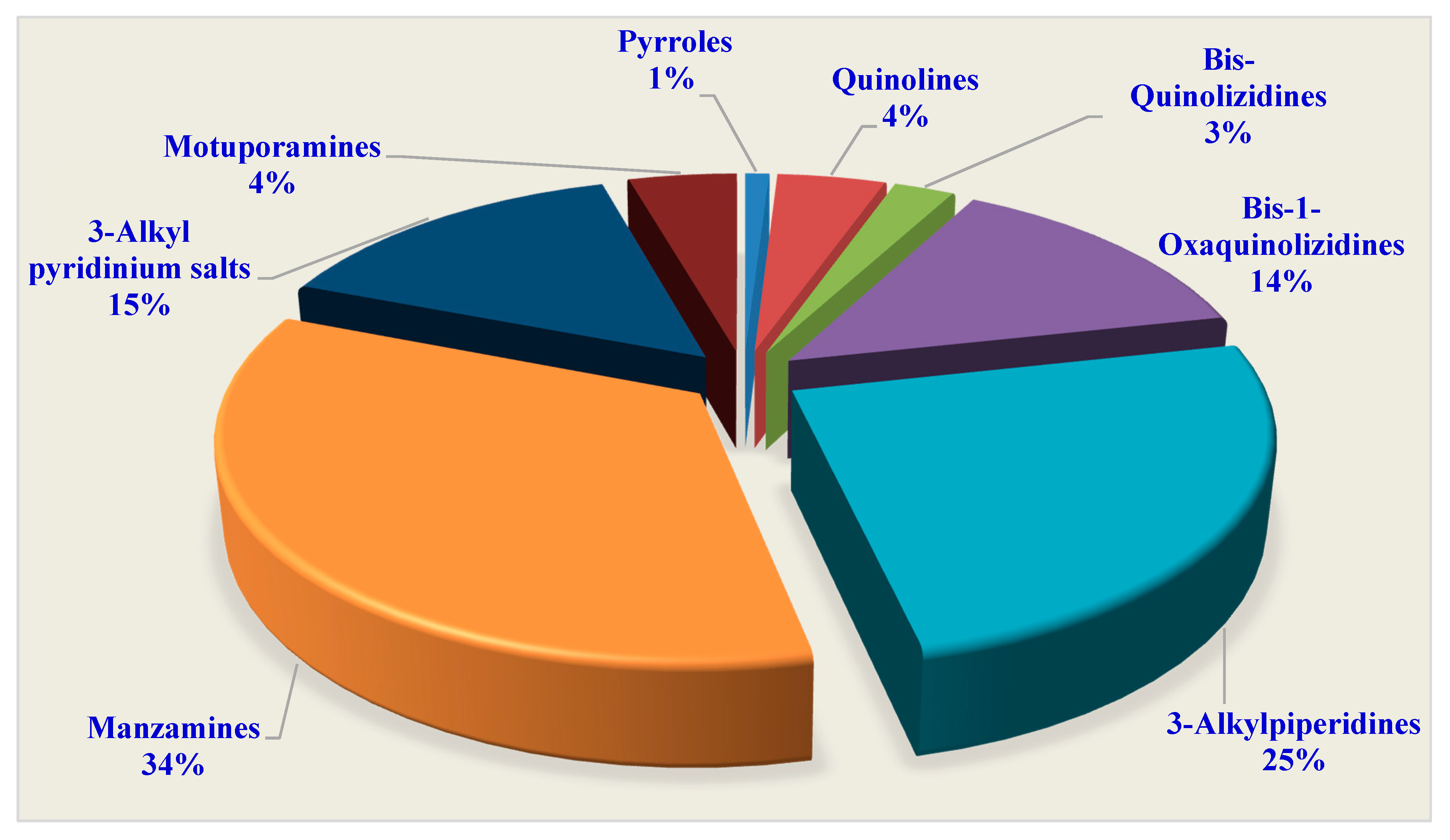
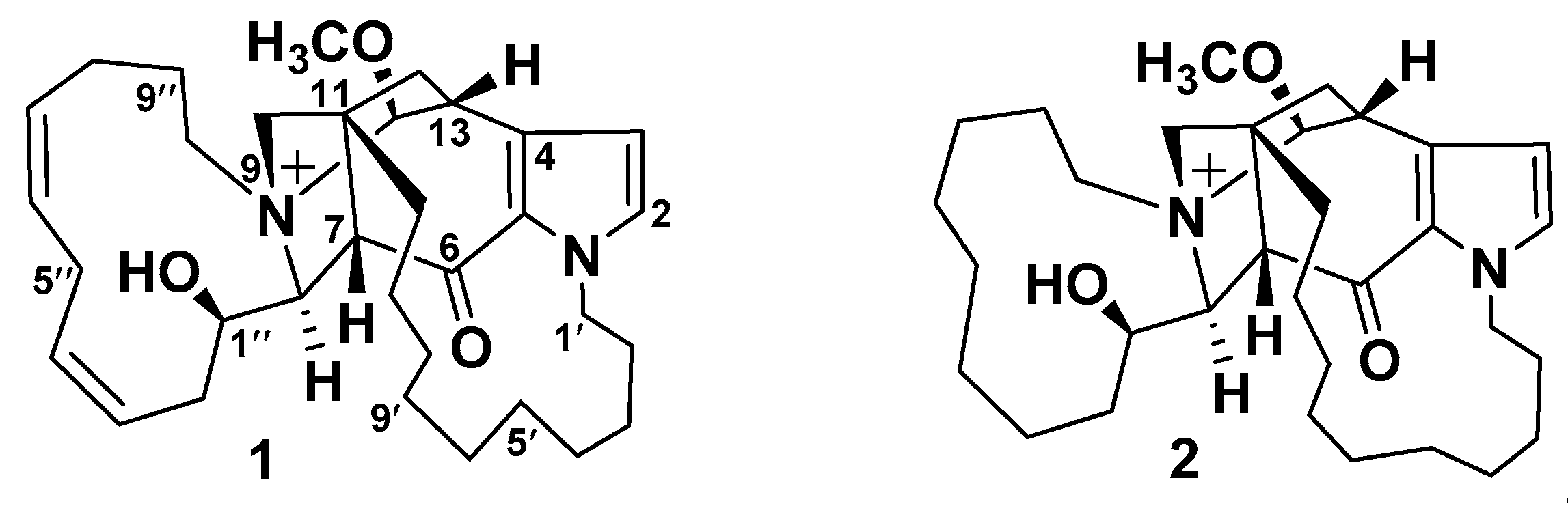
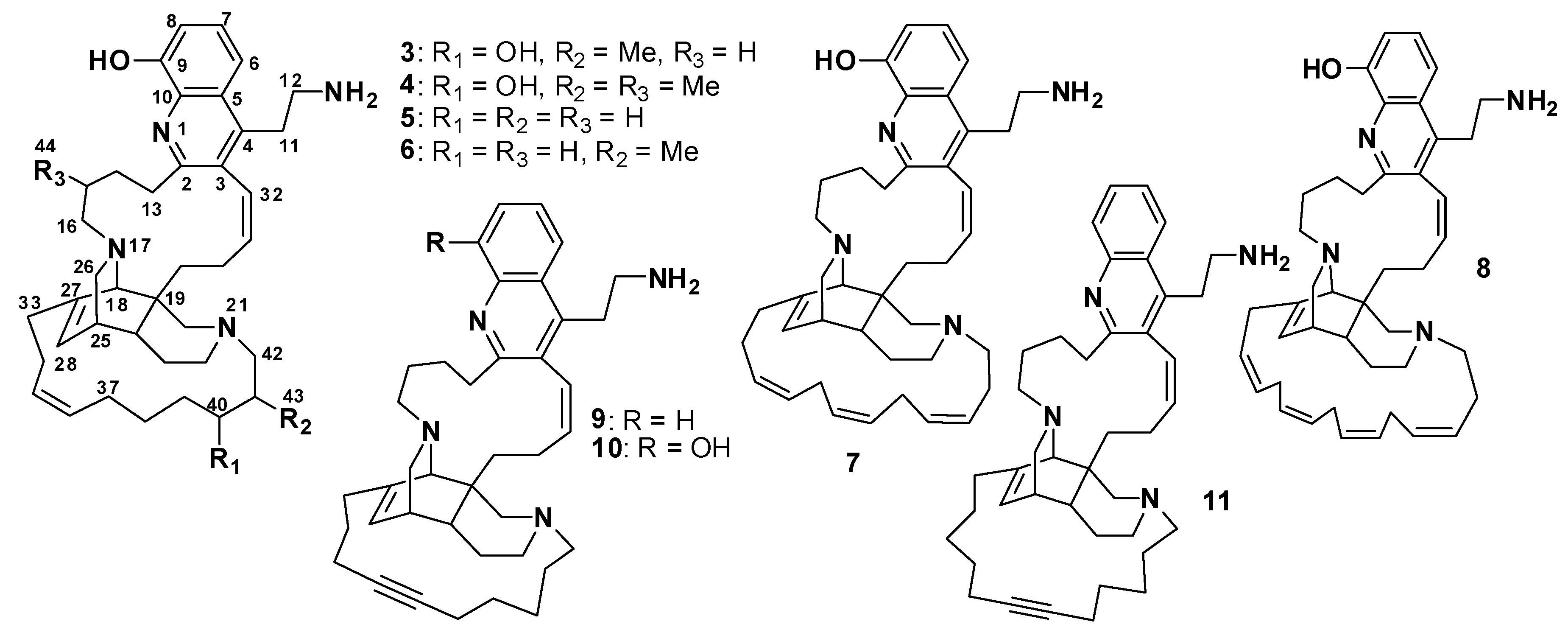
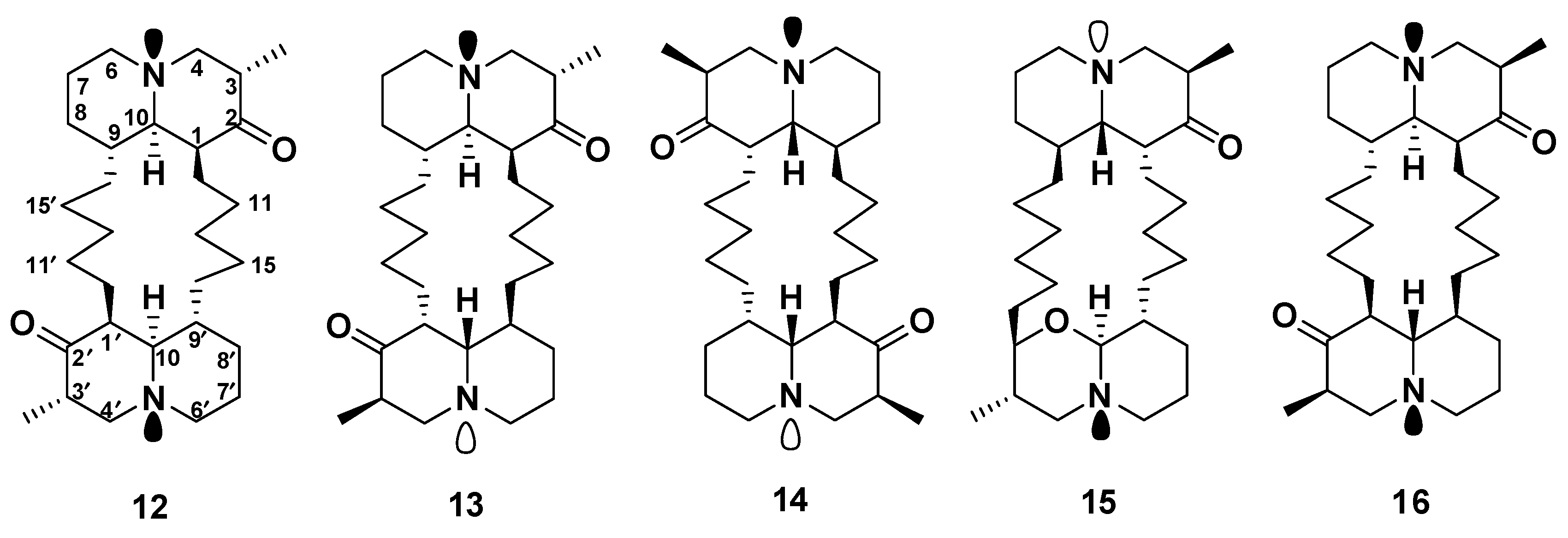
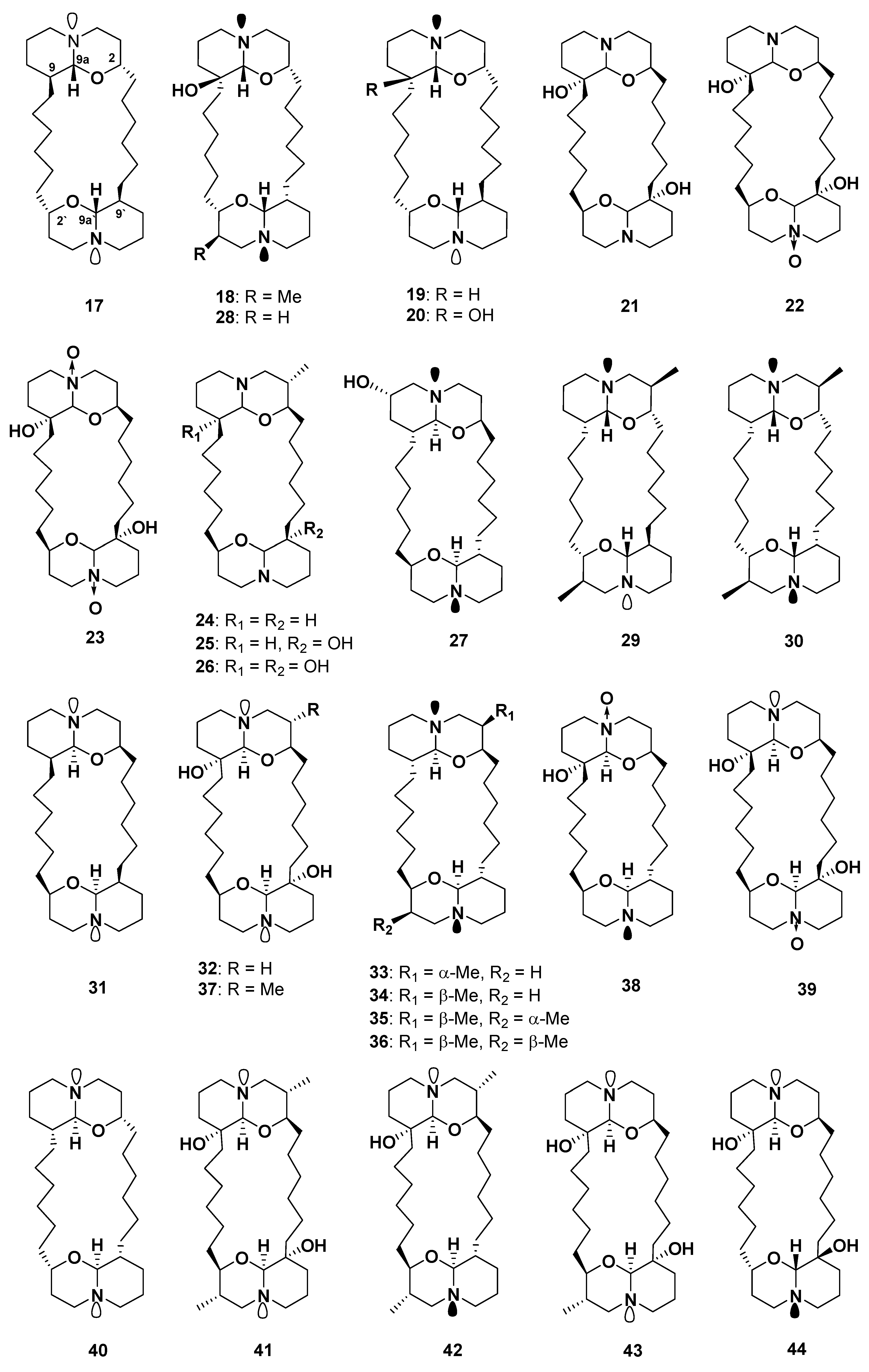
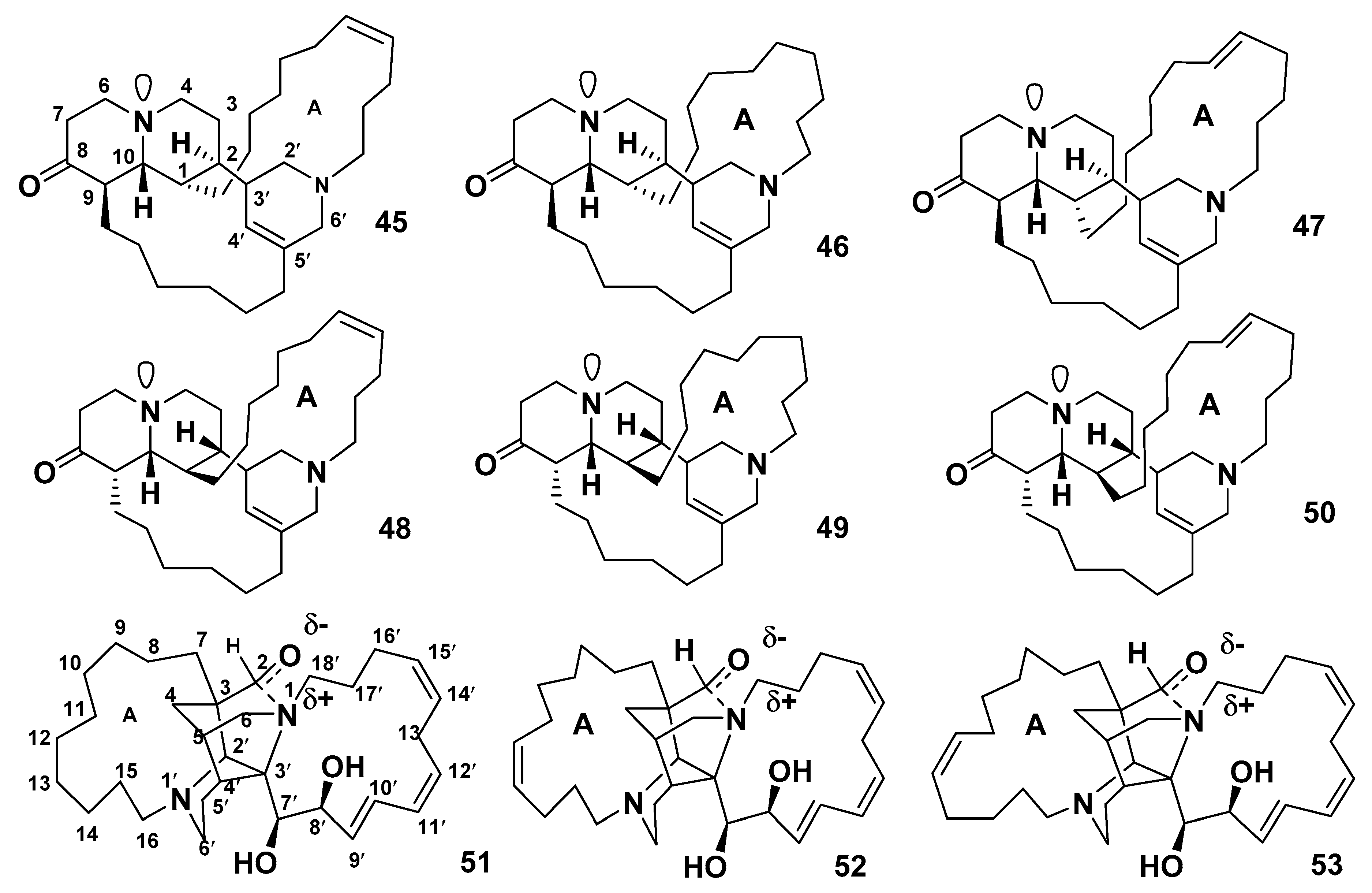
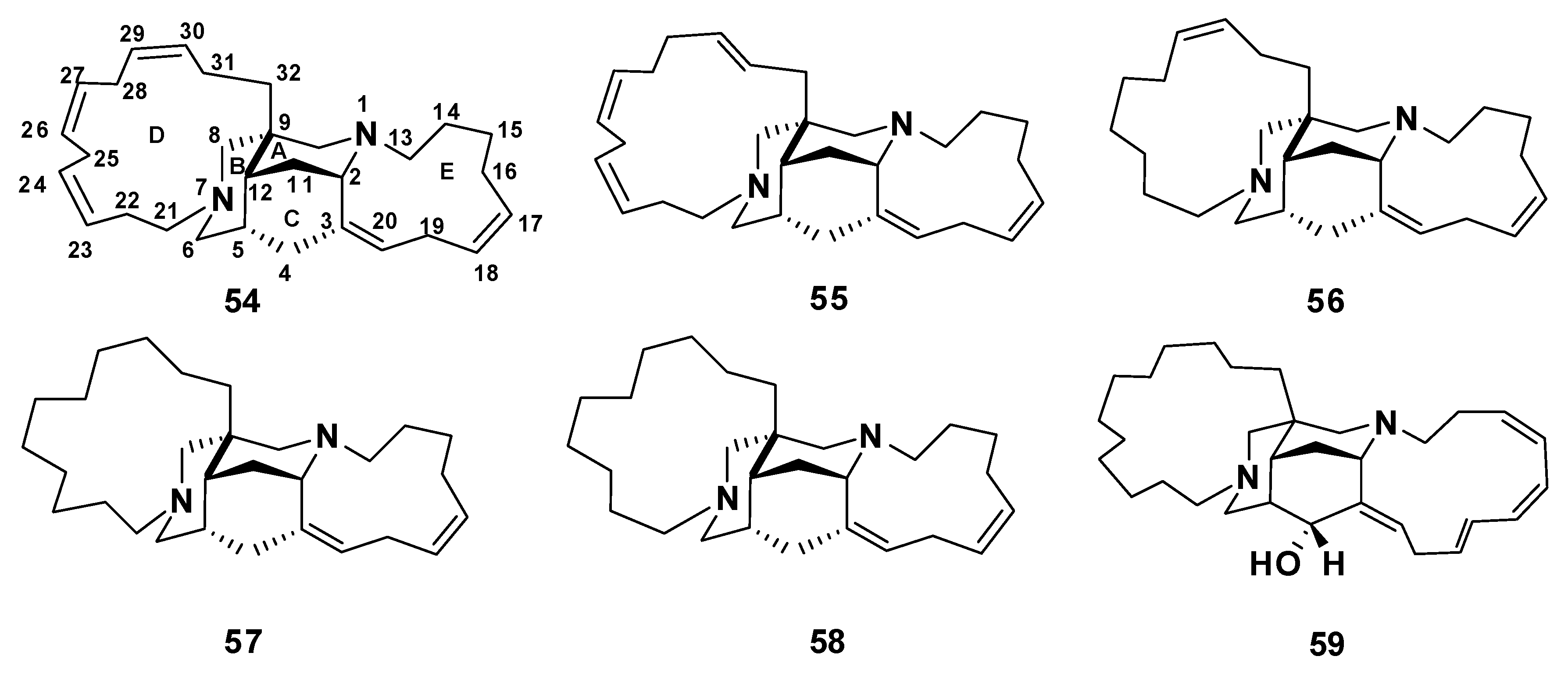
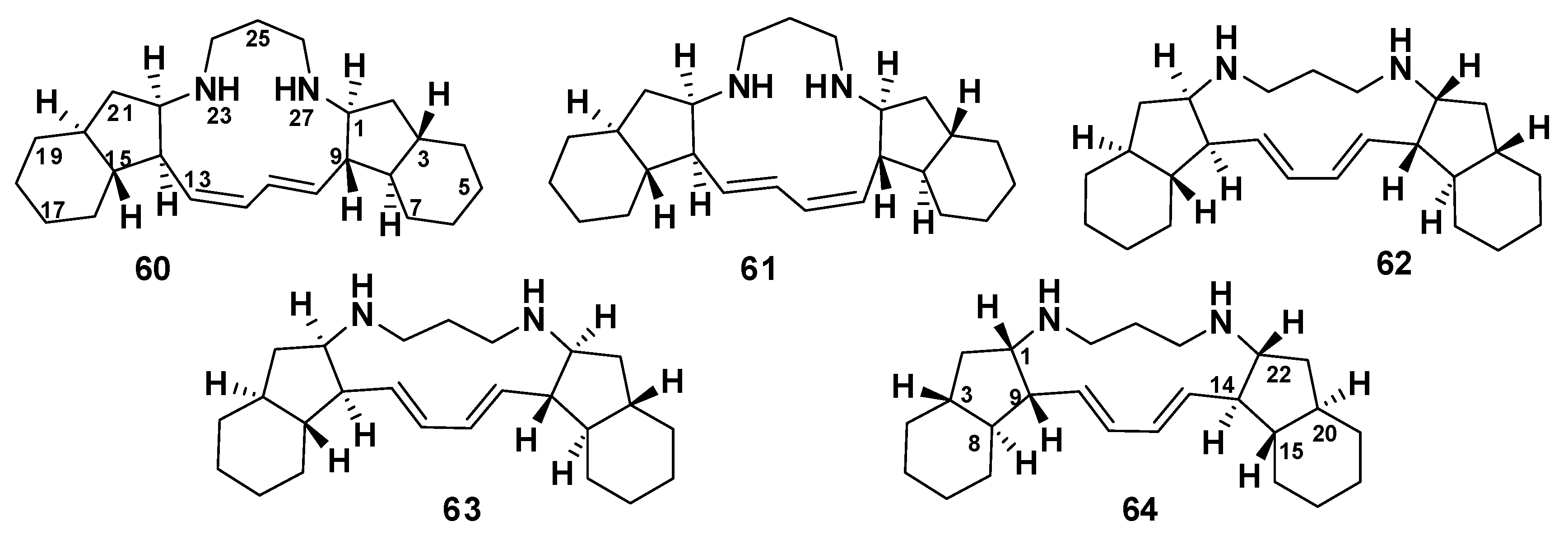
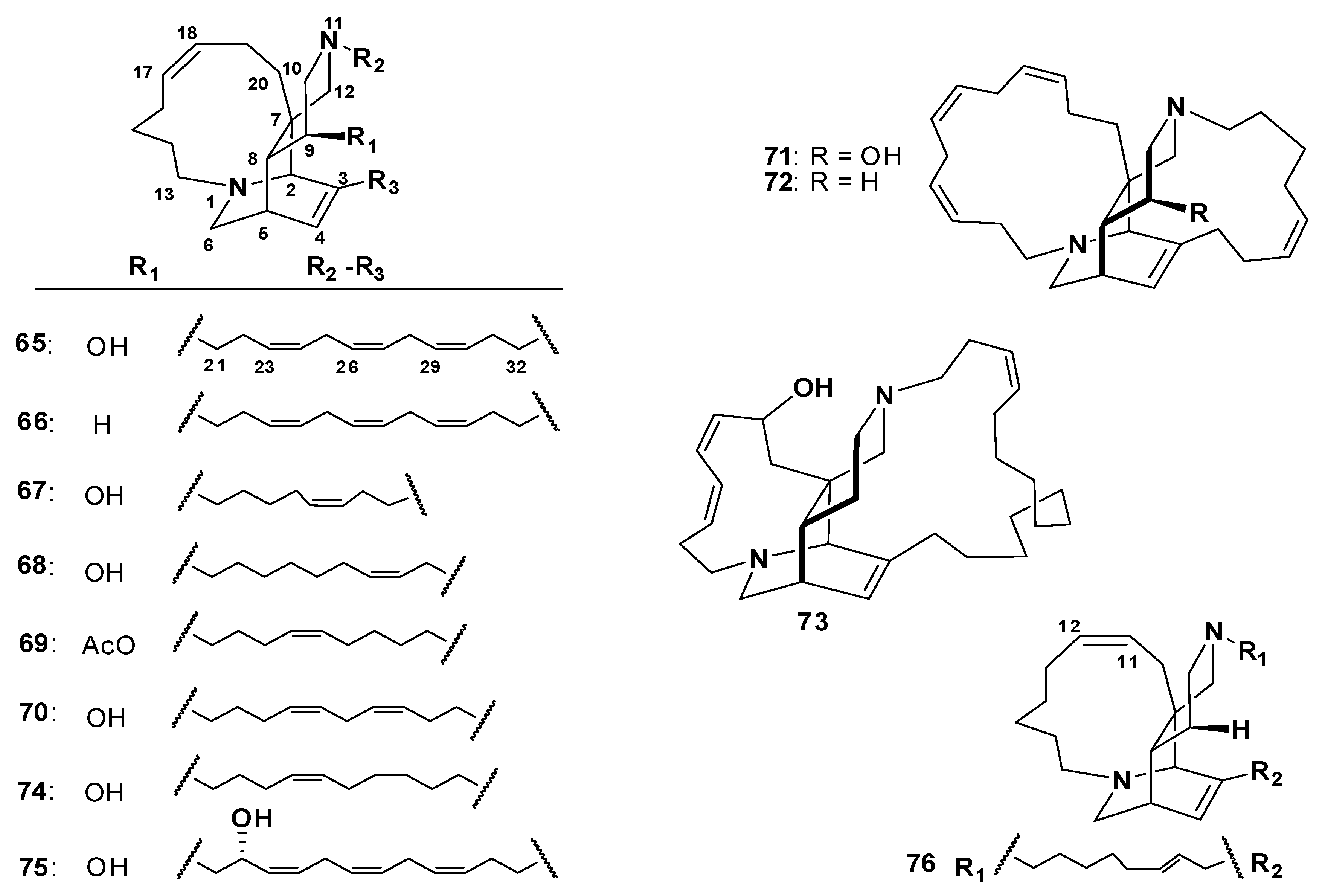
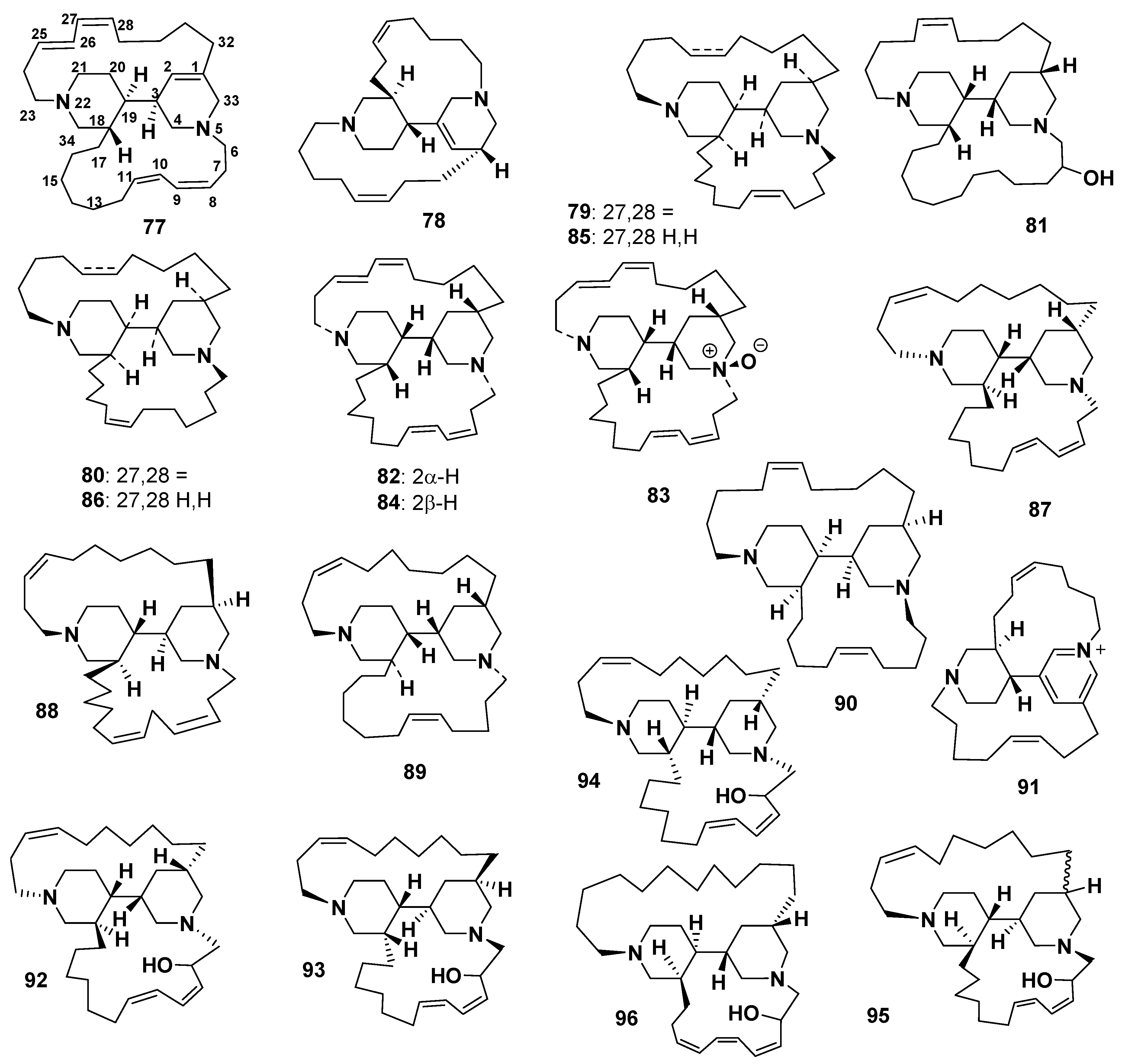
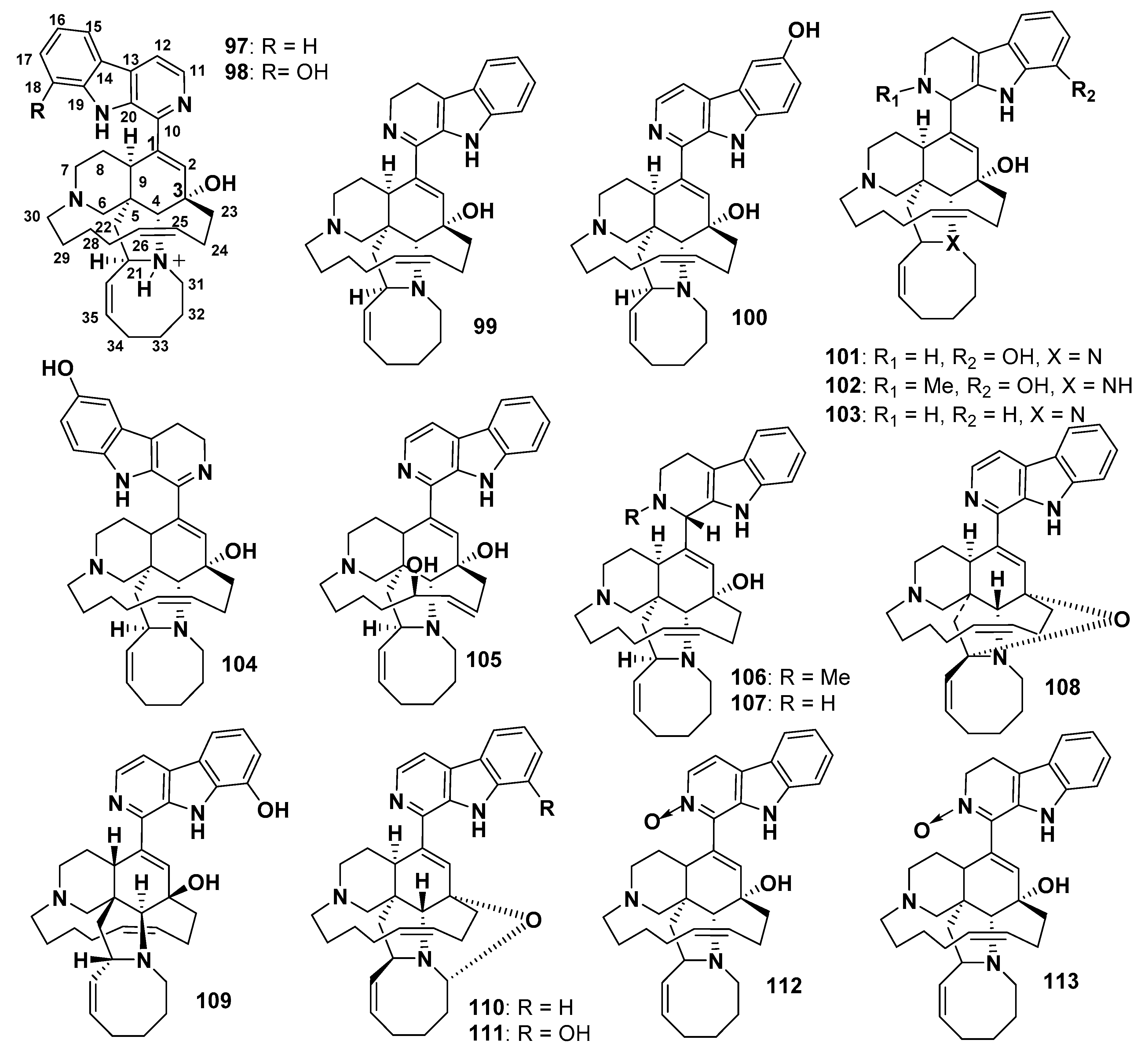
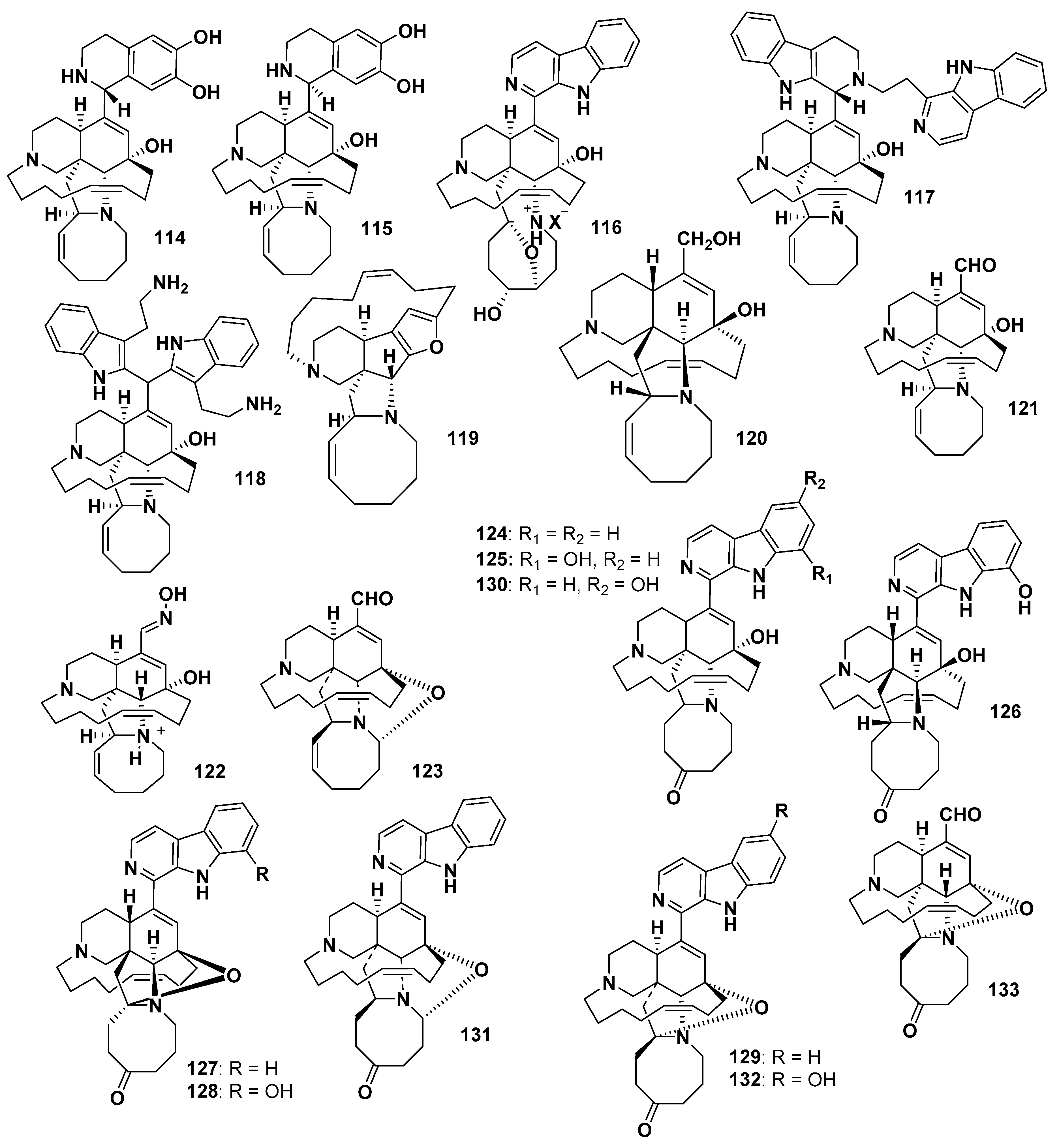
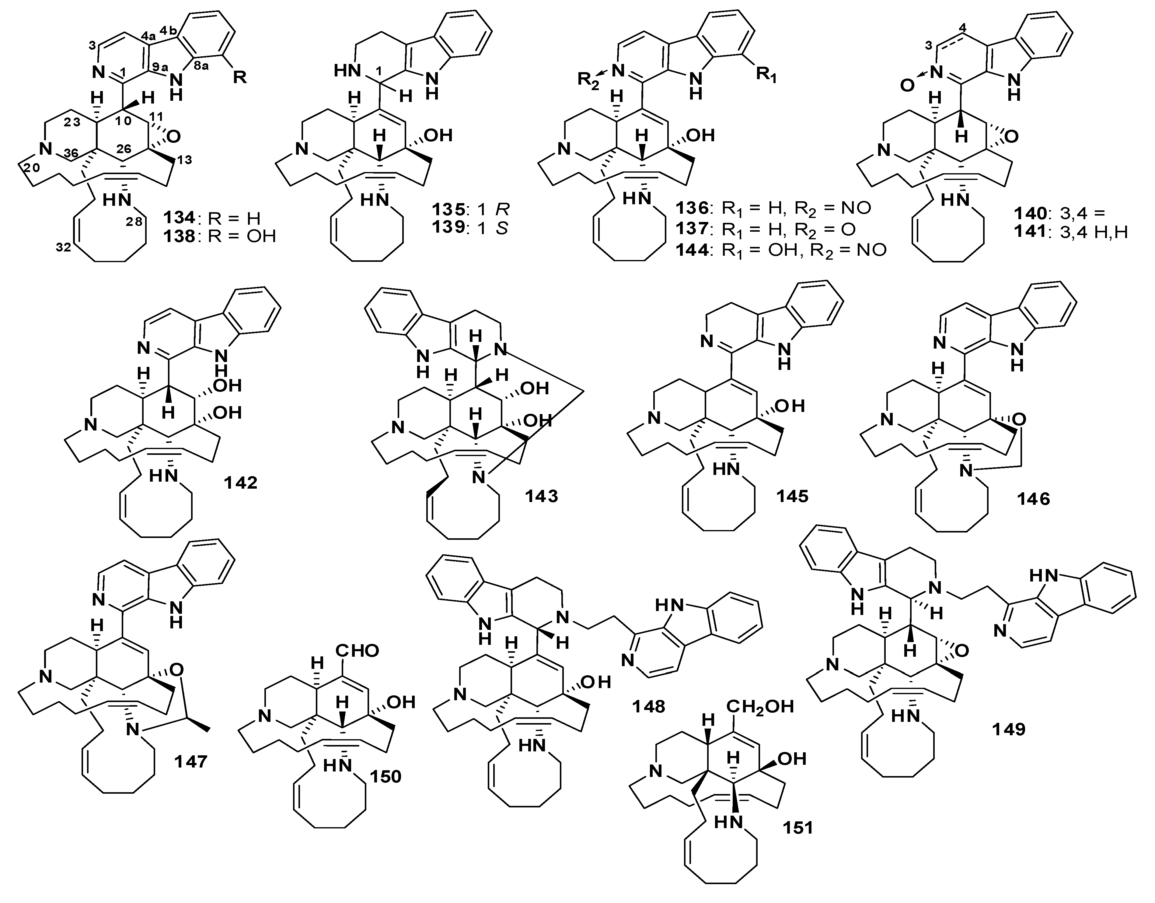
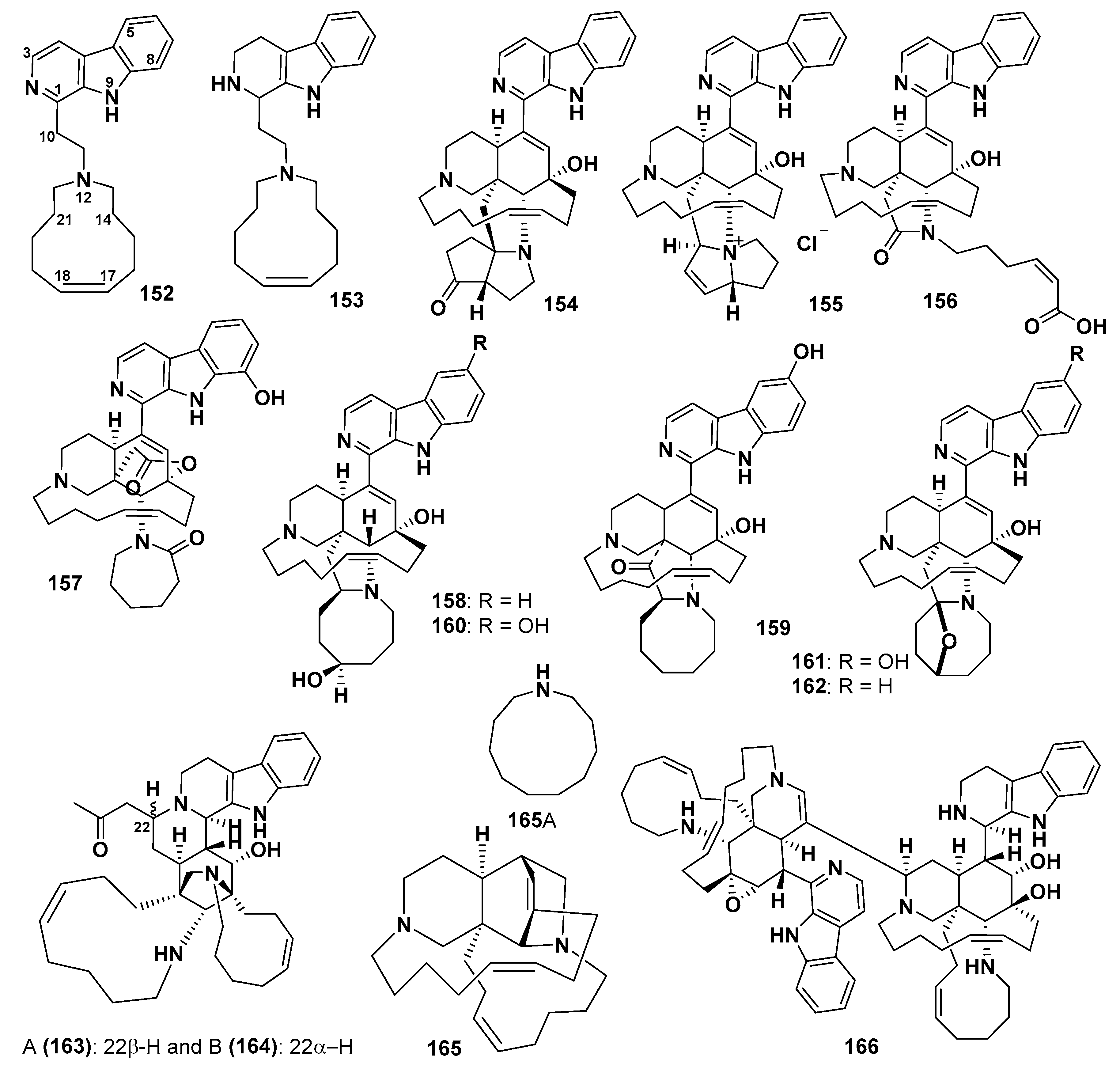
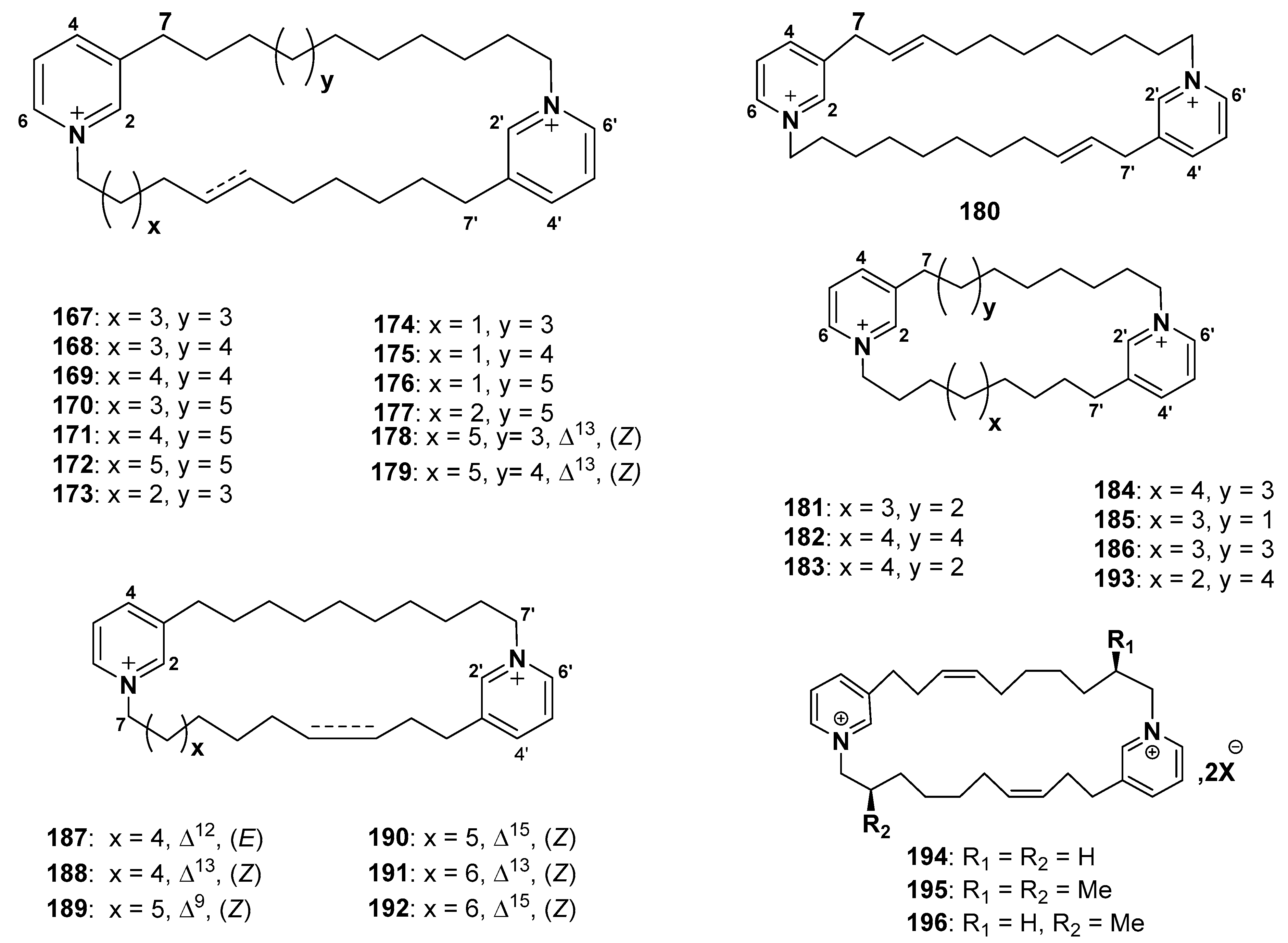
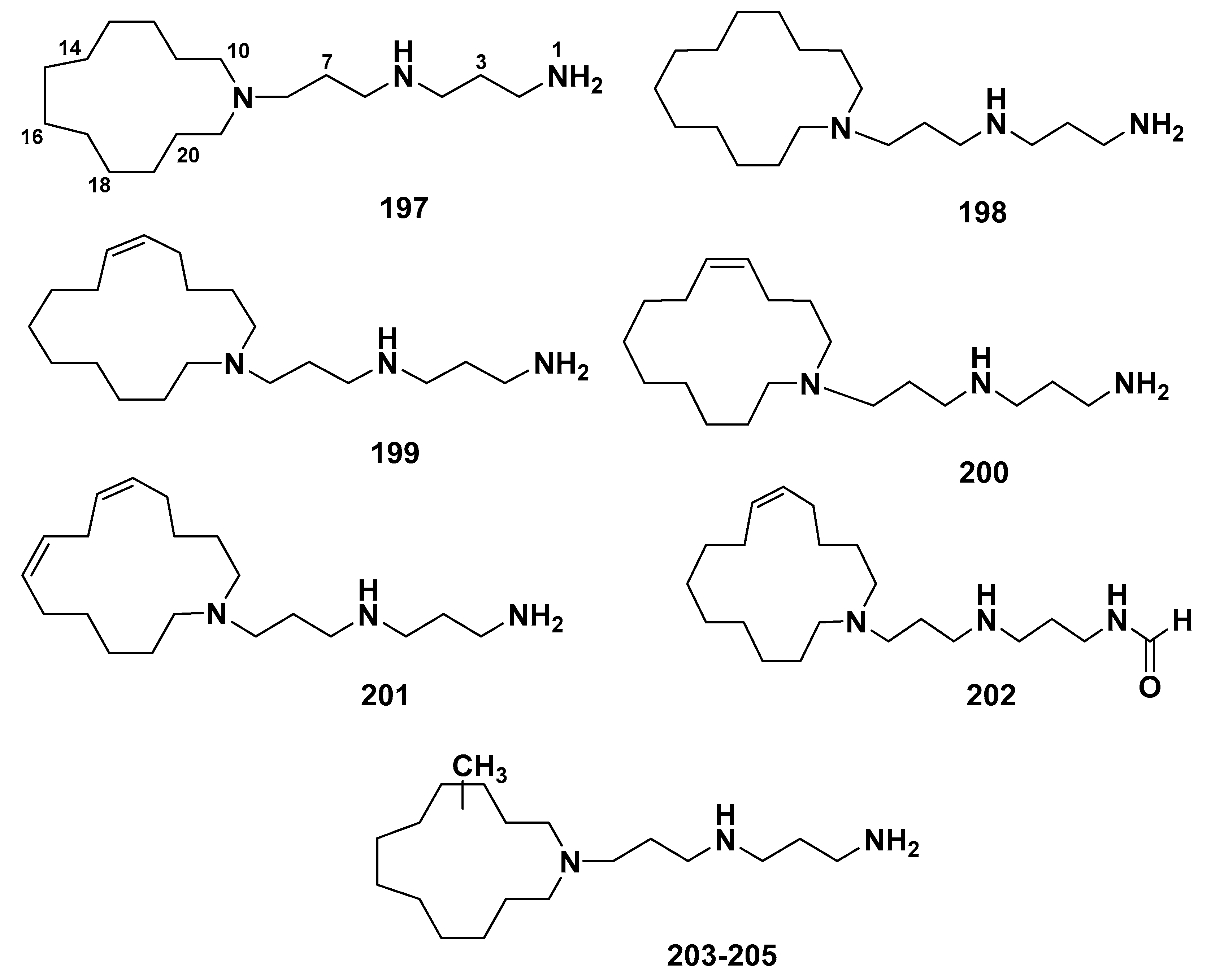
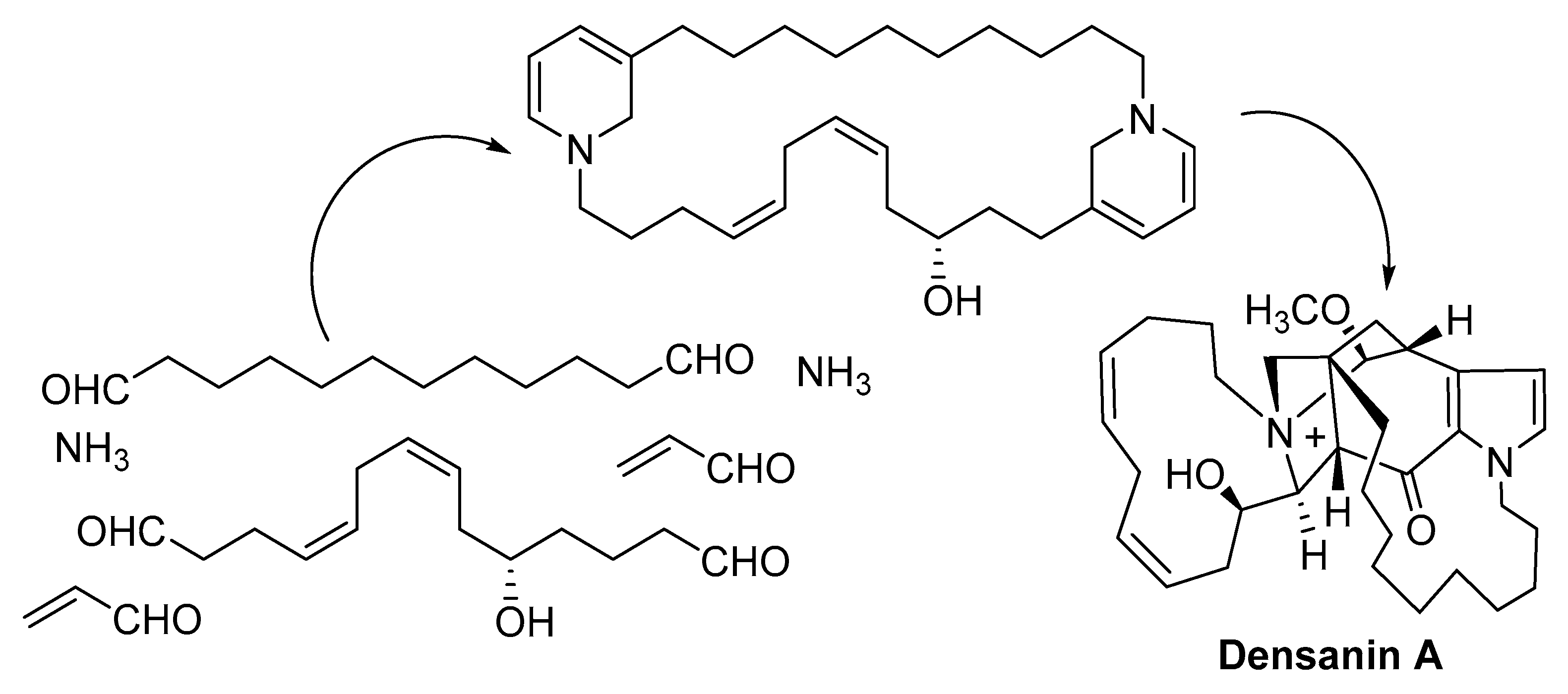
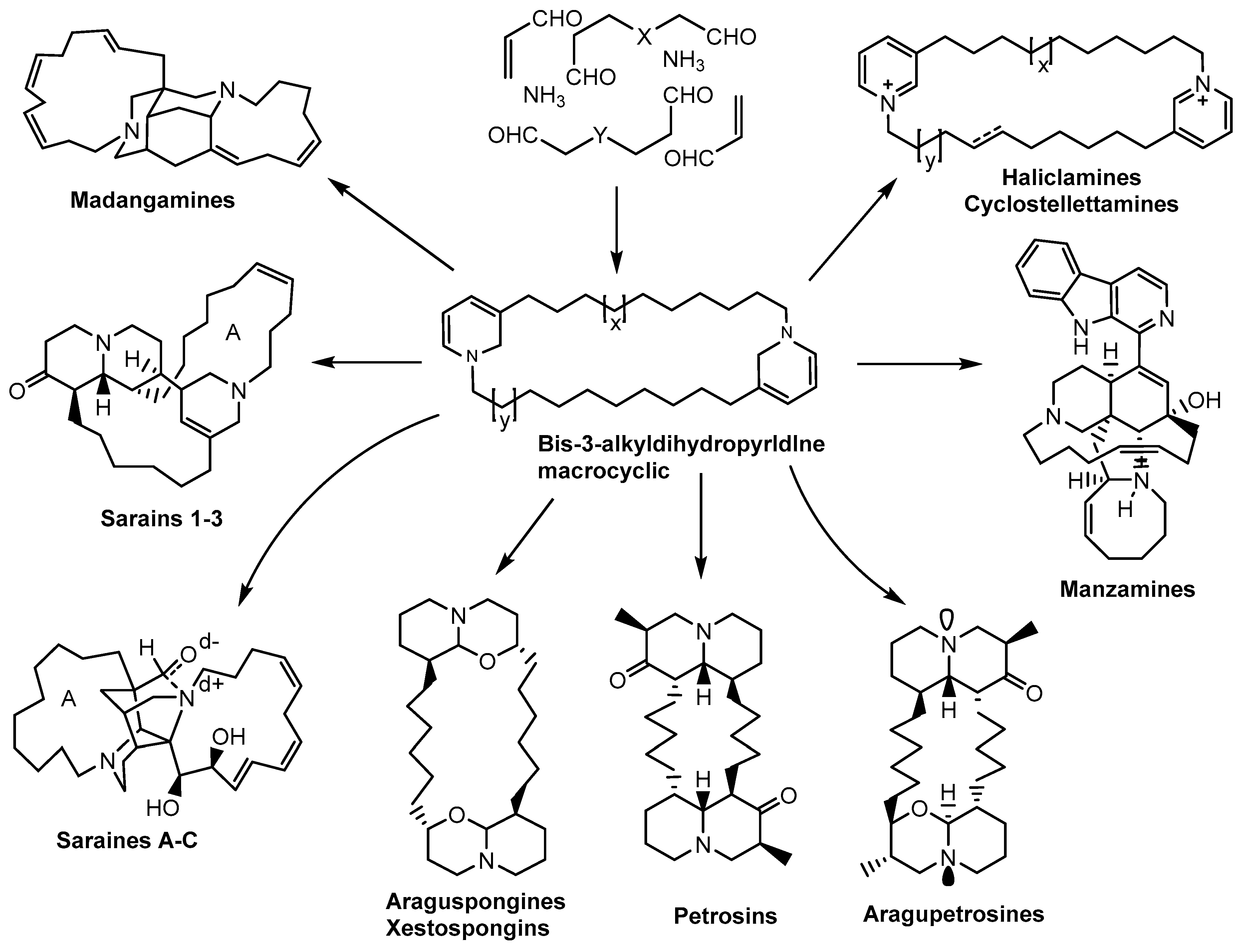
Table 1.
List of marine-derived macrocyclic alkaloids.
| Compound No. | Subclasses | Name of Compounds | Marine Organism | Biological Activities |
|---|---|---|---|---|
| 1–2 | Pyrroles | Densanins A and B | Haliclona densaspicula | Anti-inflammatory |
| 3–8 | Quinolines | Njaoamines A–F | Reniera sp. | Cytotoxic and Anti-HIV |
| 9–10 | Njaoamines G–H | Neopetrosia sp. | ||
| 11 | Njaoamine I | Reniera sp. | ||
| 12 | Bis-Quinolizidines | Petrosin | Petrosia seriata | |
| 13–14 | Petrosins A and B | |||
| 15 | Aragupetrosine A | Xestospongia sp. | ||
| 16 | Xestosin A | Xestospongia exigua | ||
| 17 | Xestospongin A (Araguspongine D) | |||
| 18 | Xestospongin B | |||
| 19 | Xestospongin C (Araguspongine E) | |||
| 20 | Xestospongin D (Araguspongine A) | Xestospongia sp. | ||
| 21–26 | Xestospongins E–J | Oceanapia sp. | ||
| 27 | (+)-7S-Hydroxyxestospongin A | Xestospongia sp. | ||
| 28 | Demethylxestospongin B | Xestospongia sp. and Neopetrosia exigua | ||
| 29 | 3β,3ʹβ-Dimethylxestospongin C | |||
| 30 | 9′-epi-3β,3′β–Dimethylxestospongin C | |||
| 31 | Araguspongine B | Antimicrobial and Cytotoxic | ||
| 32 | Araguspongine C | Xestospongia muta | ||
| 33–36 | Araguspongines F–H and J | Xestospongia sp. | ||
| 37 | 3a-Araguspongin C | Haliclona exigua | ||
| 38–39 | Araguspongines K and L | Neopetrosia exigua | ||
| 40 | Araguspongine M | |||
| 41–43 | Araguspongines N–P | Xestospongia muta | ||
| 44 | meso-araguspongine C | |||
| 45–47 | 3-Alkyl piperidines | Saraines 1–3 | Reniera sarai | Cytotoxic |
| 48–50 | Isosaraines 1–3 | Antimicrobial | ||
| 51–53 | Saraines A-C | |||
| 54–58 | Madangamines A–E | Xestospongia ingens | Cytotoxic | |
| 59 | Madangamine F | Pachychalina alcaloidifera | ||
| 60 | (10E,12Z)-haliclonadiamine | Halichondria panicea | Antimicrobial | |
| 61 | (10Z,12E)-Haliclonadiamine | Halichondria panacea | ||
| 62 | Papuamine | Haliclona sp. | ||
| 63–64 | Haliclonadiamine | Haliclona sp. | ||
| 65–66 | Ingamines A and B | Xestospongia ingens | Antimalarial | |
| 67 | Ingenamine | |||
| 68–72 | Ingenamines B–F | |||
| 73 | Ingenamine G | Pachychalina sp. | ||
| 74 | Dihydroingenamine D | Petrosid Ng5 Sp5 | ||
| 75 | 22(S)-Hydroxyingamine A | |||
| 76 | Xestocyclamine | Xestospongia sp. | protein kinase C inhibitor | |
| 77–78 | Halicyclamines A-B | Xestospongia sp. | Cytotoxic | |
| 79-80 | Haliclonacyclamines A–B | Haliclona sp. | ||
| 81 | 22-Hydroxyhaliclonacyclamine B | Halichondria sp. | ||
| 82 | 2-epi-Tetradehydrohaliclonacyclamine | Halichondria sp. | ||
| 83 | Tetradehydrohaliclonacyclamine A mono-N-oxide | Halichondria sp. | ||
| 84 | Tetradehydrohaliclonacyclamine A | Halichondria sp. | ||
| 85 | Haliclonacyclamine C | Haliclona sp. | ||
| 86 | Haliclonacyclamine D | Haliclona sp. | ||
| 87 | Haliclonacyclamine E | Arenosclera brasiliensis | Antimalarial, Cytotoxic, Proteasome and Immunoproteasome inhibition | |
| 88 | Haliclonacyclamine F | P. alcaloidifera | ||
| 89 | Halichondramine | Halichondria sp. | ||
| 90 | Neopetrosiamine A | Neopetrosia proxima | ||
| 91 | Tetradehydrohalicyclamine B | Acanthostrongylophora ingens | ||
| 92–94 | Arenosclerins A–C | A. brasiliensis | Cytotoxic, Anti-leishmanial, and Anti-HIV | |
| 95–96 | Arenosclerins D and E | P. alcaloidifera | ||
| 97 | Manzamines | Manzamine A (Keramamine A) | Haliclona sp. | |
| 98 | 8-Hydroxymanzamine A (Manzamine G) | Amphimedon sp. and Pachypellina sp. | ||
| 99 | 3,4-Dihydromanzamine A | Amphimedon sp. | ||
| 100 | 6-Hydroxymanzamine A (Manzamine Y) | Amphimedon sp. and Haliclona sp. | ||
| 101 | 1,2,3,4-Tetrahydro-8-hydroxymanza-mine A (8-Hydroxymanzamine D) | Cribochalina sp. and Petrosia sp. | ||
| 102 | 1,2,3,4-Tetrahydro-2-N-methyl-8-hyd-roxymanzamine A (8-Hydroxy-2-N-methylmanzamine D) | |||
| 103 | Manzamine D (1,2,3,4-Tetrahydromanzamine A) | Ircinia sp. | ||
| 104 | 3,4-Dihydro-6-hydroxymanzamine A | Amphimedon sp. | ||
| 105 | Manzamine M | |||
| 106 | N-Methyl-epi-manzamine D | Unidentified Paluan sponge | ||
| 107 | epi-Manzamine D | |||
| 108 | 12,34-Oxamanzamine A | Sponge 011ND 35 | ||
| 109 | ent-8-Hydroxymanzamine A | Unidentified Indo-Pacific sponge | ||
| 110 | 12,28-Oxamanzamine A | Acanthostrongylophora sp. | ||
| 111 | 12,28-Oxa-8-hydroxymanzamine A | |||
| 112 | Manzamine A N-oxide | Xestospongia ashmorica | ||
| 113 | 3,4-Dihydromanzamine A N-oxide | |||
| 114–115 | Acanthomanzamines A and B | Acanthostrongylophora sp. | ||
| 116 | Pre-neo-kauluamine | Acanthostrongylophora ingens | ||
| 117 | Zamamidine C | Amphimedon sp. | ||
| 118 | Zamamidine D | |||
| 119 | Nakadomarin A | |||
| 120 | Ircinol A | |||
| 121 | Ircinal A | Ircinia sp. | ||
| 122 | Ircinal E | |||
| 123 | 12,28-Oxaircinal A | |||
| 124 | Manzamine E | Xestospongia sp. | ||
| 125 | Manzamine F (Keramamine B) | |||
| 126 | ent-Manzamine F | |||
| 127–128 | ent-12,34-Oxamanzamines E and F | Sponge 011ND 35 | ||
| 129 | 12,34-Oxamanzamine E | Acanthostrongylophora sp. | ||
| 130 | 6-Hydroxymanzamine E | |||
| 131 | 12,28-Oxamanzamine E | |||
| 132 | 12,34-Oxa-6-hydroxymanzamine E | |||
| 133 | 31-Keto-12,34-oxa-32,33-dihydroircinal A | |||
| 134 | Manzamine B | Haliclona sp. | ||
| 135–136 | Manzamines H, J | Ircinia sp. | ||
| 137 | Manzamine J N-oxide | Xestospongiaashmorica | ||
| 138 | 8-Hydroxymanzamine B | Acanthostrongylophora sp. | ||
| 139 | Manzamine L | Amphimedon sp. | ||
| 140 | Manzamine B N-oxide | Acanthostrongylophora sp. | ||
| 141 | 3,4-Dihydromanzamine B N-oxide | |||
| 142 | 11-Hydroxymanzamine J | |||
| 143 | Ma’eganedin A | Amphimedon sp. | ||
| 144 | 8-Hydroxymanzamine J | Acanthostrongylophora | ||
| 145 | 3,4-Dihydromanzamine J | Amphimedon sp. | ||
| 146–147 | Acanthomanzamines D and E | Acanthostrongylophora sp. | ||
| 148–149 | Zamamidines A and B | Amphimedon sp. | ||
| 150 | Ircinal B | Ircinia sp. | ||
| 151 | Ircinol B | Amphimedon sp. | ||
| 152 | Manzamine C | Haliclona sp. | Cytotoxic | |
| 153 | Keramamine C | Amphimedon sp. | ||
| 154 | Acanthomanzamine C | Acanthostrongylophora sp. | ||
| 155 | Kepulauamine A | |||
| 156 | Acantholactam | |||
| 157 | Acantholactone | Acanthostrongylophora sp. | ||
| 158 | 32,33-Dihydro-31-hydroxymanzamine A | Indonesian sponge | ||
| 159 | 32,33-Dihydro-6-hydroxymanzamine A-35-one | |||
| 160 | 32,33-Dihydro-6,31-dihydroxymanzamine A | |||
| 161 | Manzamine X | Xestospongia sp. | ||
| 162 | 6-Deoxymanzamine X | X. ashmorica | ||
| 163–164 | Manadomanzamines A and B | Acanthostrongylophora sp. | ||
| 165 | Keramaphidin B | Amphimedon sp. | ||
| 166 | Kauluamine | Prianos sp. | ||
| 167–172 | 3-Alkyl pyridinium salts | Cyclostellettamines A–F | Stelletta maxima | Antimicrobial and Cytotoxic |
| 173–177 | Cyclostellettamines G–I, K, and L | Pachychalina sp. | ||
| 178–179 | Dehydrocyclostellettamines D, E | Xestospongia sp. | ||
| 180 | 8,8‘-Dienecyclostellettamine | Amphimedon compressa | ||
| 181–184 | Cyclostellettamines N, R, O, Q | Haliclona sp. | ||
| 185–192 | Cyclostellettamines | Haliclona sp. | ||
| 193 | Cyclostellettamine P | Xestospongia exigua | ||
| 194–196 | Njaoaminiums A–C | Reniera sp. | Cytotoxic | |
| 197–205 | Motuporamines | Motuporamines A–I | Xestospongia exigua | Anti-invasion |
© 2020 by the authors. Licensee MDPI, Basel, Switzerland. This article is an open access article distributed under the terms and conditions of the Creative Commons Attribution (CC BY) license (http://creativecommons.org/licenses/by/4.0/).
Share and Cite
MDPI and ACS Style
Althagbi, H.I.; Alarif, W.M.; Al-Footy, K.O.; Abdel-Lateff, A. Marine-Derived Macrocyclic Alkaloids (MDMAs): Chemical and Biological Diversity. Mar. Drugs 2020, 18, 368. https://0-doi-org.brum.beds.ac.uk/10.3390/md18070368
AMA Style
Althagbi HI, Alarif WM, Al-Footy KO, Abdel-Lateff A. Marine-Derived Macrocyclic Alkaloids (MDMAs): Chemical and Biological Diversity. Marine Drugs. 2020; 18(7):368. https://0-doi-org.brum.beds.ac.uk/10.3390/md18070368
Chicago/Turabian StyleAlthagbi, Hanan I., Walied M. Alarif, Khalid O. Al-Footy, and Ahmed Abdel-Lateff. 2020. "Marine-Derived Macrocyclic Alkaloids (MDMAs): Chemical and Biological Diversity" Marine Drugs 18, no. 7: 368. https://0-doi-org.brum.beds.ac.uk/10.3390/md18070368
Note that from the first issue of 2016, this journal uses article numbers instead of page numbers. See further details here.