Article

Optimization of Extraction of Bioactive Peptides from Monkfish (Lophius litulon) and Characterization of Their Role in H2O2-Induced Lesion

Zhejiang Provincial Engineering Technology Research Center of Marine Biomedical Products, School of Food and Pharmacy, Zhejiang Ocean University, Zhoushan 316022, China
*
Authors to whom correspondence should be addressed.
These authors contributed equally to this work.
Submission received: 14 August 2020 / Revised: 15 September 2020 / Accepted: 15 September 2020 / Published: 17 September 2020

Abstract

:
Background: Marine fish meat has been widely used for the extraction of bioactive peptides. This study was aimed to optimize the preparation of monkfish muscle peptides (LPs) using response surface methodology (RSM) and explore the antioxidant activities of <1 kDa LPs. Methods: Peptides were prepared from the muscles of monkfish (Lophius litulon), and five proteases were tested to hydrolyze muscle proteins. The hydrolysate that was treated using neutrase showed the highest degree of hydrolysis (DH) and 1,1-diphenyl-2-picrylhydrazyl (DPPH) scavenging activities. Results: The optimized conditions were as follows: water/material ratio of 5.4:1, a time span of 5 h, pH of 7.0, enzyme concentration of 2000 U/g, and temperature of 45 °C; the maximum DPPH scavenging activity and DH were 92.861% and 19.302%, respectively. LPs exhibited appreciable antioxidant activities, including DPPH radical, hydroxyl radical, 2,2′-azinobis-3-ethylbenzthiazoline-6-sulphonate (ABTS) radical, and superoxide anion scavenging activities. LPs attenuated H2O2-related oxidative injury in RAW264.7 cells, reduced the reactive oxygen species (ROS) and malondialdehyde (MDA) levels, and increased the superoxide dismutase (SOD), glutathione peroxidase (GSH-Px), and catalase (CAT) levels. Conclusion: We concluded that LPs could be an ideal source of bioactive peptides from monkfish and also have pharmaceutical potential.

Graphical Abstract

1. Introduction

In recent years, natural antioxidants have received growing attention with regard to their free radical scavenging property and their ability to slow down disease progression [1]. Oxidation is a key process in all living animals despite the side effect of generating free radicals [2]. These radicals are so unstable that they give rise to cell lesions and various diseases [3]. Antioxidants that are synthesized by chemical methods may have adverse effects on humans; therefore, the development of safe and effective natural antioxidants is urgently needed [4,5]. Antioxidant peptides that are released by enzymatic hydrolysis are safe and have low molecular weight and strong antioxidant activities [6]. Marine bioactive peptides are a group of proteins that have critical functions in host defense systems of marine organisms. They are now being widely used in drug research and development as antibacterial, antiviral, and anticancer agents [7]. A number of studies have used the hydrolysates of fish meat as antioxidant peptides, such as peptides from eel, cod, tuna, salmon, and croaker [8,9,10,11,12]. Monkfish (Lophius litulon) is distributed in the western part of the North Pacific Ocean and the coastal waters of China. Generally, large quantities of monkfish are exported as aquatic products for human consumption; however, smaller monkfish is manufactured as fish meal.
Response surface methodology (RSM) is a useful tool for the evaluation of independent parameters to modify experimental variables [13]. In recent years, RSM has been applied to the extraction of fish protein hydrolysates and peptides, such as yellowfin tuna (Thunnus albacares) [14], stonefish [15], halibut [16], and red tilapia (Oreochromis spp.) [17], as well as monkfish [18,19]. The preparation process has yet to be optimized.
In this study, hydrolytic effects of five enzymes were studied, and neutral protease was identified as the optimal enzyme to hydrolyze monkfish muscle. RSM was used to modify the process of purification. The molecular weight (MW) distribution of monkfish muscle polypeptide was obtained using high-performance liquid chromatography (HPLC). Monkfish muscle peptides with MW <1 kDa were prepared using ultrafiltration and freeze-drying followed by the determination of its antioxidant capacity.

2. Results and Discussion

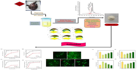
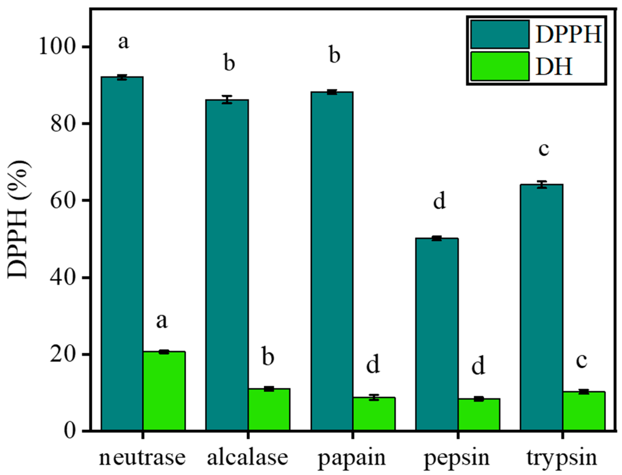
2.1. Selection of the Optimal Enzyme

To obtain hydrolysates with high efficiency, we tried to screen the best protease for hydrolysis. For each reaction, the same solid/liquid ratio, enzyme volumes, and enzymolysis time were used with the optimal pH and temperature of each enzyme. DH and DPPH scavenging ability for the five hydrolysates were as follows: neutrase > alcalase > trypsin > papain > pepsin and neutrase > alcalase > papain > trypsin > pepsin (Figure 1). Therefore, neutrase was selected for subsequent modifications.

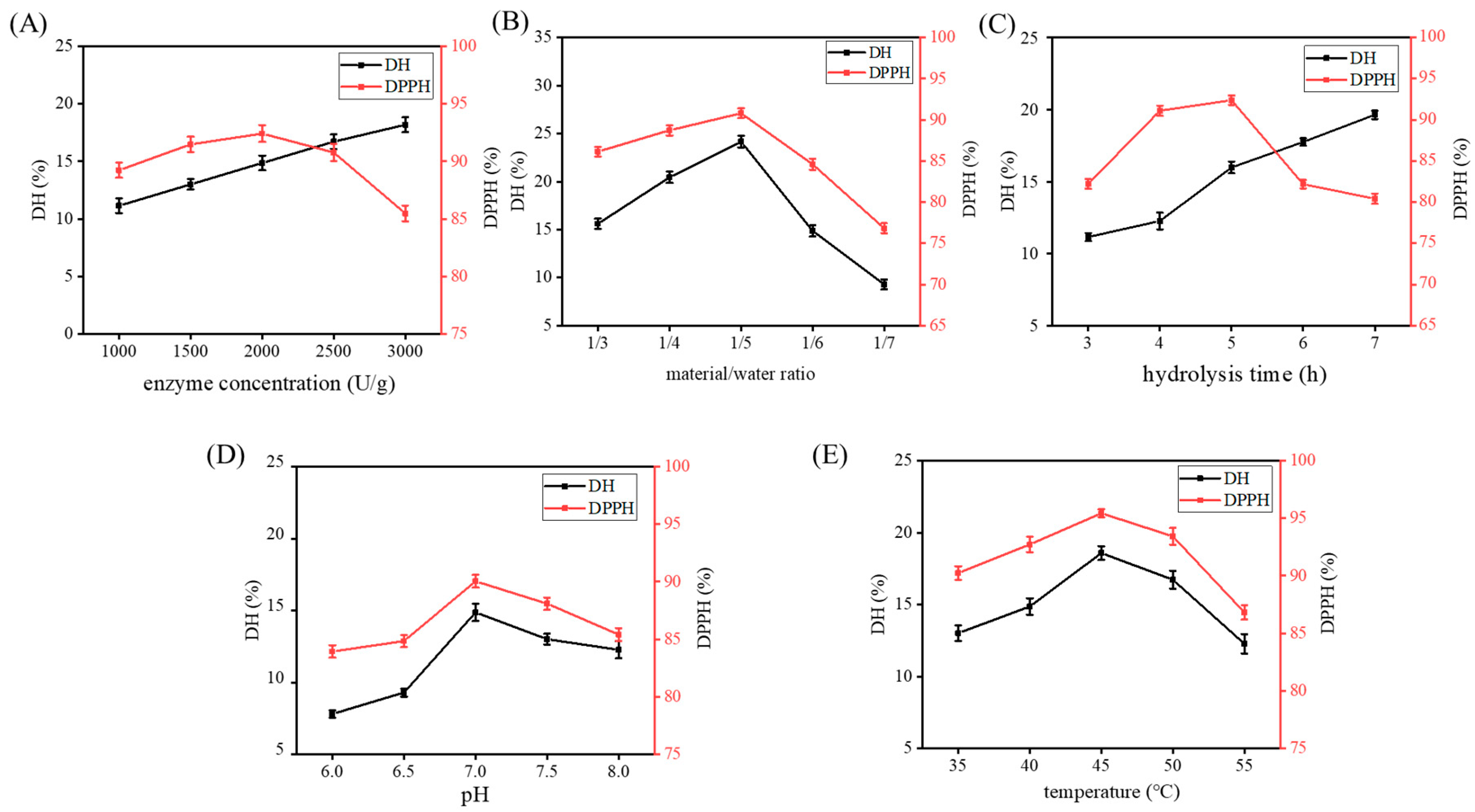
2.2. Single-Factor Experiments

Figure 2 illustrates the influence of five single factors on DH and DPPH clearance rates. The DPPH clearance rate initially reached a maximum and then decreased. DH increased with an increase in enzyme dosage and time. In the other three single factor ranges, DH first increased and then decreased. The following conditions were determined to be ideal for extraction: enzyme concentration of 2000 U/g, material/water ratio of 1:5 (w/v), hydrolysis time of 5 h, pH of 7.0, and temperature of 45 °C.

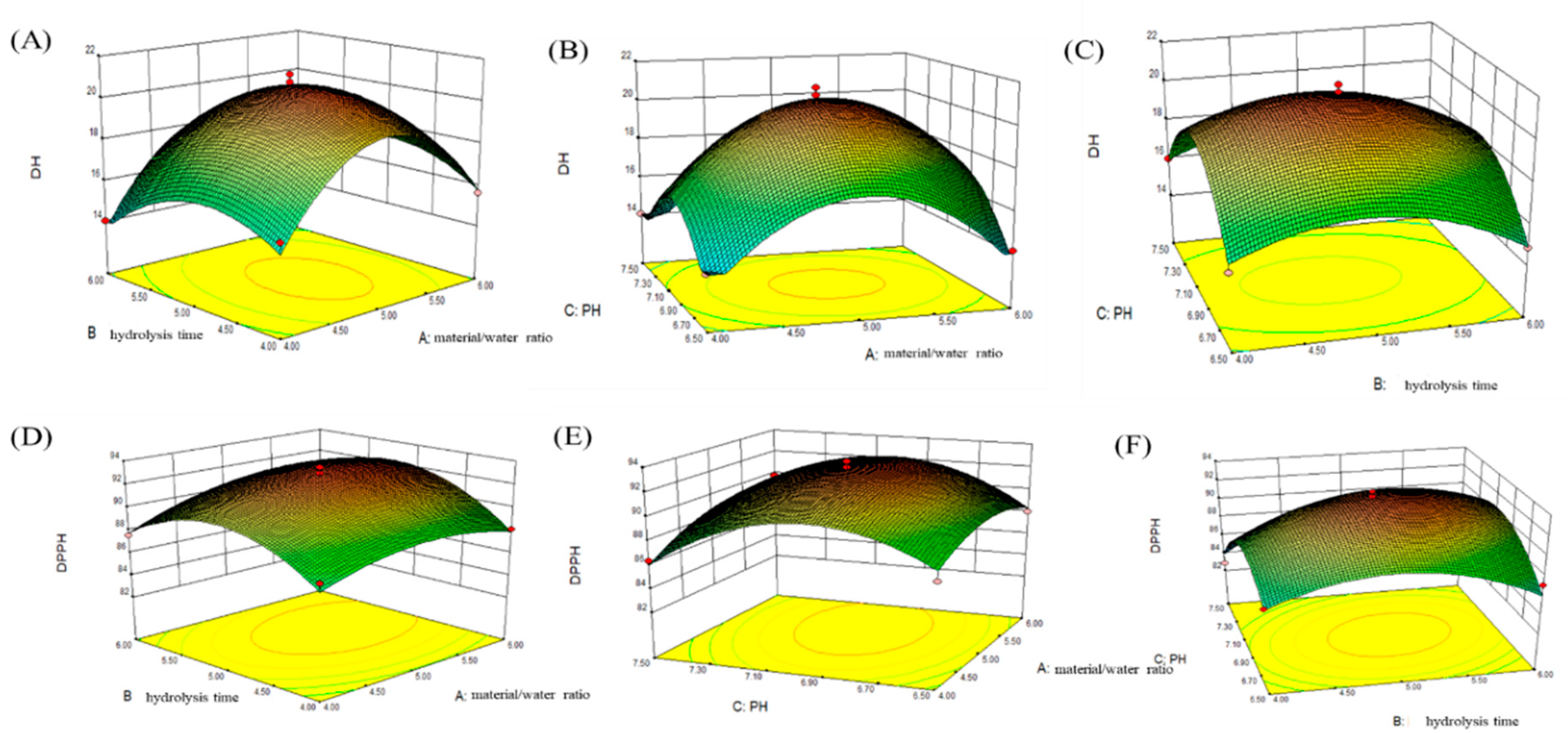
2.3. Determination of Optimal Conditions Using RSM to Produce LPs by the Action of Neutrase

Three independent parameters at three levels were used in a Box–Behnken and Design (BBD) based on single-factor experiments. The optimal bromelain treatment conditions required to produce monkfish muscle protein hydrolysate was determined using RSM. Equations were used to evaluate the effects of different enzyme conditions on DH and DPPH. Material/water ratio (A), extraction time (B), pH (C), and a statistically significant interaction (p < 0.05) on the DH and DPPH value were explained as follows:
DH = 20.590 + 0.600A − 0.460B + 0.046C + 0.280AB + 0.370AC − 0.093BC − 3.790A2
1.840B2 − 3.42C2
DPPH = 92.700 + 0.890A + 0.820B − 0.250C + 0.460AB + 0.550AC + 1.200BC −
1.420A2 − 2.990B2 − 3.56C2
ANOVA results for the model are listed in Table 1 and Table 2. The corresponding parameters were more significant as the F-value increased and the p-value decreased [20]. Based on the analysis as seen in Table 1 and Table 2 p < 0.0001 and p = 0.0006 of the two models show that the two models are extremely significant; p = 0.1663 > 0.05 and p = 0.0571 > 0.05 of the disharmony term are not significant; the regression equations of the DH and DPPH free radical scavenging rate are significantly tested, and the correlation coefficients of the two models are R2 = 0.984 3 > 0.95 and R2 = 0.956 > 0.95, respectively. The coefficients of variation for DH and DPPH activities are 3.49% and 1.04%, respectively. It shows that the fitting degree of the model is good. It can be seen that the two models can better reflect real test values. It was found that factors A, A2, B2, and C2 had significant effects on DH (p < 0.05). Factors A, B, BC, A2, B2, and C2 had significant effects on the DPPH free radical scavenging rate (p < 0.05).
Each response surface was obtained using the Box–Behnken test regression model. According to the bending degree of the response surface, the experimental points in Figure 3 fall within the experimental conditions, indicating that the optimal conditions are in this range. Figure 3A–C shows the effects of the interaction of hydrolysis time and material/water ratio, pH and material/water ratio, and pH and hydrolysis time on DH. On the whole, the p-value of each interaction was greater than 0.05 (p = 0.3604, p = 0.2333, p = 0.7538), and the interaction was not significant. The dimension reduction analysis of each factor is the trend of increasing first and then decreasing. The response value is the highest in the middle region, and the DH is the highest. Figure 3D–F shows the effects of hydrolysis time and material/water ratio, pH and material/water ratio, and pH and hydrolysis time on DPPH. The results are similar to DH. The interaction between enzymatic hydrolysis time and material/water ratio, pH and material/water ratio is not significant, and the response value reaches the maximum in the middle region, and DPPH is the highest. However, the interaction of pH and hydrolysis time had a significant effect on the proportion of oligopeptides in the hydrolysate (p < 0.05).
According to the analysis of design expert v8.0.6 software with degree of hydrolysis (DH) as the response index, the optimal experimental conditions were determined as follows: water/material ratio of 5.67:1, a time span of 4.92 h, pH of 7.02; the predicted degree of hydrolysis (DH) was 19.322%. Taking the DPPH free radical scavenging rate as the response index, the optimal experimental conditions were as follows: water/material ratio of 5.35:1, a time span of 5.17 h, pH of 7.01; the predicted DPPH scavenging rate was 92.921%. Considering the maneuverability of the experiment, the optimal conditions were adjusted as follows: water/material ratio of 5.40:1, a time span of 5 h, and pH of 7.0. The results of three experiments under this condition showed that the degrees of hydrolysis (DH) were 19.293%, 19.302%, and 19.312%, respectively. The calculated average value was 19.302%, which was close to the predicted value; the DPPH radical scavenging rates were 92.893%, 92.731%, and 92.959%, respectively. The calculated average value was 92.861%, which was close to the predicted value. Taken together, the established model is superior.

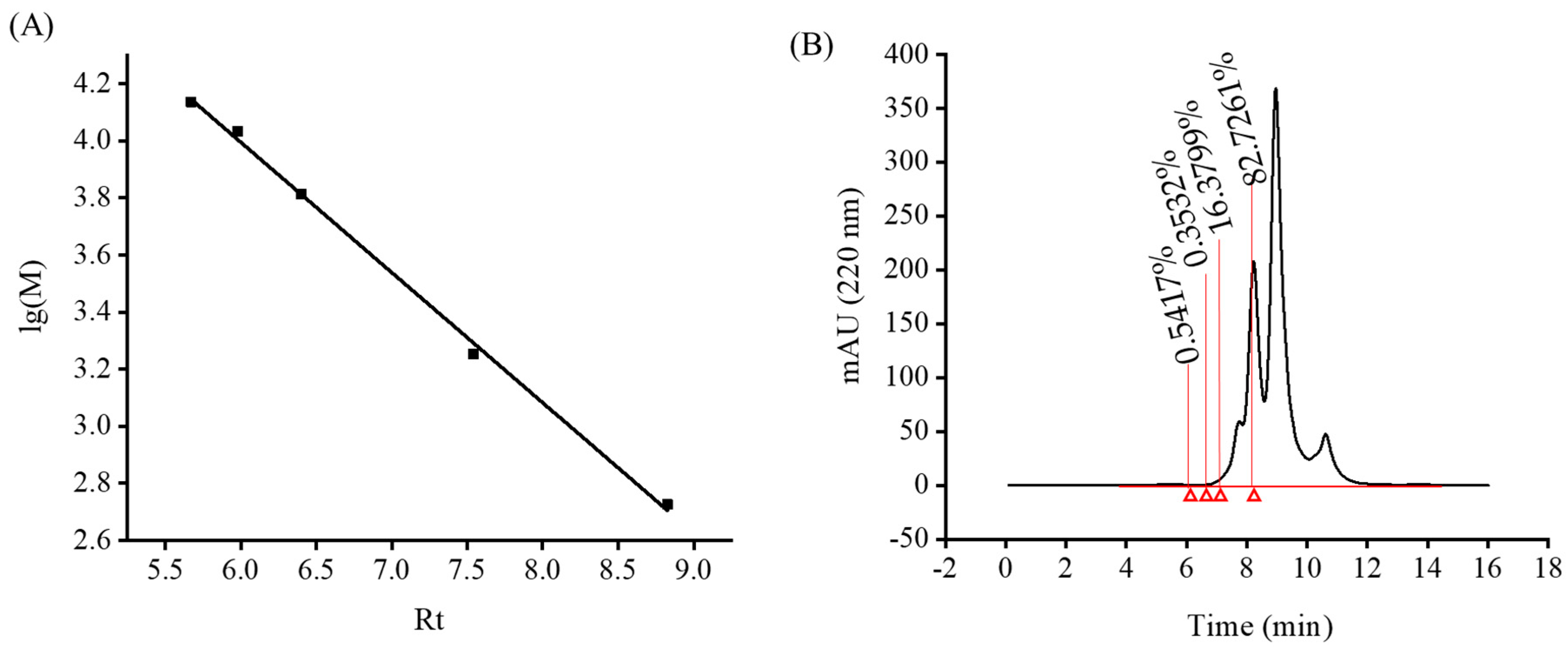
2.4. MW Distribution of Monkfish Muscle Protein Hydrolysate

In most cases, functional antioxidative peptides can only be activated by the process of hydrolysis, and most of them have small sizes that are less than 1 kDa [21,22]. The MW distribution of monkfish muscle peptides (LPs) was determined by a high-performance liquid chromatography (Agilent 1260, Palo Alto, CA, USA). A linear regression equation was made for the standard curve (Figure 4A): lg (M) = −0.4564x + 6.7339, R2 = 0.998. The MW distribution of LPs was determined using the regression equation. The relative MW distribution of monkfish muscle hydrolysate under the optimal enzymolysis conditions was small and mainly concentrated below 3000 Da, of which 82.726% was less than 1000 Da (Figure 4B). Therefore, it is concluded that LPs may have better antioxidant activity.

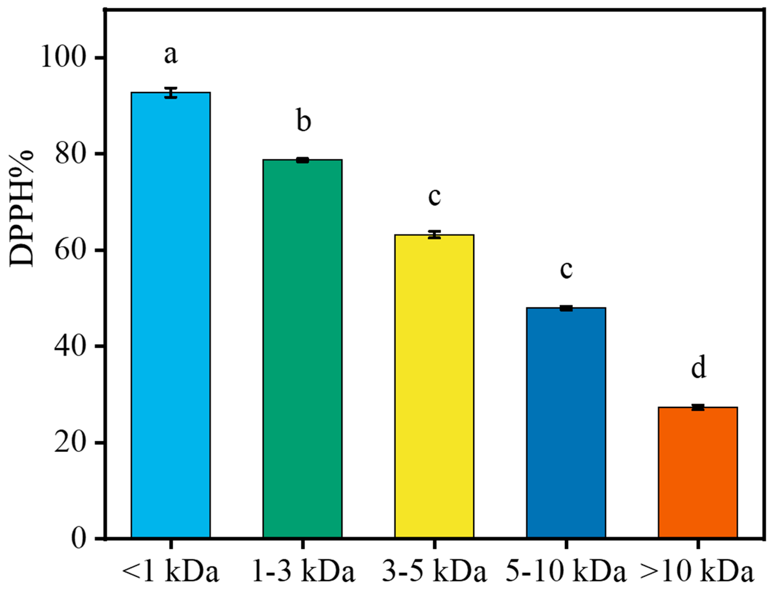
2.5. Interception of LPs with Different MW Using Ultrafiltration

DPPH is a type of stable free radical, and its response concentration is relatively low. It is usually used as a substrate to measure antioxidant activities [23]. Five different peptides were isolated from LPs using the membrane method. Figure 5 shows the DPPH radical scavenging activities of different LPs at 5 mg/mL. Low MW peptides were more effective than the high MW peptides in scavenging DPPH free radicals, especially the fraction <1 kDa that may have contained peptides/amino acids. As hydrogen donors, low MW peptides transform free radicals into more stable products, which show a higher DPPH-scavenging effect. This is consistent with the results of high DPPH scavenging activity of low MW peptides isolated from the protein hydrolysate of tuna by Hsu [24] and that from salmon hydrolysate by Ahn et al. [25].

2.6. Amino Acid Content of LPs <1 kDa

The hydrophobic nature of peptides helps elevate their antioxidant properties. They increase the interaction with lipid targets by virtue of hydrophobic associations or peptide entry into target organs, which are conducive to the realization of antioxidant effects [26,27]. Some studies have shown that negatively charged amino acids (NCAAS), such as glutamic and aspartic acids, exhibit strong antioxidant capacity due to a surplus of electrons that can be used during a free radical reaction [26,28,29]. As shown in Table 3, the percentage of hydrophobic amino acids (HAA) and NCAAS of <1 kDa LPs were relatively high, which were 25.74 (g/100 g) and 15.59 (g/100 g), respectively. The content of essential amino acids in <1 kDa LPs was 28.43 (g/100 g). We found that tryptophan, asparagine, and glutamine were not detectable due to the acidic conditions of analysis. These results indicated that <1 kDa LPs had appreciable antioxidant capacity and nutritional value.

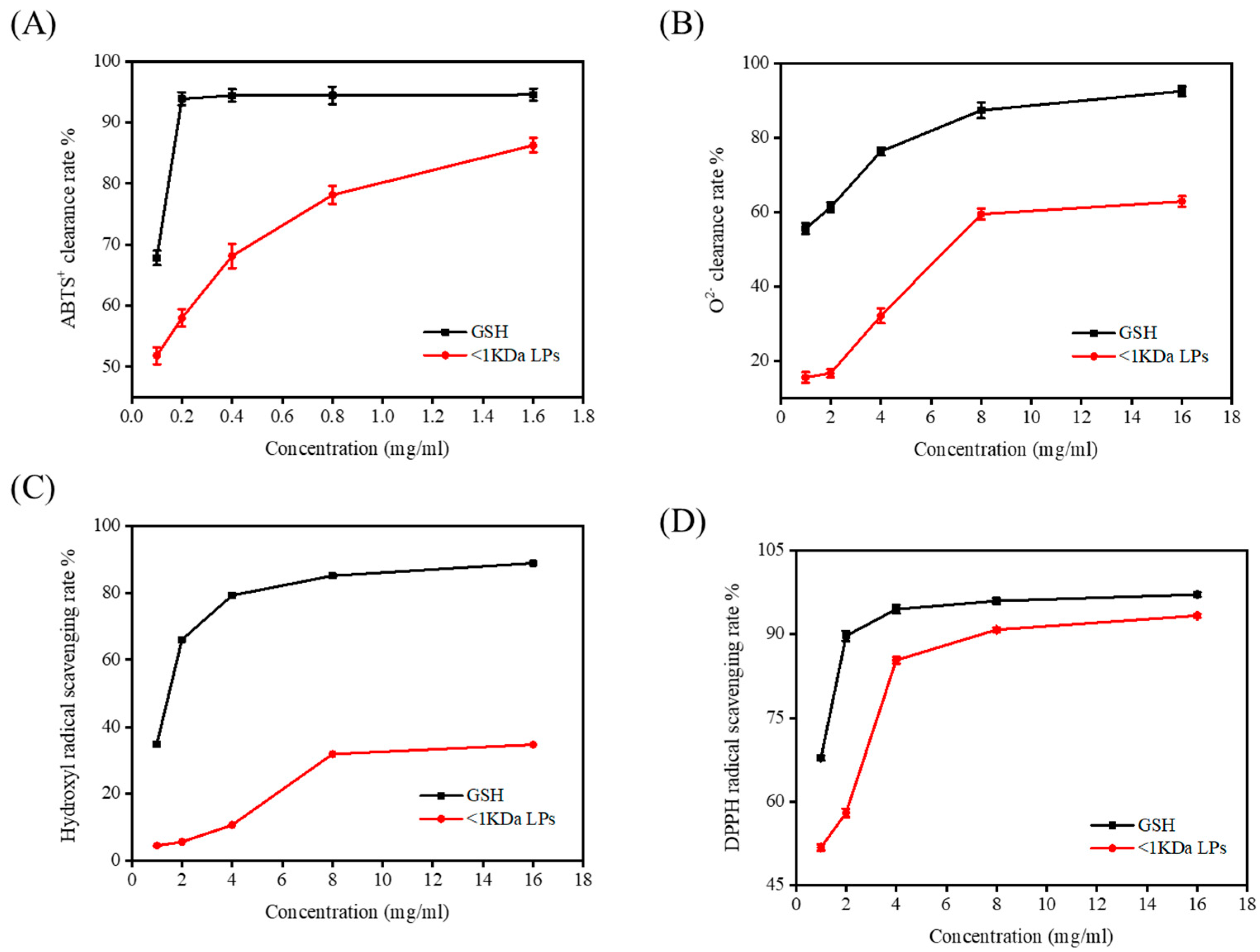
2.7. Antioxidant Activity of <1 kDa LPs

LPs were purified using an ultrafiltration membrane with MW of 1000 kD, and <1 kDa LPs were obtained and freeze-dried. The clearance rates of 2,2′-azinobis-3-ethylbenzthiazoline-6-sulphonate (ABTS), superoxide anion, DPPH radical, and hydroxyl radical were determined (Figure 6). The antioxidant capacity of <1 kDa LPs increased with increasing concentrations and was therefore dose-dependent. Among them, the ABTS scavenging effect was strong and provided a theoretical basis for the development of functional foods with antioxidant effects by using monkfish resources.

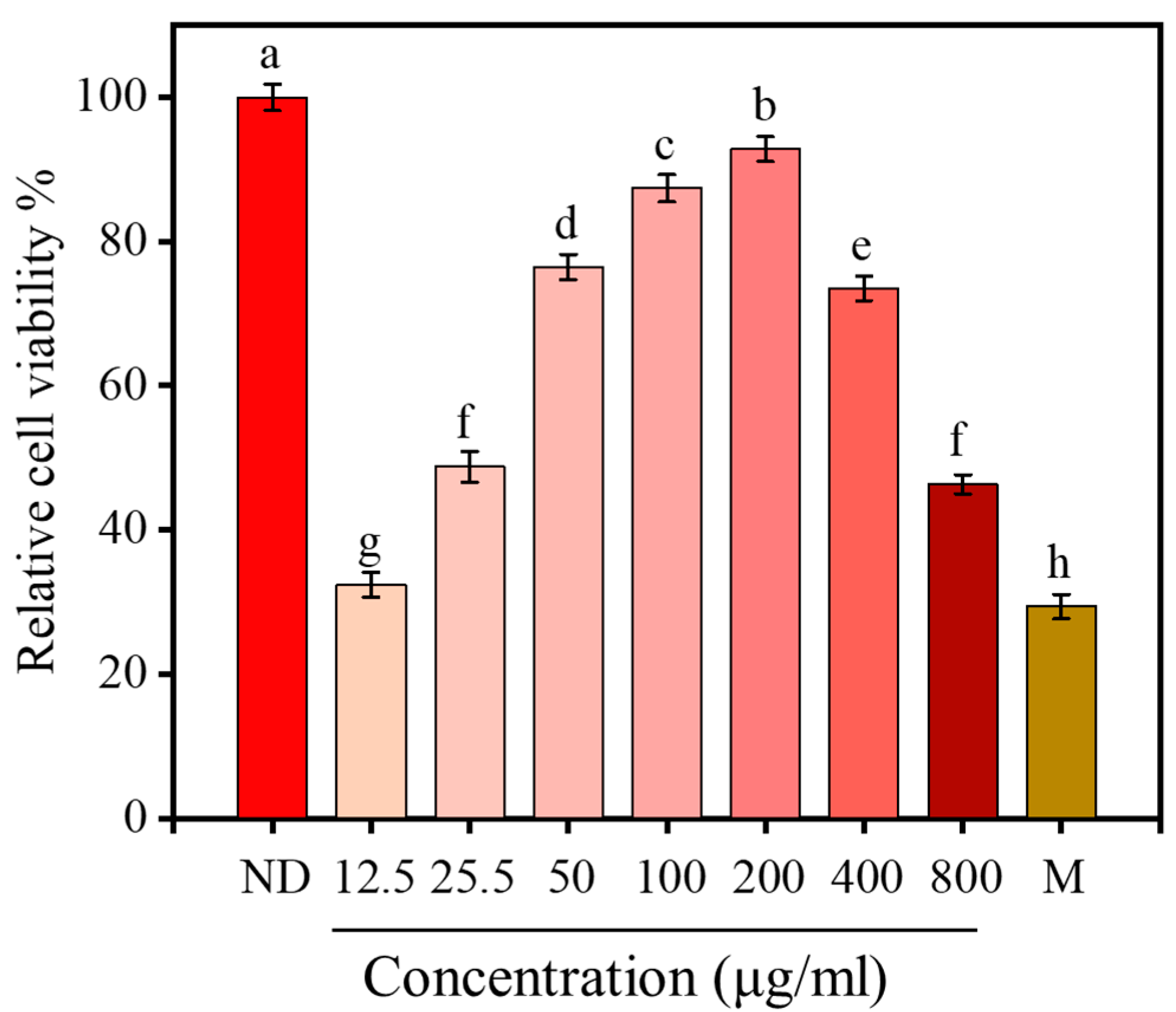
2.8. Effect of <1 kDa LPs on the Viability of H2O2-Stimulated Cells

H2O2 treatment significantly reduced the viability of RAW264.7 cells (p < 0.05), which decreased to 29.20% after 12 h of H2O2 treatment, indicating that H2O2 stimulation could cause oxidative damage. After 12 h of pretreatment with <1 kDa LPs, the activity of macrophages significantly increased (p < 0.05) and showed a concentration-dependent effect (Figure 7). When the concentration of <1 kDa LPs reached 200 μg/mL, the activity of macrophages reached 92.29%. Therefore, the concentration of 50, 100, and 200 μg/mL were selected for further studies.

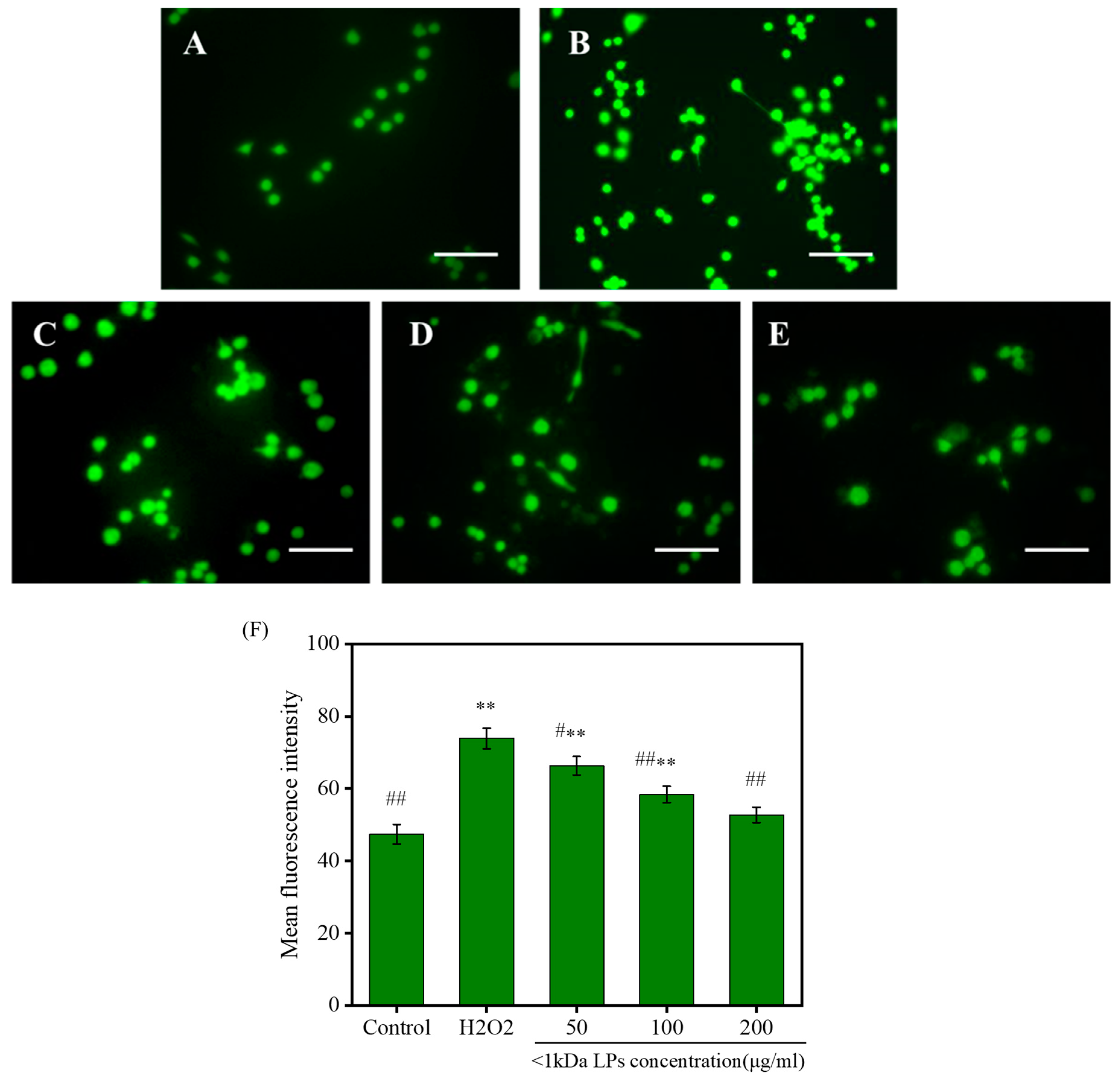
2.9. Effects of <1 kDa LPs on the ROS Levels

Reactive oxygen species (ROS) levels in the cells that were treated with <1 kDa LPs are shown in Figure 8. H2O2-treated cells showed higher fluorescence intensity, indicating that H2O2 treatment could significantly increase intracellular ROS levels. Compared to the cells treated with H2O2, the fluorescence intensity of the cells treated with <1 kDa LPs was lower than that of the cells treated with H2O2, which indicated that pretreatment with <1 kDa LPs could reduce the level of ROS in the cells stimulated by H2O2, and this effect showed a concentration-dependent effect. These results showed that <1 kDa LPs could act as a potential ROS scavenger.

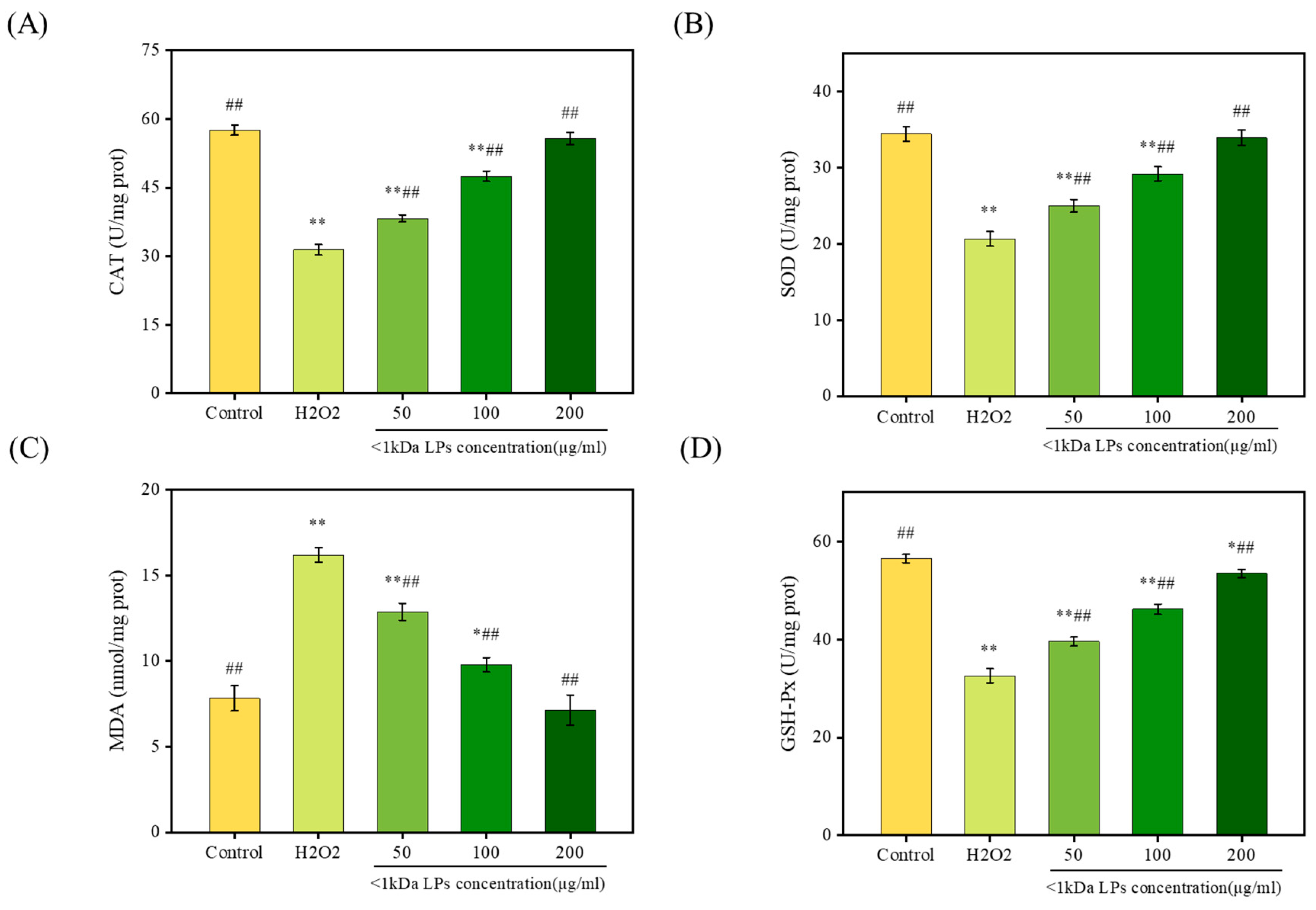
2.10. Effect of <1 kDa LPs on Antioxidant Activities

Superoxide dismutase (SOD), catalase (CAT), and glutathione peroxidase (GSH-Px), as intracellular antioxidant enzymes, can inhibit free-radical attack on cell membranes and maintain cell health. Malondialdehyde (MDA) is an oxidative metabolite of lipid oxidation that can be used as a marker for lipid peroxidation and cell lesions [30]. To measure the antioxidant activity of <1 kDa LPs, we studied the effects of pretreatment with <1 kDa LPs on GSH-Px, SOD, CAT, and MDA levels after H2O2 injury. H2O2 significantly reduced the levels of SOD, CAT, and GSH-Px, indicating that oxidative stress induced a serious damage to the antioxidant enzymes (Figure 9). The levels of SOD, CAT, and GSH-Px in cells that were pretreated with <1 kDa LPs increased dose-dependently, which were significantly higher than those in the H2O2-injured cells (p < 0.01). Compared to the control group, the MDA level in cells treated with H2O2 was markedly increased (p < 0.01), indicating that H2O2 was responsible for damage to these cells. Therefore, <1 kDa LPs could reduce oxidative stress injury and abate lipid peroxidation. The exact mechanism of how LPs work inside the cell to reduce oxidative stress needs to be thoroughly studied by analyzing the expression of antioxidant enzymes in the follow-up studies.

3. Materials and Methods

3.1. Pretreatment of Monkfish (Lophius litulon) Muscle

Monkfish was purchased from the Pearl fish market in Zhoushan, Zhejiang. Muscles were separated from the bones and then ground. Degreasing was carried out with 95% ethanol (1:5, w/v) in a water bath (50 °C) for 2 h [31]. The supernatant was discarded, and the precipitate was washed with distilled water. The defatted muscle was centrifuged at 12,000 r/min for 15 min, and the precipitate was collected.

3.2. Optimization of Preparative Conditions of LPs

Trypsin (≥250 U/mg), neutrase (≥60 U/mg), alcalase (≥200 U/mg), pepsin (≥3000 U/mg), and papain (≥500 U/mg) were ordered from YTHX Biotech (Beijing, China). The defatted muscle was added with distilled water (1:5, w/v) and protease (2000 U/g). The pH and temperature were adjusted to the appropriate value of protease (Table 4) and then hydrolyzed for 5 h. Enzymes were inactivated at 100 °C for 10 min and centrifuged at 12,000 r/min for 10 min. A portion of the supernatant was used for determining the degree of hydrolysis (DH), while the rest was freeze-dried for determining free radical activities.
DPPH, ABST, and phenazine methosulfate (PMS) were purchased from Sigma-Aldrich (Shanghai, China). According to the results of DPPH and DH, neutrase was selected as the best protease. We selected five parameters (protease concentration, pH, temperature, hydrolysis time, and muscle/water ratio) to conduct single-factor experiments using DPPH radical scavenging ability and DH as indicators. Based on single-factor experiments and three other factors (the extraction time, muscle/water ratio, and pH), a three-level and three-factor response surface test was designed using the Box–Behnken and Design (BBD) Expert (Design-Expert® 8.0). The test factors and levels are shown in Table 5. The antioxidant activities of the extracted peptides were evaluated using the degree of hydrolysis and DPPH clearance rate. Response surface test design and results are shown in Table 6. Responses were analyzed using multiple regressions to fit the following equation:
γ = β 0 + i = 1 k β i X i + i = 1 k β i i X i 2 + i < j β i j X i X j
where γ is the predicted response, β0 is the intercept, βi, βii, and βij are the linear, quadratic, and interaction coefficients, respectively, and both Xi and Xj are independent factors.
Using the design expert 8.0, an analysis of variance table was compiled. The influence coefficient, the regression coefficient of the linear term, quadratic term, and interactive term were determined.

3.3. Determining the MW Distribution of LPs

The MW distribution of LPs was analyzed using HPLC (Agilent 1260, Palo Alto, CA, USA). To make the mobile phase, 55% (v/v) water, 15% (v/v) acetonitrile, and 0.1% trifluoroacetic acid were mixed [32]. The flow rate was 0.5 mL/min, and the column was calibrated using cytochrome C (12,400 Da), aprotinin (6511.44 Da), and bacitracin (1422.69 Da) (Songon Biotech, Shanghai, China), ribonuclease A (13,700 Da) (Fushen Biotech, Shanghai, China), and Anthopleura anjunae oligopeptide (Tyr-Val-Pro-Gly-Pro, 531.61 Da) [33]. According to the relationship between the logarithm molecular weight and elution time, the molecular weight distribution of LPs was calculated.

3.4. DH and Antioxidant Activity

DH was titrated using the method by Noman et al. with some modifications [34]. Five milliliter protein hydrolysate was mixed with 50 mL of distilled water and the pH was adjusted to 8.2 with 0.05 M NaOH. Formaldehyde solution (10 mL) was added, and the hydrolysate was subsequently placed at 25 °C for 5 min. Then, the mixture was titrated with 0.05 M NaOH at pH 9.2. The content of the free amino group was determined based on the volume of NaOH used. The total nitrogen content was calculated using the Kjeldahl method [35]. The free amino content and DH were analyzed using the following equations:
%   free   amino   groups = A × B × 0.014 C / D × 100 %
DH = %   Free   amino   groups %   total   nitrogen × 100 %
where A = mL of NaOH used; B = the concentration of the solution used for titration (0.05 M NaOH); C = volume of sample diluent (mL); D = constant volume of sample (mL).
The DPPH, hydroxyl, ABTS, and superoxide anion radical scavenging activities of LPs <1 kDa were performed using the methods described by Chen et al. [36].

3.5. Preparation of LPs with Different Molecular Weight

The muscle hydrolysate was purified using a series of ultrafiltration membranes and a separation system (GM-18, Bona Biotech, Jinan, China) with a MW cut-off of 10, 5, 3, and 1 kDa. This process generated five parts as follows: >10 kDa, 5–10 kDa, 3–5 kDa, 1–3 kDa, and <1 kDa. The fractions were freeze-dried, and components with the best DPPH scavenging activity were selected. Amino acid content was determined with the method described by Tang et al. [37]. LPs were hydrolyzed with 6 mol/L HCl in an oven at 110 °C for 24 h. Approximately 1 mL of the sample was dried by blowing nitrogen, dissolved in 1 mL buffer solution (pH = 2.2), and measured by an amino acid analyzer (Hitachi L-8800, Tokyo, Japan).

3.6. Effects of <1 kDa LPs on theH2O2-Injured Cells

Mouse RAW264.7 cells were purchased from the Cell Bank of the Chinese Academy of Sciences (Shanghai, China). Cells were and seeded in 96-well plates at a density of 5 × 10⁴ cells per well. After culturing for 24 h, the medium was gently aspirated and replaced with LPs <1 kDa at different concentrations (12.5, 25, 50, 100, 200, 400, 600, 800, and 1000 μg/mL). Cells were cultured in the medium for 24 h and treated with H2O2 (final concentration of 500 μmol/L) for 8 h [38]. Then, 20 μL of 3-(4,5-dimethylthiazol-2-yl)-2,5-diphenyltetrazolium bromide (MTT) was added into the culture solution. The medium was gently aspirated from the plate, and 100 μL of dimethyl sulfoxide was added into each well. The absorbance was detected at 490 nm. The cell viability was determined as the absorbance of LPs group divided by the absorbance of control group. The cell viability was calculated using the following formula:
Cell   viability   ( % ) = A 1 A 2 × 100 %
where A1 is the absorbance of the LPs group and A2 is the absorbance of the control group.

3.7. Determination of ROS Levels after H2O2 Treatment

Cells were seeded in 6-well plates at a density of 2 × 10⁵ cells per well. After culturing for 24 h, cells were incubated with LPs for another 24 h. H2O2 (500 μmol/L) was added for 8 h. The medium was discarded and cells were rinsed with PBS three times. The fluorescent probe DCFH-DA solution (150 μL; 50 μmol/L) was added and maintained at 37 °C for 60 min. Subsequently, cells were rinsed with PBS three times [39]. Cells were visualized using fluorescence microscopy and photographed. The fluorescence intensity was analyzed using Image J software.

3.8. Antioxidant Enzyme Activity of <1 kDa LPs in H2O2-Induced Stress

First, 500 μL cell lysis buffer was added to each well, and the mixture was centrifuged at 12,000× g at 4 °C for 10 min. The supernatant was cooled to 4 °C. The levels of GSH-Px, SOD, CAT, and MDA were measured with commercial test kits (Nanjing Jiancheng Bioengineering Institute, Nanjing, China).

3.9. Statistical Analysis

The data were presented as the mean ± SD. Multiple-group comparisons were determined using ANOVA in SPSS 19.0. p = 0.05 was considered as a threshold with statistical significance.

4. Conclusions

In this study, the extraction procedure of antioxidative peptides from monkfish muscle were optimized. LPs less than 1 kDa had a high antioxidant activity and could effectively scavenge DPPH, hydroxyl, ABTS, and superoxide anion radicals. LPs possess good antioxidant capacity and protect RAW264.7 cells from H2O2-induced injury, which provides a theoretical basis for the use of <1 kDa LPs as natural antioxidants. Further studies are required to determine the peptide sequence of <1 kDa LPs and verify the protective functions in animals. Our optimized procedures for extracting LPs may also be useful for future scale-up production.

Author Contributions

Conceptualization, X.T. and Z.Y.; methodology, X.T. and Z.Y.; investigation, X.T., J.Z., B.X. and J.Y.; writing—original draft preparation, X.T.; writing—review and editing, F.Y.; supervision, Z.Y. All authors have read and agreed to the published version of the manuscript.

Funding

This study was supported by National Natural Science Foundation of China (grant number 81673349) and College Students’ Science, Technology and Innovation (Xinmiao) Plan of Zhejiang (grant number 2019R411021).

Conflicts of Interest

The authors declare no conflict of interest.

References

  1. Elias, R.J.; Kellerby, S.S.; Decker, E.A. Antioxidant activity of proteins and peptides. Crit. Rev. Food Sci. Nutr. 2008, 48, 430–441. [Google Scholar] [CrossRef] [PubMed]
  2. Bernardini, R.D.; Harnedy, P.; Bolton, D.; Kerry, J.; O’Neill, E.; Mullen, A.M.; Hayes, M. Antioxidant and antimicrobial peptidic hydrolysates from muscle protein sources and by-products. Food Chem. 2011, 124, 1296–1307. [Google Scholar] [CrossRef]
  3. Randy, C.; Tzi, N.; Jack, W. Marine peptides: Bioactivities and applications. Mar. Drugs 2015, 13, 4006–4043. [Google Scholar]
  4. Bougatef, A.; Hajji, M.; Balti, R.; Lassoued, I.; Triki-Ellouz, Y.; Nasri, M. Antioxidant and free radical-scavenging activities of smooth hound (Mustelus mustelus) muscle protein hydrolysates obtained by gastrointestinal proteases. Food Chem. 2009, 114, 1198–1205. [Google Scholar] [CrossRef]
  5. Cheung, I.W.Y.; Cheung, L.K.Y.; Tan, N.Y.; Li-Chan, E.C.Y. The role of molecular size in antioxidant activity of peptide fractions from pacific hake (Merluccius productus) hydrolysates. Food Chem. 2012, 134, 1297–1306. [Google Scholar] [CrossRef] [PubMed]
  6. Sarmadi, B.H.; Ismail, A. Antioxidative peptides from food proteins: A review. Peptides 2010, 31, 1949–1956. [Google Scholar] [CrossRef] [PubMed]
  7. Rushikesh, S.; Pravin, P.; Seetharama, J. Peptides, peptidomimetics, and polypeptides from marine sources: A wealth of natural sources for pharmaceutical applications. Mar. Drugs 2017, 15, 124–160. [Google Scholar]
  8. Jonsdottir, R.; Geirsdottir, M.; Hamaguchi, P.Y.; Jamnik, P.; Kristinsson, H.G.; Undeland, I. The ability of in vitro antioxidant assays to predict the efficiency of a cod protein hydrolysate and brown seaweed extract to prevent oxidation in marine food model systems. J. Sci. Food Agric. 2016, 96, 2125–2135. [Google Scholar] [CrossRef]
  9. Chi, C.; Hu, F.; Wang, B.; Li, Z.; Luo, H. Influence of amino acid compositions and peptide profiles on antioxidant capacities of two protein hydrolysates from skipjack tuna (Katsuwonus pelamis) dark muscle. Mar. Drugs 2015, 13, 2580–2601. [Google Scholar] [CrossRef]
  10. Borawska, J.; Darewicz, M.; Pliszka, M.; Vegarud, G.E. Antioxidant properties of salmon (Salmo salar L.) protein fraction hydrolysates revealed following their ex vivo digestion and in vitro hydrolysis. J. Sci. Food Agric. 2016, 96, 2764–2772. [Google Scholar] [CrossRef]
  11. Nazeer, R.A.; Kumar, N.S.S.; Ganesh, R.J. In vitro and in vivo studies on the antioxidant activity of fish peptide isolated from the croaker (Otolithes ruber) muscle protein hydrolysate. Peptides 2012, 35, 261–268. [Google Scholar] [CrossRef]
  12. Ranathunga, S.; Rajapakse, N.; Kim, S.K. Purification and characterization of antioxidative peptide derived from muscle of conger eel (Conger myriaster). Eur. Food Res. Technol. 2006, 222, 310–315. [Google Scholar] [CrossRef]
  13. Jaswir, I.; Noviendri, D.; Taher, M.; Mohamed, F.; Octavianti, F.; Lestari, W.; Mukti, A.; Nirwandar, S.; Hamad Almansori, B. Optimization and formulation of fucoxanthin-loaded microsphere (F-LM) using response surface methodology (RSM) and analysis of its fucoxanthin release profile. Molecules 2019, 24, 947. [Google Scholar] [CrossRef] [PubMed] [Green Version]
  14. Ovissipour, M.; Kenari, A.A.; Motamedzadegan, A.; Nazari, R.M. Optimization of enzymatic hydrolysis of visceral waste proteins of yellowfin tuna (Thunnus albacares). Food Bioprocess Technol. 2012, 5, 696–705. [Google Scholar] [CrossRef]
  15. Muhammad, A.S.; Mohammad, Z.; Azizah, A.H.; Nazamid, S. Response surface optimisation for the production of antioxidant hydrolysates from stone fish protein using bromelain. Evid. Based Complem. Altern. Med. 2017, 2017, 1–10. [Google Scholar]
  16. Yang, S.Y.; Kim, S.W.; Kim, Y.; Lee, S.H.; Jeon, H.; Lee, K.W. Optimization of Maillard reaction with ribose for enhancing anti-allergy effect of fish protein hydrolysates using response surface methodology. Food Chem. 2015, 176, 420–425. [Google Scholar] [CrossRef]
  17. Gómez, L.J.; Gómez, N.A.; Zapata, J.E.; López-García, G.; Cilla, A.; Alegría, A. Optimization of the red tilapia (Oreochromis spp.) viscera hydrolysis for obtaining iron-binding peptides and evaluation of in vitroiron bioavailability. Foods 2020, 9, 883. [Google Scholar] [CrossRef]
  18. Chi, C.F.; Wang, B.; Deng, Y.Y.; Wang, Y.M.; Deng, S.G.; Ma, J.Y. Isolation and characterization of three antioxidant pentapeptides from protein hydrolysate of monkfish (Lophius litulon) muscle. Food Res. Int. 2014, 55, 222–228. [Google Scholar] [CrossRef]
  19. Hu, X.M.; Wang, Y.M.; Zhao, Y.Q.; Chi, C.F.; Wang, B. Antioxidant peptides from the protein hydrolysate of monkfish (Lophius litulon) muscle: Purification, identification, and cytoprotective function on hepg2 cells damage by H2O2. Mar. Drugs 2020, 18, 153. [Google Scholar] [CrossRef] [PubMed] [Green Version]
  20. Zhao, Z.Y.; Zhang, Q.; Li, Y.F.; Dong, L.L.; Liu, S.L. Optimization of ultrasound extraction of Alisma orientalis polysaccharides by response surface methodology and their antioxidant activities. Carbohydr. Polym. 2015, 119, 101–109. [Google Scholar] [CrossRef]
  21. Raghavan, S.; Kristinsson, H.; Leeuwenburgh, B. Radical scavenging and reducing ability of tilapia (Oreochromis niloticus) protein hydrolysates. J. Agric. Food Chem. 2008, 56, 10359–10367. [Google Scholar] [CrossRef]
  22. Sonklin, C.; Laohakunjit, N.; Kerdchoechuen, O. Assessment of antioxidant properties of membrane ultrafiltration peptides from mungbean meal protein hydrolysates. PeerJ 2018, 6, 17–36. [Google Scholar] [CrossRef] [PubMed]
  23. Saidi, S.; Deratani, A.; Belleville, M.; Amar, R.B. Antioxidant properties of peptide fractions from tuna dark muscle protein by-product hydrolysate produced by membrane fractionation process. Food Res. Int. 2014, 65, 329–336. [Google Scholar] [CrossRef]
  24. Hsu, K. Purification of antioxidative peptides prepared from enzymatic hydrolysates of tuna dark muscle by-product. Food Chem. 2010, 122, 42–48. [Google Scholar] [CrossRef]
  25. Ahn, C.; Kim, J.; Je, J. Purification and antioxidant properties of octapeptide from salmon byproduct protein hydrolysate by gastrointestinal digestion. Food Chem. 2014, 147, 78–83. [Google Scholar] [CrossRef] [PubMed]
  26. He, R.; Girgih, A.T.; Malomo, S.A.; Ju, X.; Aluko, R.E. Antioxidant activities of enzymatic rapeseed protein hydrolysates and the membrane ultrafiltration fractions. J. Funct. Foods 2013, 5, 219–227. [Google Scholar] [CrossRef]
  27. Zheng, J.; Tian, X.; Xu, B.; Yuan, F.; Gong, J.; Yang, Z. Collagen Peptides from Swim Bladders of Giant Croaker (Nibea japonica) and Their Protective Effects against H2O2-Induced Oxidative Damage toward Human Umbilical Vein Endothelial Cells. Mar. Drugs 2020, 18, 430. [Google Scholar] [CrossRef]
  28. Samaranayaka, A.G.P.; Lichan, E.C.Y. Food-derived peptidic antioxidants: A review of their production, assessment, and potential applications. J. Funct. Foods 2011, 3, 229–254. [Google Scholar] [CrossRef]
  29. Udenigwe, C.C.; Aluko, R.E. Chemometric analysis of the amino acid requirements of antioxidant food protein hydrolysates. Int. J. Mol. Sci. 2011, 12, 3148–3161. [Google Scholar] [CrossRef] [Green Version]
  30. Xiao, X.; Liu, J.; Hu, J.; Zhu, X.; Yang, H.; Wang, C.; Zhang, Y. Protective effects of protopine on hydrogen peroxide-induced oxidative injury of pc12 cells via Ca2+ antagonism and antioxidant mechanisms. Eur. J. Pharmacol. 2008, 591, 21–27. [Google Scholar] [CrossRef]
  31. Hu, X.; Li, W.; Kong, X.; Han, H.; Tang, Y.; Yu, F.; Yang, Z.; Ding, G. Optimization of extraction technology of immunologically active peptides from Nibea japonica by response surface methodology. Sci. Technol. Food Ind. 2019, 17, 173–178. [Google Scholar]
  32. Hu, Z.; Yang, P.; Zhou, C.; Li, S.; Hong, P. Marine collagen peptides from the skin of Nile tilapia (Oreochromis niloticus): Characterization and wound healing evaluation. Mar. Drugs 2017, 15, 102. [Google Scholar] [CrossRef]
  33. Wu, Z.; Ding, G.; Huang, F.; Yang, Z.; Yu, F.; Tang, Y.; Jia, Y.; Zheng, Y.; Chen, R. Anticancer activity of Anthopleura anjunae oligopeptides in prostate cancer du-145 cells. Mar. Drugs 2018, 16, 125. [Google Scholar] [CrossRef] [PubMed] [Green Version]
  34. Noman, A.; Xu, Y.; Al-Bukhaiti, W.Q.; Abed, S.M.; Ali, A.H.; Ramadhan, A.H.; Xia, W. Influence of enzymatic hydrolysis conditions on the degree of hydrolysis and functional properties of protein hydrolysate obtained from Chinese sturgeon (Acipenser sinensis) by using papain enzyme. Process Biochem. 2018, 67, 19–28. [Google Scholar] [CrossRef]
  35. Horwitz, W. Official methods of analysis of AOAC international. Trends Food Sci. Technol. 1995, 6, 382. [Google Scholar]
  36. Chen, Y.; Jin, H.; Yang, F.; Jin, S.; Liu, C.; Zhang, L.; Huang, J.; Wang, S.; Yan, Z.; Cai, X. Physicochemical, antioxidant properties of giant croaker (Nibea japonica) swim bladders collagen and wound healing evaluation. Int. J. Biol. Macromol. 2019, 138, 483–491. [Google Scholar] [CrossRef]
  37. Tang, Y.; Jin, S.; Li, X.; Li, X.; Hu, X.; Chen, Y.; Huang, F.; Yang, Z.; Yu, F.; Ding, G. Physicochemical properties and biocompatibility evaluation of collagen from the skin of giant croaker (Nibea japonica). Mar. Drugs 2018, 16, 222. [Google Scholar] [CrossRef] [Green Version]
  38. Li, Y.; Li, J.; Lin, S.; Yang, Z.; Jin, H. Preparation of antioxidant peptide by microwave-assisted hydrolysis of collagen and its protective effect against H2O2-induced damage of raw264.7 cells. Mar. Drugs 2019, 17, 642. [Google Scholar] [CrossRef] [Green Version]
  39. Takamiya, R.; Fukunaga, K.; Arita, M.; Miyata, J.; Seki, H.; Minematsu, N.; Suematsu, M.; Asano, K. Resolvin e1 maintains macrophage function under cigarette smoke-induced oxidative stress. FEBS Open Bio 2012, 2, 328–333. [Google Scholar] [CrossRef] [Green Version]
Figure 1. Degree of hydrolysis (DH) and 1,1-diphenyl-2-picrylhydrazyl (DPPH) scavenging ability of five proteases after hydrolysis. Data are represented as mean ± SD (n = 3). Letters a–d denote statistically significant differences among treatments (p < 0.05).
Marinedrugs 18 00468 g001
Figure 2. DPPH clearance rate and degree of hydrolysis (DH) vary with enzyme concentration (A), material/water ratio (B), hydrolysis time (C), pH (D), and temperature (E). Data are represented as mean ± SD (n = 3).
Marinedrugs 18 00468 g002
Figure 3. Response surface of hydrolysis rate (DH) and DPPH free radical scavenging rate of enzymatic hydrolysate, (A) the degree of hydrolysis (DH) is the response value of the interaction between hydrolysis time and material/water ratio, (B) degree of hydrolysis (DH) is the interaction of response value pH and material/water ratio, (C) degree of hydrolysis (DH) is the interaction of response value, hydrolysis time, and pH, (D) DPPH free radical scavenging rate is the interaction of response value, hydrolysis time, and material/water ratio, (E) DPPH radical scavenging rate is the interaction of response value, pH, and material/water ratio, (F) The scavenging rate of DPPH free radical is dependent on response value, and the interaction of hydrolysis time and pH value.
Marinedrugs 18 00468 g003
Figure 4. Relative molecular mass standard curve (A) and chromatogram of relative molecular mass distribution of monkfish muscle peptides (LPs) (B). Rt: retention time; lg(M): logarithm of MW.
Marinedrugs 18 00468 g004
Figure 5. DPPH clearance of peptides with different molecular weights. Data are represented as mean ± SD (n = 3). Different superscript letters (a–d) denote statistically significant differences among treatments (p < 0.05).
Marinedrugs 18 00468 g005
Figure 6. Scavenging effects of the <1 kDa LPs on 2,2′-azinobis-3-ethylbenzthiazoline-6-sulphonate (ABTS) radical (A), superoxide anion radical (B), hydroxyl radical (C), and DPPH radical (D). Data are represented as mean ± SD (n = 3).
Marinedrugs 18 00468 g006
Figure 7. Protective effects of <1 kDa LPs on the viability of H2O2-stimulated cells. Results are expressed as means ± SD (n = 8). Letters a–h denote statistically significant differences among treatments (p < 0.05).
Marinedrugs 18 00468 g007
Figure 8. Inhibitory effects of <1 kDa LPs on intracellular reactive oxygen species (ROS) levels in H2O2-stimulated cells. (A) Control group; (B) model group (500 μM H2O2); (C) 50 µg/mL <1 kDa LPs+ 500 µM H2O2; (D) 100 µg/mL <1 kDa LPs+ 500 µM H2O2; (E) 200 µg/mL <1 kDa LPs+ 500 µM H2O2. (F) Mean fluorescence intensity of cells in different groups. Data are presented as the mean ± SD (n = 3). ** p < 0.01 vs. the control group, # p < 0.05, ## p < 0.01 vs. the H2O2 group. Scale bars = 50 μm.
Marinedrugs 18 00468 g008
Figure 9. Effect of <1 kDa LPs on levels of catalase (CAT) (A), superoxide dismutase (SOD) (B), malondialdehyde (MDA) (C), and glutathione peroxidase (GSH-Px) (D) in H2O2-induced RAW264.7 injury model. Data are represented as mean ± SD (n = 3). * p < 0.05, ** p < 0.01 vs. control group, # p < 0.05, ## p < 0.01 vs. H2O2 group.
Marinedrugs 18 00468 g009
Table 1. Analysis of variance of regression equation for degree of hydrolysis (DH).
VariablesSum of SquaresdfMean SquareF-Valuep-Value
A (material/water ratio)2.9212.928.990.0200
B (extraction time)1.7311.735.320.0545
C (PH)0.01710.0170.0530.8242
AB0.3110.310.960.3604
AC0.5510.551.700.2333
BC0.03510.0350.110.7538
A260.52160.52186.41<0.0001
B214.25114.2543.900.0003
C249.23149.23151.65<0.0001
Model142.64915.8548.82<0.0001
Lack of fit4.0270.57
Residual1.5530.522.880.1663
Pure error0.7240.18
Cor. total144.9116
R2 = 0.9843RAdj2 = 0.9642CV = 3.49%
Table 2. Analysis of variance of regression equation for degree of hydrolysis (DPPH).
VariablesSum of SquaresdfMean SquareF-Valuep-Value
A (material/water ratio)5.4315.436.400.0291
B (extraction time)6.3516.357.480.0393
C (PH)0.4810.480.570.4757
AB0.8410.840.990.3539
AC5.7815.786.800.2701
BC1.2211.221.430.0350
A^237.56137.5644.240.0161
B^28.4318.439.930.0003
C^253.45153.4562.95<0.0001
Model129.48914.3916.950.0006
Lack of fit5.9470.85
Residual4.8731.626.060.0571
Pure error1.0740.27
Cor. total135.4216
R2 = 0.9561RAdj2 = 0.8997CV = 1.04%
Table 3. Amino acid composition (g/100 g) of <1 kDa LPs.
Amino AcidsLPs
Asp5.82 ± 0.190
Thr2.83 ± 0.083
Ser2.89 ± 0.080
Glu9.75 ± 0.297
Gly2.53 ± 0.080
Ala3.45 ± 0.107
Cys0.00
Val3.51 ± 0.096
Met2.42 ± 0.051
Lle2.79 ± 0.079
Leu5.57 ± 0.152
Tyr1.81 ± 0.014
Phe6.19 ± 0.079
Lys5.48 ± 0.167
His1.55 ± 0.102
Arg3.70 ± 0.105
Pro0.00
HAA25.74
PCAA10.73
NCAA15.59
EAA28.43
Table 4. Optimal reaction conditions of proteases.
Types of ProteaseOptimum Temperature (°C)Optimum pH
neutrase45 °C7.0
alcalase45 °C10.0
papain60 °C6.0
pepsin37 °C2.0
trypsin37 °C8.0
Table 5. Box–Behnken test factors and levels.
VariablesLevels and Range
−101
A muscle/water ratio (g·mL−1)1:41:51:6
B extraction time (h)456
C pH 6.577.5
Table 6. Response surface test design and results.
Run NumbersA Extraction Time (h)B Muscle/Water Ratio (g/mL)C PHDH%DPPH/%
1−10−113.008886.7955
200020.442593.4659
30−1−115.238986.9091
4−10111.893986.375
500020.070892.1818
61−1015.610688.1136
700020.814292.4659
801−114.867387.0568
900021.185892.4091
10−1−1015.610687.7386
1110114.495689.752
1211014.867389.7727
13−11013.752287.5682
1410−114.123987.9659
1501115.238987.7955
160−1115.982382.841
1700020.442592.9773

