Article

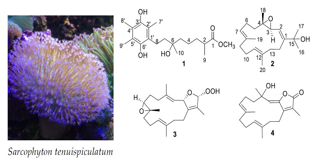
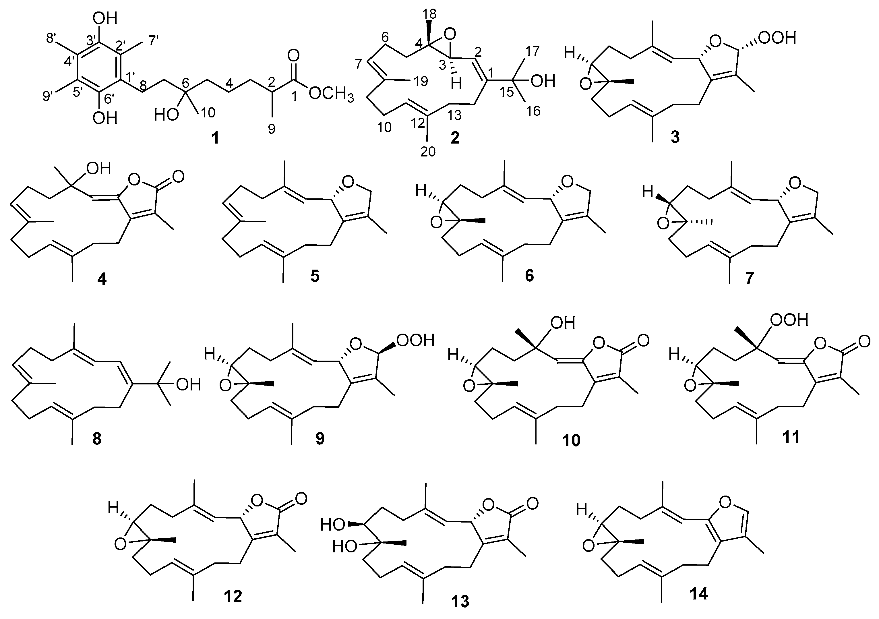
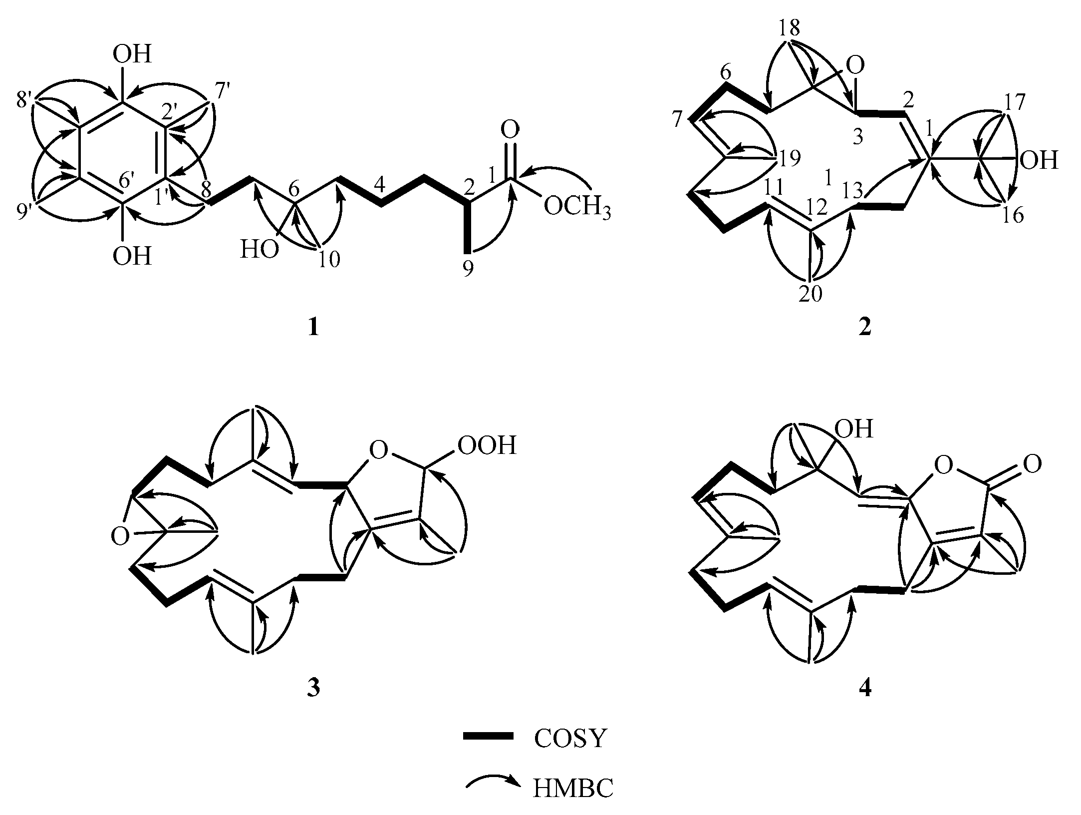
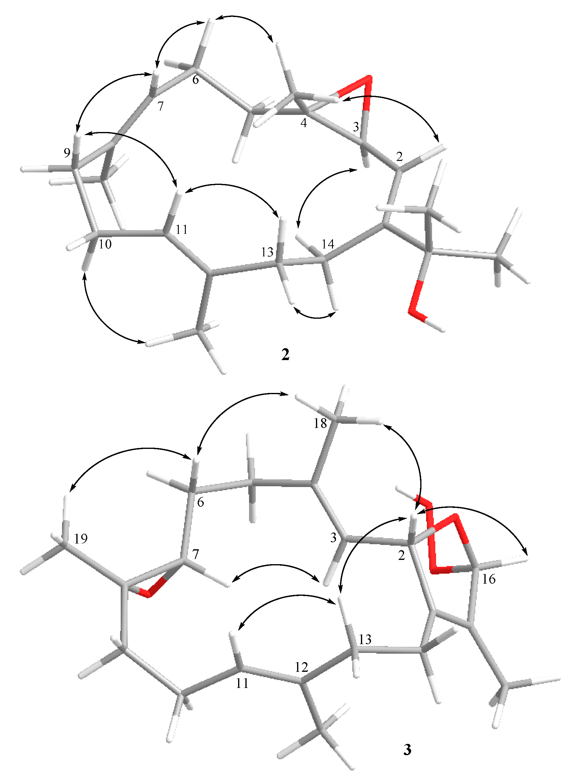
New Hydroquinone Monoterpenoid and Cembranoid-Related Metabolites from the Soft Coral Sarcophyton tenuispiculatum

1
Doctoral Degree Program in Marine Biotechnology, National Sun Yat-Sen University, Kaohsiung 804, Taiwan
2
Department of Marine Biotechnology and Resources, National Sun Yat-sen University, Kaohsiung 804, Taiwan
3
Graduate Institute of Natural Products, College of Pharmacy, Kaohsiung Medical University, Kaohsiung 807, Taiwan
4
Department of Oceanography, National Sun Yat-sen University, Kaohsiung 804, Taiwan
5
Institute of Biological Chemistry, Academia Sinica, Taipei 11529, Taiwan
6
Department of Medical Research, China Medical University Hospital, China Medical University, Taichung 404, Taiwan
*
Author to whom correspondence should be addressed.
Submission received: 4 December 2020 / Revised: 21 December 2020 / Accepted: 23 December 2020 / Published: 27 December 2020
(This article belongs to the Special Issue Bioactive Natural Products from Soft Corals)

Abstract

:
Chemical investigation of the marine soft coral Sarcophyton tenuispiculatum resulted in the isolation of a 1,4-dihydrobenzoquinone, sarcotenuhydroquinone (1), three new cembranoids, sarcotenusenes A‒C (24), and ten previously reported metabolites 514. The chemical structures of all isolated metabolites were determined by detailed spectroscopic analyses. In biological assays, anti-inflammatory, cytotoxic, and peroxisome proliferator-activated receptor γ (PPAR-γ) transcription factor assays of all compounds were performed. None of the isolated compounds were found to exhibit activity in the PPAR-γ transcription factor assay. The anti-inflammatory assays showed that (+)-7α,8β-dihydroxydeepoxysarcophine (13) inhibited the production of IL-1β to 56 ± 1% at a concentration of 30 µM in lipopolysaccharide (LPS)-stimulated J774A.1 macrophage cells. In addition, 1 and 2 were found to exhibit cytotoxicity towards a panel of cancer cell lines.

Graphical Abstract

1. Introduction

Marine organisms, especially sessile marine invertebrates, such as soft corals, have been found to produce secondary metabolites as defensive substances to protect themselves from predators or stressful environments [1]. Sarcophyton, one of the widely studied genera of soft corals, is considered an abundant source of bioactive diterpenes, especially of cembranoids. For example, cembranoid-type metabolite sarcophine, isolated from the marine soft coral Sarcophyton glaucum in 1974 [2], was found to play an important role in the defense mechanism against environmental predators [3]. Subsequently, a series of analogs were discovered from different Sarcophyton species. Importantly, these compounds have been proven to display diverse biological activities, including cytotoxic [4,5,6,7,8,9,10], anti-inflammatory [11,12,13,14], and antiviral [15] activities. In our study of discovering bioactive natural products, we focused on the investigation of secondary metabolites obtained from the soft coral Sarcophyton tenuispiculatum and examined the biological activities of the isolates. Moreover, the lack of published studies focused on S. tenuispiculatum encouraged us to investigate this soft coral in detail. This study led to the discovery of 1,4-dihydrobenzoquinone, sarcotenuhydroquinone (1), three new cembranoids, sarcotenusenes A–C (24), and ten previously reported metabolites (513), including sarcophytonin A (5) [16], (2S, 7S, 8S)-sarcophytoxide (6) [16], (2S, 7R, 8R)-sarcophytoxide (7) [17], dehydronephthenol (8) [18], sarcophytonin F (9) [16], 3,4-dihydro-4α-hydroxy-Δ2-sarcophine (10) [19], a hydroperoxide obtained by autoxidation of dihydrofuranocembranoid (11) [20], (+)-sarcophine (12) [3], (+)-7α,8β-dihydroxydeepoxysarcophine (13) [6], and 2,16:7S, 8S-diepoxy-1,3,11,15-cembratetraene (14) [21]. All of these structures are shown in Figure 1.
Peroxisome proliferator-activated receptor-γ (PPAR-γ) is a nuclear protein involved in many biological processes, such as cellular differentiation, lipid metabolism, and insulin sensitization [22]. A study revealed that overexpression of PPAR-γ leads to the inhibition of various tumor cells and cell apoptosis via degradation of nuclear factor-κB (NF-κB) [23]. Therefore, to identify more PPAR-γ agonists for developing anti-cancer medicines, the biological activities, including cytotoxicity and PPAR-γ transcription factor assay activity, of all the isolated compounds were examined.
Furthermore, the anti-inflammatory activity of all metabolites was assessed by screening the production of pro-inflammatory cytokines in lipopolysaccharide (LPS)-stimulated murine macrophage J774A.1 cells. LPS is the major component of the outer membrane in gram-negative bacteria and stimulates macrophage cells to produce pro-inflammatory mediators, such as tumor necrosis factor α (TNF-α) and interleukin-1β (IL-1β) [24]. Recent research demonstrated that IL-1β plays an important role in therapeutic targets for auto-inflammatory diseases, such as familial Mediterranean fever, Schnitzler syndrome, and adult-onset Still′s disease [25]. Further, many common diseases, including rheumatoid arthritis [25,26,27], cardiovascular disease [25,28], heart failure [25], and type 2 diabetes [25,29], have been shown to be related to IL-1β production. Accordingly, the mediation of IL-1β production could assist in reducing the severity of these diseases. In the present study, none of the isolated metabolites exhibited activity in the PPAR-γ transcription factor assay, whereas in anti-inflammatory assays, (+)-7α,8β-dihydroxydeepoxysarcophine (13) exhibited inhibition of IL-1β production in LPS-stimulated J774A.1 macrophage cells. In addition, 1 and 2 exhibited cytotoxicity towards a panel of cancer cell lines.

2. Results and Discussion

Soft coral S. tenuispiculatum was sliced then exhaustedly extracted by acetone. The oily residue was continuously purified by column chromatography to obtain four new compounds 14 and ten previously reported metabolites 514. The structures of all new compounds were determined by analyzing the IR, MS, 1D NMR, and 2D NMR spectra (Supplementary Materials Figures S1–S41). Furthermore, the 13C and 1H NMR spectroscopic data are listed in Table 1, Table 2 and Table 3.
Sarcotenuhydroquinone (1) was found to be a colorless oil. The molecular formula of 1, C20H32O5, was deduced from the high-resolution electrospray ionization mass spectrometry (HRESIMS) spectrum (calculated: 375.2142; found: 375.2149, [ M   +   Na ] + ). IR absorptions at 3434, 1726, and 1647 cm−1 indicated the presence of hydroxy, ester carbonyl, and olefinic groups, respectively. The 13C NMR (Table 1) spectrum showed signals of 20 carbons, which were identified from the distortionless enhancement by transfer (DEPT) spectrum as six methyl groups, five sp3 methylenes, one sp3 methine group, seven sp2 quaternary carbons, and one sp3 quaternary carbon. The 1H and13C NMR spectra of 1 showed signals of one methoxy group (δH 3.66, s; δC 51.5), three olefinic methyl groups (δH 2.16, s; 2.11, s; 2.10, s, and δC 12.2, 11.3, 11.8, respectively), one methyl singlet (δH 1.21, s; δC 23.7), one methyl doublet (δH 1.14, d, J = 7.2 Hz; δC 17.0), one sextet methine (δH 2.46, sext, J = 6.8 Hz; δC 39.4), and three tetrasubstituted double bonds (δC 145.3, 144.6, 122.6, 121.0, 118.5, and 117.2).
The planar structure of 1 was established by a detailed analysis of the 2D NMR spectra. The COSY data revealed two separate spin systems, from H3-9 via H-2, H2-3, H2-4 to H2-5 and H2-7 to H2-8. From the HMBC signals from H3-9 to C-1, C-2, and C-3; H3-10 to C-5, C-6, and C-7, a linear monoterpenoid functional group was deduced. In addition, the HMBC from both H-2 (δH 2.46, sext, J = 6.8 Hz) and the methoxy group (δH 3.66, s) to the ester carbonyl group (δC 177.3) indicated the presence of a methoxycarbonyl group at C-1. Furthermore, the structure of a hydroquinone moiety was established by HMBC correlations from H3-7′ to C-1′, C-2′, C-3′; H3-8′ to C-3′, C-4′, C-5′, and H3-9′ to C-4′, C-5′, C-6′. A hydroquinone moiety fused to the monoterpene structure was also determined from HMBC from H2-8 to C-1′, C-2′, C-6′ and from H2-7 to C-1′. The above evidence was used to establish the planar structure of 1 as a hydroquinone monoterpenoid, as shown in Figure 2, which was closely related to that of a previous metabolite, flexibiliquinone, isolated from the marine soft coral Sinularia flexibilis [30]. This was the first discovery of hydroquinone of 3,5,6-trimethyl-1,4-dihydroxybenzene substituted with a rare C10 side chain group of flexibiliquinone and this compound which was named sarcotenuhydroquinone (1).
Sarcotenusene A (2), with the molecular formula C20H32O2 determined by the HRESIMS spectrum (calculated: 305.2475; found: 305.2474, [ M   +   H ] + ), was isolated as a colorless oil. Its IR spectrum showed broad absorption at 3399 cm−1, suggesting the presence of a hydroxy group. The 13C NMR and DEPT (Table 2) spectra displayed 20 carbon signals, including five methyl groups, six methylenes, four methines, and five quaternary carbons. Accordingly, the 1H (Table 3) and 13C NMR spectra revealed two olefinic methyl groups (δH 1.64, s; δC 16.0 and δH 1.57, s; δC 15.9, respectively), two methyl groups linked to oxygen-bearing quaternary carbons (δH 1.37, s; δC 30.0 and δH 1.37, s; δC 29.9, respectively), one oxygen-bearing quaternary carbon (δC 73.7), one epoxy group (δH 3.42, d, J = 7.6 Hz; δC 58.5, CH, and 61.9, C), and three trisubstituted double bonds (δH 5.37, d, J = 7.6 Hz; δC 119.8, CH, 153.8, C; δH 5.07, t, J = 6.8 Hz; δC 125.1, CH, 135.1, C, and δH 5.00, t, J = 6.4 Hz; δC 125.4, CH, 134.5, C, respectively). The molecular structure of 2 was further determined by 2D NMR spectra (COSY and HMBC). The COSY spectrum showed four partial structures from H-2 to H-3, from H2-5 to H-7, from H2-9 to H-11, and from H2-13 to H2-14, respectively. These partial structures were connected by HMBC from H3-16 to C-1, C-15, and C-17; H3-18 to C-3, C-4, and C-5; H3-19 to C-7, C-8, and C-9; H3-20 to C-11, C-12, and C-13; and H2-13 to C-1 and C-14. The HMBC from H3-18 to C-3 (δC 58.5) and C-4 (δC 61.9) also revealed the presence of an epoxy group at C-3 and C-4. Accordingly, the planar structure of 2 was established as a cembranoid skeleton derived from dehydronephthenol (8) [18].
The relative configuration of 2 (Figure 3) was determined by detailed analyses of correlations recorded by nuclear Overhauser effect (NOE) spectroscopy (NOESY). Assuming the β-orientation of H3-18 (δH 1.28, s), NOE correlations of H-2 (δH 5.37, d, J = 7.6 Hz) and H3-18 rather than H-3 (δH 3.42, d, J = 7.6 Hz) were observed, suggesting the α-orientation of H-3. Furthermore, the upfield chemical shifts of C-19 (δC 15.9) and C-20 (δC 16.0) indicated the E geometry for both of the trisubstituted C-7/C-8 and C-11/C-12 double bonds. Consequently, the relative configuration of 2 was determined as 2R* and 3S*.
Sarcotenusene B (3) appeared to be a pale amorphous oil. The HRESIMS spectrum showed a sodiated molecular ion peak at m/z 357.2032 [ M   +   Na ] + (calculated for C20H30O4Na: 357.2036), suggesting the molecular formula of C20H30O4. In the IR spectrum, absorptions at 3452 and 1670 cm−1 arose from hydroxy and olefinic functional groups. The 13C NMR and DEPT spectra showed signals of 20 carbons: four methyls, six sp3 methylenes, three sp3 methines, two sp2 methines, an sp3 quaternary carbon, and four sp2 quaternary carbons. 1H and 13C NMR spectra of the 3 revealed signals of two trisubstituted double bonds (δH 5.13, d, J = 10.0 Hz; δC 124.5, CH, 141.2, C, and δH 5.11, m; δC 123.9, CH, 136.4, C), one trisubstituted epoxy group (δH 2.69, t, J = 3.6 Hz; δC 61.8, CH; 59.9, C) and three olefinic methyl groups (δH 1.85, s; δC 15.7; δH 1.73, s; δC 10.2 and δH 1.60, s; δC 15.1, respectively). The upfield 13C chemical shift of the olefinic methyl groups (δH 15.7 and 15.1) indicated the E geometry of the two trisubstituted double bonds. Further, a downfield signal at δH 8.47 (1H, brs) suggested the presence of a hydroperoxy group. The gross structure of 3 was established from the COSY and HMBC and was found to possess a dihydrofuranocembranoid skeleton close to sarcophytonin F (9) [16].
The relative configurations of the stereogenic centers of 3 were determined by inspection of NOE correlations, which revealed that the configuration of 3 was close to 9, except that 3 possessed a weak NOE correlation between H-2 and H-16, which was not found in the NOESY spectrum of 9. Further, it was found that 3 showed a W coupling signal of H-2 and H-16 (J = 3.0 Hz) and there are correlations between both protons in the COSY spectrum. On the basis of the above evidence, the orientation of H-16 in 3 was assigned to have the same β-orientation as H-2. On the other hand, 9 did not show any correlation between H-2 and H-16, demonstrating an α–orientation of H-16.
Sarcotenusene C (4), a colorless oil, had a pseudomolecular ion peak at m/z 339.1933 [ M   +   Na ] + (calculated for C20H28O3Na: 339.1931) in the HRESIMS spectrum, indicating a molecular formula of C20H28O3. The IR spectrum revealed absorption bands at 3430, 1752, and 1661 cm−1, demonstrating hydroxy, carbonyl, and olefinic functional groups, respectively. The planar structure of 4 was determined by detailed analyses of the NMR spectroscopic data (Table 2 and Table 3) and COSY and HMBC correlations (Figure 2). Accordingly, the HMBC of H3-19 (δH 1.57, s) to C-7 (δC 124.5, CH), C-8 (δC 133.9, C), and C-9 (δC 38.4, CH2) suggested the presence of a trisubstituted double bond at C-7 and C-8. The more shielded 13C signals of two olefinic methyl groups (δC 16.5 and 16.4) indicated the E geometry of two trisubstituted double bonds. Thus, except for the C-4 configuration, the structure of 4 was determined.
It is worth mentioning that in this investigation, a rare hydroquinone monoterpenoid of 3,5,6-trimethyl-1,4-dihydroxybenzene substituted with a rare monoterpenoid side chain was discovered. To explore natural marine products as new drug leads, the anti-inflammatory activity, cytotoxicity, and PPAR-γ transcription factor assay of all the isolated metabolites were examined. The results demonstrated that all compounds were inactive in the PPAR-γ transcription factor assay, while anti-inflammatory assays revealed that (+)-7α,8β-dihydroxydeepoxysarcophine (13) potentially inhibited IL-1β production to 56 ± 1% in LPS-stimulated murine macrophage J774A.1 cells at a concentration of 30 μm. A previous study showed that a derivative of 1, flexibiliquinone, potentially inhibited the production of iNOS and COX-2 proteins in LPS-stimulated RAW264.7 macrophage cells [30]; however, in this study, 1 did not inhibit the production of pro-inflammatory cytokines such as TNF-α and IL-1β in LPS-stimulated J774A.1 macrophage cells. Importantly, 1 showed cytotoxicity towards breast cancer cell lines MCF-7 and MDA-MB-231 with IC50 values of 25.3 ± 2.8 and 36.4 ± 3.6 µM, respectively. In addition, 2 was the only compound to exhibit cytotoxicity against the MCF-7 cell line with an IC50 value of 34.3 ± 3.7 µM. Furthermore, dihydrofuranocembranoids 10 and 11 that exhibited in vitro cytotoxicity against MCF-7 and HepG2 cancer cell lines were ascertained. The results for active compounds are shown in Table 4.
The autoxidation of 2S, 7S, 8S-sarcophytoxide (6) was examined by storing acetone solution of 6 at −20 °C for two weeks. It was found that the amount of 6 was decreased; on the contrary, the conversion of 6 into 10, 11, 12, and 14 was found in this study (Figure 4). This result was compatible with the previous work by Kobayashi et al. in 1991 [20]. Moreover, our investigation led to the isolation of 2S, 7R, 8R-sarcophytoxide (7) in a relatively smaller quantity than 6 in S. tenuispiculatum. On the basis of this result, it was expected that the massive extraction of S. tenuispiculatum can lead to the isolation of more diversified dihydrofuranocembranoids.

3. Experimental Section

3.1. General Experimental Procedures

Values of optical rotations of all the compounds were measured on a JASCO P-1020 polarimeter (JASCO Corporation, Tokyo, Japan). IR spectra were recorded on an FT/IR-4100 infrared spectrophotometer (JASCO Corporation, Tokyo, Japan). NMR spectra were recorded on a Varian 300 and 400 MR FT-NMR and Varian Unity INOVA500 FT-NMR (Varian Inc., Palo Alto, CA, USA) instruments at 300 MHz, 400 MHz, and 500 MHz for 1H and 100 MHz and 125 MHz for 13C in CDCl3 at 25 °C. Low-resolution electrospray ionization mass spectrometry (LRESIMS) and HRESIMS spectra were recorded on a Bruker APEX II (Bruker, Nremen, Germany) mass spectrometer. Silica gel (Merck, 230–400 mesh) was used for normal-phase column chromatography. Pre-coated silica gel plates (Merck, Kieselgel 60 F-254, 0.2 mm, Merck, Darmstadt, Germany) were used for analytical thin layer chromatography (TLC). High-performance liquid chromatography was performed using a Hitachi L-2455 HPLC apparatus with a Supelco C18 column (250 × 21.2 mm, 5 μm; Supelco, Bellefonte, PA, USA).

3.2. Animal Material

The soft coral S. tenuispiculatum was collected from southern Taiwan in 2013 and transplanted in a 90 × 60 × 45 cm3 aquarium with artificial coral reefs (Figure 5) at the Graduate Institute of Natural Products, Kaohsiung Medical University. The salinity of the seawater was controlled at 36 psu, and the temperature was maintained between 24 and 26 °C by a cooling system. Illumagic ComboRay CR-60 LED lighting was utilized to simulate sunshine and a TMS-SK-160 foam fractionator was used to remove waste particles. This organism was harvested in January 2018 and was stored in a freezer until extraction. For this study, a specimen was collected from the culture tank and sliced immediately in January 2018. The species was identified by Tzu–Hsuan Tu of the Department of Oceanography, National Sun Yat-sen University.

3.3. Extraction and Isolation

Animal sample of S. tenuispiculatum (wet weight: 180 g) was sliced followed by extraction with acetone (1 L × 3). The resulted oily residue was partitioned with ethyl acetate and H2O. The ethyl acetate soluble fraction (0.902 g) was purified over silica gel by column chromatography with elution of EtOAc in n-hexane (10−100%, stepwise) and then with MeOH in EtOAc (0−100%, stepwise) to yield 10 fractions. Fraction 3 (44.2 mg) eluted with n-hexane–EtOAc (7:1) was further purified by reversed-phase HPLC with acetonitrile–H2O (3:1) to yield 1 (3.5 mg). Fraction 2 was eluted with n-hexane–EtOAc (8:1) to obtain 6 subfractions (A–F). Subfraction 2A (17.3 mg) was further purified by reversed-phase HPLC with MeOH–H2O (8:1) to obtain 2 (2.0 mg) and 8 (6.3 mg). Subfraction 2B (77.2 mg) was also purified by reversed-phase HPLC with acetonitrile–H2O (5:1) to obtain 6 (17.5 mg), 7 (3.0 mg), 12 (23.5 mg), 13 (1.1 mg), and 14 (0.9 mg). Subfraction 2C (12.7 mg) was purified by reversed-phase HPLC with acetonitrile–H2O (4:1) to obtain 3 (3.4 mg) and 9 (4.5 mg) and subfraction 2D (9.8 mg) was purified by reversed-phase HPLC to obtain 4 (0.8 mg), 10 (1.2 mg), and 11 (3.2 mg). Furthermore, fraction 1 (33.2 mg) eluted with n-hexane–EtOAc (9:1) was further purified over silica gel with n-hexane–acetone (7:1) to yield 5 (13.1 mg).
Sarcotenuhydroquinone (1): colorless amorphous, [ α ] D 23 +15.5 (c 0.033, CHCl3), IR (KBr) vmax 3434, 2936, 2857, 1726, 1647, 1451, 1376, 1254, 1161, 1082, 636 cm–1; for 13C and 1H data see Table 1; electrospray ionization mass spectrometry (ESIMS) m/z 375; HRESIMS 375.2149 [ M   +   Na ] + (calculated for C20H32O5Na: 375.2142).
Sarcotenusene A (2): colorless amorphous, [ α ] D 23 +11.3 (c 0.027, CHCl3), IR (KBr) vmax 3399, 2970, 2927, 1639, 1451, 1372, 794, 719 cm–1; for 13C and 1H data see Table 2 and Table 3; ESIMS m/z 305; HRESIMS 305.2474 [ M   +   H ] + (calculated for C20H33O2: 305.2475).
Sarcotenusene B (3): colorless amorphous, [ α ] D 23 +80.9 (c 0.033, CHCl3), IR (KBr) vmax 3452, 2922, 2860, 1749, 1670, 1451, 1376, 1013 cm–1; for 13C and 1H data see Table 2 and Table 3; ESIMS m/z 357; HRESIMS 357.2032 [ M   +   Na ] + (calculated for C20H30O4Na: 357.2036).
Sarcotenusene C (4): colorless amorphous, [ α ] D 23 +21.8 (c 0.08, CHCl3), IR (KBr) vmax 3430, 2927, 2857, 1752, 1661, 1451, 1372, 1065, 758 cm–1; for 13C and 1H data see Table 2 and Table 3; ESIMS m/z 339; HRESIMS 339.1933 [ M   +   Na ] + (calculated for C20H28O3Na: 339.1931).

3.4. Cytotoxicity Testing

Cell lines were purchased from the American Type Culture Collection (Rockville, MD, USA). Cytotoxicity testing for all compounds 114 was carried out using 3-(4,5-dimethylthiazol-2-yl)-2,5-diphenyltetrazolium bromide (MTT) assay as in the previous report [31,32]. Doxorubicin, the positive control in the MTT assay, showed cytotoxicity towards MCF-7, MDA-MB-231, HepG2, and HeLa cancer cell lines, with IC50 values of 6.8 ± 1.4, 6.3 ± 1.2, 9.6 ± 1.8, and 8.1 ± 2.1 (µM), respectively. The compound was regarded as inactive when IC50 > 40 µM.

3.5. Anti-Inflammatory Assay

Murine macrophage cell line J774A.1 was purchased from the American Type Culture Collection. Screening about the generation of pro-inflammatory cytokines in LPS-stimulated J774A.1 macrophage cells was carried out according to the previous report [32]. The concentrations of the produced cytokines were measured using ELISA according to the previous work [33] and manufacturer′s protocol.

3.6. PPAR-γ Transcription Factor Assay

The PPAR-γ transcription factor assay of all the compounds was carried out as described previously [34].

4. Conclusions

Marine soft corals have been regarded as an important source of bioactive secondary metabolites [35,36,37]. Chemical study of S. tenuispiculatum led to the isolation of one new monoterpenoidal hydroquinone 1 and three new cembranoids 24, along with ten known cembranoids 514. Compound 1 was found to exhibit significant cytotoxicity towards human breast cancer cell lines MCF-7 and MDA-MB-231. Cembranoids 2, 6, 7, 9, 10, and 11 also displayed various cytotoxicity towards a limited panel of cancer cells. In addition, at a concentration of 30 µM, 13 could downregulate the production of IL-1β to 56 ± 1% in LPS-stimulated J774A.1 macrophage cells. On the basis of the above evidence, we suggest that S. tenuispiculatum is a good source of natural bioactive products.

Supplementary Materials

The following ESIMS, HRESIMS, IR, 1H, 13C , DEPT , HSQC, COSY, HMBC, and NOESY NMR spectra of new compounds 1--4 are available online at https://0-www-mdpi-com.brum.beds.ac.uk/1660-3397/19/1/8/s1; Figure S1: ESIMS spectrum of 1; Figure S2: HRESIMS spectrum of 1; Figure S3: IR spectrum of 1; Figure S4: 1H NMR spectrum of 1 in CDCl3 at 400 MHz; Figure S5: 1H NMR spectrum (from 1.0 to 3.0 ppm) of 1 in CDCl3 at 400 MHz; Figure S6: 13C NMR spectrum of 1 in CDCl3 at 100 MHz; Figure S7: DEPT spectrum of 1; Figure S8: HSQC spectrum of 1; Figure S9: COSY spectrum of 1; Figure S10: HMBC spectrum of 1; Figure S11: NOESY spectrum of 1; Figure S12: ESIMS spectrum of 2; Figure S13: HRESIMS spectrum of 2; Figure S14: IR spectrum of 2; Figure S15: 1H NMR spectrum of 2 in CDCl3 at 400 MHz; Figure S16: 1H NMR spectrum (from 1.0 to 3.5 ppm) of 2 in CDCl3 at 400 MHz; Figure S17: 13C NMR spectrum of 2 in CDCl3 at 100 MHz; Figure S18: DEPT spectrum of 2; Figure S19: HSQC spectrum of 2; Figure S20: COSY spectrum of 2; Figure S21: HMBC spectrum of 2; Figure S22: NOESY spectrum of 2; Figure S23: ESIMS spectrum of 3; Figure S24: HRESIMS spectrum of 3; Figure S25: IR spectrum of 3; Figure S26: 1H NMR spectrum of 3 in CDCl3 at 300 MHz; Figure S27: 1H NMR spectrum of 3 in CDCl3 at 400 MHz; Figure S28: 1H NMR spectrum (from 0.5 to 3.0 ppm) of 3 in CDCl3 at 400 MHz; Figure S29: 13C NMR spectrum of 3 in CDCl3 at 100 MHz; Figure S30: DEPT spectrum of 3; Figure S31: HSQC spectrum of 3; Figure S32: COSY spectrum of 3; Figure S33: HMBC spectrum of 3; Figure S34: NOESY spectrum of 3; Figure S35: ESIMS spectrum of 4; Figure S36: HRESIMS spectrum of 4; Figure S37: IR spectrum of 4; Figure S38: 1H NMR spectrum of 4 in CDCl3 at 500 MHz; Figure S39: 1H NMR spectrum (from 1.2 to 3.0 ppm) of 4 in CDCl3 at 500 MHz; Figure S40: 13C NMR spectrum of 4 in CDCl3 at 125 MHz; Figure S41: DEPT spectrum of 4; Figure S42: HSQC spectrum of 4; Figure S43: COSY spectrum of 4; Figure S44: HMBC spectrum of 4; Figure S45: NOESY spectrum of 4.

Author Contributions

Conceptualization, J.-H.S.; investigation, T.-Y.H., C.-Y.H., S.-R.C., J.-R.W., T.-H.T., Y.-B.C. and J.-H.S.; writing—original draft, T.-Y.H., C.-Y.H., S.-R.C., Y.-B.C. and S.-H.W.; writing—review and editing, T.-Y.H. and J.-H.S.; material resources, S.-R.C., J.-R.W., T.-H.T., Y.-B.C. and S.H.-W. All authors have read and agreed to the published version of the manuscript.

Funding

This study was funded in the main by the Ministry of Science and Technology of Taiwan (MOST; 104-2113-M-110-006, 104-2320-B-110-001-MY2, and 107-2320-B-110-001-MY3).

Institutional Review Board Statement

“Not applicable” for studies not involving human and vertebrate animal subjects.

Informed Consent Statement

“Not applicable” for studies not involving human subjects.

Data Availability Statement

Data available in a publicly accessible repository.

Conflicts of Interest

The authors declare no conflict of interest.

References

  1. Wei, W.C.; Sung, P.J.; Duh, C.Y.; Chen, B.W.; Sheu, J.H.; Yang, N.S. Anti-inflammatory activities of nature products isolated from soft corals of Taiwan between 2008 and 2012. Mar. Drugs 2013, 11, 4083–4126. [Google Scholar] [CrossRef] [PubMed] [Green Version]
  2. Bernstein, J.; Shmeuli, U.; Zadock, E.; Kashman, Y.; Néeman, I. Sarcophine, a new epoxy cembranolide from marine origin. Tetrahedron 1974, 30, 2817–2824. [Google Scholar] [CrossRef]
  3. Katsuyama, I.; Fahmy, H.; Zjawiony, J.K.; Khalifa, S.I.; Kilada, R.W.; Konoshima, T.; Takasaki, M.; Tokuda, H. Semisynthesis of new sarcophine derivatives with chemopreventive activity. J. Nat. Prod. 2002, 65, 1809–1814. [Google Scholar] [CrossRef] [PubMed]
  4. Hegazy, M.E.F.; Elshamy, A.I.; Mohamed, T.A.; Hamed, A.R.; Ibrahim, M.A.A.; Ohta, S.; Paré, P.W. Cembrene diterpenoids with ether linkages from Sarcophyton ehrenbergi: An anti-proliferation and molecular-docking assessment. Mar. Drugs 2017, 15, 192. [Google Scholar] [CrossRef]
  5. Elkhateeb, A.; El-Beih, A.A.; Gamal-Eldeen, A.M.; Alhammady, M.A.; Ohta, S.; Paré, P.W.; Hegazy, M.E.F. New terpenes from the Egyptian soft coral Sarcophyton ehrenbergi. Mar. Drugs 2014, 12, 1977–1986. [Google Scholar] [CrossRef]
  6. Abou El-Ezz, R.F.; Ahmed, S.A.; Radwan, M.M.; Ayoub, N.A.; Afifi, M.S.; Ross, S.A.; Szymanski, P.T.; Fahmy, H.; Khalifa, S.I. Bioactive cembranoids from the Red Sea soft coral Sarcophyton glacucum. Tetrahedron Lett. 2013, 54, 989–992. [Google Scholar] [CrossRef]
  7. Wang, S.K.; Hsieh, M.K.; Duh, C.Y. Three new cembranoids from the Taiwanese soft coral Sarcophyton ehrenbergi. Mar. Drugs 2012, 10, 1433–1444. [Google Scholar] [CrossRef] [Green Version]
  8. Hegazy, M.E.F.; Eldeen, A.M.G.; Shahat, A.A.; Abdel-Latif, F.F.; Mohamed, T.A.; Whittlesey, B.R.; Paré, P.W. Bioactive hydroperoxyl cembranoids from the Red Sea soft coral Sarcophyton glaucum. Mar. Drugs 2012, 10, 209–222. [Google Scholar] [CrossRef] [Green Version]
  9. Huang, H.C.; Ahmed, A.F.; Su, J.H.; Chao, C.H.; Wu, Y.C.; Chiang, M.Y.; Sheu, J.H. Crassocolides A−F, cembranoids with a trans-fused lactone from the soft coral Sarcophyton crassocaule. J. Nat. Prod. 2006, 69, 1554–1559. [Google Scholar] [CrossRef]
  10. Zhang, C.; Li, J.; Su, J.; Liang, Y.; Yang, X.; Zheng, K.; Zeng, L. Cytotoxic diterpenoids from the soft coral Sarcophyton crassocaule. J. Nat. Prod. 2006, 69, 1476–1480. [Google Scholar] [CrossRef]
  11. Peng, C.C.; Huang, C.Y.; Ahmed, A.F.; Hwang, T.L.; Dai, C.F.; Sheu, J.H. New cembranoids and a biscembranoid peroxide from the soft coral Sarcophyton cherbonnieri. Mar. Drugs 2018, 16, 276. [Google Scholar] [CrossRef] [PubMed] [Green Version]
  12. Ahmed, A.F.; Chen, Y.W.; Huang, C.Y.; Tseng, Y.J.; Lin, C.C.; Dai, C.F.; Wu, Y.C.; Sheu, J.H. Isolation and structure elucidation of cembranoids from a Dongsha Atoll soft coral Sarcophyton stellatum. Mar. Drugs 2018, 16, 210. [Google Scholar] [CrossRef] [PubMed] [Green Version]
  13. Lin, W.Y.; Chen, B.W.; Huang, C.Y.; Wen, Z.H.; Sung, P.J.; Su, J.H.; Dai, C.F.; Sheu, J.H. Bioactive cembranoids, sarcocrassocolides P–R, from the Dongsha Atoll soft coral Sarcophyton crassocaule. Mar. Drugs 2014, 12, 840–850. [Google Scholar] [CrossRef] [PubMed]
  14. Lin, W.Y.; Su, J.H.; Lu, Y.; Wen, Z.H.; Dai, C.F.; Kuo, Y.H.; Sheu, J.H. Cytotoxic and anti-inflammatory cembranoids from the Dongsha Atoll soft coral Sarcophyton crassocaule. Bioorg. Med. Chem. 2010, 18, 1936–1941. [Google Scholar] [CrossRef]
  15. Cheng, S.Y.; Wang, S.K.; Hsieh, M.K.; Duh, C.Y. Polyoxygenated cembrane diterpenoids from the soft coral Sarcophyton ehrenbergi. Int. J. Mol. Sci. 2015, 16, 6140–6152. [Google Scholar] [CrossRef]
  16. Chen, S.P.; Chen, B.W.; Dai, C.F.; Sung, P.J.; Wu, Y.C.; Sheu, J.H. Sarcophytonins F and G, new dihydrofuranocembranoids from a Dongsha Atoll soft coral Sarcophyton sp. Bull. Chem. Soc. Jpn. 2012, 85, 920–922. [Google Scholar] [CrossRef]
  17. Nii, K.; Tagami, K.; Kijima, M.; Munakata, T.; Ooi, T.; Kusumi, T. Acid-catalyzed reactions of sarcophytoxide, a marine cembranoid: An apparently enantio-directive reaction, unusual products and stereochemical reconsideration of epoxide–ketone rearrangement. Bull. Chem. Soc. Jpn. 2008, 81, 562–573. [Google Scholar] [CrossRef] [Green Version]
  18. Kato, T.; Kobayashi, T.; Kitahara, Y. Cyclization of polyenes XVI. Biogenetic type synthesis of cembrane type compounds. Tetrahedron Lett. 1975, 38, 3299–3302. [Google Scholar] [CrossRef]
  19. El Sayed, K.A.; Hamann, M.T.; Waddling, C.A.; Jensen, C.; Lee, S.K.; Dunstan, C.A.; Pezzuto, J.M. Structurally novel bioconversion products of the marine natural product sarcophine effectively inhibit JB6 cell transformation. J. Org. Chem. 1998, 63, 7449–7455. [Google Scholar] [CrossRef]
  20. Kobayashi, M. Marine terpenes and terpenoids. Part 12. Autoxidation of dihydrofuranocembranoids. J. Chem. Res. Synop. 1991, 11, 310–311. [Google Scholar]
  21. Frincke, J.M.; Mcintyre, D.E.; Faulkner, D.J. Deoxosarcophine from a soft coral Sarcophyton sp. Tetrahedron Lett. 1980, 21, 735–738. [Google Scholar] [CrossRef]
  22. Kotta-Loizou, I.; Giaginis, C.; Theocharis, S. The role of peroxisome proliferator-activated receptor-γ in breast cancer. Anticancer Agents Med. Chem. 2012, 12, 1025–1044. [Google Scholar] [CrossRef] [PubMed]
  23. Hou, Y.; Moreau, F.; Chadee, K. PPAR-γ is an E3 ligase that induces the degradation of NF-κB/p65. Nat. Commun. 2012, 3, 1300. [Google Scholar] [CrossRef] [PubMed] [Green Version]
  24. Myokai, F.; Takashiba, S.; Lebo, R.; Amar, S. A novel lipopolysaccharide-induced transcription factor regulating tumor necrosis factor α gene expression: Molecular cloning, sequencing, characterization, and chromosomal assignment. Proc. Natl. Acad. Sci. USA 1999, 96, 4518–4523. [Google Scholar] [CrossRef] [Green Version]
  25. Dinarello, C.A. Interleukin-1 in the pathogenesis and treatment of inflammatory diseases. Blood 2011, 117, 3720–3732. [Google Scholar] [CrossRef] [Green Version]
  26. Dayer, J.M.; Oliviero, F.; Punzi, L. A brief history of IL-1 and IL-1 Ra in rheumatology. Front. Pharmacol. 2017, 8, 293. [Google Scholar] [CrossRef]
  27. Kay, J.; Calabrese, L. The role of interleukin-1 in the pathogenesis of rheumatoid arthritis. Rheumatology 2004, 43, iii2–iii9. [Google Scholar] [CrossRef] [Green Version]
  28. Szekely, Y.; Arbel, Y. A review of interleukin-1 in heart disease: Where do we stand today? Cardiol. Ther. 2018, 7, 25–44. [Google Scholar] [CrossRef] [Green Version]
  29. Dinarello, C.A.; Donath, M.Y.; Mandrup-Poulsen, T. Role of IL-1β in type 2 diabetes. Curr. Opin. Endocrinol. Diabetes Obes. 2010, 17, 314–321. [Google Scholar] [CrossRef]
  30. Lin, Y.F.; Kuo, C.Y.; Wen, Z.H.; Lin, Y.Y.; Wang, W.H.; Su, J.H.; Sheu, J.H.; Sung, P.J. Flexibilisquinone, a new anti-inflammatory quinone from the cultured soft coral Sinularia flexibilis. Molecules 2013, 18, 8160–8167. [Google Scholar] [CrossRef]
  31. Cole, S.P.C. Rapid chemosensitivity testing of human lung tumor cells using the MTT assay. Cancer Chemother. Pharmacol. 1986, 17, 259–263. [Google Scholar] [CrossRef] [PubMed]
  32. Huang, T.Y.; Huang, C.Y.; Chao, C.H.; Lin, C.C.; Dai, C.F.; Su, J.H.; Sung, P.J.; Wu, S.H.; Sheu, J.H. New biscembranoids sardigitolides A−D and known cembranoid-related compounds from Sarcophyton digitatum: Isolation, structure elucidation, and bioactivities. Mar. Drugs 2020, 18, 452. [Google Scholar] [CrossRef] [PubMed]
  33. Chen, L.; Lin, S.X.; Agha-Majzoub, R.; Overbergh, L.; Mathieu, C.; Chan, L.S. CCL27 is a critical factor for the development of atopic dermatitis in the keratin-14 IL-4 transgenic mouse model. Int. Immunol. 2006, 18, 1233–1242. [Google Scholar] [CrossRef] [PubMed] [Green Version]
  34. Weng, J.R.; Bai, L.Y.; Chiu, C.F.; Hu, J.L.; Chiu, S.J.; Wu, C.Y. Cucurbitane triterpenoid from Momordica charantia induces apoptosis and autophagy in breast cancer cells, in part, through peroxisome proliferator-activated receptor γ activation. Evid. Based Complement. Alternat. Med. 2013, 2013, 935675. [Google Scholar] [CrossRef] [Green Version]
  35. Carroll, A.R.; Copp, B.R.; Davis, R.A.; Keyzers, R.A.; Prinsep, M.R. Marine natural products. Nat. Prod. Rep. 2020, 37, 175–223. [Google Scholar] [CrossRef]
  36. Khalifa, S.A.M.; Elias, N.; Farag, M.A.; Chen, L.; Saeed, A.; Hegazy, M.E.F.; Moustafa, M.S.; El-Wahed, A.A.; Al-Mousawi, S.M.; Musharraf, S.G.; et al. Marine natural products: A source of novel anticancer drugs. Mar. Drugs 2019, 17, 491. [Google Scholar] [CrossRef] [Green Version]
  37. Sang, V.T.; Dat, T.T.H.; Vinh, L.B.; Cuong, L.C.V.; Oanh, P.T.T.; Ha, H.; Kim, Y.H.; Anh, H.L.T.; Yang, S.Y. Coral and coral-associated microorganisms: A prolific source of potential bioactive natural products. Mar. Drugs 2019, 17, 468. [Google Scholar] [CrossRef] [Green Version]
Figure 1. Compounds 114 isolated from S. tenuispiculatum.
Marinedrugs 19 00008 g001
Figure 2. COSY and selective HMBC of 14.
Marinedrugs 19 00008 g002
Figure 3. Selective NOESY correlations of 2 and 3.
Marinedrugs 19 00008 g003
Figure 4. Autoxidation of 2S, 7S, 8S-sarcophytoxide (6) and the yield of final products.
Marinedrugs 19 00008 g004
Figure 5. The soft coral S. tenuispiculatum cultured in an aquarium.
Marinedrugs 19 00008 g005
Table 1. 13C, 1H, correlation spectroscopy (COSY), and heteronuclear multiple bond correlation (HMBC) spectroscopic data of 1.
Position1
13Cα1H bCOSYHMBC
1177.3 (C)
239.4 (CH)c2.46, sext (6.8)dH2-3, H3-9C-1, C-3
334.2 (CH2)1.41, m; 1.65 mH2-2, H2-4
421.3 (CH2)1.42, mH2-3, H2-5
539.3 (CH2)1.49, m; 1.56, mH2-4
674.2 (C)
731.5 (CH2)1.76, mH2-8C-6, C-8, C-1′
820.7 (CH2)2.60, t (7.2)H2-7C-6, C-7, C-1′, C-2′, C-6′
917.0 (CH3)1.14, d (7.2) C-1, C-2, C-3
1023.7 (CH3)1.21, s C-5, C-6, C-7
1′117.2 (C)
2′118.5 (C)
3′144.6 (C)
4′121.0 (C)
5′122.6 (C)
6′145.3 (C)
7′11.3 (CH3)2.11, s C-1′, C-2′, C-3′
8′12.2 (CH3)2.16, s C-3′, C-4′, C-5′
9′11.8 (CH3)2.10, s C-4′, C-5′, C-6′
OMe51.5 (CH3)3.66, s C-1
α The 13C spectroscopic data of 1 were recorded at 100 MHz in CDCl3. b The 1H spectroscopic data of 1 were recorded at 400 MHz in CDCl3. c Attached protons were deduced from the results of DEPT experiments. d J values (in Hz) in parentheses.
Table 2. 13C NMR spectroscopic data of 24.
Position2 α3 α4 b
1153.8 (C)142.5 (C)151.9 (C)
2119.8 (CH) c82.6 (CH)148.1 (C)
358.5 (CH)124.5 (CH)124.5 (CH)
461.9 (C)141.2 (C)72.9 (C)
536.8 (CH2)37.6 (CH2)42.5 (CH2)
621.8 (CH2)25.2 (CH2)24.5 (CH2)
7125.4 (CH)61.8 (CH)124.5 (CH)
8134.5 (C)59.9 (C)133.9 (C)
939.1 (CH2)39.7 (CH2)38.4 (CH2)
1024.2 (CH2)23.5 (CH2)23.0 (CH2)
11125.1 (CH)123.9 (CH)126.8 (CH)
12135.1 (C)136.4 (C)131.5 (C)
1340.8 (CH2)36.5 (CH2)36.7 (CH2)
1428.0 (CH2)26.3 (CH2)22.7 (CH2)
1573.7 (C)124.4 (C)123.2 (C)
1629.9 (CH3)114.3 (CH)170.1 (C)
1730.0 (CH3)10.2 (CH3)9.1 (CH3)
1818.9 (CH3)15.7 (CH3)30.3 (CH3)
1915.9 (CH3)16.9 (CH3)16.4 (CH3)
2016.0 (CH3)15.1 (CH3)16.5 (CH3)
α Spectroscopic data of 2 and 3 were recorded at 100 MHz in CDCl3. b Spectroscopic data of 4 were recorded at 125 MHz in CDCl3. c Attached protons were deduced from DEPT experiments.
Table 3. 1H NMR spectroscopic data of 24.
Position2 α3 α4 b
25.37, d (7.6) c5.67, d (10.0)
33.42, d (7.6)5.13, d (10.0)5.30, s
51.68, m 2.37, m1.96, m
1.99, m
61.98, m 1.65, m2.10, m
2.15, m1.94, m
75.00, t (6.4)2.69, t (3.6)4.90, t (7.0)
92.01, m1.01, t (12.8) 1.97, m
2.12, m2.12, dt (3.6, 12.8)
102.12, m1.91, m 2.12 m
2.18, m2.27 m2.38 m
115.07, t (6.8)5.11, m4.92, t (7.0)
132.20, m1.92, m 2.31, m
2.23, m2.00, m
142.25, m 1.72, m 2.52 m
2.50, m2.59, m2.57 m
161.37, s5.97, d (4.0)
171.37, s1.73, s1.94, s
181.28, s1.85, s1.45, s
191.57, s1.28, s1.57, s
201.64, s1.60, s1.58, s
OOH 8.47, brs
α Spectroscopic data of 2 and 3 were recorded at 400 MHz in CDCl3. b Spectroscopic data of 4 were recorded at 500 MHz in CDCl3. c J values (in Hz) in parentheses.
Table 4. Cytotoxicity (µM) of compounds 114.
CompoundMCF-7MDA-MB-231HepG2HeLa
125.3 ± 2.836.4 ± 3.6a
234.3 ± 3.7
637.6 ± 4.235.2 ± 4.4
733.3 ± 3.528.6 ± 3.4
930.1 ± 3.138.6 ± 5.0
1024.3 ± 3.034.5 ± 4.2
1127.2 ± 4.036.4 ± 5.3
Doxorubicin6.8 ± 1.46.3 ± 1.29.6 ± 1.88.1 ± 2.1
a > 40 µM. Values are means ± SEM (n = 3).
Publisher’s Note: MDPI stays neutral with regard to jurisdictional claims in published maps and institutional affiliations.

