
Decreasing Significance of Early Allograft Dysfunction with Rising Use of Nonconventional Donors

 ,
,
Abstract
:1. Introduction
2. Materials and Methods
Statistical Methods
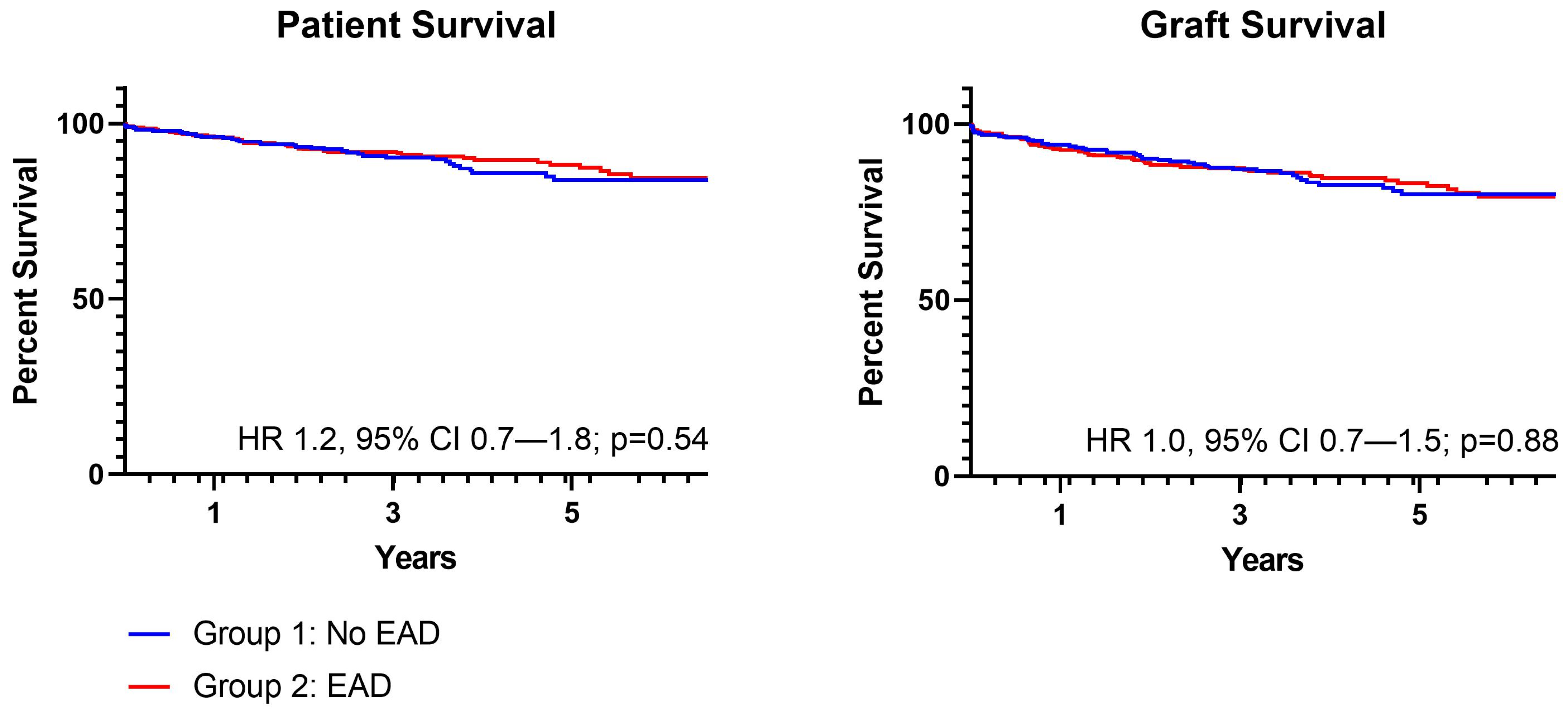
3. Results
Graft Type Subgroup Analysis
4. Discussion
5. Conclusions
Author Contributions
Funding
Institutional Review Board Statement
Informed Consent Statement
Data Availability Statement
Conflicts of Interest
References
- Jadlowiec, C.C.; Taner, T. Liver transplantation: Current status and challenges. World J. Gastroenterol. 2016, 22, 4438–4445. [Google Scholar] [CrossRef] [PubMed]
- Busuttil, R.W.; Tanaka, K. The utility of marginal donors in liver transplantation. Liver Transplant. 2003, 9, 651–663. [Google Scholar] [CrossRef] [PubMed] [Green Version]
- Feng, S.; Goodrich, N.P.; Bragg-Gresham, J.L.; Dykstra, D.M.; Punch, J.D.; DebRoy, M.A.; Greenstein, S.M.; Merion, R.M. Characteristics associated with liver graft failure: The concept of a donor risk index. Am. J. Transplant. 2006, 6, 783–790, Erratum in Am. J. Transplant. 2018, 18, 3085. [Google Scholar] [CrossRef] [PubMed] [Green Version]
- Jadlowiec, C.C.; Macdonough, E.; Pont, K.; Valenti, K.; Lizaola-Mayo, B.; Brooks, A.; Das, D.; Heilman, R.; Mathur, A.K.; Hewitt, W.; et al. Donation After Circulatory Death Transplant Outcomes Utilizing Livers Recovered by Local Surgeons. Liver Transplant. 2022, 25. epub ahead of print. [Google Scholar] [CrossRef]
- Nunez-Nateras, R.; Reddy, K.S.; Aqel, B.A.; Heilman, R.; Morgan, P.; Mathur, A.K.; Hewitt, W.; Heimbach, J.; Rosen, C.; Moss, A.A.; et al. Simultaneous liver-kidney transplantation from donation after cardiac death donors: An updated perspective. Am. J. Transplant. 2020, 20, 3582–3589. [Google Scholar] [CrossRef]
- Jackson, K.R.; Bowring, M.G.; Holscher, C.; Haugen, C.E.; Long, J.J.; Liyanage, L.; Massie, A.B.; Ottmann, S.; Philosophe, B.; Cameron, A.M.; et al. Outcomes After Declining a Steatotic Donor Liver for Liver Transplant Candidates in the United States. Transplantation 2020, 104, 1612–1618. [Google Scholar] [CrossRef]
- Lai, J.C.; Roberts, J.P.; Vittinghoff, E.; Terrault, N.A.; Feng, S. Patient, center and geographic characteristics of nationally placed livers. Am. J. Transplant. 2012, 12, 947–953. [Google Scholar] [CrossRef]
- Foley, D.P.; Fernandez, L.A.; Leverson, G.; Chin, L.T.; Krieger, N.; Cooper, J.T.; Shames, B.D.; Becker, Y.T.; Odorico, J.S.; Knechtle, S.J.; et al. Donation after cardiac death: The University of Wisconsin experience with liver transplantation. Ann. Surg. 2005, 242, 724–731. [Google Scholar] [CrossRef]
- Abt, P.L.; Desai, N.M.; Crawford, M.D.; Forman, L.M.; Markmann, J.W.; Olthoff, K.M.; Markmann, J.F. Survival following liver transplantation from non-heart-beating donors. Ann. Surg. 2004, 239, 87–92. [Google Scholar] [CrossRef]
- Croome, K.P.; David, D.L.; Croome, S.; Chadha, R.; Livingston, D.; Abader, P.; Keaveny, A.P.; Taner, C.B. The impact of postreperfusion syndrome during liver transplantation using livers with significant macrosteatosis. Am. J. Transplant. 2019, 19, 2550–2559. [Google Scholar] [CrossRef]
- Olthoff, K.M.; Kulik, L.; Samstein, B.; Kaminski, M.; Abecassis, M.; Emond, J.; Shaked, A.; Christie, J.D. Validation of a current definition of early allograft dysfunction in liver transplant recipients and analysis of risk factors. Liver Transplant. 2010, 16, 943–949. [Google Scholar] [CrossRef] [PubMed]
- Lee, D.D.; Croome, K.P.; Shalev, J.A.; Musto, K.R.; Sharma, M.; Keaveny, A.P.; Taner, C.B. Early allograft dysfunction after liver transplantation: An intermediate outcome measure for targeted improvements. Ann. Hepatol. 2016, 15, 53–60. [Google Scholar] [CrossRef] [PubMed]
- Wadei, H.M.; Lee, D.D.; Croome, K.P.; Mai, L.; Leonard, D.; Mai, M.L.; Taner, C.B.; Keaveny, A.P. Early Allograft Dysfunction Is Associated with Higher Risk of Renal Nonrecovery After Liver Transplantation. Transplant. Direct. 2018, 4, e352. [Google Scholar] [CrossRef] [PubMed]
- Hudcova, J.; Scopa, C.; Rashid, J.; Waqas, A.; Ruthazer, R.; Schumann, R. Effect of early allograft dysfunction on outcomes following liver transplantation. Clin. Transplant. 2017, 31, e12887. [Google Scholar] [CrossRef]
- Croome, K.P.; Hernandez-Alejandro, R.; Chandok, N. Early allograft dysfunction is associated with excess resource utilization after liver transplantation. Transplant. Proc. 2013, 45, 259–264. [Google Scholar] [CrossRef]
- Lee, D.D.; Singh, A.; Burns, J.M.; Perry, D.K.; Nguyen, J.H.; Taner, C.B. Early allograft dysfunction in liver transplantation with donation after cardiac death donors results in inferior survival. Liver Transplant. 2014, 20, 1447–1453. [Google Scholar] [CrossRef] [PubMed]
- Croome, K.P.; Wall, W.; Quan, D.; Vangala, S.; McAlister, V.; Marotta, P.; Hernandez-Alejandro, R. Evaluation of the updated definition of early allograft dysfunction in donation after brain death and donation after cardiac death liver allografts. Hepatobiliary Pancreat Dis. Int. 2012, 11, 372–376. [Google Scholar] [CrossRef]
- Croome, K.P.; Lee, D.D.; Keaveny, A.P.; Taner, C.B. Improving National Results in Liver Transplantation Using Grafts From Donation After Cardiac Death Donors. Transplantation 2016, 100, 2640–2647. [Google Scholar] [CrossRef]
- Croome, K.P.; Mao, S.; Yang, L.; Pungpapong, S.; Wadei, H.M.; Taner, C.B. Improved National Results With Simultaneous Liver-Kidney Transplantation Using Donation After Circulatory Death Donors. Liver Transplant. 2020, 26, 397–407. [Google Scholar] [CrossRef]
- Jadlowiec, C.; Smith, M.; Neville, M.; Mao, S.; Abdelwahab, D.; Reddy, K.; Moss, A.; Aqel, B.; Taner, T. Acute Kidney Injury Patterns Following Transplantation of Steatotic Liver Allografts. J. Clin. Med. 2020, 9, 954. [Google Scholar] [CrossRef] [Green Version]
- Ito, T.; Naini, B.V.; Markovic, D.; Aziz, A.; Younan, S.; Lu, M.; Hirao, H.; Kadono, K.; Kojima, H.; DiNorcia, J., 3rd; et al. Ischemia-reperfusion injury and its relationship with early allograft dysfunction in liver transplant patients. Am. J. Transplant. 2021, 21, 614–625. [Google Scholar] [CrossRef] [PubMed]
- Wang, K.; Lu, D.; Liu, Y.; Li, W.; Zhuang, L.; Ma, Z.; Xie, Q.; Pan, B.; Wu, Y.; Chen, J.; et al. Severity of early allograft dysfunction following donation after circulatory death liver transplantation: A multicentre study. Hepatobiliary Surg. Nutr. 2021, 10, 9–19. [Google Scholar] [CrossRef] [PubMed]
- Mazilescu, L.I.; Kotha, S.; Ghanekar, A.; Lilly, L.; Reichman, T.W.; Galvin, Z.; Cattral, M.S.; Bhat, M.; McGilvray, I.D.; Sapisochin, G.; et al. Early Allograft Dysfunction after Liver Transplantation with Donation after Circulatory Death and Brain Death Grafts: Does the Donor Type Matter? Transplant. Direct. 2021, 7, e727. [Google Scholar] [CrossRef] [PubMed]
- Mihaylov, P.; Mangus, R.; Ekser, B.; Cabrales, A.; Timsina, L.; Fridell, J.; Lacerda, M.; Ghabril, M.; Nephew, L.; Chalasani, N.; et al. Expanding the donor pool with the use of extended criteria donation after circulatory death livers. Liver Transplant. 2019, 25, 1198–1208. [Google Scholar] [CrossRef]
- Chan, E.; Logan, A.J.; Sneddon, J.M.; Singh, N.; Brock, G.N.; Washburn, W.K.; Schenk, A.D. Dynamic impact of liver allocation policy change on donor utilization. Am. J. Transplant. 2022. epub ahead of print. [Google Scholar] [CrossRef]
- Fodor, M.; Woerdehoff, A.; Peter, W.; Esser, H.; Oberhuber, R.; Margreiter, C.; Maglione, M.; Cardini, B.; Resch, T.; Weissenbacher, A.; et al. Reassessment of Relevance and Predictive Value of Parameters Indicating Early Graft Dysfunction in Liver Transplantation: AST Is a Weak, but Bilirubin and INR Strong Predictors of Mortality. Front. Surg. 2021, 8, 693288. [Google Scholar] [CrossRef]


| Group 1: No EAD (n = 290) | Group 2: EAD (n = 321) | p Value | |
|---|---|---|---|
| Recipient | |||
| Age (years) | 56.4 ± 10.2 (58.0) | 57.2 ± 10.5 (59.0) | 0.32 |
| Female | 108 (37.2%) | 102 (31.8%) | 0.16 |
| Hispanic | 54 (18.6%) | 43 (13.4%) | 0.08 |
Race
| 222 (76.6%) 0 (0.0%) 14 (4.8%) | 258 (80.4%) 7 (2.2%) 13 (4.0%) | 0.04 |
| MELD | 23 (17, 30) | 21 (15, 26) | 0.0002 |
Diagnosis
| 126 (43.4%) 67 (23.1%) 40 (13.8%) 22 (7.6%) 35 (12.1%) | 148 (46.1%) 59 (18.4%) 68 (21.2%) 29 (9.0%) 17 (5.3%) | 0.004 |
| PVT | 43 (14.8%) | 61 (19.0%) | 0.17 |
| TIPS | 27 (9.3%) | 41 (12.8%) | 0.17 |
| Re-Transplant | 24 (8.3%) | 9 (2.8%) | 0.003 |
| Donor | |||
| Age (years) | 48.0 ± 18.1 (50.0) | 48.5 ± 13.0 (52.0) | 0.65 |
| DRI | 1.6 ± 0.5 (1.6) | 1.9 ± 0.5 (1.9) | <0.0001 |
| BMI (kg/m2) | 28.4 ± 7.4 (26.8) | 31.0 ± 8.3 (30.0) | <0.0001 |
Sharing
| 103 (35.5%) 137 (47.2%) 50 (17.2%) | 65 (20.2%) 198 (61.7%) 58 (18.1%) | <0.0001 |
| DCD donor | 40 (13.8%) | 154 (48.0%) | <0.0001 |
| DCD dWIT (minutes) | 23.0 (16.0, 26.0) | 21.0 (18.0, 25.0) | 0.53 |
| CIT (hours) | 5.9 ± 1.9 (5.5) | 6.4 ± 1.9 (6.0) | 0.001 |
| Group 1: No EAD (n = 290) | Group 2: EAD (n = 321) | p Value | |
|---|---|---|---|
| ICU LOS (days) | 1.0 | 2.0 | 0.60 |
| Hospital LOS (days) | 5.0 | 6.0 | 0.24 |
EAD
| 286 (89.1%) 28 (8.7%) 7 (2.2%) | --- | |
| --- | |||
| Peak AST (U/L) | 913 (554, 7700) | 4010 (2853, 7700) | <0.0001 |
| AKI | 117 (40.3) | 140 (43.6%) | 0.41 |
| PNF | 1 (0.3%) | 4 (1.3%) | 0.22 |
Early Allograft Dysfunction
| 15 (4.7%) 21 (6.5%) 321 (100.0%) | --- | |
| --- |
| Group 1: No EAD | Group 2: EAD | p Value | |
|---|---|---|---|
1 Week Post-LT
| 1.5 (1.0, 2.5) 115.0 (77.8, 190.3) 41.0 (29.0, 62.3) 140.5 (94.0, 211.3) | 1.9 (1.1, 4.5) 229.0 (155.0, 338.0) 55.0 (38.0, 81.0) 167.0 (117.0, 257.0) | <0.0001 <0.0001 0.03 0.01 |
6 Weeks Post-LT
| 0.6 (0.4, 0.8) 19.0 (13.0, 31.5) 21.0 (16.0, 27.0) 100.0 (79.0, 143.0) | 0.6 (0.4, 1.1) 26.0 (16.0, 45.0) 22.0 (17.0, 32.0) 125.0 (87.0, 198.0) | 0.004 0.05 0.001 <0.0001 |
1 Year Post-LT
| 0.6 (0.3, 0.7) 23.0 (17.0, 38.0) 27.0 (20.0, 34.0) 114.0 (85.0, 164.8) | 0.5 (0.3, 0.7) 23.0 (16.0, 38.0) 25.0 (21.0, 33.0) 113.0 (88.0, 156.0) | 0.29 0.42 0.18 0.08 |
| Group A: EAD DCD (n = 154) | Group B: EAD DBD (n = 167) | Group C: No EAD DCD (n = 40) | Group D: No EAD DBD (n = 250) | p Value | |
|---|---|---|---|---|---|
| Recipient | |||||
| Age (years) | 58.5 ± 9.1 | 56.0 ± 11.5 | 58.1 ± 9.4 | 56.1 ± 10.4 | 0.08 |
| MELD (median) | 20.5 ± 6.0 (22.0) | 21.0 ± 9.0 (21.0) | 21.5 ± 6.1 (23.0) | 23.6 ± 9.6 (23.0) | 0.001 |
Diagnosis
| 73 (47.4%) 41 (26.6%) 29 (18.8%) 10 (6.5%) 1 (0.6%) | 75 (44.9%) 18 (10.8%) 39 (23.4%) 19 (11.4%) 16 (9.6%) | 17 (42.5%) 12 (30.0%) 7 (17.5%) 1 (2.5%) 3 (7.5%) | 110 (44.0%) 55 (22.0%) 33 (13.2%) 21 (8.4%) 31 (12.4%) | 0.001 |
| Re-Transplant | --- | 9 (5.4%) | --- | 24 (9.6%) | 0.12 |
| Donor | |||||
| Age (years) | 49.2 ± 11.4 (51.5) | 47.8 ± 18.9 | 49.6 ± 15.2 (53.0) | 47.6 ± 15.2 (50.0) | 0.62 |
| DRI | 2.3 ± 0.4 (2.3) | 1.6 ± 0.4 (1.5) | 2.3 ± 0.3 (2.3) | 1.5 ± 0.4 (1.5) | <0.0001 |
| BMI (kg/m2) | 20.5 ± 6.0 (21.5) | 32.2 ± 8.5 (31.5) | 26.3 ± 5.2 (25.9) | 28.7 ± 7.6 (27.2) | <0.0001 |
Sharing
| 27 (17.5%) 106 (68.8%) 21 (13.6%) | 38 (22.7%) 92 (55.1%) 37 (22.2%) | 10 (25.0%) 23 (57.5%) 7 (17.5%) | 93 (37.2%) 114 (45.6%) 43 (17.2%) | <0.0001 |
| CIT (hours) | 5.6 ± 1.2 (5.4) | 7.2 ± 2.1 (6.9) | 5.5 ± 1.0 (5.4) | 6.0 ± 2.0 (5.5) | <0.0001 |
| Post-LT Outcomes | |||||
| ICU LOS (days) | 2.0 (1.0, 2.0) | 1.0 (1.0, 2.0) | 2.0 (1.0, 3.0) | 1.0 (1.0, 2.0) | 0.37 |
| Hospital LOS (days) | 6.0 (5.0, 8.0) | 6.0 (5.0, 9.0) | 5.0 (4.0, 8.0) | 5.0 (4.0, 8.0) | 0.12 |
| PNF | 0 (0.0%) | 4 (2.4%) | 1 (2.5%) | 0 (0.0%) | 0.02 |
| PNF Case | MELD | Donor Age (Years) | BMI (kg/m2) | Allocation | DRI | Post-Cross Clamp Offer | CIT (Hours) | Liver Allograft Biopsy |
|---|---|---|---|---|---|---|---|---|
| #1 | 14 | 49 | 42.1 | National | 1.4 | Yes | 8.1 | Steatosis, 20–30% |
| #2 | 32 | 57 | 26.2 | Local | 1.4 | No | 4.1 | Severe arteriolar hyaline |
| #3 | 8 | 63 | 24.2 | National | 1.9 | Yes | 7.7 | >70% hepatocellular necrosis |
| #4 | 39 | 36 | 36.1 | Regional | 1.2 | No | 6.6 | Normal biopsy |
Publisher’s Note: MDPI stays neutral with regard to jurisdictional claims in published maps and institutional affiliations. |
© 2022 by the authors. Licensee MDPI, Basel, Switzerland. This article is an open access article distributed under the terms and conditions of the Creative Commons Attribution (CC BY) license (https://creativecommons.org/licenses/by/4.0/).
Share and Cite
Ohara, S.; Macdonough, E.; Egbert, L.; Brooks, A.; Lizaola-Mayo, B.; Mathur, A.K.; Aqel, B.; Reddy, K.S.; Jadlowiec, C.C. Decreasing Significance of Early Allograft Dysfunction with Rising Use of Nonconventional Donors. Medicina 2022, 58, 821. https://0-doi-org.brum.beds.ac.uk/10.3390/medicina58060821
Ohara S, Macdonough E, Egbert L, Brooks A, Lizaola-Mayo B, Mathur AK, Aqel B, Reddy KS, Jadlowiec CC. Decreasing Significance of Early Allograft Dysfunction with Rising Use of Nonconventional Donors. Medicina. 2022; 58(6):821. https://0-doi-org.brum.beds.ac.uk/10.3390/medicina58060821
Chicago/Turabian StyleOhara, Stephanie, Elizabeth Macdonough, Lena Egbert, Abigail Brooks, Blanca Lizaola-Mayo, Amit K. Mathur, Bashar Aqel, Kunam S. Reddy, and Caroline C. Jadlowiec. 2022. "Decreasing Significance of Early Allograft Dysfunction with Rising Use of Nonconventional Donors" Medicina 58, no. 6: 821. https://0-doi-org.brum.beds.ac.uk/10.3390/medicina58060821