Review

Parameters Influencing Zinc in Experimental Systems in Vivo and in Vitro

1
Institute of Immunology, Faculty of Medicine, RWTH Aachen University Hospital, Pauwelsstr. 30, 52074 Aachen, Germany
2
Department of Food Chemistry and Toxicology, Berlin Institute of Technology, Gustav-Meyer-Allee 25, 13355 Berlin, Germany
*
Author to whom correspondence should be addressed.
Submission received: 31 January 2016 / Revised: 7 March 2016 / Accepted: 17 March 2016 / Published: 21 March 2016
(This article belongs to the Special Issue Metallomics)

Abstract

:
In recent years, the role of zinc in biological systems has been a subject of intense research. Despite wide increase in our knowledge and understanding of zinc homeostasis, numerous questions remain to be answered, encouraging further research. In particular, the quantification of intracellular zinc ions and fluctuation, as well as the function of zinc in signaling processes are being intensely investigated. The determination of free intracellular zinc ions is difficult and error-prone, as concentrations are extremely low (in the pico- to nanomolar range), but techniques exist involving fluorescent probes and sensors. In spite of zinc deficiency being accepted as a global problem, causing death and disease worldwide, to date there are no markers to reliably assess a person’s zinc status. This review summarizes the difficulties and major pitfalls when working with zinc in in vitro and in vivo research. Additionally, it specifies important aspects for zinc substitution and supplementation, including the bioavailability of zinc and its intestinal absorption. In particular, it is intended to help researchers with yet minor experience working with zinc efficiently set up experiments and avoid commonly occurring mistakes, starting with the choice and preparation of reagents and instrumentation, and concluding with possibilities for measuring the status of zinc in humans.

Graphical Abstract

1. Introduction

Zinc is an essential trace element with an immense importance for human health. The human body contains 2–4 g zinc in total, and the major part is found in the musculoskeletal system. There is no significant difference between the concentration of zinc in serum and plasma [1]. The plasma or serum concentration of zinc is 12–16 µM, representing a small pool of whole-body zinc at only 0.1%, mainly bound to albumin [2]. Nearly all intracellular zinc, some hundred micromolar in total [3], is bound to cytosolic zinc-binding proteins, resulting in an estimated picomolar concentration of loosely bound, but exchangeable zinc ions [3,4,5]. This pool, commonly called free zinc or free Zn2+, must be strictly controlled, as already nanomolar concentrations cause cell death in cell cultures [3]. Zinc has particular significance in the immune, nervous and endocrine systems; a role in the cardiovascular system is suggested [6].
Even though it is a transition metal, zinc ions are only present at the oxidation state +2. Zinc may have coordination numbers from two to eight, enabling it to bind a wide variety of ligands [7], to serve as a cofactor for about 300 enzymes [2,8], and to stabilize protein structures [9]. Furthermore, zinc ions are direct modulators of cell functions [9] as they participate in diverse signaling processes [10]. They are mobilized from exocytotic vesicles, intracellular organelles or directly from zinc proteins [11]. Zinc ions are indispensable for gene expression, enzymatic activity, and cell signaling [4], and moreover for proliferation, DNA and RNA synthesis, and apoptosis [8]. Accordingly, zinc deficiency, both acquired and inherited, leads to immunodeficiency, dermatitis, gastrointestinal problems, reduced wound healing, neurological and psychiatric disorders, growth impairment [12], and other pathological conditions. Additionally, tumorigenesis might be indirectly affected [13].
It might be obvious that zinc, its homeostasis and changes are decisive for the understanding of human health and pathology, and worth being studied in detail. While we know the problems relating to zinc deficiency, we still lack an accepted and widely applicable marker for human zinc status. The zinc status is integral to zinc requirements in the body, thus it reflects a sufficient zinc saturation for all functions and not just the normal value of serum zinc. Due to the lack of a reliable marker, so far zinc status is just a technical term to indicate the necessity of such a marker. Serum zinc does not allow for a reliable statement about a possible zinc deficiency, as will be discussed later, though it is often used [14]. In the absence of better options, serum zinc is the best marker for the zinc status to date [15]. The flow cytometric measurement of free Zn2+ in peripheral blood mononuclear cells (PBMC) seems to be promising, but the method requires special equipment that might not be available for field studies [16].
Scientists should be aware of some difficulties when working with zinc. The majority of experimental steps in vitro as well as in vivo is prone to errors including the selection and preparation of cell culture media and chemicals, the addition of supplements or chelators, and the measurement of zinc. The following review will list some of the avoidable mistakes, but also some problems, which are yet unsolved.

2. Considerations When Working with Zinc

The zinc compounds usually used show different solubility in water. Whereas zinc chloride (ZnCl2) and zinc sulfate (ZnSO4) are soluble in water (ZnCl2: 432 g in 100 mL water [17] and ZnSO4: 57 g in 100 mL water, both at 25 °C [18]), zinc oxide (ZnO) is not soluble in water but in acids and alkalis [19]. If a stock solution of ZnSO4 is prepared accidentally with buffers containing relevant amounts of phosphate, e.g., phosphate buffered saline (PBS) or cell culture media such as Roswell Park Memorial Institute medium (RPMI) 1640, zinc phosphate precipitates because it is hardly soluble in water (Zn3(PO4)2: 0.27 g in 100 mL water at 20 °C [20]). If such a stock solution is sterile filtered to be used subsequently in an experiment, the precipitates become removed and the resulting zinc content might be overestimated. Figure 1 illustrates the low recovery of sterile filtered zinc orotate and ZnO from a 100 mM solution prepared in water. A reduced recovery of ZnSO4, if solved in phosphate buffer is depicted as well. A lower pH of the solvent would improve the solubility of zinc salts. However, sufficiently low pH-values are not compatible with the commonly required conditions for cultured cells. Figure 2 clearly shows the effect of sterile filtration of cell culture medium after the addition of various zinc concentrations.
Great attention should always be paid to the exact chemical composition of a zinc preparation because the relative amount of zinc may differ. For example, the relative content of zinc in ZnSO4, ZnSO4·H2O and ZnSO4·7H2O ranges from 40.5% to 22.7%, which has to be taken into account in zinc substitution or supplementation [21]. Metal ion chelators can reduce the availability of metals in media depending on their affinity to a particular metal, thus they are frequently used additives in experiments. Common chelators are CHELEX resin, TPEN (N,N,N′,N′-tetrakis (2-pyridylmethyl) ethylenediamine), DTPA (diethylenetriamine pentaacetic acid), and also others such as BAPTA (1,2-bis(2-aminophenoxy)ethane-N,N,N′,N′-tetraacetic acid), EDTA (ethylenediaminetetraacetic acid) or EGTA (ethylene glycol-bis(2-aminoethylether)-N,N,N′,N′-tetraacetic acid). Even if some of these chelators show a high selectivity for one metal, e.g., TPEN for zinc, they do not always only bind their target metal, but others as well [22], making it difficult to assign an observed effect to altered zinc conditions. Therefore, a control with the same saturated chelator is essential. Other aspects to consider are the direct effects of some chelators. The direct effect of TPEN on enzymes and other proteins is not yet clear because, for example, the zinc saturated complex of TPEN is still able to induce apoptosis in cells and this effect is therefore independent of zinc chelation [23]. The ion exchange resin CHELEX seems to induce cytokine production and to activate monocytes independently from zinc through an unknown mechanism. This possibly involves a direct activation of surface receptors or an observed increase of molybdenum in a medium treated with CHELEX resin. Moreover, there exist contradictory data in the literature regarding the rate of depletion of different trace metals by CHELEX resin [24]. All substances added to a zinc preparation or medium in an experiment with the intention to measure zinc or its effects must be selected carefully to prevent an unwanted chelation or addition of zinc and thereby an alteration of experimental outcomes.
Depending on their structure, chelators work at different cellular sites, which needs to be considered: TPEN, for example, chelates intracellular zinc, whereas DTPA does not. Nakatani et al. (2000) were able to trigger apoptosis in cultured rat hepatocytes through the chelation of intracellular zinc with membrane-permeable TPEN, whereas DTPA, which is not membrane permeable, could not induce apoptosis, even if it was used in higher concentrations than TPEN [25]. However, the mobility of zinc ions across the cell membrane relativizes the classification into intracellular and extracellular chelators, because the depletion at one side of the membrane might cause a redistribution of ions as shown for the “extracellular” chelator CaEDTA in vivo and in vitro in neurons [26]. Furthermore, TPEN is hardly soluble in water, requiring attention when preparing stock solutions. Once it is added to cell cultures, it rapidly loses its activity because of biological degradation, shown by the increasing concentration of intracellular zinc after prior depletion (Figure 3). An upregulation of zinc importers due to prior zinc depletion might be an explanation for higher zinc concentrations in TPEN-treated cells after an incubation time exceeding 24 h compared to the control cells [27].
TPEN is toxic for cells, but the tolerable concentration depends on the cell type as shown in Figure 4. Therefore, a viability test should always be performed to define the adequate concentration of TPEN.

3. Zinc in Cell Culture Experiments

Many experiments are executed in vitro as it is impossible to realize them in vivo, or as part of a pilot study. Though one seeks to create comparable conditions to tissue, some variables usually differ in cell culture. Cultured cells do not have the same physiological barriers like the original cells in their primary environment. They are directly exposed to the conditions of the surrounding medium, showing a different pH-value and metal buffering capacities [11]. Though it would be important to know, the metal buffering capacity is usually disregarded. Proteins such as albumin, as well as single amino acids, buffer zinc ions effectively in human plasma, resulting in nanomolar concentrations of free Zn2+ [3]. Albumin as the major binding protein ensures 60% of the total zinc binding [28], and is physiologically available in substantial molar excess to Zn2+ [29]. Also, in media the concentration of proteins regulates the amount of free Zn2+ and its biological activity. For example, the cytokine secretion of monocytes stimulated with zinc increased in medium with a minor protein content, albeit not in protein-free medium as transferrin seems to be necessary as a transport protein [30].
Most of the commonly used media (RPMI1640, Iscove’s Modified Dulbecco’s medium (IMDM)) do not include zinc [11], although there exist exceptions such as Neurobasal media and Ham’s F-12 medium. The essential zinc as well as the buffering proteins are provided by added fetal calf serum (FCS), typically at around 10% (range 5%–20%), which thereby represents only one-tenth of the physiological serum concentration (see Figure 5). In addition, FCS is a natural product; its composition differs, which makes an exact prediction of effects impossible. An insufficient buffering might lead to an overestimation of the effects of free Zn2+ in cell cultures. Other metal ions like Cu2+, Pb2+, Cd2+, Hg2+, Ni2+, and Ag+ are also influenced by the protein concentration in culture medium [3]. Different lots of FCS contain different zinc concentrations (Figure 6), therefore resulting in the necessity to adapt experimental systems if the lot of FCS is changed. Apart from this, differences in the serum zinc concentration of different species can be observed (see Figure 7).
Buffers containing thiol-based reducing agents or EDTA might also alter the intended zinc concentration, or even totally eliminate zinc [11].
Though zinc itself is redox inert, it changes the thiol redox status of the protein and interacts with sulfur in protein cysteine clusters. Zinc can be released from its binding sites, when zinc proteins are exposed to thiol-oxidants and other molecules such as NO, H2O2, oxidized glutathione or some metals [31]. Aldehydes are able to release zinc from zinc-binding proteins like metallothionein and induce zinc release from vesicles, thereby enhancing the concentration of intracellular unbound free Zn2+. They are formed when cells are exposed to oxidative stress, during lipid peroxidation and hyperglycemia-induced glycation [32]. If such a substance is already contained in a medium or supplement, or unknowingly added with a stabilized enzyme or produced in a cell culture, signal processes might vary, unexpected effects might result from the altered speciation of zinc ions, and measurements might become falsified. An agent that reduces protein disulfides is dithiothreitol (DTT), a strong chelator of zinc, which is used as a thiol group protector in many commercial protein preparations [30,31]. Therefore, alternative reagents like TCEP hydrochloride (tris(2-carboxyethyl)phosphine hydrochloride) should be used to avoid zinc chelating artifacts. Aldehydes, which are also used for fixation of tissues for microscopic examinations, might influence the zinc distribution as well and could cause wrong results. Commonly used incubators provide hyperoxic surroundings compared to the conditions in tissues or blood. They might alter the environmental redox status of surrounding elements, and therefore indirectly change the behavior of zinc ions.
Diverse enzymes are inhibited by zinc in physiological concentrations [33,34]. Thionein, an important endogenous chelator with the capacity to bind seven zinc ions, is capable to remove zinc from these inhibitory sites, where it is bound with stability constants in the nanomolar range. Though thionein is very potent, it is selective as it neither takes zinc from the catalytically active site of zinc metalloenzymes, where the stability constants are within the picomolar range, nor is it able to remove structural, fully coordinated zinc atoms [34]. When enzyme activities are investigated, the presence or absence of free Zn2+ as well as of thionein and other factors like the metal buffering capacity of the surrounding medium should be considered. Even the lowest fluctuations of the zinc ion concentration might influence the results, albeit not completely predictably thus far.

4. Measurement of Zinc

4.1. Measurement of Total Zinc

Measurement of total zinc in organic samples started with chemical methods, which employed colored zinc-chelator complexes for a quantification of zinc. However, they were too insensitive for current questions with a detection limit of 1 µg/g [7]. A more sensitive examination of the total zinc content was subsequently possible via atomic emission, absorption or fluorescence spectroscopy, but also with other techniques such as inductively coupled plasma mass spectrometry [7,11,34] and laser ablation-inductively coupled plasma mass spectrometry. The latter even allow a two-dimensional mapping of the distribution of zinc in organic samples [35].

4.2. Measurement of the Zinc Status

Currently, there is no consensus regarding a sensitive and specific biochemical indicator of the zinc status in humans. Therefore, serum zinc seems to be the best indicator of the zinc status in a population [15,36]. The measurement of different proteins in peripheral blood cells to determine the long-term zinc status is promising, but still difficult to implement on a large scale in a clinical and especially field setting in particular [37]. Plasma or serum zinc levels can be determined by atomic absorption spectroscopy. They show acute responses to metabolic events: Hemolysis distorts the measurements as the intracellular zinc exceeds the plasma or serum zinc content [38], an acute febrile infection in contrast lowers the blood and serum zinc concentration, but the total body amount does not necessarily decrease, and the zinc concentration normalizes after recovering from the infection [39]. Therefore, when serum zinc is measured, it should be normalized in relation to albumin levels and C-reactive protein (CRP) content.

4.3. Measurement of Free Zn2+

Since the identification of the immense importance of intracellular free Zn2+ as a signaling molecule, efforts have increased to visualize free Zn2+ and to measure the zinc concentrations and fluxes within cells. Metal-responsive fluorophores are used to perform fluorescence microscopy or spectroscopy. While probes are low-molecular-weight chelators that change their fluorescence intensity when binding zinc ions, protein sensors are metal binding or chelating proteins, which are fused to a fluorescent protein or low molecular weight ligands [11]. The concentration of intracellular free Zn2+ can be determined by ion-induced changes in dye signal [40] using the formula:
[ Zn 2 + ] = K D ( F F min F max F ) [ 41 ]
The autofluorescence of the dye (Fmin) and the maximum fluorescence of the zinc-saturated dye (Fmax) are needed for the quantification of free Zn2+. TPEN is usually used to generate the depleted sample for the determination of the autofluorescence, and zinc is used in combination with pyrithione to measure the maximum fluorescence [16]. Pyrithione is an ionophore that transports zinc, copper and lead across biological membranes [42]. Given in combination with zinc, it is able to induce apoptosis by facilitated intracellular zinc uptake, resulting in toxic intracellular zinc concentrations [43]. The optimal pyrithione concentration and incubation time should be determined for each cell type. In particular, zinc pyrithione changes the appearance of cells as demonstrated in Figure 8. This requires careful gating when analyzing data obtained by flow cytometry, because different gate settings change the values of the estimated intracellular zinc concentration (e.g., in Jurkat cells 0.223 nM with a smaller gate (Figure 8A), compared to 0.280 nM with a larger gate (Figure 8B), including the entire population of cells treated with ZnSO4 and pyrithione).
The bioimaging of free Zn2+ generates high demands on the design of fluorophores, which should be selective to zinc even if there is the possibility of other abundant divalent ions existing. Once activated, they should emit brightly to minimize the needed amount of the dye, they should respond fast and reversely, and the wavelength for excitation and emission should be in the visible range to prevent cell damage [44,45]. Some probes like Zinquin ethyl ester and TSQ (6-methoxy-(8-p-toluenesulfonamido)quinoline) need ultraviolet light for excitation, which harms cells [11]. Moreover, the dyes must be soluble in water, stable and nontoxic [44,45]. Some of the demands are difficult to fulfill and the technology using fluorescent metal-responsive dyes still has limitations which must be considered [11]. Cell-permeable probes like FluoZin-3 AM accumulate within cells because once entered they are hydrolyzed by intracellular esterases, which prevents crossing the cellular membrane again. Consequently, influencing the expression of cellular esterases will result in changes in fluorescent dye accumulation. These probes therefore reach higher intracellular concentrations than one would expect, if an equilibrium as during regular diffusion is assumed. The amount of zinc required for saturation of the probe to determinate the maximum fluorescence is underestimated by this effect [5]. The dyes also locate in different concentrations depending on the cell type, and can even locate to specific compartments [40]. Because probes act as chelators themselves, they alter the zinc buffering capacity. Thus, the higher the utilized concentration of a probe, the lower appears the zinc concentration, making quantitative zinc measurements complicated, and extrapolation is needed [5,11,40]. The same problem should occur when ratiometric probes are used [40]. Furthermore, the probes compete for zinc with cellular proteins, depending on the respective dissociation constant (Table 1). When fluorescent dyes are commercially generated, the binding properties are usually determined and specified in buffered solutions (Table 2). In an intracellular environment with variable pH and components like membranes or proteins, these properties might change, and with them the fluorescence signal [46]. Though the sensitivity of probes is high, the specificity and selectivity must be considered. FluoZin-3 AM, Newport Green DCF and Zinquin ethyl ester respond not only to free Zn2+, but also and even more strongly to zinc bound to larger biomolecules [46]. Zinc ions interfere with calcium probes, and other divalent ions bind to zinc probes might in the same manner, depending on the dissociation constants. This effect complicates the detection or even induces false-positive fluorescence and compromises the quantification of free Zn2+ [11,47]. Fluorophores reduce the concentration of unbound ions by buffering. Therefore, the dye might influence signaling processes, and even harm cells when applied in higher concentrations.

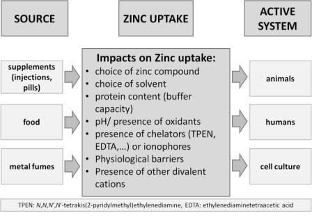
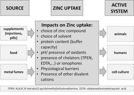
5. Influences on Zinc Bioavailability and Functions in Experimental Systems in Vivo

Various factors must be considered when in vivo-experiments are planned. The careful choice and preparation of equipment is indispensable, as well as the thorough selection and composition of reagents, as has been described earlier for in vitro experiments. Frequent errors are caused by using blood tubes with chelating substances such as EDTA. To be able to define exact zinc uptake, zinc sources other than the intended must be avoided, including zinc contained in cages, water flasks and in material for wound closure in animal studies. Dietary questionnaires are a useful instrument in human studies, whereas depending on the study’s composition, the questionnaire’s usability depends on the compliance of the proband.
To evaluate possible effects of zinc, it is necessary to know the amount of absorbed and required zinc. Hereby, the intestinal zinc resorption plays a decisive role. However, the affecting factors are more difficult to estimate than the amount of dietary zinc. The bioavailability of both nutritional zinc and different zinc compounds used as supplements is not explored in detail. The utilization of a standard bioavailability factor for calculation would involve the inclusion of all the factors that might change bioavailability during physiological adaptations like growth, pregnancy, lactation or low zinc intake [64]. The requirement for zinc also varies: there is, for example, a physiological peak during the pubertal growth spurt [65]. The most important factor for dietary zinc absorption from protein rich meals seems to be the amount of ingested zinc, because the fractional zinc absorption decreases with increasing amounts of ingested zinc [66]. The zinc absorption is saturable at higher doses, an existing diffusion effect after a meal seems to be negligible [67]. However, the utilization of zinc from composited meals is altered by various substances. Meanwhile, it is commonly known that phytates (e.g., in corn, soy beans, legumes and whole wheat bread) notably reduce the intestinal zinc resorption [67,68], whereupon this effect is just elicited by inositol hexa- and pentaphosphates. Lower phosphates induce no effect, or at least a lower one [67]. Miller et al. developed a mathematical model to estimate zinc absorption including basic characteristics of the intestinal biochemistry [69], and according to this model, the negative effect of phytates explains more than 80% of the variation found when measuring absorption of dietary zinc in adults [70]. However, for children, data on this effect is rare [36]. Miller et al. even showed an independence between zinc absorption and phytate:zinc molar ratio in young children. Therefore, this ratio should not be applied for them without further research [71]. Unlike the phytate:zinc ratio, length and maturity of the intestine are important factors for zinc absorption in children. The zinc absorption adjusted for gut length was comparable with that of adults [72]. Zinc absorption in infants with an immature intestine is less regulated than it is in adults, as it seems to be increased at higher dietary intake in contrast to the negative correlation between zinc intake and fractional zinc absorption in adults. The regulation of zinc homeostasis seems to require developmental maturation at least in cell culture experiments [73].
For lack of systematic studies and reliable markers of zinc status, there is no consensus on the bioavailability of different zinc supplements. At least some organic compounds of zinc, such as zinc aspartate and zinc orotate, seem to have a better bioavailability compared to ZnO and ZnSO4 [74]. However, there is no study comparing those organic compounds, although both are approved supplements. Zinc aspartate is less stable than zinc orotate (log10KZn aspartate = 2.9, log10KZn orotate = 6.42) [75,76], and therefore might provide more absorbable zinc ions. Other chemical factors affecting a substance’s bioavailability in addition to its stability: there are, for example, its solubility in water, the charge density, its reduction potential, the environmental pH and the formation of complexes. Despite its very low solubility at least at neutral pH, ZnO is the most commonly used zinc supplement by the US food industry as it is the cheapest form and does not significantly change the taste or appearance of a meal [77]. Though ZnSO4 is much more soluble than ZnO, the absorption from fortified wheat products was equal in two studies [78,79], which might result from the physiological gastric conditions. The low gastric pH increases the solubility of zinc salts [80] and enhances zinc absorption [81] because it forms hydrates by protonation of the nearly insoluble hydroxides [77]. Hypochlorhydria and achlorhydria, the intake of proton pump inhibitors or antacids, as well as pernicious anemia, increase the gastric pH and accordingly lead to a decreased absorption of zinc [81]. This effect is especially to consider, if ZnO is used as supplement, whereas the more soluble zinc acetate is absorbed more consistently [81].
Some divalent cations like copper, iron, cobalt, manganese and tin also decrease the intestinal zinc absorption in animal models [82], whereas heme iron in physiological doses does not inhibit zinc uptake in humans [83]. High doses of non-heme iron [83], or zinc and non-heme iron together as supplements [67] could notably decrease the absorption of zinc in humans. However, this effect seems to be weaker or absent when both are added in the same amount to food as fortifiers, maybe because of the influence of other components such as proteins [67,77,82,84]. Furthermore, calcium might impair zinc absorption, but in an indirect manner compared to iron, as it forms insoluble complexes in phytate-containing meals with zinc and phytate. Alternatively, calcium might even improve the absorption by complexing with phytate and thereby reducing the amount of chelated and insoluble zinc [67]. The content of some proteins and amino acids, in particular, just like the overall content of proteins, improves the absorption of zinc, whereas some other proteins inhibit it, such as casein, for example [67].
Besides the composition of a meal or supplement, the resorption of zinc and other micro- and macronutrients depends on the gastrointestinal function. Already mentioned is the influence of the gastric acidity. Enteropathies may decrease zinc absorption and therefore elicit or aggravate an existing zinc deficiency. Zinc deficiency itself affects the immune system and worsens, e.g., an environmental enteropathy. High-dose supplemental zinc improves the function of deranged tight junctions in cell culture and animal experiments, and reduces the intestinal permeability in different populations with supposable environmental enteropathy [85]. Environmental enteropathy occurs mostly in developing countries under unhygienic conditions, but there also exist various forms of enteropathies in industrialized countries. The possible effects of environmental and other enteropathies on zinc homeostasis should be considered when studies are performed.
A method for quantifying the amount of absorbed zinc is meant to determine the difference between dietary and fecal content of zinc, but reliable results can only be received under steady state conditions. A more reliable and more complex possibility for quantification of absorbed zinc is to label meals with stable zinc isotopes and to determine the difference between digested and excreted amounts of the isotope by mass spectrometry. The endogenous excretion can be corrected and possible zinc contamination is prevented. Zinc isotopes with low natural abundance can also be measured in fecal samples to determine the amount of non-absorbed zinc using mass spectrometry. However, this method requires special equipment [37].

6. Conclusions

Zinc and its role in health and disease are worthy of extensive study, and the techniques for research continue to increase. However, the measurement of zinc, especially of free Zn2+ and its effects, is difficult and provides sources of errors. Researchers who start to dedicate themselves to this fascinating topic should be very careful and aware when exercising old or establishing new methods, to avoid at least the preventable faults, and possibly to find new solutions for unsolved problems.

Acknowledgments

L.R. is a member of the European COST action Zinc-Net (TD1304).

Author Contributions

J.O. performed the literature search and wrote the manuscript. V.K., I.W. and H.H. performed the experiments and corrected the manuscript. L.R. designed the experiments and corrected the manuscript.

Conflicts of Interest

The authors declare no conflict of interest.

References

  1. Chen, W.J.; Zhao, C.Y.; Zheng, T.L. Comparison of zinc contents in human serum and plasma. Clin. Chim. Acta 1986, 155, 185–187. [Google Scholar] [PubMed]
  2. Rink, L.; Gabriel, P. Zinc and the immune system. Proc. Nutr. Soc. 2000, 59, 541–552. [Google Scholar] [CrossRef] [PubMed]
  3. Haase, H.; Hebel, S.; Engelhardt, G.; Rink, L. The biochemical effects of extracellular Zn2+ and other metal ions are severely affected by their speciation in cell culture media. Metallomics 2015, 7, 102–111. [Google Scholar] [CrossRef] [PubMed]
  4. Colvin, R.A.; Holmes, W.R.; Fontaine, C.P.; Maret, W. Cytosolic zinc buffering and muffling: Their role in intracellular zinc homeostasis. Metallomics 2010, 2, 306–317. [Google Scholar] [CrossRef] [PubMed]
  5. Krezel, A.; Maret, W. Zinc-buffering capacity of a eukaryotic cell at physiological pZn. J. Biol. Inorg. Chem. 2006, 11, 1049–1062. [Google Scholar] [CrossRef] [PubMed]
  6. Lee, S.R.; Noh, S.J.; Pronto, J.R.; Jeong, Y.J.; Kim, H.K.; Song, I.S.; Xu, Z.; Kwon, H.Y.; Kang, S.C.; Sohn, E.H.; et al. The critical roles of zinc: Beyond impact on myocardial signaling. Korean J. Physiol. Pharmacol. 2015, 19, 389–399. [Google Scholar] [CrossRef] [PubMed]
  7. Vallee, B.L.; Falchuk, K.H. The biochemical basis of zinc physiology. Physiol. Rev. 1993, 73, 79–118. [Google Scholar] [PubMed]
  8. Kloubert, V.; Rink, L. Zinc as a micronutrient and its preventive role of oxidative damage in cells. Food Funct. 2015, 6, 3195–3204. [Google Scholar] [CrossRef] [PubMed]
  9. Haase, H.; Rink, L. Functional significance of zinc-related signaling pathways in immune cells. Annu. Rev. Nutr. 2009, 29, 133–152. [Google Scholar] [CrossRef] [PubMed]
  10. Maret, W. Zinc in the biosciences. Metallomics 2014, 6, 1174. [Google Scholar] [CrossRef] [PubMed]
  11. Maret, W. Analyzing free zinc(II) ion concentrations in cell biology with fluorescent chelating molecules. Metallomics 2015, 7, 202–211. [Google Scholar] [CrossRef] [PubMed]
  12. Ackland, M.L.; Michalczyk, A. Zinc deficiency and its inherited disorders—A review. Genes Nutr. 2006, 1, 41–49. [Google Scholar] [CrossRef] [PubMed]
  13. Murakami, M.; Hirano, T. Intracellular zinc homeostasis and zinc signaling. Cancer Sci. 2008, 99, 1515–1522. [Google Scholar] [CrossRef] [PubMed]
  14. Haase, H.; Mocchegiani, E.; Rink, L. Correlation between zinc status and immune function in the elderly. Biogerontology 2006, 7, 421–428. [Google Scholar] [CrossRef] [PubMed]
  15. Lowe, N.M.; Fekete, K.; Decsi, T. Methods of assessment of zinc status in humans: A systematic review. Am. J. Clin. Nutr. 2009, 89, 2040s–2051s. [Google Scholar] [CrossRef] [PubMed]
  16. Haase, H.; Hebel, S.; Engelhardt, G.; Rink, L. Flow cytometric measurement of labile zinc in peripheral blood mononuclear cells. Anal. Biochem. 2006, 352, 222–230. [Google Scholar] [CrossRef] [PubMed]
  17. National center for biotechnology information. Pubchem compound database; cid = 5727. Available online: https://pubchem.Ncbi.Nlm.Nih.Gov/compound/5727 (accessed on 21 December 2015).
  18. National center for biotechnology information. Pubchem compound database; cid = 24424. Available online: https://pubchem.Ncbi.Nlm.Nih.Gov/compound/24424 (accessed on 21 December 2015).
  19. National center for biotechnology information. Pubchem compound database; cid = 14806. Available online: https://pubchem.Ncbi.Nlm.Nih.Gov/compound/14806 (accessed on 21 December 2015).
  20. Zinc phosphate safety data sheet. Available online: Http://www.Sigmaaldrich.Com/msds/msds/displaymsdspage.Do?Country=de&language=en-generic&productnumber=ps803&brand=supelco&pagetogotourl=http%3a%2f%2fwww.Sigmaaldrich.Com%2fcatalog%2fproduct%2fsupelco%2fps803%3flang%3dde (accessed on 21 December 2015).
  21. Haase, H.; Overbeck, S.; Rink, L. Zinc supplementation for the treatment or prevention of disease: Current status and future perspectives. Exp. Gerontol. 2008, 43, 394–408. [Google Scholar] [CrossRef] [PubMed]
  22. Cho, Y.E.; Lomeda, R.A.; Ryu, S.H.; Lee, J.H.; Beattie, J.H.; Kwun, I.S. Cellular Zn depletion by metal ion chelators (TPEN, DTPA and chelex resin) and its application to osteoblastic MC3T3-E1 cells. Nutr. Res. Pract. 2007, 1, 29–35. [Google Scholar] [CrossRef] [PubMed]
  23. Bozym, R.A.; Thompson, R.B.; Stoddard, A.K.; Fierke, C.A. Measuring picomolar intracellular exchangeable zinc in PC-12 cells using a ratiometric fluorescence biosensor. ACS Chem. Biol. 2006, 1, 103–111. [Google Scholar] [CrossRef] [PubMed]
  24. Mayer, L.S.; Uciechowski, P.; Meyer, S.; Schwerdtle, T.; Rink, L.; Haase, H. Differential impact of zinc deficiency on phagocytosis, oxidative burst, and production of pro-inflammatory cytokines by human monocytes. Metallomics 2014, 6, 1288–1295. [Google Scholar] [CrossRef] [PubMed]
  25. Nakatani, T.; Tawaramoto, M.; Kennedy, D.O.; Kojima, A.; Matsui-Yuasa, I. Apoptosis induced by chelation of intracellular zinc is associated with depletion of cellular reduced glutathione level in rat hepatocytes. Chem.-Biol. Interact. 2000, 125, 151–163. [Google Scholar] [CrossRef]
  26. Frederickson, C.J.; Suh, S.W.; Koh, J.Y.; Cha, Y.K.; Thompson, R.B.; LaBuda, C.J.; Balaji, R.V.; Cuajungco, M.P. Depletion of intracellular zinc from neurons by use of an extracellular chelator in vivo and in vitro. J. Histochem. Cytochem. 2002, 50, 1659–1662. [Google Scholar] [CrossRef] [PubMed]
  27. Daaboul, D.; Rosenkranz, E.; Uciechowski, P.; Rink, L. Repletion of zinc in zinc-deficient cells strongly up-regulates IL-1beta-induced IL-2 production in T-cells. Metallomics 2012, 4, 1088–1097. [Google Scholar] [CrossRef] [PubMed]
  28. Scott, B.J.; Bradwell, A.R. Identification of the serum binding proteins for iron, zinc, cadmium, nickel, and calcium. Clin. Chem. 1983, 29, 629–633. [Google Scholar] [PubMed]
  29. Foote, J.W.; Delves, H.T. Albumin bound and alpha 2-macroglobulin bound zinc concentrations in the sera of healthy adults. J. Clin. Pathol. 1984, 37, 1050–1054. [Google Scholar] [CrossRef] [PubMed]
  30. Driessen, C.; Hirv, K.; Wellinghausen, N.; Kirchner, H.; Rink, L. Influence of serum on zinc, toxic shock syndrome toxin-1, and lipopolysaccharide-induced production of IFN-gamma and IL-1 beta by human mononuclear cells. J. Leukoc. Biol. 1995, 57, 904–908. [Google Scholar] [PubMed]
  31. Oteiza, P.I. Zinc and the modulation of redox homeostasis. Free Radic. Biol. Med. 2012, 53, 1748–1759. [Google Scholar] [CrossRef] [PubMed]
  32. Hao, Q.; Maret, W. Aldehydes release zinc from proteins. A pathway from oxidative stress/lipid peroxidation to cellular functions of zinc. FEBS J. 2006, 273, 4300–4310. [Google Scholar] [CrossRef] [PubMed]
  33. Wilson, M.; Hogstrand, C.; Maret, W. Picomolar concentrations of free zinc(II) ions regulate receptor protein-tyrosine phosphatase beta activity. J. Biol. Chem. 2012, 287, 9322–9326. [Google Scholar] [CrossRef] [PubMed]
  34. Maret, W.; Jacob, C.; Vallee, B.L.; Fischer, E.H. Inhibitory sites in enzymes: Zinc removal and reactivation by thionein. Proc. Natl. Acad. Sci. USA 1999, 96, 1936–1940. [Google Scholar] [CrossRef] [PubMed]
  35. Kindness, A.; Sekaran, C.N.; Feldmann, J. Two-dimensional mapping of copper and zinc in liver sections by laser ablation-inductively coupled plasma mass spectrometry. Clin. Chem. 2003, 49, 1916–1923. [Google Scholar] [CrossRef] [PubMed]
  36. Krebs, N.F.; Miller, L.V.; Hambidge, K.M. Zinc deficiency in infants and children: A review of its complex and synergistic interactions. Paediatr. Int. Child Health 2014, 34, 279–288. [Google Scholar] [CrossRef] [PubMed]
  37. Brown, K.H.; Rivera, J.A.; Bhutta, Z.; Gibson, R.S.; King, J.C.; Lonnerdal, B.; Ruel, M.T.; Sandtrom, B.; Wasantwisut, E.; Hotz, C. International zinc nutrition consultative group (IZINCG) technical document #1. Assessment of the risk of zinc deficiency in populations and options for its control. Food Nutr. Bull. 2004, 25, S99–S203. [Google Scholar] [PubMed]
  38. Davies, I.J.; Musa, M.; Dormandy, T.L. Measurements of plasma zinc. I. In health and disease. J. Clin. Pathol. 1968, 21, 359–363. [Google Scholar] [CrossRef] [PubMed]
  39. Bresnahan, K.A.; Tanumihardjo, S.A. Undernutrition, the acute phase response to infection, and its effects on micronutrient status indicators. Adv. Nutr. 2014, 5, 702–711. [Google Scholar] [CrossRef] [PubMed]
  40. Dineley, K.E.; Malaiyandi, L.M.; Reynolds, I.J. A reevaluation of neuronal zinc measurements: Artifacts associated with high intracellular dye concentration. Mol. Pharmacol. 2002, 62, 618–627. [Google Scholar] [CrossRef] [PubMed]
  41. Grynkiewicz, G.; Poenie, M.; Tsien, R.Y. A new generation of Ca2+ indicators with greatly improved fluorescence properties. J. Biol. Chem. 1985, 260, 3440–3450. [Google Scholar] [PubMed]
  42. Ding, W.Q.; Lind, S.E. Metal ionophores—An emerging class of anticancer drugs. IUBMB Life 2009, 61, 1013–1018. [Google Scholar] [CrossRef] [PubMed]
  43. Haase, H.; Watjen, W.; Beyersmann, D. Zinc induces apoptosis that can be suppressed by lanthanum in C6 rat glioma cells. Biol. Chem. 2001, 382, 1227–1234. [Google Scholar] [CrossRef] [PubMed]
  44. Domaille, D.W.; Que, E.L.; Chang, C.J. Synthetic fluorescent sensors for studying the cell biology of metals. Nat. Chem. Biol. 2008, 4, 168–175. [Google Scholar] [CrossRef] [PubMed]
  45. Tomat, E.; Lippard, S.J. Imaging mobile zinc in biology. Curr. Opin. Chem. Biol. 2010, 14, 225–230. [Google Scholar] [CrossRef] [PubMed] [Green Version]
  46. Figueroa, J.A.; Vignesh, K.S.; Deepe, G.S., Jr.; Caruso, J. Selectivity and specificity of small molecule fluorescent dyes/probes used for the detection of Zn2+ and Ca2+ in cells. Metallomics 2014, 6, 301–315. [Google Scholar] [CrossRef] [PubMed]
  47. Martin, J.L.; Stork, C.J.; Li, Y.V. Determining zinc with commonly used calcium and zinc fluorescent indicators, a question on calcium signals. Cell Calcium 2006, 40, 393–402. [Google Scholar] [CrossRef] [PubMed]
  48. Barnett, J.P.; Blindauer, C.A.; Kassaar, O.; Khazaipoul, S.; Martin, E.M.; Sadler, P.J.; Stewart, A.J. Allosteric modulation of zinc speciation by fatty acids. Biochim. Biophys. Acta 2013, 1830, 5456–5464. [Google Scholar] [CrossRef] [PubMed] [Green Version]
  49. Krezel, A.; Maret, W. Dual nanomolar and picomolar Zn(II) binding properties of metallothionein. J. Am. Chem. Soc. 2007, 129, 10911–10921. [Google Scholar] [CrossRef] [PubMed]
  50. Kehl-Fie, T.E.; Chitayat, S.; Hood, M.I.; Damo, S.; Restrepo, N.; Garcia, C.; Munro, K.A.; Chazin, W.J.; Skaar, E.P. Nutrient metal sequestration by calprotectin inhibits bacterial superoxide defense, enhancing neutrophil killing of staphylococcus aureus. Cell Host Microbe 2011, 10, 158–164. [Google Scholar] [CrossRef] [PubMed]
  51. Brophy, M.B.; Hayden, J.A.; Nolan, E.M. Calcium ion gradients modulate the zinc affinity and antibacterial activity of human calprotectin. J. Am. Chem. Soc. 2012, 134, 18089–18100. [Google Scholar] [CrossRef] [PubMed]
  52. Harris, W.R. Thermodynamic binding constants of the zinc-human serum transferrin complex. Biochemistry 1983, 22, 3920–3926. [Google Scholar] [CrossRef] [PubMed]
  53. Burdette, S.C.; Walkup, G.K.; Spingler, B.; Tsien, R.Y.; Lippard, S.J. Fluorescent sensors for Zn2+ based on a fluorescein platform: Synthesis, properties and intracellular distribution. J. Am. Chem. Soc. 2001, 123, 7831–7841. [Google Scholar] [CrossRef] [PubMed] [Green Version]
  54. Gee, K.R.; Zhou, Z.L.; Ton-That, D.; Sensi, S.L.; Weiss, J.H. Measuring zinc in living cells. A new generation of sensitive and selective fluorescent probes. Cell Calcium 2002, 31, 245–251. [Google Scholar] [CrossRef]
  55. Fluorescent indicators for zinc. Available online: https://tools.Thermofisher.Com/content/sfs/manuals/mp07990.Pdf (accessed on 10 January 2016).
  56. Gee, K.R.; Zhou, Z.L.; Qian, W.J.; Kennedy, R. Detection and imaging of zinc secretion from pancreatic beta-cells using a new fluorescent zinc indicator. J. Am. Chem. Soc. 2002, 124, 776–778. [Google Scholar] [CrossRef] [PubMed]
  57. Canzoniero, L.M.; Turetsky, D.M.; Choi, D.W. Measurement of intracellular free zinc concentrations accompanying zinc-induced neuronal death. J. Neurosci. 1999, 19, RC31. [Google Scholar] [PubMed]
  58. Hechtenberg, S.; Beyersmann, D. Differential control of free calcium and free zinc levels in isolated bovine liver nuclei. Biochem. J. 1993, 289, 757–760. [Google Scholar] [CrossRef] [PubMed]
  59. Atar, D.; Backx, P.H.; Appel, M.M.; Gao, W.D.; Marban, E. Excitation-transcription coupling mediated by zinc influx through voltage-dependent calcium channels. J. Biol. Chem. 1995, 270, 2473–2477. [Google Scholar] [CrossRef] [PubMed]
  60. Simons, T.J. Measurement of free Zn2+ ion concentration with the fluorescent probe mag-fura-2 (furaptra). J. Biochem. Biophys. Methods 1993, 27, 25–37. [Google Scholar] [CrossRef]
  61. Sensi, S.L.; Canzoniero, L.M.; Yu, S.P.; Ying, H.S.; Koh, J.Y.; Kerchner, G.A.; Choi, D.W. Measurement of intracellular free zinc in living cortical neurons: Routes of entry. J. Neurosci. 1997, 17, 9554–9564. [Google Scholar] [PubMed]
  62. Zalewski, P.D.; Forbes, I.J.; Seamark, R.F.; Borlinghaus, R.; Betts, W.H.; Lincoln, S.F.; Ward, A.D. Flux of intracellular labile zinc during apoptosis (gene-directed cell death) revealed by a specific chemical probe, zinquin. Chem. Biol. 1994, 1, 153–161. [Google Scholar] [CrossRef]
  63. Meeusen, J.W.; Tomasiewicz, H.; Nowakowski, A.; Petering, D.H. Tsq (6-methoxy-8-p-toluenesulfonamido-quinoline), a common fluorescent sensor for cellular zinc, images zinc proteins. Inorg. Chem. 2011, 50, 7563–7573. [Google Scholar] [CrossRef] [PubMed]
  64. Aggett, P.J. Population reference intakes and micronutrient bioavailability: A european perspective. Am. J. Clin. Nutr. 2010, 91, 1433s–1437s. [Google Scholar] [CrossRef] [PubMed]
  65. Moran, V.H.; Stammers, A.L.; Medina, M.W.; Patel, S.; Dykes, F.; Souverein, O.W.; Dullemeijer, C.; Perez-Rodrigo, C.; Serra-Majem, L.; Nissensohn, M.; et al. The relationship between zinc intake and serum/plasma zinc concentration in children: A systematic review and dose-response meta-analysis. Nutrients 2012, 4, 841–858. [Google Scholar] [CrossRef] [PubMed]
  66. Sandstrom, B.; Cederblad, A. Zinc absorption from composite meals. II. Influence of the main protein source. Am. J. Clin. Nutr. 1980, 33, 1778–1783. [Google Scholar] [PubMed]
  67. Lonnerdal, B. Dietary factors influencing zinc absorption. J. Nutr. 2000, 130, 1378s–1383s. [Google Scholar] [PubMed]
  68. Gibson, R.S. A historical review of progress in the assessment of dietary zinc intake as an indicator of population zinc status. Adv. Nutr. 2012, 3, 772–782. [Google Scholar] [CrossRef] [PubMed]
  69. Miller, L.V.; Krebs, N.F.; Hambidge, K.M. A mathematical model of zinc absorption in humans as a function of dietary zinc and phytate. J. Nutr. 2007, 137, 135–141. [Google Scholar] [PubMed]
  70. Hambidge, K.M.; Miller, L.V.; Westcott, J.E.; Sheng, X.; Krebs, N.F. Zinc bioavailability and homeostasis. Am. J. Clin. Nutr. 2010, 91, 1478s–1483s. [Google Scholar] [CrossRef] [PubMed]
  71. Miller, L.V.; Hambidge, K.M.; Krebs, N.F. Zinc absorption is not related to dietary phytate intake in infants and young children based on modeling combined data from multiple studies. J. Nutr. 2015, 145, 1763–1769. [Google Scholar] [CrossRef] [PubMed]
  72. Hambidge, K.M.; Krebs, N.F.; Westcott, J.E.; Miller, L.V. Changes in zinc absorption during development. J. Pediatr. 2006, 149, S64–S68. [Google Scholar] [CrossRef] [PubMed]
  73. Jou, M.Y.; Philipps, A.F.; Kelleher, S.L.; Lonnerdal, B. Effects of zinc exposure on zinc transporter expression in human intestinal cells of varying maturity. J. Pediatr. Gastroenterol. Nutr. 2010, 50, 587–595. [Google Scholar] [CrossRef] [PubMed]
  74. Overbeck, S.; Rink, L.; Haase, H. Modulating the immune response by oral zinc supplementation: A single approach for multiple diseases. Arch. Immunol. Ther. Exp. 2008, 56, 15–30. [Google Scholar] [CrossRef] [PubMed]
  75. Martin, R.B. pH as a variable in free zinc ion concentration from zinc-containing lozenges. Antimicrob. Agents Chemother. 1988, 32, 608–609. [Google Scholar] [CrossRef] [PubMed]
  76. Tucci, E.R.; Ke, C.H.; Li, N.C. Linear free energy relationships for proton dissociation and metal complexation of pyrimidine acids. J. Inorg. Nucl. Chem. 1967, 29, 1657. [Google Scholar] [CrossRef]
  77. Rosado, J.L. Zinc and copper: Proposed fortification levels and recommended zinc compounds. J. Nutr. 2003, 133, 2985s–2989s. [Google Scholar] [PubMed]
  78. Herman, S.; Griffin, I.J.; Suwarti, S.; Ernawati, F.; Permaesih, D.; Pambudi, D.; Abrams, S.A. Cofortification of iron-fortified flour with zinc sulfate, but not zinc oxide, decreases iron absorption in indonesian children. Am. J. Clin. Nutr. 2002, 76, 813–817. [Google Scholar] [PubMed]
  79. De Romana, D.L.; Lonnerdal, B.; Brown, K.H. Absorption of zinc from wheat products fortified with iron and either zinc sulfate or zinc oxide. Am. J. Clin. Nutr. 2003, 78, 279–283. [Google Scholar]
  80. Degerud, E.M.; Manger, M.S.; Strand, T.A.; Dierkes, J. Bioavailability of iron, vitamin A, zinc, and folic acid when added to condiments and seasonings. Ann. N. Y. Acad. Sci. 2015, 1357, 29–42. [Google Scholar] [CrossRef] [PubMed]
  81. Henderson, L.M.; Brewer, G.J.; Dressman, J.B.; Swidan, S.Z.; DuRoss, D.J.; Adair, C.H.; Barnett, J.L.; Berardi, R.R. Effect of intragastric pH on the absorption of oral zinc acetate and zinc oxide in young healthy volunteers. J. Parenter. Enter. Nutr. 1995, 19, 393–397. [Google Scholar] [CrossRef]
  82. Valberg, L.S.; Flanagan, P.R.; Chamberlain, M.J. Effects of iron, tin, and copper on zinc absorption in humans. Am. J. Clin. Nutr. 1984, 40, 536–541. [Google Scholar] [PubMed]
  83. Solomons, N.W.; Jacob, R.A. Studies on the bioavailability of zinc in humans: Effects of heme and nonheme iron on the absorption of zinc. Am. J. Clin. Nutr. 1981, 34, 475–482. [Google Scholar] [PubMed]
  84. Whittaker, P. Iron and zinc interactions in humans. Am. J. Clin. Nutr. 1998, 68, 442s–446s. [Google Scholar] [PubMed]
  85. Lindenmayer, G.W.; Stoltzfus, R.J.; Prendergast, A.J. Interactions between zinc deficiency and environmental enteropathy in developing countries. Adv. Nutr. 2014, 5, 1–6. [Google Scholar] [CrossRef] [PubMed]
Figure 1. Zinc sulfate (ZnSO4), zinc chloride (ZnCl2), zinc oxide (ZnO) and zinc orotate (ZnOr) were dissolved in water (H2O), or in phosphate buffered saline (PBS) to a final concentration of 100 mM (6.5 g/L) without pH adjustment. Zinc content of the solutions was measured by atomic absorption spectrometry after an incubation of 30 min at room temperature and sterile filtration (0.22 µm), and the amount of recovered zinc from the initial 6.5 g/L was calculated. As for ZnSO4 and ZnCl2 solutions, concentrations of 100 mM exceeded the detectable maximum; solutions were diluted with water until detection was possible (0.1 mM; 6.5 mg/L). One representative experiment is shown.
Metals 06 00071 g001
Figure 2. ZnSO4 solution (100 mM in water) was used to generate zinc supplemented media (RPMI1640) with concentrations ranging between 0–16.25 mg/L. Zinc concentrations before and after sterile filtration (0.22 µm) were measured. Atomic absorption spectrometry was used to measure total zinc in the solutions. One representative experiment is shown.
Metals 06 00071 g002
Figure 3. Chelation of zinc by TPEN (N,N,N′,N′-tetrakis(2-pyridylmethyl)ethylenediamine) is time dependent, but not stable. The murine T cell line EL-4 was incubated for either 3 h, 24 h, or 48 h with TPEN (1.5 µM) to induce zinc deficiency. Intracellular zinc was measured with flow cytometry, using the fluorescent probe FluoZin-3 AM. After 3 h of incubation, the TPEN-treated cells showed significantly less intracellular zinc compared to the untreated cells. However, cells treated for 24 h with TPEN showed significantly more intracellular zinc compared to the control cells, which might be the result of a previous upregulation of zinc importers due to zinc depletion. Cells treated for 48 h with TPEN showed no difference in zinc content in comparison to the control cells. Mean values ± SEM (standard error of mean) of n = 13; significant differences are shown as * (p < 0.05, t-test).
Metals 06 00071 g003
Figure 4. Measurement of TPEN-induced cell death in various cell lines. The human cell lines THP-1, Raji, and Jurkat, and the murine cell line EL-4 were incubated for 24 h with different concentrations of TPEN (0 µM, 1 µM, 2 µM, and 3 µM). Afterwards, the amount of dead cells was measured with flow cytometry, using propidium iodide (PI). The T cell line EL-4 shows significantly more dead cells after incubation with 3 µM TPEN for 24 h compared to the control. Mean values ± SEM of n = 6 (THP-1, Raji, and Jurkat) and n = 4 (EL-4); significant differences are shown as ** (p < 0.01, t-test).
Metals 06 00071 g004
Figure 5. Measurement of the zinc buffering capacity of different media compositions. ZnSO4 was added in different concentrations (0 µM, 30 µM, 50 µM, 100 µM, and 150 µM) to only RPMI1640 medium (A), RPMI1640 medium supplemented with either 20% (B) fetal calf serum (FCS) or 50% FCS (C), and only FCS (D). Zinc was measured using the fluorescent probe FluoZin-3 in a concentration of 1 µM. The calculation of free Zn2+ was performed using the formula [Zn2+] = KD × [(FFmin)/(FmaxF)] as previously published [16]. The measured zinc concentration (Y-axis) is lower than the concentration of zinc originally added into the media (X-axis), comprising concentrations within the nanomolar range. Moreover, it is observed that the zinc concentrations are decreasing the higher the FCS content gets. Mean values ± SEM of n = 2–3.
Metals 06 00071 g005
Figure 6. Zinc content of different FCS samples. Different commercially available FCS samples (1SB008 (A); A04304 (B); A10111 (C); A10212 (D) and Biochrom (E)) were diluted 1:5 in 0.2% ultrapure HNO3 and analyzed by flame atomic absorption spectrometry. Data are shown as means of at least n = 3 independent measurements from the same samples ± SEM.
Metals 06 00071 g006
Figure 7. Zinc and copper content of serum samples from different species. Serum samples from three different species were diluted 1:5 in 0.2% ultrapure nitric acid (HNO3) and analyzed for their zinc and copper content by flame atomic absorption spectrometry. Data are shown as means of at least n = 2 independent samples.
Metals 06 00071 g007
Figure 8. Change of size and granularity in flow cytometry. Flow cytometric measurements of the human T cell line Jurkat in medium were performed. The cells were incubated either with 50 µM TPEN or with a combination of 100 µM ZnSO4 and 50 µM pyrithione for 15 min prior to flow cytometric determination of size (FSC) and granularity (SSC). The cells changed their size and granularity after ZnSO4 and pyrithione had been added. Analyses were performed with two different gates, using a small gate (A) and a large gate (B). The intracellular zinc concentration was calculated as previously explained: A = 0.223 nM and B = 0.280 nM. Representative results of at least n = 3 experiments are shown.
Metals 06 00071 g008
Table 1. Dissociation constants (KD) of selected zinc-binding proteins.
Zinc-Binding ProteinKDConditionsReference
Human serum albumin30–100 nMpH dependent[48]
Metallothionein~1.6 nM, ~10 nM and ~0.2 µM for the different binding sitespH 7.4[49]
Calprotectin S100A8, S100A91.35 nM for the first and 5.6 nM for the second zinc ionin the absence of Ca2+[50]
≤10 pM for the first and ≤240 pM for the second zinc ionin the presence of Ca2+[51]
Human serum transferrin16 nM and 0.4 µMin 0.1 M N-(2-hydroxyethyl)piperazine-N′-2-ethanesulfonic acid (HEPES), 15 mM bicarbonate at pH 7.4 and 25 °C[52]
Table 2. Dissociation constants (KD) of the most important Zn2+-probes.
Zn2+ IndicatorKDBuffer SystemReference
Zinpyr-10.7 ± 0.1 nMCa2+/EDTA/Zn2+ buffer at pH 7.0[53]
Zinpyr-20.5 ± 0.1 nMCa2+/EDTA/Zn2+ buffer at pH 7.0[53]
FluoZin-17.8 µMunbuffered Zn2+ solution at pH 7.0[54]
8 µM50 mM MOPS at pH 7.0 and 22 °C[55]
Fluozin-22.1 µMunbuffered Zn2+ solution at pH 7.0[54]
2 µM50 mM MOPS at pH 7.0 and 22 °C[55]
FluoZin-38.9 nM1mM metal buffer containing 0.05–0.95 mM ZnCl2 in 50 mM HEPES at pH 7.4 and 25 °C, I = 0.1 M[5]
15 nM20 mM HEPES, 135 mM NaCl, 1.1 mM total EGTA, and 0−1.1 mM ZnCl2 at pH 7.4 and 22 °C[56]
135 mM NaCl, 1.1 mM EGTA, 20 mM HEPES, 0–10 µM free Zn2+ at pH 7.4 and 22 °C[55]
Newport Green DCF1 µMEGTA-buffered Zn2+[57]
50 mM 3-morpholinopropane-1-sulfonic acid (MOPS) pH 7.0 at 22 °C[55]
Newport Green PDX40 µMunbuffered Zn2+ solutions at pH 7.0[54]
30 µM50 mM MOPS at pH 7.0 and 22 °C[55]
FuraZin3.4 µMunbuffered Zn2+ solution at pH 7.0[54]
Fura-2 AM1.5 nM125 mM KCI, 2 mM K2HPO4, 25 mM HEPES, 4 mM MgCl2, 0.5 mM EGTA, 0.5 mM EDTA, 2 mM NTA at pH 7.0 and 25 °C[58]
Fura-23 nM100 mM KCl, 10 mM HEPES, 10 mM EGTA, 20 mM NTA, 1 mM CaCl2, 10−3 mM fura-2 and varied amounts of ZnCl2 at pH 7.15[59]
Mag-fura-220 nMat pH 7.0–7.8 and 37 °C[60]
Mag-fura-527 nM100 mM KCl, 0.1 mM EGTA, 10 mM MOPS and 0–0.2 mM ZnCl2 at pH 7 and room temperature[61]
RhodZin23.0 µMunbuffered Zn2+ solution at pH 7.0[54]
RhodZin-365 nM50 mM MOPS at pH 7.0 and 22 °C[55]
Zinquin370 ± 60 nM (1:1 complex) and 85 ± 16 nM (2:1 complex)in physiological medium[62]
TSQ15.5 µMdissociation of Zn-carbonic anhydrase[63]

