Article

Cellular Responses Required for Oxidative Stress Tolerance of the Necrotrophic Fungus Alternaria alternata, Causal Agent of Pear Black Spot

1
College of Food Science and Engineering, Gansu Agricultural University, Lanzhou 730070, China
2
Department of Postharvest Science of Fresh Produce, Agricultural Research Organization, Rishon LeZion 50250, Israel
*
Author to whom correspondence should be addressed.
Submission received: 11 February 2022 / Revised: 11 March 2022 / Accepted: 12 March 2022 / Published: 15 March 2022
(This article belongs to the Section Plant Microbe Interactions)

Abstract

:
To establish successful infections in host plants, pathogenic fungi must sense and respond to an array of stresses, such as oxidative stress. In this study, we systematically analyzed the effects of 30 mM H2O2 treatment on reactive oxygen species (ROS) metabolism in Alternaria alternata. Results showed that 30 mM H2O2 treatment lead to increased O2− generation rate and H2O2 content, and simultaneously, increased the activities and transcript levels of NADPH oxidase (NOX). The activities and gene expression levels of enzymes related with ascorbic acid-glutathione cycle (AsA-GSH cycle) and thioredoxin systems, including superoxide dismutase (SOD), catalase (CAT), glutathione reductase (GR), ascorbate peroxidase (AXP) and thioredoxin (TrxR), were remarkably enhanced by 30 mM H2O2 stress treatment. Additionally, 30 mM H2O2 treatment decreased the glutathione (GSH) content, whereas it increased the amount of oxidized glutathione (GSSG), dehydroascorbate (DHA) and ascorbic acid (AsA). These results revealed that cellular responses are required for oxidative stress tolerance of the necrotrophic fungus A. alternata.

1. Introduction

Zaosu pear (Pyrus bretchneideri cv. Zaosu) is greatly popular with consumers due to its thin skin, rich juicy flesh, abundant aroma, crispy texture, and high nutritional value [1]. However, pear fruits are peculiarly prone to postharvest diseases due to fungal infection [2]. Black pot caused by Alternaria alternata is the most serious postharvest disease of pear fruit [3]. A. alternata is a common genus of necrotrophic fungi that attack more than 100 plant species [4]. The pathogenic mechanisms of A. alternata in plants are varied and complex [5]. The host-specific toxins AK-Ⅰ and AK-Ⅱ are released by A. alternata during spore germination, which could restrain host defense systems or kill host cells [6,7]. The pathogen also secretes cell-wall-degrading enzymes during infection, which makes a great contribution to the destruction of the cell wall, producing suppressor and non-host-specific toxin metabolites, thereby accelerating the formation of black spot symptoms [8,9]. Recently, a dynamic balance of reactive oxygen species (ROS) was found playing an important role in fungal pathogenicity [4].
ROS, including superoxide (O2), hydrogen peroxide (H2O2), hydroxyl radical (OH), and singlet oxygen (1O2), are highly reactive, reduced forms of oxygen [10]. In general, a low concentration of exogenous or pathogen-derived ROS acts as a signal molecule to participate in programmed cell death and various stress responses, as well as to regulate the growth, development, and pathogenicity of the microorganism [11,12]. On the other hand, a high concentration of ROS causes oxidative cell damage; therefore, pathogens must strike a balance between these extremes by mitigating oxidative stress. Studies have reported that the growth and development of Cryptococcus neoformans, Saccharomyces cerevisiae, S. pombe, Penicillium digitatum of citrus, and P. expansum of apples were slowed to adapt to exogenous H2O2 stress [13,14,15].
During pathogen infection, plants rapidly generate ROS, resulting in an oxidative burst that limits pathogen spread through hypersensitive response (HR) or cell death [11,16]. In order to establish successful infections in host plants, pathogenic fungus have thus evolved many strategies to neutralize, scavenge, or repair the damage caused by ROS [17,18]. Evidence suggests that fungi, e.g., Colletotrichum gloeosporioides, Magnaporthe grisea, Fusarium oxysporum, and Metarhizium acridum, can scavenge excess ROS by using non-enzymatic systems, including the ascorbic acid-glutathione cycle (AsA-GSH cycle) and thioredoxin system, and enzymatic systems such as superoxide dismutase (SOD), catalase (CAT), and peroxidase (POD) [19,20,21]. H2O2, as an excellent intracellular redox signaling molecule, diffuses easily, and is relatively stable, which allows it time to interact with more targets compared with other ROS [22]. H2O2 treatment was reported to be able to activate the antioxidant systems of pathogenic fungi in vitro. In M. oryzae, H2O2 stress treatment upregulates the expression level of the antioxidant related genes MoAPX3, NOX3, CATA, MoPRX1, TPX1 and HYR1 [23]. In Beauveria bassiana, the activities of SOD and CAT enzymes significantly increase in the hyphae after H2O2 stress treatment [24]. Some studies have revealed the mechanisms of response to the oxidative burst in many fungi during the interaction between host and pathogen. Furthermore, previous studies suggest that there was strong tolerance to high concentration H2O2 in A. alternata. However, information is limited on the specific mechanism(s) deployed by A. alternata to maintain the intracellular redox balance under conditions causing oxidative stress.
The aim of this study was to evaluate intracellular reactive oxygen generation, enzymatic activities related to the antioxidant system (AsA-GSH cycle and thioredoxin system), and gene expression of A. alternata in response to exogenous H2O2 stress treatment in vitro. We reveal how A. alternata activates the intracellular ROS scavenging system to maintain the intracellular redox balance after responding to oxidation burst.

2. Materials and Methods

2.1. Fungal Strains and Culture Conditions

The A. alternata was originally isolated from a naturally decayed Zaosu pear fruit and characterized (KY397985.1) and was incubated on potato dextrose agar (PDA) at 28 °C. A spore suspension of A. alternata was configured according to Tang et al. [25], and the spore’s concentrations were determined by a hemocytometer.

2.2. Sample Collection of the ROS Metabolism Evaluation

The spore suspension (2 µL, 1 × 10⁶ spores mL−1) of A. alternata was inoculated on PDA plates, cultured for 72 h, and then a 5 mm plug of mycelial agar was transferred to Cha’s liquid medium (2 g/L sodium nitrate, 1 g/L dipotassium hydrogen phosphate, 0.5 g/L magnesium sulfate, 0.5 g/L potassium chloride, 0.01 g/L ferrous sulfate, 30 g/L sucrose) containing 30 mM H2O2 for 0, 1, 2, 3, and 4 h. The hyphae of A. alternata was washed with sterile water and filtered with a Buchner funnel (Shanghai Laboratory Reagent Co., Ltd., Shanghai, China), and 1 g of the filtered mycelium was immediately used or frozen in liquid nitrogen and stored at −80 °C for the following assays [26].

2.3. Assessment of the O2− Generation Rate and H2O2 Content of A. alternata

O2− generation rate and H2O2 content were quantified as previously described by Zhang et al. [26], with a kit from Suzhou Comin Biotechnology (www.cominbio.com, accessed on 11 March 2020). The measure of O2− generation rate was based on O2− reactions with hydroxylamine hydrochloride to generate NO2. NO2 generates red azo compounds, which have a characteristic absorption peak at 530 nm under the action of p-aminobenzenesulfonamide and naphthalene ethylenediamine hydrochloric acid (Art.No: SA-1-G). The O2− content in the sample was calculated according to the A530 value. The measure of H2O2 was based on H2O2 and titanium sulfate forming a yellow, titanium peroxide complex with characteristic absorption peak at 415 nm (Art.No: H2O2-1-Y). According to the manufacturer’s instructions, the 0.1 g of hyphae were collected to determine O2− generation rate and H2O2 content using a spectrophotometer (UV-2450, Shimizu Company, Kyoto, Japan), against a standard curve; they were expressed as nmol/g·min and μmol/g FW, respectively. Assays were run in triplicates.

2.4. ROS Metabolism Key Enzyme Activity and Antioxidant Substances Content of A. alternata Assay

NOX activity was based on the method previously described by Ge et al. [27], with minor modifications. A 1 g frozen hyphae from both the H2O2-treated and control group were suspended in 3 mL extract solution (25 mmol/L Mes-Tris, 250 mM sucrose, 3 mM ethylenedia-minetetraacetic acid (EDTA), 0.9% polyvinyl pyrrolidone (PVP), 5 mM dithiothreitol (DTT), and 1 mM phenylmethanesulfonyl fluoride (PMSF)) for 30 min in an ice bath, then the homogenates were centrifuged at 12,000× g for 30 min. The precipitate obtained was suspended in 1 mL supernatant and used for assaying NOX activity. The reaction mixture contained 50 mM Tris-HCl buffer (pH 7.5), 0.5 mM XTT, 100 µM NADPH, 30 µL enzyme extraction, with a final addition of NADPH. NOX activity was expressed as U/g FW (fresh weight), where 1 U = 0.01 A 600 min−1.
The assays of SOD and CAT activities were based on the methods previously described by Ren et al. [28], with minor modifications. A 1 g frozen hyphae from both the H2O2-treated and control group were suspended in 3 mL 50 mM phosphate buffer (pH 7.5 containing 20 g/L PVP and 5 mM DTT), and fully ground in an ice bath. The supernatant was taken for testing after centrifugation (4 °C, 12,000× g, 30 min). SOD activity was expressed as U/g FW, where U was defined as the 50% inhibition of nitrotetrazolium chloride blue (NBT) by the enzyme at 560 nm. CAT activity was measured at 240 nm and expressed as nmol/min/g FW.
The assay of GR and APX activities was based on the methods previously described by Sun et al. [29], with minor modifications. A 1 g frozen hyphae from both the H2O2-treated and control group were suspended in 3 mL 100 mM phosphate buffer (pH 7.5 containing 1 mM EDTA), and fully ground in an ice bath. The supernatant was taken for testing after centrifugation (4 °C, 12,000× g, 30 min). GR and APX activities were measured at 340 nm and 290 nm, respectively, and expressed as μmol/min/g FW.
TrxR activity was quantified with a kit (Art.No: TRXR-1-W) from Suzhou Comin Biotechnology (www.cominbio.com, accessed on 11 March 2020). The TNB and NADP+ were generated by TrxR catalytic reduction in NADPH and DTNB. TNB has a characteristic absorption peak at 412 nm. TrxR activity was calculated according to the increase rates of TNB at the wavelength of 412 nm and expressed as nmol/min/g FW. The entire experiment was conducted three times.
The amounts of ASA and DHA were assayed by following the methods of Turcsanyi et al. [30]. In total, 0.5 g of frozen hyphae from both the H2O2-treated and control group was suspended in 2 mL 100 mM HCl, and fully ground in an ice bath. The supernatant was taken for testing after centrifugation (4 °C, 7800× g, 10 min). The ASA and DHA contents were measured at 216 nm and expressed as μmol/g FW.
The amounts of GSH and GSSG were assayed with a kit (Art.No: GSH-1-W/GSSG-1-W) from Suzhou Comin Biotechnology (www.cominbio.com, accessed on 11 March 2020). The GSH and GSSG contents were measured at 412 nm expressed as μmol/g FW. The thioredoxin concentration of A. alternata was measured by the thioredoxin concentration kit (Meilian Biotechnology Co., Ltd., Shanghai, China) and expressed as ng/L.

2.5. Real-Time qPCR Analysis

Total RNA was extracted from hyphae at different points after H2O2 treatment using the TRIzol reagent (QIAGEN, Shanghai, China) according to the manufacturers’ protocol. Reverse transcription was performed using 2 µg of RNA. GAPDH was used as an internal control. For qRT-PCR analysis, amplifications were performed using a Bio-Rad CFX96 real-time thermal cycler (Takara Biological Technology Co., Ltd., Dalian, China) and the QIAGEN QuantiNova TM SYBR®R Green PCR Kit (Takara Biological Technology Co., Ltd., Dalian, China). Three replicates were performed for each sample, and the relative transcript levels of NOX, SOD, CAT, GR, APX and TrxR were calculated using the 2ΔΔct method as described by Livak and Schmittgen [31]. The primers are shown in Table 1.

2.6. Statistical Analysis

Experimental data were expressed as the means ± the standard errors. Statistical analyses were performed with SPSS 19.0 (SPSS Inc., Chicago, IL, USA), and the data were tested by a one-way analysis of variance (ANOVA).

3. Results

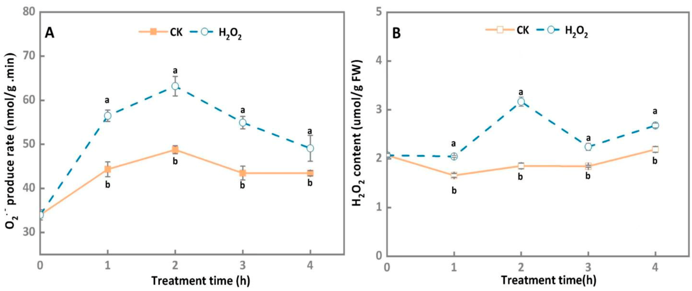
3.1. O2− Generation Rate and H2O2 Content of A. alternata Increased under Exogenous H2O2 Stress

In general, the O2− generation rate of A. alternata treated by the exogenous H2O2 stress and the control group increased at first; then they steadily declined during 4 h of incubation, whereas O2− generation rate in the H2O2-treated A. alternata significantly increased compared with the control group, peaking at 2 h after treatment, which was 22.9% higher than the control group (Figure 1A).
As shown in Figure 1B, the H2O2 contents of the control group maintained at a relatively constant level during 4 h of incubation, whereas the H2O2 contents of A. alternata treated by the exogenous H2O2 stress increased at first and then declined, which were significantly higher than the control group. Peak H2O2 content was observed 2 h after treatment in H2O2-treated A. alternata, where its value was 41.4% higher than that of the control samples (p < 0.05).

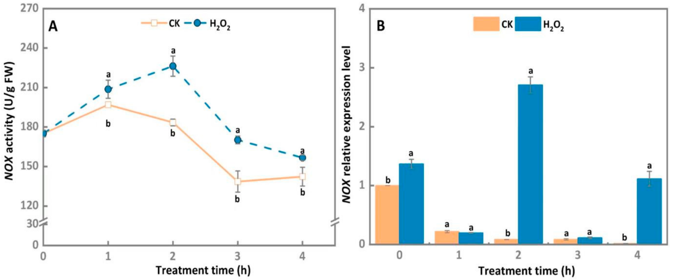
3.2. NOX Activity and Gene Expression Level of A. alternata Increased after Exogenous H2O2 Stress

In comparison with the control group, nicotinimide adenine dinucleotide phosphate oxidase (NOX) activity increased constantly during the 2 h of incubation in 30 mM H2O2-treated A. alternata. Subsequently, it significantly declined in both control group and H2O2-treated A. alternata. The NOX activity of A. alternata treated by the exogenous H2O2 stress was always higher than that of the control group. Peak, at 2 h, was observed in the 30 mM H2O2-treated A. alternata, which was increased by 18.9% compared with the control group (Figure 2A). Further studies showed that H2O2 treatment significantly upregulated the gene expression levels of NOX at 2 h and 4 h of incubation (Figure 2B). Correspondingly, the NOX expression level of 30 mM H2O2-treated A. alternata was significantly increased by 97% compared with the control group after 2 h of incubation (p < 0.05).

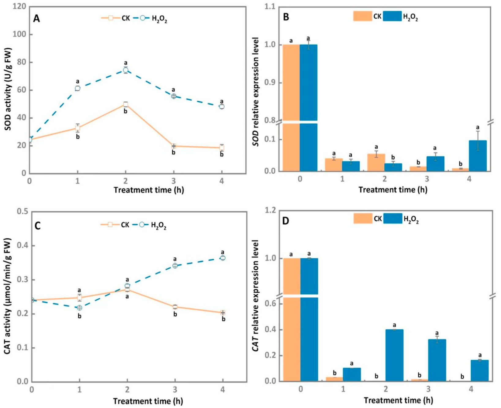
3.3. SOD and CAT Activities, and Transcript Level of A. alternata Increased after Exogenous H2O2 Stress

H2O2 treatments obviously increased the SOD activity of A. alternata from 1 to 4 h compared to the control group, with SOD activity peaking on 2 h and then slowly decreasing during later incubation of the H2O2-treated A. alternata (Figure 3A). In comparison with the control group, SOD activity of H2O2-treated A. alternata was increased by 33% after 2 h of incubation. Furthermore, SOD relative gene expression level of the A. alternata treated 30 mM H2O2 at 4 h upregulated by 10.8 times compared with the corresponding control group (Figure 3B).
As shown in Figure 3C, CAT activity of 30 mM H2O2-treated A. alternata raised steadily after 1 h of incubation, whereas SOD activity of the control group reduced rapidly from 2 to 4 h, and CAT activity of the H2O2 treatment group was significantly higher than that of the control group after 2 h of incubation. The highest level of CAT activity was observed at 4 h after the H2O2 treatments and was 1.8 times higher than that of the control group. Gene expression results showed CAT relative expression levels in H2O2-treated A. alternata were significantly higher than those of the control and peaked at 2 h (Figure 3D).

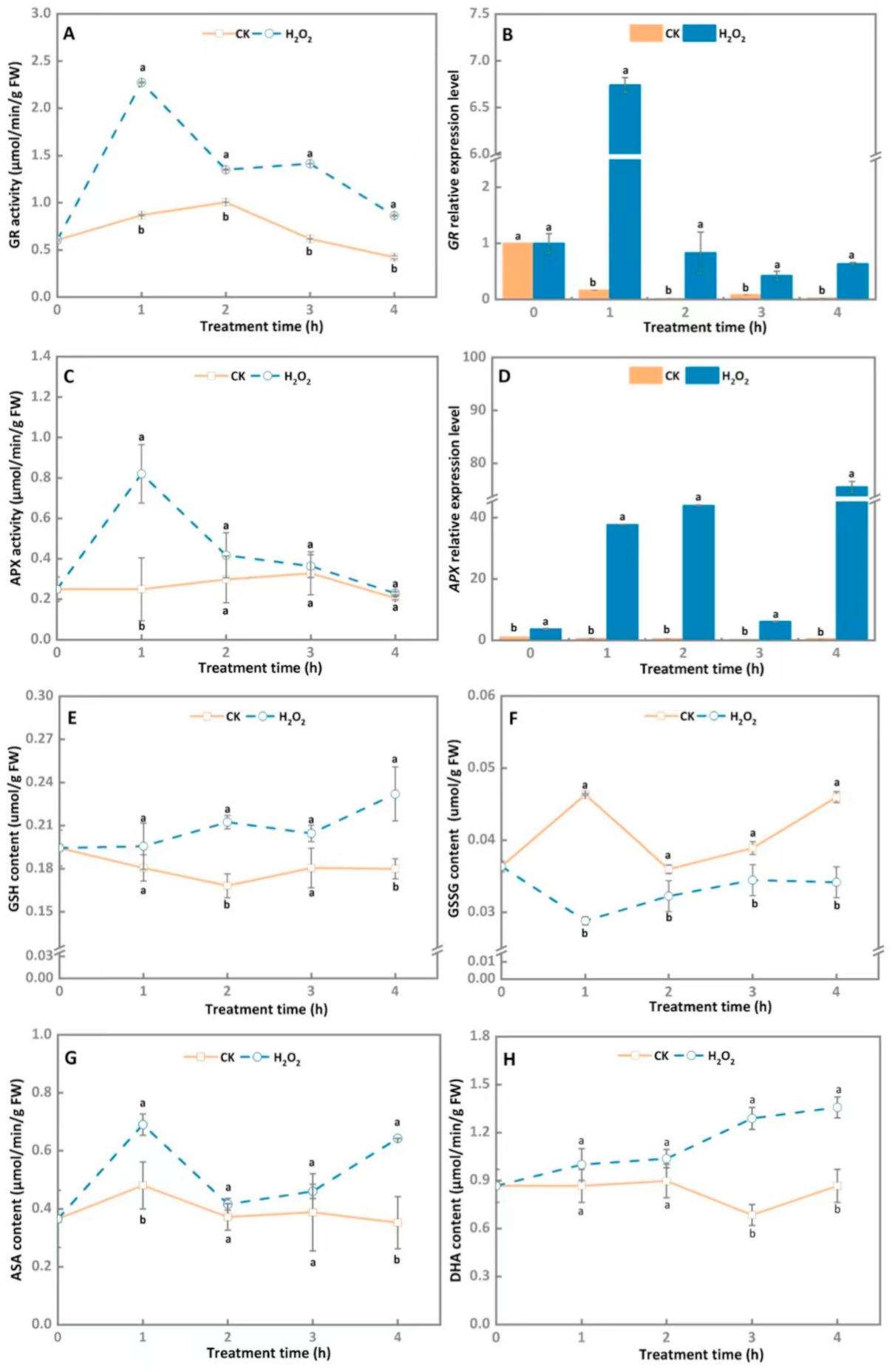
3.4. AsA-GSH Cycle System Involved in Maintaining the Intracellular Redox Balance of A. alternata after Exogenous H2O2 Stress

In general, the GR activity of A. alternata treated by the exogenous H2O2 stress rapidly increased at first and then gradually declined during 4 h of incubation, and the GR activity of the treatment group was significantly higher than the control group throughout the 4 h of incubation, with an increase of 61.6% at 1 h of incubation in contrast with the control (Figure 4A). Similarly, the maximum expression level of GR in H2O2-stress-treated A. alternata was observed at 1 h (Figure 4B) and shows an enhancement of 41.8 times in the H2O2-treated group compared with the control group (p < 0.05).
As shown in Figure 4, there was no significant difference in APX activity between the H2O2-treated group and the control during the entire incubation time, expect at 1 h (Figure 4C). The peak of the APX activity was discovered in the H2O2-stress-treated hypha at 1 h, which was raised by 69.5% after H2O2 treatment (p < 0.05). By contrast, H2O2 treatment obviously upregulated APX relative expression level during 4 h of incubation, which was 56.9 and 78.6 times higher than the control after 1 h and 2 h of incubation, respectively (Figure 4D).
To further analyze the effect of H2O2 stress on the antioxidant system of A. alternata, the GSH and GSSG contents were determined during 4 h of incubation. The GSH content of 30 mM H2O2-stress-treated A. alternata increased continuously throughout the 4 h of cultivation, and the treatment group was significantly higher than the control group in the same period (Figure 4E). The high GSH content in the H2O2-treated A. alternata was observed at 4 h, which was accelerated by 26% compared to the control. Additionally, H2O2 treatment also caused a decline in GSSG content, which was reduced by 26.2% compared with the control after 4 h of cultivation (Figure 4F).
As shown in Figure 4G, the ASA content of A. alternata evidently increased after the 30 mM H2O2 treatment, and the highest content was detected at 1 h, which was increased by 30.4% in contrast with the control (p < 0.05). The DHA content in H2O2-treated A. alternata increased rapidly during 4 h of cultivation, and the treatment group maintained noticeably higher values than the control group over the entire cultivation period. Compared with the control, the DHA content was enhanced by 36.7% after 4 h of incubation (Figure 4H).

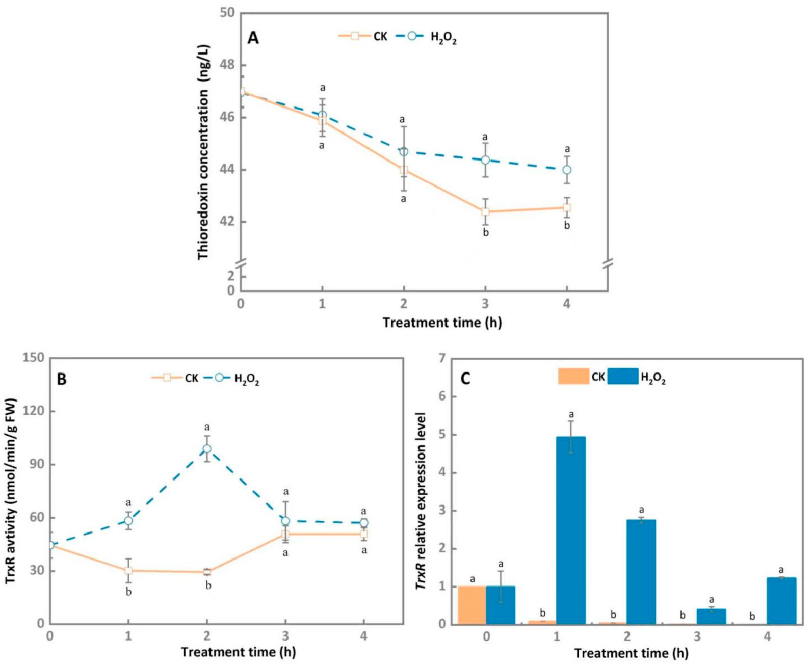
3.5. Thioredoxin System Involved in Maintaining the Intracellular Redox Balance of A. alternata after Exogenous H2O2 Stress

The thioredoxin concentration of 30 mM H2O2-treated A. alternata and the control group declined steadily, showing similar trends. However, H2O2 treatment significantly decelerated the decrease rate of thioresdoxin concentration of A. alternata, which was 4.5% higher than the control group after 3 h of cultivation (Figure 5A).
The TrxR activity peaked on 2 h and then rapidly decreased during later incubation in 30 mM H2O2-treated A. alternata, and H2O2 stress effectively enhanced the TrxR activity of A. alternata compared to the control group. After 2 h, the TrxR activity of treatment group was noticeably increased by 3.3 folds (Figure 5B). Similarly, the TrxR expression level of the H2O2-treated A. alternata were markedly upregulated at 1 h and 2 h compared with the control group, which were increased by 53.3 folds and 56 folds, respectively (Figure 5C).

4. Discussion

ROS are the core of host plant defense, and also play a vital role in the successful invasion of host plants by pathogenic fungi [32]. During the disease resistance process of the host, the pathogen accumulates high levels of endogenous ROS, and this may create an imbalance in the cell redox state [33]. The data presented in this study showed that 30 mM H2O2 treatment significantly increased the O2− generation rate and H2O2 content of A. alternata (Figure 1), indicating that oxidative stress induced the intracellular ROS production of A. alternata. Previous reports have demonstrated that NOX is the key enzyme source for the generation of ROS in pathogens, which use FADH2 and two heme molecules as cofactors. In P. expansum and Epichloë festucae, the deletion of NoxA reduced the contents of O2− and H2O2 [26,34]. In A. alternata of citrus, the deletion of AaNoxA increased sensibility to exogenous oxidants such as H2O2 and menadione [35]. In this study, we found that NOX activity and gene expression levels of A. alternata were significantly enhanced after it was 30 mM H2O2-treated (Figure 2). These results indicate that exogenous H2O2 treatment leads to initial accumulation of intracellular ROS via increasing NOX activity in A. alternata. However, pathogenic fungi can adapt to high concentrations of H2O2 and have a certain degree of tolerance to exogenous oxidative stress [23,36]. In our results, we found that A. alternata can continue to grow after 30 mM H2O2 treatment (data not shown), indicating that there was strong tolerance to high concentrations of H2O2 in A. alternata.
In order to establish successful infections in host plants, pathogenic fungus can neutralize, scavenge, or repair the damage caused by ROS though activating its own antioxidant defense system [17,18]. The excessive ROS can be scavenged by sophisticated enzymatic systems such as SOD and CAT by pathogens [36]. In the present study, 30 mM H2O2 treatment significantly increased SOD activity, as well as the SOD gene expression levels, in A. alternata (Figure 3A,B), which may be conducive to the decrease in the content of O2− and generation of more H2O2, since excess H2O2 is decomposed into H2O and O2 under the catalysis of CAT [37]. The observation in our study revealed that CAT activity and gene expression levels were also rapidly increased (Figure 3C,D), which was in accordance with the phenomenon in Aspergillus niger, where H2O2 treatment enhanced CAT activity [38]. In addition, the rapid increase in CAT activity proved it has a central role in counteracting the burst of ROS generated by the host cells to kill the invading fungal pathogens.
GR is a flavoprotein oxidoreductase that may convert GSSG into GSH under the action of NADPH in the AsA-GSH cycle, whereas the GSH/GSSG ratio presents the redox state mediated by the intracellular glutathione system [39]. In this study, the GR activity and transcriptional levels were elevated by H2O2 treatment in A. alternata (Figure 4A,B). Simultaneously, 30 mM H2O2 stress treatment increased GSH content but decreased GSSG content (Figure 4E,F). Similar results were also reported in A. niger, that H2O2 treatment increased the GSH/GSSG ratio [38]. However, in P. chrysogenum, there were no significant differences in GSH levels in response to H2O2 stress [40], indicating that different pathogens vary in their tolerance to H2O2 stress. APX is another key antioxidant enzyme in the AsA-GSH cycle, which breaks down H2O2 to form H2O and monodehydroascorbate (MDHA) that can be further converted to DHA [41]. The present results showed that H2O2 treatment induced the activity and transcriptional levels of APX in A. alternata (Figure 4C,D). In addition, the AsA and DHA contents were also enhanced after 4 h of incubation (Figure 4G,H). Results further confirmed that the AsA-GSH cycle system is involved in the A. alternata response to oxidative stress. In the Alternaria pathosystem, AsA-GSH cycle system may be activated immediately during the early infection stage, and scavenge ROS produced by the host to facilitate further infection of A. alternata
Thioredoxin (TRX) is a type of small molecule protein that can provide electrons for nucleotide reductase and peroxidase, which are involved in ROS scavenging to reduce oxidative damage [42]. In Candida albicans, the deletion of thioredoxin trx1 leads to increased sensitivity to H2O2 stress [43]. In this study, the thioredoxin concentration and TrxR activity were higher in H2O2-treated A. alternata, compared to the control group (Figure 5), indicating that the thioredoxin system is involved in maintaining the intracellular redox balance of A. alternata. However, Wang et al. [44] reported that the redox state of Trichoderma reesei was maintained via the GSH system, whereas Trtrx only plays a synergistic or backup role in oxidative stress resistance. Interestingly, in Botrytis cinerea, the thioredoxin system, not the GSH pool, is a central player in the maintenance of the redox status [45]. Therefore, the molecular mechanism of TRX in the response to oxidative stress in A. alternata needs further elucidation.

5. Conclusions

Collectively, the present study showed that the activity of key enzymes and gene expression levels of ROS production systems in A. alternata increased initially upon oxidative stress, and then A. alternata effectively eliminated exogenous H2O2 and kept intracellular redox balance through increasing the activity of key enzymes and gene expression levels, and the antioxidant content of the AsA-GSH cycle and thioredoxin scavenging systems. The results suggested that A. alternata can effectively adapt to oxidative stress, but further work needs to be addressed for clarifying the underlying molecular mechanisms.

Author Contributions

M.Z., Y.Z., Y.L., Y.B. and D.P. conceived and designed the experiments. M.Z., Y.Z., R.M., Y.Y. and Q.J. performed the experiments. M.Z. analyzed the data and wrote the manuscript. All authors have read and agreed to the published version of the manuscript.

Funding

This research was funded by National Natural Science Foundation of China (31860456 and 32060567); the Fostering Foundation for the Excellent Ph.D. Dissertation of Gansu Agricultural University (YB2021004).

Institutional Review Board Statement

Not applicable.

Informed Consent Statement

Not applicable.

Data Availability Statement

The data presented in this study are available upon request from the corresponding authors.

Conflicts of Interest

The authors declare no conflict of interest.

References

  1. Li, Y.C.; Yin, Y.; Bi, Y.; Wang, D. Effect of riboflavin on postharvest disease of Asia pear and the possible mechanisms involved. Phytoparasitica 2012, 40, 261–268. [Google Scholar] [CrossRef]
  2. Mari, M.; Bertolini, P.; Pratella, G.C. Non-conventional methods for the control of post-harvest pear diseases. J. Appl. Microbiol. 2003, 94, 761–766. [Google Scholar] [CrossRef] [PubMed] [Green Version]
  3. Pan, T.T.; Pu, H.B.; Sun, D.W. Insights into the changes in chemical compositions of the cell wall of pear fruit infected by Alternaria alternata with confocal aman microspectroscopy. Postharvest Biol. Technol. 2017, 132, 119–129. [Google Scholar] [CrossRef]
  4. Wang, P.H.; Wu, P.C.; Huang, R.; Chung, K.R. The role of a nascent polypeptide associated complex subunit alpha in siderophore biosynthesis, oxidative stress response, and virulence in Alternaria alternata. Mol. Plant Microbe Interact. 2019, 33, 668–679. [Google Scholar] [CrossRef]
  5. Yang, X.P.; Hu, H.J.; Yu, D.Z.; Sun, Z.H.; He, X.J.; Zhang, J.G.; Chen, Q.L.; Tian, R.; Fan, J.; Liu, J.H. Candidate resistant genes of sand pear (Pyrus pyrifolia nakai) to Alternaria alternata revealed by transcriptome sequencing. PLoS ONE 2015, 10, e0135046. [Google Scholar] [CrossRef]
  6. Saccon, F.A.M.; Parcey, D.; Paliwal, J.; Sherif, S.S. Assessment of fusarium and deoxynivalenol using optical methods. Food Bioprocess Technol. 2017, 10, 34–50. [Google Scholar] [CrossRef]
  7. Tanaka, A.; Shiotani, H.; Yamamoto, M.; Tsuge, T. Insertional mutagenesis and cloning of genes required for biosynthesis of the host-specific AK-toxin in the Japanese pear pathotype of Alternaria alternata. Mol. Plant Microbe Interact. 1999, 12, 691–702. [Google Scholar] [CrossRef] [Green Version]
  8. Kubicek, C.P.; Starr, T.L.; Glass, N.L. Plant cell wall-degrading enzymes and their secretion in plant-pathogenic fungi. Annu. Rev. Phytopathol. 2014, 52, 427–451. [Google Scholar] [CrossRef]
  9. Takao, K.; Akagi, Y.; Tsuge, T.; Harimoto, Y.; Yamamoto, M.; Kodama, M. The global regulator LaeA controls biosynthesis of host-specific toxins, pathogenicity and development of Alternaria alternata pathotypes. J. Gen. Plant Pathol. 2016, 82, 121–131. [Google Scholar] [CrossRef]
  10. Camejo, D.; Guzman-Cedeno, A.; Moreno, A. Reactive oxygen species, essential molecules, during plant-pathogen interactions. Plant Physiol. Biochem. 2016, 103, 10–23. [Google Scholar] [CrossRef]
  11. Bhattacharjee, S. An inductive pulse of hydrogen peroxide pretreatment restores redox- homeostasis and oxidative membrane damage under extremes of temperature in two rice cultivars. Plant Growth Regul. 2012, 68, 395–410. [Google Scholar] [CrossRef]
  12. Tsukagoshi, H.; Busch, W.; Benfey, P.N. Transcriptional regulation of ROS controls transition from proliferation to differentiation in the root. Cell 2010, 143, 606–616. [Google Scholar] [CrossRef] [PubMed] [Green Version]
  13. Zaragoza, O.; Chrisman, C.J.; Castelli, M.V.; Frases, S.; Casadevall, A. Capsule enlargement in Cryptococcus neoformans confers resistance to oxidative stress suggesting a mechanism for intracellular survival. Cell Microbiol. 2008, 10, 2043–2057. [Google Scholar] [CrossRef] [PubMed] [Green Version]
  14. Ganetta, E.; Walker, G.M.; Adya, A.K. Nanoscopic morphological changes in Yyt cell surfaces caused by oative stress: An atomic force microscopic study. Microbiol. Biotechnol. 2009, 19, 547–555. [Google Scholar] [CrossRef]
  15. Buron-Moles, G.; Torres, R.; Teixidó, N.; Usall, J.; Vilanova, L.; Vinas, I. Characterization of H2O2 production to study compatible and non-host pathogen interactions in orange and apple fruit at different maturity stages. Postharvest Biol. Technol. 2015, 99, 27–36. [Google Scholar] [CrossRef]
  16. Liu, X.; Williams, C.E.; Nemacheck, J.A.; Wang, H.Y. Reactive oxygen species are involved in plant defense against a gall midge. Plant Physiol. 2010, 152, 985–999. [Google Scholar] [CrossRef] [Green Version]
  17. Heller, J.; Tudzynski, P. Reactive oxygen species in phytopathogenic fungi: Signaling, development and disease. Annu. Rev. Phytopathol. 2011, 49, 369–390. [Google Scholar] [CrossRef]
  18. Kou, Y.; Qiu, J.; Tao, Z. Every coin has two sides: Reactive oxygen species during rice Magnaporthe oryzae interaction. Int. J. Mol. Sci. 2019, 20, 1191. [Google Scholar] [CrossRef] [Green Version]
  19. Eloy, Y.R.; Vasconcelos, I.M.; Barreto, A.L.; Freire-filho, F.R.; Oliveira, J.T.A. H2O2 plays an important role in the lifestyle of Colletotrichum gloeosporioides during interaction with cowpea Vigna unguiculata (L.) walp. Fungal Biol. 2015, 119, 747–757. [Google Scholar] [CrossRef] [Green Version]
  20. Qi, X.Z.; Guo, L.J.; Yang, L.Y.; Huang, J. Foatf1, a ZIP transcription factor of Fusarium oxysporum f. sp. cubense, is involved in pathogenesis by regulating the oxidative stress responses of Cavendish banana (Musa spp.). Physiol. Mol. Plant Path. 2013, 84, 76–85. [Google Scholar] [CrossRef]
  21. Li, G.F.; Peng, G.X.; Keyhani, N.O.; Xin, J.; Cao, Y.; Xia, Y.X. A bifunctional catalase-peroxidase, MakatG1, contributes to virulence of Metarhizium acridum by overcoming oxidative stress on the host insect cuticle. Environ. Microbiol. 2017, 19, 4365–4378. [Google Scholar] [CrossRef] [PubMed]
  22. Russell, E.G.; Cotter, T.G. New insight into the role of reactive oxygen species (ROS) in cellular signal-transduction processes. Int. Rev. Cell Mol. Biol. 2015, 319, 221. [Google Scholar] [CrossRef] [PubMed]
  23. Mir, A.A. Systematic characterization of the peroxidase gene family provides new insights into fungal pathogenicity in Magnaporthe oryzae. Sci. Rep. 2015, 5, 11831. [Google Scholar] [CrossRef] [Green Version]
  24. Zhang, L.B.; Tang, L.; Ying, S.H.; Feng, M.G. Regulative roles of glutathione reductase and four glutaredoxins in glutathione redox, antioxidant activity, and iron homeostasis of Beauveria bassiana. Appl. Microbiol. Biotechnol. 2016, 100, 5907–5917. [Google Scholar] [CrossRef] [PubMed]
  25. Tang, Y.; Li, Y.C.; Bi, Y.; Wang, Y. Role of pear fruit cuticular wax and surface hydrophobicity in regulating the prepenetration phase of Alternaria alternata infection. J. Phytopathol. 2017, 165, 313–322. [Google Scholar] [CrossRef]
  26. Zhang, X.M.; Zong, Y.Y.; Gong, D.; Yu, L.R.; Sionov, E.; Bi, Y.; Prusky, D. NADPH oxidase regulates the growth and pathogenicity of Penicillium expansum. Front. Plant Sci. 2021, 12, 696210. [Google Scholar] [CrossRef]
  27. Ge, Y.H.; Deng, H.W.; Bi, Y.; Li, Y.C.; Liu, Y.Y. The role of reactive oxygen species in ASM-induced disease resistance in apple fruit. In Postharvest Pathology; Prusky, D., Gullino, M.L., Eds.; Springer International Publishing: Cham, Switzerland, 2014; pp. 39–52. [Google Scholar] [CrossRef]
  28. Ren, Y.L.; Wang, Y.F.; Bi, Y.; Deng, H. Postharvest BTH treatment induced disease resistance and enhanced reactive oxygen species metabolism in muskmelon (Cucumis melo L.) fruit. Eur. Food Res. Technol. 2012, 234, 963–971. [Google Scholar] [CrossRef]
  29. Sun, J.Z.; Lin, H.T.; Zhang, S.; Lin, Y.F.; Wang, H.; Lin, M.S.; Chen, Y.H. The roles of ROS production-scavenging system in Lasiodiplodia theobromae (Pat.) Griff. & Maubl. -induced pericarp browning and disease development of harvested longan fruit. Food Chem. 2018, 247, 16–22. [Google Scholar] [CrossRef]
  30. Turcsanyi, E.; Lyons, T.; Plöchl, M.; Barnes, J. Does ascorbate in the mesophyll cell walls form the first line of defence against ozone? Testing the concept using broad bean (Vicia faba L.). J. Exp. Bot. 2000, 51, 901–910. [Google Scholar] [CrossRef]
  31. Livak, K.J.; Schmittgen, T.D. Analysis of relative gene expression data using real-time quantitative PCR and the 2ΔΔCT method. Methods 2001, 25, 402–408. [Google Scholar] [CrossRef]
  32. Brown, D.I.; Griendling, K.V.K. Nox proteins in signal transduction. Free Radic. Biol. Med. 2009, 47, 1239–1253. [Google Scholar] [CrossRef] [PubMed] [Green Version]
  33. Miller, G.; Shulaev, V.; Mittler, R. Reactive oxygen signaling and abiotic stress. Physiol. Plant. 2008, 133, 481–489. [Google Scholar] [CrossRef] [PubMed]
  34. Segal, L.M.; Wilson, R.A. Reactive oxygen species metabolism and plant-fungal interactions. Fungal Genet. Biol. 2018, 110, 1–9. [Google Scholar] [CrossRef] [PubMed] [Green Version]
  35. Yang, S.L.; Chung, K.R. The NADPH oxidase-mediated production of hydrogen peroxide (H2O2) and resistance to oxidative stress in the necrotrophic pathogen Alternaria alternata of citrus. Mol. Plant Microbe Interact. 2012, 13, 900–914. [Google Scholar] [CrossRef]
  36. Angelova, M.B.; Pashova, S.B.; Spasova, B.K.; Vassilev, S.V.; Slokoska, L.S. Oxidative stress response of filamentous fungi induced by hydrogen peroxide and paraquat. Mycol. Res. 2005, 109, 150–158. [Google Scholar] [CrossRef]
  37. Wei, M.L.; Ge, Y.H.; Li, C.Y.; Han, X.; Qin, S.C.; Chen, Y.R.; Tang, Q.; Li, J.R. G6PDH regulated NADPH production and reactive oxygen species metabolism to enhance disease resistance against blue mold in apple fruit by acibenzolar-S-methyl. Postharvest Biol. Technol. 2019, 148, 228–235. [Google Scholar] [CrossRef]
  38. Li, Q.; Mcneil, B.; Harvey, L.M. Adaptive response to oxidative stress in the filamentous fungus Aspergillus niger B1-D. Free Radic. Biol. Med. 2008, 44, 394–402. [Google Scholar] [CrossRef] [Green Version]
  39. Chumyam, A.; Shank, L.; Faiyue, B.; Uthaibutra, J.; Saengnil, K. Effects of chlorine dioxide fumigation on redox balancing potential of antioxidative ascorbate-glutathione cycle in ‘Daw’ longan fruit during storage. Sci. Hortic. 2017, 222, 76–83. [Google Scholar] [CrossRef]
  40. Emri, T.; Pocsi, I.; Szentirmai, A. Glutathione metabolism and protection against oxidative stress caused by peroxides in Penicillium chrysogenum. Free Radic. Biol. Med. 1997, 23, 809–814. [Google Scholar] [CrossRef]
  41. Jiang, H.; Wang, Y.; Li, C.J.; Wang, B.; Ma, L.; Ren, Y.Y.; Bi, Y.; Li, Y.C.; Xue, H.L.; Prusky, D. The effect of benzo-(1,2,3)-thiadiazole-7-carbothioic acid S-methyl ester (BTH) treatment on regulation of reactive oxygen species metabolism involved in wound healing of potato tubers during postharvest. Food Chem. 2020, 309, 125608. [Google Scholar] [CrossRef]
  42. Ying, W.H. NAD+/NADH and NADP+/NADPH in cellular functions and cell death: Regulation and biological consequences. Antioxid. Redox Signal. 2008, 10, 179–206. [Google Scholar] [CrossRef] [PubMed] [Green Version]
  43. Dantas, A.D.S.; Patterson, M.J.; Smith, D.A.; Maccallum, D.M.; Quinn, J. Thioredoxin regulates multiple hydrogen peroxide-induced signaling pathways in Candida albicans. Cell Mol. Biol. 2010, 30, 4550–4563. [Google Scholar] [CrossRef] [Green Version]
  44. Wang, G.; Wang, H.; Xiong, X.; Chen, S.; Zhang, D. Mitochondria thioredoxin"s backup role in oxidative stress resistance in Trichoderma reesei. Microbiol. Res. 2015, 171, 32–38. [Google Scholar] [CrossRef] [PubMed]
  45. Viefhues, A.; Heller, J.; Temme, N.; Tudzynski, P. Redox systems in Botrytis cinerea: Impact on development and virulence. Mol. Plant Mocrobe Interact. 2014, 27, 858–874. [Google Scholar] [CrossRef] [PubMed] [Green Version]
Figure 1. Effect of H2O2 stress treatment on O2− generation rate (A) and H2O2 contents (B) of A. alternata. Different letters represent significant differences (p < 0.05), bars indicate standard error (±SE).
Microorganisms 10 00621 g001
Figure 2. Effect of H2O2 stress treatment on NOX activity (A) and gene expression level (B) of A. alternata. GAPDH was used as an internal control; different letters represent significant differences (p < 0.05), bars indicate standard error (±SE).
Microorganisms 10 00621 g002
Figure 3. Effect of H2O2 stress treatment on SOD (A) and CAT activities (C), and gene expression level (B,D) of A. alternata. GAPDH was used as an internal control; different letters represent significant differences (p < 0.05), bars indicate standard error (±SE).
Microorganisms 10 00621 g003
Figure 4. Effect of H2O2 stress treatment on AsA-GSH cycle system related enzymes activities and gene expression, as well as related metabolite contents of A. alternata (A: GR activity; B: GR relative expression; C: APX activity; D: APX relative expression; E: GSH content; F: GSSG content; G: ASA content; H: DHA content). GAPDH was used as an internal control; different letters represent significant differences (p < 0.05), bars indicate standard error (±SE).
Microorganisms 10 00621 g004
Figure 5. Effect of H2O2 stress treatment on thioredoxin concentration (A), TrxR activity (B) and gene expression level (C) of A. alternata. GAPDH was used as an internal control; different letters represent significant differences (p < 0.05), bars indicate standard error (±SE).
Microorganisms 10 00621 g005
Table 1. Sequence of primer design.
GeneSequence of Primer (5′ to 3′)TM
NOXF: AACGAAGTCGCAGTTCTTATTGR: GGCGGAGGTGGTAGATAGATT60
SODF: AACAACTTCAGCGAGCAAATCR: TTGATGGCAGCAGATAGCG60
CATF: CCACGGCACCTTTGTTTCTR: ATCTCGCACTGTGTCAGCACT60
TrxRF: GCGGTATCGTCAGGCTATCAR: CTATCCCTATGCTGTCATCTTGC60
GRF: GCCAAACACGGTGCAAAAGTR: TTTGAAGGTCTCGGCAATCG60
APXF: AATGCTGGTCTCAAGGCTGCR: GACCCTGCATCTCCTGGATG60
Publisher’s Note: MDPI stays neutral with regard to jurisdictional claims in published maps and institutional affiliations.

