Article

Dynamics of Changes in the Gut Microbiota of Healthy Mice Fed with Lactic Acid Bacteria and Bifidobacteria

1
Laboratory of Metagenomics and Food Biotechnology, Voronezh State University of Engineering Technologies, 394036 Voronezh, Russia
2
Department of Genetics, Cytology and Bioengineering, Voronezh State University, 394018 Voronezh, Russia
3
FSBSI All-Russian Veterinary Research Institute of Pathology, Pharmacology and Therapy, 394061 Voronezh, Russia
*
Author to whom correspondence should be addressed.
Submission received: 19 April 2022 / Revised: 10 May 2022 / Accepted: 11 May 2022 / Published: 12 May 2022
(This article belongs to the Special Issue Antimicrobial Impact of Probiotic Bacteria)

Abstract

:
Probiotics are living microorganisms that provide numerous health benefits for their host. Probiotics have various effects on the body; for example, they change gut microbiota, improve the integrity of the epithelial barrier and have anti-inflammatory effects. The use of probiotic supplements that are based on lactic acid bacteria and bifidobacteria is one of the approaches that are used to balance gut microflora. In our study, we evaluated the effects of supplements, which were based on members of the Lactobacillaceae family and bifidobacteria, on the gut microbiome of healthy mice using the 16S rRNA sequencing method. The data that were obtained demonstrated that when mice received the probiotic supplements, statistically significant changes occurred in the composition of the microbiome at the phylum level, which were characterized by an increase in the number of Actinobacteriota, Bacteroidota, Verrucomicrobia and Proteobacteria, all of which have potentially positive effects on health. At the generic level, a decrease in the abundance of members of the Nocardioides, Helicobacter and Mucispirillum genus, which are involved in inflammatory processes, was observed for the group of mice that was fed with lactic acid bacteria. For the group of mice that was fed with bifidobacteria, a decrease was seen in the number of members of the Tyzzerella and Akkermansia genus. The results of our study contribute to the understanding of changes in the gut microbiota of healthy mice under the influence of probiotics. It was shown that probiotics that are based on members of the Lactobacillaceae family have a more positive effect on the gut microbiome than probiotics that are based on bifidobacteria.

1. Introduction

The gastrointestinal tract of mammals contains a complex community of microorganisms. The taxonomy of this community can vary significantly from individual to individual, but the main microbiome has a high functional redundancy and plays an important role in maintaining the health of its host [1].
It is well known that representatives of probiotic bacteria, such as lactic acid bacteria and bifidobacteria, are beneficial for human and animal health [2]. Their use in food has various positive effects, including the prevention of infections [3], the modulation of lipid metabolism to prevent and control obesity, reductions in allergic symptoms and the alleviation of diarrhea [4] and constipation, because they stimulate the immune system of the mucous membrane and the systemic immune response and/or change the gut microbiota [5].
The use of probiotic supplements, which contain members of the Lactobacillaceae family and the Bifidobacterium genus, is one of the modern approaches that are used to positively change the balance of gut microflora [6]. However, the effects of probiotic interventions on the microbiome as a whole cannot be fully assessed without a study on healthy organisms.
The effects of probiotics on the composition, diversity and function of gut microbiota has been studied using various tools and methods, ranging from targeted culture-dependent methods to metagenomic sequencing [7]. Previous studies have demonstrated significant changes in microbiota after probiotic treatment [8]. For example, Khan et al. showed the beneficial effects of Lactobacillus plantarum on the microbiome of mice, which were demonstrated by an increase in the beneficial bacteria that are associated with the production of short-chain fatty acids and a reduction in pro-inflammatory cytokines [9]. The addition of L. fermentum prevented hyperlipidemia, inflammation and oxidative stress in the colon and heart tissues of rats that were fed a high-fat diet. Studies have also shown that Lactobacillus acidophilus can inhibit pathogens and modulate immunity [10]. It has been shown that B. bifidum can have beneficial effects on cholesterol metabolism through the modulation of gut microbiota, as well as alleviating the symptoms of irritable bowel syndrome [11]. The beneficial effects of B. longum are based on a high carbohydrate metabolism followed by the production of acetate, which is a short-chain fatty acid that activates the barrier function of the intestinal epithelium of the host [12]. B. adolescentis is recognized as one of the dominant anaerobes that are found in adults and is considered to be beneficial to health. Some strains can regulate the Proteobacteria to Bacteroidetes ratio in gut microbiota and inhibit NF-κB activation in the colon [13,14].
Despite the fact that the effects of probiotic bacteria on organisms with various pathologies have been widely studied, there have been very limited studies on the effects of probiotic bacteria on the gut microbiome of healthy organisms. Thus, it has been shown that the addition of a probiotic of one species (Bifidobacterium infantis) does not affect the diversity and composition of the microbiome of the gastrointestinal tract of a healthy adult [15]. The intake of a yogurt with Bifidobacterium animalis subsp. lactis BB-12 increases the presence of beneficial bacteria, such as those in the Bifidobacterium genus, Slackia isoflavoniconvertens and Adlercreutzia equolifaciens [16]. L. rhamnosus CNCM I-4036 significantly increases the abundance of members of the Lactobacillus genus in the gut of a healthy adult [17]. No significant changes were found in the microbiological composition of the gut of healthy Japanese people following the consumption of probiotics that contained either Lactobacillus or Bifidobacterium strains [18]. Similarly, no effects of probiotics (Lactobacillus rhamnosus (LGG®) and Bifidobacterium animalis subsp. lactis (BB-12®)) were found on the bacterial composition of the gut of Danish infants [19].
The effects of probiotics on the bacterial composition of the gut of healthy animals have also been relatively poorly studied. However, there have been studies that show the effectiveness of the use of probiotic supplements for preventive purposes [20,21,22,23]. For example, Yang et al. showed that the combination of Cochlearius cochlearium and L. acidophilus has beneficial effects on body weight control and glucose homeostasis in high-fat DIO mice [24]. However, the dynamics of the changes in gut microbiota during long-term feed intake with the addition of probiotic bacteria have not been studied. The aim of this research was to study the effects of two types of probiotics (one based on lactic acid bacteria and the other on bifidobacteria) on the microbiome composition of mouse feces, depending on the duration of their use as a dietary supplement, using the next-generation sequencing method (NGS).

2. Materials and Methods

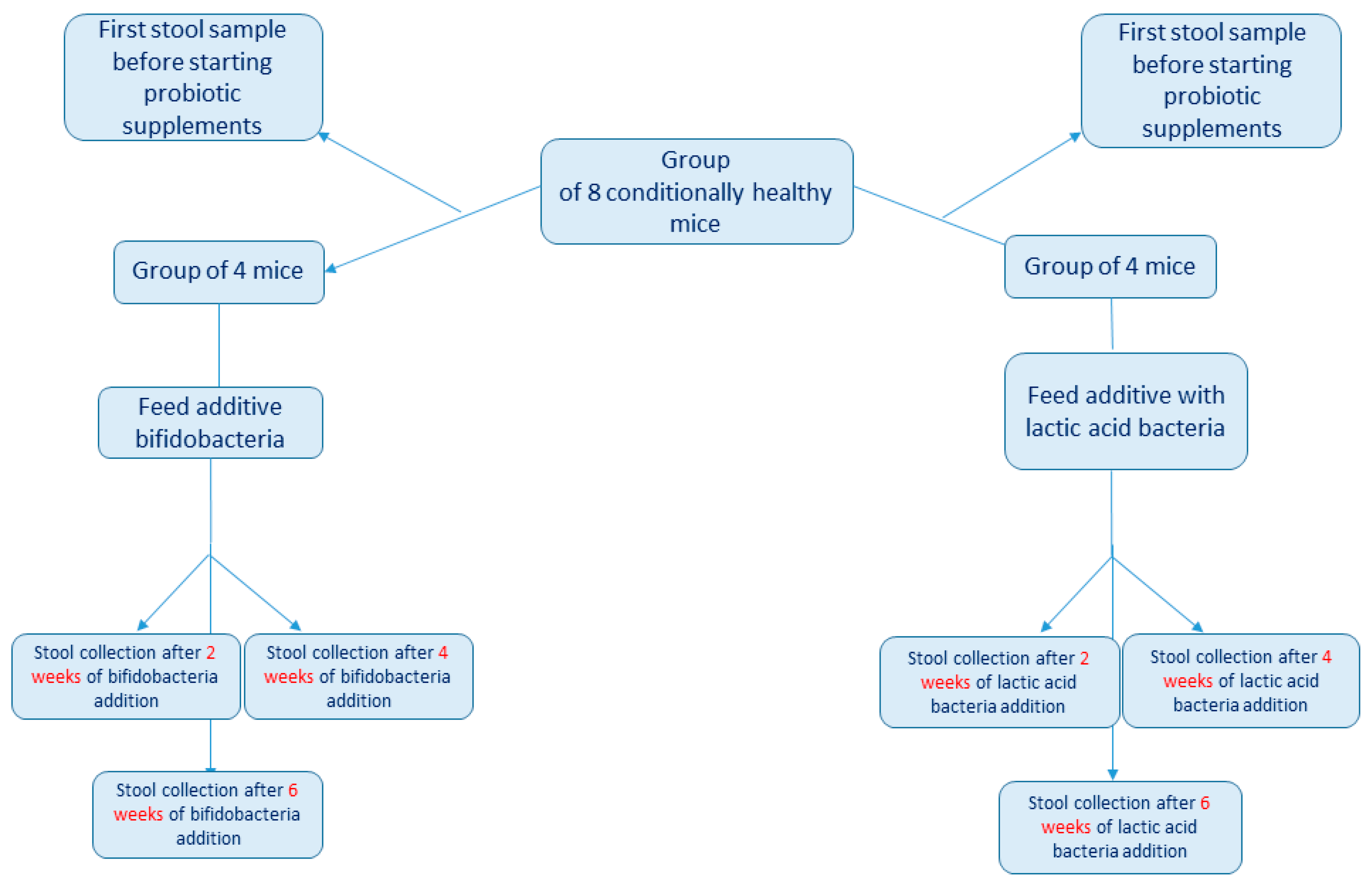
2.1. Experiment Design

Our study involved six-week-old Mus musculus of the C57BL/6 line. The mice were delivered from the farm of the Scientific Center for Biomedical Technologies (Stolbovaya branch, Russia). Each mouse was kept in a separate cage in a temperature-controlled room at 25 ± 2 °C and with a 12 h light–dark cycle. All animals had free access to water and were fed an ad libitum diet. The animals were fed a standard laboratory diet (Ssniff-Spezialdiäten GmbH, Soest, Germany). The composition of the diet is presented in Supplementary Materials File S1 (diet composition).
Initially, commercial probiotic bacteria (White Lily Ltd., Voronezh, Russia) were used. Pure bacterial cultures were deposited in the scientific microbiological museum of the Department of Biochemistry and Biotechnology at the Voronezh State University of Engineering Technologies and the following numbers were assigned: Bifidobacterium bifidum, VSUET01; Bifidobacterium longum, VSUET02; Bifidobacterium adolescentis, VSUET03; Lactobacillus acidophilus, VSUET12; Lactiplantibacillus plantarum, VSUET13; and Limosilactobacillus fermentum, VSUET14. Bacteria were added into 100 mL of sterilized skimmed milk. To activate the microbial cells, the concentrate was thoroughly mixed and kept for 4.0 h at a temperature of 37 ± 1 °C. Then, 1 and 2 h after the start of activation, the bacterial suspension was stirred again (by shaking) to encourage the distribution of bacterial cells throughout the solution. Immediately after activation, the resulting activated concentrate was introduced into pasteurized (92 ± 2 °C, 2–8 min exposure) and cooled (37–42 °C) skimmed milk by stirring. The concentration of microorganisms in the finished product was brought to 108 CFU/mL. The prepared cultures of bacteria were stored at −4 °C for no more than 3 days. The number of viable cells of lactic acid bacteria was calculated using the MRS agar.
The feed was crushed to a particle size of no more than 5 mm and then mixed with the prepared bacterial cultures in sterile conditions at a ratio of 9 g of feed to 1 g of bacterial culture. The feed in cage was replaced with freshly prepared feed every day.
The test animals were divided into two main research groups, each of which included four healthy male mice. The first stool collection was carried out before the addition of the probiotic supplements into the feed for all mice. Then, the first group received supplements with a mixture of bifidobacteria (B. bifidum, B. longum and B. adolescentis) and the second group received supplements with a mixture of Lactobacillaceae species (Lactobacillus acidophilus, Lactiplantibacillus plantarum and Limosilactobacillus fermentum). The concentration of each bacterial species in the supplement was 107 CFU per gram of feed. Then, 0.2 ± 0.01 g of fecal matter was collected from each mouse after two, four and six weeks for the subsequent estimation of changes in the composition of their gut microbiome under the influence of probiotics (Figure 1). All fecal samples were immediately placed into sterile 1.5-mL Eppendorf tubes and stored at −80 °C until DNA isolation.

2.2. Sequencing

DNA was extracted from each fecal sample using the ZymoBiomics DNA Miniprep Kit (Zymo Research, Los Angeles, CA, USA), according to the protocol. The V3 region of the 16S rRNA gene was selected for library preparation and further sequencing using the Ion Torrent PGM platform. The targeted amplification of this region was performed using the universal primers 337F (5′-GACTCCTACGGGAGGCWGCAG-3′) and 518R (5′-GTATTACCGCGGCTGCTGG-3′) and the 5X ScreenMix-HS Master Mix Kit (Evrogen, Moscow, Russia). The amplification protocol consisted of the following temperature regimes: 94 °C for 4 min; 37 cycles of 94 °C for 30 s, 53 °C for 30 s and 72 °C for 30 s; and then, final elongation at 72 °C for 5 min. After that, the PCR products were purified using AMPureXP magnetic particles (Beckman Coulter, Brea, CA, USA) and proceeded directly into the preparation of sequencing libraries.
Libraries were prepared using the NEBNext Fast DNA Library Prep Kit (New England Biolabs, Ipswich, MA, USA), according to the manufacturer’s instructions. The resulting libraries were barcoded using NEXTflex DNA Barcodes (Ion Torrent; 64 adapters; PerkinElmer, Inc., Waltham, MA, USA). After the ligation of the adapters, the purification of the finished libraries using the AMPureXP magnetic particles (Beckman Coulter, Brea, CA, USA) was repeated.
Sequencing was performed on the IonTorrent PGM platform using the Ion PGM Hi-Q View OT2 Kit and the Ion PGM Hi-Q View Sequencing Kit, according to the protocol using the Ion 318™ Chip v2 BC chip (ThermoFisher Scientific, Madison, WI, USA).

2.3. Statistical Analysis

Based on the results of sequencing, BAM files were obtained for each sample, which were then converted into the FastQ format using the FileExporter plugin and further analyzed using the R programming language in the RStudio environment (VSEARCH v.2.8.2 software). For all reads, low-quality reads were filtered out using the maximum expected error threshold of 1.0 (DADA2 package). We also trimmed the reads to the optimal equal length and subjected them to demultiplexing. After preparatory manipulations, dereplication was carried out and we achieved the combination of all identical readings into unique sequences and proceeded to search for the operational taxonomic units (OTUs) using the UNOISE2 algorithm. The taxonomy was determined to the genus using the SILVA database, version 132 (https://www.arb-silva.de/, accessed 14 March 2022). For taxonomy assignment, a maximum threshold of 100% identity was set with variant amplicon sequences.
A statistical comparison of the relative abundance in each study group was performed in the RStudio environment using the DeSEQ2 R package, which employed the generalized linear modelling (GLM) method [25]. In our study, we compared the changes in the microbiome of a group of mice before adding lactic acid bacteria to their feed (point 0) and then again after two (point 1), four (point 2) and six weeks (point 3). We carried out the same comparisons for a group of mice that were fed with bifidobacteria supplements. The p values were obtained using the Wald test. The results were expressed as the mean ± standard error of the mean (SEM). The alpha diversity for each group was calculated using the Shannon index.

2.4. Ethical Statements
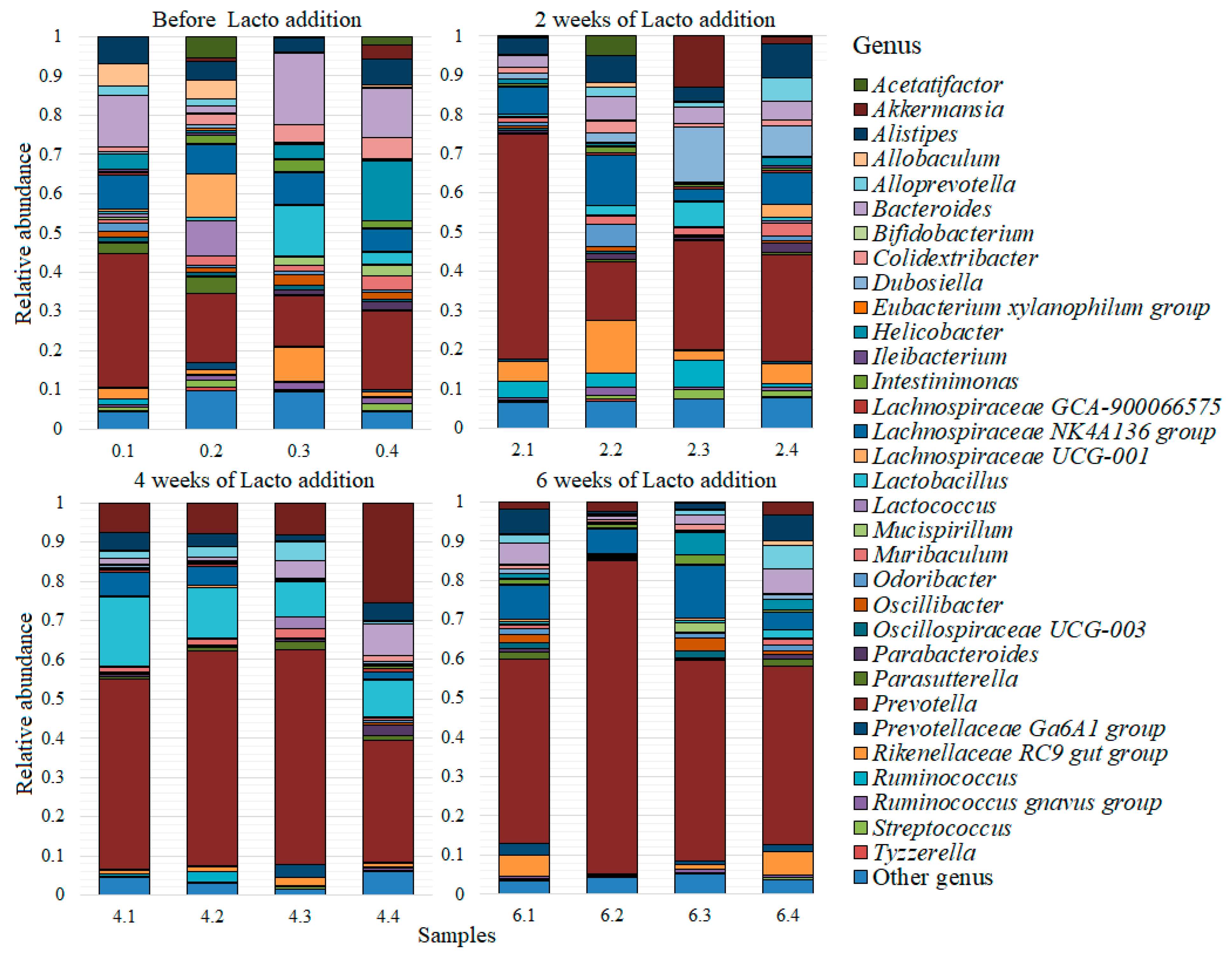
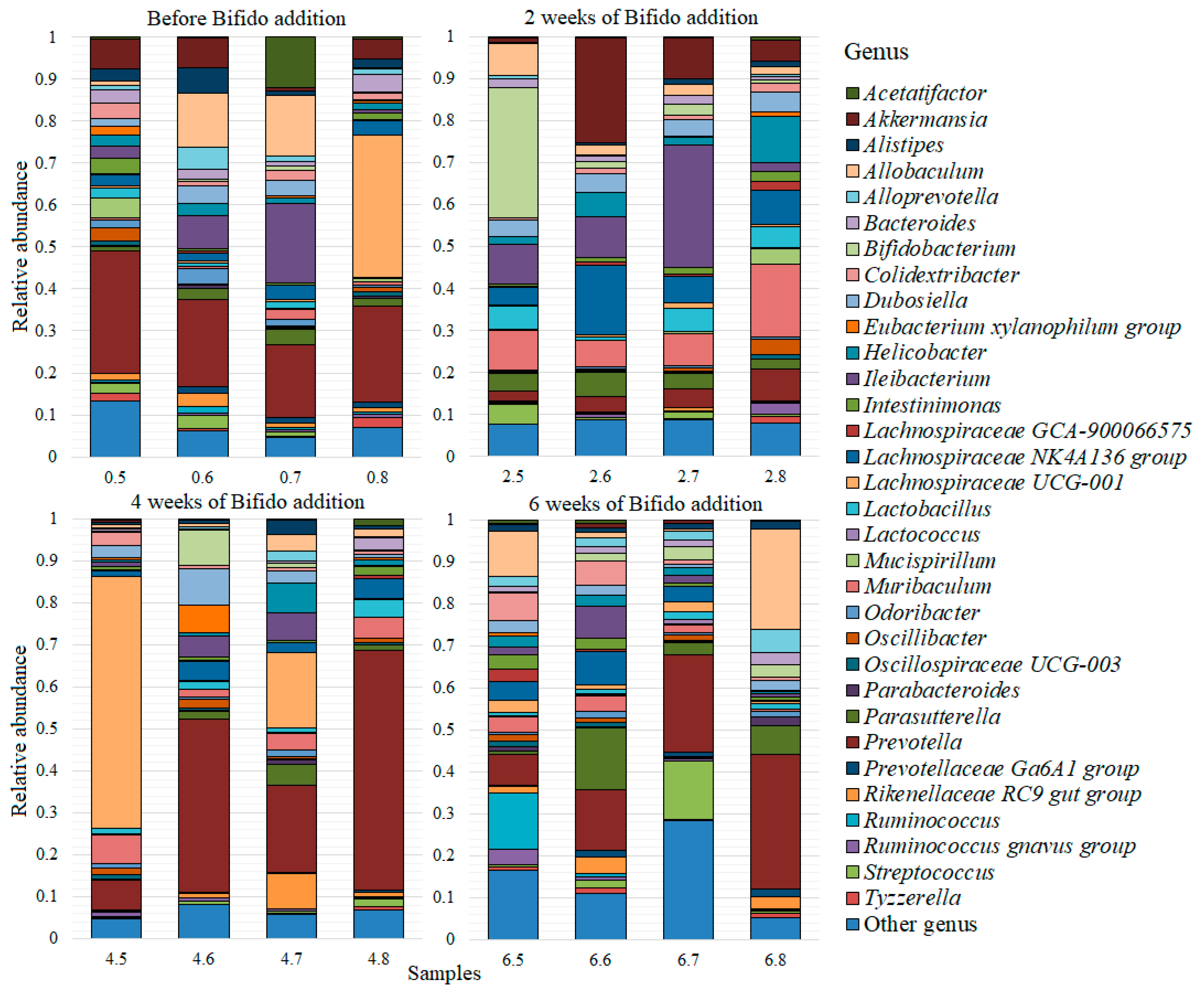
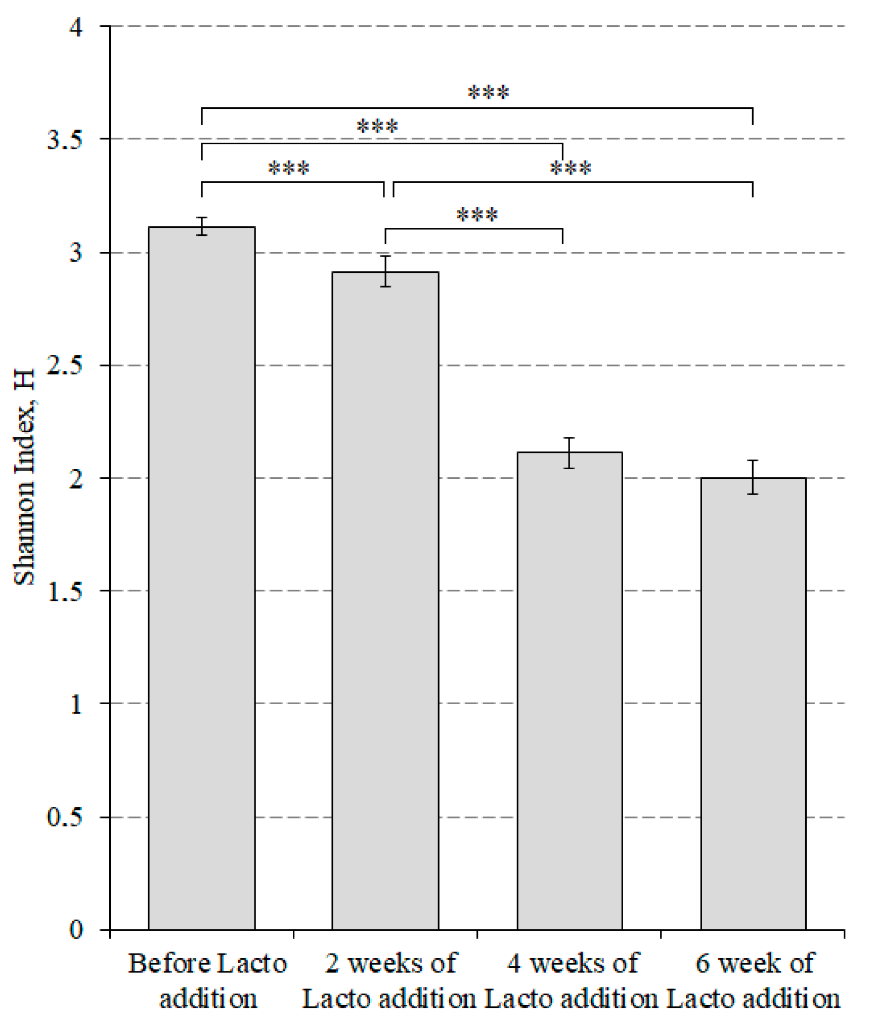
The experiments on the animals were approved by the Ethics Commission of the FSBSI “All-Russian Veterinary Research Institute of Pathology, Pharmacology and Therapy” (FSBSI “ARVRIPP&T”) (protocol number 7-12/21, 15 December 2021).

3. Results

As a result of the analysis of 32 fecal samples from mice from before and after two, four and six weeks of the addition of two probiotic supplements, a total of 81,385 unique reads were obtained, which corresponded to 99 genera of bacteria. A complete list of the discovered genera is presented in Supplementary Materials (Table S1). All genera that had an average abundance of less than 0.005 were combined into the “Other” group, thus forming a list of the 32 most common genera, which is presented for each sample from the lactic acid bacteria (Figure 2) and bifidobacteria (Figure 3) groups.
For the group of mice that received lactic acid bacteria, the most common genus was Prevotella, with an abundance of 0.39 ± 0.04. The next in order of abundance were: Lachnospiraceae NK4A136 group (0.07 ± 0.01); Bacteroides (0.06 ± 0.01); Lactobacillus (0.05 ± 0.01); Akkermansia (0.05 ± 0.02); Alistipes (0.05 ± 0.01); Rikenellaceae RC9 gut group (0.04 ± 0.01); Helicobacter (0.02 ± 0.01); Alloprevotella (0.02 ± 0.004); Dubosiella (0.02 ± 0.01); Colidextribacter (0.02 ± 0.004); and Muribaculum (0.02 ± 0.002). The average numbers of Ruminococcus, Parasutterella, Lachnospiraceae UCG-001, Odoribacter, Oscillibacter and Intestinimonas were within 0.01–0.02. The average number of other genera was less than 0.01.
For the group of mice that received bifidobacteria, the most common genus was also Prevotella (0.2 ± 0.04) and the second largest was Lachnospiraceae UCG-001 (0.08 ± 0.04). However, the further distribution of genus was significantly different from that of the group of mice that received lactic acid bacteria. The remaining genera of bacteria were distributed, in terms of abundance, as follows: Ileibacterium (0.07 ± 0.02); Allobaculum (0.05 ± 0.02); Lachnospiraceae NK4A136 group (0.05 ± 0.01); Muribaculum (0.05 ± 0.01); Akkermansia (0.04 ± 0.02); Parasutterella (0.04 ± 0.01); Bifidobacterium (0.04 ± 0.02); Dubosiella (0.03 ± 0.01); and Helicobacter (0.03 ± 0.01). The average relative abundance of the genera Lactobacillus, Streptococcus, Colidextribacter, Bacteroides, Rikenellaceae RC9 gut group, Alistipes, Alloprevotella, Intestinimonas, Oscillibacter, Ruminococcus, Acetatifactor and Odoribacter was within 0.01–0.02 and the average number of other genera was less than 0.01.
In addition, we estimated the way in which changes in alpha diversity occurred in mice that received lactic acid bacteria (Figure 4) and bifidobacteria (Figure 5) using the Shannon index.
Figure 4 shows that the maximum value of alpha diversity in the group of mice before the addition of members of Lactobacillaceae family was 3.11 ± 0.04: two weeks later, it was 2.92 ± 0.06; four weeks later, it was 2.11 ± 0.07; and six weeks later, it was 2.00 ± 0.08. Only the difference between the fourth and sixth weeks was not statistically significant.
Until the fourth week, we observed a decrease in alpha diversity. Before taking bifidobacteria, the indicator was 3.18 ± 0.05. Then, two weeks later, it became 3.0037 ± 0.0474 and by the fourth week, it reached 2.60 ± 0.09. However, after six weeks, the alpha diversity reached its maximum value of 3.62 ± 0.05.
A phylogenetic analysis of the microbiome of the studied groups at the phylum level also revealed several differences in bacterial composition. In the mice that received members of the Lactobacillaceae family during the feeding, the phylum Bacteroidota increased: 0.48 ± 0.06 before the addition of the probiotic; 0.58 ± 0.07 after two weeks of supplements; 0.62 ± 0.05 after four weeks for supplements; and 0.73 ± 0.05 after six weeks of supplements. At the same time, the phylum Firmicutes decreased: 0.41 ± 0.08 before the addition of the probiotic; 0.35 ± 0.05 after two weeks of supplements; 0.23 ± 0.03 after four weeks of supplements; and 0.20 ± 0.04 after six weeks of supplements. These two phyla accounted for the main share of the bacterial composition of the gut of the mice. Bacteroidota remained the dominant phylum throughout the lactic acid bacteria feeding.
In the mice that received bifidobacteria during the study, an uneven distribution of bacterial phyla was observed. As in the lactic acid bacteria-treated mice, the two main phyla throughout the study were Firmicutes (the abundance of which was 0.51 ± 0.06 before probiotic addition, 0.49 ± 0.05 after two weeks of supplements, 0.47 ± 0.12 after four weeks of supplements and 0.48 ± 0.09 after six weeks of supplements) and Bacteroidota (0.37 ± 0.04 before the addition of the probiotic, 0.19 ± 0.03 after two weeks of supplements, 0.44 ± 0.11 after four weeks of supplements and 0.34 ± 0.06 after six weeks of supplements).
We also estimated the statistical significance of differences in the microbiome composition at the phylum level after two, four and six weeks of lactic acid bacteria feeding with a control group of mice. It was found that after two weeks of feeding with probiotics, statistically significant changes were observed for two bacterial phyla: Campylobacterota, the abundance of which decreased 2.71-fold, and Patescibacteria, the abundance of which increased 4.07-fold compared to the control group (p < 0.01 in both cases).
After four weeks of feeding with lactic acid bacteria, statistically significant changes were observed for the following phyla: Bacteroidota, which increased 1.41-fold (p < 0.001); Verrucomicrobiota, which increased 4.15-fold (p < 0.001); Campylobacterota, which decreased 4.4-fold (p < 0.001); and Deferribacterota, which decreased 4.7-fold (p < 0.01) (Figure 6).
After six weeks of feeding with lactic acid bacteria, statistically significant changes were observed for the following: Bacteroidota, which increased 1.25-fold (p < 0.01); Cyanobacteria, which decreased 2.64-fold (p < 0.01); and Abditibacteriota, which significantly increased 24.97-fold (p < 0.001) (Figure 7).
The statistically significant differences in the microbiome composition at the phylum level that were revealed after two weeks of feeding with a probiotic mixture of bifidobacteria showed an increase in the number of phyla Actinobacteriota (4.2-fold (p < 0.001)) and Spirochaetota (4.26-fold (p < 0.01)).
After four weeks of probiotic feeding, the mice showed a significant decrease in the number of Abditibacteriota phyla (19.46-fold (p < 0.001)) and Verrucomicrobiota (2.9-fold (p < 0.01)), as well as an increase in the abundance of the genera Bacteroidota (1.21-fold (p < 0.001)) and Actinobacteriota (2.69-fold (p < 0.01)), compared to the control (Figure 8).
After six weeks of adding bifidobacteria to the feed, we continued to observe a decrease in the phylum Verrucomicrobiota (2.62-fold (p < 0.01)) and the phylum Desulfobacterota (1.92-fold) compared to the control group (p < 0.05), while the abundance of the phylum Proteobacteria increased 1.77-fold (p < 0.001) and Actinobacteriota increased 3.17-fold (p < 0.001) (Figure 9).
An analysis of the changes at the genus level in the composition of the microbiome of mice that received lactic acid bacteria showed that after two weeks, there were statistically significant changes, which were characterized by an increase in the abundance of the genera Ruminococcus (3.57-fold), Dubosiella (4.56-fold) and Candidatus Saccharimonas (4.7-fold) and a decrease in Helicobacter (2.43-fold) and Nocardioides (20.5-fold) compared to the microbiome before the addition of lactic acid bacteria to the feed (p < 0.001 in all cases) (Figure 10).
Four weeks later, we registered a decrease in the genera Nocardioides (19.55-fold (p < 0.01)), Mucispirillum (4.05-fold (p < 0.01)) and Helicobacter (3.82-fold (p < 0.001)), as well as an increase in the genera Prevotella (2.72-fold (p < 0.001)), Alloprevotella (2.93-fold (p < 0.001)), Akkermansia (4.75-fold (p < 0.001)), Faecalibaculum (4.99-fold (p < 0.01)) and Veillonella (5.97-fold (p < 0.001)), compared to the microbiome before the addition of lactic acid bacteria to the feed (Figure 11).
After six weeks, in the lactic acid bacteria-fed mice, we observed a 20.39-fold decrease in the Nocardioides (** p < 0.01) and a 6.08-fold decrease in the Lactococcus (*** p < 0.001) genera, as well as a 2.59-fold increase in the Prevotella (*** p < 0.001), a 4.31-fold increase in the Lachnospiraceae UCG-003 (*** p < 0.001) and a 24.91-fold increase in the Abditibacterium (*** p < 0.001) genera, compared to the control (Figure 12).
Statistically significant decreases were revealed two weeks later in the abundance of Lachnospiraceae UCG-001 (5.01-fold (p < 0.001)), Prevotellaceae Ga6A1 group (3.41-fold (p < 0.001)), Acetatifactor (3.33-fold (p < 0.01)), Anaerotruncus (3.32-fold (p < 0.01)), Lachnospiraceae ASF356 (3.26-fold (p < 0.05)), Tyzzerella (3.04-fold (p < 0.05)), Odoribacter (2.45-fold (p < 0.001)), Prevotella (2.43-fold (p < 0.001)), Rikenellaceae RC9 gut group (1.95-fold (p < 0.05)) and Alistipes (1.56-fold (p < 0.001)). Statistically significant increases were observed in the abundance of Lachnospiraceae NK4A136 group (1.47-fold (p < 0.05)), Lactobacillus (2.37-fold (p < 0.01)), Candidatus Saccharimonas (3.2-fold (p < 0.01)), Lachnospiraceae A2 (3.27-fold (p < 0.05)), Muribaculum (3.42-fold (p < 0.001)), Brachyspira (3.86-fold (p < 0.01)) and Bifidobacterium (5.04-fold (p < 0.001)) (Figure 13).
Four weeks later, we observed a 20.3-fold decrease in Nocardioides, a 19.1-fold decrease in Abditibacterium, a 3.59-fold increase in Akkermansia and a 2.78-fold increase in Muribaculum (*** p < 0.001 for all changes) (Figure 14).
After six weeks of probiotic supplementation, the abundance of the genus Escherichia-Shigella significantly increased 4.74-fold (p < 0.001) compared to the microbiome before the addition of bifidobacteria to the feed.

4. Discussion

In this study, we studied the effects of lactic acid bacteria- and bifidobacteria-supplemented diets on the gut microbiome of healthy mice.
Statistically significant changes were observed in two phyla of bacteria after two weeks of feeding with members of the Lactobacillaceae family: Campylobacterota, the abundance of which decreased, and Patescibacteria, the abundance of which increased. We observed an increase in the abundance of the phyla Bacteroidota and Verrucomicrobiota and a decrease in the abundance of Campylobacterota and Deferribacterota after four weeks of lactic acid bacteria feeding (Figure 6). After six weeks, the abundance of the genera Bacteroidota and Abditibacteriota increased, whereas the abundance of the phylum Cyanobacteria decreased (Figure 7).
An increase in the Campylobacterota phylum has been associated with the development of gastrointestinal diseases, such as ulcerative colitis (UC) [26]. Additionally, members of the phylum Helicobacter are involved in the development of IBD.
The members of the phylum Bacteroidota are the main colonizers of the gastrointestinal tract. They play an important role in maintaining the integrity of the interbacterial bonds in the gut. The members of the phylum Bacteroidota produce butyrate, which has anti-cancer properties and plays a role in maintaining gut health. In addition, the members of the phylum Bacteroidota are involved in the metabolism of bile acids and the transformation of toxic compounds, which also emphasizes their positive role [27]. We observed an increase in their abundance after two weeks of the addition of both types of probiotics.
The phylum Verrucomicrobia is a mucin-degrading bacterial community. This phylum is thought to promote gut health and is involved in glucose homeostasis. It can represent 3–5% of the bacterial community and primarily resides in the intestinal mucosa. The abundance of this bacterial phylum significantly reduces body weight and reduces the risk of obesity [28]. The phylum Verrucomicrobiota has the potential to induce regulatory immunity and is a possible target for interventions that are aimed at improving regulatory immunity in the gut microbiome [29]. However, in our study, we observed contrasting effects of the probiotics on the abundance of this phylum: the addition of members of the Lactobacillaceae family contributed to its increase, while the addition of bifidobacteria led to a decrease.
The phylum Deferribacterota is involved in the activation of systemic inflammation in the host organism [30].
According to the available literature data, the members of the phylum Cyanobacteria have potential pro-inflammatory, as well as neurotoxic, activity [31].
The influence of members of the phylum Abditibacteriota on the host organism has been so far poorly studied. Despite a significant increase in this phylum in the group of mice that was treated with a mixture of lactic acid bacteria for six weeks, this phylum was not included in the list of the most common bacteria, which indicated that there were only trace amounts of microorganisms from this phylum in the studied samples.
The phylum Desulfobacterota consists of many organisms that can reduce sulfur compounds through the sulfite reduction pathway for the dissimilation of DsrAB (dissimilatory sulfite reductase). These organisms are involved in the degradation of butyrate through the beta-oxidation pathway, which indicates the involvement of Desulfobacterota in the equilibrium of the catabolic reaction [32]. Therefore, it can be assumed that bacteria of this genus can cause inflammatory damage and exacerbate energy metabolism disorders, both of which are pathological signs of diabetes [33].
The phylum Proteobacteria is one of the first colonizers of the mammalian gastrointestinal tract. Proteobacteria reduce redox potential and also play a key role in the preparation of the gut for subsequent colonization by the strict anaerobes that are necessary for healthy intestinal function [34].
We observed an increase in the genera Ruminococcus, Dubosiella and Candidatus Saccharimonas and a decrease in Helicobacter and Nocardioides after two weeks of lactic acid bacteria feeding (Figure 10). After four weeks, we continued to notice declines in the genera Nocardioides and Helicobacter, whose role in the development of IBD is well known. The population of the genus Mucispirillum also decreased. At the same time, the abundance of the members of the genera Prevotella, Alloprevotella, Akkermansia, Faecalibaculum and Veillonella increased (Figure 11). The data that were obtained demonstrated a significant decrease in the genus Nocardioides throughout the feeding of mice with lactic acid bacteria. We also found a decline in the genus Lactococcus and an increase in the genera Prevotella, Lachnospiraceae UCG-003 and Abditibacterium (Figure 12).
The members of the genus Ruminococcus, which increased after two weeks of feeding with lactic acid bacteria, have ambiguous effects on the host organism. On one hand, there is evidence that the members of this genus are associated with the risk of obesity and their abundance also directly correlates with the development of irritable bowel syndrome (IBS) and the exacerbation of IBD [35]. However, on the other hand, there is also evidence that demonstrates that they can synthesize butyric acid, reduce the risk of cancer, increase immunological protection and have anti-inflammatory effects [36].
According to studies, the genus Dubosiella is associated with negative effects on the host organism, which include a direct relationship with the development of obesity [37]. We observed its increase after two weeks of supplemented feeding.
The members of the genus Candidatus Saccharimonas produce lactate and acetate. They are associated with inflammatory mucosal disease and can modulate the immune response by downregulating TNF-α gene expression in macrophages. Lower numbers of Candidatus Saccharimonas have been noted in acute necrotizing pancreatitis, which is associated with hypertriglyceridemia in rats and mice that have been fed a high-fat diet. This may indicate the potential anti-inflammatory role of Candidatus Saccharimonas, an increase in which we observed after two weeks of supplementation with both types of probiotics that were studied [38].
It is known that the members of the genus Helicobacter can have multiple negative effects on their host organism, including rodents. Most Helicobacter species are long, narrow and slightly curved rods with membrane-covered bipolar flagella. Naturally acquired Helicobacter infections have been reported in all commonly used laboratory rodent species. Most Helicobacter species in rodents are urease negative and, therefore, preferentially colonize the gut; although in some cases, they may translocate to the liver and biliary system, the stomach or other tissues. The presence of Helicobacter spp. deeply affects the host organism. Overall, Helicobacter organisms in mice have been associated with IBD and cancers of the breast, liver, stomach and colon [39]. We registered a decrease in this genus after two and four weeks of feeding with lactic acid bacteria.
We also observed a decrease in the abundance of the genus Nocardioides at all three test points in the group of mice that was treated with lactic acid bacteria, as well as in the group that was treated with bifidobacteria for four weeks. Nocardioides is a rare and poorly studied taxon. There is evidence of this type of increase in the gut of asthmatic mice that were infected with a respiratory syncytial virus [40]. In addition, this genus seems to be inversely correlated with the abundance of the genus Lactobacillus and is involved in the development of gut dysbiosis [41].
At the moment, there are no data that describe the role of the genus Alloprevotella in the gut of mammals. Our data showed that the increase in the abundance of this genus could be associated with the addition of probiotic bacteria to the diet.
The genus Akkermansia has been reported to be correlated with metabolic diseases; however, there are also studies that have proved its beneficial effects on the metabolism of its host [42,43]. We obtained contrasting data for this genus, depending on the type of probiotic that was supplemented: The Akkermansia genus increased with the addition of lactic acid bacteria and decreased with the addition of bifidobacteria.
The members of the genus Faecalibaculum are known to have antitumor potential in the gut of mice and are capable of producing the metabolites of SCFAs (mainly butyrate) [9,44]. We observed an increase in this genus in mice after four weeks of supplementation with lactic acid bacteria.
The members of the genus Veillonella are bacteria that decompose lactate through the formation of propionate SCFAs [45]. Veillonella may be sensitive to bile acids since its enrichment is an adaptive response to luminal bile acid deficiency, as in cirrhosis and cholestasis. This adaptation of the microbiota appears to be beneficial as the fermentation of lactate into propionate benefits the microbe by producing additional energy and also benefits the host by eliminating toxic lactate, the accumulation of which is associated with mortality [46].
The bacteria of the genus Lactococcus have not been widely recognized for their beneficial effects on gut health and are commonly used in dairy products. However, some data have shown that the members of this genus still have anti-inflammatory efficacy and can reduce oxidative stress in the colonic epithelium of mice that have colitis [47].
It is known that bacteria belonging to the Lachnospiraceae UCG-003 family can protect against the development of colon cancer by producing butyric acid. At the same time, it has been found that this genus can contribute to the development of diabetes in genetically predisposed mice [48].
With the addition of bifidobacteria, we registered an increase in the abundance of the phyla Actinobacteriota and Spirochaetota after two weeks. After four weeks, we observed a decrease in the abundance of the phyla Abditibacteriota and Verrucomicrobiota, as well as an increase in the abundance of the genera Bacteroidota and Actinobacteriota (Figure 8). After six weeks, we continued to observe a decrease in the phylum Verrucomicrobiota. The abundance of the phylum Desulfobacterota also decreased, while the abundance of Proteobacteria and Actinobacteriota increased (Figure 9). We also observed an increase in bifidobacteria belong to the phylum Actinobacteriota. Some members of these bacteria are known to have positive effects on the health of the host. It is important to note that a decrease in the levels of bifidobacteria is associated with IBD [49]. Indeed, the use of a combination of bifidobacteria as a supplement causes an accumulation of this phylum of bacteria, which emphasizes the effectiveness of this probiotic.
It is known that some bacteria of the phylum Spirochaetota are pathogenic. However, studies have shown a strong decrease in the number of these bacteria in the cases of intestinal diseases [50]. Feeding with bifidobacteria stimulated an increase in the abundance of members of this genus in our experiment.
In the group of mice that were fed bifidobacteria for two weeks, we found significant changes in the composition of their microbiome. These changes were characterized by a decrease in the genus Lachnospiraceae UCG-001, Prevotellaceae Ga6A1 group, Acetatifactor, Anaerotruncus, Lachnospiraceae ASF356, Tyzzerella, Odoribacter, Prevotella, Rikenellaceae RC9 gut group and Alistipes and an increase in Lachnospiraceae NK4A136 group, Lactobacillus, Candidatus Saccharimonas, Lachnospiraceae A2, Muribaculum and Bifidobacterium (Figure 13). After four weeks, we observed a decrease in the abundance of the genera Nocardioides, Abditibacterium and Akkermansia and an increase in the abundance of members of the genus Muribaculum (Figure 14). After six weeks, only the abundance of the genus Escherichia-Shigella increased significantly.
The Lachnospiraceae UCG-001 bacteria belong to the family Lachnospiraceae, the members of which are known to be potent producers of short-chain fatty acids (SCFAs). In addition, one study showed that Lachnospiraceae UCG-001 may play a significant positive role in the alleviation of type 2 diabetes [51]. However, against the background of a two-week intake of bifidobacteria, the abundance of this genus decreased significantly.
The Prevotellaceae-Ga6A1 bacteria, which belong to the Prevotellaceae family, are bacteria that produce SCFAs and have beneficial effects on the host. However, their abundance also decreased after two weeks of supplementation with bifidobacteria [52].
The bacteria of the genus Acetatifactor are strictly anaerobic Gram-positive rods, which produce acetate and butyrate at a ratio of approximately 3:1 [53]. We observed a decrease in this genus after two weeks of probiotic feeding.
Anaerotruncus is a recently discovered genus of bacteria, the only species of which is A. colihominis [54]. In the literature, no association has been found between this genus and indicators of gut health. In our study, a decrease in this genus was shown for the first time during the intake of bifidobacteria.
The literature that describes Tyzzerella is very limited, but there is evidence that this genus increases in patients who have a high-risk profile for cardiovascular disease (CVD). Tyzzerella is also overrepresented in patients who have unhealthy diets and Crohn’s disease. Interestingly, numerous recent studies have identified IBD as a risk factor for CVD [55]. Our data demonstrated a decrease in this genus in mice after two weeks of bifidobacteria ingestion.
The bacteria of the genus Odoribacter are atypical opportunistic pathogens. We found that the abundance of this genus decreased after two weeks of taking probiotics. The members of Odoribacter spp. are associated with several pathologies, such as abdominal abscesses in humans and periodontal problems in other animals. More often, however, their presence appears to be beneficial in preventing some CVDs, such as hypertension [56]. Maintaining the proper number of Odoribacter spp. is particularly critical for a healthy gut [57]. A loss of Odoribacter spp. leads to a decrease in the availability of SCFAs, which in turn leads to the development of inflammation in the host.
A decrease in the genus Prevotella is associated with atrophic gastritis and gastric cancer and an increase in this genus is noted in esophagitis, Barrett’s esophagus, colorectal cancer and in delayed neurodegeneration in mice that have Parkinson’s disease [15,58]. However, in our study, we observed a decrease in the Prevotella genus after two weeks of taking bifidobacteria, as well as an increase in this genus after four and six weeks of taking lactic acid bacteria.
The members of the Rikenellaceae RC9 gut group can neutralize cytotoxic reactive oxygen species and protect cells from oxidative stress, thereby reducing the likelihood of inflammation. In addition, it has been noted that a high-fat diet affects the composition of the microbiome in mouse models, which includes an increase in the abundance of the members of the Rikenellaceae RC9 gut group [59]. We observed a decrease in this group of bacteria in mice after only two weeks of dietary supplementation with bifidobacteria.
Some studies have reported the protective role of Alistipes against diseases such as colitis, autism spectrum disorders and various fibrotic diseases of the liver and cardiovascular system. Despite this, these bacteria can also play a pathogenic role in diseases such as myalgic encephalomyelitis (also known as chronic fatigue syndrome) and depression [60]. In our study, the abundance of this genus significantly decreased after two weeks of adding bifidobacteria to feed.
Some strains of the genus Lactobacillus are probiotic, present in relatively high amounts in the gastrointestinal tract of mice and thought to play a beneficial role in healthy animals [61]. However, despite this, the abundance of this genus declined after two weeks of adding bifidobacteria to feed.
The members of the genus Brachyspira are thought to be pathogenic to pigs, birds, dogs and humans and may be a cause of gastroenteritis, UC, acute diarrhea and IBS [62]. It is also known that some of these species can colonize the gut of mice; however, it is not known whether this genus has a pathogenic effect in this case. We observed an increase in the genus Brachyspira in mice that were treated with bifidobacteria for two weeks.
The presence of the members of the genus Bifidobacterium in the mammalian gut promotes the development of the immune system, helps to maintain the integrity of the intestinal barrier, limits the occurrence of certain gut diseases and provides protection against the multiplication of pathogens [63]. After two weeks of adding this genus to the feed of mice, we registered its direct increase.
Abditibacterium is a genus of Gram-negative, aerobic, non-motile and non-spore-forming bacteria, which are extremely resistant to antibiotics and toxic compounds [64]. There have been no data in the literature on the role of this genus in the gut of mammals. However, in our study, data were obtained that showed a significant decrease in the abundance of this genus after four weeks of adding bifidobacteria to feed. At the same time, we observed an increase in the abundance of this genus in mice that received a probiotic mixture of lactic acid bacteria for six weeks.
The genus Escherichia–Shigella is extremely diverse and includes a large number of species, ranging from beneficial gut inhabitants to obligate extraintestinal pathogens [65].

5. Conclusions

The data that we obtained demonstrated that when mice received probiotic bacteria, there were statistically significant changes in the composition of their gut microbiome at the phylum level. These changes were characterized by an increase in the number of representatives of microorganisms that have potentially positive impacts on health, such as Actinobacteriota, Bacteroidota, Verrucomicrobia and Proteobacteria. At the same time, we observed a decrease in the bacterial phylum of Campylobacteria, Deferribacteria, Cyanobacteria and Desulfobacteria, which are potentially capable of having negative effects on the health of their host.
A study into the changes in generic microbiome composition during feeding with probiotics showed that when lactic acid bacteria were added to feed, the abundance of the genus Nocardioides, the role of which in the gut is not completely clear, decreased. The abundance of the genus Helicobacter, the inflammatory activity of which is beyond doubt, decreased. After four weeks of taking a probiotic supplement, a decrease was also observed in the genus Mucispirillum, which is involved in inflammatory bowel processes. After six weeks, the genus Lactococcus also decreased, which is a potential probiotic but requires further research to prove its anti-inflammatory effectiveness.
Our analysis of the generic composition of the gut microbiota of mice that were fed bifidobacteria for six weeks showed rather ambiguous results. On one hand, we observed that the addition of probiotic bacteria to the diet could actually indirectly modulate the microbiota by increasing the abundance of useful bacteria, such as Lachnospiraceae (some groups), Lactobacillus and Bifidobacterium. However, we also observed a decrease in some bacteria for which their beneficial effects on the host organism have been proven, such as Prevotellaceae-Ga6A1, the Prevotella gut group and Rikenellaceae RC9. We also observed a decrease in the bacteria genera that are associated with an involvement in the development of inflammatory diseases: Tyzzerella and Akkermansia. For the majority of the genera that we found, it was impossible to unambiguously determine their contribution to the state of gut health, due to either contradictory or limited literature data.
In general, this study contributes to our understanding of the mechanisms of modulation in gut microbiota for various types of probiotic supplements. It was shown that probiotics that are based on Lactobacillaceae family members have more pronounced positive effects on the gut microbiome than probiotics that are based on bifidobacteria. Our study provides a rationale for the development of trials that aim to study the effects of probiotics on the composition of the gut microbiome in healthy individuals, which is a topic that has not yet been widely studied.

Supplementary Materials

The following supporting information can be downloaded at https://0-www-mdpi-com.brum.beds.ac.uk/article/10.3390/microorganisms10051020/s1, Table S1: The abundance of microorganisms in the samples. File S1: Diet composition.

Author Contributions

Conceptualization, M.S. and V.P.; methodology, M.S., E.P. and A.K.; software, M.G.; validation, M.G. and Y.D.; formal analysis, M.G. and Y.D.; investigation, M.G.; resources, V.P.; data curation, M.S. and V.P.; writing—original draft preparation, M.G. and I.B.; writing—review and editing, M.S., E.M. and V.P.; visualization, M.G.; supervision, M.S.; project administration, V.P.; funding acquisition, V.P. All authors have read and agreed to the published version of the manuscript.

Funding

This work was supported by the Russian Science Foundation (project number 19-76-10023).

Institutional Review Board Statement

The animal study protocol was approved by the Ethics Committee of FSBSI “All-Russian Veterinary Research Institute of Pathology, Pharmacology and Therapy” (protocol number 7-12/21, 15 December 2021).

Informed Consent Statement

Not applicable.

Data Availability Statement

Sequencing data are available in the NCBI BioProject database (BioProjectID: PRJNA822353).

Conflicts of Interest

The authors declare no conflict of interest.

References

  1. Grazul, H.; Kanda, L.L.; Gondek, D. Impact of probiotic supplements on microbiome diversity following antibiotic treatment of mice. Gut Microbes 2016, 7, 101–114. [Google Scholar] [CrossRef] [PubMed] [Green Version]
  2. Yamanaka, T.; Helgeland, L.; Farstad, I.N.; Fukushima, H.; Midtvedt, T.; Brandtzaeg, P. Microbial colonization drives lymphocyte accumulation and differentiation in the follicle-associated epithelium of Peyer’s patches. J. Immunol. 2003, 170, 816–822. [Google Scholar] [CrossRef] [PubMed]
  3. Weizman, Z.; Asli, G.; Alsheikh, A. Effect of a probiotic infant formula on infections in child care centers: Comparison of two probiotic agents. Pediatrics 2005, 115, 5–9. [Google Scholar] [CrossRef] [PubMed]
  4. De Vrese, M.; Marteau, P.R. Probiotics and prebiotics: Effects on diarrhea. J. Nutr. 2007, 137, 205–227. [Google Scholar] [CrossRef] [PubMed] [Green Version]
  5. Makioka, Y.; Tsukahara, T.; Ijichi, T.; Inoue, R. Oral supplementation of Bifidobacterium longum strain BR-108 alters cecal microbiota by stimulating gut immune system in mice irrespectively of viability. Biosci. Biotechnol. Biochem. 2018, 82, 1180–1187. [Google Scholar] [CrossRef] [PubMed]
  6. Collins, M.D.; Gibson, G.R. Probiotics, prebiotics, and synbiotics: Approaches for modulating the microbial ecology of the gut. Am. J. Clin. Nutr. 1999, 69. [Google Scholar] [CrossRef] [Green Version]
  7. Kim, J.K.; Choi, M.S.; Jeong, J.J.; Lim, S.M.; Kim, I.S.; Yoo, H.H.; Kim, D.H. Effect of Probiotics on pharmacokinetics of orally administered acetaminophen in mice. Drug Metab. Dispos. 2018, 46, 122–130. [Google Scholar] [CrossRef]
  8. Khan, M.T.; Nieuwdorp, M.; Bäckhed, F. Microbial modulation of insulin sensitivity. Cell Metab. 2014, 20, 753–760. [Google Scholar] [CrossRef] [Green Version]
  9. Khan, I.; Wei, J.; Li, A.; Liu, Z.; Yang, P.; Jing, Y.; Chen, X.; Zhao, T.; Bai, Y.; Zha, L.; et al. Lactobacillus plantarum strains attenuated DSS-induced colitis in mice by modulating the gut microbiota and immune response. Int. Microbiol. 2022, 1–17. [Google Scholar] [CrossRef]
  10. Li, Z.; Wang, W.; Liu, D.; Guo, Y. Effects of Lactobacillus acidophilus on the growth performance and intestinal health of broilers challenged with Clostridium perfringens. J. Anim. Sci. Biotechnol. 2018, 9, 1–10. [Google Scholar] [CrossRef] [Green Version]
  11. Wang, L.J.; Yang, C.Y.; Kuo, H.C.; Chou, W.J.; Tsai, C.S.; Lee, S.Y. Effect of Bifidobacterium bifidum on Clinical Characteristics and Gut Microbiota in Attention-Deficit/Hyperactivity Disorder. J. Pers. Med. 2022, 12, 227. [Google Scholar] [CrossRef] [PubMed]
  12. Sugahara, H.; Odamaki, T.; Fukuda, S.; Kato, T.; Xiao, J.Z.; Abe, F.; Kikuchi, J.; Ohno, H. Probiotic Bifidobacterium longum alters gut luminal metabolism through modification of the gut microbial community. Sci. Rep. 2015, 5, 1–11. [Google Scholar] [CrossRef] [PubMed] [Green Version]
  13. Guo, Y.; Xie, J.P.; Deng, K.; Li, X.; Yuan, Y.; Xuan, Q.; Xie, J.; He, X.M.; Wang, Q.; Li, J.J.; et al. Prophylactic effects of Bifidobacterium adolescentis on anxiety and depression-like phenotypes after chronic stress: A role of the gut microbiota-inflammation axis. Front. Behav. Neurosci. 2019, 13, 126. [Google Scholar] [CrossRef] [PubMed]
  14. Wang, L.; Hu, L.; Xu, Q.; Yin, B.; Fang, D.; Wang, G.; Zhao, J.; Zhang, H.; Chen, W. Bifidobacterium adolescentis Exerts Strain-Specific Effects on Constipation Induced by Loperamide in BALB/c Mice. Int. J. Mol. Sci. 2017, 18, 318. [Google Scholar] [CrossRef] [Green Version]
  15. Washburn, R.L.; Sandberg, D.; Gazdik Stofer, M.A. Supplementation of a single species probiotic does not affect diversity and composition of the healthy adult gastrointestinal microbiome. Hum. Nutr. Metab. 2022, 28, 200148. [Google Scholar] [CrossRef]
  16. Volokh, O.; Klimenko, N.; Berezhnaya, Y.; Tyakht, A.; Nesterova, P.; Popenko, A.; Alexeev, D. Human gut microbiome response induced by fermented dairy product intake in healthy volunteers. Nutrients 2019, 11, 547. [Google Scholar] [CrossRef] [Green Version]
  17. Plaza-Díaz, J.; Fernández-Caballero, J.Á.; Chueca, N.; García, F.; Gómez-Llorente, C.; Sáez-Lara, M.J.; Fontana, L.; Gil, Á. Pyrosequencing analysis reveals changes in intestinal microbiota of healthy adults who received a daily dose of immunomodulatory probiotic strains. Nutrients. 2015, 7, 3999–4015. [Google Scholar] [CrossRef] [Green Version]
  18. Kim, S.-W.; Suda, W.; Kim, S.; Oshima, K.; Fukuda, S.; Ohno, H.; Morita, H.; Hattori, M. Robustness of gut microbiota of healthy adults in response to probiotic intervention revealed by high-throughput pyrosequencing. DNA Res. 2013, 20, 241–253. [Google Scholar] [CrossRef]
  19. Laursen, M.F.; Laursen, R.P.; Larnkjær, A.; Michaelsen, K.F.; Bahl, M.I.; Licht, T.R. Administration of two probiotic strains during early childhood does not affect the endogenous gut microbiota composition despite probiotic proliferation. BMC Microbiol. 2017, 17, 175. [Google Scholar] [CrossRef] [Green Version]
  20. Wang, L.; Zhao, Z.; Zhao, L.; Zhao, Y.; Yang, G.; Wang, C.; Gao, L.; Niu, C.; Li, S. Lactobacillus plantarum DP189 reduces α-SYN aggravation in MPTP-Induced Parkinson’s disease mice via regulating oxidative damage, inflammation, and gut microbiota disorder. J. Agric. Food Chem. 2022, 70, 1163–1173. [Google Scholar] [CrossRef]
  21. He, Q.; Zhang, Y.; Ma, D.; Zhang, W.; Zhang, H. Lactobacillus casei Zhang exerts anti-obesity effect to obese glut1 and gut-specific-glut1 knockout mice via gut microbiota modulation mediated different metagenomic pathways. Eur. J. Nutr. 2022, 1–12. [Google Scholar] [CrossRef] [PubMed]
  22. Chen, Y.T.; Chiou, S.Y.; Hsu, A.H.; Lin, Y.C.; Lin, J.S. Lactobacillus rhamnosus Strain LRH05 intervention ameliorated body weight gain and adipose inflammation via modulating the gut microbiota in high-fat diet-induced obese mice. Mol. Nutr. Food Res. 2022, 66, 2100348. [Google Scholar] [CrossRef] [PubMed]
  23. Kong, C.; Akkerman, R.; Klostermann, C.E.; Beukema, M.; Oerlemans, M.M.P.; Schols, H.A.; De Vos, P. Distinct fermentation of human milk oligosaccharides 3-FL and LNT2 and GOS/inulin by infant gut microbiota and impact on adhesion of Lactobacillus plantarum WCFS1 to gut epithelial cells. Food Funct. 2021, 12, 12513–12525. [Google Scholar] [CrossRef] [PubMed]
  24. Yang, F.; Zhu, W.J.; Edirisuriya, P.; Ai, Q.; Nie, K.; Ji, X.M.; Li, Y.; Zhou, K. Beneficial effects of a combination of Clostridium cochlearium and Lactobacillus acidophilus on body weight gain, insulin sensitivity, and gut microbiota in high-fat diet–induced obese mice. Nutrition 2022, 93, 111439. [Google Scholar] [CrossRef] [PubMed]
  25. Love, M.I.; Huber, W.; Anders, S. Moderated estimation of fold change and dispersion for RNA-seq data with DESeq2. Genome Biol. 2014, 15, e02804-20. [Google Scholar] [CrossRef] [PubMed] [Green Version]
  26. Shao, J.; Li, Z.; Gao, Y.; Zhao, K.; Lin, M.; Li, Y.; Wang, S.; Liu, Y.; Chen, L. Construction of a “Bacteria-Metabolites” Co-Expression network to clarify the anti-ulcerative colitis effect of flavonoids of sophora flavescens aiton by regulating the “Host-Microbe” interaction. Front. Pharmacol. 2021, 12, 1–17. [Google Scholar] [CrossRef]
  27. Kim, Y.S.; Milner, J.A. Dietary modulation of colon cancer risk. J. Nutr. 2007, 137, 2576S–2579S. [Google Scholar] [CrossRef] [Green Version]
  28. Ulker, I.; Yildiran, H. The effects of bariatric surgery on gut microbiota in patients with obesity: A review of the literature. Biosci. Microbiota Food Health 2019, 38, 3. [Google Scholar] [CrossRef] [Green Version]
  29. Lindenberg, F.; Krych, L.; Fielden, J.; Kot, W.; Frøkiær, H.; van Galen, G.; Nielsen, D.S.; Hansen, A.K. Expression of immune regulatory genes correlate with the abundance of specific Clostridiales and Verrucomicrobia species in the equine ileum and cecum. Sci. Rep. 2019, 9, 12674. [Google Scholar] [CrossRef] [Green Version]
  30. Shi, Y.; Zou, Y.; Xiong, Y.; Zhang, S.; Song, M.; An, X.; Liu, C.; Zhang, W.; Chen, S. Host Gasdermin D restrains systemic endotoxemia by capturing Proteobacteria in the colon of high-fat diet-feeding mice. Gut Microbes 2021, 13, 1946369. [Google Scholar] [CrossRef]
  31. Di Gioia, D.; Bozzi Cionci, N.; Baffoni, L.; Amoruso, A.; Pane, M.; Mogna, L.; Gaggìa, F.; Lucenti, M.A.; Bersano, E.; Cantello, R.; et al. A prospective longitudinal study on the microbiota composition in amyotrophic lateral sclerosis. BMC Med. 2020, 18, 153. [Google Scholar] [CrossRef] [PubMed]
  32. Hao, L.; Michaelsen, T.Y.; Singleton, C.M.; Dottorini, G.; Kirkegaard, R.H.; Albertsen, M.; Nielsen, P.H.; Dueholm, M.S. Novel syntrophic bacteria in full-scale anaerobic digesters revealed by genome-centric metatranscriptomics. ISME J. 2020, 14, 906–918. [Google Scholar] [CrossRef] [PubMed]
  33. Huang, Y.; Wang, Z.; Ma, H.; Ji, S.; Chen, Z.; Cui, Z.; Chen, J.; Tang, S. Dysbiosis and implication of the gut microbiota in diabetic retinopathy. Front. Cell. Infect. Microbiol. 2021, 11, 215. [Google Scholar] [CrossRef] [PubMed]
  34. Shin, N.R.; Whon, T.W.; Bae, J.W. Proteobacteria: Microbial signature of dysbiosis in gut microbiota. Trends Biotechnol. 2015, 33, 496–503. [Google Scholar] [CrossRef] [PubMed]
  35. Blaser, M.J. Antibiotic use and its consequences for the normal microbiome. Science 2016, 352, 544–545. [Google Scholar] [CrossRef] [PubMed] [Green Version]
  36. Popenko, A.S. Bioinformatic Study of the Taxonomic Composition of the Human Intestinal Microbiota. Ph.D. Thesis, Institute of Biomedical Chemistry, Moscow, Russia, 26 March 2015. [Google Scholar]
  37. Bai, Y.F.; Wang, S.W.; Wang, X.X.; Weng, Y.Y.; Fan, X.Y.; Sheng, H.; Zhu, X.T.; Lou, L.J.; Zhang, F. The flavonoid-rich Quzhou Fructus Aurantii extract modulates gut microbiota and prevents obesity in high-fat diet-fed mice. Nutr. Diabetes 2019, 9, 42825. [Google Scholar] [CrossRef] [Green Version]
  38. Wang, J.; Wang, Y.; Wang, S.; Cai, J.; Shi, J.; Sui, X.; Cao, Y.; Huang, W.; Chen, X.; Cai, Z.; et al. Bone marrow-derived mesenchymal stem cell-secreted IL-8 promotes the angiogenesis and growth of colorectal cancer. Oncotarget 2015, 6, 42825–42837. [Google Scholar] [CrossRef] [Green Version]
  39. Erdman, S.E.; Poutahidis, T.; Tomczak, M.; Rogers, A.B.; Cormier, K.; Plank, B.; Horwitz, B.H.; Fox, J.G. CD4+ CD25+ regulatory T lymphocytes inhibit microbially induced colon cancer in Rag2-deficient mice. Am. J. Pathol. 2003, 162, 691–702. [Google Scholar] [CrossRef] [Green Version]
  40. Wang, J.; Lu, H.; Yu, L.; Cheng, W.; Yan, W.; Jing, X. Aggravation of airway inflammation in RSV-infected asthmatic mice following infection-induced alteration of gut microbiota. Ann. Palliat. Med. 2021, 10, 5084–5097. [Google Scholar] [CrossRef]
  41. Zhang, W.; Zhu, Y.H.; Yang, G.Y.; Liu, X.; Xia, B.; Hu, X.; Su, J.H.; Wang, J.F. Lactobacillus rhamnosus GG Affects Microbiota and Suppresses Autophagy in the Intestines of Pigs Challenged with Salmonella Infantis. Front. Microbiol. 2018, 8, 2705. [Google Scholar] [CrossRef] [Green Version]
  42. Plovier, H.; Everard, A.; Druart, C.; Depommier, C.; Van Hul, M.; Geurts, L.; Chilloux, J.; Ottman, N.; Duparc, T.; Lichtenstein, L.; et al. A purified membrane protein from Akkermansia muciniphila or the pasteurized bacterium improves metabolism in obese and diabetic mice. Nat. Med. 2017, 23, 107–113. [Google Scholar] [CrossRef] [PubMed] [Green Version]
  43. Depommier, C.; Everard, A.; Druart, C.; Plovier, H.; Van Hul, M.; Vieira-Silva, S.; Falony, G.; Raes, J.; Maiter, D.; Delzenne, N.M.; et al. Supplementation with Akkermansia muciniphila in overweight and obese human volunteers: A proof-of-concept exploratory study. Nat. Med. 2019, 25, 1096–1103. [Google Scholar] [CrossRef] [PubMed]
  44. Zagato, E.; Pozzi, C.; Bertocchi, A.; Schioppa, T.; Saccheri, F.; Guglietta, S.; Fosso, B.; Melocchi, L.; Nizzoli, G.; Troisi, J.; et al. Endogenous murine microbiota member Faecalibaculum rodentium and its human homologue protect from intestinal tumour growth. Nat. Microbiol. 2020, 5, 511–524. [Google Scholar] [CrossRef] [PubMed]
  45. Loomba, R.; Ling, L.; Dinh, D.M.; DePaoli, A.M.; Lieu, H.D.; Harrison, S.A.; Sanyal, A.J. The Commensal Microbe Veillonella as a Marker for Response to an FGF19 Analog in NASH. Hepatology 2021, 73, 126–143. [Google Scholar] [CrossRef]
  46. Wang, Y.; Gao, X.; Zhang, X.; Xiao, Y.; Huang, J.; Yu, D.; Li, X.; Hu, H.; Ge, T.; Li, D.; et al. Gut Microbiota Dysbiosis Is Associated with Altered Bile Acid Metabolism in Infantile Cholestasis. mSystems 2019, 4, e00463-19. [Google Scholar] [CrossRef] [Green Version]
  47. Weber, C. IBD: Lactococcus lactis alleviates oxidative stress and colitis in mice. Nat. Rev. Gastroenterol. Hepatol. 2015, 12, 429. [Google Scholar] [CrossRef]
  48. Ai, D.; Pan, H.; Li, X.; Gao, Y.; Liu, G.; Xia, L.C. Identifying gut microbiota associated with colorectal cancer using a zero-inflated lognormal model. Front. Microbiol. 2019, 10, 826. [Google Scholar] [CrossRef]
  49. Hughes, K.R.; Harnisch, L.C.; Alcon-Giner, C.; Mitra, S.; Wright, C.J.; Ketskemety, J.; Van Sinderen, D.; Watson, A.J.M.; Hall, L.J. Bifidobacterium breve reduces apoptotic epithelial cell shedding in an exopolysaccharide and MyD88-dependent manner. Open Biol. 2017, 7, 160155. [Google Scholar] [CrossRef] [Green Version]
  50. Qi, M.; Cao, Z.; Shang, P.; Zhang, H.; Hussain, R.; Mehmood, K.; Chang, Z.; Wu, Q.; Dong, H. Comparative analysis of fecal microbiota composition diversity in Tibetan piglets suffering from diarrheagenic Escherichia coli (DEC). Microb. Pathog. 2021, 158, 105106. [Google Scholar] [CrossRef]
  51. Wei, X.; Tao, J.; Xiao, S.; Jiang, S.; Shang, E.; Zhu, Z.; Qian, D.; Duan, J. Xiexin Tang improves the symptom of type 2 diabetic rats by modulation of the gut microbiota. Sci. Rep. 2018, 8, 3685. [Google Scholar] [CrossRef]
  52. Zhang, X.; Li, C.; Cao, W.; Zhang, Z. Alterations of Gastric Microbiota in Gastric Cancer and Precancerous Stages. Front. Cell. Infect. Microbiol. 2021, 11, 69. [Google Scholar] [CrossRef]
  53. Pfeiffer, N.; Desmarchelier, C.; Blaut, M.; Daniel, H.; Haller, D.; Clavel, T. Acetatifactor muris gen. nov., sp. nov., a novel bacterium isolated from the intestine of an obese mouse. Arch. Microbiol. 2012, 194, 901–907. [Google Scholar] [CrossRef] [PubMed]
  54. Lawson, P.A.; Song, Y.; Liu, C.; Molitoris, D.R.; Vaisanen, M.L.; Collins, M.D.; Finegold, S.M. Anaerotruncus colihominis gen. nov., sp. nov., from human faeces. Int. J. Syst. Evol. Microbiol. 2004, 54, 413–417. [Google Scholar] [CrossRef] [PubMed] [Green Version]
  55. Olaisen, M.; Flatberg, A.; Granlund, A.V.B.; Røyset, E.S.; Martinsen, T.C.; Sandvik, A.K.; Fossmark, R. Bacterial Mucosa-associated Microbiome in Inflamed and Proximal Noninflamed Ileum of Patients with Crohn’s Disease. Inflamm. Bowel Dis. 2021, 27, 12–24. [Google Scholar] [CrossRef] [PubMed]
  56. Göker, M.; Gronow, S.; Zeytun, A.; Nolan, M.; Lucas, S.; Lapidus, A.; Hammon, N.; Deshpande, S.; Cheng, J.F.; Pitluck, S.; et al. Complete genome sequence of Odoribacter splanchnicus type strain (1651/6). Stand. Genom. Sci. 2011, 4, 200–209. [Google Scholar] [CrossRef] [Green Version]
  57. Morgan, X.C.; Tickle, T.L.; Sokol, H.; Gevers, D.; Devaney, K.L.; Ward, D.V.; Reyes, J.A.; Shah, S.A.; LeLeiko, N.; Snapper, S.B.; et al. Dysfunction of the intestinal microbiome in inflammatory bowel disease and treatment. Genome Biol. 2012, 13, 1–18. [Google Scholar] [CrossRef]
  58. Kraler, M.; Ghanbari, M.; Domig, K.J.; Schedle, K.; Kneifel, W. The intestinal microbiota of piglets fed with wheat bran variants as characterised by 16S rRNA next-generation amplicon sequencing. Arch. Anim. Nutr. 2016, 70, 173–189. [Google Scholar] [CrossRef]
  59. Okeke, F.; Roland, B.C.; Mullin, G.E. The role of the gut microbiome in the pathogenesis and treatment of obesity. Glob. Adv. Health Med. 2014, 3, 44–57. [Google Scholar] [CrossRef] [Green Version]
  60. Parker, B.J.; Wearsch, P.A.; Veloo, A.C.M.; Rodriguez-Palacios, A. The Genus Alistipes: Gut Bacteria with Emerging Implications to Inflammation, Cancer, and Mental Health. Front. Immunol. 2020, 11, 906. [Google Scholar] [CrossRef]
  61. Schaedler, R.W.; Dubos, R.; Costello, R. The development of the bacterial flora in the gastrointestinal tract of mice. J. Exp. Med. 1965, 122, 59–66. [Google Scholar] [CrossRef]
  62. Westerman, L.J.; De Boer, R.F.; Roelfsema, J.H.; Friesema, I.H.M.; Kortbeek, L.M.; Wagenaar, J.A.; Bonten, M.J.M.; Kusters, J.G. Brachyspira species and gastroenteritis in humans. J. Clin. Microbiol. 2013, 51, 2411–2413. [Google Scholar] [CrossRef] [PubMed] [Green Version]
  63. Alessandri, G.; Ossiprandi, M.C.; MacSharry, J.; van Sinderen, D.; Ventura, M. Bifidobacterial Dialogue with Its Human Host and Consequent Modulation of the Immune System. Front. Immunol. 2019, 10, 2348. [Google Scholar] [CrossRef] [PubMed] [Green Version]
  64. Fritz, B.; Bier-Kirkegaard, J.; Nielsen, C.H.; Kirketerp-Møller, K.; Malone, M.; Bjarnsholt, T. Transcriptomic Fingerprint of Bacterial Infection in Lower Extremity Ulcers. medRxiv 2021. [Google Scholar] [CrossRef]
  65. Sims, G.E.; Kim, S.H. Whole-genome phylogeny of Escherichia coli/Shigella group by feature frequency profiles (FFPs). Proc. Natl. Acad. Sci. USA 2011, 108, 8329–8334. [Google Scholar] [CrossRef] [Green Version]
Figure 1. Design of the experiment to collect biomaterial for sequencing. Lactic acid bacteria refers to feed with the addition of lactic acid bacteria species and bifidobacteria refers to feed with the addition of Bifidobacterium species.
Microorganisms 10 01020 g001
Figure 2. Most common bacterial genera for the group of mice that was supplemented with lactic acid bacteria. Lacto addition refers to feed with the addition of lactic acid bacteria (members of the Lactobacillaceae family).
Microorganisms 10 01020 g002
Figure 3. Most common bacterial genera for the group of mice that was supplemented with bifidobacteria. Bifido addition refers to feed with the addition of bifidobacteria.
Microorganisms 10 01020 g003
Figure 4. Alpha diversity index for the group of mice that received lactic acid bacteria. *** p < 0.001. Lacto addition refers to feed with the addition of lactic acid bacteria.
Microorganisms 10 01020 g004
Figure 5. Alpha diversity index for the group of mice that received bifidobacteria. *** p < 0.001. Bifido addition refers to feed with the addition of bifidobacteria.
Microorganisms 10 01020 g005
Figure 6. Microbiome changes at the phylum level in mice that were supplemented with lactic acid bacteria for four weeks (** p < 0.01; *** p < 0.001).
Microorganisms 10 01020 g006
Figure 7. Microbiome changes at the phylum level in mice that were supplemented with lactic acid bacteria for six weeks (** p < 0.01; *** p < 0.001).
Microorganisms 10 01020 g007
Figure 8. Changes in bacterial composition at the phylum level after four weeks of feeding mice with bifidobacteria (** p < 0.01; *** p < 0.001).
Microorganisms 10 01020 g008
Figure 9. Changes in bacterial composition at the phylum level after six weeks of feeding mice with bifidobacteria (* p < 0.05; ** p < 0.01; *** p < 0.001).
Microorganisms 10 01020 g009
Figure 10. Microbiome changes at the genus level in mice that were supplemented with lactic acid bacteria for two weeks (*** p < 0.001).
Microorganisms 10 01020 g010
Figure 11. Microbiome changes at the genus level in mice that were supplemented with lactic acid bacteria for four weeks (** p < 0.01; *** p < 0.001).
Microorganisms 10 01020 g011
Figure 12. Microbiome changes at the genus level in mice that were supplemented with lactic acid bacteria for six weeks (** p < 0.01; *** p < 0.001).
Microorganisms 10 01020 g012
Figure 13. Microbiome changes at the genus level in mice that were supplemented with bifidobacteria for two weeks (* p < 0.05; ** p < 0.01; *** p < 0.001).
Microorganisms 10 01020 g013
Figure 14. Microbiome changes at the genus level in mice that were supplemented with bifidobacteria for four weeks (*** p < 0.001).
Microorganisms 10 01020 g014
Publisher’s Note: MDPI stays neutral with regard to jurisdictional claims in published maps and institutional affiliations.

