Article

Root-Knot Disease Suppression in Eggplant Based on Three Growth Ages of Ganoderma lucidum

1
Department of Botany, Aligarh Muslim University, Aligarh 202002, India
2
Regional Ayurveda Research Institute, Central Council for Research in Ayurvedic Science, Ranikhet 263645, India
3
Department of Biotechnology, College of Science, Taif University, P.O. Box 11099, Taif 21944, Saudi Arabia
4
School of Life Sciences, Jawaharlal Nehru University, New Delhi 110067, India
5
Central Council for Research in Unani Medicine, New Delhi 110058, India
*
Authors to whom correspondence should be addressed.
Submission received: 28 April 2022 / Revised: 17 May 2022 / Accepted: 18 May 2022 / Published: 23 May 2022
(This article belongs to the Special Issue Microorganisms as Biocontrol Agents)

Abstract

:
This investigation presents a novel finding showing the effect of culture filtrates (CFs) of macrofungi, Ganoderma lucidum, against Meloidogyne incognita evaluated in vitro and in planta. To determine the nematicidal activity, juveniles of M. incognita were exposed to Ganoderma CFs of three different ages (Two, four and eight weeks old) of pileus and stipe at different concentrations, i.e., 100%, 50%, 10% and 1% for different time intervals (12, 24, 48 and 72 h). Ganoderma species were examined morphologically based on external appearance and analytically using SEM. The ethanolic samples of basidiocarp were prepared and analyzed for in vitro nematicidal assay and different bioactive compounds. The in vitro experiment results revealed that among all three ages of pileus and stipe, two-week-old pileus and stipe exhibited great nematotoxic potency and caused 83.8% and 73.8% juveniles’ mortality at 100% concentration after 72 h of exposure time, respectively. Similarly, the two-week-old pileus and stipe showed the highest egg hatching inhibition of 89.2% and 81.0% at the 100% concentration after five days. The eight-week-old pileus and stipe were not more effective than the two- and four-week-old pileus and stipe. The metabolites were characterized using GC-MS, including sugar alcohol, steroids, silanes, glucosides, pyrones, ester, oleic acid, phthalic acid, linoleic acid, palmitates and ketones. The in planta study conducted in the greenhouse demonstrated that the root dip treatment for 30 min with Ganoderma CFs curtailed the infection level of M. incognita and promoted the eggplant plant growth. The maximum percent increase in plant length, plant fresh weight, plant dry weight, total chlorophyll, carotenoids and yield/plant was obtained at 100% conc. of fungus CFs, whereas a reduction was observed in nematode infestation parameters. It was concluded from the study that Ganoderma CFs can be explored as an effective and eco-friendly antinemic biocontrol agent in fields infected with root-knot nematodes.

1. Introduction

Eggplant is one of the most prevalent and economically important vegetable crops in India and elsewhere. This Solanaceae family member is cultivated throughout the world for its high nutritional importance. Eggplant ranks among the top ten vegetables having high antioxidants such as phenols and flavonoids [1]. The global production of eggplant in the year 2020 exceeded 56 million tons [2]. According to the FAO, in 2020, 94% of eggplant yield was produced by ten countries, namely, China, India, Egypt, Turkey, Iran, Indonesia, Italy, Japan, the Philippines and Spain. Eggplant is the most susceptible crop to common biotic stresses [3] and soil-borne pathogens or other pests may lead to 78% of the destruction of eggplant production if not tracked and managed properly [4].
Among these biotic stresses, phytoparasitic nematodes cause considerable damage and huge losses to a wide range of crops. The most dominating group of phytonematodes belongs to the genus Meloidogyne, a root-knot nematode. The Meloidogyne species are soil-borne pests that damage the host roots severely and get their nutrition by sucking up vascular tissues of the root, forming the giant cells. The physiology of the plant is badly disturbed; symptoms developed by the formation of root galls on the roots include reduced height of the plant and poor quality of the fruit developed. According to the All India Coordinated Research Project (AICRP), it was estimated that the loss of eggplant yield due to the economically important Meloidogyne species in India from 2014–2015 was 21% [5].
Plant-parasitic nematodes (PPNs) are the most difficult crop pests to control [6]. Unfortunately, due to their high rate of reproduction, it is not possible to eradicate PPNs from the field completely. We can only manage these field infections at the threshold level to protect the agronomy from huge damages. There are a number of management methods such as cultural practices, chemical methods, resistant varieties, organic amendment and biological control. Among them, the use of chemical nematicides are one of the primary means of control, but their incessant use makes the environment unhealthy and unsuitable for the survival of other living organisms. Therefore, there is a need to use a safer, cheaper and eco-friendly natural weapon against the Meloidogyne incognita. Biological management is one of the best strategies to control PPNs because antagonistic organisms have nematotoxic metabolites to reduce the nematode population. To date, fungi and bacteria remain the pre-eminent antagonists for PPNs biological control [7].
Macrofungi that are Basidiomycetes produce several bioactive compounds, such as polysaccharides [8], as well as have nematicidal properties in Pleurotus spp. [9]. G. lucidum or the mushroom of immortality is a basidiomycetous macrofungus. The genus Ganoderma consists of about 80 species and belongs to the family Ganodermataceae [10]. It is a popular medicinal mushroom that causes white rot disease on wood, which is why it is also called a wood-degrading fungus or decomposer. G. lucidum has a broad range of properties that make it unique from other fungi, such as being antibacterial, antifungal, antimicrobial, antiviral, etc. [11]. Several detailed studies and testing through HPLC, LCMS and GC-MS techniques were performed to explore the hidden qualities of bioactive macromolecules in G. lucidum. A group of research has been conducted on the basidiocarp, mycelia and spores of G. lucidum and explored the approximately 400 different bioactive compounds that exist in it [12]. Many studies of G. lucidum demonstrated its antibacterial nature and that its chemical constituents can provoke Gram-positive and Gram-negative bacteria [13,14]. An investigation showed the antifungal activities of ganodermin, a protein isolated from the fruiting body of G. lucidum [15], demonstrates its great potential to inhibit the growth of some harmful fungi such as Botrytis cinerea, Physalospora oiricola and Fusarium oxysporum [12]. Thus, Ganoderma continues to be frequently used to treat or inhibit a variety of disease causing pathogens [16,17,18,19]. However, there is no report yet on the nematicidal effect of G. lucidum on infected plants except two in vitro studies [20,21].
In the current study, the nematicidal ability of the macrofungus, Ganoderma lucidum, was evaluated in vitro and in planta as an eco-friendly approach for the management of M. incognita-infesting eggplant. In addition, an analysis of volatile organic compounds (VOCs) was carried out using the GC-MS technique. The present study highlights how effective are the extracts of different body parts (pileus and stipe) from different stages of G. lucidum at different concentrations against root-knot nematode, M. incognita.

2. Materials and Methods

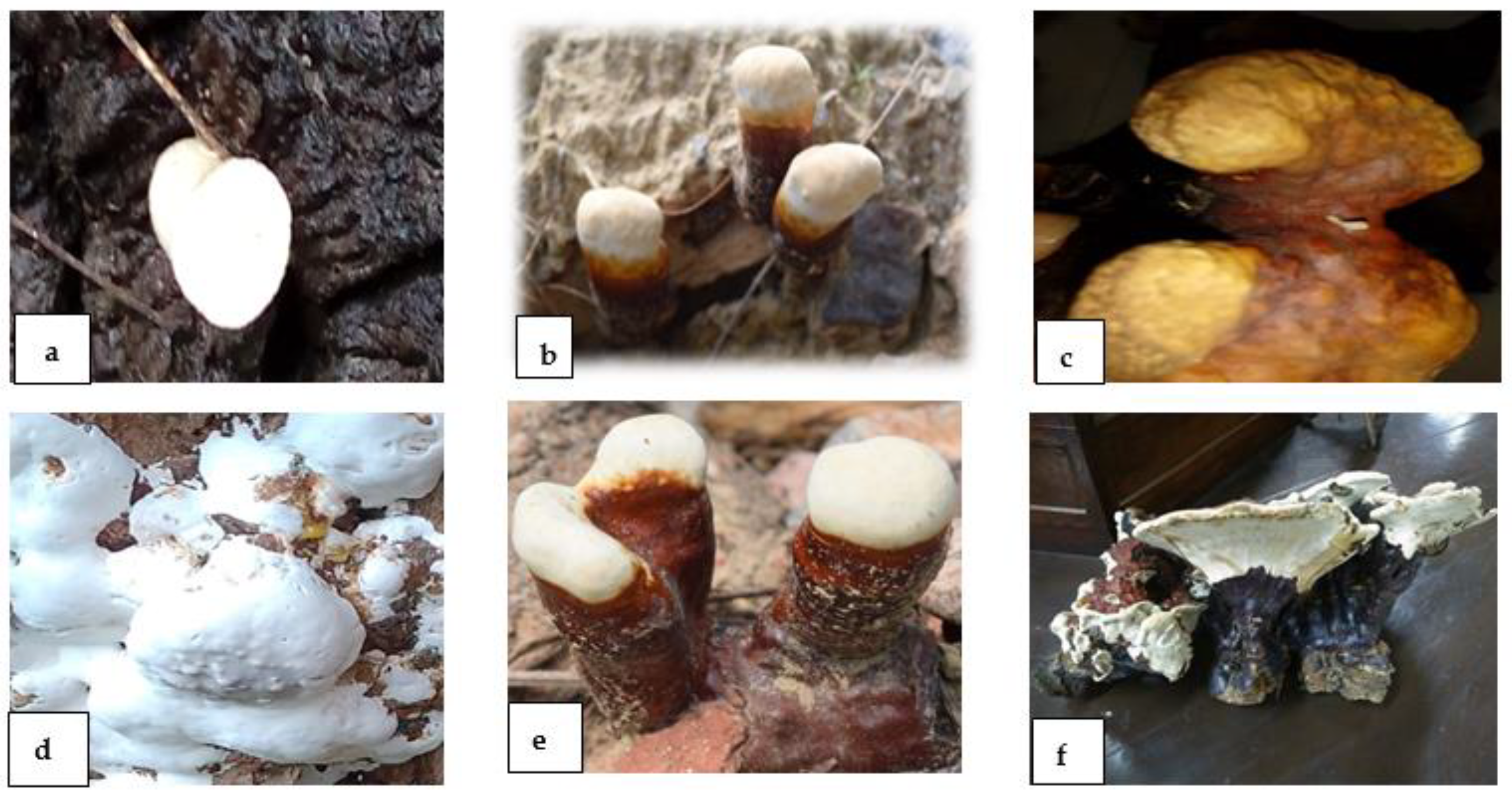
2.1. Collection, Identification and Microscopic Analysis of G. lucidum

The macrofungus, Ganoderma lucidum, was grown in monsoon season (July to September) on the campus of Aligarh Muslim University (AMU), Aligarh. Ganoderma was collected from the campus of AMU and brought to the laboratory for further examination. The morphological identification of G. lucidum was based on the shape, color and size of the basidiocarp and basidiospores. The pileus or fruiting body is reniform, kidney or concave in shape and dark reddish-brown, whereas the stalk-like stipe or stem is cylindrical in shape and has a dark brown or brownish-black stalk. The species was found to be G. lucidum based on the presence of a brilliant or shiny surface of the fruiting body, which was confirmed by the morphological investigation conducted by Wang et al. [22] (Figure 1).
For further identification, the morphology of basidiospores was examined by following the technique of [23]. Basidiospores from the basidiome of G. lucidum were dusted on a clean microscopic slide and stained with 1–2 drops of lactophenol. The structure or shape of basidiospores was observed under an inverted fluorescence microscope (Leica DMi 8, Wetzlar, Germany) magnified at 40× and 100× (Figure 2a,b). Basidiospores were found to be small, ovoid, truncated at the apex and yellowish-brown with a dark brown eusporium bearing thick echinulae, surrounded by a hyaline myxosporium.

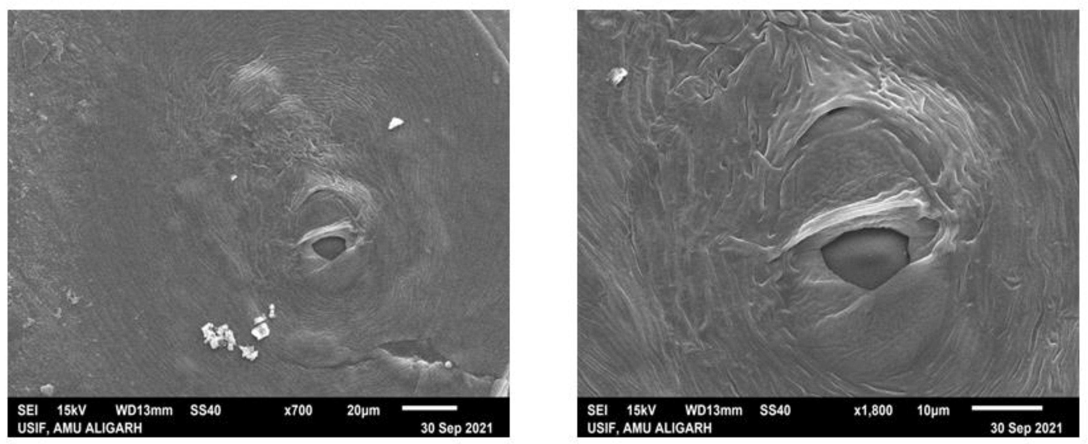
2.2. Scanning Electron Microscopy of Basidiocarp (Pileal Surface and Stipe) of G. lucidum

The ultrastructure of the basidiocarp of G. lucidum was analyzed using scanning electron microscopy (SEM, JSM 6510 LV, JEOL, Tokyo, Japan). Samples must be free from moisture to study the microstructure of hyphae of the pileus and stipe surfaces of G. lucidum. Samples were prepared using the method of Singh et al. [24]. Fresh Ganoderma was divided into two parts, i.e., the pileus and stipe. The pileus was then cut into small pieces 2 mm × 2 mm in size and the stipe was cut into small pieces of 4 mm × 4 mm. Specimens were fixed in a fixative (2.5% glutaraldehyde in 0.1% sodium phosphate buffer pH 7.2) for 24 h at 4 °C. Thereafter, samples were washed three times in a sodium phosphate buffer for 10–15 min in each wash. The second time, fixation was performed with 1% osmium tetraoxide for 2 h at 4 °C, followed by dehydration with 30% and 100% ethanol for 10 min. The samples were taken for CPD (critical point drying), and after CPD, dried specimens were mounted on an aluminum stub that had a piece of double-sided tape. Specimens were gold-coated (JEOL JFC-1600 Auto Fine Coater) with a layer about 14 nm thick [24]. The samples were now ready to be loaded into the SEM for basidiocarp surface viewing.

2.3. Cultivation and Maintenance of G. lucidum

The sample of the collected Ganoderma was inoculated into the three different heat-sealed cultivation bags, which contained substrate and had microfilter windows. The substrate of each bag consisted of rice bran (93 g), wheat bran (69 g), wood chips (1563 g) and water 775 g that resulted in a final moisture up to 55% in the mushroom bed. The temperature of the cultivated site was maintained at 23–25 °C, with air circulation for more than 12 h/day and humidity 80%. One sample of each developmental age, i.e., two-week-old (TW), four-week-old (FW) and eight-week-old (EW) G. lucidum, was harvested from separate bags [25].

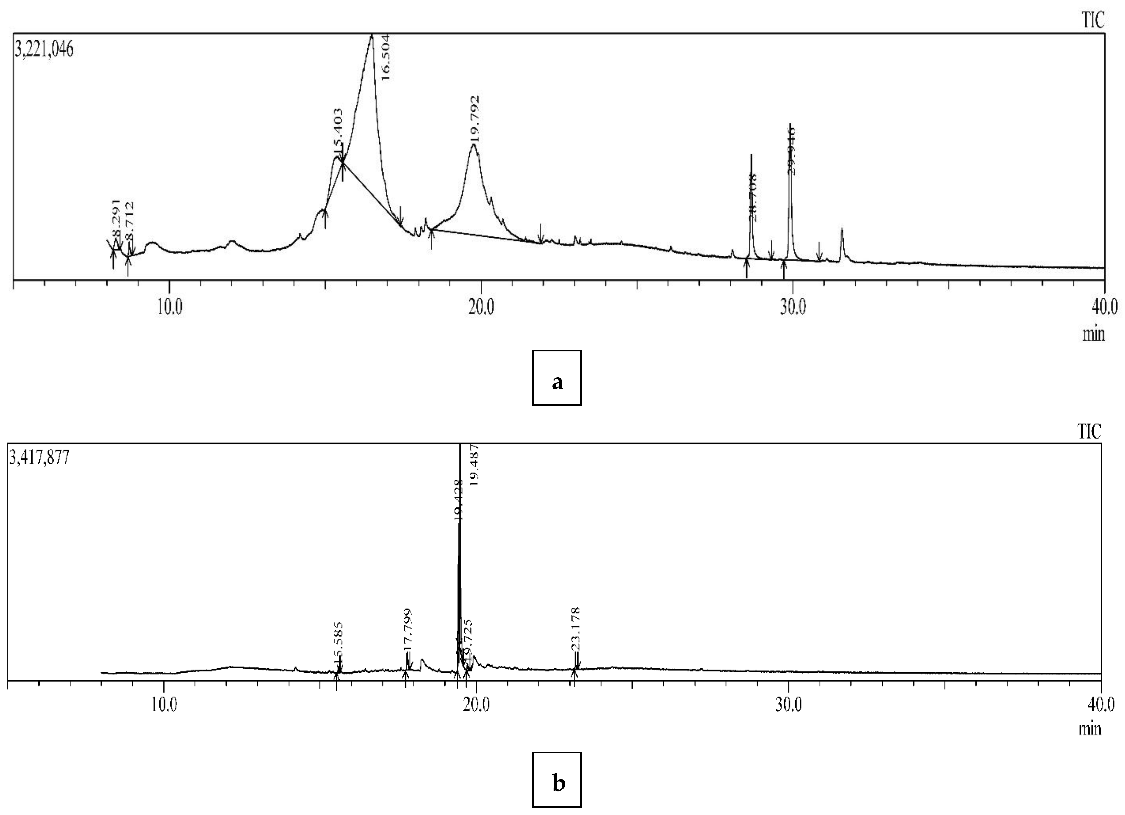
2.4. GC-MS Analysis of G. lucidum

2.4.1. Sample Preparation

For the analysis of the VOCs present in G. lucidum, a crude extract was prepared under laboratory conditions. The samples for the GC-MS analysis were prepared using the previously described method with slight modifications [26]. One hundred grams of basidiocarp (pileus and stipe separately) were washed with DW four times and air-dried on clean blotting sheets. The material was cut into small blocks and ground in a sterilized electric blender until fine. The grounded material (50 g) was soaked in a 1000 mL flask containing 500 mL of 100% ethanol. The mixture was kept for 72 h, then strained using a muslin cloth. The obtained decoction was centrifuged at 6000 rpm for 20 min, the pellet was discarded and the supernatant was filtered again using Whatman No. 1 filter paper. To avoid any contamination, the supernatant was treated with 2–3 drops of 0.1% streptomycin sulfate solution. Next, the filtrate was concentrated at 27 °C using a rotary evaporator to evaporate the ethanol. The resulting extract was stored at 4 °C in a glass vial until further use.

2.4.2. Gas Chromatography-Mass Spectrometry (GC-MS) Conditions

The metabolites were detected and quantified by gas chromatography coupled with mass spectrometry (Shimadzu QP2010 Plus, Kyoto, Japan), equipped with a Rtx-5 MS capillary column (30 m in length, internal diameter and film thickness each 0.25 mm). The derivatization procedure was performed using N-Methyl-N-(trimethylsilyl) trifluoroacetamide (MSTFA). A sample volume of 250 µL was transferred into GC glass vials and 1 µL was injected into the column with a split ratio of 10 at the rate of 1.21 mL per minute using helium as a carrier gas. The temperature of the oven was set at 100 °C, then increased to 250 °C by an increment of 5 °C per min and, finally, to 280 °C by an increment of 10 °C per min. Distinct peak fragmentation patterns of metabolites were detected using an MS detector in a full scan mode. The data were analyzed using the software GCMS Solutions (Lab Solutions ver. 2.5 Shimadzu, Kyoto, Japan); the peaks were integrated manually and the chromatograms were deconvoluted in the same way. The identification of metabolites was performed based on the retention times. The quantification of various metabolites was performed using their respective peak areas and the molecular masses were obtained. The metabolites identified were confirmed by comparing the peak spectra with standard mass spectra from three library databases, including the National Institute of Standards and Technology’s NIST 14 and NIST 14s (https://www.nist.gov/, accessed on 28 February 2022) and Wiley 8. The metabolite compounds were normalized against the internal standards.

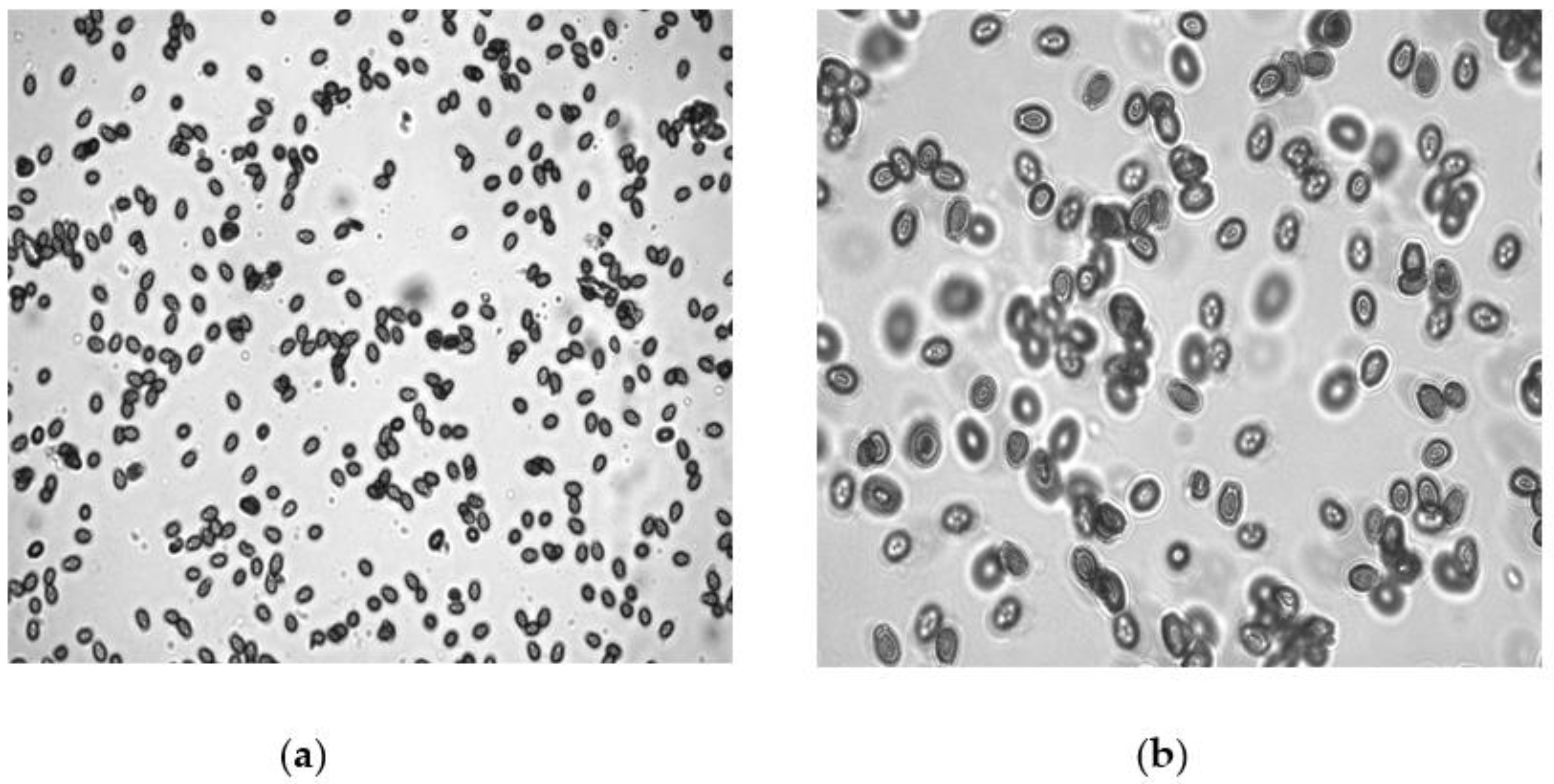
2.5. Inoculums Maintenance and Species Identification of M. incognita

The pure population of root-knot nematodes (RKNs), Meloidogyne incognita, was maintained and cultured on eggplant plants under greenhouse conditions (temperature 27 ± 2 °C, adequate photoperiod of 11 ± 1 h and humidity 65% to 80%) at the Department of Botany, AMU, Aligarh (India). Scanning electron microscopy (SEM) was utilized for species identification of the root-knot nematode. The sample was prepared using the perineal pattern technique given by [27]. A healthy female was isolated from the infected eggplant root and placed on a clean slide under a stereoscopic microscope. The section of female was cut in such a manner that the posterior portion exhibited a perineal pattern that was completely visible. Three sharp cuts were made on the posterior side of the female body; the first cut was made transversely and two cuts were made longitudinally, with one on the left and the other on the right side. The specimen was cleaned and put in 1–2 drops of lactophenol. The specimen was transferred to a new slide and kept in different series of ethanol (70%, 90% and 100% ethanol). Further steps such as CPD and the mounting and coating of samples were performed in a similar manner, using scanning electron microscopy as described above. The presence of a high dorsal arch and wavy lines, key features of Meloidogyne incognita, were observed (Figure 3).

2.6. Inoculum Preparation of M. incognita Juveniles

Infected roots were washed delicately to free them from adhering soil. The egg masses were handpicked and collected into coarse sieves, which were already layered with tissue paper, then kept in a petri plate containing DW. The petri plates were incubated at 27 ± 2 °C in the BOD incubator. After every 2 days, the juveniles’ suspension was collected and freshwater was added. The collected juveniles’ suspension was standardized as per requirement for in vitro and in planta experiments.

2.7. Toxicity Testing of G. lucidum against M. incognita Juveniles: In Vitro

2.7.1. Preparation of Culture Filtrates (CFs) of G. lucidum

Culture filtrates of each age of the basidiocarp (pileus and stipe separately) of G. lucidum were prepared for in vitro testing. G. lucidum was surface sterilized with 0.1% of NaOCl and washed gently with distilled water (DW) four times. The cleaned pileus and stipe were placed on blotting sheets for drying at room temperature. The pileus and stipe were cut into small pieces and both materials were ground separately with the help of an electric blender. An amount of 50 g of each part was transferred into a 1 L flask containing 1000 mL of distilled water and left for 24 h. The mixture of fungus was filtered with the help of a cheese cloth, followed by Whatman No. 1 filter papers. The filtrate was centrifuged at 6000 rpm for 20 min to obtain a clear solution and was considered as stock for further use.
The stock solution of the pileus and stipe of each stage of G. lucidum was considered as standard or as a treatment of 100% concentration. This standard concentration was diluted with DW to obtain 50%, 10% and 1% concentrations. Two drops of 0.1% streptomycin sulfate solution was added to avoid bacterial contamination.

2.7.2. Inhibition in Hatching of M. incognita Eggs Exposed to the CFs of G. lucidum

For hatching purposes, five fresh and healthy egg masses were transferred into each petri dish containing 5 mL of CF of all three ages with different concentrations (100%, 50%, 10% and 1%) of the pileus and stipe of the Ganoderma. All petri dishes were kept in an incubator at 28 ± 2 °C for 5 days. The total number of hatched second-stage juveniles (J2) was counted in a counting dish under a stereoscopic microscope.

2.7.3. Mortality and Immobility of J2 of M. incognita Exposed to the CFs of G. lucidum

The culture filtrate (4.8 mL) of each dilution of TW-, FW- and EW-old pileus and stipe extracts was put into petri dishes. An amount of 0.2 mL of pure suspension of M. incognita containing one hundred freshly hatched juveniles was added to the treated and control petri dishes, in which the latter had only DW. All the petri dishes were kept on a laboratory bench at room temperature and the total number of dead juveniles was counted using a counting dish after 12, 24, 48 and 72 h of exposure time under the stereo microscope. According to El-Rokiek and El-Nagdi, [28], if there is any movement and the shape of the nematode is winding, then it is considered as alive. The nematode is found dead when there is no movement or it is straight. Keeping these points in mind, the number of both alive and dead nematodes was recorded. The percent mortality or inhibition in egg hatching was calculated by using the formula:
% inhibition or mortality = {(C0 − Tα)/C0} × 100
where
in the case of hatching,
C0 = number of juveniles hatched in the control and Tα = number of juveniles hatched in each concentration of CF.
In the case of mortality,
C0 = number of alive nematodes in the control and Tα = number of alive nematodes after the 12, 24, 48 and 72 h exposure period.

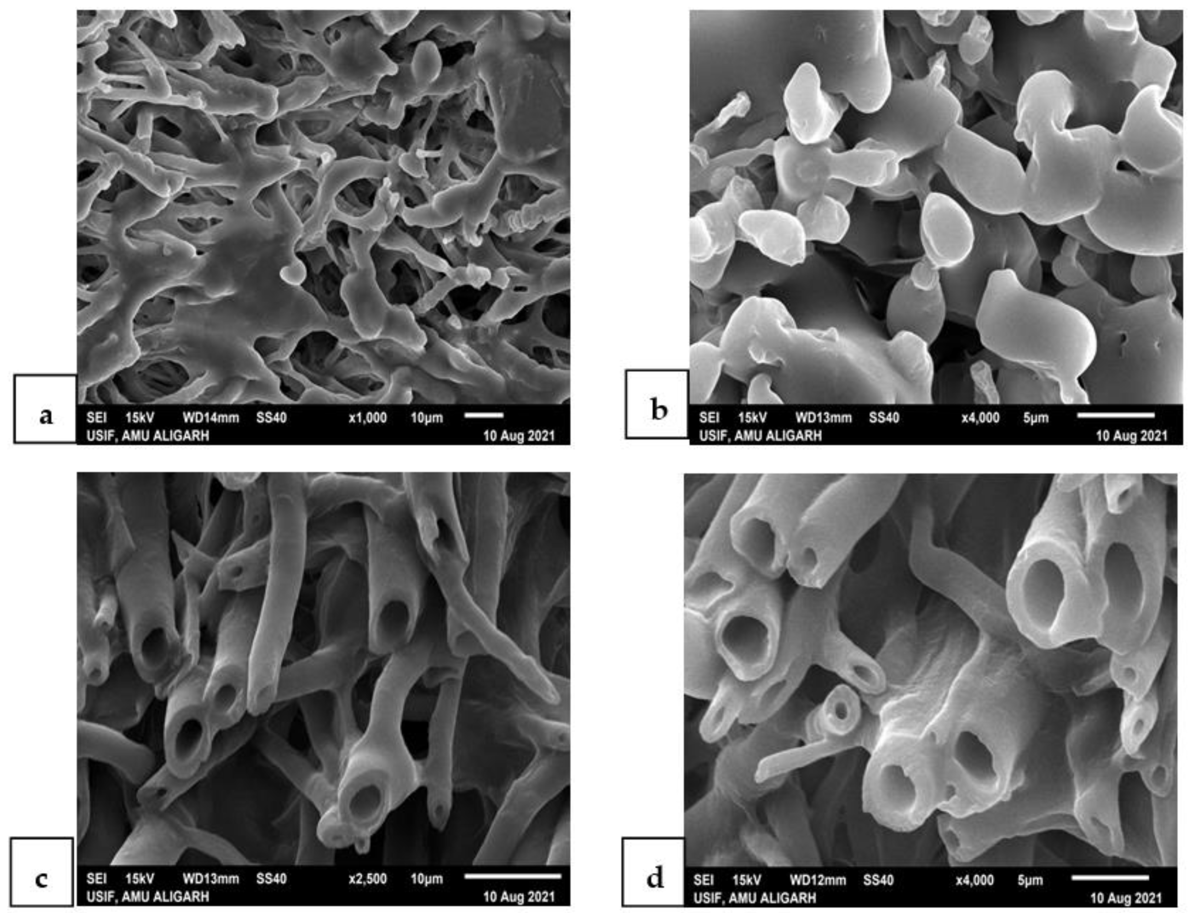
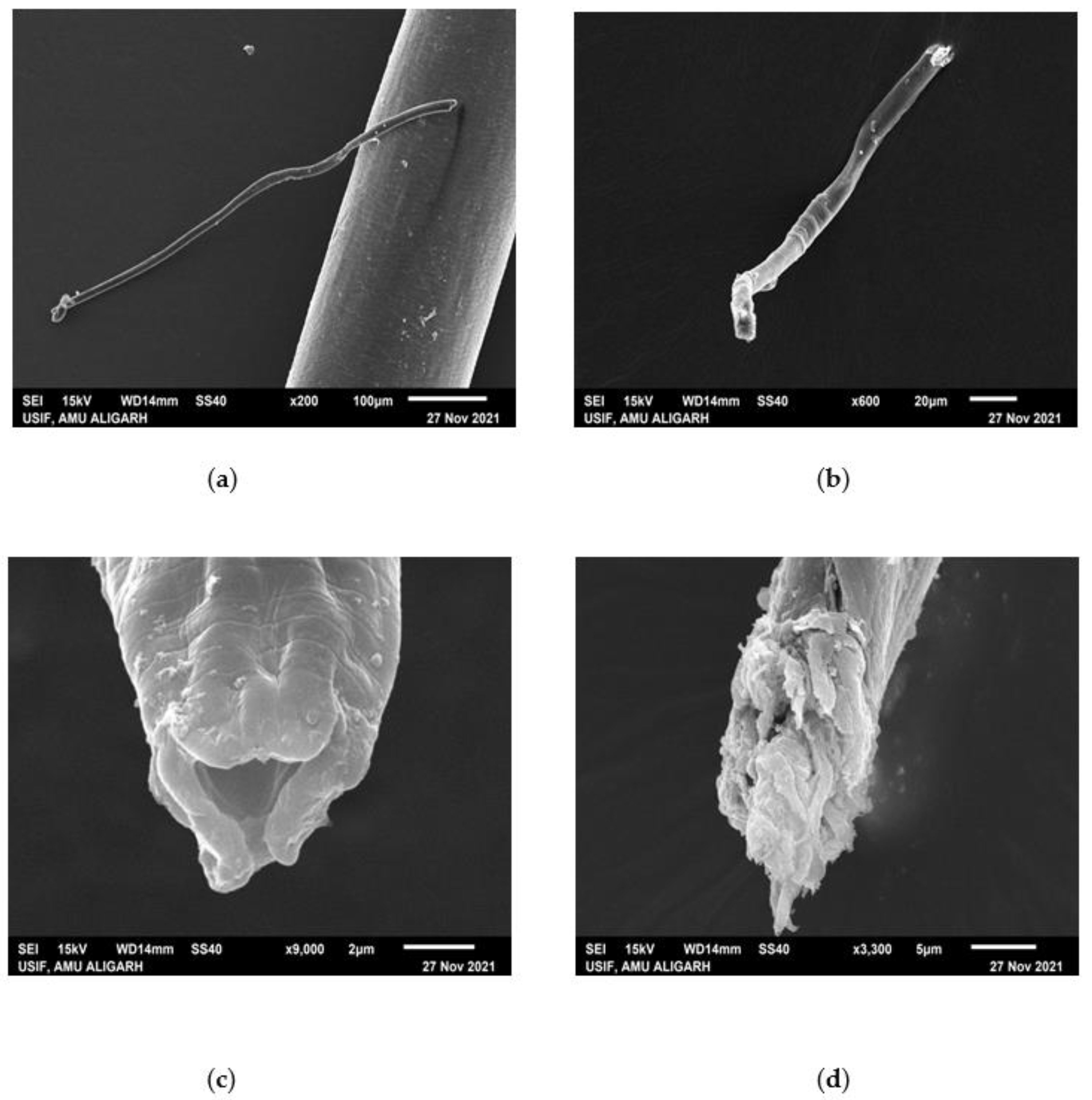
2.8. SEM of Treated Juveniles (TJ) and Untreated Juveniles (UTJ) of M. incognita

To explore the impact of G. lucidum on the external surface of the root-knot nematode, M. incognita, juveniles were treated with the CF of the TW-old pileus of G. lucidum. Specimens from treated and control petri dishes (healthy juveniles) were handpicked with the help of a horse tail hair attached to a needle and fixed in 40% formaldehyde for 24 h. Fixed specimens were transferred into 3% glutaraldehyde [29] for 48 h. After that, specimens were transferred to capsules and washed with a 0.05 M sodium phosphate buffer twice. The specimens were dehydrated in a graded ethanolic series (30, 50, 70, 80, 90 and 100%) and carried for CPD (critical point drying) in carbon dioxide [27]. Dried specimens were kept in a desiccator and mounted on stubs, coated with gold. The nematode surface was analyzed after loading the specimens in the SEM (model, JOEL JSM 6510 LV) at a different resolution at 15 KV.

2.9. In Planta Study (Pot Experiment)

The pot study was conducted under greenhouse conditions. Three-week-old healthy eggplant seedlings were exposed to the CFs of two-week-old Ganoderma (100% conc.), which were applied as a root dipping treatment for 30 min along with the control. Afterward, all the seedlings were transferred in autoclaved earthen pots (30 cm diameter) filled with 2 kg of sandy loam soil and manure in the ratio of 3:1. Each treatment was replicated five times, including the control (untreated uninoculated control, UUC). Three days later, each plant was inoculated with 3000 freshly hatched juveniles (J2) of M. incognita by making three holes around the soil without damaging the roots. The plants were watered and cared for time to time.

2.10. Data Collection and Observations

Three months after inoculation, all plants were uprooted gently and washed in a bucket filled with water to remove adhering soil. Care was taken while dealing with roots to ensure the safety of root galls. The data for plant growth were analyzed in terms of shoot and root fresh weight, shoot and root dry weight, yield and physiological (total chlorophyll and carotenoids) and pathological parameters (juveniles/kg soil, females/root system, no. of galls/root system and number of eggs/egg-mass). Daykin and Hussey’s [30] methodology was adopted for examining egg masses. Cobb’s sieving and decanting procedure [31] was used to determine the total population of the nematodes, followed by a modified Baermann funnel technique [32]. The total amount of chlorophyll and carotenoids in the fresh leaf tissues was analyzed following the protocol given by Mackinney [33]. The following formula was used to determine the chlorophyll and carotenoid content of the sample (mg/g fresh leaf).
Total chlorophyll = {20.2(A645) + 8.02(A663)} × (V/1000 × W).
Carotenoid = {7.6(A480) − 1.49(A510)} × (V/1000 × W).
A480, A510, A645, A663 = absorbance of extract at given wavelengths (480, 510, 645, and 663 nm, respectively).
V = final volume of the extract
W = fresh weight of the leaf sample
D = length of the path of light

2.11. Statistical Analysis

Each treatment was conducted in five replications. Data on hatching and mortality were displayed in terms of values of percent mortality over the control. Experimental data from the pots study were analyzed by a one-way analysis of variance (ANOVA) using the SPSS-17.0 statistical software (SPSS Inc., Chicago, IL, USA). The variability between mean values of treatments was determined by Duncan’s multiple range test (DMRT) at p ≤ 0.05.

3. Results

The morphological changes of the fruiting bodies of G. lucidum during its development were categorized into three successive stages or ages, as can be seen in Figure 1. The scanning electron microscopic analysis of the pileus and stipe and inverted fluorescence microscopic investigation of the spores of the fruiting body of Ganoderma gave conformity to the species of G. lucidum (Figure 2 and Figure 4). Basidiospores appeared small, ovoid, truncated at the apex and yellowish-brown with a dark brown eusporium bearing thick echinulae, surrounded by a hyaline myxosporium. Perineal pattern identification via scanning electron microscopy provided confirmation about the species of Meloidogyne incognita, showing the presence of a high dorsal arch with wavy lines. For the in vitro test, four variable concentrations (100%, 50%, 10% and 1%) of CFs of both parts, i.e., the pileus and stipe, were taken as treatments, along with DW as the control. The ascendancy of different treatments of CFs of two isolated parts (pileus and stipe) of G. lucidum acted differently on the hatching and mortality of the second-stage (J2) juveniles of M. incognita at different exposure times (12, 24, 48 and 72 h). The evaluation of the toxicity of pileus and stipe CFs of G. lucidum on the hatching and mortality of J2 of M. incognita was found significant.

3.1. Effect of Different Concentrations of G. lucidum CFs on the Egg Hatching of M. incognita

It is apparent by the perusal of data presented in Table 1 and Table 2 that after five days of incubation the culture filtrate of the pileus and stipe of G. lucidum showed the nematicidal effect to varying degrees on egg hatching of the root-knot nematode, M. incognita. At 100% concentration, the highest inhibition in the emergence of M. incognita juveniles was recorded in TW-old pileus CFs, followed by TW stipe, FW pileus, FW stipe, EW pileus and EW-old stipe CFs. The corresponding percentage of suppression in egg hatching was recorded as 89.2, 81.0, 77.1, 69.1, 55.9, and 45.0%, as compared to the control (0.0%). Similarly, at 50% conc., inhibition in the emergence of juveniles from the eggs in respective CFs was observed as 72.8, 68.5, 51.0, 34.0, 39.0, and 31.0%, as compared to the control (Table 1 and Table 2).
In the 10% treatment, the greatest reduction in egg hatching was noticed in TW-old pileus CF (53.0%), followed by TW stipe (42.3%), FW pileus (33.1%), FW stipe (25.1%), EW pileus (29.9%) and EW stipe (22.3%), as compared to the control (0.0%). The least inhibition in egg hatching was found at 1% conc. and was recorded as 24.0, 20.0, 18.7, 14.9, 14.0 and 7.6% in the corresponding CFs of different parts of G. lucidum (Table 1 and Table 2). However, there was no significant difference in the inhibition of egg hatching noticed in the 1% treatment of FW- and EW-old stipe CFs.
There was a relatively significant decrease in the eggs hatching with the corresponding increase in the concentration of fungus CFs. Inhibition in egg hatching at different treatments significantly declined with the increase in the age of the parts of G. lucidum. Among the different parts (pileus and stipe) of G. lucidum, the pileus CFs showed the most significant and greatest inhibition in the eggs hatching as compared to the stipe of the fungus.

3.2. Effect of Different Concentrations of G. lucidum CFs on the Mortality of M. incognita

The data presented in Table 3 and Table 4 indicated that the mortality of juveniles of M. incognita was observed to be significant (p ≤ 0.05) at different concentrations of pileus and stipe CFs at different exposure periods. The percent mortality of J2 was directly proportional to the concentration of fungal extracts of pileus and stipe and the period for which the nematode was exposed to the extract. No percent mortality was recorded in all the treatments of G. lucidum, even after 12 h of the exposure period. For the pileus, 83.8% mortality was found at a 72-h exposure time in 100% concentration of CFs of two-week-old (TW) Ganoderma. Certainly, the CFs of the pileus was more efficacious in killing juveniles of M. incognita than CFs of the stipe. TW pileus in 100% treatment for 72 h of incubation occupied the supremacy of the reduction in nematode juveniles over other TW-, FW- and EW-old treatments (Table 3). The rate of mortality was low in the beginning, but an appreciable increase was found only after 24 h of the exposure. Mortality of juveniles reached up to 67% on FW-old pileus in 100% treatment for a 72-h exposure period. The least amount of mortality of 47% was found on EW-old pileus in the 100% treatment for a 72-h incubation period, compared to 0% in the control.
A nearly similar trend of mortality was recorded for the set-ups treated with CFs of different ages of the stipe (Table 4). The highest mortality (73.8%) was seen in TW-old stipe with the 100% treatment and a 72-h exposure time. The FW- and EW-old stipe showed 61.6% and 38.9% of J2 mortality in the 100% treatment with a 72-h exposure period, respectively. After 12 h of the exposure period, there was no percent mortality observed in the EW-old pileus- and stipe-treated nematodes in all concentrations. The mortality of juveniles of M. incognita was found nil in all the 1% treatments of TW-, FW- and EW-old pileus and stipe at 12 h. In most of the treatment concentrations and exposure periods, the mortality of M. incognita was significantly decreased with the increase in age of G. lucidum.

3.3. GC-MS Analysis for Bioactive Compounds

A total of 13 metabolites were identified in the basidiocarp (pileus and stipe) of the G. lucidum. The results revealed different bioactive compounds; seven in the pileus and six in the stipe of the G. lucidum were detected. The GC-MS analysis showed that the metabolites obtained belonged to diverse chemical functional groups, including pyrones (2,3—Dihydro—3,5—Dihydroxy—6—Methyl—(4H)—pyran—4—one), alkenes (2—Hexene—3,4,4—Trimethyl), glucosides (Beta—D—Glucopyranoside, methyl), sugar alcohols (DL—Arabinitol and D—Mannitol), silanes (Silane, dimethyl (3—fluorophenoxy) tetradecyloxy) and steroids (Cholesta—8,14—dien—3—ol, 3 beta, 5—alpha) present in the pileus (Table 5, Figure 5a), and cyclic ketone ((3aR, 4R, 7R)—1,4,9,9—Tetramethy l—3,4,5,6,7,8—hexahydro—2H—3a, 7—methanoazulen—2—one), palmitates (hexadecanoic acid, methyl ester), linoleic acids (9,12—Octadecadienoic acid (Z, Z)-, methyl ester), oleic acids (9—Octadecenoic acid, methyl ester, (E), Ester (Methyl stearate) and Phthalic acid (Bis (2—ethylhexyl) phthalate) present in the stipe of G. lucidum (Table 5, Figure 5b). The important information, such as the name of the compound, retention time, peak area, peak area (%), chemical formula and chemical functional group, were listed in Table 5.

3.4. SEM of the Treated Juveniles (TJ) and Untreated Juveniles (UTJ)

SEM analysis of the TJ and UTJ showed a great difference in their surface morphology. Figure 6 indicated that treated juveniles were deformed in their structure, appearance and size as compared to the untreated control. As we can see in Figure 6, the entire body of the UTJ (a) was found as it is, whereas the TJ (6b) was missing the anterior portion. In comparison, a high magnification view of the anterior side depicts that there was a huge disruption of the mouth region in the TJ (6c), whereas the anterior shape was perfect in the UTJ (6d). These morphological alterations in TJ specimens were due to the toxicity of the culture filtrate of G. lucidum. The GC-MS identified some metabolites that have a nematotoxic effect on the morphology of the nematode.

3.5. Effect of Root Dip Treatment with CFs of G. lucidum on Multiplication of M. incognita Parasitized Eggplant (in Planta Study)

The data analysis of the in vitro test on hatching and mortality of M. incognita revealed that CFs of G. lucidum can curtail the population of nematodes. To examine whether the root dip treatment of Ganoderma CFs affects M. incognita parasitism during a compatible interaction, a pot study was conducted where the CFs of TW-old Ganoderma were applied as a root dip treatment on eggplant susceptible to M. incognita. Significantly, the highest decline in the number of galls (68), number of egg masses (97), number of eggs/egg mass (129), juveniles/kg soil (5293) and adult females per root system (123) was achieved when roots of healthy seedlings were dipped in 100% CFs of G. lucidum for 30 min (Table 6). The lowest reduction in nematode infestation was observed at 10 and 1% concentrations with the minimum number of galls (126 and 132), the number of egg masses (131 and 137), the number of eggs per egg mass (330 and 342), juveniles/kg soil (11,942 and 12,349) and females/root system (259 and 278), respectively (Table 6). The values of pathological parameters for 10 and 1% treatments were closely related to values of the untreated inoculated (UIC) control, which exhibit the highest number of galls (139), number of egg masses (146), number of eggs/egg mass (355), juveniles/kg soil (12,844) and females/root system (327) (Table 6). The concentrated form of Ganoderma CFs with 100% conc. significantly suppressed the infection intensity of M. incognita as compared to the UIC.

3.6. Effects of Root Dip Treatment with CFs of G. lucidum on Eggplant Growth

Treatments of Ganoderma CFs as a root dip for 30 min significantly enhanced the plant growth and yield of eggplant. The treatment (100%) improved the highest plant growth in terms of shoot length (48.39 cm), root length (18.86 cm), shoot fresh weight (220 g), root fresh weight (92.0 g), shoot dry weight (34.20 g), root dry weight (10.45 g) and yield per plant (605) compared to the control (UIC), followed by the treatment of 50% that presented the second-best result of promoting plant growth, shown in Table 7. The UIC gave the lowest growth for the shoot (25.45 cm) and root (13.3 cm) length, shoot (110 g) and root (40.0 g) fresh weights, shoot (22.63 g) and root (6.45 g) dry weights and yield (347). The diluted forms of CFs of fungus, i.e., 10 and 1%, resulted in slightly improved plant growth compared to the UIC. The treatment of 10% conc. showed better growth than the UIC in terms of shoot length (45.32 cm), root length (17.25 cm), shoot fresh weight (147 g), root fresh weight (61.0 g), shoot dry weight (23.50 g), root dry weight (7.40 g), and yield per plant (432) (Table 7). The smallest growth among all the concentrations of Ganoderma was recorded in the 1% treatment, with shoot length, root length, shoot fresh weight, root fresh weight, shoot dry weight, root dry weight and yield per plant having the corresponding values of 43.56 cm, 16.99 cm, 132 g, 48 g, 22.10 g, 6.40 g and 376, respectively. The values of plant growth of the 1% treatment were not equal to, but were almost the same as the growth values of the untreated inoculated control (Table 7). The data presented in Table 7 depicted that the best significant growth promotion was observed in the 100% treatment compared to the untreated uninoculated control (UUC).

3.7. Effect of CFs of G. lucidum on the Physiology of Eggplant

Similarly, the use of G. lucidum CFs as a root dip treatment for 30 min stimulated the physiological activity of the plant. The highest content of total chlorophyll (1.93 mg/g) and carotenoids (0.81 mg/g) was observed in the 100% treatment of G. lucidum (Table 7). On the other hand, the lowest value of total chlorophyll and carotenoids, i.e., 1.49 mg/g and 0.58 mg/g, respectively, were found in the 1% concentration of the Ganoderma. The values of the 1% treatment were slightly greater than the values of the UIC, which were 1.45 for chlorophyll and 0.54 for carotenoids (Table 7).

4. Discussion

The motive of the current investigation was to deduce important information for the effective formulation of the king of the herb, ‘G. lucidum’. This macrofungus was utilized at three different ages to reduce the disease-causing intensity of the root-knot nematode, M. incognita. Among the different developmental stages of the root-knot nematode, second-stage juvenile (J2) is the infective stage where it hatches out and searches for a loving host to penetrate root tissues. They obtain their nourishment by destroying the root system of the host plant (eggplant). To overcome this problem, we tested the impact of different concentrations of CFs of TW-, FW- and EW-old G. lucidum on the egg hatching and mortality of M. incognita. It was clear from the results that after five days, different treatments of G. lucidum CFs showed inhibition in the hatching of eggs of M. incognita to a varying degree, irrespective of the age of G. lucidum. There was a relatively significant decrease in the number of eggs hatching that corresponded with an increase in the concentration of fungus CFs. Inhibition in egg hatching with different treatments significantly decreased with the increase in the age of G. lucidum, except with the 1% treatment of four- and eight-week-old stipe. The pileus of G. lucidum CFs showed significantly the greatest inhibition in egg hatching as compared to the stipe of the fungus. In the “100%” treatment, the highest inhibition in the juvenile emergence of M. incognita was recorded in the two-week-old pileus CF, followed by two-week stipe, four-week pileus, four-week stipe, eight-week pileus and eight-week-old stipe CFs.
Similarly, the extracts of pileus and stipe of G. lucidum manifest the nematicidal effect to varying degrees on M. incognita. The percentage of mortality of nematodes was directly proportional to the treatment of fungal CFs and the period for which the nematode was exposed to the extract. In most of the concentrations of the treatment and exposure periods, the mortality of M. incognita significantly decreased with the increase in age of G. lucidum. Generally, the pileus CF showed significantly the greatest mortality of juveniles as compared to the stipe extract of G. lucidum in different concentrations and exposure periods. The nematicidal potential of the extract of fungus on the plant-parasitic nematode may be attributed to the production of certain toxic metabolites, such as triterpenes, sesquiterpenes, applannoxidic acid, steroids and bioactive peptidoglycans by Ganoderma species [34,35,36]. Akshaya et al. [20] and Themuhi et al. [21] observed 93.2% juvenile mortality and 92.6% egg hatching inhibition at a 1000 ppm concentration of G. lucidum extract. These results are also in agreement with the findings of Bua-art et al. [37] in which the bioactive compound extracted from Neonothopanus nambi at a concentration of 500 mg/L caused 100% mortality of J2 of Meloidogyne incognita in 1 min. The concentrations of 100 and 50 mg/L affected the J2 and resulted in a significant mortality of 100% in 30 min and 48 h, respectively. The effect of the bioactive compound on J2 was also confirmed in a screened-house experiment and found that concentrations of 100 and 500 mg/l suppressed J2 without root-knot symptoms on tomato plants. The adverse effect of the culture filtrate of several other fungi on the mortality and hatching of nematodes has been reported by other workers also [38,39,40,41].
The variable effect of pileus and stipe CFs on the hatching and mortality of root-knot nematodes observed in the present studies can be attributed to the varied nature of the toxic metabolites produced by G. lucidum in different parts of the fruiting body. GC-MS analysis of the pileus and stipe of G. lucidum explored thirteen different bioactive compounds that belong to diverse groups of secondary metabolites, including pyrones, glucosides, sugar alcohols, silanes and steroids present in the pileus, and cyclic ketone, palmitates, linoleic acids, oleic acids, ester and phthalic acid present in the stipe of G. lucidum. It can be concluded that the presence of bioactive compounds is responsible for curtailing the nematode population effectively. The antimicrobial properties of G. lucidum were found effective in inhibiting the development of nematodes. The surface investigation of the TJ and UTJ using scanning electron microscopy provided confirmation on the lethal effect of G. lucidum toxicity against nematodes. It was crystal clear from comparing the TJ with the UTJ that the extract of G. lucidum interferes with the metabolic activities of nematodes, resulting in the degradation of juveniles.
There are only two findings available regarding the nematicidal potential of Ganoderma lucidum [20,21], in which they investigated the inhibitory potential of an ethyl acetate extract of Ganoderma lucidum and Lentinus edodes on M. incognita. Akshaya et al. [20] also showed twenty-two bioactive metabolites of G. lucidum by GC-MS analysis. In the present study, it was noted that the three bioactive compounds of the pileus ethanolic extract, including hexadecanoic acid, methyl ester; methyl stearate and Bis (2—Ethylhexyl) Phthalate, resembled the same compounds identified already by investigators Akshaya et al. [20]. Zhao et al. [42] explored the toxicity of bioactive compounds released by Simplicillium chinense Snef5 on the suppression of the root-knot nematode, M. incognita. Limited research has been conducted on the fungicidal activity of it. The adverse effect of G. lucidum against different fungi has also been reported by Innocenti et al. [43]. Badalyan et al. [44] reported the antagonistic activity of 17 species (C. versicolor, F. velutipes, G. lucidum, H. fasciculare, H. sublateritium, K. mutabilis, L. edodes, P. alnicola, P. aurivella, P. destruens, P. ostreatus, P. cornucopiae, P. squamosus, P. subarcularius, P. varius and S. commune) against four fungi (B. sorokiniana, F. culmorum, G. graminis var. tritici and R. cerealis), responsible for foot and root disease of cereals. Almost all tested mushroom species markedly inhibited mycelia growth of the four phytopathogenic fungi; however, the antagonistic activity of P. ostreatus, G. lucidum, H. fasciculare, L. tigrinus and S. commune was stronger. Sridhar et al. [45] evaluated the antifungal activity of the fruiting body of Ganoderma lucidum against five fungi, viz., Penicillium spp., Aspergillus fumigatous, A. niger, A. flavus and Mucor indicus. For antifungal activity, the zone of inhibition of microorganisms was measured in mm. The methanolic extract showed the maximum antifungal activity and a 30 mm inhibition zone was recorded in 200 mg of the extract against Mucor indicus, whereas a minimum 3 mm inhibition zone was recorded in 50 mg of the extract against Aspergillus flavus. Liu et al. [46] also conducted studies on various nematophagous fungi.
There is no pot study available regarding plant disease management to manage or control Meloidogyne incognita in greenhouse or field. In the present study, G. lucidum was used as a root dip treatment for the first time in the greenhouse condition. The data analysis indicates that the TW-old G. lucidum significantly regulated growth and physiology in terms of fresh shoot and root weights, dry shoot and root weights and chlorophyll and carotenoid content of eggplant plants. There was a great increase in yield per plant. Ganoderma lucidum showed a significantly negative correlation towards nematode development. As the concentration of G. lucidum increased, there was a significant decrease in juvenile numbers, adult females, number of galls and number of egg masses.
In the present investigation, the extract of G. lucidum could be used as a biological control for the management of nematodes. The interest of scientists can adhere to the development of novel nematicidal/fungicidal compounds derived from fungal culture filtrates. These toxic fungus metabolites can effectively target the nematodes/fungi and are less hazardous to the environment than traditional pest control chemical methods. Due to its significant nematicidal activity shown under laboratory and greenhouse conditions, the G. lucidum CFs may be tested for their ability to control M. incognita under field conditions. More research can be conducted to isolate and identify the effective components present in fungal extract which are responsible for their pesticidal activity.

5. Conclusions

The fungus can colonize a plant’s roots and secrete secondary metabolites against pathogens and stimulate plant growth. The entry of the phytopathogens into the root can be inhibited by the interference of metabolites secreted by biocontrol agents, including the root-knot nematodes. The present investigation revealed that the production of different bioactive compounds by Ganoderma lucidum has a toxic effect on M. incognita juveniles, inhibiting egg hatching and reducing the nematode population. Two-week-old pileus and stipe were found most effective against the nematode activity. SEM analysis of Ganoderma-treated juveniles indicated that the compounds present in Ganoderma significantly degraded the entire body of the nematode. It was concluded that the bioactive compounds released from G. lucidum can be used as a promising biocontrol product or TW-old G. lucidum as a biocontrol agent for future root-knot nematode management strategies, and also provide an excellent alternative approach in place of nematicides.

Author Contributions

S.F.: conceptualization, investigation, visualization, writing—original draft; F.K. and M.A.: conceptualization, investigation, writing—original draft; S.S.A. and K.I.: software, writing—review and editing; M.S. and A.K.: formal analysis, writing—review and editing; M.I. and F.A.: formal analysis, writing—review and editing; T.A.K., R.M. and M.A.S.: conceptualization, supervision, software, writing—review and editing; S.S.A.: resources, writing—review and editing. All authors have read and agreed to the published version of the manuscript.

Funding

This research received no external funding.

Institutional Review Board Statement

Not applicable.

Informed Consent Statement

Not applicable.

Data Availability Statement

The data presented in this study are available within the article.

Acknowledgments

Authors extend their appreciation to USIF, Aligarh Muslim University, Aligarh and AIRF, Jawaharlal Nehru University, New Delhi for providing the instrumentation facility. Saqer S. Alotaibi is highly thankful to the Taif University Researchers Supporting Project Number (TURSP-2020/38), Taif University, Taif Saudi Arabia, for financial support.

Conflicts of Interest

The authors declare no conflict of interest.

References

  1. Kandoliya, U.K.; Bajaniya, V.K.; Bhadja, N.K.; Bodar, N.P.; Golakiya, B.A. Antioxidant and nutritional components of eggplant (Solanum melongena L.) fruit grown in Saurastra region. Int. J. Curr. Microbiol. Appl. Sci. 2015, 4, 806–813. [Google Scholar]
  2. FAOSTAT. Production of Lentils, “Crops/World Regions/Production Quantity, from Picklists”. 2020. Available online: http://faostat3.fao.org/browse/Q/QC/E (accessed on 22 January 2022).
  3. Abd-Elgawad, M.M. Biological control of nematodes infecting eggplant in Egypt. Bull. Nat. Res. Cent. 2021, 45, 6. [Google Scholar] [CrossRef]
  4. Peng, D.; Chen, Q. Nematode chitin and application. Adv. Exp. Med. Biol. 2019, 1142, 209–219. [Google Scholar]
  5. Kumar, A.; Patil, J.A.; Yadav, S.; Ram, S. Screening, confirmation and field evaluation of promising resistant germplasm of different pulses against root knot nematode, Meloidogyne javanica. J. Environ. Biol. 2020, 41, 1594–1598. [Google Scholar] [CrossRef]
  6. Chitwood, D.J. Phytochemical based strategies for nematode control. Ann. Rev. Phytopathol. 2002, 40, 221–249. [Google Scholar] [CrossRef] [Green Version]
  7. Xiang, N.; Lawrence, K.S.; Donald, P.A. Biological control potential of plant growth-promoting rhizobacteria suppression of Meloidogyne incognita on cotton and Heterodera glycines on soybean: A review. J. Phytopathol. 2018, 166, 449–458. [Google Scholar] [CrossRef] [Green Version]
  8. Wasser, S.P. Current findings, future trends, and unsolved problems in studies of medicinal mushrooms. App. Microbiol. Biotech. 2011, 89, 1323–1332. [Google Scholar] [CrossRef]
  9. Li, G.H.; Yu, Z.F.; Li, X.; Wang, X.B.; Zheng, L.J.; Zhang, K.Q. Nematicidal metabolites produced by the endophytic fungus Geotrichum sp. AL4. Chem. Biodiv. 2007, 4, 1520–1524. [Google Scholar] [CrossRef]
  10. Kirk, P.M.; Cannon, P.F.; David, J.C.; Stalpers, J.A. Ainsworth and Bisby’s Dictionary of the Fungi, 10th ed.; CABI Publishing: Wallingford, UK, 2008; p. 771. [Google Scholar]
  11. Ahmad, M.F. Ganoderma lucidum: Persuasive biologically active constituents and their health endorsement. Biomed. Pharmacol. 2018, 107, 507–519. [Google Scholar] [CrossRef]
  12. Sanodiya, B.S.; Thakur, G.S.; Baghel, R.K.; Prasad, G.B.K.S.; Bisen, P.S. Ganoderma lucidum: A potent pharmacological macrofungus. Curr. Pharma. Biotechnol. 2009, 10, 717–742. [Google Scholar] [CrossRef]
  13. Smith, J.; Rowan, N.; Sullivan, R. Medicinal Mushrooms: Their Therapeutic Properties and Current Medical Usage with Special Emphasis on Cancer Treatment; Special Report Commissioned by Cancer Research UK, The University of Strathclyde in Glasgow; Johns Hopkins University Press: Baltimore, MD, USA, 2002; p. 256. [Google Scholar]
  14. Gao, Y.; Zhou, S.H.; Huang, M.; Xu, A. Antibacterial and antiviral value of the genus Ganoderma P. Karst. species (Aphyllophoromycetideae): A review. Int. J. Med. Mushrooms 2003, 5, 235–246. [Google Scholar] [CrossRef]
  15. Wang, H.; Ng, T.B. Ganodermin, an antifungal protein from fruiting bodies of the medicinal mushroom Ganoderma lucidum. Peptides 2006, 27, 27–30. [Google Scholar] [CrossRef] [PubMed]
  16. Bishop, K.S.; Kao, C.H.; Xu, Y.; Glucina, M.P.; Paterson, R.R.M.; Ferguson, L.R. From 2000 years of Ganoderma lucidum to recent developments in nutraceuticals. Phytochem 2015, 114, 56–65. [Google Scholar] [CrossRef] [PubMed] [Green Version]
  17. Zhao, C.; Fan, J.; Liu, Y.; Guo, W.; Cao, H.; Xiao, J.; Liu, B. Hepatoprotective activity of Ganoderma lucidum triterpenoids in alcohol-induced liver injury in mice, an iTRAQ-based proteomic analysis. Food Chem. 2019, 271, 148–156. [Google Scholar] [CrossRef]
  18. Ahmad, M.F. Ganoderma lucidum: A rational pharmacological approach to surmount cancer. J. Ethnopharmacol. 2020, 260, 113047. [Google Scholar] [CrossRef]
  19. Ren, L.; Zhang, J.; Zhang, T. Immunomodulatory activities of polysaccharides from Ganoderma on immune effector cells. Food Chem. 2021, 340, 127933. [Google Scholar] [CrossRef]
  20. Akshaya, S.B.; Krishnamoorthy, A.S.; Nakkeeran, S.; Poornima, K.; Sivakumar, U. Inhibitory potential of ethyl acetate extract from mushrooms against root-knot nematode (Meloidogyne incognita). J. Entomol. Zool. Stud. 2021, 9, 528–534. [Google Scholar]
  21. Themuhi, M.; Shanthi, A.; Krishnamoorthy, A.S.; Swarnakumari, N.; Amirtham, D. In vitro screening of macro basidiomycetous fungi against root-knot nematode, Meloidogyne incognita. J. Entomol. Zool. Stud. 2020, 8, 641–644. [Google Scholar] [CrossRef]
  22. Wang, X.C.; Xi, R.J.; Li, Y.; Wang, D.M.; Yao, Y.J. The species identity of the widely cultivated Ganoderma lucidum (Ling-zhi), in China. PLoS ONE 2012, 7, 40857. [Google Scholar] [CrossRef] [Green Version]
  23. Wang, Q.B.; Yao, Y.J. Boletus reticuloceps, a new combination for Aureoboletus reticuloceps. Sydowia-Horn 2005, 57, 131–136. [Google Scholar]
  24. Singh, S.; Kuca, K.; Kalia, A. Alterations in Growth and Morphology of Ganoderma lucidum and Volvariella volvaceae in Response to Nanoparticle Supplementation. Mycobiology 2020, 48, 383–391. [Google Scholar] [CrossRef] [PubMed]
  25. Satria, D.; Amen, Y.; Niwa, Y.; Ashour, A.; Allam, A.E.; Shimizu, K. Lucidumol D, a new lanostane-type triterpene from fruiting bodies of Reishi (Ganoderma lingzhi). Nat. Prod. Res. 2019, 33, 189–195. [Google Scholar] [CrossRef] [PubMed]
  26. Oh, K.K.; Adnan, M.; Cho, D.H. A network pharmacology analysis on drug-like compounds from Ganoderma lucidum for alleviation of atherosclerosis. J. Food Biochem. 2021, 45, e13906. [Google Scholar] [CrossRef] [PubMed]
  27. Eisenback, J.D. Detailed morphology and anatomy of second-stage juveniles, males, and females of the genus Meloidogyne (root-knot nematodes). In An Advanced Treatise on Meloidogyne; Sasser, J.N., Carter, C.C., Eds.; Biology and Control, North Carolina State University Graphics: Raleigh, NC, USA, 1985; Volume 1, pp. 47–77. [Google Scholar]
  28. El-Rokiek, K.G.; El-Nagdi, W.M. Dual effects of leaf extracts of Eucalyptus citriodora on controlling purslane and root-knot nematode in sunflower. J. Plant Prot. Res. 2011, 51, 121–129. [Google Scholar] [CrossRef]
  29. Kiontke, K.; Manegold, A.; Sudhaus, W. Redescription of Diplogasteroides nasuensis Takaki, 1941 and D. magnus Volk, 1950 (Nematoda: Diplogastrina) associated with Scarabaeidae (Coleoptera). Nematology 2001, 3, 817–832. [Google Scholar]
  30. Daykin, M.E.; Hussey, R.S. Staining and Histopathological techniques in nematology. In An Advanced Treatise on Meloidogyne, Methodology, Raleigh; Baker, K.R., Carter, C.C., Sasser, J.N., Eds.; North Carolina State University Graphics: Elizabeth City, NC, USA, 1985; Volume II, pp. 39–48. [Google Scholar]
  31. Cobb, N.A. Estimating the Nema Populations of the Soil. Agriculture Technical Circulation Bulletin of Plant Industries; US Department of Agriculture: Washington, DC, USA, 1918; p. 48.
  32. Southey, J.F. Laboratory Methods for Work with Plant and Soil Nematodes; HMSO: London, UK, 1986.
  33. MacKinney, G. Absorption of light by chlorophyll solutions. J. Biol. Chem. 1941, 140, 315–322. [Google Scholar] [CrossRef]
  34. Smania, J.R.A.; Monache, F.D.; Smania, E.D.F.A.; Cuneo, R.S. Antibacterial activity of steroidal compounds isolated from Ganoderma applanatum (Pers.) Pat. (Aphyllophoromycetideae) fruit body. Int. J. Med. Mush. 1999, 1, 325–330. [Google Scholar] [CrossRef]
  35. Boh, B.; Berovic, M.; Zhang, J.; Zhi-Bin, L. Ganoderma lucidum and its pharmaceutically active compounds. Biotech. Ann. Rev. 2007, 13, 265–301. [Google Scholar]
  36. Zhou, X.; Lin, J.; Yin, Y.; Zhao, J.; Sun, X.; Tang, K. Ganodermataceae: Natural products and their related pharmacological functions. Amer. J. Chin. Med. 2007, 35, 559–574. [Google Scholar] [CrossRef]
  37. Bua-art, S.; Saksirirat, W.; Kanokmedhakul, S.; Hiransalee, A.; Lekphrom, R. Extraction of bioactive compounds from luminescent mushroom (Neonothopanus nambi) and its effect on Root-Knot Nematode (Meloidogyne incognita). Asia. Pac. J. Sci. Tech. 2010, 15, 726–737. [Google Scholar]
  38. Meyer, S.; Huettel, R.; Liu, X.Z.; Humber, R.; Juba, J.; Nitao, J. Activity of fungal culture filtrates against soybean cyst nematode and root-knot nematode egg hatch and juvenile motility. Nematology 2004, 6, 23–32. [Google Scholar] [CrossRef] [Green Version]
  39. Akram, M.; Khan, M.R. Interaction of Meloidogyne incognita and Fusarium oxysporum f. sp. lycopersici on tomato. Ann. Plant Prot. Sci. 2006, 14, 448–451. [Google Scholar]
  40. Khalil, M.S.; Kenawy, A.; Gohrab, M.A.; Mohammed, E.E. Impact of microbial agents on Meloidogyne incognita management and morphogenesis of tomato. J. Biopest. 2012, 5, 28. [Google Scholar]
  41. Arya, R. Effect of culture filtrates of Epicoccum purpurascens and Penicillium vermiculatum grown on different carbon sources on hatching and mortality of larvae of root knot nematode (Meloidogyne incognita). Indian J. Pure Appl. Biosci. 2011, 26, 9–14. [Google Scholar]
  42. Zhao, D.; Zhu, X.; Chen, L.; Liu, W.; Chen, J.; Wang, S.; Liu, X. Toxicity of a secondary metabolite produced by Simplicillium chinense Snef5 against the root-knot nematode Meloidogyne incognita. Acta Agric. Scand. Sect. B-Soil Plant Sci. 2020, 70, 550–555. [Google Scholar] [CrossRef]
  43. Innocenti, M.; Tenca, P.; Frittoli, E.; Faretta, M.; Tocchetti, A.; Di Fiore, P.P.; Scita, G. Mechanisms through which Sos-1 coordinates the activation of Ras and Rac. J. Cell Biol. 2002, 156, 125–136. [Google Scholar] [CrossRef] [PubMed] [Green Version]
  44. Badalyan, S.M.; Garibyan, N.G.; Innocenti, G. Antagonistic activity of xylotrophic mushrooms against pathogenic fungi of cereals in dual culture. Antagonistic activity of xylotrophic mushrooms against pathogenic fungi of cereals in dual culture. Phytopathol. Med. 2002, 41, 220–225. [Google Scholar]
  45. Sridhar, S.; Sivaprakasam, E.; Balakumar, R.; Kavitha, D. Evaluation of antibacterial and antifungal activity of Ganoderma lucidum (Curtis) P. Karst fruit bodies extracts. World J. Sci. Tech. 2011, 1, 8–11. [Google Scholar]
  46. Liu, X.; Xiang, M.; Che, Y. The living strategy of nematophagous fungi. Mycoscience 2009, 50, 20–25. [Google Scholar] [CrossRef]
Figure 1. Morphology of different developmental ages, i.e., TW (two-week-old) (a,d), FW (four-week-old) (b,e) and EW (eight-week-old) (c,f) G. lucidum.
Microorganisms 10 01068 g001
Figure 2. Inverted fluorescence microscope micrograph of basidiospores of G. lucidum at 40× and 100×. (a) 40× shows small, brown and ovoid basidiospores of G. lucidum (b) 100× shows that basidiospores appeared ovoid, truncated at the apex, with a dark brown eusporium bearing thick echinulae, surrounded by a hyaline myxosporium.
Microorganisms 10 01068 g002
Figure 3. SEM micrograph of perineal pattern of Meloidogyne incognita at different magnifications.
Microorganisms 10 01068 g003
Figure 4. Scanning electron microscopy (SEM) micrograph of (a,b) upper body (pileus) and (c,d) lower body (stipe) of G. lucidum.
Microorganisms 10 01068 g004
Figure 5. GC-MS chromatogram showing peaks of bioactive compounds obtained from ethanolic extract of G. lucidum parts: (a) pileus and (b) stipe.
Microorganisms 10 01068 g005
Figure 6. SEM micrograph of untreated (a,c) and treated (b,d) Meloidogyne incognita. Untreated (a,c) shows no splitting and no deformation in the body of the nematode, whereas (b,d) when treated with G. lucidum for 3 days, clear deformation was found on the body of the nematode.
Microorganisms 10 01068 g006
Table 1. Effect of different stages of pileus of Ganoderma lucidum on egg hatching inhibition of root-knot nematode, Meloidogyne incognita.
Age of FungusConcentration (%)Inhibition (%)
Two week10058 (89.2) a
50146 (72.8) b
10253 (53.0) c
1409 (24.0) f
Four week100123 (77.1) b
50264 (51.0) c
10360 (33.1) e
1437(18.7) g
Eight week100237 (55.9) c
50328 (39.0) d
10377 (29.9) e
1462 (14.0) h
Control-538 (0.0) i
Each value is the mean of five replicates; values in parentheses are percent inhibition over control; values in each column followed by the same in the letter are not significantly different according to Duncan’s multiple range test (DMRT) at p ≤ 0.05.
Table 2. Effect of different stages of stipe of Ganoderma lucidum on egg hatching inhibition of root-knot nematode, Meloidogyne incognita.
Age of FungusConcentration (%)Inhibition (%)
Two week100102 (81.0) a
50169 (68.5) b
10312 (42.3) c
1430 (20.0) f
Four week100166 (69.1) b
50355 (34.0) d
10403 (25.1) e
1458 (14.9) g
Eight week100296 (45.0) c
50371 (31.0) d
10418 (22.3) f
1497 (7.6) h
Control-538 (0.0) i
Each value is the mean of five replicates; values in parentheses are percent inhibition over control; values in each column followed by the same in the latter are not significantly different according to Duncan’s multiple range test (DMRT) at p ≤ 0.05.
Table 3. Effect of different stages of pileus of Ganoderma lucidum on juvenile mortality of Meloidogyne incognita.
Age of FungusExposure Period (h)Concentration (%)Juvenile Mortality (%)
Two week1210025.9 j
5018.9 n
1012.2
10.0 w
2410040.5 f
5028.6 i
1019.3 mn
18.4 stu
4810058.6 c
5036.1 g
1023.6 kl
18.1 tu
7210083.8 a
5054.3 d
1036.3 g
120.9 mn
Four week1210019.5 mn
5011.4 qr
100.0 w
10.0 w
2410024.4 kl
5016.5 op
107.0 u
10.0 w
4810039.9 d
5027.7 ij
1015.9 p
13.9 v
7210067.3 b
5039.0 f
1029.0 i
115.0 p
Eight week121000.0 w
500.0 w
100.0 w
10.0 w
2410016.7 op
509.1 rst
102.1 vw
10.0 w
4810029.2 i
5021.9 lm
1010.0 qrst
10.0 w
7210047.0 e
5031.9 h
1019.6 mn
111.0 qrs
Control--0.0 w
Each value is the mean of five replicates; values in each column followed by the same in the latter are not significantly different according to Duncan’s multiple range test (DMRT) at p ≤ 0.05.
Table 4. Effect of different stages of stipe of Ganoderma lucidum on juvenile mortality of Meloidogyne incognita.
Age of FungusExposure Period (h)Concentration (%)Juvenile Mortality (%)
Two week1210023.8 j
5016.4 n
109.1 qr
10.0 x
2410032.4 ef
5019.5 lm
1013.0 o
15.3 uv
4810047.8 c
5028.8 gh
1017.5 mn
16.7 tu
7210073.8 a
5046.6 c
1030.9 fg
116.9 n
Four week1210010.2 pqrs
503.4 vw
100.0 x
10.0 x
2410018.7 mn
5010.4 pqr
104.0 vw
12.3 wx
4810033.6 e
5025.3 ij
1011.5 opq
14.1 vw
7210061.6 b
5033.7 e
1021.3 kl
113.0 o
Eight week121000.0 x
500.0 x
100.0 x
10.0 x
2410012.2 op
507.1 tu
101.9 wx
10.0 x
4810027.4 hi
5018.4 mn
107.8 st
10.0 x
7210038.9 d
5023.3 jk
1011.6 op
18.0 rst
Control--0.0 x
Each value is the mean of five replicates; values in each column followed by the same in the latter are not significantly different according to Duncan’s multiple range test (DMRT) at p ≤ 0.05.
Table 5. The major compounds detected in the ethanolic extract of G. lucidum parts (pileus and stipe) by GC-MS analysis.
Fungus PartPeak NumberRetention Time (Min)AreaCompoundMolecular FormulaChemical Functional Group
Pileus18.291911,2592,3-Dihydro- 3,5-Dihydroxy- 6-Methyl -(4H)-pyran-4-oneC6H8O4Pyrones
28.712574,4712-Hexene-3,4,4-TrimethylC9H18Alkenes
315.4035,934,223Beta-D-Glucopyranoside, methylC7H14O6Glucosides
416.50483,965,777DL-ArabinitolC5H12O5Sugar Alcohol
519.79266,792,284D-MannitolC6H14O6Sugar Alcohol
628.7086,731,054Silane, dimethyl (3-fluorophenoxy) tetradecyloxyC22H39FO2SiSilanes
729.94610,642,382Cholesta-8,14-dien-3-ol, 3 beta, 5-alphaC27H44OSteroids
Stipe115.585169,985(3aR,4R,7R)-1,4,9,9-Tetramethyl-3,4,5,6,7,8-hexahydro-2H-3a,7-methanoazulen-2-oneC5H22OCyclic Ketones
217.799531,800Hexadecanoic acid, methyl esterC17H34O2Palmitates
319.4283,143,2129,12-Octadecadienoic acid (Z,Z)-, methyl esterC19H34O2Linoleic Acids
419.4875,395,8709-Octadecenoic acid, methyl ester, (E)C19H36O2Oleic Acids
519.725173,129Methyl stearateC19H38O2Ester
623.178454,082Bis(2-ethylhexyl) phthalateC24H38O4Phthalic Acid
Table 6. Effect of G. lucidum CFs on the multiplication of M. incognita infesting eggplant.
TreatmentsJuveniles/kg SoilFemales/Root SystemNumber of Galls/Root SystemNumber of Egg Masses/Root SystemNumber of Eggs/Egg Mass
100%5293 e123 e68 e97 de129 f
50%6342 d153 d83 d103 d165 e
10%11,942 c259 bc126 bc131 bc330 bcd
1%12,349 b278 b132 ab137 ab342 bc
UIC12,844 a327 a139 a146 a355 a
UUC-----
Each value is the mean of five replicates. Values in each column followed by the same in the latter are not significantly different according to Duncan’s multiple range test (DMRT) at p ≤ 0.05. UIC—untreated inoculated control; UUC—untreated uninoculated control.
Table 7. Effect of G. lucidum CFs on the growth and physiological parameters of eggplant in relation to M. incognita.
TreatmentPlant Length (cm)Plant Fresh Weight (g)Plant Dry Weight (g)Physiological ParametersYield/Plant (g)
ShootRootShootRootShootRootTotal Chlorophyll
(mg/g Fresh Leaf)
Carotenoid
(mg/g Fresh Leaf)
100%48.39 b18.86 b220 b92.0 b34.20 b10.45 b1.93 b0.81 b605 b
50%46.98 bc18.52 b192 c79.5 c28.25 c9.10 c1.67 c0.69 c530 c
10%45.32 bc17.25 bc147 d61.0 d23.50 d7.40 d1.50 d0.60 cd432 d
1%43.56 cd16.99 cd132 e48.0 e22.10 de6.40 de1.49 de0.58 cd376 e
UIC25.45 e13.3 e110 f40.0 f22.63 de6.45 de1.45 d0.54 de347 ef
UUC54.26 a21.57 a267 a106.0 a46.33 a16.55 a2.72 a0.96 a876 a
Each value is the mean of five replicates. Values in each column followed by the same in the latter are not significantly different according to Duncan’s multiple range test (DMRT) at p ≤ 0.05. UIC—untreated inoculated control; UUC—untreated uninoculated control.
Publisher’s Note: MDPI stays neutral with regard to jurisdictional claims in published maps and institutional affiliations.

