Article

A New Kayfunavirus-like Escherichia Phage vB_EcoP-Ro45lw with Antimicrobial Potential of Shiga Toxin-Producing Escherichia coli O45 Strain

1
Produce Safety and Microbiology Research Unit, U.S. Department of Agriculture, Agricultural Research Service, Western Regional Research Center, Albany, CA 94710, USA
2
College of Food and Biological Engineering, Zhengzhou University of Light Industry, Zhengzhou 450001, China
3
Henan Key Laboratory of Cold Chain Food Quality and Safety Control, Zhengzhou University of Light Industry, Zhengzhou 450001, China
4
Collaborative Innovation Center of Food Production and Safety, Zhengzhou 450001, China
*
Author to whom correspondence should be addressed.
These authors contributed equally to this work.
Submission received: 24 November 2022 / Revised: 22 December 2022 / Accepted: 23 December 2022 / Published: 27 December 2022
(This article belongs to the Special Issue Microorganisms as Biocontrol Agents)

Abstract

:
Lytic bacteriophages are re-considered as a solution to resolve antibiotic-resistant rampage. Despite frequent foodborne outbreaks caused by the top six non-O157 Shiga-toxin-producing Escherichia coli (STEC), the current interventions are not sufficiently effective against each serogroup, particularly O45. Therefore, this study aimed to characterize a new short-tailed phage, vB_EcoP-Ro45lw (or Ro45lw), as an alternative antimicrobial agent for STEC O45 strains. Phage Ro45lw belongs to the Kayfunavirus genus within the Autographiviridae family and shares no close evolutionary relationship with any reference phages. Ro45lw contains a tail structure composed of a unique tail fiber and tail tubular proteins A and B, likely to produce enzymatic activity against the target bacterial cells besides structural function. Additionally, the phage genome does not contain virulent, antibiotic-resistant, or lysogenic genes. The phage has a latent period of 15 min with an estimated burst size of 55 PFU/CFU and is stable at a wide range of pH (pH4 to pH11) and temperatures (30 °C to 60 °C). Regardless of the MOIs (MOI = 0.1, 1, and 10) used, Ro45lw has a strong antimicrobial activity against both environmental (E. coli O45:H-) and clinical (E. coli O45:H2) strains at 25 °C. These findings indicate that phage Ro45lw has antimicrobial potential in mitigating pathogenic STEC O45 strains.

1. Introduction

Shiga-toxin-producing Escherichia coli (STEC) strains, including O157 and the top six non-O157 serogroups, are renowned for producing Shiga toxin, capable of causing severe illnesses, such as hemolytic–uremic syndrome (HUS), and are implicated in many foodborne outbreaks in the United States and around the world [1]. STEC, in general, has contributed to approximately 265,000 illnesses, 3600 hospitalizations, and 30 deaths yearly [2]. Although the O157 serogroup is the primary pathogen contributing to STEC-associated outbreaks, the rate of non-O157 STEC infections has increased in recent years [3]. Among non-O157 STEC serogroups, STEC O45 strains have scarcely been implicated in any recent incidences but are still regulated as adulterants for certain types of food [4,5]. Despite the low prevalence of STEC O45 infection, precautions for this pathogen are still needed and can easily be overlooked. E. coli O45:H2 was associated with sporadic cases of bloody diarrhea from previous foodborne outbreaks, deriving from ill food workers and consuming contaminated meat products [4,6,7]. Our previous study also showed that E. coli O45:H2 of clinical or outbreak origin shared a close evolutionary relationship with the outbreak strain E. coli O103:H2, one of the most prevalent serogroups associated with recent foodborne outbreaks [8]. Most importantly, our genomic results showed that E. coli O45:H2 harbored similar pathogenicity factors as E. coli O103:H2 and could still cause severe illness if food producers or administrators overlook the precautionary interference to prevent human infection. A previous study found the genetic correlation of E. coli O104:H4 strain associated with a 2011 outbreak in Germany with the same strain, E. coli O104:H4 (HUSEC041), isolated 10 years ago in the same area where this particular serogroup of E. coli was not even within the radar [9]. The food industry has established standardized antimicrobial measurements, such as lactic acid and peroxyacetic acid, to prevent contamination of STEC O157 for years. However, these intervention technologies may not be sufficient to control each top six non-O157 STEC serogroups, and the efficacy varies depending on the contamination levels [10]. Therefore, an alternative option of antimicrobial interventions for these pathogens is needed.
Bacteriophages, also known as phages, are bacteria-killing viruses with high diversity and are ubiquitous in the ecosystem [11]. Phages can recognize their receptor proteins on the target bacterial cell walls to initiate phage infection [12]. For a lytic phage, phage DNA will then hijack the genomic machinery of the bacterial host to produce phage progenies, released via lysis of the bacterial cell [13]. Therefore, lytic phages are host-specific and are very helpful in treating bacterial infections, especially those with the development of antibiotic resistance, without affecting the biennial background flora [14]. Many studies have indicated the antimicrobial potential of lytic phages against various STEC strains, particularly O157 serogroup [15,16]. In contrast to chemical-based antimicrobial agents, the phage application has few adverse effects on the quality of food products, such as sprouts [17]. Consumer awareness of phage application in food is low; however, the Generally Recognized As Safe (GRAS) approved phage products shed new light on alternative antimicrobial agents for foodborne pathogens [18]. Thus, this study aimed to characterize Escherichia phage vB_EcoP-Ro45lw (or Ro45lw) isolated from non-fecal compost for the potential antimicrobial activity against STEC O45.

2. Materials and Methods

2.1. Phage Isolation

Escherichia phage Ro45lw was previously isolated with E. coli O45:H- from a non-fecal compost sample [19]. Phage propagation was performed by mixing 50 μL phage lysate (~106 PFU/mL) with 100 μL of overnight E. coli O45:H- culture per plate using a double-layer plaque assay. After incubation at 37 °C for 20 h, 5 mL SM buffer was poured on top of the plate and then incubated at room temperature overnight to elute the phage. Later, the elution was centrifuged at 8000× g for 5 min, followed by filtration through a 0.22 μm filter membrane prior to downstream analysis.

2.2. Bacterial Culture

A bacterial panel consisting of the top 6 non-O157 STEC—the serogroups O26, O45, O103, O111, O121, O145—E. coli O157:H7, generic E. coli, and Salmonella enterica strains was obtained from the Produce Safety and Microbiology (PSM) Research Unit at the U.S. Department of Agriculture (USDA), Agricultural Research Service (ARS), Western Regional Research Center (WRRC), Albany, CA, USA for the host range tests in this study. E. coli O45:H- was used for phage propagation, quantification, and the one-step growth curve. Fresh bacterial culture was prepared in 10 mL tryptic soy broth (TSB; Becton Dickinson, Sparks, MD, USA) inoculated with 1 μL loopful of individual strain and incubated overnight at 37 °C before use.

2.3. Genomic Analysis

Phage Ro45lw was purified through a CsCl gradient and subsequently subjected to DNA extraction, followed by DNA library preparation before sequencing as previously described [20]. Later, the samples were loaded into a MiSeq Reagent Kit v2 (500-cycle) and sequenced on MiSeq platform (Illumina, San Diego, CA, USA), generating approximately 14 million 2 × 250 bp paired-end reads. The updated assembly and annotation were generated using the previous methods [21] and deposited in National Center for Biotechnology Information (NCBI) database. Briefly, raw sequence reads were subjected to FASTQC and trimming via Trimmomatic with a setting of Q30. De novo assembly was conducted on the resulting quality reads using Unicycler Galaxy v0.4.6.0 (SPAdes v2.5.1), followed by annotation via the Prokka pipeline Galaxy 1.13 [22] with default settings. Subsequently, the annotation was manually curated with Universal Protein Resource (UniProt) database [23] using Geneious (v11.0.3, Biomatters, Auckland, New Zealand). tRNAscan-SE (v2.0) server was used to confirm the predicted tRNAs in the phage genome [24]. Genome map of Ro45lw was performed using the CGview server beta (https://proksee.ca/, accessed on 15 November 2022).
The evolutionary tree of Ro45lw with its close-related reference phages was analyzed using the Virus Classification and Tree Building Online Resource (VICTOR) based on the whole-genome sequence at the amino acid level [25]. Phylogenetic analyses of the amino acid sequences were conducted on the putative tail fiber protein (ORF_44), tail tubular protein B (ORF_37), lysozyme (ORF_17), and putative class II holin (ORF_45) using the methods as previously described [20]. In brief, the amino acid sequences were aligned using the ClustalW (version 1.2.3), and the phylogenetic tree was performed using the MEGA11 program, with the maximum composite likelihood method and 500 bootstrap replications.

2.4. Transmission Electron Microscopy (TEM)

Purified phage Ro45lw was CsCl-purified before being subjected to negative staining and examined in a transmitted electron microscope (FEI Tecnai G2), as previously described [20].

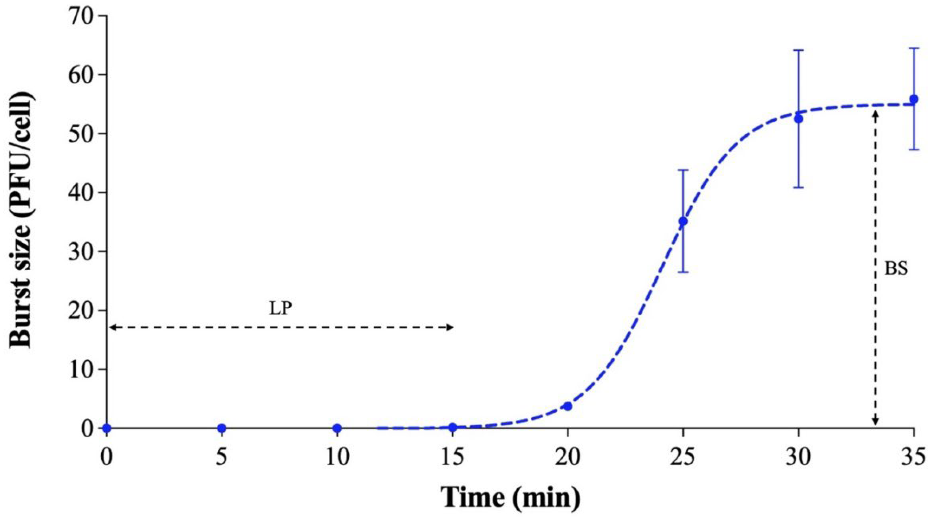
2.5. One-Step Growth Curve

A one-step growth curve of phage Ro45lw was conducted on E. coli O45:H1-, based on the previous method with subtle modification [20]. In brief, fresh bacterial culture was prepared in 10 mL of TSB at 37 °C for 20 h; the next day, 0.2 mL of the overnight culture was sub-cultured in 19.8 mL of TSB and incubated for 2 h at 37 °C to reach log phase of the bacterial growth. Thereafter, phage lysate of Ro45lw was added to the log-phase bacterial solution (MOI of 0.01) supplemented with CaCl2 at 10 mM and incubated at 37 °C for 5 min to allow phage adsorption onto the membranes of the bacterial cells. The phage–bacterial mixture was centrifuged at 10,000× g for 5 min at 4 °C to discard supernatant. Later, the bacterial pellet was washed with 2 mL of TSB and then resuspended into 20 mL of fresh TSB. Next, 0.3 mL of resuspended culture was added to 29.7 mL of TSB and incubated at 37 °C throughout the duration of the experiment (35 min). Meanwhile, quantification of phage-infected bacteria was determined at incubation time 0 by mixing 50 μL of the 30 mL phage–bacterial mixture (no filtration) with 100 μL of fresh overnight bacterial culture and 5 mL of molten 50% tryptic soy agar (TSA) and pouring into a Petri dish with 10 mL bottom TSA (also known as double-layer plaque assay). Additionally, 1 mL of the sample was obtained from the 30 mL phage–bacterial mixture every five min and subjected to 0.22-µm membrane filtration. The titers of Ro45lw were determined using the double-layer plaque assay, where plates were incubated at 37 °C overnight. The experiment was conducted in three replications to estimate the latent period and burst size of phage Ro45lw.

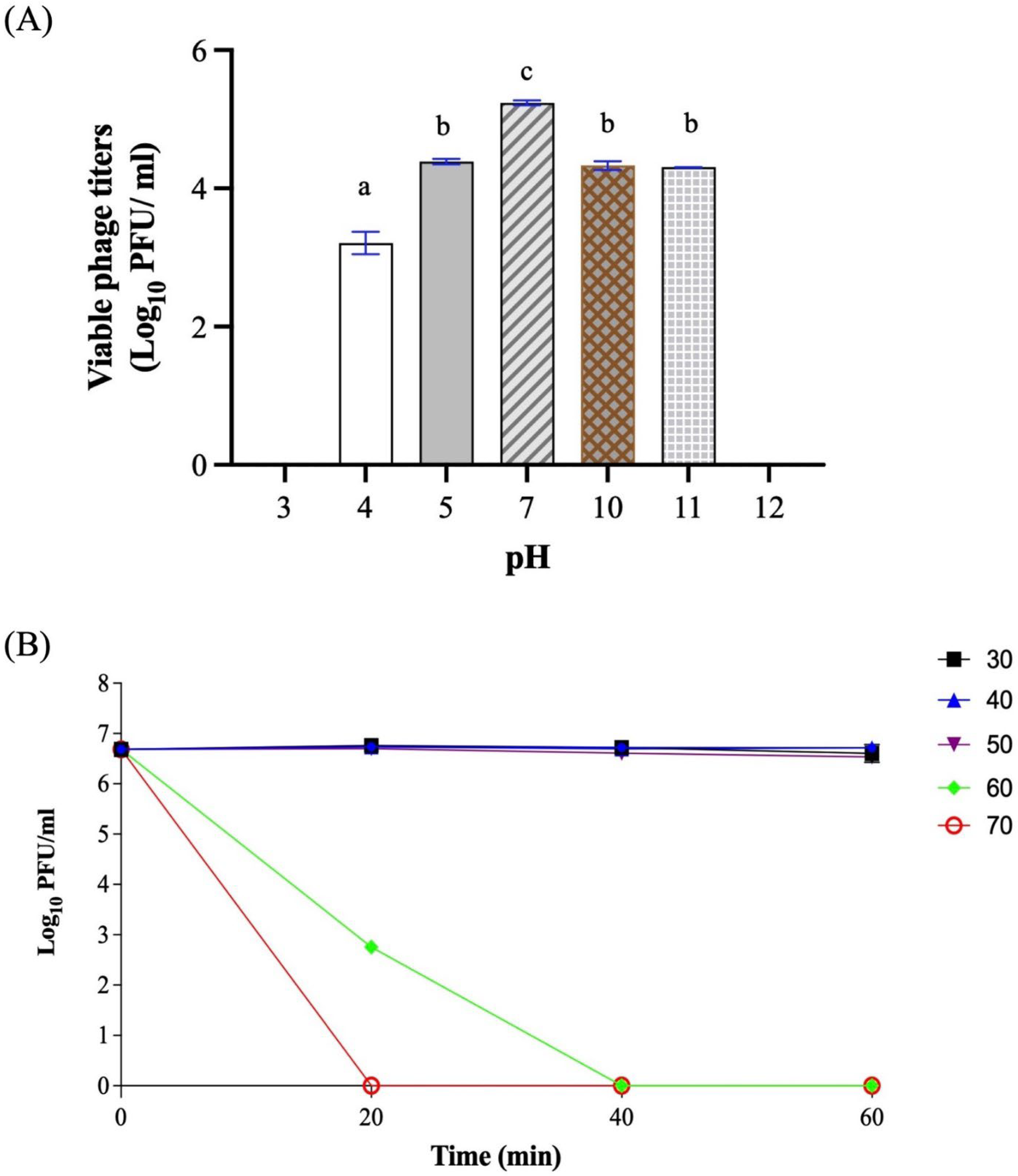
2.6. pH and Temperature Stability

A range of pH values from pH3 to pH12 was used to test the stability of Ro45lw at 37 °C for 20 h using the method as previously described with minor changes [20]. In brief, 100 μL of phage Ro45lw was added to 5 mL of SM buffer with final pH values of 3, 4, 5, 7, 10, and 12 and incubated at 37 °C for 20 h. Viable phage particles of Ro45lw were determined against E. coli O45:H- via the double-layer plaque assay.
For the temperature stability test, bulk phage lysate of Ro45lw was prepared by mixing the original phage lysate with SM buffer (pH = 7) at 1:9 (v/v) ratio. Later, an aliquot of 1 mL Ro45lw solution was dispensed in several sterile microcentrifuge tubes and subjected to heat treatment, including 30, 40, 50, 60, and 70 °C, for 20 min, 40 min, and 60 min. The temperatures covered were inclusive of the general conditions encountered in food-related environments. Phage titers were determined using the double-layer plaque assay.

2.7. Host Range and Efficiency of Plating (EOP)

The host range test of Ro45lw against non-pathogenic E. coli, E. coli O157:H7, top six non-O157 STEC, and three Salmonella strains was conducted using a spot test assay with three different dilutions of phage lysate (10−1 to 10−3) as previously described [26]. The bacterial strains showing lysis were further subjected to the efficiency of plating (EOP) assay to determine productive infection of phage Ro45lw from the tested bacterial strains versus the primary host strain for producing the phage progenies [27]. Briefly, fresh bacterial cultures were prepared in TSB at 37 °C overnight and used for quantification of Ro45lw by the double-layer plaque assay with diluted phage lysate using four successive dilutions (10−3 to 10−7). The plates were incubated at 37 °C for 18 h. The experiment was conducted in three replications. Generally, a high phage-producing efficiency had an EOP of 0.5 or more; a medium-producing efficiency had an EOP above 0.1 but below 0.5; a low-producing efficiency had an EOP between 0.001 and 0.1; inefficient phage production was any value lower than 0.001.

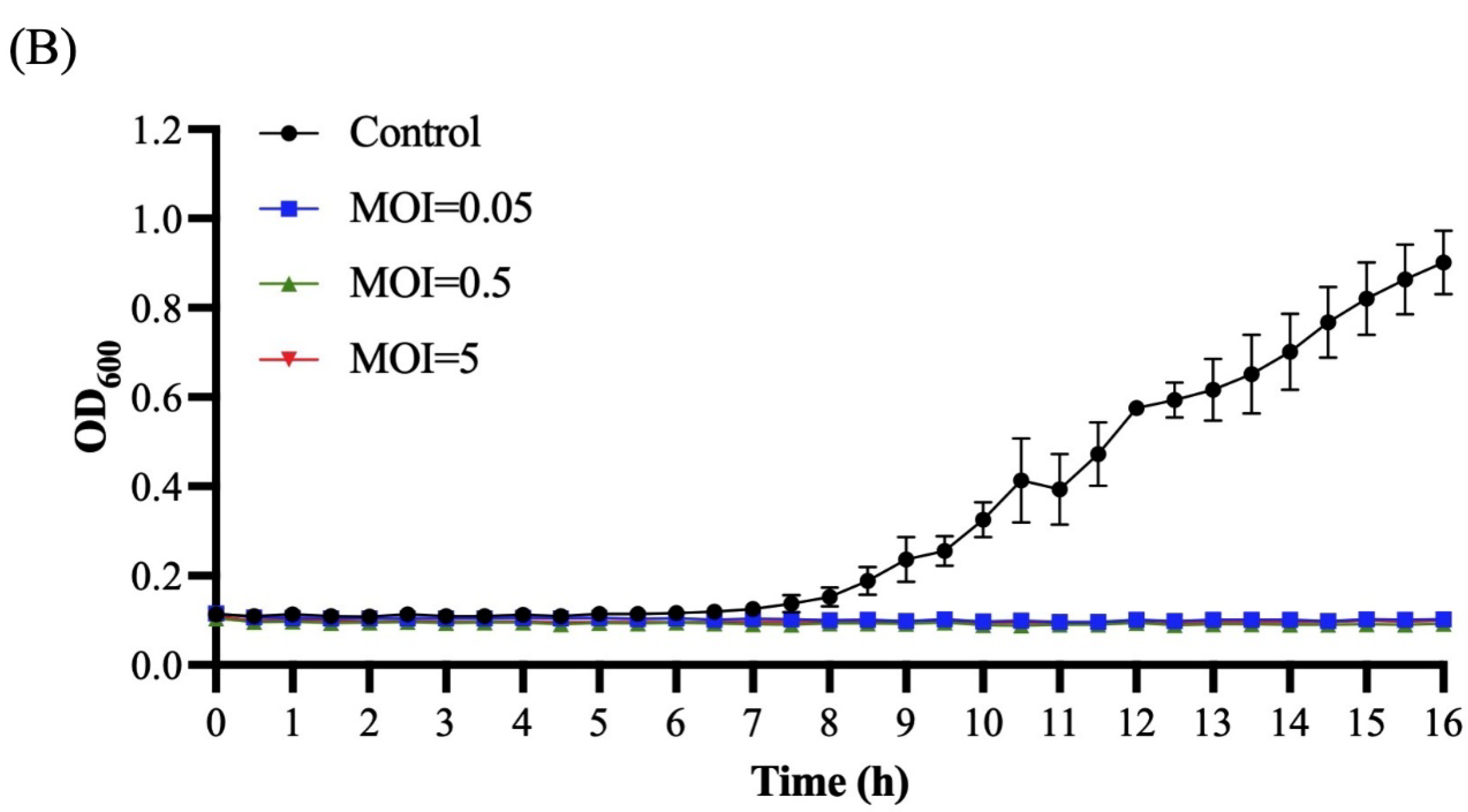
2.8. Bacterial Challenge Assay

The bacterial challenge assay was performed to measure the effects of phage Ro45lw with different MOIs on bacterial growth based on bacterial optical density at a wavelength of 600 nm (OD600) as previously described with minor changes [21]. In brief, fresh bacterial culture of E. coli O45:H- (RM10729), E. coli O45:H16 (RM13752), and E. coli O45:H2 (SJ7) was prepared in TSB at 37 °C overnight and further diluted in TSB to 1 × 10⁴ CFU/mL. An aliquot of 190 µL of diluted bacterial solution per well was added to a 96-well plate. Later, 10 µL of phage Ro45lw with different titers was added to each sample well to reach MOIs of 0.05, 0.5, and 5; the control group contained only bacterial solution without phage. At 25 °C, readings programmed to OD600 were measured using a spectrophotometer (Promega, Madison, WI, USA) every 30 min for 17 h. The experiment was conducted in 3 replications.

2.9. Statistical Analysis

Experiments subjected to statistical analysis were conducted in three individual repeats. The quantification of phages was calculated as PFU/mL, with logarithmical conversion for statistical analysis. The stress effect of pH (pH4 to pH10) was determined using a one-way analysis of variance (ANOVA) with statistical significance at a 5% level [28].

3. Results

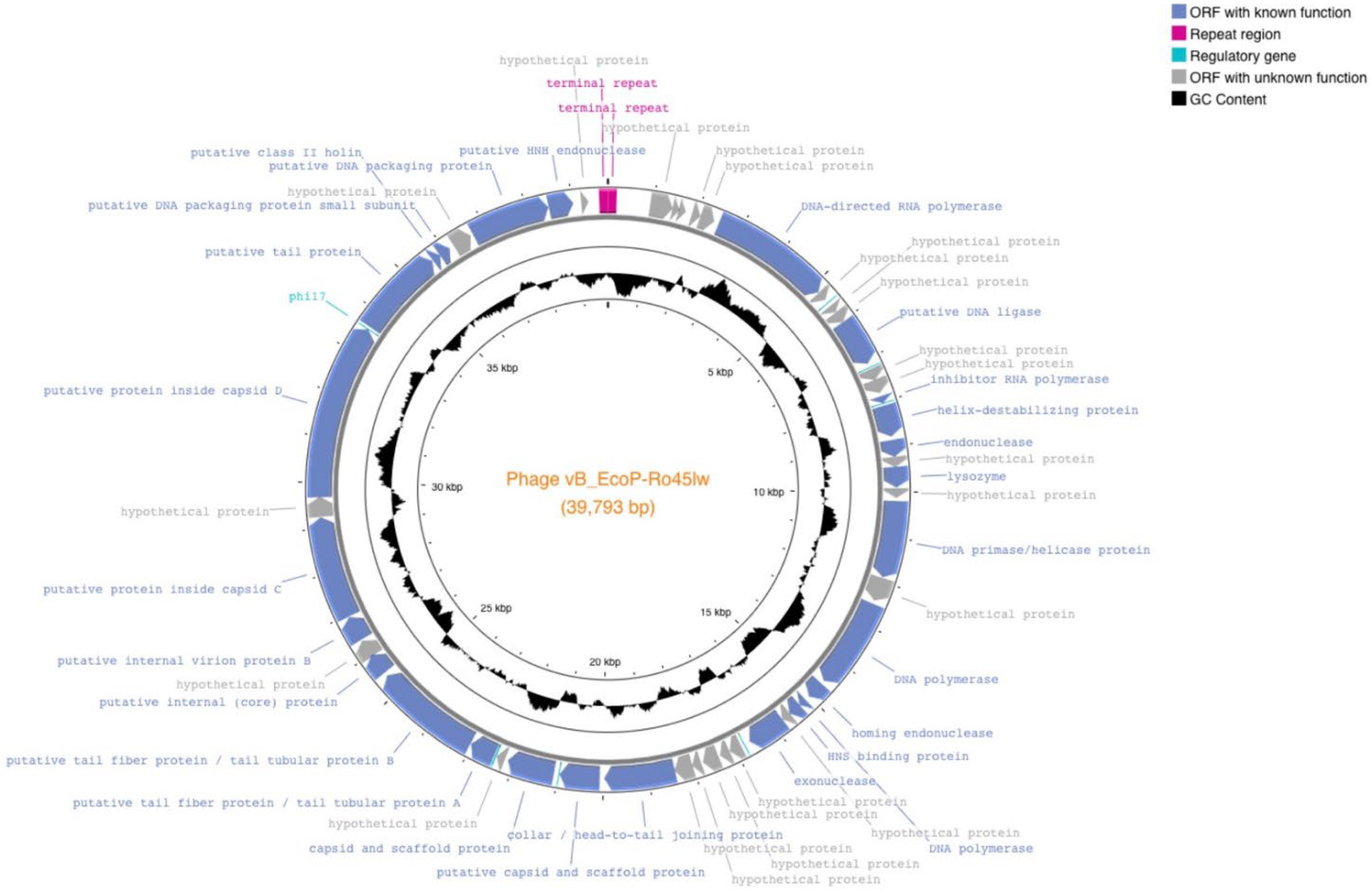
3.1. Genomic Features of Ro45lw

Phage Ro45lw had double-stranded DNA with 39,793 bp genome size and an average GC content of 52.2%. The phage was predicted to have the packaging mechanism belonging to direct terminal repeats, also known as DTR [29], with 190 bp terminal repeat region at both sides of the genome end [19]. Ro45lw also contained 50 open reading frames (ORFs), 6 promoters, and 1 terminator without tRNA (Figure 1). Ro45lw had been classified in the genus of Kayfunavirus under the Autographiviridae family by the International Committee on Taxonomy of Virus (ICTV). Based on the ICTV taxonomy guideline, a new phage classified in the same genus shares a minimum 70% nucleotide identity of the full genome length of the phages in the particular genera via analysis of genomic tools, such as BLASTn or VIRIDIC [30]. The evolutionary tree of the VICTOR analysis showed that Ro45lw had a unique amino acid sequence in comparison to 65 reference phages belonging to the Kayfunavirus genus (Figure 2). Among 50 ORFs, 26 were annotated with known functions, including structural proteins, DNA replication, DNA packaging, and cell lysis (Table 1). The phage also contained regulatory genes, such as phi1.6, phi2.5, phi6.5, and phi17, which were also found in Enterobacteria phage T7.

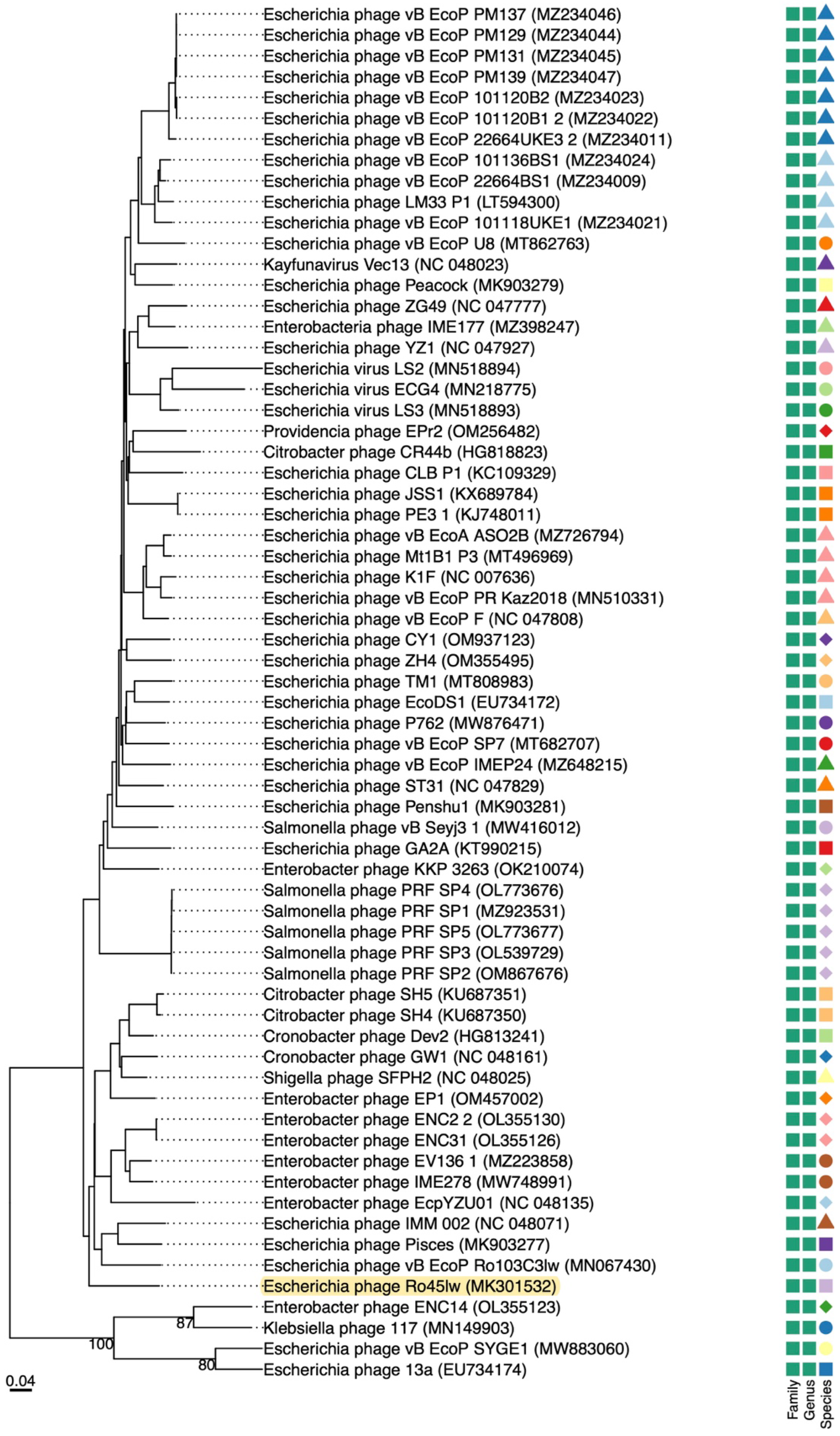
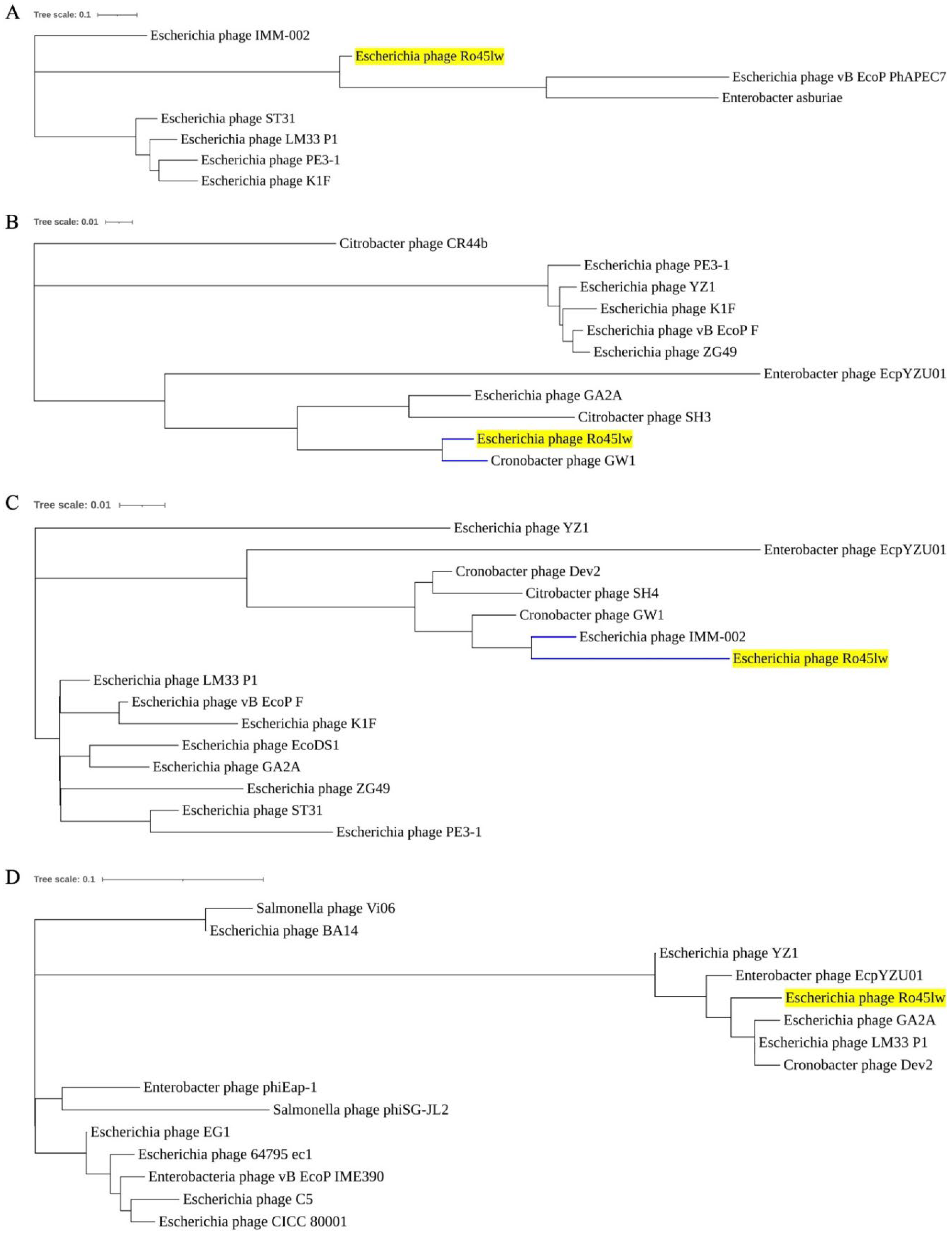
Phylogenetic Analysis

Phylogenetic analyses were conducted on the ORFs encoding the proteins associated with host range and bacterial cell lysis. Ro45lw contained a unique putative tail fiber protein sharing no close evolutionary relationship at the amino acid level with the counterfeit of any reference phage (Figure 3A). Moreover, Ro45lw contained ORF_37 encoding tail tubular protein B, which shared a close evolutionary relationship with Cronobacter phage GW1 (Figure 3B). The phage also harbored the gene encoding tail tubular protein A, with high amino acid similarity to the counterpart of Cronobacter phage GW1 (data not shown). Tail fiber, tail tubular proteins A and B are parts of the tail structure of a podophage [31]. For lysozyme (cell lysis), Ro45lw shared a high amino acid identity with the counterpart of Escherichia phage IMM-002 (Figure 3C). On the contrary, the phage contained putative class II holin protein that did not share a close evolutionary relationship with that of the reference phages (Figure 3D).

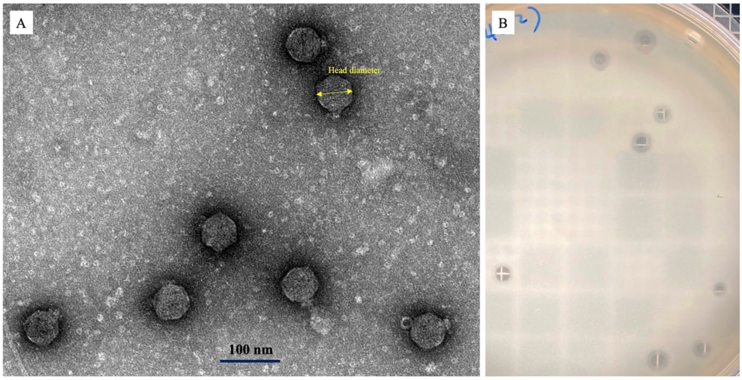
3.2. Phage Morphology

Phage Ro45lw had the morphology containing an icosahedral head of approximately 95 ± 5 nm in diameter and a short tail, showing the Podoviridae morphology (Figure 4A). The phage also produced plaques with a distinct halo on the plaque assay plate (Figure 4B).

3.3. One-Step Growth Curve and Stability Test

Phage Ro45lw had a 15 min latent period against E. coli O45:H- (RM10729) with an average burst size of 55 progeny virions per infected cell after the incubation at 37 °C for 35 min (Figure 5).
For pH tolerance, the titers of phage Ro45lw dropped approximately 2 log PFU/mL (p < 0.05) at pH4 and 1 log PFU/mL (p < 0.05) at pH5, pH10, and pH11 after 20 h incubation at 37 °C (Figure 6A). No phage was detected at either pH3 or pH12. For temperature stability, Ro45lw was stable at temperatures ranging from 30 °C to 50 °C for 1 h treatment but completely inactivated at 60 °C and 70 °C after 40 min and 20 min, respectively (Figure 6B).

3.4. Host Range and Productive Infection

Phage Ro45lw was able to infect various STEC O45 strains of environmental and clinical origins and generic E. coli (ATCC 13706 & DH5α) strains; no Salmonella strains were subjected to the phage infection (Table 2). Except for the generic E. coli with inefficiency, both environmental E. coli O45:H16 strains had a high phage-producing efficiency, and one clinical E. coli O45:H2 possessed a medium phage-producing efficiency (Table 2).

3.5. Bacterial Challenge Assay of Ro45lw

The in vitro antimicrobial activities of Ro45lw at MOIs of 0.1, 1, and 10 were determined against environmental E. coli O45:H16 (RM13752) and clinical E. coli O45:H2 (SJ7) strains at 25 °C using a spectrophotometer (Figure 7). The results showed that the bacterial growth of both E. coli O45:H16 and E. coli O45:H6 strains were completely inhibited by Ro45lw regardless of MOIs used during the 17.5 h incubation (Figure 7).

4. Discussion

Phage Ro45lw is a new member of podophages with a short tail, showing genomic classification in the genus of Kayfunavirus within the Autographiviridae family. Ro45lw does not share a close evolutionary relationship at the amino acid level with any of the 65 reference phages used in this study which also aligns with the VICTOR classification analysis, showing no similarity to Ro45lw at the species level. The Ro45lw does not contain tRNA in the phage genome, which could be associated with a narrow host range of the phage [32]. This is related to high codon usage by the phages containing more tRNAs than those with few or none [32].
For podophages, T7-like phages in particular, the tail structure is composed of at least four proteins, including connector protein, tail tubular proteins, and fiber proteins [31]. Previous studies found that tail tubular protein A was associated with phage adherence to its bacterial hosts, with hydrolytic activity targeting polysaccharides on the bacterial capsule [33,34]. However, the enzymatic function of tail tubular protein A, also known as depolymerase enzyme [35], likely contributes to the halo surrounding a plaque caused by phage Ro45lw. The same research team also found the additional enzymatic activity of tail tubular protein B, extracted from Yersinia phage, to inhibit bacterial growth and biofilm formation of Yersinia enterocolitica, besides structural function [36]. Due to the close evolutionary relationship to Cronobacter phage GW1 on both tail tabular protein A and B, phage Ro45lw might be able to infect Cronobacter commonly found in dry food, such as powdered milk [37]. The current results show that Ro45lw has a unique tail fiber protein sequence. The phage also encodes a lysozyme sharing high amino acid similarity to Escherichia phage IMM-002, which infects multiple STEC O6 strains [38]. This protein likely contributes to a strong affinity toward STEC strains because its C-terminal domain is related to recognizing and adsorbing bacterial lipopolysaccharide on the cell membrane [39]. The special tail structure and cell lysis enzyme likely render the antimicrobial activity of Ro45lw against clinical E. coli O45:H2 strains, which were resistant to the infection of another STEC O45-infecting phage, Sa45lw, previously isolated in our lab [20]. Furthermore, Ro45lw can suppress both environmental and clinical STEC O45 with various MOIs in this study. The genome of Ro45lw does not have virulent genes, any lysogenic genes, or any antibiotic-resistance genes that can jeopardize the phage application as an alternative antimicrobial agent.
Regarding growth parameters, Ro45lw has a short latent period of 15 min and a small burst size (55 PFU/CFU) against E. coli O45:H-. Ro45lw encodes a unique class II holin protein, one of the factors affecting the latent period of the phage [40]. Ro45lw contains transcriptional terminators and regulated genes found in T7 phage and may be associated with the phage’s lysis time [41]. Despite Ro45lw having a small burst size and short latent period, it may rapidly establish multiple parallel infections on the target bacteria if the bacterial concentration is high [42]. Therefore, the initial titers of the phage may be more critical upon application than the subsequent infection by the phage progenies produced from the first infection to mitigate the target bacteria. The current results showed that Ro45lw is stable at a wide pH range but is more sensitive to thermal treatment than phage Sa45lw, which could sustain up to 65 °C for 1 h [20]. The overall phage stability of Ro45lw is adequate to support further various applications in the food-processing environment.
For antimicrobial activity, although Ro45lw has a narrow host range and a small burst size compared with the phage Sa45lw [20], the phage characterized in this study can infect STEC O45 of both environmental and clinical sources. The finding indicates that Ro45lw can target different receptor proteins on these STEC O45 strains from phage Sa45lw, and both phages may be used for developing a phage cocktail to enhance antimicrobial activity. Further study regarding in vitro bacterial reduction and phage application in a food model is necessary.
To conclude, the genetic evidence indicates that phage Ro45lw is a novel STEC O45-infecting phage in the Kayfunavirus genus. The current findings show that phage Ro45lw has the antimicrobial potential to control pathogenic STEC O45 strains. Additionally, the phage-derived depolymerases with enzymatic activity encoded by Ro45lw may also be promising phage-derived antimicrobial agents to prevent bacterial pathogens.

Author Contributions

Conceptualization, Y.-T.L. and V.C.H.W.; methodology, X.S. and Y.-T.L.; software, Y.Z. and Y.-T.L.; validation, Y.Z. and Y.-T.L.; formal analysis, Y.Z. and Y.-T.L.; investigation, X.S., A.S. and K.-J.H.; resources, V.C.H.W.; data curation, Y.-T.L.; writing—original draft preparation, Y.-T.L.; writing—review and editing, X.S., Y.-T.L. and V.C.H.W.; visualization, Y.-T.L.; supervision, V.C.H.W.; project administration, V.C.H.W.; funding acquisition, V.C.H.W.; Y.-T.L. and X.S. share the same contribution as the first authorship. All authors have read and agreed to the published version of the manuscript.

Funding

This research was funded by the USDA-ARS CRIS projects 2030-42000-0055-000-D and Henan major public welfare project 222102520041.

Data Availability Statement

The phage Ro45lw sequence used in this study can be found in GenBank, with the accession number MK301532.

Acknowledgments

We thank Leslie A. Harden for his assistance in CsCl gradient purification.

Conflicts of Interest

The authors declare no conflict of interest.

References

  1. Karmali, M.A. Emerging Public Health Challenges of Shiga Toxin-Producing Escherichia coli Related to Changes in the Pathogen, the Population, and the Environment. Clin. Infect. Dis. 2017, 64, 371–376. [Google Scholar] [CrossRef] [PubMed] [Green Version]
  2. CDC. National Shiga Toxin-Producing Escherichia coli (STEC) Surveillance Overview; US Department of Health and Human Services, CDC: Atlanta, GA, USA, 2012.
  3. CDC. National Enteric Disease Surveillance: Shiga Toxin-Producing E. coli (STEC) Annual Report. 2017. Available online: https://www.cdc.gov/ecoli/surv2017/index.html (accessed on 30 September 2022).
  4. Luna-Gierke, R.E.; Griffin, P.M.; Gould, L.H.; Herman, K.; Bopp, C.A.; Strockbine, N.; Mody, R.K. Outbreaks of non-O157 Shiga toxin-producing Escherichia coli infection: USA. Epidemiol. Infect. 2014, 142, 2270–2280. [Google Scholar] [CrossRef] [PubMed]
  5. Institutional Meat Purchase Specifications for Fresh Beef Products; Agricultural Marketing Service, U.S. Department of Agriculture: Washington, DC, USA, 2011.
  6. Schaffzin, J.K.; Coronado, F.; Dumas, N.B.; Root, T.P.; Halse, T.A.; Schoonmaker-Bopp, D.J.; Lurie, M.M.; Nicholas, D.; Gerzonich, B.; Johnson, G.S.; et al. Public health approach to detection of non-O157 Shiga toxin-producing Escherichia coli: Summary of two outbreaks and laboratory procedures. Epidemiol. Infect. 2011, 40, 283–289. [Google Scholar] [CrossRef] [Green Version]
  7. CDC. Importance of culture confirmation of shiga toxin-producing Escherichia coli infection as illustrated by outbreaks of gastroenteritis—New York and North Carolina, 2005. MMWR Morbility Mortal. Wkly. Rep. 2006, 55, 1042–1045. [Google Scholar]
  8. Zhang, Y.; Liao, Y.-T.; Sun, X.; Wu, V.C.H. Is Shiga Toxin-Producing Escherichia coli O45 No Longer a Food Safety Threat? The Danger is Still Out There. Microorganisms 2020, 8, 782. [Google Scholar] [CrossRef]
  9. Bielaszewska, M.; Mellmann, A.; Zhang, W.; Köck, R.; Fruth, A.; Bauwens, A.; Peters, G.; Karch, H. Characterisation of the Escherichia coli strain associated with an outbreak of haemolytic uraemic syndrome in Germany, 2011: A microbiological study. Lancet Infect. Dis. 2011, 11, 671–676. [Google Scholar] [CrossRef] [Green Version]
  10. Liao, Y.-T.; Brook, J.C.; Martin, J.N.; Echeverry, A.; Loneragan, G.H.; Brashears, M.M. Antimicrobial interventions for O157:H7 and non-O157 Shiga toxin-producing Escherichia coli on beef subprimal and mechanically tenderized steaks. J. Food Prot. 2015, 78, 511–517. [Google Scholar] [CrossRef] [Green Version]
  11. O’Sullivan, L.; Bolton, D.; McAuliffe, O.; Coffey, A. Bacteriophages in Food Applications: From Foe to Friend. Annu. Rev. Food Sci. Technol. 2019, 10, 151–172. [Google Scholar] [CrossRef]
  12. Milho, C.; Silva, M.D.; Alves, D.; Oliveira, H.; Sousa, C.; Pastrana, L.M.; Azeredo, J.; Sillankorva, S. Escherichia coli and Salmonella Enteritidis dual-species biofilms: Interspecies interactions and antibiofilm efficacy of phages. Sci. Rep. 2019, 9, 18183. [Google Scholar] [CrossRef] [Green Version]
  13. Clokie, M.R.J.; Millard, A.D.; Letarov, A.V.; Heaphy, S. Phages in nature. Bacteriophage 2011, 1, 31–45. [Google Scholar] [CrossRef] [Green Version]
  14. Hassan, A.Y.; Lin, J.T.; Ricker, N.; Anany, H. The Age of Phage: Friend or Foe in the New Dawn of Therapeutic and Biocontrol Applications? Pharmaceuticals 2021, 14, 199. [Google Scholar] [CrossRef] [PubMed]
  15. Amarillas, L.; Chaidez, C.; Gonzalez-Robles, A.; Lugo-Melchor, Y.; Leon-Felix, J. Characterization of novel bacteriophage phiC119 capable of lysing multidrug-resistant Shiga toxin-producing Escherichia coli O157:H7. PeerJ 2016, 4, e2423. [Google Scholar] [CrossRef] [PubMed] [Green Version]
  16. Snyder, A.B.; Perry, J.J.; Yousef, A.E. Developing and optimizing bacteriophage treatment to control enterohemorrhagic Escherichia coli on fresh produce. Int. J. Food Microbiol. 2016, 236, 90–97. [Google Scholar] [CrossRef] [PubMed] [Green Version]
  17. Mohammad, Z.; Kalbasi-Ashtari, A.; Riskowski, G.; Castillo, A. Reduction of Salmonella and Shiga toxin-producing Escherichia coli on alfalfa seeds and sprouts using an ozone generating system. Int. J. Food Microbiol. 2019, 289, 57–63. [Google Scholar] [CrossRef] [PubMed]
  18. Vikram, A.; Tokman, J.I.; Woolston, J.; Sulakvelidze, A. Phage Biocontrol Improves Food Safety by Significantly Reducing the Level and Prevalence of Escherichia coli O157:H7 in Various Foods. J. Food Prot. 2020, 83, 668–676. [Google Scholar] [CrossRef]
  19. Liao, Y.-T.; Liu, F.; Wu, V.C.H. Complete Genome Sequence of a Lytic T7-Like Phage, Escherichia Phage vB_EcoP-Ro45lw, Isolated from Nonfecal Compost Samples. Microbiol. Resour. Announc. 2019, 8, e00036-00019. [Google Scholar] [CrossRef] [Green Version]
  20. Liao, Y.-T.; Zhang, Y.; Salvador, A.; Harden, L.A.; Wu, V.C.H. Characterization of a T4-like Bacteriophage vB_EcoM-Sa45lw as a Potential Biocontrol Agent for Shiga Toxin-Producing Escherichia coli O45 Contaminated on Mung Bean Seeds. Microbiol. Spectr. 2022, 10, e02220–e02221. [Google Scholar] [CrossRef]
  21. Zhang, Y.; Liao, Y.T.; Salvador, A.; Lavenburg, V.M.; Wu, V.C.H. Characterization of Two New Shiga Toxin-Producing Escherichia coli O103-Infecting Phages Isolated from an Organic Farm. Microorganisms 2021, 9, 1527. [Google Scholar] [CrossRef]
  22. Seemann, T. Prokka: Rapid prokaryotic genome annotation. Bioinformatics 2014, 30, 2068–2069. [Google Scholar] [CrossRef] [Green Version]
  23. Bairoch, A.; Apweiler, R.; Wu, C.H.; Barker, W.C.; Boeckmann, B.; Ferro, S.; Gasteiger, E.; Huang, H.; Lopez, R.; Magrane, M.; et al. The Universal Protein Resource (UniProt). Nucleic Acids Res. 2005, 33, D154–D159. [Google Scholar] [CrossRef] [Green Version]
  24. Lowe, T.M.; Chan, P.P. tRNAscan-SE On-line: Integrating search and context for analysis of transfer RNA genes. Nucleic Acids Res. 2016, 44, W54–W57. [Google Scholar] [CrossRef]
  25. Meier-Kolthoff, J.P.; Göker, M. VICTOR: Genome-based phylogeny and classification of prokaryotic viruses. Bioinformatics 2017, 33, 3396–3404. [Google Scholar] [CrossRef] [Green Version]
  26. Liao, Y.-T.; Salvador, A.; Harden, L.A.; Liu, F.; Lavenburg, V.M.; Li, R.W.; Wu, V.C.H. Characterization of a Lytic Bacteriophage as an Antimicrobial Agent for Biocontrol of Shiga Toxin-Producing Escherichia coli O145 Strains. Antibiotics 2019, 8, 74. [Google Scholar] [CrossRef] [Green Version]
  27. Liao, W.C.; Ng, W.V.; Lin, I.H.; Syu, W., Jr.; Liu, T.-T.; Chang, C.H. T4-Like genome organization of the Escherichia coli O157:H7 lytic phage AR1. J. Virol. 2011, 85, 6567–6578. [Google Scholar] [CrossRef] [Green Version]
  28. Heiberger, R.M.; Neuwirth, E. One-way Anova. In R through Excel; Springer: Berlin/Heidelberg, Germany, 2009; pp. 165–191. [Google Scholar]
  29. Bardina, C.; Colom, J.; Spricigo, D.A.; Otero, J.; Sanchez-Osuna, M.; Cortes, P.; Llagostera, M. Genomics of Three New Bacteriophages Useful in the Biocontrol of Salmonella. Front. Microbiol. 2016, 7, 545. [Google Scholar] [CrossRef] [Green Version]
  30. Turner, D.; Kropinski, A.M.; Adriaenssens, E.M. A Roadmap for Genome-Based Phage Taxonomy. Viruses 2021, 13, 506. [Google Scholar] [CrossRef]
  31. Cuervo, A.; Pulido-Cid, M.; Chagoyen, M.; Arranz, R.; González-García, V.A.; Garcia-Doval, C.; Castón, J.R.; Valpuesta, J.M.; van Raaij, M.J.; Martín-Benito, J.; et al. Structural characterization of the bacteriophage T7 tail machinery. J. Biol. Chem. 2013, 288, 26290–26299. [Google Scholar] [CrossRef] [Green Version]
  32. Yoshikawa, G.; Askora, A.; Blanc-Mathieu, R.; Kawasaki, T.; Li, Y.; Nakano, M.; Ogata, H.; Yamada, T. Xanthomonas citri jumbo phage XacN1 exhibits a wide host range and high complement of tRNA genes. Sci. Rep. 2018, 8, 4486. [Google Scholar] [CrossRef] [Green Version]
  33. Pyra, A.; Brzozowska, E.; Pawlik, K.; Gamian, A.; Dauter, M.; Dauter, Z. Tail tubular protein A: A dual-function tail protein of Klebsiella pneumoniae bacteriophage KP32. Sci. Rep. 2017, 7, 2223. [Google Scholar] [CrossRef] [Green Version]
  34. Pyra, A.; Urbańska, N.; Filik, K.; Tyrlik, K.; Brzozowska, E. Biochemical features of the novel Tail Tubular Protein A of Yersinia phage phiYeO3-12. Sci. Rep. 2020, 10, 4196. [Google Scholar] [CrossRef] [Green Version]
  35. Drulis-Kawa, Z.; Majkowska-Skrobek, G.; Maciejewska, B. Bacteriophages and Phage-Derived Proteins—Application Approaches. Curr. Med. Chem. 2015, 22, 1757–1773. [Google Scholar] [CrossRef] [PubMed]
  36. Pyra, A.; Filik, K.; Szermer-Olearnik, B.; Czarny, A.; Brzozowska, E. New Insights on the Feature and Function of Tail Tubular Protein B and Tail Fiber Protein of the Lytic Bacteriophage φYeO3-12 Specific for Yersinia enterocolitica Serotype O:3. Molecules 2020, 25, 4392. [Google Scholar] [CrossRef] [PubMed]
  37. CDC. Cronobacter Infection and Infants. Available online: https://www.cdc.gov/cronobacter/infection-and-infants.html (accessed on 30 September 2022).
  38. Chakraborty, S.; von Mentzer, A.; Begum, Y.A.; Manzur, M.; Hasan, M.; Ghosh, A.N.; Hossain, M.A.; Camilli, A.; Qadri, F. Phenotypic and genomic analyses of bacteriophages targeting environmental and clinical CS3-expressing enterotoxigenic Escherichia coli (ETEC) strains. PLoS ONE 2018, 13, e0209357. [Google Scholar] [CrossRef] [PubMed] [Green Version]
  39. Bhardwaj, A.; Olia, A.S.; Cingolani, G. Architecture of viral genome-delivery molecular machines. Curr. Opin. Struct. Biol. 2014, 25, 1–8. [Google Scholar] [CrossRef] [Green Version]
  40. Abedon, S.T.; Herschler, T.D.; Stopar, D. Bacteriophage latent-period evolution as a response to resource availability. Appl. Environ. Microbiol. 2001, 67, 4233–4241. [Google Scholar] [CrossRef] [Green Version]
  41. Nguyen, H.M.; Kang, C. Lysis delay and burst shrinkage of coliphage T7 by deletion of terminator Tφ reversed by deletion of early genes. J. Virol. 2014, 88, 2107–2115. [Google Scholar] [CrossRef] [Green Version]
  42. Abedon, S.T. Selection for bacteriophage latent period length by bacterial density: A theoretical examination. Microb. Ecol. 1989, 18, 79–88. [Google Scholar] [CrossRef]
Figure 1. Genome map of phage Ro45lw was generated using the CGview server beta. ORFs are annotated and colored based on predicted molecular functions of identified genes (Table 1). The center of the genome map provides % GC content (black).
Microorganisms 11 00077 g001
Figure 2. Phylogenetic analysis of whole-genome sequences of Ro45lw and its closely related reference phages belonging to the Kayfunavirus genus under the Autographiviridae family at the amino acid level using VICTOR (formula d6). Annotations, including family, genus, and species cluster predicted by VICTOR, with different shapes and colors, are different in classification.
Microorganisms 11 00077 g002
Figure 3. Phylogenetic tree of phage Ro45lw (with yellow highlight) and the close-related reference phages based on the Clustal Omega alignment of the amino acid sequences of putative tail fiber protein (ORF_44) (A), tail tubular protein B (ORF_37) (B), lysozyme (ORF_17) (C), and putative class II holin (ORF_45) (D). Blue lines are used to indicate the closest evolutionary relationship. The scale represents the homology percentage.
Microorganisms 11 00077 g003
Figure 4. Phage morphology under (A) transmission electron microscopy with a capsid (95 ± 5 nm in diameter) and a short tail, and (B) plaque morphology on a plaque-assay plate.
Microorganisms 11 00077 g004
Figure 5. One-step growth curve of phage Ro45lw using generic E. coli O45:H- strain (RM10729). The growth parameters of the phage indicate a latent period (LP) of 15 min and an average burst size (BS) of 55 phages per infected cell. The error bars present the standard error of the mean (SEM) for each time point of the one-step growth curve.
Microorganisms 11 00077 g005
Figure 6. Stability of phage Ro45lw at (A) different pH levels (pH 3, pH 5, pH 7.5, pH 9, pH 10.5, and pH 12) at 37 °C for 20 h, and (B) various temperatures (30 °C to 70 °C) for up to 1 h. For the pH test, the means of phage titers that lack common letters (a, b, and c) differ (p < 0.05). The error bars show the SEM.
Microorganisms 11 00077 g006
Figure 7. Bacterial challenge assay for E. coli O45:H16 (A) and E. coli O45:H2 (B) treated with phage Ro45lw at MOIs of 0.1, 1 and 10 at 25 °C for 17.5 h based on the bacterial optical density at wavelength 600 nm (OD600). The Control group only contains bacterial culture.
Microorganisms 11 00077 g007aMicroorganisms 11 00077 g007b
Table 1. List of annotated ORFs with known predicted functions, size, and location in the Ro45lw genome.
TypePredicted FunctionFunctional CategoryMinimum bpMax bp
Repeat regionterminal repeat region-1190
ORF_6DNA-directed RNA polymerase CDSDNA replication24985179
regulatorypredicted promoter regulatory-54835505
ORF_10putative DNA ligase CDSDNA replication60267093
regulatoryphi1.6 regulatory-71787200
ORF_13inhibitor RNA polymerase CDSDNA replication78648022
regulatoryphi2.5 regulatory-80188040
ORF_14helix-destabilizing protein CDSDNA replication80808778
ORF_15endonuclease CDSDNA replication88599233
ORF_17lysozyme CDSCell lysis94509908
ORF_19DNA primase/helicase protein CDSDNA replication10,20311,903
ORF_21DNA polymerase CDSDNA replication12,52314,541
ORF_22homing endonuclease CDSDNA replication14,62115,016
ORF_23DNA polymerase CDSDNA replication15,11915,292
ORF_24HNS binding protein CDSDNA replication15,29215,582
ORF_26exonuclease CDSDNA replication15,78516,651
regulatoryphi6.5 regulatory-16,77416,796
ORF_32collar/head-to-tail joining protein CDSStructural protein18,41619,984
ORF_33putative capsid and scaffold protein CDSStructural protein20,08120,962
regulatoryphi regulatory-20,96720,989
ORF_34capsid and scaffold protein CDSStructural protein21,09322,142
regulatoryphiTE regulatory-22,40922,444
ORF_36putative tail fiber protein/tail tubular protein A CDSStructural protein22,46423,030
ORF_37putative tail fiber protein/tail tubular protein B CDSStructural protein23,04225,411
ORF_38putative internal (core) protein CDSStructural protein25,49825,980
ORF_40putative internal virion protein B CDSStructural protein26,38126,944
ORF_41putative protein inside capsid C CDSStructural protein26,95629,238
ORF_43putative protein inside capsid D CDSStructural protein29,67533,571
regulatoryphi17 regulatory-33,56733,589
ORF_44putative tail proteinStructural protein33,63735,646
ORF_45putative class II holin CDSCell lysis35,66235,856
ORF_46putative DNA packaging protein small subunit CDSDNA packaging35,85336,116
ORF_48putative DNA packaging protein CDSDNA packaging36,71038,476
ORF_49putative HNH endonuclease CDSDNA replication38,48639,028
Repeat regionterminal repeat region-39,60439,793
Table 2. Host range and efficiency of plating of phage Ro45lw against various serogroups of Shiga-toxin-producing E. coli and Salmonella enterica strains.
GroupStrain IDEOP α
Non-O157 STECSTEC O26, O103, O111, O121 and O145R *
E. coli O45:H- (RM10729)H ^
E. coli O45:H2 (SJ7)0.10
E. coli O45:H16 (RM13752)0.77
E. coli O45:H16 (RM13745)0.84
STEC O157E. coli O157:H7 (RM18959, RM18974 & ATCC 43888)R
Generic E. coliATCC 13706Inefficiency
DH5aR
Salmonella entericaSalmonella NewportR
Salmonella EnteritidisR
Salmonella TyphimuriumR
α EOP was conducted for the spot test-positive strains, with a value calculated by the ratio of phage titer on the test bacterium versus the phage titer on the primary host bacterium. High production efficiency is EOP ≥ 0.5, medium production efficiency is 0.5 > EOP ≥ 0.1, low production efficiency is 0.1 > EOP > 0.001, and inefficiency of phage production is EOP ≤ 0.001. * R denotes no lysis in the spot test assay. ^ H was the primary host bacterial strain used for isolation.
Disclaimer/Publisher’s Note: The statements, opinions and data contained in all publications are solely those of the individual author(s) and contributor(s) and not of MDPI and/or the editor(s). MDPI and/or the editor(s) disclaim responsibility for any injury to people or property resulting from any ideas, methods, instructions or products referred to in the content.

