Article

The Type III Accessory Protein HrpE of Xanthomonas oryzae pv. oryzae Surpasses the Secretion Role, and Enhances Plant Resistance and Photosynthesis

1
Department of Plant Pathology, College of Plant Protection, Nanjing Agricultural University, Nanjing 210095, China
2
Department of Plant Pathology, Shandong Agricultural University, Taian 271018, China
3
State Key Laboratory of Crop Biology, Taian 271018, China
4
Department of Plant Pathology, University of Agriculture, Faisalabad, P.O. Box 38040, Pakistan
*
Authors to whom correspondence should be addressed.
Submission received: 30 September 2019 / Revised: 5 November 2019 / Accepted: 14 November 2019 / Published: 18 November 2019
(This article belongs to the Special Issue Plant Microbial Interactions)

Abstract

:
Many species of plant-pathogenic gram-negative bacteria deploy the type III (T3) secretion system to secrete virulence components, which are mostly characteristic of protein effectors targeting the cytosol of the plant cell following secretion. Xanthomonas oryzae pv. oryzae (Xoo), a rice pathogen causing bacterial blight disease, uses the T3 accessory protein HrpE to assemble the pilus pathway, which in turn secretes transcription activator-like (TAL) effectors. The hrpE gene can execute extensive physiological and pathological functions beyond effector secretion. As evidenced in this study, when the hrpE gene was deleted from the Xoo genome, the bacteria incur seriouimpairments in multiplication, motility, and virulence. The virulence nullification is attributed to reduced secretion and translocation of PthXo1, which is a TAL effector that determines the bacterial virulence in the susceptible rice varieties. When the HrpE protein produced by prokaryotic expression is applied to plants, the recombinant protein is highly effective at inducing the defense response. Moreover, leaf photosynthesis efficiency is enhanced in HrpE-treated plants. These results provide experimental avenues to modulate the plant defense and growth tradeoff by manipulating a bacterial T3 accessory protein.

1. Introduction

Bacteria adopt different life styles for adapting to their surroundings; alone, cooperative or synergetic, commensal and parasitic. Bacterial cells utilize their flagella to swim and look for a better environment for their survival [1]. The interactions among gregarious and extroverted cells with their host tissues are established by means of pili or fimbriae, and colonies and biofilm formation for their survival [2,3]. Pathogenic bacteria adhere to the eukaryotic cells, which is considered an important step in effective colonization of the host tissue. There are certain structures present on the bacterial cell surface that are involved in the adhesion of bacteria, including a vast group of fimbrial and non-fimbrial adhesions. Fimbrial adhesions include pili, which are hair-like appendages present on the surface of most of the bacteria. However, they are different in the mechanism of their assembly, structure and function [4]. Bacteria are thought to use various types of secretion systems to translocate the toxins and effector proteins from the source to the sink [5]. There are six different types of secretion systems (type I to type VI) that have been reported to be responsible for substrate secretion in gram-negative bacteria. Out of these, the type III (T3) secretion system plays a predominant role in secretion of toxins and proteic effectors that makes them virulent against their host [6].
In Xanthomonas, a chromosomal region of 23 kb length contains the hrp gene cluster where it is organized into 6 different operons. The harpins included in the hrp gene cluster are nominated as hrpA to hrpF [7]. These two proteins are substrates of the T3 secretion system where they play a predominant role in the delivery of T3 proteic and toxic effectors through and towards the plasma membrane of the plant cell. The hrpE gene encrypts the HrpE protein of 9 kDa size, which formulates a slender pilus with a length and diameter of 4 µm and 8 to 10 nm, respectively. However, the Hrp pilus acts as an appendage of the cell surface in the T3 secretion system. On the basis of various logics including mutational analyses and electron microscopy, HrpE was considered to be the structural constituent of the hrp pilus, which is made up of pilin subunits while HrpA is regarded as the best hrp pilus subunit [8]. Mutations in the hrp genes of bacteria affect the virulence, so they neither produce disease in susceptible hosts nor elicit a hypersensitive response (HR) in resistant plants [8,9]. A hrpA mutant from Pseudomonas syringae DC3000 failed to elicit a HR despite the fact that the bacteria had avr gene, which interact with an R gene in the plant [9]. In Xanthomonas citri pv. citri, biofilm formation and motility of the bacteria was affected by the deletion of the hrpB gene [10]. Gene deletion from the genome of Riemerella anatipestifer affected the bacterial virulence, gene expression and growth patterns in liquid media [11].
The T3 secretion system is extremely conserved in pathogenic gram-negative bacteria including plant pathogens. Xanthomonas oryzae pv oryzae (Xoo) is a gram-negative bacterial plant pathogen that causes a disease named bacterial blight in rice [12]. The plant and bacteria undergo several layers of interactions to confer plant susceptibility (disease) or insusceptibility (resistance), as a common feature of pathogenesis. The first interaction that occurs between the pathogens and their hosts takes place through a structure called a pilus [13]. The pilus is responsible for bacterial effectors and protein translocation from the two bacterial membranes into the interior of the host cell, by the trafficking across the plasma membrane of the host and in some circumstances across the cell wall of the plants [14]. The structure of the pilus is almost similar to that of the flagella, but additionally has the cap formed on the tip during assembly [5]. Almost ten T3 secretory proteins are similar in membrane topology and sequence to the flagellar proteins [15]. Although the pilus and flagella share some functions, the pilus is distinct from the flagella in regards to induction of T3 secretion by host sensing and the ability to translocate the proteins into eukaryotic cells [3]. The HrpE protein in Xanthomonas species and pathovars is highly conserved, including X. campestris pv. vesicatoria, X. citri pv. citri and X. campestris pv. campestris [16]. Therefore, HrpE assembly into the pilus is crucial for effector secretion by these bacteria. Transcription activator-like (TAL) effectors play essential roles in the interactions between Xanthomonas and their hosts. Several TAL effectors from Xoo have been reported to be major virulence factors that may activate the expression of susceptibility genes (S) during infection. The key virulence factor of the Xoo strain PXO99A in rice is PthXo1 [17].
Harpins have been characterized to elicit disease resistance through the NIM1 (non-inducible immunity)-mediated SAR (systemic acquired resistance) signal transduction pathway. The action site of harpin is upstream of salicylic acid (SA) in the SAR regulatory pathway [18]. Moreover, harpins have been considered as a potential component of pathogen associated molecular patterns (PAMPs) based on studies demonstrating the role of T3 accessory proteins in inducing PAMP-triggered immunity (PTI) in plants externally treated with these proteins [18,19,20,21]. PTI is activated once a PAMP molecule is recognized by one of the pattern recognition receptors situated in the plasma membrane of plant cells [22]. A physical role of PTI is to limit the growth of the pathogen, thus hindering tissue colonization [23]. Accompanying immune events are multiple, such as induced expression of defense response genes, callose deposition, and the oxidative burst, including hydrogen peroxide (H2O2) accumulation [24].
In the present study, we constructed a mutant of Xoo ΔhrpE and studied its effect on growth patterns, motility and virulence of the bacteria. Overall, in our study, we premeditated the connotation of hrpE in pathogen virulence and integrity as well as the functional description of Xoo HrpE as an elicitor of the plant immune response in rice. For this purpose, we infiltrated the plant leaves with HrpE protein produced by prokaryotic expression, and studied its roles in eliciting the HR and in inducing PTI responses. In addition, this study sheds light on the role of HrpE protein in leaf photosynthesis.

2. Materials and Methods

2.1. Bacterial Strains, Growth Conditions and Antibodies

The bacterial strains used in the present study, constructed recombinant vectors and the information about the different antibiotic resistances are given in Table S1. Different strains of Xanthomonas oryzae pv oryzae (Xoo) were grown on nutrient broth (NB) or nutrient agar (NA) medium at 28 °C [25]. Luria–Bertani broth (LB) or LB agar (LA) was used to culture engineered Escherichia coli strains on media supplemented with 100 μg/mL spectinomycin, or 50 μg/mL kanamycin or 100 μg/mL ampicillin [26].

2.2. Bacterial Gene Alterations

The hrpE gene was knocked-out from Xoo PXO99A by adopting the marker-less deletion method [12]. Four hundred base pairs (bp) upstream and 400 bp downstream of the hrpE gene, with flanking partial sequence fragments, were amplified from the PXO99A. The amplified eGFP gene fragment also contained overhang sequences of up and down stream of the hrpE gene. These fragments were then joined to each other by overlapped fusion-PCR using fragment-specific primers listed in Table S2. Gel electrophoresis and sequencing was done to confirm each PCR product. The fragments were then cloned into the vector pK18sacB by restriction digestion using XbaI ((Takara Bio, Beijing, China), and BamHI ((Takara Bio, Beijing, China). Ligation was done by using the T4 Ligation system (Thermo Scientific, Wilmington, DE, USA) and the recombinant vector was then transformed in competent cells of DH5α ((Takara Bio, Beijing, China). Then, electroporation was done to transform the recombinant vector carrying the cloned fragment into PXO99A competent cells. Single-colonies were selected from kanamycin-containing and sugar-absent NA plates. The single crossover colonies; semi-integrated into the Xoo genome were shifted to NB liquid media, grown at 28 °C for 12 h and then transferred onto NA plates containing 10% sucrose. The colonies able to grow on sucrose-supplemented media were streaked onto NA plates with and without kanamycin. This double crossover event resulted in selection of colonies showing kanamycin-negative and sucrose-positive traits, and again PCR amplification was done for the confirmation of unmarked mutants.
The protocol for construction of the cya-fused gene to analyze effector translocation was as previously described [27]. Briefly, the hrpE gene with the sacI recognition site was amplified by PCR and then inserted in the sequence of pthXo1-cya at the sacI site. The recombinant sequence of pthXo1-cya was inserted into ΔhrpE by electroporation. The complementation strain (ΔhrpE/hrpE-pthXo1-cya) was formed by inserting the hrpE-pthXo1-cya gene into the competent cell of ΔhrpE by electroporation.

2.3. Growth Curves

A single colony of Xoo (PXO99A), three independent ΔhrpE mutants (ΔhrpEI, ΔhrpEII, and ΔhrpE III to achieve accuracy) and their complementation strain (ΔhrpE/hrpE) were cultured at 28 °C in NB medium with shaking at 200 rpm overnight. Optical density (OD600) of the cultures was measured, set as equal for all the strains and then transferred to new NB medium, and bacterial growth was measured at 0, 8, 16, 24, and 32 h. The colony forming units (CFUs) of the bacterial strains were analyzed by spreading the serial dilution of the bacterial cultures on NA plates.

2.4. Bacterial Motility Assays

The cellular motility of PXO99A, ΔhrpE and ΔhrpE/hrpE was assessed by using the cultures with different concentrations of agar in NA media. OD600 was normalized to 1. The NA media was supplemented with different concentrations of agar (for swarming 0.7%, for twitching 1.6% and for swimming motility 0.2% agar was used) [28] and spotted with 2 µL of bacterial culture for analyzing twitching and swarming motility. A sterile tooth pick was dipped in bacterial culture and tinged gently in the center of the plate containing NA media to check the swimming. The plates were analyzed for swimming and swarming motility after 24 h of incubation at 28 ℃, whereas the twitching was observed at 48-hour post-incubation. The results were confirmed by repeating the same experiment three times with five replicates for each treatment.

2.5. In-Planta Growth Asssay

The germination of rice seeds was carried out in plastic trays filled with a mixture of vermiculite, sand, and peat (1:1:1 v/v/v). The germinated seedlings were transferred to 12-L pots (3 plants/pot) after 3 days. These pots were pre-filled with soil from a local rice field. The seed germination and plant growth was undertaken in temperature-controlled growth chambers at 28 °C, relative humidity of 85%, and 12-h light at 250 ± 50 μmol quanta/m2/sec. The tobacco plants were used after growing for 2 months in a greenhouse at 25 °C [20].

2.6. Bacterial Virulence Evaluation

The inoculum suspensions of PXO99A, ΔhrpE and ΔhrpE/hrpE were prepared with an OD600 = 0.5 by washing the cultures twice and re-suspending in autoclaved water, while sterilized double distilled water was used as a control in this experiment. Leaves of 2-month-old rice plants were inoculated with bacterial suspension by using the leaf-clip method [25]. The disease symptoms in all treatments were recorded by means of the leaf lesion length after 15 days post-inoculation. Hypersensitive response (HR) in tobacco leaves was observed at the inoculated sites 34-hour post-inoculation. For each treatment, the growth of bacteria was measured from infiltrated rice leaves as log cfu/leaf 15 days post leaf-center-infiltration inoculations [27].

2.7. Translocation Assays

Two-week-old rice seedlings inoculated with pthXo1-Cya transformed strains of Xoo were used for studying the cya reporter assay. The bacterial culture grown in NB media (OD600 = 0.5) was infiltrated into three sites/leaf. The leaf from the infiltrated site were cut 12 h post inoculation (hpi), ground to fine powder in liquid nitrogen by using pestle and mortar. The ground sample was suspended in 350 μL of 0.1 M HCl and centrifuged [27]. A cAMP ELISA detection kit (GenScript, Piscataway, NJ, USA) was used to analyze the obtained supernatant for determination of intracellular cAMP concentrations [26,27].

2.8. Expression and Purification of Recombinant Proteins

The primers phrpE-f-phrpE-r (Table S2) were used to amplify the full-length hrpE gene PCR from PXO99A genomic DNA. Similarly, the gene encoding red fluorescent protein (RFP) was amplified by using PCR primers pRFPf and pRFPr (Table S2). The pET30a vector (Novagen®, St. Louis, USA) digested by BamHI and HindIII restriction enzymes was used to clone the amplified products. Colony PCR and sequencing was used to confirm the transformants. The recombinant vector was then transformed into E. coli strain BL21(DE3) pLysS and 0.5 mM isopropyl-β-d-1-thiogalactopyranoside (IPTG) was used for 5 h at 37 °C to induce the expression of recombinant protein. The soluble portion of bacterial lysate was subjected to affinity chromatography for the purification of protein. The Ni2+-nitrilotriacetate (Ni-NTA) agarose column (Qiagen, Hilden, Germany) was used to purify the recombinant proteins HrpE-RFP-His (HrpE) and RFP-His (RFP) and PBS buffer was used to dialyze the proteins for 24 h. The quantification of purified proteins was done by using a BCA protein assay kit (TransGen Biotech, Beijing, China) [29].

2.9. Callose Staining

The callose of rice leaves was stained on the infiltration site with aniline blue and then the samples were cytologically observed by using UV fluorescence microscopy. The leaves of rice were infiltrated with 2.0 µM of HrpE, while RFP protein served as control in this study, and the callose staining for leaves of each treatment was observed by using UV fluorescence microscopy 24 hpi as reported [30]. Image J software was used to estimate the callose intensity from digital photographs by the number of blue pixels as compared to the total number of pixels covering the leaves. The presented results are relative to the callose intensity of control treatments, which was considered to be one for this study [31].

2.10. Accumulation of H2O2

The accumulation of H2O2 in rice leaves was detected by infiltrating the samples with 2.0 µM of HrpE, and RFP proteins. After 24 h post inoculation, H2O2 accumulation was visualized after staining the leaves with DAB (Sigma, St Louis, MO, USA) as reported by [32]. An optical microscope was used to observe and photograph the stained leaves. The accumulation of H2O2 was observed as the intensity of DAB calculated from the captured images by counting the brown pixels as reported [24].

2.11. Xoo Growth in Rice Leaves Pretreated with HrpE

The growth of Xoo in leaves of the rice plants infiltrated with HrpE and RFP proteins at the concentration of 2 µM and 15 mM NaCl was observed at 24 hpi after infiltrating these pre-treated leaves with 106 cfu/mL Xoo culture by using sterile syringes. Treatment with RFP and 15 mM NaCl served as a control. The infection caused by Xoo was observed on the leaves at 2, 4 and 8 days post inoculation (dpi). Leaf discs of 0.5 cm diameter from each sample were ground in 1 ml of 15 mM NaCl to perform bacterial growth assays for each time interval and the suspension was plated onto NA following serial dilutions. The results are presented as log cfu/cm2 of leaf tissue after counting the number of colonies 48 h post-incubation at 28 °C.

2.12. Expression Analysis of Defense-Related Genes in Rice

Two-week-old rice plants were treated with 2.0 µM of HrpE or RFP and the samples were collected after 24 h. TRIzol® reagent (Invitrogen Biotechnology Co., Carlsbad, CA, U.S.A.) was used for the extraction of total RNA from treated leaves and subjected to DNase I (Invitrogen) treatment for removing DNA as described [27]. The synthesis of first-strand cDNA was carried out by using RT enzymes (TaKaRa Bio, Beijing, China). The SYBR Premix Ex-Taq kit (TaKaRa) was used for performing quantitative PCR on the Quant studio 6.0 real-time PCR system (Applied Biosystems, CA, USA) by using specific oligonucleotide primers listed in Table S2. The genes analyzed for differential expression were OsGST (Glutathione-S-Transferase), OsSOD (Superoxide Dismutase), OsMKK4 Kinase 4 (Mitogen Activated Protein Kinase), OsPR1 and OsPR4 (Pathogenesis Related 1 and 4), OsHMGR (3-Hydroxy-Methylglutaryl CoA Reductase) and OsPAL gene encoding Phenylalanine Ammonia Lyase. The obtained values were generated as means of three biological replicates and three technical replicates for each sample as reported by [26]. The fold change or relative expression was calculated based on the comparative CT method [33].

2.13. Photosynthetic Activity, Transpiration Rate and Stomatal Conductance of the Rice Plant

The photosynthetic parameters were observed by treating the two-week-old rice plants via spraying with 2.0 µM of HrpE protein for 24 h followed by inoculation of the PXO99A strain (HrpE P.T.). The rice plants treated with PXO99A and sterilized with double distilled water were used as controls. The time frame study for consumption of CO2 as well as light usage depicting photosynthetic activity, transpiration rate and stomatal conductance of rice plants was performed by using the readings from seven maximum light exposed leaves from three plants of each treatment post inoculation (at an interval of 2, 4 and 6 dpi) by using an open IRGA LI-COR 6400 XT portable photosynthesis system (LI-6400, Li-Cor Inc., Lincoln, NE, USA) [34]. All parameters under study were recorded under light saturated conditions at 380 mol mol−1 CO2 concentration and a photosynthetic photon flux density of 1000 mmol photons m−2 s−1.

2.14. Statistical Analysis

The statistical analysis for all experiments was based on a completely randomized design. The SPSS statistical package was used for statistical analysis, Tukey’s HSD test was applied following ANOVA, and standard deviation from the means was taken at p ≤ 0.05.

3. Results

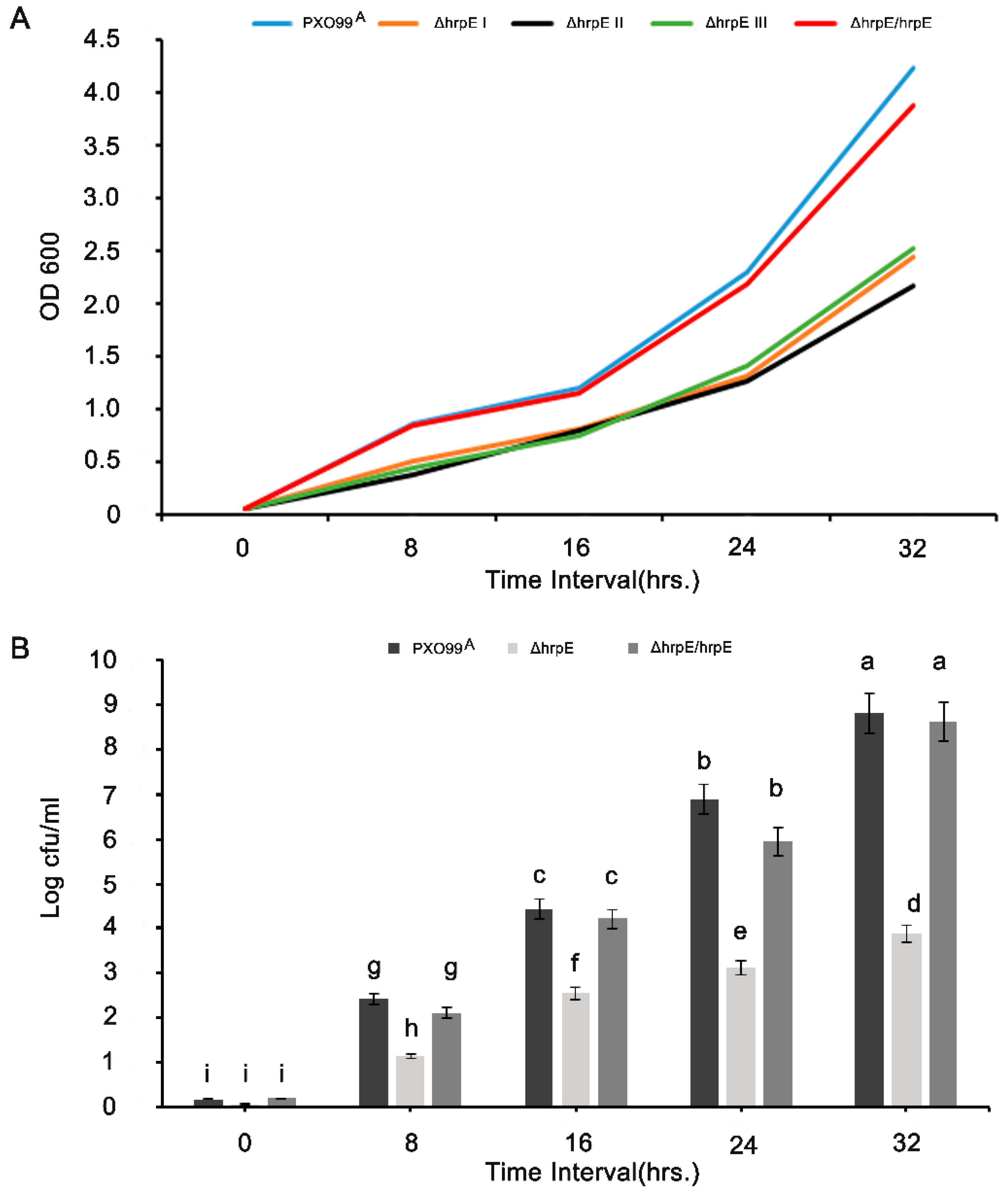
3.1. HrpE Gene Deletion Affects the Growth and Colony Forming Units (CFUs)

To analyze the function of HrpE, the PXO99A mutant ΔhrpE was used. The mutations were confirmed by PCR (Figure S1). The ΔhrpE mutants grew slower than PXO99A and the complementation strain (Figure 1A). The delayed growth of the ΔhrpE mutant was recovered by the introduction of hrpE back into the mutant, demonstrating that hrpE deletion affects bacterial growth. The time-interval study for evaluating the colony forming units (CFU) suggested that the hrpE deletion mutant had the lowest CFU as compared to the other strains (Figure 1B). The growth patterns and CFU of all strains indicated that hrpE is involved in regulation of bacterial growth.

3.2. Deletion of hrpE Affects the Bacterial Motility

The ΔhrpE mutants showed significantly less growth in NA media. All three types of motility were affected in the ΔhrpE mutant compared to the wild-type strain PXO99A (Figure 2). Swimming motility was constrained in ΔhrpE compared to the PXO99A, and a significantly smaller diameter was recorded in ΔhrpE than that in WT (wild type) PXO99A and ΔhrpE/hrpE complementation strains (Figure 2A). Swarming motility was also reduced to ½-fold of PXO99A and ΔhrpE/hrpE strains (Figure 2B). Moreover, twitching motility was most affected in the ΔhrpE mutant (Figure 2C).

3.3. Deletion of hrpE Affects the Pathogen Virulence in a Host and the Hypersensitive Response in a Non-Host

The leaves inoculated with ΔhrpE showed significantly less symptoms compared to those inoculated with PXO99A (Figure 3A). The lesion length was 2.1 cm in ΔhrpE-inoculated leaves, whereas it was 16.9 cm in the wild type strain (Figure 3B). The ΔhrpE/hrpE produced symptoms on rice similar to PXO99A, suggesting that hrpE deletion affects PXO99A virulence. To further confirm this result, bacterial populations were measured. The ΔhrpE mutant multiplied slower in rice leaves compared to PXO99A and ΔhrpE/hrpE (Figure 3C). These results demonstrated that hrpE is involved in PXO99A virulence.
To evaluate whether HrpE is able to elicit HR, Nicotiana benthamiana leaves were infiltrated with ΔhrpE, PXO99A, and ΔhrpE/hrpE. Tobacco leaves infiltrated with PXO99A and ΔhrpE/hrpE showed HR, while ΔhrpE failed to elicit HR (Figure 3D), indicating that HrpE induces HR in N. benthamiana.

3.4. HrpE Pilus Serves as Conduit for Effector Translocation

Leaves were collected 12 h after infiltration, and translocation of the effectors was measured by quantifying the concentration of cAMP in the leaf cells. Higher levels of pthXo1 translocation was detected from hrpE-pthXo1-cya bacteria at 12 hpi, whereas cAMP concentration was significantly decreased (p < 0.05) in ΔhrpE-pthXo1-cya in which hrpE is deleted (Figure 4A). However, cya activity was increased when the leaves were infiltrated with ΔhrpE/hrpE-pthXo1-cya bacteria showing the higher concentrations of cAMP indicating that complementing the mutant strain recovered its ability to translocate the effector pthXo1. Bacterial strains without pthXo1-cya served as controls. There was no significant difference in the CFU of all bacterial treatments (Figure 4B), this indicates the reduced cya activity in ΔhrpE-pthXo1-cya was mainly caused by efficiency of effector translocation and not by growth reduction. These results demonstrate that HrpE is involved in effector translocation.

3.5. Defense Responses Triggered by HrpE Protein

To evaluate if Xoo HrpE could elicit defense responses in rice, recombinant HrpE-RFP-His (HrpE) protein was purified from E. coli. Western blot analysis validated the degree of purity and reliability of the recombinant proteins (Figure S2), and RFP was used as a negative control. Rice leaves were infiltrated with different concentrations of the recombinant proteins. The infiltrated leaves showed chlorotic lesions 24 h after infiltration when HrpE protein concentration was increased to 2 µM. The intensity of chlorotic lesions on the leaves were directly proportional to the dose of the HrpE protein, whereas the leaves infiltrated with the RFP showed no obvious lesions (Figure 5A). At 24 hpi, rice leaves showed significant deposition of callose, while control leaves showed less callose deposition (Figure 5B,C). Similarly, H2O2 accumulation was significantly higher in the leaves infiltrated with HrpE than in the control leaves treated with RFP (Figure 5D,E).

3.6. Pre-Treatment of Rice Leaves with HrpE Supresses Xanthomonas oryzae pv oryzae (Xoo) Growth

HrpE pre-treatment promotes plant resistance against Xoo. The leaves were infiltrated with 2 µM of HrpE or RFP recombinant protein. Later, the infiltrated leaves were inoculated with PXO99A 24 h after treatment of the recombinant protein. The disease symptoms in HrpE-pretreated leaves were significantly reduced compared to controls (Figure 6A). The bacterial population measured as cfu log was significantly less in the leaves pre-treated with HrpE compared to RFP and mock controls (Figure 6B).

3.7. Expression Profiling of the Plant Defense Related Genes under the Influence of HrpE

The genes analyzed for differential expression were OsGST (Glutathione-S-Transferase), OsSOD (Superoxide Dismutase), OsMKK4 Kinase 4 (Mitogen Activated Protein Kinase), OsPR1 and OsPR4 (Pathogenesis Related 1 and 4), OsHMGR (3-Hydroxy-Methylglutaryl CoA Reductase) and OsPAL gene encoding Phenylalanine Ammonia Lyase. In HrpE infiltrated rice plants, there was a significant increase in the expression levels of all the genes under study as compared to the rice plants treated with the RFP protein as a control (p < 0.05) (Figure 7).

3.8. Pre-Treatment with HrpE Influences the Photosynthetic Activity, Transpiration Rate and Stomatal Conductance of the Rice Plant

Rice leaves were pre-treated with HrpE or RFP proteins, and then inoculated with PXO99A, and leaves pre-treated with water alone were used as a control. Photosynthetic efficiency of the plants was determined at different time intervals. Significant increases in the stomatal conductance (Figure 8B), photosynthetic activity (Figure 8A) and transpiration rate (Figure 8C) were observed in PXO99A infected leaves pre-treated with the HrpE compared with those inoculated with PXO99A alone, which is comparable to those treated with water alone.

4. Discussion

In Xanthomonas, the HrpE protein has been identified as a major structural component required for pilus formation [35]. The protein sequence analysis showed that the Xoo HrpE protein is differentiated from other Xanthomonas species based on sequence dissimilarity (Figure S3). Bacterial effectors are translocated into plant cytosol by the T3 secretory pilus, causing disease in host plants [27]. Based on the already established facts about the critical role of HrpE in plant disease onset, in the present study we constructed a hrpE gene deletion mutant of Xoo (PXO99A) and analyzed the effect of this deletion on the physiological parameters related to the growth and motility of the bacteria and its virulence.
In order to develop plant–pathogen interactions, bacteria use their pili or fimbriae to communicate and interact with each other, as well as with plants [9]. In this study, the deletion of the hrpE gene resulted in a significantly lower growth rate of Xoo ΔhrpE as compared to PXO99A and the complementation strain. To the best of our knowledge, there are no studies reporting the effect of the hrp gene deletion on bacterial growth and the underlying mechanism is still unknown. However, there are certain reports that demonstrate that deletion of the AS87-03730 gene associated with virulence may negatively affect bacterial growth in liquid media [11]. The gram-negative bacterium, Xoo, is capable of three different types of motility: swimming, swarming and twitching [5]. The results from our study demonstrated that bacterial motility was significantly reduced by hrpE deletion, which is in accordance with a previous study stating that the deletion of the pilin-like gene pilA negatively affected the motility of Xanthomonas citri [36]. Overall, our results showed that deletion of hrpE reduced the growth of Xoo in liquid media, as well as its motility.
Apart from affecting the growth and motility, hrpE deletion resulted in disruption in the translocation of effectors, which are extremely essential for disease onset in the plant. Several hrp genes that are responsible for translocating effectors, can also induce HR in non-host plants [37]. The present study indicated that the hrpE mutant was deficient in both HR elicitations in a non-host plant (Nicotiana benthamiana) and pathogenicity in a host plant (Nipponbare rice). ΔhrpE failed to translocate the pathogenesis-linked effectors and other toxic proteins into host cells and resulted in a decrease, or almost failure, of disease. This demonstrates that in addition to having an essential role in effector translocation, HrpE also serves as one of the key virulence factors. A complete loss of pathogenicity occurred when any of the hrp genes were mutated [38]. The current study highlights that the translocation of the TAL effector pthXol was blocked in Xoo ΔhrpE based on the Cya reporter assay. Therefore, we can conclude that HrpE functions as a type III translocator for effector translocation. Our findings are supported by a previous study stating that the harpin protein Hpa1 functions in mediating effector translocation from bacterial cells to the cytosol of the rice plant [27].
Additionally, harpins have been considered as activators of the plant immune response and confer plant disease resistance through induced systemic resistance pathways [18]. Hpa1 protein from Xoo plays an important role in induction of defense response in rice plants and it has been illustrated to affect disease resistance in Arabidopsis [21]. In our study, we used purified HrpE protein to elicit a defense response in rice plants. Rice leaves infiltrated with the HrpE protein resulted in activation of various defense responses such as developing visible leaf lesions, enhancing callose deposition and promoting H2O2 accumulation in rice leaf tissues. Pathogen challenged rice leaves pre-infiltrated with HrpE protein resulted in significantly reduced bacterial growth compared to the mock and RFP treated control, further confirming that HrpE functions as an elicitor of the defense response. HrpE from Xcc has been reported to initiate the expression of defense-related genes [31]. Similarly, infiltration of Xoo HrpE induced systemic resistance in rice by modulating the expression of various defense-related genes.
Plants are highly sensitive to stress and respond either by inducing systemic resistance or by affecting plant growth through altering metabolic activities [39,40]. Pre-infiltration of the rice plants with HrpE protein activated their defense response. When pre-infiltrated rice plants were challenged with Xoo, fluctuation occurred in various physiological activities of the plants. The photosynthetic parameters of the plants were reduced in the infected control leaves, while the plants pre-infiltrated with the HrpE protein were already primed to tackle the pathogen; hence, the photosynthetic efficiency was also enhanced in pathogen challenged plants. These results indicate that HrpE treatment significantly improves the plant photosynthetic parameters. Hpa1 has been reported to promote the vegetative growth of the plants and increase their photosynthetic efficiency [41], which is in concordance with our findings.
The significance of this study is highlighted by the fact that hrpE gene deletion affected the bacterial growth patterns and motility. The gene deletion impaired the secretion of T3 effectors, and thus affected pathogen virulence. Our study also demonstrated that plants have developed a capacity to identify the HrpE protein as an immune signal, enabling them to protect themselves against pathogens and aiding in enhanced photosynthetic efficiency (Figure S4). Based on the potential of this protein to elicit the defense response, it is highly desirable to investigate the transgenic expression of this protein in plants, aiding the development of resistant rice cultivars.

Supplementary Materials

Author Contributions

Conceptualization, T.M.M.S., H.D.; methodology, T.M.M.S., M.Z. (Muhammad Zubair), A.H.; software, A.F., L.Z., H.A.; validation, H.D., M.Z. (Meixiang Zhang), C.S.; formal analysis, M.Z. (Muhammad Zubair), C.S., P.L., Y.H.; data curation, X.C., P.L., M.S.B., H.A.; writing-original draft preparation, T.M.M.S.; writing-review and editing, T.M.M.S., M.Z. (Meixiang Zhang), A.H., M.Z. (Muhammad Zubair); visualization, T.M.M.S., A.F., L.Z., M.S.B.; supervision, H.D., M.Z. (Meixiang Zhang); project administration, H.D.; funding acquisition, H.D.

Funding

China National Key Research and Development Plan (Grant no. 2017YFD0200901), The Natural Science Foundation of China 31772247) and Talent Recruitment Funding of Shandong Agricultural University (0171226) to HD.

Conflicts of Interest

The authors declare no conflict of interest.

References

  1. Büttner, D.; Bonas, U. Port of entry-the type III secretion translocon. Trends Microbiol. 2002, 10, 186–192. [Google Scholar] [CrossRef]
  2. Alfano, J.R.; Collmer, A. Type III secretion system effector proteins: Double agents in bacterial disease and plant defense. Annu. Rev. Phytopathol. 2004, 42, 385–414. [Google Scholar] [CrossRef] [PubMed]
  3. Blocker, A.; Komoriya, K.; Aizawa, S.-I. Type III secretion systems and bacterial flagella: Insights into their function from structural similarities. Proc. Natl. Acad. Sci. USA 2003, 100, 3027–3030. [Google Scholar] [CrossRef] [PubMed]
  4. Giltner, C.L.; Nguyen, Y.; Burrows, L.L. Type IV pilin proteins: Versatile molecular modules. Microbiol. Mol. Biol. Rev. 2012, 76, 740–772. [Google Scholar] [CrossRef] [PubMed]
  5. Aizawa, S.I. Bacterial flagella and type III secretion systems. FEMS Microbiol. Lett. 2001, 202, 157–164. [Google Scholar] [CrossRef] [PubMed]
  6. Buttner, D.; He, S.Y. Type III protein secretion in plant pathogenic bacteria. Plant Physiol. 2009, 150, 1656–1664. [Google Scholar] [CrossRef] [PubMed]
  7. Hienonen, E.; Roine, E.; Romantschuk, M.; Taira, S. Taira mRNA stability and the secretion signal of HrpA, a pilin secreted by the type III system in Pseudomonas syringae. Mol. Genet. Genom. 2002, 266, 973–978. [Google Scholar]
  8. Weber, E.; Koebnik, R. Domain Structure of HrpE, the Hrp Pilus Subunit of Xanthomonas campestris pv. vesicatoria. J. Bacteriol. 2005, 187, 6175–6186. [Google Scholar] [CrossRef]
  9. Alfano, J.R.; Collmer, A. The type III (Hrp) secretion pathway of plant pathogenic bacteria: Trafficking harpins, Avr proteins, and death. J. Bacteriol. 1997, 179, 5655–5662. [Google Scholar] [CrossRef]
  10. Granato, L.M.; Picchi, S.C.; De Oliveira Andrade, M.; Takita, M.A.; De Souza, A.A.; Wang, N.; MacHado, M.A. The ATP-dependent RNA helicase HrpB plays an important role in motility and biofilm formation in Xanthomonas citri subsp. Citri. BMC Microbiol. 2016, 16, 55. [Google Scholar] [CrossRef]
  11. Wang, X.; Yue, J.; Ding, C.; Wang, S.; Liu, B.; Tian, M.; Yu, S. Deletion of AS87-03730 gene changed the bacterial virulence and gene expression of Riemerella anatipestifer. Sci. Rep. 2016, 6, 1–9. [Google Scholar]
  12. Ji, Z.; Song, C.; Lu, X.; Wang, J. Two coiled-coil regions of Xanthomonas oryzae pv. oryzae harpin differ in oligomerization and hypersensitive response induction. Amino Acids 2011, 40, 381–392. [Google Scholar] [CrossRef] [PubMed]
  13. Brown, I.R.; Mansfield, J.W.; Taira, S.; Roine, E.; Romantschuk, M. Immunocytochemical localization of HrpA and HrpZ supports a role for the Hrp pilus in the transfer of effector proteins from Pseudomonas syringae pv. tomato across the host plant cell wall. Mol. Plant. Microbe. Interact. 2001, 14, 394–404. [Google Scholar] [CrossRef]
  14. Haapalainen, M.; Engelhardt, S.; Küfner, I.; Li, C.M.; Nürnberger, T.; Lee, J.; Romantschuk, M.; Taira, S. Functional mapping of harpin HrpZ of Pseudomonas syringae reveals the sites responsible for protein oligomerization, lipid interactions and plant defence induction. Mol. Plant Pathol. 2011, 12, 151–166. [Google Scholar] [CrossRef] [PubMed]
  15. Cambronne, E.D.; Roy, C.R. Recognition and delivery of effector proteins into eukaryotic cells by bacterial secretion systems. Traffic 2006, 7, 929–939. [Google Scholar] [CrossRef] [PubMed]
  16. Nguyen, H.P.; Chakravarthy, S.; Velásquez, A.C.; McLane, H.L.; Zeng, L.; Nakayashiki, H.; Park, D.-H.; Collmer, A.; Martin, G.B. Methods to Study PAMP-Triggered immunity using tomato and Nicotiana benthamiana. Mol. Plant-Microbe Interact. 2010, 23, 991–999. [Google Scholar] [CrossRef] [PubMed]
  17. Li, R.; Wang, S.; Sun, R.; He, X.; Liu, Y. Xanthomonas oryzae pv. oryzae type III effector PthXo3 JXOV suppresses innate immunity, induces susceptibility and binds to multiple targets in rice. FEMS Microbiol. Lett. 2018, 365, fny037. [Google Scholar] [CrossRef] [PubMed]
  18. Dong, H.; Delaney, T.P.; Bauer, D.W.; Beer, S.V. Harpin induces disease resistance in Arabidopsis through the systemic acquired resistance pathway mediated by salicylic acid and the NIM1 gene. Plant J. 1999, 20, 207–215. [Google Scholar] [CrossRef] [PubMed]
  19. Choi, M.; Kim, W.; Lee, C.; Oh, C.; Resources, E.N.; Hee, K. Harpins, Multi-functional proteins secreted by gram-negative plant pathogenic bacteria. Mol. Plant-Microbe Interact. 2013, 26, 1115–1122. [Google Scholar] [CrossRef]
  20. Chen, L.; Qian, J.; Qu, S.; Long, J.; Yin, Q.; Zhang, C.; Wu, X.; Sun, F.; Wu, T.; Hayes, M.; et al. Identification of specific fragments of HpaG Xooc, a Harpin from Xanthomonas oryzae pv. oryzicola, that induce disease resistance and enhance growth in plants. Phytopathology 2008, 98, 781–791. [Google Scholar] [CrossRef]
  21. Sang, S.; Li, X.; Gao, R.; You, Z.; Lü, B.; Liu, P.; Ma, Q.; Dong, H. Apoplastic and cytoplasmic location of harpin protein Hpa1 Xoo plays different roles in H2O2 generation and pathogen resistance in Arabidopsis. Plant Mol. Biol. 2012, 79, 375–391. [Google Scholar] [CrossRef] [PubMed]
  22. Boller, T.; Felix, G. A renaissance of elicitors: Perception of microbe-associated molecular patterns and danger signals by pattern-recognition receptors. Annu. Rev. Plant Biol. 2009, 60, 379–406. [Google Scholar] [CrossRef] [PubMed]
  23. Chisholm, S.T.; Coaker, G.; Day, B.; Staskawicz, B.J. Host-microbe interactions: Shaping the evolution of the plant immune response. Cell 2006, 124, 803–814. [Google Scholar] [CrossRef] [PubMed]
  24. Tian, S.; Wang, X.; Li, P.; Wang, H.; Ji, H.; Xie, J.; Qiu, Q.; Shen, D.; Dong, H. Plant aquaporin AtPIP1;4 Links Apoplastic H2O2 induction to disease immunity pathways. Plant Physiol. 2016, 171, 1635–1650. [Google Scholar] [CrossRef]
  25. Li, Y.; Che, Y.; Zou, H.; Cui, Y.; Guo, W.; Zou, L.; Biddle, E.M.; Yang, C.; Chen, G. Hpa2 Required by HrpF to translocate Xanthomonas oryzae transcriptional activator-like effectors into rice for pathogenicity. Appl. Environ. Microbiol. 2011, 77, 3809–3818. [Google Scholar] [CrossRef]
  26. Li, P.; Zhang, L.; Mo, X.; Ji, H.; Bian, H.; Hu, Y.; Majid, T.; Pang, H.; Tao, Y.; Dong, H. Aquaporin PIP1;3 of rice and harpin Hpa1 of bacterial blight pathogen cooperate in a type III effector translocation. J. Exp. Botony 2019, 70, 3057–3073. [Google Scholar] [CrossRef]
  27. Wang, X.; Zhang, L.; Ji, H.; Mo, X.; Li, P.; Wang, J.; Dong, H. Hpa1 is a type III translocator in Xanthomonas oryzae pv. Oryzae. BMC Microbiol. 2018, 18, 105. [Google Scholar] [CrossRef]
  28. Tahir, H.A.S.; Gu, Q.; Wu, H.; Niu, Y.; Huo, R.; Gao, X. Bacillus volatiles adversely affect the physiology and ultra-structure of Ralstonia solanacearum and induce systemic resistance in tobacco against bacterial wilt. Sci. Rep. 2017, 7, 1–15. [Google Scholar] [CrossRef]
  29. van Gijsegem, F.; Gough, C.; Zischek, C.; Niqueux, E.; Arlat, M.; Genin, S.; Barberis, P.; German, S.; Castello, P.; Boucher, C. The hrp gene locus of Pseudomonas solanacearum, which controls the production of a type III secretion system, encodes eight proteins related to components of the bacterial flagellar biogenesis complex. Mol. Microbiol. 1995, 15, 1095–1114. [Google Scholar] [CrossRef]
  30. Fernández-Crespo, E.; Navarro, J.A.; Serra-Soriano, M.; Finiti, I.; García-Agustín, P.; Pallás, V.; González-Bosch, C. Hexanoic acid treatment prevents systemic MNSV movement in cucumis melo plants by priming callose deposition correlating SA and OPDA accumulation. Front. Plant Sci. 2017, 8, 1–15. [Google Scholar] [CrossRef]
  31. Gottig, N.; Vranych, C.V.; Sgro, G.G.; Piazza, A.; Ottado, J. HrpE, the major component of the Xanthomonas type three protein secretion pilus, elicits plant immunity responses. Sci. Rep. 2018, 8, 1–13. [Google Scholar] [CrossRef] [PubMed]
  32. Piazza, A.; Zimaro, T.; Garavaglia, B.S.; Ficarra, F.A.; Thomas, L.; Marondedze, C.; Feil, R.; Lunn, J.E.; Gehring, C.; Ottado, J.; et al. The dual nature of trehalose in citrus canker disease: A virulence factor for Xanthomonas citri subsp. citri and a trigger for plant defence responses. J. Exp. Bot. 2015, 66, 2795–2811. [Google Scholar] [CrossRef] [PubMed]
  33. Livak, K.J.; Schmittgen, T.D. Analysis of relative gene expression data using real-time quantitative PCR and the 2-ΔΔCT method. Methods 2001, 25, 402–408. [Google Scholar] [CrossRef] [PubMed]
  34. Zubair, M.; Hanif, A.; Farzand, A.; Sheikh, T.M.M.; Khan, A.R.; Suleman, M.; Ayaz, M.; Gao, X. Genetic screening and expression analysis of psychrophilic Bacillus spp. reveal their potential to alleviate cold stress and modulate phytohormones in wheat. Microorganisms 2019, 7, 337. [Google Scholar] [CrossRef] [PubMed] [Green Version]
  35. Weber, E.; Ojanen-Reuhs, T.; Huguet, E.; Hause, G.; Romantschuk, M.; Korhonen, T.K.; Bonas, U.; Koebnik, R. The type III-dependent Hrp pilus is required for productive interaction of Xanthomonas campestris pv. vesicatoria with pepper host plants. J. Bacteriol. 2005, 187, 2458–2468. [Google Scholar] [CrossRef] [PubMed] [Green Version]
  36. Petrocelli, S.; Arana, M.R.; Cabrini, M.N.; Casabuono, A.C.; Moyano, L.; Beltramino, M.; Moreira, L.M.; Couto, A.S.; Orellano, E.G. Deletion of pilA, a minor pilin-like gene, from Xanthomonas citri subsp. citri influences bacterial physiology and pathogenesis. Curr. Microbiol. 2016, 73, 904–914. [Google Scholar] [CrossRef]
  37. Lindgren, P.B.; Peet, R.C.; Panopoulos, N.J. Gene cluster of Pseudomonas syringae pv. “phaseolicola” controls pathogenicity of bean plants and hypersensitivity of nonhost plants. J. Bacteriol. 1986, 168, 512–522. [Google Scholar]
  38. Tian, D.; Wang, J.; Zeng, X.; Gu, K.; Qiu, C.; Yang, X.; Zhou, Z.; Goh, M.; Luo, Y.; Murata-Hori, M.; et al. The rice TAL effector-dependent resistance protein XA10 triggers cell death and calcium depletion in the endoplasmic reticulum. Plant Cell 2014, 26, 497–515. [Google Scholar] [CrossRef] [Green Version]
  39. Zhang, H.; Xie, X.; Kim, M.S.; Kornyeyev, D.A.; Holaday, S.; Paré, P.W. Soil bacteria augment Arabidopsis photosynthesis by decreasing glucose sensing and abscisic acid levels in planta. Plant J. 2008, 56, 264–273. [Google Scholar] [CrossRef]
  40. Park, Y.S.; Dutta, S.; Ann, M.; Raaijmakers, J.M.; Park, K. Promotion of plant growth by Pseudomonas fluorescens strain SS101 via novel volatile organic compounds. Biochem. Biophys. Res. Commun. 2015, 461, 361–365. [Google Scholar] [CrossRef]
  41. Li, X.; Han, L.; Zhao, Y.; You, Z.; Dong, H.; Zhang, C. Hpa1 harpin needs nitroxyl terminus to promote vegetative growth and leaf photosynthesis in Arabidopsis. J. Biosci. 2014, 39, 127–137. [Google Scholar] [CrossRef] [PubMed]
Figure 1. Growth curves of three independent Xoo ΔhrpE colonies in comparison with WT Xoo (PXO99A) at different time intervals; the complementation strain Xoo (ΔhrpE/hrpE) served as a control (A). Quantification of the log colony forming unit (log cfu/mL) of different bacterial strains on NA media (B). The error bars on the graph indicate standard deviations. Different alphabets on the bars describe significant differences among the treatments at p < 0.05. The experiment was conducted three times with similar results.
Microorganisms 07 00572 g001
Figure 2. Effect of hrpE deletion on swimming, swarming and twitching motility. Swimming motility of different bacterial strains recorded 24 h after inoculation (A), swarming motility of different bacterial strains recorded 24 h after inoculation (B) and twitching motility of bacterial strains recorded 48 h after inoculation (C). Error bars in graph indicate standard deviations of five replicates. Different letters on the bars describe significant differences at p < 0.05. All experiments were repeated thrice with similar results each time.
Microorganisms 07 00572 g002
Figure 3. Deletion of the hrpE gene reduces the virulence of Xoo strain PXO99A in Nipponbare. Symptoms of the bacterial blight on rice leaves after 15 days of leaf-top-clipping on Nipponbare rice; 1. PXO99A 2. ΔhrpE 3. CK (water) 4. ΔhrpE/hrpE (A). Lesion length of bacterial blight symptoms of the leaves from A (B). Bacterial population count presented as CFU (colony formation unit) of bacterial cultures from rice leaves after 15 days of leaf-center inoculations (C). Hypersensitive response in tobacco leaves after 34 h of inoculation against Xoo strains; (a) Ck (Sterilized double distilled Water), (b) PXO99A, (c) ΔhrpE/hrpE, (d) ΔhrpE, (e)15 mM NaCl (D). Error bars on graph shows standard deviations. Values are presented as the mean of three replicates. Different letters on the bars in the graph states significant differences at p < 0.05. The experiments were repeated thrice with similar results each time.
Microorganisms 07 00572 g003
Figure 4. Concentrations of cAMP in leaves of Nipponbare rice plants 12 hpi with different strains of Xoo (A). The CFU of inoculated bacteria in rice leaves 12 hpi with different Xoo strains (B). Error bars on the graph represents standard deviations of three replicates. Different letters refer to significant differences at p < 0.05. The experiments were repeated three times with similar results.
Microorganisms 07 00572 g004
Figure 5. Analysis of rice leaf response to Xoo HrpE. Leaf response to infiltration of purified HrpE at different concentrations and 6 µM RFP as a control 24 hpi (A). Photographs representing the deposition of callose in rice leaves center-infiltrated with HrpE, with RFP as a control (B). Quantification of callose deposition intensity in rice leaves (C). Representative photographs for H2O2 accumulation in leaves of rice infiltrated with HrpE, with RFP as a control (D). Quantification of H2O2 accumulation in rice leaves by DAB staining in rice leaves (E). For callose quantification and DAB staining quantification, values are the mean calculated from 10 different photographs. Each experiment was repeated three times. Error bars represent standard deviations. Different letters on the graph bars represent significant differences among the treatments at p < 0.05.
Microorganisms 07 00572 g005
Figure 6. Xoo infections in rice leaves pre-infiltrated with HrpE, at 2, 4 and 8 days post infiltration (dpi) where RFP and Mock (15 mM NaCl) served as a control (A). Calculation of log bacterial population in the leaves of rice plants at 2,4 and 8 dpi (B). Error bars show the standard deviations of the means calculated from three different replicates. Lower case letters in the graph bars represent a significant difference at p < 0.05. Experiments were conducted thrice with similar results each time.
Microorganisms 07 00572 g006
Figure 7. Expression profiling of the rice defense-related genes in the leaves infiltrated with HrpE by q RT-PCR. Values represent the average of three biological replicates with three technical replicates. Error bars signify standard deviations of the means. Different letters above the columns in the graph show a significant difference among the treatments at p < 0.05. The experiment was repeated three times.
Microorganisms 07 00572 g007
Figure 8. Photosynthesis rate of rice leaves pre-treated with HrpE at different time intervals (A); stomatal conductance (B); and transpiration rate (C). dpi = days post inoculation. Error bars show standard deviations of the means. Lower case letters above the columns indicate a significant difference at p < 0.05. All the experiments were conducted in triplicate with similar results each time.
Microorganisms 07 00572 g008

