Article

Antimicrobial Sensitivity Testing of Mycoplasma bovis Isolates Derived from Western Canadian Feedlot Cattle

1
Western College of Veterinary Medicine, University of Saskatchewan, Saskatoon, SK S7N 5B4, Canada
2
Lethbridge Research and Development Centre, Agriculture and Agri-Food Canada, Lethbridge, AB T1J 4B1, Canada
3
Ontario Veterinary College, University of Guelph, Guelph, ON N1G 2W1, Canada
*
Author to whom correspondence should be addressed.
Submission received: 21 December 2019 / Revised: 11 January 2020 / Accepted: 14 January 2020 / Published: 16 January 2020
(This article belongs to the Special Issue Antimicrobial Resistance in Livestock)

Abstract

:
Mycoplasma bovis is particularly adept at evading the immune system, resulting in chronic infections of the lungs and joints of feedlot cattle. The chronicity of the lesions results in prolonged antimicrobial therapy, possibly exacerbating antimicrobial resistance. This cross-sectional study generated in vitro antimicrobial susceptibility testing (AST) data on 211 M. bovis isolates recovered from 159 healthy, diseased, and dead cattle, spanning the period of 2006–2018. Nine antimicrobials commonly administered to western Canadian feedlot cattle were assessed. The data were analyzed with non-parametric statistical tests with a level of significance of p < 0.05 (two-tailed). Minimum inhibitory concentration (MIC) values tended to increase between the isolates from healthy versus dead cattle and over time (2006–2018). Isolates from dead versus healthy cattle were more likely to be resistant to tulathromycin, gamithromycin, tylosin and enrofloxacin. There was no difference in the distributions of the MICs generated from the isolates recovered from the lungs and joints (p ≥ 0.124) and the lungs and deep nasal passages (p ≥ 0.157) of the same animals.

1. Introduction

Canada’s beef industry is comprised of two main sectors, cow–calf and feedlot. Calves are born in the spring of the year and then raised on pasture until weaned in the fall. Most are transported to an auction where they are commingled, sorted, and sold into feedlots. Commingling and transport predisposes cattle to developing bovine respiratory disease (BRD) or “Shipping Fever,” which is the leading cause of morbidity and mortality in weaned beef calves. The most common bacterial pathogens associated with BRD are Mannheimia haemolytica, Pasteurella multocida, Histophilus somni and Mycoplasma bovis [1]. Furthermore, the polymicrobial nature of BRD makes it difficult to ascribe a specific pathogen(s) to an individual case of BRD, since affected animals typically present non-specific clinical signs such as anorexia, depression, coughing and fever. The epidemiology of BRD is well known, with cases occurring within 6–10 day of entry into the feedlot [2]. Therefore, antimicrobial metaphylaxis is routine for on-arrival cattle that are deemed a high risk of developing BRD [3,4]. A study of antimicrobial usage (AMU) in western Canadian feedlots for the years 2008–2012 found that tetracyclines and macrolides were the classes of drugs that were most commonly administered parenterally. Furthermore, 39% of all cattle arriving to the feedlots were categorized as high risk; 95% of these cattle received parenterally administered BRD metaphylaxis, while 59% of the low risk cattle also received parenterally administered metaphylaxis [5]. Macrolides are the primary antimicrobials that are administered to high risk cattle, whereas tetracyclines are administered to low risk cattle. Fluoroquinolones and ceftiofur are used sparingly, comprising <1% of the total amount of AMU of medically important antimicrobials used in feedlots.
There is some debate as to whether M. bovis is a primary pathogen, a secondary invader, or a predisposing factor for other BRD bacterial agents [6,7,8,9]. However, it is accepted that M. bovis can be routinely recovered from the lungs of cattle with subacute-to-chronic BRD [1,6,10,11]. Furthermore, the finding of chronic caseonecrotic bronchopneumonia on postmortem examination is considered pathognomonic of an M. bovis infection [6,12,13]. Cattle with a concurrent septic polyarthritis and caseonecrotic pneumonia are given a diagnosis of chronic pneumonia and polyarthritis syndrome (CPPS) [14], which was previously known as the ‘pneumo-arthritis syndrome of calves’ [15]. The chronicity of mycoplasmosis is salient because diseased cattle receive repeated antimicrobial therapies [10] that may lead to increased antimicrobial resistance (AMR) within bacterial communities.
There are four Canadian cattle studies of interest wherein antimicrobial susceptibility testing (AST) data were generated from M. bovis isolates. Two were conducted on M. bovis isolates that had been derived from cattle in eastern Canada. However, in both studies, the isolates were primarily from dairy cattle and the most contemporaneous isolates were from 2009 [16,17]. The third study provides AST results from 51 M. bovis isolates recovered from western Canadian feedlot cattle; however, all isolates originated from one feedlot [18]. In addition, the isolates predated some of the newer macrolide products currently used to treat BRD. More recently, AST data were generated from 226 M. bovis isolates that were derived from 211 western Canadian feedlot cattle [19]. However, the majority of tested antimicrobials are not routinely used in feedlot cattle (aminoglycosides, lincosamides, and pleuromutilin) or have no antibacterial activity to mycoplasmas (β-lactams, trimethoprim, and sulphonamides) [20,21].
The objective of this retrospective cross-sectional study was to conduct AST on 211 M. bovis isolates that were recovered over a multi-year period (2006–2018) from western Canadian feedlot cattle. Isolates were collected from multiple feedlots, three anatomical sites (i.e., nasopharynx, lung, and joint) and from healthy, diseased, and dead cattle. A customized 96-well broth microdilution test plate was used for AST of nine antimicrobials most commonly administered to cattle in western Canadian feedlots for the metaphylaxis and treatment of BRD.

2. Materials and Methods

2.1. Sample Collection and Handling

This study was approved by the University of Saskatchewan’s Animal Research Ethics Board (Protocols 20070023 and 20170021) and the Lethbridge Research Centre’s Animal Care Committee (Protocol 1641).
This cross-sectional study obtained clinical samples from cattle of varying health statuses (healthy, diseased, and dead), from different anatomical regions (deep nasopharyngeal passages, lungs and joints), and over a multi-year period (2006–2018). The long timeframe coupled with the involvement of multiple private veterinary practices resulted in minor changes to the sampling procedures, culturing methods, and M. bovis isolation techniques. However, all antimicrobial susceptibility testing (AST) was standardized, with samples being batch processed by two individuals over a six month period.
Diseased cattle were identified by the feedlots’ trained personnel. A diagnosis of BRD was based on a constellation of clinical signs such as depression, nasal discharge, elevated respiratory rate, anorexia and fever. Guarded uterine swabs (Reproduction Resources, Walworth, WI, USA) were used to obtain deep nasopharyngeal (DNP) samples from healthy animals and from cattle who showed clinical signs of BRD (diseased). Immediately following sampling, the swabs were placed in Ames media (Mai, Ames Media, Product Number 49203, Spring Valley, WI, USA).
Three feedlot veterinary practices, who manage the health programs of the majority of feedlot cattle in Canada, were recruited to provide clinical material from necropsied animals. A purposive sampling strategy was followed with the veterinary practices being provided sampling kits and written standard operating procedures for selecting and sampling cases. The target population was animals that died or were euthanized as a result of M. bovis pneumonia or chronic pneumonia and polyarthritis syndrome (CPPS). The lungs had to have a gross pathology that was consistent with chronic bronchopneumonia and/or a caseonecrotic pneumonia. If the animals had concurrent septic arthritis, then, whenever possible, the joint(s) was sampled by swabbing, aspirating joint fluid into a syringe, or excising synovial tissue. Veterinarians were instructed to excise a minimum 3 × 3 cm block of healthy and diseased lung tissue. Because of the varying distance to the laboratory and the random nature of the occurrence of the cases, samples were frequently stored at −20 °C until they were batch shipped.
The animal’s feedlot record accompanied the sample and provided the date of death, number of days on feed (DOF), and treatment history. The following metadata were also recorded: date of sampling, type of sample (swab, tissue, or joint fluid), anatomical region (nasopharynx, lung, or joint), and health status (healthy, diseased, or dead). Because most feedlot cattle enter the feedlot in the fall of the year and are carried into the following year, sampling was defined by production year (cohort) [5]. For example, the 2006 production year (cohort) included samples obtained between 1 November 2006 and 30 June 2007.

2.2. Mycoplasma spp. Isolation

The DNP swabs and swabs taken from fresh cut tissue surfaces (lung and joint) were used to inoculate either Hayflick’s (prepared in-house) or PPLO (pleuropneumonia-like organisms) broth (BD Difco, Fisher Scientific, Waltham, MA, USA). Samples collected from 2006 to 2008 and were cultured with Hayflick’s media, while all subsequent samples were cultured with neat PPLO media supplemented with 20% (v/v) horse serum (Invitrogen, Fisher Scientific) and 10 g/L of yeast extract (BD Diagnostic Systems, Fisher Scientific). Further media supplementation is indicated where applicable. The cultures were filtered at 0.45 and 0.20 µm (Basix, VWR International, Radnor, PA, USA) prior to inoculating in a PPLO broth containing 0.05% thallium (I) acetate, 500 U/mL penicillin G, and 0.5% sodium pyruvate (Sigma-Aldrich, St. Louis, MO, USA. Sodium pyruvate was added during initial growth to further enrich for M. bovis. All broth and agar plate incubations were conducted at 37 °C with 5% CO2 and 75% humidity. Cultures with visible growth were streaked onto PPLO agar (BD Difco, Fisher Scientific) with 0.05% thallium (I) acetate and 500 U/mL penicillin G, and they were then incubated for 3–6 day. An isolated putative M. bovis colony, based on the characteristic “fried-egg” morphology, was picked, plated, and incubated for 72 h. Up to three individual colonies were selected and inoculated separately into a PPLO broth with 0.05% thallium (I) acetate and 500 U/mL penicillin G. Cultures were incubated for 48 h and stored in a PPLO broth with glycerol (20%, v/v) at −80 °C. One individual culture was processed for DNA extraction and identification. After confirmation of the isolate as M. bovis, the culture was subjected to AST.

2.3. Species Identification

Cultures were centrifuged (10 min at 2500× g), and the DNA was extracted with a GenElute Bacterial Genomic DNA Kit (Sigma-Aldrich). The manufacturer’s instructions for Gram-negative bacterial DNA extraction were followed with the final elution buffer replaced with 10 mM Tris (pH 8.5). To confirm a pure culture of Mycoplasma bovis, DNA excision repair gene uvrC and 16S ribosomal (rRNA) genes were amplified by using specific primers, as per previous reports [22,23]. The PCR product corresponding to the 16S rRNA gene was purified with the QIAquick PCR Purification Kit (Qiagen, Venlo, Netherlands) and sent to Macrogen (Seoul, South Korea) for Sanger sequencing. The consensus sequence was generated with Staden (version 1.6-r) and verified as M. bovis via the NCBI Blastn Suite.

2.4. Antimicrobial Susceptibility Testing

Antimicrobial susceptibility testing was performed by using a customized 96-well Sensititre plate (Trek Diagnostics, Oakwood, GA, USA) with the following antimicrobials: enrofloxacin (ENRO), gamithromycin (GAM), tulathromycin (TUL), tildipirosin (TIP), tilmicosin (TIL), tylosin tartrate (TYL), florfenicol (FFN), oxytetracycline (OXY), chlortetracycline (CTET), and penicillin (PEN). Serial two-fold dilutions were prepared as follows: ENRO, 0.12–128 µg/mL; TIP, 0.12–128 µg/mL; GAM, 0.25–256 µg/mL; TUL, 0.25–256 µg/mL; TIL, 1–256 µg/mL; TYL, 1–128 µg/mL; FFN, 0.25–256 µg/mL; OXY, 0.5–256 µg/mL; and CTET, 1–256 µg/mL. PEN (2–8 µg/mL) served as a control. Growth was assessed by using a color redox indicator, alamarBlue (Invitrogen, Fisher Scientific), based on a blue-to-pink color change.
For AST, the reserve culture was inoculated into the PPLO broth with 0.5% sodium pyruvate and incubated for 72 h. Following incubation, the actively growing broth cultures of isolates were subcultured into a neat PPLO broth and incubated for 24 h. Next, the optical density (OD) at 450 nm was determined by using a NanoDrop One Spectrophotometer (Fisher Scientific) and cultures were normalized to an OD450 = 0.1. Cultures were further diluted up to 10× in neat PPLO media prior to preparation of the inoculum. This inoculum (culture: media, 1:50) was prepared in a 20% alamarBlue solution in neat PPLO media and added to each well according to the manufacturer’s procedure (Trek Diagnostics, Thermo Fischer Scientific, Oakwood, OH, USA). This provided a final concentration of 103 to 5 × 10⁵ CFU/mL in a 10% alamarBlue solution (as per manufacturer’s instructions). The plates were sealed with a CO2 permeable film and incubated for 48–72 h. Minimum inhibitory concentrations (MIC) were visually determined at 48 and 72 h and reported as per the Clinical and Laboratory Standards Institute (CLSI) guidelines [24].
If growth was observed in the positive control wells (no antibiotics), then the MIC values for that isolate were accepted. The MIC was defined as the lowest concentration of antimicrobial that prevented visible growth of the inoculated M. bovis culture. A M. bovis reference strain (Mycoplasma bovis ATCC® 25523™) was tested five times for quality control.

2.5. Breakpoint Interpretation Guidelines

The CLSI has no approved MIC breakpoint values for the mycoplasmas of livestock, but it has established interpretive breakpoints for human mycoplasmas and three respiratory pathogens of cattle: Mannheimia haemolytica, Pasteurella multocida, and Histophilus somni [25,26]. The suggested breakpoint interpretations provided herein were derived from three sources: CLSI guidelines [26], previous publications, and the MIC data generated in the current study. The Clinical and Laboratory Standards Institute (CLSI) has assigned the same breakpoints to Mannheimia haemolytica, Pasteurella multocida, and Histophilus somni for TUL, GAM, ENRO, FFN, and OXY [26]. Only Mannheimia haemolytica has breakpoints for TIL. All breakpoints were adopted for this study. Furthermore, the OXY breakpoints were applied to CTET. The suggested intermediate and resistance interpretative breakpoints for TIP are lower for Mannheimia haemolytica (8 and ≥16 µg/mL) than for Pasteurella multocida and Histophilus somni (16 and ≥32 µg/mL). The lower set of breakpoints was chosen because they did not bisect the unimodal MIC distributions. No CLSI breakpoints exist for TYL. However, M. bovis isolates with an MIC of ≤4 µg/mL have no point mutations associated with resistance, whereas single point mutations in one or more alleles are associated with MIC values of 8–32 µg/mL [27]. Therefore, ≥8 µg/mL was used as the breakpoint for TYL.
Epidemiological cut-off values (ECV) were assigned to antimicrobials with a bi-modal MIC distribution.

2.6. Data Analysis

Data were captured in a commercial spreadsheet (Microsoft Excel version 12; Microsoft Corporation, Redmond, Washington, WA, USA) and then imported into a statistical program for analyses (IBM SPSS Statistics version 26, IBM Corporation, Armonk, NY, USA). The MIC data of each antimicrobial were tabulated, graphed, and visually assessed. The MIC data were considered ordinal data and analyzed by using non-parametric test statistics. The Kruskal–Wallis test statistic with a Bonferroni corrected post-hoc analyses and the Mann–Whitney test statistic were used to assess for differences in MIC distributions. The Wilcoxon signed-rank test was used to analyze the MIC distributions of the paired lung–joint and paired DNP–lung isolates. Mean ranks provided a measure of effect. Spearman’s correlation assessed whether the antimicrobials’ MICs were correlated, where Spearman’s correlation coefficient rho (ρ) value of > 0.8 was considered a strong correlation, 0.6–0.8 as moderate, 0.3–0.5 as fair, and 0.1–0.2 as poor. Suggested susceptibility (S), intermediate (I), and resistance (R) breakpoints were provided, and the Chi-square statistic was used to analyze for differences in the percentage of resistant isolates by health status and over time. The level of significance for all statistical tests was p-value < 0.05 (two-tailed).

3. Results

3.1. Sample History

Mycoplasma bovis isolates were derived from a broad cross-section of cattle in time and place. Of the 211 clinical isolates, 14 were recovered from cattle imported from Idaho, USA, and the remaining 197 came from cattle raised in western Canada and sold to feedlots via auctions or directly from the ranch. Feedlot records were used to associate the cattle to a feedlot and cohort, where a cohort was a group of animals in the same feedlot at the same time. The 211 isolates were recovered from cattle that represented 31 feedlots, 40 cohorts, and a 12 year time interval (Appendix A).
Table 1 is a cross tabulation of the M. bovis isolates by anatomical region, health status, and production year (placement cohort). The 211 clinical isolates were recovered from 159 different animals, 111 of which yielded one isolate (66 DNP, 36 lung, nine joint), 36 yielded a pair of lung–joint isolates (n = 72), eight yielded a pair of lung–DNP isolates (n = 16), and four animals provided a set of DNP, lung, and joint isolates (n = 12). Thus, 40 animals provided paired lung–joint isolates (n = 80 isolates), and 12 animals provided paired DNP–lung isolates (n = 24 isolates). These pairings were separately analyzed to determine if the MIC distributions differed by anatomical location.
Forty-three healthy cattle from 14 cohorts yielded DNP isolates. The 14 American cattle were all healthy at the time of DNP sampling, and all were sampled in 2017 and 2018. The mean (median) DOF at sampling was 14 DOF (0 DOF) with a range of 0–140. Over half (58.1%; n = 25) were sampled on-arrival (0 DOF).
Nineteen DNP isolates came from diseased cattle, representing five cohorts, with a mean (median) DOF at sampling of 55 DOF (21 DOF) and range of 0–150. Ten (52.6%) of the diseased cattle were ≤21 DOF at the time of sampling.
Most isolates (n = 149) came from 122 cattle that died of pneumonia between 9 and 217 DOF, with mean (median) of 47 DOF (42 DOF). These mortalities came from 24 cohorts of cattle placed in 20 different feedlots, with 76 (51.0%) isolates coming from four cohorts within one feedlot over a four-year period.

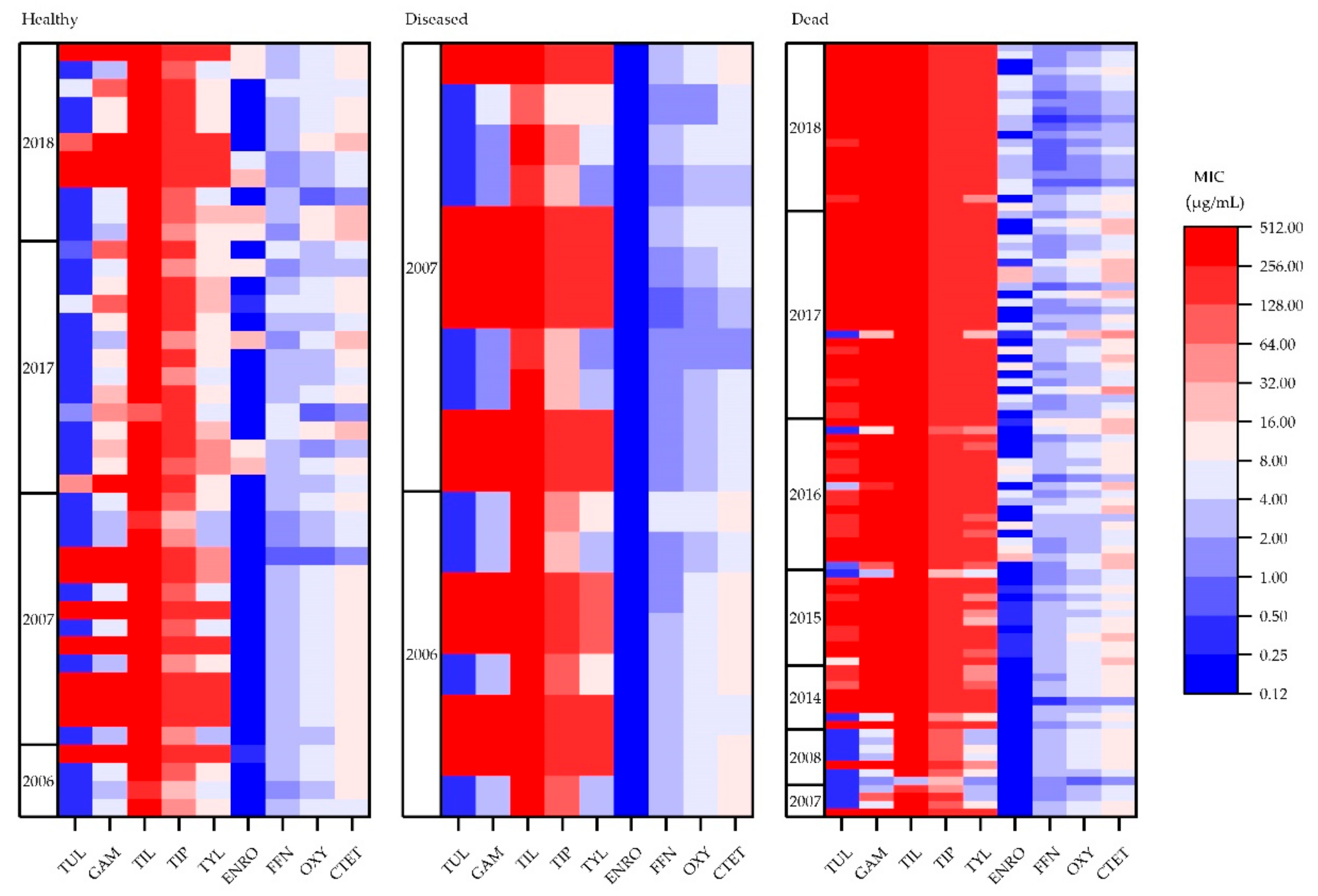
3.2. Antimicrobial Susceptibility Testing

The minimum inhibitory concentration (MIC) values for the M. bovis reference strain (ATCC® 25523™) were consistent across the AST processing period: TUL 0.25 µg/mL, GAM 8–16 µg/mL, TIL 1 µg/mL, TIP 4–8 µg/mL, TYL 1–2 µg/mL, ENRO 0.12–0.25 µg/mL, FFN 1–2 µg/mL, OXY 1–2 µg/mL, and CTET 1 µg/mL.
The heat maps in Figure 1 provide an overall impression of how MICs changed by health status and over time. In general, MIC values were lowest for the isolates from healthy cattle and highest for the dead cattle isolates. There was also a trend for MIC values to increase in time. There were also some interesting changes in the MIC values that were derived from the dead cattle isolates for the years 2016–2018. From 2016 onwards, nearly all the isolates were highly resistant to all macrolides. In addition, resistance to ENRO appeared to increase from 2016 to 2018, while the opposite occurred for OXY and CTET.
Table 2 is a crude summary of the MICs from all 211 isolates, ignoring health status, anatomical location, year, and the fact that some animals provided up to three isolates. The macrolide MICs were right censored, with TUL and TYL being both right and left censored. That is, the distributions suggest that the MICs were higher (right censored) or lower (left censored) than the tested range. Due to the right censoring, there was no resolution between the MIC50 and MIC90 values for the macrolides. The dark vertical line represents the resistance breakpoint for each antimicrobial and where applicableare based on CLSI’s suggested intermediate (I) breakpoints. Most isolates were resistant to all five macrolides.
Statistical analyses were used to compare the MIC distributions of each antimicrobial by health status and production year. Only one isolate (lung) per animal (n = 159) was used for these analyses. The lung isolate was chosen because it was the most consistently sampled tissue andbecause of its role in BRD. There were only 19 isolates from diseased cattle, with the mean rank for each antimicrobial generally being between those of the healthy and dead cattle; therefore, this group was omitted for subsequent analyses. In order to assess if the MIC values changed over time, the data displayed in heat maps were analyzed by health status. Theisolates obtained from the nasal cavities of healthy cattle (n = 43) were dichotomized into two time-points, “early” (2006–2007) and “late” (2017–2018). A similar manipulation was performed for the MIC data that were obtained from the lung isolates of the dead cattle. In this instance, the “early” isolates were from 2007–2008, while the “late” isolates were derived from cattle that died in the 2017 and 2018 production years. An additional analysis also compared the MIC values from the dead cattle isolates from 2016 versus 2018. These years were chosen because of the changes in the MICs for ENRO, OXY and CTET in these later years.
Table 3 provides the measure of effect (mean ranks) and the level of significance with respect to how the MIC values changed by health status over time. The MIC values that were obtained from the healthy cattle isolates remained relatively constant over a 10 year period, save one exception. The MIC distributions for ENRO changed over time (p = 0.004), with the means ranks shifting towards higher MIC values. Analyses of the MICs from the dead cattle isolates, comparing the early and later isolates, found that most of the mean ranks increased over time. The exceptions were FFN, OXY and CTET, whose mean ranks actually dropped; however, only OXY was significantly lower. A similar analysis for the years 2016 versus 2018 found that the MICs for TUL increased over this two-year period, whereas those for FFN, OXY, and CTET decreased.
To summarize, the isolates obtained from the DNP of healthy cattle on arrival to the feedlot did not appreciably change over the 10-year period, with the exception of ENRO. As for the dead cattle isolates, the MICs for the macrolides increased over the 10-year period; the exception was TIL, which had high MICs throughout the 10-year period. Significantly, the mean ranks for FFN, OXY and CTET all decreased in time. This was particularly evident when comparing MICs from the lung isolates of cattle that died in 2016 versus 2018.
Table 4 and Table 5 provide the antimicrobial MIC distributions for the healthy and dead cattle isolates, respectively. A visual inspection found a bimodal distribution of MIC values for TUL, GAM, TYL, and ENRO. The estimated epidemiological cut-off values were TUL 16 µg/mL, GAM 32 µg/mL, TYL 16 µg/mL, and ENRO 0.50 µg/mL.
A Chi-square analysis assessed for differences in the level of resistance (%R) between the healthy and dead cattle isolates. Isolates from the dead cattle were more likely to be resistant to TUL (OR = 11.68; 95% CI 5.03, 27.15; p ≤ 0.001), GAM (OR = 5.69; 95% CI 2.32, 13.93; p ≤ 0.001), TYL (OR = 3.90; 95% CI 1.37, 11.08; p = 0.007), and ENRO (OR = 2.3; 95% CI 1.03, 5.23; p = 0.041).
Forty-two DNP isolates from the healthy cattle had corresponding DOF data; one isolate had missing metadata. The data were dichotomized into 0 DOF and ≥14 DOF (range 14–140). The majority (n = 25) of the isolates were recovered at 0 DOF, with 17 isolates obtained ≥14 DOF. The mean ranks of the MIC distributions for the isolates from these two populations were similar, with the exception of ENRO (p = 0.007, 0 DOF mean rank = 25.27, ≥14 DOF mean rank = 17.00).
The MIC distributions for the macrolides (GAM, TIP, TUL and TYL) were highly correlated, with the exception being TIL (Table 6). The tetracyclines were highly correlated to each other (ρ = 0.895), and moderately correlated to FFN (ρ ≥ 0.653).
As an incidental finding, alamarBlue interfered with OXY at concentrations of ≥32 µg/mL; however, this did not affect the results, since all MICs for OXY were ≤16 µg/mL.

3.3. Antimicrobial Susceptibility Testing―Anatomical Region

The MIC distributions for all antimicrobials were compared across 80 paired lung–joint isolates that were derived from 40 animals and 20 isolates from 10 paired lung–DNP isolates. There were no differences in the MIC distributions for any of the antimicrobials between the lung–joint isolates (p ≥ 0.124) or the paired lung–DNP isolates (p ≥ 0.157).

4. Discussion

There are a number of unique features and findings associated with this study. This is the first report of MIC data that were generated from M. bovis isolates recovered from western Canadian feedlot cattle over an extended time period. Furthermore, a customized AST panel was developed to test only the antimicrobials that are most commonly used in the metaphylaxis and therapeutic treatment of BRD. This is notable since researchers frequently report using the standard Sensititre Bovine/Porcine MIC Plate (Trek Diagnostics), which tests antimicrobials that are used in both swine and cattle. While the standard plate has utility for conducting AST on multiple different bacterial pathogens, it is not an ideal plate for M. bovis. Many of the antimicrobials either have no activity against mycoplasmas (β-lactams and trimethoprim–sulphonamides) or are not routinely used in feedlot cattle (aminoglycosides). Though danofloxacin, a fluoroquinolone, was registered in Canada in 2004 for the treatment of BRD [28], it is used much less than ENRO, so it was not included in the panel. While focusing on fewer antimicrobials allowed for a broader range of serial dilutions, there was still significant right censoring of the MIC values for the macrolides, which is a common finding [19,29,30,31]. This is an issue because the magnitude of resistance and the potential presence of different subpopulations may be missed. Further optimization could be realized by reducing the range of dilutions for ENRO, FFN, OXY and CTET while increasing the upper range of the macrolides.
One of the strengths, but perhaps also a weakness of the study, is the wide-range of isolates recovered from healthy, diseased and dead cattle over multiple years. Sampling in the formative years was designed to answer questions regarding the genetic relatedness of isolates within groups of cattle. Over time, additional samples were obtained to examine how strains varied depending on the anatomical niche they occupied. More recently, increased attention has been given to M. bovis and its role in BRD cases that are refractory to standard antimicrobial therapy. Therefore, it is recognized that the purposive and convenience sampling methodologies that were used to acquire the isolates for other research objectives did not constitute a structured random sampling; hence, caution is warranted when interpreting the data. Furthermore, the data were cross-sectional in nature, and, hence, no inference can be made about causality; however, identifying relationships can lead to the generation of hypotheses for future research. That said, important conclusions can be drawn from the data. Specifically, these data highlight the very high levels of antimicrobial resistance to macrolides. This is undoubtedly related to decades of macrolide use in Canadian feedlots. In addition, mycoplasmosis is typically a chronic disease that requires serial courses of therapy, often involving multiple classes of antimicrobials. Thus, significant antimicrobial selection pressure is placed on the strains of M. bovis that circulate within a feedlot. While this may explain the very high levels of resistance found in the dead cattle, it is more difficult to reconcile why 100% of the isolates from the 43 healthy cattle were resistant to TIL and TIP.
The finding of high levels of resistance in the healthy cattle was unexpected since most of the cattle would have been sourced from cow–calf operations. This is salient because these animals would have been raised on rangeland during the preceding summer months, where disease and AMU is comparably low in these production settings. However, previous research has shown that M. bovis can be recovered from the nasopharyngeal passages and bronchoalveolar lavages of cattle within days of arrival at a feedlot [32]. This may explain the rapid emergence and horizontal transmission of resistant clones within the feedlot, particularly in cattle that were ≥14 DOF. However, this does not account for the level of resistance that was seen in the healthy cattle that were sampled on arrival. Perhaps the clonal spread of resistant bacteria may occur during commingling at auctions and during transport. Consideration must also be given to herd level factors, such as antimicrobial use, which may have selected for resistant strains that circulated within cow–calf operations. Regardless of the reservoir of resistant strains, the MIC data were disconcerting, since, even after controlling for the number of DOF, these healthy cattle arrived at the feedlot harboring M. bovis with significant levels of resistance to macrolides. Based on the data, the use of TIP and TIL for treating putative M. bovis infections such as CPPS should be avoided. This recommendation is certainly not novel. As early as 2002, Rosenbusch et al. [29] noted that erythromycin and TIL should not be considered for the treatment of M. bovis infections. This contention is supported by multiple studies and review articles [33,34,35,36].
While the current study has a number of limitations, the results are quite similar to two previous studies that included AST data from M. bovis isolates that were derived from western Canadian feedlot cattle [18,19]. In the first study, Hendrick et al. [18] conducted AST on 51 M. bovis isolates that were derived from sick and dead western Canadian feedlot cattle in 2007 and 2008 and reported lower MIC50 values than those seen in the current study, which may have been related to the isolates coming from a single feedlot. However, TUL, OXY, CTET and FFN all had MIC50 values in the 2–4 µg/mL range, which is comparable to the results reported herein. In both studies, TIL had very high MICs, whereas the isolates from 2007 and 2008 had very low MICs for TUL. These findings can be explained, in part, by AMU. Tilmicosin was registered in Canada in 1990 and, for many years, was the only long-acting macrolide used for BRD, whereas TUL came to the market in the autumn of 2007 [28]. Therefore, very few, if any, of the cattle that were sampled in 2007 and 2008, in either study, would have been treated with TUL. Thus, the heat maps provide a historical perspective on the increasing resistance to TUL over the last decade.
Though there was significant levels of resistance to OXY and CTET, it is important to appreciate that the mean ranks for FFN and the tetracyclines have been decreasing over time. Unlike TUL and TIL, these results cannot be explained by when the products came to market, since FFN was licensed in 1996 and the tetracyclines predate this registration by decades. More likely, the entry of newer macrolides into the marketplace has displaced the use of FFN, OXY and CTET. A study of AMU in western Canadian feedlots found that CTET, which has historically been incorporated into the feed for the control of Histophilus somni has been replaced with more efficacious parenterally administered therapies [5]. Further to this reduction, in 2018 the pharmaceutical industry voluntarily removed growth promotion claims from all medically important antimicrobials (MIAs). In addition, Health Canada mandated that, by the end of 2018, all MIAs for veterinary use must be sold by prescription only. These prescribing practices and regulatory changes may account for the lower tetracycline MICs seen in 2018.
A second and more recent western Canadian study reported on the AST results that were generated from 226 M. bovis isolates recovered from 211 feedlot cattle that were sampled in the 2014 production year [19]. While many of the data were right censored, the overall results were similar to the current study. Specifically, macrolides had the highest MICs, while ENRO, FFN, OXY and CTET were relatively comparable but with a bias towards slightly higher MICs. These results are relevant because they confirm or validate the results reported herein with respect to the AST findings from the more recently recovered isolates. The slightly higher MICs could be real or may reflect the inherent challenges of comparing AST data across different laboratories. What is clear, however, is that the macrolides had very high levels of resistance, whereas resistance to FFN, ENRO, and the tetracyclines was much lower.
Though the current study was not designed a priori as an AST surveillance study, the results are very consistent with the findings of the two previous studies of western Canadian feedlot cattle [18,19]. In general, all three studies have arrived at the same conclusions. The vast majority of M. bovis isolates are resistant to macrolides but susceptible to ENRO, FFN, and OXY. The low level of resistance to ENRO is an important finding since the World Health Organization (WHO) classifies fluoroquinolones as Class I antimicrobials, which are deemed critically important [37]. It is also noteworthy that ENRO AMU has actually been declining in western Canadian feedlots [5], which may explain the low levels of resistance. That said, it was clear from the heat maps that the MICs for ENRO have been increasing in time within both the healthy and dead cattle isolates. Presumably, ENRO is being used when all other treatments have failed to resolve the BRD, resulting in selection pressure on the mycoplasma community. All three studies underscore the need for alternatives to antimicrobials for controlling BRD. Macrolide resistance is very high and is unlikely to change in the near future because most feedlots rely on this class of antimicrobials for the metaphylaxis and treatment of BRD. Potential overuse is compounded by the fact that there is a limited number of therapeutic options, and this may result in increased use of ENRO as a last resort antimicrobial.
Macrolide resistance has been attributed to single nucleotide polymorphisms (SNPs) or point mutations that are common to all pathogenic mycoplasmas [38,39]. These modifications alter how macrolides interact with the 50S rRNA subunit to inhibit protein synthesis. Specifically, macrolides bind within the 50 s tunnel and interact with the A2058 and A2059 nucleotides [40,41]. It is noteworthy that TIL and TIP are derivatives of TYL, and all three bind at the same site. However, minor changes in how they bind may modify antimicrobial activities [40]. This may explain the lower levels of resistance to TYL versus TIL and TIP in the healthy cattle isolates. Sulyok et al. [35] also reported a cross-resistance between TYL and TIL and speculated, based on similar MIC distributions, that cross-resistance extended to GAM and TUL. Thus, all four macrolides share common point mutations that confer resistance, which explains the highly correlated MICs distributions for GAM, TIP, TUL and TYL. The finding that TIL was not as highly correlated to the other macrolide is a spurious finding. The Spearman correlation compares the distributions of the MICs versus specific values. In the case of TIL, all the isolates essentially had the same MIC (≥256 µg/mL), whereas the other four macrolides had a range of MICs. Thus, the test statistic found that the TIL was not correlated with the others. In this situation, it was best to look at the MIC50 and MIC90 values, which were identical for all five macrolides for dead cattle isolates. In reality, all the macrolides were indeed highly correlated. This is an important finding because the intense use of macrolides to control and treat BRD [5] invariably leads to an increased AMR [19] in all BRD pathogens. Thus, the results of the current study do not bode well for the continued use of macrolides on a routine basis for the metaphylaxis and therapeutic treatment of mycoplasmosis. There is a compelling argument that macrolides should not be used in animals with chronic BRD, and perhaps even in healthy arrivals to the feedlot, unless the AST data support such therapy.
Macrolide resistance is certainly not unique to this study; rather, this is a growing worldwide phenomenon in human and veterinary medicine. A review article of macrolide resistance of human mycoplasmas noted that Mycoplasma pneumoniae has emerged as a major cause of community-acquired pneumonia and, not unlike the current findings, tetracyclines and fluoroquinolones remain effective, whereas macrolide resistance is becoming widespread [38]. As a result, some countries have implemented antimicrobial stewardship programs to curtail macrolide use in humans. Here in Canada, macrolides are classified as Category II drugs; however, the WHO considers them Category I, or highest priority-critically important antimicrobials (HP-CIAs). Furthermore, the WHO states that HP-CIAs should not be used prophylactically or as a first line therapy for animals. Rather, they should only be used if no effective alternative treatment is available. In many instances, macrolides are being administered in feedlots for metaphylaxis and not prophylaxis, with the distinguishing feature being that the cohort of animals being treated is manifesting varying levels of disease. However, data from multiple studies show that OXY or FFN should be considered for first line therapy, ahead of macrolides. This also applies to Mannheimia haemolytica and Pasteurella multocida [19]. The current state of macrolide resistance in veterinary medicine is a clarion call for developing alternative and sustainable methods for controlling BRD.
Despite the challenges and caveats of extrapolating breakpoints, there is utility in providing veterinary practitioners with a guarded interpretation of the MIC values. A common practice has been to extrapolate the CLSI guidelines from other bacterial members of the BRD complex, specifically Mannheimia haemolytica, Pasteurella multocida, and Histophilus somni [19,28,36,42]. As stated previously, all the CLSI guidelines suggested macrolide breakpoints were adopted with slight modifications. With respect to TIP, the MIC data lacked a bimodal distribution and all MICs were ≥16 µg/mL, suggesting that this population of isolates were non-wild-type. That is, they had acquired some mechanisms of resistance. It also needs to be stressed that the suggested resistant breakpoints encompassed the intermediate breakpoints. The rationale being that isolates in the intermediate interpretative category may require a higher approved dosage of drug [43]. Therefore, from a clinical standpoint, veterinary practitioners want to know which antimicrobial will provide the most successful clinical outcome with a typical dosage regimen.
The same breakpoints were assigned to both OXY and CTET, even though the MIC distributions for CTET were higher than for OXY. This decision was based on a review of MIC data from three other Canadian studies [17,18,19]. In all studies, the MIC values for OXY and CTET were similar, which is understandable since both antimicrobials are impacted by point mutations in the Tet-1 tetracycline binding pocket of the 30S ribosomal unit [35,44]. Until the CLSI establishes interpretative criteria for MIC data for M. bovis, researchers and veterinarians will be required to extrapolate from the guidelines for other BRD pathogens. It is worth noting that the CLSI does have guidelines for human mycoplasmas [24], and the suggested resistance breakpoint for tetracycline and M. hominis is ≥8 µg/mL; the current study used ≥4 µg/mL, which is a difference of one serial dilution.
Singh et al. [45] reported the first outbreaks of M. bovis arthritis in Canadian calves in 1971; however, it was Haines et al. in 2001 who found M. bovis in 71% of lungs and 45% of joints of western Canadian feedlot cattle that failed to respond to antimicrobial therapy [46]. Despite a growing body of evidence pointing to the role of M. bovis in chronic BRD cases in Canadian feedlot cattle [7,10], AST is not typically performed. Rather, feedlot veterinarians have historically been more concerned with M. haemolytica, Pastuerella multicoda, and Histophilus somni, which together account for the majority of early arrival peracute to acute cases of BRD. Thus, the choice of antimicrobials is determined by algorithms that take the level of risk assigned to a cohort of animals arriving at the feedlot into account. Others, however, have posited that AST should be performed when animals fail to respond to multiple classes of antimicrobials [34,47]. The question then becomes how often and how many isolates are needed, as well as from what animals (healthy, sick or dead). Previous research has shown that during BRD outbreaks, a single dominant clone of M. bovis will emerge and spread within a pen [48]. However, in large scale studies of M. pneumoniae associated with endemics and epidemics of human mycoplasmosis, the infections were polyclonal [49]. Therefore, in large feedlots, multiple clones, rather than a single clone, may be spreading and infecting cattle. This has implications in respect to AST, AMR, AMU, and vaccine development.
Another interesting feature of this study was the analyses of the paired lung–joint and lung–DNP isolates, which found no differences in the MIC distributions for any antimicrobial. Previously, researchers noted that respiratory tract isolates had a higher MIC50 for TUL compared to isolates from the lung, milk, and synovial fluids [50]. In another study, M. bovis isolates from milk samples had higher MICs than did those from the lungs for spectinomycin, though not for other antimicrobials [51]. However, in both studies, the samples were not paired, but isolates came from samples submitted to diagnostic laboratories. The current study examined the AST profiles of isolates that were obtained from the same animals. Whether these isolates represent a single clone that colonizes the DNP and then gains entry to the lung and spreads hematogenously to the joints and other tissues is unknown. Molecular techniques should be able to answer whether the isolates in the DNP, lungs and joints are mono or polyclonal.

5. Conclusions

Mycoplasma bovis is an important pathogen of the bovine respiratory disease complex. While the colonization of the respiratory tract occurs within days, cattle with mycoplasmosis often linger in the feedlot for many weeks, during which time they receive multiple courses of antimicrobial therapy. Additionally, high-risk cattle are routinely given macrolides for metaphylaxis. As a result, there is widespread antimicrobial resistance to the five most commonly used macrolides. The challenges facing the feedlot sector is that this class of antimicrobials is extensively used for the prevention and treatment of all causes of BRD. As a result, the resistance issue is not unique to mycoplasmas, but it is a cause for concern in the management of BRD in general. One potential solution would be to rotate macrolides as first-choice treatment to second or third choice, elevating the use of OXY and FFN. However, this is not a long-term solution; rather, a paradigm shift is needed if the industry is to remain sustainable. It also needs to be stated that AMR is not just an economic issue. Chronic mycoplasmosis leads to pain and suffering, especially if the cattle have CPPS. Therefore, AMR is very much an animal welfare issue.

Author Contributions

Conceptualization, M.J., S.W., and T.A.M.; methodology, M.J., A.K., K.G., S.A.-L., and R.Z.; formal analysis, M.J.; investigation, M.J., A.K., K.G., S.A.-L., and R.Z.; resources, M.J. and T.A.M.; data curation, M.J., A.K., K.G., S.A.-L., and R.Z.; writing—original draft preparation, M.J.; writing—review and editing, M.J., A.K., S.A.-L., R.Z.K., S.W., and T.A.M.; visualization, M.J., S.W., and T.A.M.; supervision, M.J. and T.A.M.; project administration, M.J. and T.A.M.; funding acquisition, M.J., S.W., and T.A.M. All authors have read and agreed to the published version of the manuscript.

Funding

This study was made possible by the Saskatchewan Agricultural Development Fund (Grants #20060116, #20130170, #20160253) and the Canadian Cattlemen’s Association, Beef Cattle Research Council Division (Grant ANH.30.17).

Acknowledgments

The authors would like to express their appreciation to: Karen Register, Research Microbiolgist, USDA, Ames, IA and Manual Chirino-Trejo, Professor Emeritus, Western College of Veterinary Medicine, for their expertise in the development of the culturing, isolation, and identification techniques for Mycoplasma bovis.

Conflicts of Interest

The authors declare no conflict of interest.

Appendix A

YearFeedlotCohortIsolate IDAnimal #Anatomical RegionHealth StatusDOF
2006F6C13MPLM06481DNPDiseased90
MPLM06542DNPDiseased90
F14C21MPLM06493DNPDiseased90
MPLM07864DNPDiseased90
F16C23MPLM08455DNPHealthy
F20C29MPLM08106DNPDiseased150
MPLM06427DNPDiseased150
MPLM06518DNPDiseased150
F30C40MPLM06439DNPHealthy140
MPLM081110DNPHealthy14
MPLM081311DNPDiseased
MPLM064012DNPHealthy14
N = 5N = 5N = 12N = 12
2007F1C1MPLM078813LungDead47
F8C15MPLM063214DNPDead
F9C16MPLM079515LungDead
MPLM0631JointDead60
F15C22MPLM066416DNPDiseased15
MPLM066017DNPDiseased0
MPLM066518DNPDiseased21
MPLM064419DNPDiseased21
MPLM066920DNPDead41
MPLM066121DNPDiseased12
MPLM064722DNPDiseased16
MPLM065723DNPDiseased45
MPLM071324DNPHealthy15
MPLM071425DNPHealthy15
MPLM068426DNPHealthy15
MPLM065227DNPDiseased10
MPLM066228DNPDiseased21
MPLM070029DNPHealthy15
MPLM070330DNPHealthy15
MPLM071531DNPHealthy15
MPLM070632DNPHealthy15
MPLM064533DNPDiseased10
MPLM066634DNPDiseased10
MPLM069235DNPHealthy15
MPLM069836DNPHealthy15
F16C24MPLM067037DNPHealthy14
MPLM067138DNPHealthy14
C25MPLM066739DNPHealthy90
MPLM066840DNPHealthy90
MPLM064641DNPHealthy90
N = 5N = 6N = 30N = 29
2008F7C14MPLM078142LungDead
MPLM0602DNPDead60
MPLM078443LungDead
F8C15MPLM063044JointDead30
F22C31MPLM078945LungDead
MPLM079246LungDead
F23C32MPLM078347LungDead
MPLM079048LungDead
N = 4N = 4N = 8N = 7
2014F2C5MYCO06649LungDead35
MYCO09650LungDead96
F3C6MP021951LungDead39
MP0209JointDead39
F13C20MYCO08152JointDead31
F17C26MP006453LungDead193
F19C28MPLM063654LungDead93
F21C30MYCO06255LungDead76
MYCO07656LungDead24
N = 6N = 6N = 9N = 8
2015F3C7MP007157LungDead42
MP006358LungDead42
MPLM063759LungDead31
MP005860LungDead31
MP007361LungDead67
MP007762LungDead61
MP007963LungDead67
MPLM0638JointDead67
MP018364JointDead217
F11C18MPLM063465LungDead
F13C20MP005766LungDead80
F21C30MP007067LungDead20
F27C37MP007568LungDead46
N = 5N = 5N = 13N = 12
2016F1C2MPLM004169LungDead46
MPLM0042JointDead46
MPLM003370LungDead48
MPLM0034JointDead48
MPLM002971LungDead
MPLM0030JointDead
F18C27MPLM002172LungDead55
MPLM0022JointDead55
MPLM000973LungDead22
MPLM0010JointDead22
MPLM001374LungDead26
MPLM0014JointDead26
MPLM002575LungDead15
MPLM0026JointDead15
MPLM003776LungDead81
MPLM001777LungDead43
MPLM0018JointDead43
MPLM001578LungDead31
MPLM0016JointDead31
MPLM000779LungDead9
MPLM0008JointDead9
MPLM003580LungDead38
MPLM0036JointDead38
MPLM003181LungDead34
MPLM001982LungDead55
MPLM0020JointDead55
MPLM001183LungDead22
MPLM0012JointDead22
MPLM003984LungDead50
MPLM0040JointDead50
F19C28MPLM013485LungDead60
MPLM0135JointDead60
MPLM013286LungDead30
F28C38MPLM000387LungDead28
MPLM0004JointDead28
N = 4N = 4N = 35N = 19
2017F1C3MPLM011488LungDead33
MPLM010289LungDead40
MPLM0103JointDead40
MPLM016090LungDead52
MPLM009391LungDead37
MPLM0094JointDead37
MPLM009092LungDead45
MPLM0091JointDead45
MPLM011193LungDead17
MPLM0112JointDead17
MPLM008794LungDead60
MPLM0088JointDead60
MPLM010595LungDead54
MPLM0106JointDead54
MPLM006496JointDead
MPLM016497JointDead30
MPLM014398JointDead24
MPLM006699LungDead
MPLM0067JointDead
MPLM0108100LungDead44
MPLM0145101LungDead42
MPLM0146JointDead42
MPLM0154102LungDead40
MPLM0155JointDead40
MPLM0073103JointDead
MPLM0157104LungDead46
MPLM0158JointDead46
MPLM0084105LungDead33
MPLM0085JointDead33
MPLM0148106LungDead48
MPLM0149JointDead48
MPLM0069107LungDead
MPLM0070JointDead
F4C8MPLM0831108DNPHealthy0
F5C10MPLM0833109DNPHealthy0
MPLM0834110DNPHealthy0
MPLM0837111DNPHealthy0
MPLM0838112DNPHealthy0
C11MPLM0832113DNPHealthy0
MPLM0835114DNPHealthy0
C12MPLM0839115DNPHealthy0
F10C17MPLM0081116LungDead180
F12C19MPLM0167117JointDead
MPLM0057118LungDead
MPLM0058JointDead
MPLM0060119LungDead
MPLM0061JointDead
MPLM0054120LungDead
MPLM0076121JointDead
F24C33MPLM0815122DNPHealthy0
MPLM0819123DNPHealthy0
MPLM0820124DNPHealthy0
F26C36MPLM0821125DNPHealthy0
MPLM0822126DNPHealthy0
F29C39MPLM0826127DNPHealthy0
N = 8N = 10N = 55N = 40
2018F1C4MPLM0563128LungDead
MPLM0533129LungDead65
MPLM0534JointDead65
MPLM0535DNPDead65
MPLM0569130LungDead27
MPLM0552131LungDead36
MPLM0553DNPDead36
MPLM0611132LungDead15
MPLM0622133DNPDead33
MPLM0566134LungDead30
MPLM0567DNPDead30
MPLM0537135LungDead27
MPLM0538JointDead27
MPLM0539DNPDead27
MPLM0593136LungDead44
MPLM0555137LungDead60
MPLM0556JointDead60
MPLM0549138LungDead34
MPLM0587139LungDead34
MPLM0588DNPDead34
MPLM0584140LungDead44
MPLM0624141LungDead33
MPLM0625DNPDead33
MPLM0541142LungDead71
MPLM0542JointDead71
MPLM0543DNPDead71
MPLM0545143LungDead56
MPLM0546JointDead56
MPLM0547DNPDead56
MPLM0608144LungDead61
MPLM0609DNPDead61
MPLM0582145DNPDead61
MPLM0559146LungDead33
MPLM0560JointDead33
MPLM0578147LungDead15
MPLM0579DNPDead15
F4C9MPLM0827148DNPHealthy0
MPLM0828149DNPHealthy0
MPLM0829150DNPHealthy0
MPLM0830151DNPHealthy0
F24C34MPLM0816152DNPHealthy0
MPLM0817153DNPHealthy0
MPLM0818154DNPHealthy0
F25C35MPLM0627155LungDead
MPLM0628DNPDead
F29C40MPLM0823156DNPHealthy0
MPLM0824157DNPHealthy0
MPLM0825158DNPHealthy0
F31C12MPLM0836159DNPHealthy0
N = 6N = 6N = 49N = 32

References

  1. Griffin, D.; Chengappa, M.M.; Kuszak, J.; McVey, D.S. Bacterial pathogens of the bovine respiratory disease complex. Vet. Clin. N. Am. Food Anim. 2010, 26, 381–394. [Google Scholar] [CrossRef] [PubMed]
  2. Panciera, R.J.; Confer, A.W. Pathogenesis and pathology of bovine pneumonia. Vet. Clin. N. Am. Food Anim. Pract. 2010, 26, 191–214. [Google Scholar] [CrossRef] [PubMed]
  3. Nickell, J.S.; White, B.J. Metaphylactic antimicrobial therapy for bovine respiratory disease in stocker and feedlot cattle. Vet. Clin. N. Am. Food Anim. 2010, 26, 285–301. [Google Scholar] [CrossRef] [PubMed]
  4. Brault, S.A.; Hannon, S.J.; Gow, S.P.; Otto, S.J.G.; Booker, C.W.; Morley, P.S. Calculation of antimicrobial use indicators in beef feedlots—Effects of choice of metric and standardized values. Front. Vet. Sci. 2019, 6, 330. [Google Scholar] [CrossRef]
  5. Brault, S.A.; Hannon, S.J.; Gow, S.P.; Warr, B.N.; Withell, J.; Song, J.; Williams, C.M.; Otto, S.J.G.; Booker, C.W.; Morley, P.S. Antimicrobial use on 36 beef feedlots in western Canada: 2008–2012. Front. Vet. Sci. 2019, 6, 329. [Google Scholar] [CrossRef] [Green Version]
  6. Nicholas, R.A.J.; Ayling, R.D. Mycoplasma bovis: Disease, diagnosis, and control. Res. Vet. Sci. 2003, 74, 105–112. [Google Scholar] [CrossRef]
  7. Gagea, M.I.; Bateman, K.G.; Shanahan, R.A.; van Dreumel, T.; McEwen, B.J.; Carman, S.; Archambault, M.; Caswell, J.L. Naturally occurring mycoplasma bovis—Associated pneumonia and polyarthritis in feedlot beef calves. J. Vet. Diagn. Investig. 2006, 18, 29–40. [Google Scholar] [CrossRef] [PubMed] [Green Version]
  8. Horwood, P.; Schibrowski, M.; Fowler, E.; Gibson, J.; Barnes, T.; Mahony, T. Is mycoplasma bovis a missing component of the bovine respiratory disease complex in australia? Aust. Vet. J. 2014, 92, 185–191. [Google Scholar] [CrossRef] [PubMed]
  9. Grissett, G.P.; White, B.J.; Larson, R.L. Structured literature review of responses of cattle to viral and bacterial pathogens causing bovine respiratory disease complex. J. Vet. Intern. Med. 2015, 29, 770–780. [Google Scholar] [CrossRef]
  10. Booker, C.W.; Abutarbush, S.M.; Morley, P.S.; Jim, G.K.; Pittman, T.J.; Schunicht, O.C.; Perrett, T.; Wildman, B.K.; Fenton, R.K.; Guichon, P.T.; et al. Microbiological and histopathological findings in cases of fatal bovine respiratory disease of feedlot cattle in western canada. Can. Vet. J. 2008, 49, 473–481. [Google Scholar]
  11. Radaelli, E.; Luini, M.; Loria, G.R.; Nicholas, R.A.J.; Scanziani, E. Bacteriological, serological, pathological and immunohistochemical studies of mycoplasma bovis respiratory infection in veal calves and adult cattle at slaughter. Res. Vet. Sci. 2008, 85, 282–290. [Google Scholar] [CrossRef]
  12. Caswell, J.L.; Bateman, K.G.; Cai, H.Y.; Castillo-Alcala, F. Mycoplasma bovis in respiratory disease of feedlot cattle. Vet. Clin. N. Am. Food Anim. 2010, 26, 365–379. [Google Scholar] [CrossRef] [PubMed]
  13. Nicholas, R.A.J. Bovine mycoplasmosis: Silent and deadly. Vet. Rec. 2011, 168, 459–462. [Google Scholar] [CrossRef] [PubMed]
  14. Caswell, J.L.; Archambault, M. Mycoplasma bovis pneumonia in cattle. Anim. Health Res. Rev. 2007, 8, 161–186. [Google Scholar] [CrossRef] [PubMed]
  15. Romváry, J.; Rózsa, J.; Stipkovits, L.; Mészaros, J. Incidence of diseases due to Mycoplasma bovis in a cattle herd. Pneumo-arthritis syndrome in calves. Acta Vet. Acad. Sci. Hung 1979, 27, 29–37. [Google Scholar]
  16. Francoz, D.; Fortin, M.; Fecteau, G.; Messier, S. Determination of mycoplasma bovis susceptibilities against six antimicrobial agents using the E test method. Vet. Microbiol. 2005, 105, 57–64. [Google Scholar] [CrossRef]
  17. Cai, H.Y.; McDowall, R.; Parker, L.; Kaufman, E.I.; Caswell, J.L. Changes in antimicrobial susceptibility profiles of mycoplasma bovis over time. Can. Vet. J. 2019, 83, 34–41. [Google Scholar]
  18. Hendrick, S.H.; Bateman, K.G.; Rosengren, L.B. The effect of antimicrobial treatment and preventive strategies on bovine respiratory disease and genetic relatedness and antimicrobial resistance of mycoplasma bovis isolates in a western canadian feedlot. Can. Vet. J. 2013, 54, 1146. [Google Scholar]
  19. Anholt, R.M.; Klima, C.; Allan, N.; Matheson-Bird, H.; Schatz, C.; Ajitkumar, P.; Otto, S.J.; Peters, D.; Schmid, K.; Olson, M.; et al. Antimicrobial susceptibility of bacteria that cause bovine respiratory disease complex in alberta, canada. Front. Vet. Sci. 2017, 4, 207. [Google Scholar] [CrossRef] [Green Version]
  20. Taylor-Robinson, D.; Bébéar, C. Antibiotic susceptibilities of mycoplasmas and treatment of mycoplasmal infections. J. Antimicrob. Chemother. 1997, 40, 622–630. [Google Scholar] [CrossRef] [Green Version]
  21. Lysnyansky, I.; Ayling, R.D. Mycoplasma bovis: Mechanisms of resistance and trends in antimicrobial susceptibility. Front. Microbiol. 2016, 7, 595. [Google Scholar] [CrossRef] [PubMed]
  22. Register, K.B.; Boatwright, W.D.; Gesy, K.M.; Thacker, T.C.; Jelinski, M.D. Mistaken identity of an open reading frame proposed for PCR-based identification of Mycoplasma bovis and the effect of polymorphisms and insertions on assay performance. J. Vet. Diagn. Investig. 2018, 30, 637–641. [Google Scholar] [CrossRef] [PubMed] [Green Version]
  23. Miles, K.; McAuliffe, L.; Ayling, R.D.; Nicholas, R.A.J. Rapid detection of Mycoplasma dispar and M. bovirhinis using allele specific polymerase chain reaction protocols. FEMS Microbiol. Lett. 2004, 241, 103–107. [Google Scholar] [CrossRef] [Green Version]
  24. CLSI. Performance Standards for Antimicrobial Disk and Dilution Susceptibility Tests for Bacteria Isolated From Animals, 5th ed.; CLSI standard VET01; Clinical and Laboratory Standards Institute: Wayne, PA, USA, 2018. [Google Scholar]
  25. CLSI. Methods for Antimicrobial Susceptibility Testing for Human Mycoplasmas Approved Guideline; CLSI document M43-A.; Clinical and Laboratory Standards Institute: Wayne, PA, USA, 2011. [Google Scholar]
  26. CLSI. Performance Standards for Antimicrobial Disk and Dilution Susceptibility Tests for Bacteria Isolated From Animals, 4th ed.; CLSI supplement VET08; Clinical and Laboratory Standards Institute: Wayne, PA, USA, 2018. [Google Scholar]
  27. Lerner, U.; Amram, E.; Ayling, R.D.; Mikula, I.; Gerchman, I.; Harrus, S.; Teff, D.; Yogev, D.; Lysnyansky, I. Acquired resistance to the 16-membered macrolides tylosin and tilmicosin by Mycoplasma bovis. Vet. Microbiol. 2014, 168, 365–371. [Google Scholar] [CrossRef] [PubMed]
  28. Government of Canada, Notice of Compliance Information. Available online: https://health-products.canada.ca/noc-ac/index-eng.jsp (accessed on 10 December 2019).
  29. Rosenbusch, R.F.; Kinyon, J.M.; Apley, M.; Funk, N.D.; Smith, S.; Hoffman, L.J. In vitro antimicrobial inhibition profiles of mycoplasma bovis isolates recovered from various regions of the united states from 2002 to 2003. J. Vet. Diagn. Investig. 2005, 17, 436–441. [Google Scholar] [CrossRef] [PubMed] [Green Version]
  30. Gautier-Bouchardon, A.V.; Ferré, S.; Le Grand, D.; Paoli, A.; Gay, E.; Poumarat, F. Overall decrease in the susceptibility of mycoplasma bovis to antimicrobials over the past 30 years in france. PLoS ONE 2014, 9, e87672. [Google Scholar] [CrossRef] [PubMed] [Green Version]
  31. Sulyok, K.M.; Kreizinger, Z.; Fekete, L.; Hrivnák, V.; Magyar, T.; Jánosi, S.; Schweitzer, N.; Turcsányi, I.; Makrai, L.; Erdélyi, K.; et al. Antibiotic susceptibility profiles of mycoplasma bovis strains isolated from cattle in hungary, central europe. BMC Vet. Res. 2014, 10, 256. [Google Scholar] [CrossRef] [PubMed] [Green Version]
  32. Allen, J.W.; Viel, L.; Bateman, K.G.; Rosendal, S. Changes in the bacterial flora of the upper and lower respiratory tracts and bronchoalveolar lavage differential cell counts in feedlot calves treated for respiratory diseases. Can. Vet. J. 1992, 56, 177–183. [Google Scholar]
  33. Ayling, R.D.; Baker, S.E.; Nicholas, R.A.J.; Peek, M.L.; Simon, A.J. Comparison of in vitro activity of danofloxacin, florfenicol, oxytetracycline, spectinomycin and tilmicosin against recent field isolates of mycoplasma bovis. Vet. Rec. 2000, 146, 745–747. [Google Scholar] [CrossRef]
  34. Maunsell, F.P.; Donovan, G.A. Mycoplasma bovis infections in young calves. Vet. Clin. N. Am. Food Anim. 2009, 25, 139–177. [Google Scholar] [CrossRef] [PubMed]
  35. Sulyok, K.M.; Kreizinger, Z.; Wehmann, E.; Lysnyansky, I.; Bányai, K.; Marton, S.; Jerzsele, Á.; Rónai, Z.; Turcsányi, I.; Makrai, L.; et al. Mutations associated with decreased susceptibility to seven antimicrobial families in field and laboratory-derived mycoplasma bovis strains. Antimicrob. Agents Chemother. 2017, 61, e01983-16. [Google Scholar] [CrossRef] [PubMed] [Green Version]
  36. Gautier-Bouchardon, A.V. Antimicrobial resistance in mycoplasma spp. Microbiol. Spectrum. 2018, 6, 425–446. [Google Scholar] [CrossRef] [PubMed]
  37. Who Critically Important Antimicrobials for Human Medicine. 5th Revision. Available online: https://www.who.int/foodsafety/publications/cia2017.pdf?ua=1 (accessed on 21 December 2019).
  38. Cao, B.; Zhao, C.; Yin, Y.; Zhao, F.; Song, S.; Bai, L.; Zhang, J.; Liu, Y.; Zhang, Y.; Wang, H.; et al. High prevalence of macrolide resistance in mycoplasma pneumoniae isolates from adult and adolescent patients with respiratory tract infection in china. Clin. Infect. Dis. 2010, 51, 189–194. [Google Scholar] [CrossRef] [PubMed] [Green Version]
  39. Chironna, M.; Sallustio, A.; Esposito, S.; Perulli, M.; Chinellato, I.; Di Bari, C.; Quarto, M.; Cardinale, F. Emergence of macrolide-resistant strains during an outbreak of Mycoplasma pneumoniae infections in children. J. Antimicrob. Chemother. 2011, 66, 734–737. [Google Scholar] [CrossRef] [PubMed] [Green Version]
  40. Andersen, N.M.; Poehlsgaard, J.; Warrass, R.; Douthwaite, S. Inhibition of protein synthesis on the ribosome by tildipirosin compared with other veterinary macrolides. Antimicrob. Agents Chemother. 2012, 56, 6033–6036. [Google Scholar] [CrossRef] [PubMed] [Green Version]
  41. Poehlsgaard, J.; Andersen, N.M.; Warrass, R.; Douthwaite, S. Visualizing the 16-Membered ring macrolides tildipirosin and tilmicosin bound to their ribosomal site. ACS Chem. Biol. 2012, 7, 1351–1355. [Google Scholar] [CrossRef]
  42. Gerchman, I.; Levisohn, S.; Mikula, I.; Lysnyansky, I. In vitro antimicrobial susceptibility of mycoplasma bovis isolated in israel from local and imported cattle. Vet. Microbiol. 2009, 137, 268–275. [Google Scholar] [CrossRef]
  43. CLSI. Generation, Presentation, and Application of Antimicrobial Susceptibility Test Data for Bacteria of Animal Origin: A Report; CLSI Document VET05-R; Clinical and Laboratory Standards Institute: Wayne, PA, USA, 2011. [Google Scholar]
  44. Amram, E.; Mikula, I.; Schnee, C.; Ayling, R.D.; Nicholas, R.A.J.; Rosales, R.S.; Harrus, S.; Lysnyansky, I. 16S rRNA gene mutations associated with decreased susceptibility to tetracycline in mycoplasma bovis. Antimicrob. Agents Chemother. 2015, 59, 796–802. [Google Scholar] [CrossRef] [Green Version]
  45. Singh, U.M.; Doig, P.A.; Ruhnke, H.L. Mycoplasma arthritis in calves. Can. Vet. J. 1971, 12, 183–185. [Google Scholar]
  46. Haines, D.M.; Martin, K.M.; Clark, E.G.; Jim, G.K.; Janzen, E.D. The immunohistochemical detection of mycoplasma bovis and bovine viral diarrhea virus in tissues of feedlot cattle with chronic, unresponsive respiratory disease and/or arthritis. Can. Vet. J. 2001, 42, 857–860. [Google Scholar]
  47. Watts, J.L.; Sweeney, M.T. Antimicrobial resistance in bovine respiratory disease pathogens: Measures, trends, and impact on efficacy. Vet. Clin. N. Am. Food Anim. 2010, 26, 79–88. [Google Scholar] [CrossRef] [PubMed]
  48. Timsit, E.; Arcangioli, M.-A.; Bareille, N.; Seegers, H.; Assié, S. Transmission dynamics of mycoplasma bovis in newly received beef bulls at fattening operations. J. Vet. Diagn. Investig. 2012, 24, 1172–1176. [Google Scholar] [CrossRef] [PubMed] [Green Version]
  49. Pereyre, S.; Charron, A.; Hidalgo-Grass, C.; Touati, A.; Moses, A.E.; Nir-Paz, R.; Bébéar, C. The spread of mycoplasma pneumoniae is polyclonal in both an endemic setting in france and in an epidemic setting in israel. PLoS ONE 2012, 7, e38585. [Google Scholar] [CrossRef] [Green Version]
  50. Heuvelink, A.; Reugebrink, C.; Mars, J. Antimicrobial susceptibility of mycoplasma bovis isolates from veal calves and dairy cattle in the netherlands. Vet. Microbiol. 2016, 189, 1–7. [Google Scholar] [CrossRef]
  51. Soehnlen, M.K.; Kunze, M.E.; Karunathilake, K.E.; Henwood, B.M.; Kariyawasam, S.; Wolfgang, D.R.; Jayarao, B.M. In vitro antimicrobial inhibition of mycoplasma bovis isolates submitted to the pennsylvania animal diagnostic laboratory using flow cytometry and a broth microdilution method. J. Vet. Diagn. Investig. 2011, 23, 547–551. [Google Scholar] [CrossRef] [PubMed] [Green Version]
Figure 1. Minimum inhibitory concentration distribution for M. bovis isolates from healthy (n = 43), diseased (n = 19), and dead (n = 97) cattle (n = 159). Tulathromycin (TUL; 0.25–256 µg/mL), gamithromycin (GAM; 0.25–256 µg/mL), tilmicosin (TIL; 1–256 µg/mL), tildipirosin (TIP; 0.12–128 µg/mL), tylosin tartrate (TYL; 1–128 µg/mL), enrofloxacin (ENRO; 0.12–128 µg/mL), florfenicol (FFN; 0.25–256 µg/mL), oxytetracycline (OXY; 0.5–16 µg/mL), chlortetracycline (CTET; 1–256 µg/mL).
Microorganisms 08 00124 g001
Table 1. Cross tabulation of 211 Mycoplasma bovis isolates by sample (anatomical region or health status) and production year.
SampleFeedlot Production Year
20062007200820142015201620172018Total
Anatomical Region
DNP12271000142478
Lung02671119201984
Joint011221621649
Total12308913355549211
Health Status
Healthy4140000141143
Diseased81100000019
Dead058913354138149
Total12308913355549211
Table 2. Antibiotic susceptibility test results of the 211 Mycoplasma bovis isolates that were recovered from 159 healthy, diseased, and dead cattle between 2006 and 2018.
AntibioticClass≤0.120.250.501248163264128≥256%RMIC Range
(µg/mL)
MIC50
(µg/mL)
MIC90
(µg/mL)
TulathromycinMacrolide 5222122 342511871.1≤0.25–≥256≥256≥256
GamithromycinMacrolide 517179316315081.51–≥256≥256≥256
TilmicosinMacrolide 1 2620299.52–≥256≥256≥256
TildipirosinMacrolide 181320169 1008–≥128≥128≥128
TylosinMacrolide 310132261211134 87.71–≥128≥128≥128
EnrofloxacinFluoroquinolone12015 2030197 36.0≤0.12–160.254
FlorfenicolPhenicol 21872100181 9.0≤0.25–822
OxytetracyclineTetracycline 6307585141 47.40.50–1624
ChlortetracyclineTetracycline 9316274323 81.0≤1–32816
The dark vertical lines denote the resistance breakpoint, whereas the shaded cells are antimicrobial concentrations that were not tested.
Table 3. Summary of the Mann–Whitney U-test showing the change in mean ranks for each antimicrobial when analyzed by health status (healthy or dead) and over time.
SampleTULGAMTILTIPTYLENROFFNOXYCTET
Healthy (N = 43)
2006–2007 (n = 18)24.3921.3121.1718.6121.8316.9219.6422.6722.86
2017–2018 (n = 25)20.2822.5022.6024.4422.1225.6623.7021.5221.38
p = 0.225p = 0.753p = 0.403p = 0.091p = 0.939p = 0.004p = 0.206p = 0.745 p = 0.676
Dead (N = 47)
2007–2008 (n = 8)7.316.9424.009.384.5610.5031.8832.5029.13
2017–2018 (n = 39)27.4227.5024.0027.0027.9926.7722.3822.2622.95
p < 0.001p < 0.001p = 1.000p < 0.001p < 0.001p = 0.001p = 0.060p = 0.044p = 0.231
Dead (N = 38)
2016 (n = 19)14.8418.0019.5019.0017.6118.0825.6125.1625.97
2018 (n = 19)24.1621.0019.5020.0021.3920.9213.3913.8413.03
p = 0.002p = 0.075p = 1.000p = 0.317p = 0.097p = 0.413p < 0.001p = 0.001p < 0.001
Table 4. Antibiotic susceptibility test results of 43 Mycoplasma bovis isolates that were recovered from the nasopharyngeal passages of healthy cattle between 2006 and 2018.
AntibioticClass≤0.120.250.501248163264128≥256%RMIC Range
(µg/mL)
MIC50
(µg/mL)
MIC90
(µg/mL)
TulathromycinMacrolide 2611 2 11 1130.2≤0.25–≥256≤0.25≥256
GamithromycinMacrolide 897213 1360.52–≥2568≥256
TilmicosinMacrolide 124010064–≥256≥256≥256
TildipirosinMacrolide 28825 10016–≥128≥128≥128
TylosinMacrolide 371544 10 76.72–≥1288≥128
EnrofloxacinFluoroquinolone312 154 23.3≤0.12–16≤0.128
FlorfenicolPhenicol 18295 11.60.5–424
OxytetracyclineTetracycline 3112234 62.8≤0.5–844
ChlortetracyclineTetracycline 3210235 88.4≤1–16816
The dark vertical lines denote the resistance breakpoint, whereas the shaded cells are antimicrobial concentrations that were not tested.
Table 5. Antibiotic susceptibility test results for 97 Mycoplasma bovis isolates that were recovered from the lungs of dead cattle between 2007 and 2018 via the broth microdilution method.
AntibioticClass≤0.120.250.501248163264128≥256%RMIC Range
(µg/mL)
MIC50
(µg/mL)
MIC90
(µg/mL)
TulathromycinMacrolide 1311 1 1206083.5≤0.25–≥256≥256≥256
GamithromycinMacrolide 14511 218289.71–≥256≥256≥256
TilmicosinMacrolide 1 19599.02–≥256≥256≥256
TildipirosinMacrolide 22786 10016–≥128≥128≥128
TylosinMacrolide 142427671 92.8≤1–≥128≥128≥128
EnrofloxacinFluoroquinolone4710 131863 41.2≤0.12–160.254
FlorfenicolPhenicol 210334471 8.2≤0.25–822
OxytetracyclineTetracycline 313373671 45.4≤0.5–≥1624
ChlortetracyclineTetracycline 4152734152 80.4≤1–32816
The dark vertical lines denote the resistance breakpoint, whereas the shaded cells are antimicrobial concentrations that were not tested.
Table 6. Correlation of the minimum inhibitory concentrations (MICs) from 140 M. bovis isolates for nine different antimicrobials.
ENROTIPGAMTULFFNOXYCTETTYLTIL
ENRO 0.166 *0.262 **0.295 **−0.357 **−0.170 *−0.1120.389 **0.163
TIP0.166 * 0.846 **0.690 **−0.027−0.091−0.0470.720 **0.336 **
GAM0.262 **0.846 ** 0.874 **−0.183 *−0.139−0.0970.876 **0.333 **
TUL0.295 **0.690 **0.874 ** −0.255 **−0.187 *−0.1450.838 **0.238 **
FFN−0.357 **−0.027−0.183 *−0.255 ** 0.694 **0.653 **−0.225 **0.085
OXY−0.170 *−0.091−0.139−0.187 *0.694 ** 0.895 **−0.1020.215 *
CTET−0.112−0.047−0.097‒0.1450.653 **0.895 ** −0.0180.171 *
TYL0.389 **0.720 **0.876 **0.838 **−0.225 **−0.102−0.018 0.335 **
TIL0.1630.336 **0.333 **0.238 **0.0850.215 *0.171 *0.335 **
** Correlation is significant at the 0.01 level (2-tailed). * Correlation is significant at the 0.05 level (2-tailed).

