Article

The Effect of Biochars and Endophytic Bacteria on Growth and Root Rot Disease Incidence of Fusarium Infested Narrow-Leafed Lupin (Lupinus angustifolius L.)

by
Dilfuza Egamberdieva
1,2,*,
Vyacheslav Shurigin
2,
Burak Alaylar
3,
Hua Ma
1,
Marina E. H. Müller
1,
Stephan Wirth
1,
Moritz Reckling
1 and
Sonoko Dorothea Bellingrath-Kimura
1,4
1
Leibniz Centre for Agricultural Landscape Research (ZALF), 15374 Müncheberg, Germany
2
Faculty of Biology, National University of Uzbekistan, Tashkent 100174, Uzbekistan
3
Faculty of Arts and Sciences, Department of Molecular Biology and Genetics, Agri Ibrahim Cecen University, 04100 Agri, Turkey
4
Faculty of Life Science, Humboldt University of Berlin, 14195 Berlin, Germany
*
Author to whom correspondence should be addressed.
Submission received: 7 March 2020 / Revised: 27 March 2020 / Accepted: 28 March 2020 / Published: 31 March 2020
(This article belongs to the Special Issue Plant Microbial Interactions)

Abstract

:
The effects of biochar on plant growth vary depending on the applied biochar type, study site environmental conditions, microbial species, and plant–microbial interactions. The objectives of the present study were therefore to assess 1) the response of growth parameters of lupin and root disease incidence to the application of three biochar types in a loamy sandy soil, and 2) the role of endophytic bacteria in biological control of root rot disease incidence in lupin after the amendment of soil with different biochar types. As biochar types we tested (i) hydrochar (HTC) from maize silage, (ii) pyrolysis char from maize (MBC), and (iii) pyrolysis char from wood (WBC) at three different concentrations (1%, 2%, and 3% of char as soil amendments). There were no significant effects in lupin shoot and root growth in soils amended with WBC at any of the concentrations. MBC did not affect plant growth except for root dry weight at 2% MBC. HTC char at 2% concentration, significantly increased the root dry weight of lupin by 54–75%, and shoot dry weight by 21–25%. Lupin plants grown in soil amended with 2% and 3% WBC and MBC chars showed 40–50% and 10–20% disease symptoms, respectively. Plants grown in soil without biochar and with HTC char were healthy, and no disease incidence occurred. Pseudomonas putida L2 and Stenotrophomonas pavanii L8 isolates demonstrated a disease reduction compared to un-inoculated plants under MBC and WBC amended soil that was infested with Fusarium solani.

Graphical Abstract

1. Introduction

Biochar is a solid by-product of biomass produced by pyrolysis or by high temperatures under limited supply of or in the complete absence of air [1]. It is considered to be a tool for carbon sequestration and to improve soil and crop health [2,3,4]. The amendment of soil with biochar was reported to enhance plant growth and yields, such as of pepper and tomato [5], soybean [6], maize [7], or wheat [8]. Moreover, biochar holds the potential as a suitable carrier of microbial inoculants to improve plant growth [9]. The biochar application improved soil cation exchange capacity [10], water holding capacity [11], and soil organic matter [12]. In addition, biochar application was found to reduce plant diseases by inducing systemic resistance in plants against different fungal pathogens [13], including Fusarium oxysporum f. sp. lycopersici on tomato [14], Botrytis cinerea on tomato [15], and Rhizoctonia solani on cucumber [16]. It has also been reported that biochar application into soil improved soil and plant microbiomes, especially beneficial microbes that can directly improve plant fitness [6,17,18]. The roles of endophytes in plant health, especially to protect their host plants against invasion and damage by soil-borne pathogens have been reported in numerous studies [19,20,21,22]. For example, root rotting of mung bean caused by Fusarium solani was reduced by endophytic bacteria Pseudomonas spp. isolates from root nodules [23].
A combined application of biochar and endophytic bacteria into the soil could provide more benefits compared to the single inoculation of plants with bacteria [9]. This was explained by the favorable conditions provided by biochar for bacterial survival and activity. However, there is increased evidence that the responses of plant growth, nutrient acquisition, disease incidence, soil biogeochemical processes, or microbial communities after biochar application depend on the type of feedstock and the thermal conditions during production (e.g., heating period and the final set point temperature) [24]. For example, Butnan et al. [25] observed that plant growth was reduced in a sandy Ultisol amended with eucalyptus wood-derived biochar produced by high (800 °C) temperatures, whereas biochar produced at 350 °C enhanced plant growth. It has also been noted that biochar types have an impact on plant susceptibility to pathogens, and it may enhance plant disease incidence and severity (reviewed by Frenkel et al. [26]). However, more detailed knowledge is needed to improve our understanding of the functional relationships operating behind the effects of different biochar types on plant disease incidence and the mechanisms used by bacteria to boost plant fitness.
Narrow-leafed lupin (Lupinus angustifolius L.) is a common grain legume crop in Europe, the Mediterranean countries [27], and Australia. Lupin forms nitrogen-fixing nodules that support plants to meet their N needs through which plant biomass and yield increase [28]. The fixed nitrogen is also available to subsequent crops, reducing the requirement for nitrogen fertilizers, and crops grown in rotation with lupin benefit from positive rotational effects [29]. Greater production of lupin and other grain legumes would benefit many ecosystem services [30]. However, lupin is susceptible to soil-borne pathogens such as F. oxysporum and F. solani [31] reducing plant growth rates, plant nutrition, and thus resulting in yield losses and relatively low yield stability [32].
Based on a pot experiment, our objectives were to assess 1) the response of growth parameters of lupin and disease incident to the application of three types of biochar applied in a loamy sandy soil, and 2) the role of endophytic bacteria in biological control of root rot disease incidence in lupin after amendment with biochar.

2. Materials and Methods

2.1. Plant, Biochar, and Soil

Seeds of the narrow-leafed lupin (L. angustifolius L.) cultivar Probor were obtained from the Fehrower Agrarbetrieb GmbH, Germany. We selected biochars produced from maize by two different pyrolysis techniques, i.e., heating at 600 °C (MBC) and batch-wise hydrothermal carbonization at 210 °C (HTC), and also a char derived from wood by pyrolysis at 850 °C (WBC), and they were amended to the loamy sandy soil. The chemical composition of the biochars is presented in Table 1 as characterized by Reibe et al. [33]. The biochar types were obtained from the Leibniz-Institute for Agrartechnik Potsdam-Bornim e.V. (ATB), Germany.
The soil used for the pot experiment was collected from an experimental field at the Leibniz Center for Agricultural Landscape Research (ZALF), Müncheberg, Germany. The soil consisted of clay and fine silt (7%), coarse and medium silt (19%), and sand (74%) and was characterized by the following properties: pH 6.2; organic C content 0.55%, total N content 0.07%, P content 0.03%, K content 1.25%, and Mg content 0.18% [34].

2.2. Plant Growth Experiment

The pot experiment was conducted under greenhouse conditions at the Leibniz Center for Agricultural Landscape Research (ZALF), Germany. Three different biochar types, WBC, MBC, and HTC, were used as soil amendments. Pots (d = 0.16 m, v = 3016 cm3) were filled with 1000 g of air-dried soil mixed with crushed char (particle size <3 mm) at 1%, 2%, and 3% (w/v) concentration. The following four treatments were set up:
i)
plants grown in soil without WBC, MBC, or HTC biochar,
ii)
plants grown in soil amended with WBC,
iii)
plants grown in soil amended with MBC,
iv)
plants grown in soil amended with HTC.
Each treatment included four replicated pots, arranged in a randomized complete block design with three replications (total of 12 plants), and three char concentrations (1%, 2%, and 3% w/v). Plants were grown for 30 days at a temperature of 24 °C/16 °C (day/night) and humidity of 50–60%. At harvest, the lupin root system was carefully removed, soil adhering to the roots was washed and examined for root rot symptoms. The dry weights of roots and shoots of healthy plants were determined.

2.3. Isolation of Fungi from Diseased Plants

The plants grown under biochar amendment (WBC) showing Fusarium root rot symptoms were collected, surface disinfected by dipping into 70% ethanol and then 1% NaOCl for 5 min, and were washed with sterile distilled water three times. The sterilized plants were cut into small pieces and placed on the surface of potato dextrose agar (PDA) (BD, Difco Laboratories, Detroit, USA) supplemented with chloramphenicol (Oxoid, UK) (150 ppm). The plates were kept in an incubator for five days, then fungal colonies with different morphological characteristics were isolated, and purified. The identification of fungi at the genus level was carried out by using macroscopic and microscopic examinations depending on the colony color, shape, hyphae, conidia, conidiophores, and arrangement of spores. The isolated fungal isolates belong to Mucor spp., Alternaria spp., and Fusarium spp.
The plant pathogenicity tests were conducted with two Fusarium spp. and Alternaria spp. isolates in order to select pathogenic fungi that cause lupin root rot disease. A single culture was prepared from each isolate. The pathogenicity of these isolates was tested by sowing lupin seeds in pots filled with sterile vermiculite and sand (1/1) mixed with spores of the pathogen (7.0 × 10⁵ spores/kg soil) and grown for six weeks. At harvest, the diseased plants were selected, and a piece of the root system was placed on a PDA plate and re-isolated. The fungal isolates were grown on sterile filter paper placed on PDA agar and then ground in liquid nitrogen. The Nucleon Phytopure kit (Amersham Biosciences GmbH, Freiburg, Germany) was used to isolate DNA. The fragments of mtSSU rDNA were amplified using MS1 and MS2 primers [35], and the sequences of the fragments were compared with those in the GenBank using the BLAST algorithm. The isolate showed 99.68% similarity with the mtSSU rDNA sequence from F. solani f. sp. glycines 91-10-1-G (GenBank Acc.N. AF125009.1) and was identified as F. solani.

2.4. Isolation of Endophytic Bacteria from Lupin Roots and Characterization

Lupin grown under field conditions at the Experimental Station of the Leibniz Center for Agricultural Landscape Research (ZALF), Germany was collected for the isolation of endophytic bacteria. Four fresh plants were separated from the shoots and roots and washed, and then surface sterilized by immersion in 70% ethanol and 5% sodium hypochlorite.
One gram of root was macerated in a mortar and mixed with 9 mL sterile buffered saline (PBS) (20 mM sodium phosphate, 150 mM NaCl, pH 7.4). The supernatant was diluted (101–105) and spread on Tryptic Soy Agar (TSA) (BD, Difco Laboratories, Detroit, USA). The plates were incubated for four days at 28 °C, and morphologically different colonies were picked up from the plates and purified.

2.4.1. DNA Isolation

For DNA extraction, the heat treatment method [36] was used. The small aliquots of the colonies were transferred into 2-mL Eppendorf tubes with 1.5 mL of sterile MQ-water and were mixed with a Biosan B-1 Vortex for 10 sec. The tubes were incubated at 90 °C for 20 min in a Dry Block Heater (IKA Works) and centrifuged at 12,000 rpm for 5 min. The DNA-containing supernatant was taken and stored at −20 °C. The presence of DNA was checked by horizontal gel electrophoresis (0.8% agarose) and was quantified using NanoDrop™ One (Thermo Fisher Scientific, Inc. Waltham, MA).

2.4.2. Polymerase Chain Reaction (PCR)

Extracted DNA was used as a template for 16S rRNA gene analysis. The 16S rRNA genes were amplified via PCR using the following primers: 16SF 5′-GAGTTTGATCCTGGCTCAG-3′ (Sigma-Aldrich, St. Louis, MO) and 16SR 5′-GAAAGGAGGTGATCCAGCC (Sigma-Aldrich, St. Louis, MO). Each 25 µL of reaction mixture contained 1 µL with 15–40 ng of DNA, 5× One Taq standard reaction buffer (BioLabs, New England)—5 µL; 10 mM dNTP mix (Thermo Scientific)—5 µL, 10 mM primer 16SF (Merck)—0.5 µL, 10 mM primer 16SR (Merck)—0.5 µL (25 pmol/mL), 0.1% bovine serum albumin (TaKaRa Bio Inc.)—1 µL, One Taq polymerase (BioLabs, New England)—0.125 µL, and MQ water—16.375 µL. The PCR was performed using a PTC-200 thermocycler (BioRad). The PCR program was as follows: a primary heating step for 30 s at 94 °C, followed by 30 cycles of denaturation for 15 s at 94 °C, annealing for 30 s at 55 °C, and extension for 1.5 min at 68 °C, followed by the final step for 20 min at 68 °C. The PCR-amplified products were examined by electrophoresis in a 0.8% agarose gel containing GelRed.

2.4.3. Restriction Fragment Length Polymorphism (RFLP) Analysis

To determine the difference of the isolates similar in their color, shape, and size, we conducted an RFLP analysis of 16S rRNA products as described by Jinneman et al. [37]. The fragments of digested RNA were checked via gel electrophoresis (1% agarose gel). After the electrophoresis, the gel was visualized using a digital gel imaging system (Gel-Doc XR TM+, Bio-Rad) to identify identical isolates to reduce the number of strains to be sequenced.

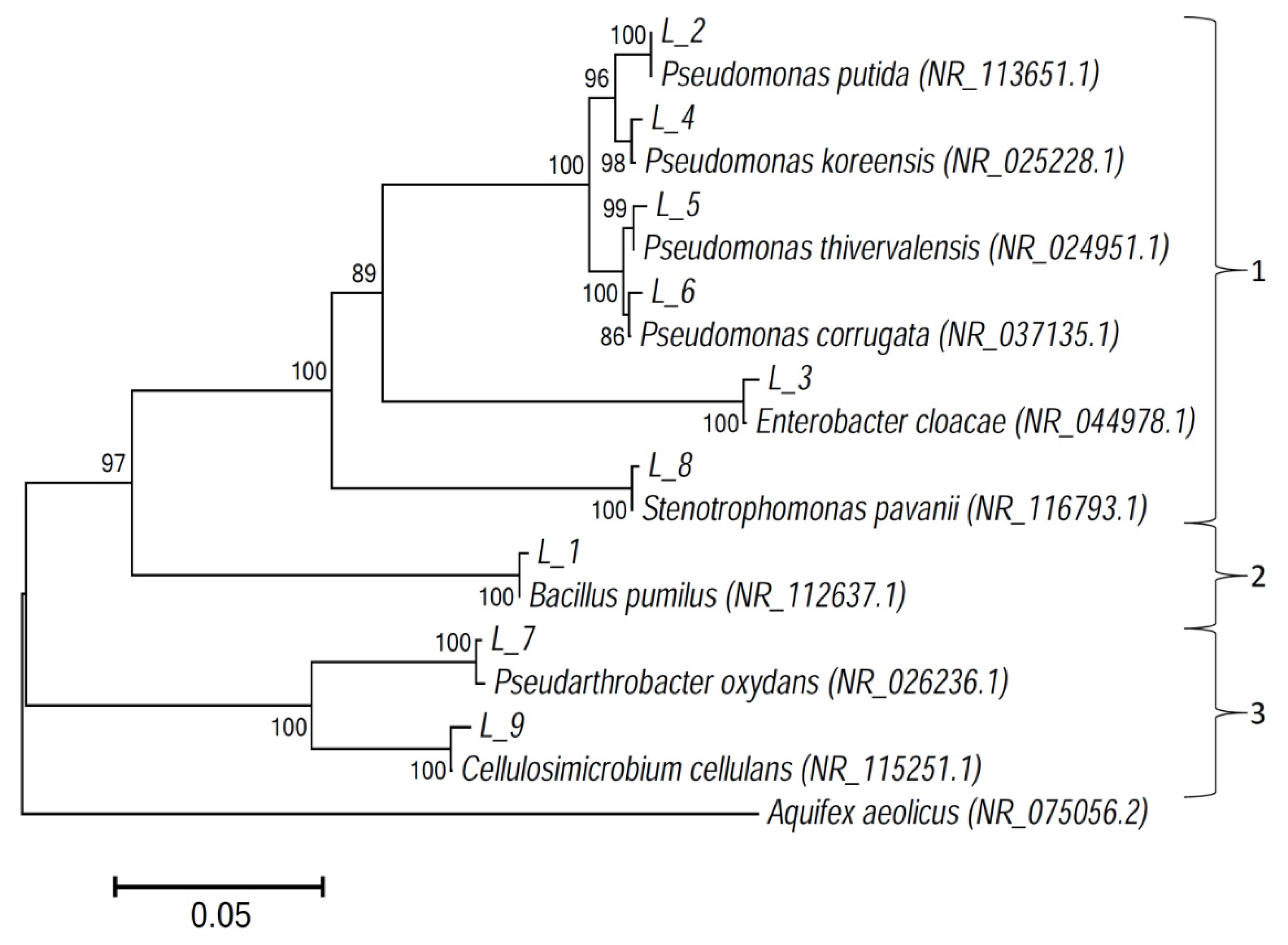
2.4.4. Sequencing and Phylogenetic Analysis

Before being sequenced, the PCR products were purified with the USB® ExoSAP-IT® PCR Product Cleanup Kit (Affymetrix, USB® Products, USA) according to the manufacturer’s protocol. Sequencing was performed using an ABI PRISM BigDye 3.1 Terminator Cycle Sequencing Ready Reaction Kit (Applied Biosystems) by the manufacturer’s protocol. Received data were analyzed and corrected using Chromas (v. 2.6.5) software. Corrected sequences were merged manually using EMBOSS Explorer (http://emboss.bioinformatics.nl/). The sequences were identified using the basic local alignment search tool (BLAST), and comparisons were made with the GenBank nucleotide data bank from the National Center for Biotechnology Information (NCBI) (http://0-www-ncbi-nlm-nih-gov.brum.beds.ac.uk/). All sequences were multiply aligned using Clustal× 2.1 software, and a FASTA format file was used to construct the phylogenetic tree. The evolutionary history was inferred using the neighbor-joining method [38]. The optimal tree with the sum of branch length = 0.76327423 is shown. The percentage of replicate trees in which the associated taxa clustered together in the bootstrap test (500 replicates) are shown next to the branches [39]. The tree is drawn to scale, with branch lengths in the same units as those of the evolutionary distances used to infer the phylogenetic tree. The evolutionary distances were computed using the maximum composite likelihood method [40] and are in the units of the number of base substitutions per site. The analysis involved 19 nucleotide sequences. All positions containing gaps and missing data were eliminated. There were a total of 1281 positions in the final dataset. Evolutionary analyses were conducted in MEGA6 [41].
The 16S rRNA sequences isolated from the endophytic bacteria of white lupin were deposited into GenBank under the accession numbers: MH636785–MH636793.

2.5. Plant Growth Traits

Several plant growth-promoting traits of endophytic bacteria were determined. Indole 3-acetic acid (IAA) production was studied following the method described in Bano and Musarrat [42]. The production of chitinase and protease activity was determined as described in Malleswari and Bagyanarayan [43]. The cellulose-degrading capacity of the bacterial isolates was analyzed as described by Gupta et al. [44]. The antagonistic capabilities of bacterial isolates against the pathogenic fungi F. oxysporum, F. solani, Fusarium culmorum, Verticillum dahlia, and R. solani were evaluated on potato dextrose agar (PDA) media as described by Egamberdieva et al. [6].

2.6. Biological Control of Lupin Root Rot

The biological control of lupin root rot caused by F. solani under different biochar soil amendments using selected endophytes was determined in a pot experiment. For soil infestation, a suspension of F. solani grown in Czapek–Dox medium for four days was filtrated with sterile glass wool, centrifuged, and diluted to 106 spores mL−1. The resulting suspension with fungal spores was mixed with soil (106 spores kg−1 soil). Two biochar types MBC and WBC were mixed with the soil at 2% (w/w) as follows:
-
un-inoculated plants grown without WBC or MBC biochar,
-
un-inoculated plants grown in soil amended with WBC,
-
un-inoculated plants grown in soil amended with MBC,
-
inoculated plants with Pseudomonas putida L2 grown in soil without WBC or MBC chars,
-
inoculated plants with P. putida L2 grown in soil amended with WBC char,
-
inoculated plants with P. putida L2 grown in soil amended with MBC char,
-
inoculated plants with Stenotrophomonas pavanii L8 grown in soil without WBC or MBC chars,
-
inoculated plants with S. pavanii L8 grown in soil amended with WBC char,
-
inoculated plants with S. pavanii L8 grown in soil amended with MBC char.
Each of the treatments contained four groups of twenty-four plants and were arranged in a randomized block design. For plant inoculation, bacterial isolates were grown in Tryptic soy broth (TSB) medium for two days, centrifuged, washed in PBS, and re-suspended with a bacterial density of 108 CFU/mL. The seedling roots were dipped into bacterial suspension prior to planting into soil. The plants were grown at a temperature range of 24–28 °C day and 18–20 °C night under greenhouse conditions for 34 days. At harvest, root rot symptoms of washed plants were determined as indicated by browning and lesions.

2.7. Plant Growth Experiment with Endophytic Bacteria and Biochar

The plant growth chamber experiments with nine treatments were set up as described above. The lupin seeds were surface-sterilized using 70% ethanol and 10% v/v NaOCl. Afterward, the seeds were washed five times with sterile distilled water and transferred to paper tissue for germination in a dark room at 25 °C for 3–4 days. The bacterial strains P. putida L2 and S. pavanii L8 were grown in TSB medium for two days, and the culture suspension was re-suspended in PBS with a final bacterial density of 108 CFU mL−1. Germinated seeds were dipped into the bacterial suspensions and transferred to pots. Three seeds were sown in each pot, and, after one week, the seedlings were thinned to one plant per pot. Each treatment included four pots and was arranged in a randomized complete block design with three replications (total of 12 plants). Plants were grown for 30 days at a temperature of 24 °C/16 °C (day/night) and humidity of 50–60%. At harvest, the roots and shoots were oven-dried at 70 °C for 48 h, and dry weight was determined.

2.8. Survival of Endophytic Bacterial Strains

The rifampicin (200 µg/mL) resistant mutants of the wild-type strain were obtained following the method of Glandorf et al. [45] and were used for the colonization studies. Mutants of P. putida L2 and S. pavanii L8 that were marked with antibiotic resistance were obtained by plating the parental strain onto TSA agar plates containing increasing concentrations of 50, 100, 150, and 200, µg/mL rifampicin. After incubation, isolates were selected based on similarities in colony morphology and growth rate with the parent strain and were re-cultured on medium containing 200 µg/mL rifampicin to ensure stability of the antibiotic resistance.
Plants were grown in pots as described above. Lupin seedlings were coated with bacteria by dipping the seedlings in bacterial suspensions that resulted in 108 CFU mL−1 seeds. Plants were grown for 30 days at a temperature of 24 °C/16 °C (day/night) and at a humidity of 50–60%. At harvest, the adhering soil was removed from plant roots, and 1 g roots was shaken in 9 mL sterile PBS. The resulting suspensions were evaluated for colony forming units (CFUs) according to the dilution-plate method in TSA agar with the addition of 200 µg/mL rifampicin. After incubation for 2–3 days at 28 °C the re-isolated, rifampicin-resistant strains were identified for their colony characteristics [46].

2.9. Statistical Analysis

Data were tested for statistical significance using the analysis of variance package included in Microsoft Excel 2010. Comparisons were performed using Student’s t-test. Mean comparisons were conducted using the least significant difference (LSD) test (P = 0.05).

3. Results

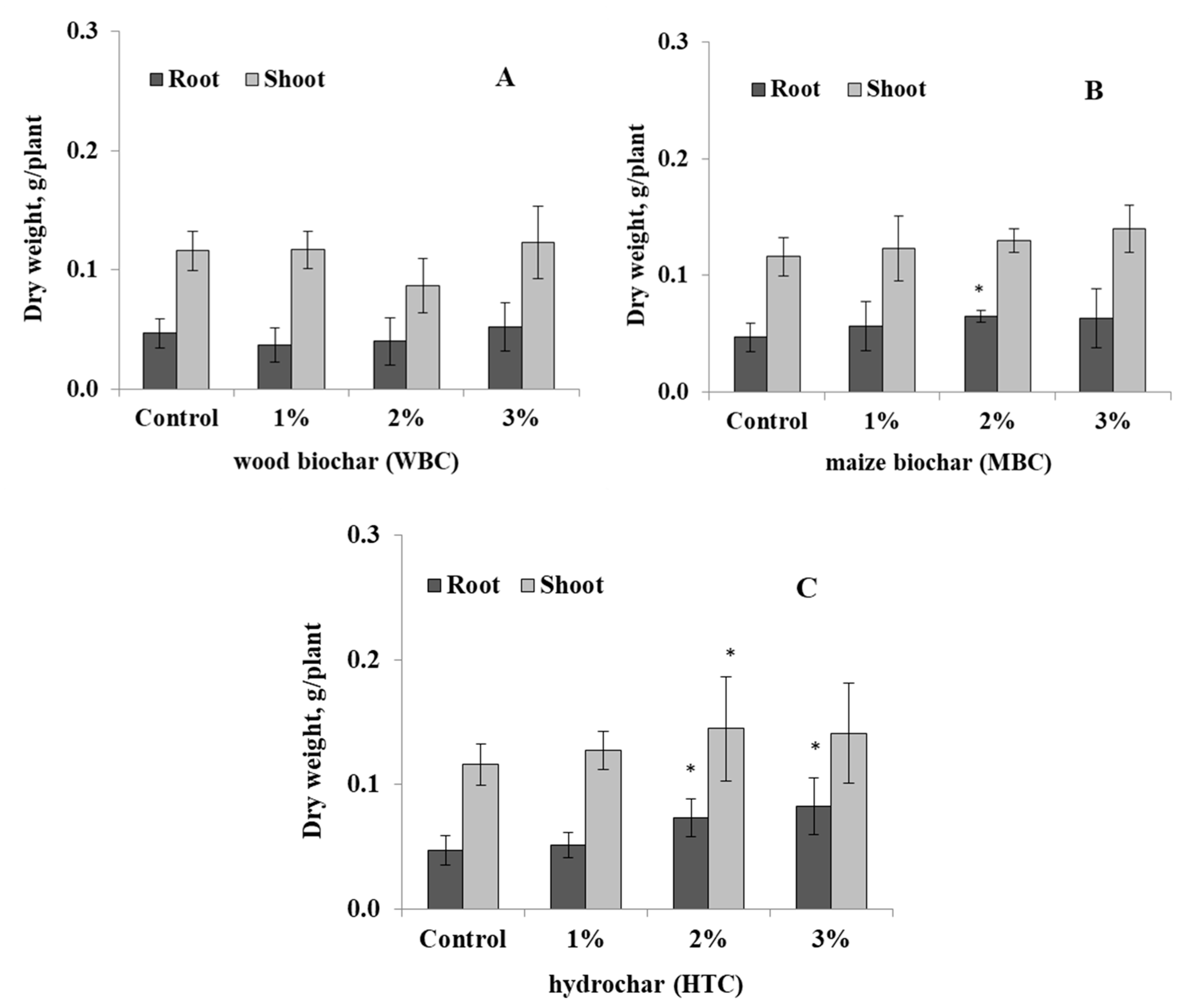
3.1. Response of Lupin to the Biochar Types

The response of narrow-leafed lupin to the type of biochar was studied at 1%, 2%, and 3% concentrations in pot experiments. The shoot and root biomass of lupin were not significantly affected by WBC soil amendments at 1–3% (Figure 1A). Correspondingly, the root and shoot growth of lupin under MBC char was not affected, but only root dry weight was increased at 2% MBC amendment (Figure 1B). However, the root dry weight of lupin was significantly increased by 54–75%, and the shoot dry weight was increased by up to 21–25% by HTC char amendment at 2% concentration, respectively (Figure 1C).
The plants grown in soils amended with 2% and 3% w/v WBC and MBC chars showed disease symptoms of 40–50% and 10–20%, respectively (Figure 2). Plants grown in soil without biochar and in soil with HTC char were healthy, and no disease incidence was found.

3.2. Fungal and Bacterial Isolates

The selected fungal isolate showed root rot symptoms after plant pathogenicity tests and was identified as F. solani. The biological control capability of endophytic bacteria against root rot of lupin caused by F. solani was examined.
A total of 25 bacterial isolates were isolated from plant tissues of lupin. The RFLP analysis was conducted to eliminate similar isolates, and as a result, only nine isolates were selected and identified using the BLAST algorithm and compared with related strains from NCBI GenBank (Table 2).
All strains were identified using BLAST and compared with similar strains from the NCBI GenBank. The 16S rRNA sequences similarities of endophytic bacteria isolated from plant tissue of lupin with sequences from GenBank are shown in Table 2. All strains were identical with a similarity of 98.72–99.25% to those registered in GenBank. The isolates were identified as Bacillus pumilus L1, P. putida L2, Enterobacter cloacae L3, Pseudomonas koreensis L4, Pseudomonas thivervalensis L5, Pseudomonas corrugata L6, Pseudarthrobacter oxydans L7, S. pavanii L8, and Cellulosimicrobium cellulans L9 (Table 2, Figure 3). A phylogenetic tree was constructed showing the closest relatives of the isolates (Figure 3).
All nine endophytes were screened for multiple plant growth-promoting traits. Most of the bacterial isolates exhibited one or more plant growth-promoting activities (Table 3). Among nine isolates only two isolates P. putida L2 and S. pavanii L8 were able to produce IAA (5.6 and 4.7 µg mL−1), were able to produce one or more cell wall degrading enzymes, and were highly effective in vitro against the F. oxysporum and F. solani (Table 3). HCN and siderophores were produced only by P. putida L2.

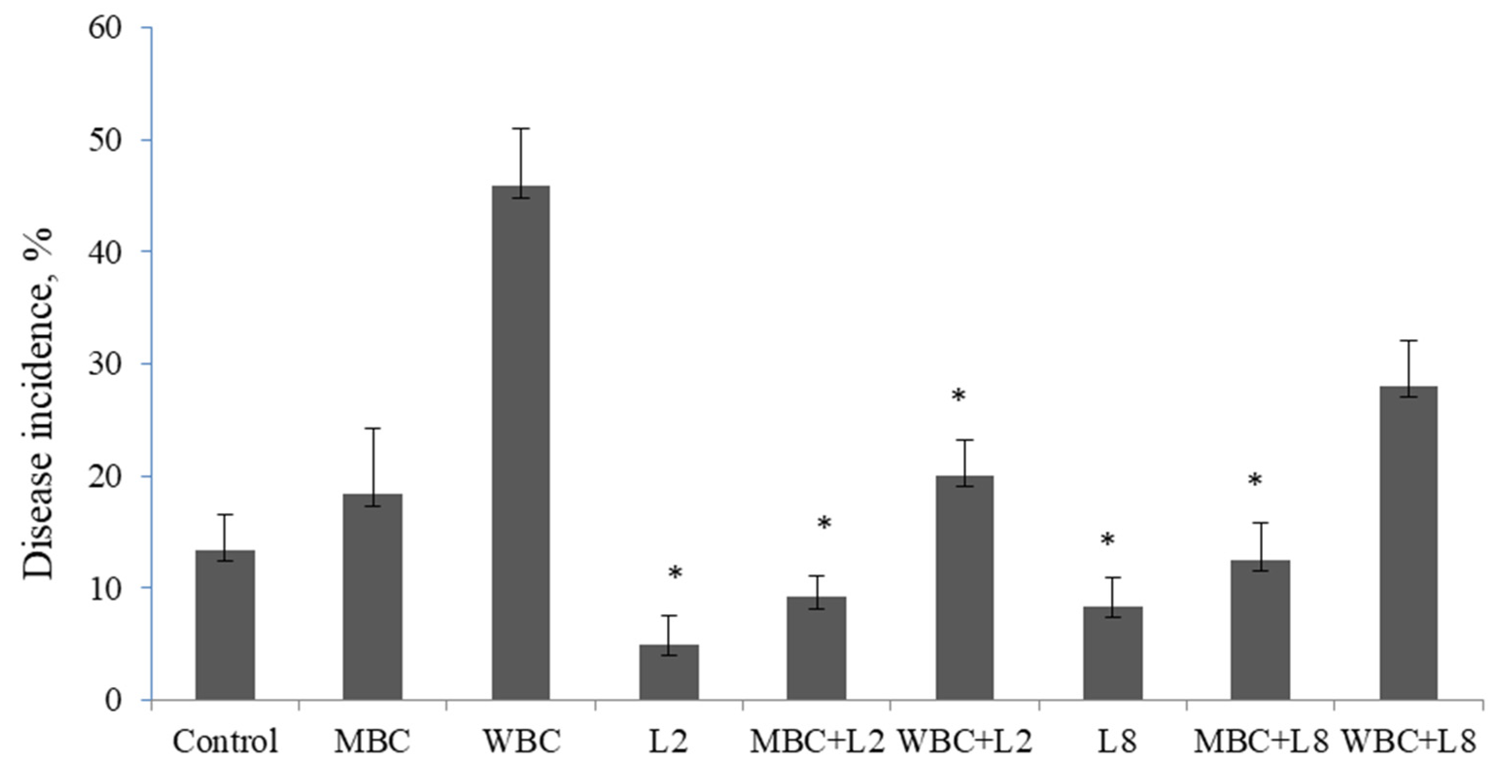
3.3. Biological Control of Lupin Root Rot under Biochar Amended Soil

The two selected isolates, P. putida L2 and S. pavanii L8, were chosen to evaluate their capability to suppress lupin root rot caused by F. solani. The portion of diseased plants grown in soil infested with the pathogen was 13%. The disease incidence was increased by 18% under MBC amended soil and by 45% under WBC soil amendment (Figure 4). The bacterial isolates, P. putida L2 and S. pavanii L8, demonstrated a disease reduction up to 9–12% under MBC amended soil compared to control plants without biochar (13%) and un-inoculated plants grown under MBC (18%) (Figure 5). The number of pathogen-infected plants was reduced up to 20% and 28% by bacterial isolates P. putida L2 and S. pavanii L8 under WBC soil amendment, whereas un-inoculated plants grown under WBC char showed a disease incidence up to 45%.

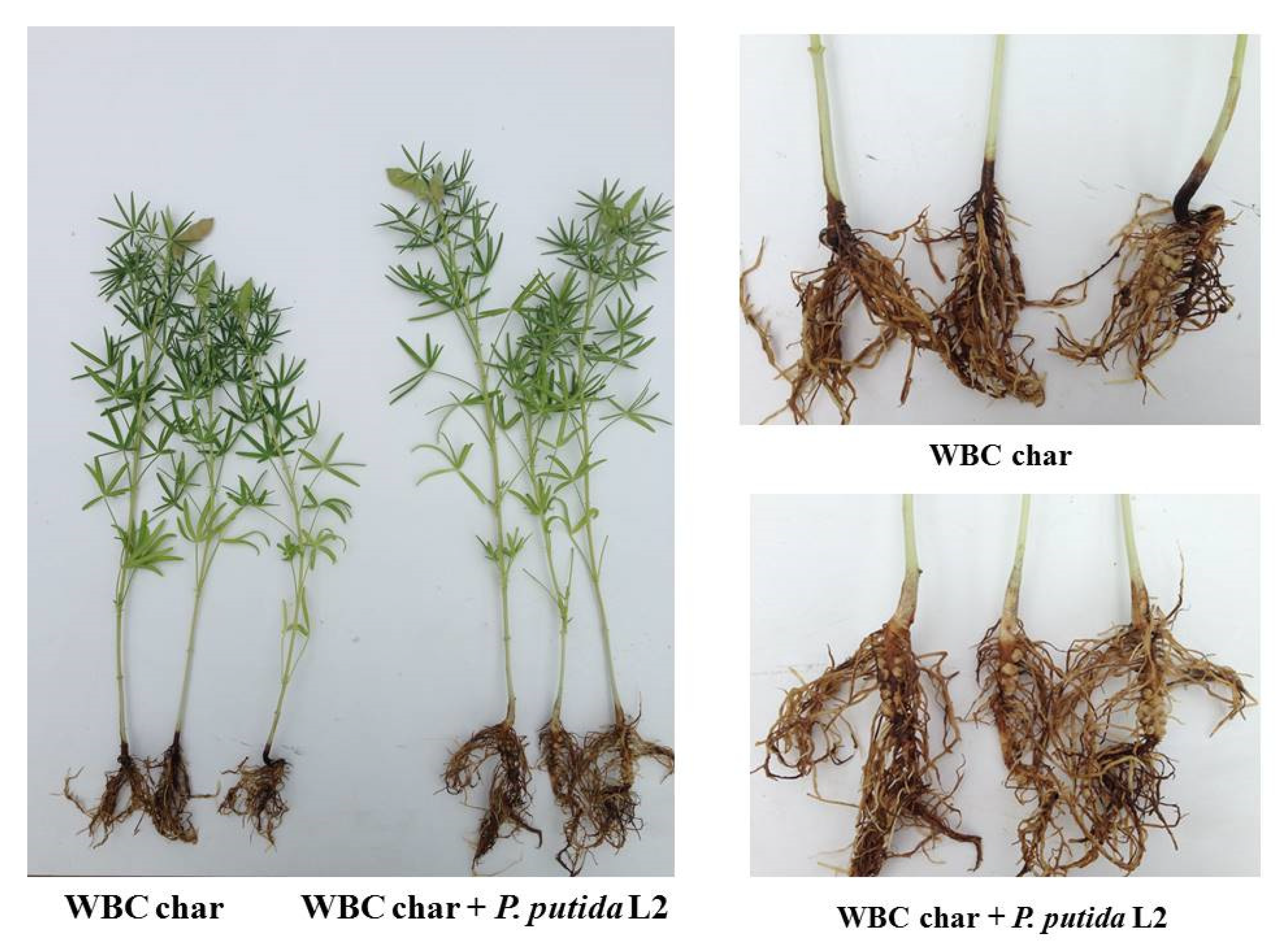
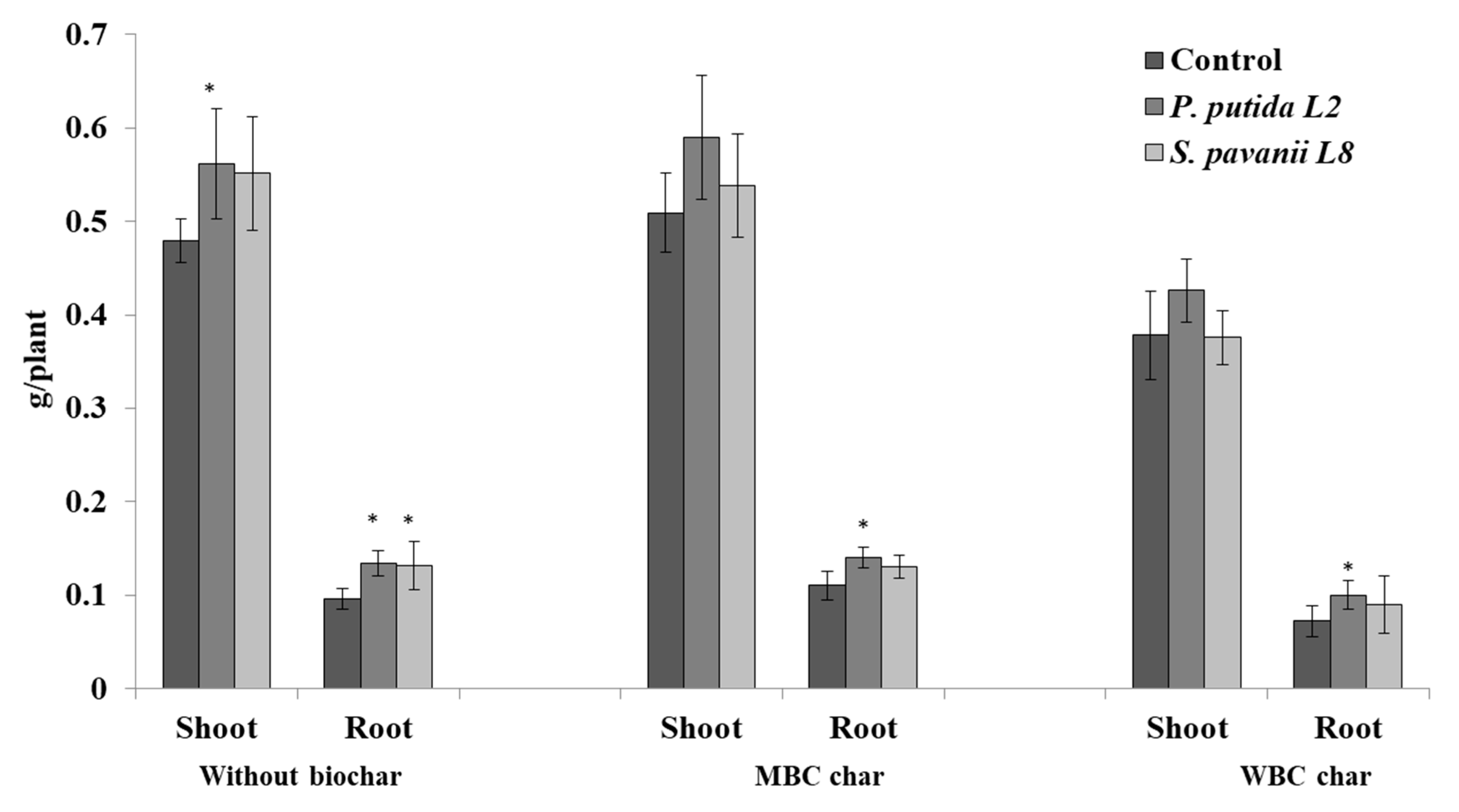
3.4. Plant Growth, Nodulation, and Bacterial Survival in the Root System

The endophytic bacteria that showed biocontrol ability to control the root rot of lupin were evaluated for their plant growth-promoting activities under the amendment of two different biochar types. The responses of lupin to the two biochar types were different, being higher in soil amended with MBC compared to those in soil amended with WBC (Figure 6). The shoot and root biomass of un-inoculated lupin grown under MBC char were increased by 6% and 14% compared to control plants, respectively. Bacterial inoculants P. putida L2 and S. pavanii L8 increased lupin root and shoot growth in soil without biochar amendment by 17–39% and 15–37%, respectively (Figure 6). However, there were no significant effects on the shoot growth of inoculated plants grown in soil amended with MBC and WBC chars. The root biomass of lupin was increased by inoculation of plants with P. putida L2 in MBC and WBC amended soil (Figure 6). Compared with plants grown in soil amended with MBC and WBC chars, the weight of shoots and roots was not affected by bacterial inoculation with S. pavanii L8.
Both endophytic bacteria P. putida (L2) and S. pavanii (L8) improved the symbiotic performance of lupin with its rhizobia under soil amended with MBC and WBC chars. The nodule numbers of lupin inoculated with P. putida L2 and S. pavanii L8 were 6.8 and 4.6 grown in soil without biochar, while under WBC and MBC biochar amendment nodule numbers increased to 9.0 and 10.0 per plant, respectively (Figure 7).
Rifampicin-resistant mutants of bacterial isolates P. putida L2 and S. pavanii L8 were tested for their survival in the root of lupin grown without and with biochar amendment. The results showed that both bacterial isolates were able to survive in the rhizosphere of lupin. The root colonization in the rhizosphere of lupin was 2.1 × 10³ ± 1.4 with P. putida L2, and 3.2 × 10³ ± 1.9 with S. pavanii L8 in soil without biochar amendment. The CFUs of bacterial isolates were slightly higher in WBC and MBC amended soils, being 3.2 × 10³ ± 0.7 and 4.2 × 10³ ± 1.2 (CFU/g of fresh root) with P. putida L2 and 3.9 × 10³ ± 1.6 and 3.8 (CFU/g of fresh root) with S. pavanii L8, respectively.

4. Discussion

Biochar amendments have been reported to elicit complex responses in plants [4,6,15], including plant growth stimulation or suppression of several soil-borne diseases [47,48]. However, the beneficial effect depends on the type of biochar feedstock, the technique of preparation (e.g., pyrolysis or carbonization), and finally, concentration [15,16]. Overall, our experiment showed that the shoot and root biomass of lupin responded differently to types of biochar and biochar concentrations, i.e., MBC char produced more benefits to lupin growth as compared to WBC char. Reibe et al. [33] reported similar observation for MBC char amendments increasing dry biomass of wheat as compared to WBC char or plants grown without any biochar application. Joseph et al. [49] reported that the interaction of biochar with environmental conditions is another crucial requirement for revealing contrasting effects, which might depend on the physicochemical properties of biochars. For example, Butnan et al. [25] observed reduced plant growth on a sandy Ultisol amended with eucalyptus wood-derived biochar produced by pyrolysis (800 °C), whereas biochar produced at a lower temperature (350 °C) provided higher benefits. Shen et al. [50] reported that the addition of biochar from willow woodchips increased plant growth of Lotus pedunculatus cv barsille, whereas pine-based biochar did not show any positive effect on plant growth.
There are also numerous reports on the positive effect of biochar in reducing plant pathogens [51], e.g., R. solani of cucumber [16] or F. oxysporum f. sp. lycopersici of tomato [14]. However, some biochar types showed an increased disease incidence by providing favorable conditions for pathogenic fungi, e.g., 3% green waste biochar increased the disease incidence of tomato wilt caused by F. oxysporum f. sp. lycopersici [14]. Correspondingly, we observed an increased disease incidence of lupin wilt caused by F. solani under WBC char, whereas no diseased plants were observed in control plots. However, the mechanisms of biochar to suppress or induce soil-borne diseases were not sufficiently revealed because they depend upon several factors, including chemical and physical characteristics of biochar [16] as well as soil conditions and properties. In earlier studies, Sanogo and Yang [52] reported that the development and growth of Fusarium species was faster at higher soil pH 8.2 compared to that at lower soil pH 5.7.
The capability of endophytic bacteria living within plant tissues to inhibit plant pathogens was reported in numerous studies [22,53,54]. Silva et al. [55] observed a biological control of coffee leaf rust caused by Hemileia vastatrix with endophytic bacteria such as Bacillus megaterium and Microbacterium testaceum. In another study, Bacillus cereus decreased wilt symptoms of tomato caused by F. oxysporum [56]. In our study, endophytic bacteria P. putida L2 and S. pavanii L8 were able to reduce lupin root rot caused by F. solani in WMB and MBC amended soil. In a previous study, the endophytic Bacillus subtilis NUU4 with antagonistic activity against F. oxysporum, F. solani, and F. culmorum reduced disease incidence of chickpea root rot [54]. The plant inoculation with P. putida L2 and S. pavanii L8 increased lupin root and shoot biomass grown in soil without biochar. The root biomass of lupin was significantly increased by P. putida L2 under both MBC and WBC char. Lupin treated with Bradyrhizobium sp. and growing in soil amended with biochar had higher root and shoot biomass as compared to un-inoculated control plants or direct inoculation with Bradyrhizobium sp., alone [9]. Correspondingly, soil amendment with biochar from citrus wood provided favorable conditions for bacterial proliferation under sweet pepper (Capsicum annuum L.) [18]. Pietikäinen et al. [57] reported that the survival of bacteria, which were adsorbed to biochar surfaces, was due to biochar properties protecting bacteria in the soil. In another study, the number of rhizobial cells in soil under soybean were increased by the addition of biochar mixed with compost [58]. Hale et al. [59] observed that pinewood biochar based inoculum of E. cloacae UW5 resulted in a significant increase in cucumber root branching and total root length giving evidence for the role of biochar in effects on root architecture. The ability of endophytic bacteria colonizing the internal part of plants to stimulate the root system by better nutrient uptake was reported in numerous studies [53,54,60,61]. The colonization of inoculated bacterial isolates in the root system is an important trait, especially in a highly competitive environment [62,63]. Moreover, it is the very first step in beneficial interactions of the plant with a microbe. The inoculated endophytic bacteria were able to survive in the rhizosphere of lupin grown in soil without biochar and biochar amendment for 30 days. It has been previously reported that improved plant–microbe interactions resulted from biochar effects on soil microbial diversity and activity [64], e.g., biochar maintains bacterial proliferation residing inside pores by supplying nutrients and improving aeration [65].
Several mechanisms by which endophytic bacteria enhance plant growth, improve stress tolerance, and protect plants from various diseases were described. These include the production of phytohormones such as indole 3-acetic acid [9,54], antifungal metabolites [62,66], and cell wall degrading enzymes such as cellulase or chitinase [67]. In our previous work, the endophytic strain B. subtilis NUU4 that reduced root rot incidence in chickpea and stimulated plant growth demonstrated an antagonistic activity against F. oxysporum, F. solani, and F. culmorum and produced IAA, as well as cell wall degrading enzymes. Similar observations were found by Abdallah et al. [56], where the wilt symptoms of tomato caused by F. oxysporum were decreased by B. cereus antagonistic against F. oxysporum f. sp. lycopersici. The endophytic bacteria that synthesized IAA stimulated the root system architecture and also increased root hairs, which resulted in better absorption of nutrients [68].

5. Conclusions

The results presented in this study showed that soil amended with 2% MBC and HTC improved root and shoot growth of lupin. However, the disease symptoms of lupin root rot occurred in soil amended with WBC char at a rate of 2% and 3%. Plants under HTC char were healthy, and no disease incidence was found. The endophytic bacteria P. putida L2 and S. pavanii L8 isolated from lupin were able to reduce root rot caused by F. solani under WBC amended soil. Furthermore, the lupin growth was also increased by inoculation with endophytic bacteria in MBC and WBC amended soil treatments. The soil amended with MBC and WBC supported better survival of inoculated bacteria in the rhizosphere of lupin. Future research is required to reveal the mechanisms of biochar-induced disease incidence in more detail, as well as the traits endophytic bacteria use to control lupin root rot caused by F. solani in soil amended with biochar, especially considering soil physicochemical properties.

Author Contributions

D.E., M.R., and S.W. designed the experiments. D.E., V.S., and M.E.H.M. conducted the experiments. B.A. and H.M. analyzed the data. D.E., S.W., M.R., and S.D.B.-K. wrote the manuscript. All authors read and approved the manuscript.

Funding

This research was funded by Alexander von Humboldt Foundation for DE, and by the Deutsche Forschungsgemeinschaft (DFG, German Research Foundation), grant number 420661662 for MR.

Acknowledgments

We thank the staff of the Experimental Station of the Leibniz Center for Agricultural Landscape Research (ZALF) in Müncheberg for implementing the field and greenhouse experiment.

Conflicts of Interest

The authors declare no conflict of interest.

References

  1. Lehmann, J. A handful of carbon. Nature 2007, 447, 143–144. [Google Scholar] [CrossRef] [PubMed]
  2. Barrow, C.J. Biochar: Potential for countering land degradation and for improving agriculture. Appl. Geogr. 2012, 34, 21–28. [Google Scholar] [CrossRef]
  3. Ippolito, J.A.; Laird, D.A.; Busscher, W.J. Environmental benefits of biochar. J. Environ. Qual. 2012, 41, 967–972. [Google Scholar] [CrossRef] [PubMed] [Green Version]
  4. Biederma, L.A.; Harpole, W.S. Biochar and its effects on plant productivity and nutrient cycling: a meta-analysis. GCB Bioenergy 2013, 5, 202–214. [Google Scholar] [CrossRef]
  5. Graber, E.R.; Meller-Harel, Y.; Kolton, M.; Cytryn, E.; Silber, A.; David, D.R.; Tsechansky, L.; Borenshtein, M.; Elad, Y. Biochar impact on development and productivity of pepper and tomato grown in fertigated soilless media. Plant Soil 2010, 337, 481–496. [Google Scholar] [CrossRef]
  6. Egamberdieva, D.; Wirth, S.; Behrendt, U.; Abd_Allah, E.F.; Berg, G. Biochar treatment resulted in a combined effect on soybean growth promotion and a shift in plant growth promoting rhizobacteria. Front. Microbiol. 2016, 7, 209. [Google Scholar] [CrossRef] [Green Version]
  7. Islami, T.; Curitno, B.; Basuki, N.; Suryanto, A. Maize yield and associated soil quality changes in cassava + maize intercropping system after 3 years of biochar application. J. Agric. Food. Techn. 2011, 1, 112–115. [Google Scholar]
  8. Alburquerque, J.A.; Salazar, P.; Barrón, V.; Torrent, J.; del Campillo, M.D.; Gallardo, A.; Villar, R. Enhanced wheat yield by biochar addition under different mineral fertilization levels. Agron. Sustain. Dev. 2013, 33, 475. [Google Scholar] [CrossRef] [Green Version]
  9. Egamberdieva, D.; Reckling, M.; Wirth, S. Biochar-based inoculum of Bradyrhizobium sp. improves plant growth and yield of lupin (Lupinus albus L.) under drought stress. Eur. J. Soil Biol. 2017, 78, 38–42. [Google Scholar] [CrossRef]
  10. Novak, J.M.; Busscher, W.J.; Laird, D.L.; Ahmedna, M.; Watts, D.W.; Niandou, M.A. Impact of biochar amendment on fertility of a southeastern coastal plain soil. Soil Sci. 2009, 174, 105–112. [Google Scholar] [CrossRef] [Green Version]
  11. Yu, O.Y.; Raichle, B.; Sink, S. Impact of biochar on the water holding capacity of loamy sand soil. Int. J. Energy Environ. Eng. 2013, 4, 44. [Google Scholar] [CrossRef] [Green Version]
  12. Chan, K.Y.; Van Zwieten, L.; Meszaros, I.; Downie, A.; Joseph, S. Agronomic values of greenwaste biochar as a soil amendment. Aust. J. Soil Res. 2007, 45, 629–634. [Google Scholar] [CrossRef]
  13. Elad, Y.; David, D.R.; Harel, Y.M.; Borenshtein, M.; Silber, K.S.A.; Graber, E.R. Induction of systemic resistance in plants by biochar, a soil applied carbon sequestering agent. Phytopathology 2010, 100, 913–921. [Google Scholar] [CrossRef] [PubMed] [Green Version]
  14. Akhter, A.; Hage-Ahmed, K.; Soja, G.; Steinkellner, S. Potential of Fusarium wilt-inducing chlamydospores, in vitro behaviour in root exudates and physiology of tomato in biochar and compost-amended soil. Plant Soil 2016, 406, 425–440. [Google Scholar] [CrossRef] [Green Version]
  15. Mehari, Z.H.; Elad, Y.; Rav-David, D.; Graber, E.R.; Harel, Y.M. Induced systemic resistance in tomato (Solanum lycopersicum) against Botrytis cinerea by biochar amendment involves jasmonic acid signaling. Plant Soil 2015, 395, 31–44. [Google Scholar] [CrossRef]
  16. Jaiswal, A.K.; Elad, Y.; Graber, E.R.; Frenkel, O. Rhizoctonia solani suppression and plant growth promotion in cucumber as affected by biochar pyrolysis temperature, feedstock and concentration. Soil Biol. Biochem. 2014, 69, 110–118. [Google Scholar] [CrossRef]
  17. Chen, J.; Liu, X.; Zheng, J.; Zhang, B.; Lu, H.; Chi, Z.; Pan, G.; Li, L.; Zhen, J.; Zhang, X.; et al. Biochar soil amendment increased bacterial but decreased fungal gene abundance with shifts in community structure in a slightly acid rice paddy from Southwest China. Appl. Soil Ecol. 2013, 71, 33–44. [Google Scholar] [CrossRef]
  18. Kolton, M.; Meller Harel, Y.; Pasternak, Z.; Graber, E.R.; Elad, Y.; Cytryn, E. Impact of biochar application to soil on the root-associated bacterial community structure of fully developed greenhouse pepper plants. Appl. Environ. Microbiol. 2011, 77, 4924–4930. [Google Scholar] [CrossRef] [Green Version]
  19. Glick, B.R. Bacteria with ACC deaminase can promote plant growth and help to feed the world. Microbiol. Res. 2014, 169, 30–39. [Google Scholar] [CrossRef]
  20. Berg, G.; Martinez, J.L. Friends or foes: can we make a distinction between beneficial and harmful strains of the Stenotrophomonas maltophilia complex? Front. Microbiol. 2015, 6, 241. [Google Scholar] [CrossRef]
  21. Hashem, A.; Abd_Allah, E.F.; Alqarawi, A.; Al-Huqail, A.A.; Wirth, S.; Egamberdieva, D. The interaction between arbuscular mycorrhizal fungi and endophytic bacteria enhances plant growth of Acacia gerrardii under salt stress. Front. Plant Sci. 2016, 7, 1089. [Google Scholar]
  22. Egamberdieva, D.; Kucharova, Z.; Davranov, K.; Berg, G.; Makarova, N.; Azarova, T.; Chebotar, V.; Tikhonovich, I.; Kamilova, F.; Validov, S.; et al. Bacteria able to control foot and root rot and to promote growth of cucumber in salinated soils. Biol. Fert. Soils 2011, 47, 197–205. [Google Scholar] [CrossRef]
  23. Noreen, R.; Ali, S.A.; Hasan, K.A.; Sultana, V.; Ara, J.; Ehteshamul-Haque, S. Evaluation of biocontrol potential of fluorescent Pseudomonas associated with root nodules of mungbean. Crop Prot. 2015, 75, 18e24. [Google Scholar] [CrossRef]
  24. Gul, S.; Whalen, J.K.; Thomas, B.W.; Sachdeva, V.; Deng, H.Y. Physico-chemical properties and microbial responses in biochar-amended soils: Mechanisms and future directions. Agric. Ecosyst. Environ. 2015, 206, 46–59. [Google Scholar] [CrossRef]
  25. Butnan, S.; Deenik, J.L.; Toomsan, B.; Antal, M.J.; Vityakon, P. Biocharcharacteristics and application rates affecting corn growth and properties of soils contrasting in texture and mineralogy. Geoderma 2015, 237, 105–116. [Google Scholar] [CrossRef]
  26. Frenkel, O.; Jaiswal, A.K.; Elad, Y.; Lew, B.; Kammann, C.; Graber, E.R. The effect of biochar on plant diseases: what should we learn while designing biochar substrates? J. Environ. Eng. Landsc. 2017, 25, 105–113. [Google Scholar] [CrossRef] [Green Version]
  27. Gresta, F.; Wink, M.; Prins, U.; Abberton, M.; Capraro, J.; Scarafoni, A.; Hill, G. Lupins in European cropping systems. In Legumes in cropping systems; Murphy-Bokern, D., Stoddard, F., Watson, C., Eds.; CABI Publishing: Wallingford, UK, 2017; pp. 88–108. [Google Scholar]
  28. Lupwayi, N.Z.; Kennedy, A.C. Grain Legumes in Northern Great Plains. Agron. J. 2007, 99, 1700–1709. [Google Scholar] [CrossRef]
  29. Reckling, M.; Bergkvist, G.; Watson, C.A.; Stoddard, F.L.; Zander, P.M.; Walker, R.; Pristeri, A.; Toncea, I.; Bachinger, J. Trade-offs between economic and environmental impacts of introducing legumes into cropping systems. Front. Plant Sci. 2016, 7, 669. [Google Scholar]
  30. Watson, C.; Reckling, M.; Preissel, S.; Bachinger, J.; Bergkvist, G.; Kuhlman, T.; Lindström, K.; Nemecek, T.; Topp, C.; Vanhatalo, A.; et al. Grain legume production and use in European agricultural systems. Adv. Agron. 2017, 144, 235–303. [Google Scholar]
  31. Jensen, B.; Bødker, L.; Larsen, J.; Knudsen, J.C.H.; Jørnsgaard, B. Specificity of soil-borne pathogens on grain legumes. In Proceedings of the 5th European Conference on Grain Legumes, Dijon, France, 7–11 June 2004. [Google Scholar]
  32. Reckling, M.; Döring, T.F.; Bergkvist, G.; Stoddard, F.L.; Watson, C.A.; Seddig, S.; Chmielewski, F.-M.; Bachinger, J. Grain legume yields are as stable as other spring crops in long-term experiments across northern Europe. Agron. Sustain. Dev. 2018, 38, 63. [Google Scholar] [CrossRef] [Green Version]
  33. Reibe, K.; Götz, K.P.; Ross, C.L.; Doering, T.F.; Ellmer, F.; Ruess, L. Impact of quality and quantity of biochar and hydrochar on soil collembola and growth of spring wheat. Soil Biol. Biochem. 2015, 8, 84–87. [Google Scholar] [CrossRef]
  34. Egamberdieva, D.; Li, L.; Ma, H.; Wirth, S.; Bellingrath-Kimura, S.D. Soil amendments with different maize biochars, to varying degrees, improve chickpea growth under drought by improving symbiotic performance with Mesorhizobium ciceri and soil biochemical properties. Front. Microbiol. 2019, 10, 2423. [Google Scholar] [CrossRef]
  35. Zeng, Q.Y.; Wang, X.R.; Blomquist, G. Development of mitochondrial ssu rDNA-based oligonucleotide probes for specific detection of common airborne fungi. Mol. Cell. Probes. 2003, 17, 281–288. [Google Scholar] [CrossRef]
  36. Dashti, A.A.; Jadaon, M.M.; Abdulsamad, A.M.; Dashti, H.M. Heat Treatment of Bacteria: A simple method of DNA extraction for molecular techniques. Kuwait Med J. 2009, 41, 117–122. [Google Scholar]
  37. Jinneman, K.C.; Wetherington, J.H.; Adams, A.M.; Johnson, J.M.; Tenge, B.J.; Dang, N.L.; Hill, W.E. Differentiation of Cyclospora sp. and Eimeria spp. by Using the Polymerase Chain Reaction Amplification Products and Restriction Fragment Length Polymorphisms; Food and Drug Admin Lab Information Bulletin LIB no. 4044: Cebu, Philippines, 1996. [Google Scholar]
  38. Saitou, N.; Nei, M. The neighbor-joining method: A new method for reconstructing phylogenetic trees. Mol. Biol. Evol. 1987, 4, 406–425. [Google Scholar]
  39. Felsenstein, J. Confidence limits on phylogenies: An approach using the bootstrap. Evolution 1985, 39, 783–791. [Google Scholar] [CrossRef]
  40. Tamura, K.; Nei, M.; Kumar, S. Prospects for inferring very large phylogenies by using the neighbor-joining method. Proc. Nat. Acad. Sci. USA 2004, 101, 11030–11035. [Google Scholar] [CrossRef] [Green Version]
  41. Tamura, K.; Stecher, G.; Peterson, D.; Filipski, A.; Kumar, S. MEGA6: Molecular Evolutionary Genetics Analysis version 6.0. Mol. Biol. Evol. 2013, 30, 2725–2729. [Google Scholar] [CrossRef] [Green Version]
  42. Bano, N.; Musarrat, J. Characterization of a new Pseudomonas aeruginosa strain NJ-15 as a potential biocontrol agent. Curr. Microbiol. 2003, 46, 324–328. [Google Scholar] [CrossRef]
  43. Malleswari, D.; Bagyanarayan, G. In vitro screening of rhizobacteria isolated from the rhizosphere of medicinal and aromatic plants for multiple plant growth promoting activities. J. Microbiol. Biotechnol. Res. 2013, 3, 84–89. [Google Scholar]
  44. Gupta, P.; Samant, K. SahuIsolation of cellulose-degrading bacteria and determination of their cellulolytic potential. Int. J. Microbiol. 2012. [Google Scholar] [CrossRef] [PubMed] [Green Version]
  45. Glandorf, D.C.M.; Brand, I.; Bakker, P.A.H.M.; Schippers, B. Stability of rifampicin resistance as a marker for root colonization studies of Pseudomonas putida in the field. Plant Soil 1992, 146, 135–142. [Google Scholar] [CrossRef]
  46. Höflich, G.; Wiehe, W.; Hecht-Buchholz, C.H. Rhizosphere colonization of different growth promoting Pseudomonas and Rhizobium bacteria. Microbiol. Res. 1995, 150, 139–147. [Google Scholar] [CrossRef]
  47. Harel, Y.M.; Elad, Y.; Rav-David, D.; Borenstein, M.; Shulchani, R.; Lew, B.; Graber, E.R. Biochar mediates systemic response of strawberry to foliar fungal pathogens. Plant Soil 2012, 357, 245–257. [Google Scholar] [CrossRef]
  48. Graber, E.R.; Elad, Y. Biochar impact on plant resistance to disease. In Biochar and Soil Biota; Ladygina, N., Rineau, F., Eds.; CRC Press (Taylor and Francis Group): Boca Raton, FL, USA, 2013; pp. 41–68. [Google Scholar]
  49. Joseph, S.D.; Camps-Arbestain, M.; Lin, Y.; Munroe, P.; Chia, H.; Hook, J.; van Zwieten, L.; Kimber, S.; Cowie, A.; Singh, P.; et al. An investigation into the reactions of biochar in soil. Aust. J. Soil Res. 2010, 48, 501–515. [Google Scholar] [CrossRef]
  50. Shen, Q.; Hedley, M.; Camps Arbestain, M.; Kirschbaum, M.U.F. Can biochar increase the bioavailability of phosphorus? J. Soil Sci. Plant Nutr. 2016, 16, 268–286. [Google Scholar] [CrossRef] [Green Version]
  51. Bonanomi, G.; Ippolito, F.; and Scala, F. A “black” future for plant pathology? Biochar as a new soil amendment for controlling plant diseases. J. Plant Pathol. 2015, 97, 223–234. [Google Scholar]
  52. Sanogo, S.; Yang, X.B. Relation of sand content, pH, and potassium and phosphorus nutrition to the development of sudden death syndrome in soybean. Can. J. Plant Pathol. 2001, 23, 174–180. [Google Scholar] [CrossRef]
  53. Eljounaidi, K.; Lee, S.K.; Bae, H. Bacterial endophytes as potential biocontrol agents of vascular wilt diseases–review and future prospects. Biol. Control 2016, 103, 62–68. [Google Scholar] [CrossRef]
  54. Egamberdieva, D.; Wirth, S.; Shurigin, V.; Hashem, A.; Abd_Allah, E.F. Endophytic bacteria improve plant growth, symbiotic performance of chickpea (Cicer arietinum L.) and induce suppression of root rot caused by Fusarium solani under salt stress. Front. Microbiol. 2017, 28, 1887. [Google Scholar] [CrossRef]
  55. Silva, M.C.; Nicole, M.; Rijo, L.; Geiger, J.P.; Rodrigues, C.J., Jr. Cytochemical aspects of the plant-rust fungus interface during the compatible interaction Coffea arabica (cv. Caturra)-Hemileia vastatrix (race III). Int. J. Plant Sci. 1999, 160, 79–91. [Google Scholar]
  56. Abdallah, R.A.B.; Jabnoun-Khiareddine, J.; Nefzi, A.; Mokni-Tlili, S.; Daami-Remadi, M. Biocontrol of Fusarium wilt and growth promotion of tomato plants using endophytic bacteria isolated from Solanum elaeagnifolium stems. J. Phytopathol. 2016, 164, 811–824. [Google Scholar] [CrossRef]
  57. Pietikäinen, J.; Kiikkila, O.; Fritze, H. Charcoal as a habitat for microbes and its effects on the microbial community of the underlying humus. Oikos 2000, 89, 231–242. [Google Scholar]
  58. Lehmann, J.; Rillig, M.C.; Thies, J.; Masiello, C.A.; Hockaday, W.C.; Crowley, D. Biochar effects on soil biota—A review. Soil Biol. Biochem. 2011, 43, 1812–1836. [Google Scholar]
  59. Hale, L.; Luth, M.; Crowley, D. Biochar characteristics relate to its utility as an alternative soil inoculum carrier to peat and vermiculite. Soil Biol. Biochem. 2015, 81, 228–235. [Google Scholar] [CrossRef]
  60. Egamberdieva, D.; Jabborova, D.; Hashem, A. Pseudomonas induces salinity tolerance in cotton (Gossypium hirsutum) and resistance to Fusarium root rot through the modulation of indole-3-acetic acid. Saudi J. Biol. 2015, 22, 773–779. [Google Scholar] [CrossRef] [Green Version]
  61. Egamberdieva, D.; Jabborova, D.; Wirth, S.; Alam, P.; Alyemeni, M.N.; Ahmad, P. Interactive Effects of Nutrients and Bradyrhizobium japonicum on the Growth and Root Architecture of Soybean (Glycine max L.). Front. Microbiol. 2018, 9, 1000. [Google Scholar]
  62. Cho, S.T.; Chang, H.H.; Egamberdieva, D.; Kamilova, F.; Lugtenberg, B.; Kuo, C.H. Genome analysis of Pseudomonas fluorescens PCL1751: a rhizobacterium that controls root diseases and alleviates salt stress for its plant host. PLoS ONE 2015, 10, e0140231. [Google Scholar]
  63. Egamberdieva, D.; Kucharova, Z. Selection for root colonising bacteria stimulating wheat growth in saline soils. Biol. Fert. Soils 2009, 45, 561–573. [Google Scholar] [CrossRef]
  64. Steiner, C.; Teixeira, W.G.; Lehmann, J.; Zech, W. Microbial response to charcoal amendments of highly weathered soils and Amazonian Dark Earths in Central Amazonia – Preliminary Results. In Amazonian Dark Earths: Explorations in Space and Time; Glaser, B., Woods, W.I., Eds.; Springer: Berlin/Heidelberg, Germany, 2004. [Google Scholar]
  65. Sangeetha, D. Survival of plant growth promoting bacterial inoculants indifferent carrier materials. Int. J. Pharm. Biol. Arch. 2012, 3, 170–178. [Google Scholar]
  66. Park, J.W.; Balaraju, K.; Kim, J.W.; Lee, S.W.; Park, K. Systemic resistance and growth promotion of chili pepper induced by an antibiotic producing Bacillus vallismortis strain BS07. Biol. Control 2013, 65, 246–257. [Google Scholar] [CrossRef]
  67. Parray, A.P.; Jan, S.; Kamili, A.N.; Qadri, R.A.; Egamberdieva, D.; Ahmad, P. Current perspectives on plant growth promoting rhizobacteria. Plant Growth Regul. 2016, 35, 877–902. [Google Scholar] [CrossRef]
  68. Abd_Allah, E.F.; Alqarawi, A.A.; Hashem, A.; Radhakrishnan, R.; Al-Huqail, A.A.; Al-Otibi, F.A.; Malik, J.A.; Alharbi, R.I.; Egamberdieva, D. Endophytic bacterium Bacillus subtilis (BERA 71) improves salt tolerance in chickpea plants by regulating the plant defense mechanisms. J. Plant Interact. 2017, 3, 37–44. [Google Scholar] [CrossRef] [Green Version]
Figure 1. Root and shoot dry weights of lupin grown in a greenhouse for 30 days in a sandy soil containing wood biochar (WBC) (A), maize biochar (MBC) (B), and HTC-char (C) at concentrations of 1%, 2%, and 3% w/v, respectively. Columns represent means for six plants (N = 6), with error bars showing the standard deviation.
Microorganisms 08 00496 g001
Figure 2. The effect of soil amendments with maize biochar (MBC, left position), wood biochar (WBC, middle position), and HTC-char (HTC, right position) on disease incidence of lupin after 30 days of growth.
Microorganisms 08 00496 g002
Figure 3. Neighbor-joining phylogenetic tree based on 16S rRNA gene sequences isolated from endophytic bacteria of lupin, showing the relationship of isolated strains to their closest relatives in GenBank. All presented strains were related to three phyla: 1—Proteobacteria, 2—Firmicutes, 3—Actinobacteria.
Microorganisms 08 00496 g003
Figure 4. Biological control of lupin root rot caused by F. solani using endophytic bacteria P. putida L2 and S. pavanii L8. The soil samples were infested with F. solani, and plants were grown in a greenhouse for 30 days in sandy soil containing either no biochar (Control), maize biochar (MBC), or wood biochar (WBC), with or without endophytic bacteria. Columns represent means for six plants (N = 6), with error bars showing the standard deviation.
Microorganisms 08 00496 g004
Figure 5. Biological control of lupin root rot caused by F. solani by endophytic bacteria P. putida L2. Plants were grown in soil amended with WBC char infested with F. solani.
Microorganisms 08 00496 g005
Figure 6. The effect of endophytic bacteria P. putida L2 and S. pavanii L8 on shoot and root growth of lupin. Plants were grown in a greenhouse for 30 days in sandy soil containing either no biochar (Control), maize biochar (MBC), or wood biochar (WBC). Columns represent means for six plants (N = 6), with error bars showing the standard deviation.
Microorganisms 08 00496 g006
Figure 7. The effect of endophytic bacteria P. putida L2 and S. pavanii L8 on the nodule number of lupin. Plants were grown in a greenhouse for 30 days in sandy soil containing either no biochar (Control), maize biochar (MBC), or wood biochar (WBC). Columns represent means for six plants (N = 6), with error bars showing the standard deviation.
Microorganisms 08 00496 g007
Table 1. Characterization of chars [33].
MaterialDM (%FM)Ash (%DM)Ct (%DM)Nt (%DM)P (g/kg FM)K (g/kg FM)pHEC
HTC-char47.393.1964.552.091.023.585.250.30
MBC92.8518.4275.161.655.2631.129.893.08
WBC55.0916.6477.620.721.247.89.351.71
FM, fresh matter; DM, dry matter; MBC, maize biochar; HTC, hydrochar; WBC, wood biochar; EC, electrical conductivity.
Table 2. Sequence similarities of endophytic bacteria isolated from the roots of lupin grown under field conditions with sequences registered in GenBank.
Sequences of Isolated Strains Deposited to GenBankClosest Match among Bacteria (16S rRNA Genes) (GenBank)
StrainLength (bp)Accession NumberReference StrainsAccession NumberPercent Identity
L_11456MH636785Bacillus pumilusNR_112637.199.25
L_21437MH636786Pseudomonas putidaNR_113651.199.24
L_31404MH636787Enterobacter cloacaeNR_044978.198.72
L_41409MH636788Pseudomonas koreensisNR_025228.199.15
L_51406MH636789Pseudomonas thivervalensisNR_024951.199.22
L_61445MH636790Pseudomonas corrugataNR_037135.199.03
L_71436MH636791Pseudarthrobacter oxydansNR_026236.199.23
L_81485MH636792Stenotrophomonas pavaniiNR_116793.199.06
L_91358MH636793Cellulosimicrobium cellulansNR_115251.198.75
Table 3. Characterization of endophytic bacterial isolates.
Bacterial Isolates Exo-EnzymesAntagonistic Activity
HCN productionSiderophoreIAA (µg/mL)ProteaseCellulaseChitinaseF.oxysporumF.solaniF.culmorumA.alternataB.cinerea
B. pumilus L1
P. putida L2++++++++
E. cloacae L3
P. koreensis L4+++
P. thivervalensis L5+
P. corrugata L6+++
P. oxydans L7+
S. pavanii L8++++++
C. cellulans L9++
“+” positive; “−“ negative”.

