Seroprevalence and Molecular Identification of Brucella spp. in Camels in Egypt

 ,
, 
 , ,
, , 
Abstract
:1. Introduction
2. Materials and Methods
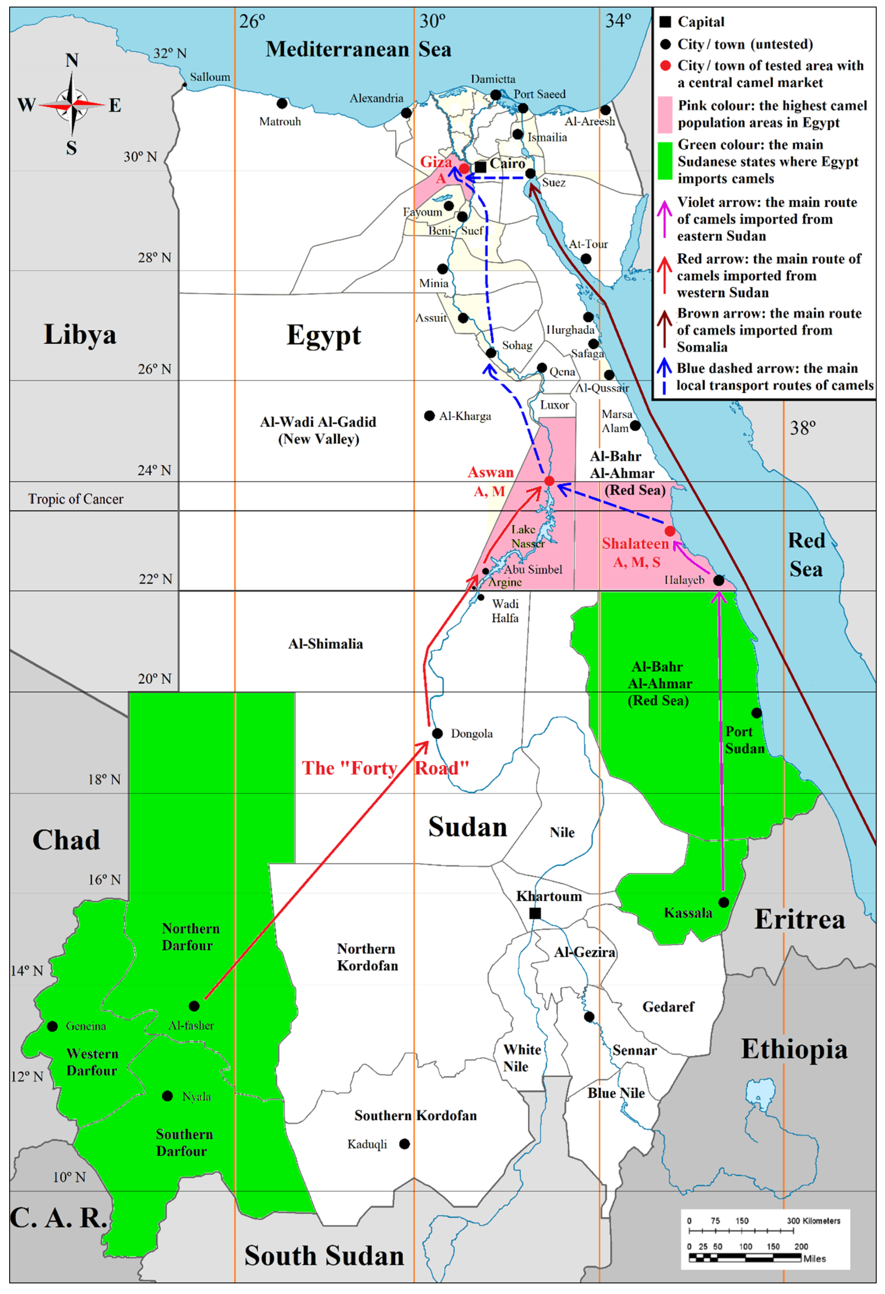
2.1. Study Area and Sera Collection
2.2. Ethics Statement
2.3. Detection of Anti-Brucella Antibodies
2.4. Molecular Detection of Brucella spp. DNA
2.5. Statistical Analysis
3. Results
3.1. Seroprevalence of Anti-Brucella Antibodies in Camel Sera
3.2. Detection of Brucella spp. DNA in Camel Sera
3.3. Statistical Analysis
4. Discussion
5. Conclusions
Author Contributions
Funding
Acknowledgments
Conflicts of Interest
References
- Kirk, M.D.; Pires, S.M.; Black, R.E.; Caipo, M.; Crump, J.A.; Devleesschauwer, B.; Döpfer, D.; Fazil, A.; Fischer-Walker, C.L.; Hald, T.; et al. World Health Organization Estimates of the Global and Regional Disease Burden of 22 Foodborne Bacterial, Protozoal, and Viral Diseases, 2010: A Data Synthesis. PLoS Med. 2015, 12, e1001921. [Google Scholar] [CrossRef] [Green Version]
- Fatima, S.; Khan, I.; Nasir, A.; Younus, M.; Saqib, M.; Melzer, F.; Neubauer, H.; El-Adawy, H. Serological, molecular detection and potential risk factors associated with camel brucellosis in Pakistan. Trop. Anim. Health Prod. 2016, 48, 1711–1718. [Google Scholar] [CrossRef] [PubMed]
- Gyuranecz, M.; Wernery, U.; Kreizinger, Z.; Juhász, J.; Felde, O.; Nagy, P. Genotyping of Brucella melitensis strains from dromedary camels (Camelus dromedarius) from the United Arab Emirates with multiple-locus variable-number tandem repeat analysis. Veter Microbiol. 2016, 186, 8–12. [Google Scholar] [CrossRef] [PubMed]
- Gwida, M.; Elgohary, A.; Melzer, F.; Khan, I.; Rösler, U.; Neubauer, H. Brucellosis in camels. Res. Veter Sci. 2012, 92, 351–355. [Google Scholar] [CrossRef] [PubMed]
- Sayed-Ahmed, M.; Mohamed, Z.S.-A.; Mohamed, M.E.-D.; Mohamed, A.E.-B.; Sherif, M.S.; Emad, E.Y.; El-Sayed, A.M.; El-Diasty, M.M.; El-Beskawy, M.A.; Shoieb, S.M.; et al. Sero-prevalence of camel brucellosis (Camelus dromedarius) and phenotypic characteristics of Brucella melitensis biovar 3 in Shalateen City, Red Sea Governorate, Egypt. Afr. J. Microbiol. Res. 2017, 11, 1259–1266. [Google Scholar] [CrossRef] [Green Version]
- Salisu, U.; Kudi, C.; Bale, J.; Babashani, M.; Kaltungo, B.; Saidu, S.; Asambe, A.; Baba, A. Seroprevalence of Brucella antibodies in camels in Katsina State, Nigeria. Trop. Anim. Health Prod. 2017, 49, 1041–1046. [Google Scholar] [CrossRef]
- Sprague, L.D.; al Dahouk, S.; Neubauer, H. A review on camel brucellosis: A zoonosis sustained by ignorance and indifference. Pathog. Glob. Health 2012, 106, 144–149. [Google Scholar] [CrossRef] [PubMed] [Green Version]
- Othman, O.E.; El-Kader, H.A.A.; Alam, S.S.; El-Aziem, S.H.A. Cytochrome b conservation between six camel breeds reared in Egypt. J. Genet. Eng. Biotechnol. 2017, 15, 1–6. [Google Scholar] [CrossRef]
- Refai, M. Incidence and control of brucellosis in the Near East region. Veter Microbiol. 2002, 90, 81–110. [Google Scholar] [CrossRef]
- Menshawy, A.; Perez-Sancho, M.; García-Seco, T.; Hosein, H.I.; García, N.; Martínez, I.; Sayour, A.; Goyache, J.; Azzam, R.A.A.; Domínguez, L.; et al. Assessment of Genetic Diversity of Zoonotic Brucella spp. Recovered from Livestock in Egypt Using Multiple Locus VNTR Analysis. BioMed Res. Int. 2014, 2014, 1–7. [Google Scholar] [CrossRef] [Green Version]
- Wareth, G.; Hikal, A.; Refai, M.; Melzer, F.; Roesler, U.; Neubauer, H. Animal brucellosis in Egypt. J. Infect. Dev. Ctries. 2014, 8, 1365–1373. [Google Scholar] [CrossRef]
- Hegazy, Y.M.; Molina-Flores, B.; Shafik, H.; Ridler, A.; Guitian, F.; Guitian, J. Ruminant brucellosis in Upper Egypt (2005–2008). Prev. Veter Med. 2011, 101, 173–181. [Google Scholar] [CrossRef] [PubMed]
- Eltholth, M.M.; Hegazy, Y.M.; El-Tras, W.F.; Rushton, J.; Bruce, M. Temporal Analysis and Costs of Ruminant Brucellosis Control Programme in Egypt Between 1999 and 2011. Transbound. Emerg. Dis. 2016, 64, 1191–1199. [Google Scholar] [CrossRef] [PubMed] [Green Version]
- Samaha, H.; Al-Rowaily, M.; Khoudair, R.M.; Ashour, H.M. Multicenter Study of Brucellosis in Egypt. Emerg. Infect. Dis. 2008, 14, 1916–1918. [Google Scholar] [CrossRef] [PubMed]
- Khan, A.U.; Melzer, F.; El-Soally, S.A.G.E.; Elschner, M.C.; Mohamed, S.A.; Ahmed, M.A.S.; Roesler, U.; Neubauer, H.; El-Adawy, H. Serological and Molecular Identification of Brucella spp. in Pigs from Cairo and Giza Governorates, Egypt. Pathogens 2019, 8, 248. [Google Scholar] [CrossRef] [Green Version]
- Nielsen, K.; Yu, W.L. Serological diagnosis of brucellosis. Prilozi 2010, 31, 65–89. [Google Scholar]
- Mathew, C.; Stokstad, M.; Johansen, T.B.; Klevar, S.; Mdegela, R.H.; Mwamengele, G.; Michel, P.; Escobar, L.; Fretin, D.; Godfroid, J. First isolation, identification, phenotypic and genotypic characterization of Brucella abortus biovar 3 from dairy cattle in Tanzania. BMC Veter Res. 2015, 11, 156. [Google Scholar] [CrossRef] [Green Version]
- Ulu-Kilic, A.; Metan, G.; Alp, E. Clinical presentations, and diagnosis of brucellosis. Recent Pat. Antiinfect Drug Dis. 2013, 8, 34–41. [Google Scholar] [CrossRef]
- Gwida, M.; Elgohary, A.; Melzer, F.; Tomaso, H.; Roesler, U.; Wernery, U.; Wernery, R.; Elschner, M.; Khan, I.; Eickhoff, M.; et al. Comparison of diagnostic tests for the detection of Brucella spp. in camel sera. BMC Res. Notes 2011, 4, 525. [Google Scholar] [CrossRef] [Green Version]
- Garcell, H.G.; Garcia, E.G.; Pueyo, P.V.; Martín, I.R.; Arias, A.V.; Serrano, R.N.A. Outbreaks of brucellosis related to the consumption of unpasteurized camel milk. J. Infect. Public Health 2016, 9, 523–527. [Google Scholar] [CrossRef]
- Rhodes, H.M.; Williams, D.N.; Hansen, G.T. Invasive human brucellosis infection in travelers to and immigrants from the Horn of Africa related to the consumption of raw camel milk. Travel Med. Infect. Dis. 2016, 14, 255–260. [Google Scholar] [CrossRef]
- Ben-Shimol, S.; Dukhan, L.; Belmaker, I.; Bardenstein, S.; Sibirsky, D.; Barrett, C.; Greenberg, D. Human brucellosis outbreak acquired through camel milk ingestion in southern Israel. Isr. Med Assoc. J. IMAJ 2012, 14, 475–478. [Google Scholar]
- Zakaria, A.M.; Ahmed, S.F.; Motawae, M.S. Seropositivity in animals and risk of occupational brucellosis among abattoirs personnel associated with poor work practices and absence of safety policy in Egypt. Int. J. Occup. Environ. Health 2018, 24, 55–60. [Google Scholar] [CrossRef] [PubMed]
- OIE. Brucellosis (Brucella abortus, B. melitensis and B. suis) (infection with B. abortus, B. melitensis and B. suis). In Manual of Diagnostic Tests and Vaccines for Terrestrial Animals 2019, OIE; World Health Organization for Animal Health World Organisation for Animal Health (Office International des Épizooties): Paris, France, 2019; pp. 355–398. [Google Scholar]
- Probert, W.S.; Schrader, K.N.; Khuong, N.Y.; Bystrom, S.L.; Graves, M.H. Real-Time Multiplex PCR Assay for Detection of Brucella spp., B. abortus, and B. melitensis. J. Clin. Microbiol. 2004, 42, 1290–1293. [Google Scholar] [CrossRef] [PubMed] [Green Version]
- Hänsel, C.; Mertens, K.; Elschner, M.C.; Melzer, F. Novel real-time PCR detection assay for Brucella suis. Veter Rec. Open 2015, 2. [Google Scholar] [CrossRef] [Green Version]
- Sanogo, M.; Abatih, E.; Thys, E.; Fretin, D.; Berkvens, D.; Saegerman, C. Importance of identification and typing of Brucellae from West African cattle: A review. Vet. Microbiol. 2013, 164, 202–211. [Google Scholar] [CrossRef] [Green Version]
- Godfroid, J.; al Dahouk, S.; Pappas, G.; Roth, F.; Matope, G.; Muma, J.; Marcotty, T.; Pfeiffer, D.U.; Skjerve, E. A “One Health” surveillance and control of brucellosis in developing countries: Moving away from improvisation. Comp. Immunol. Microbiol. Infect. Dis. 2013, 36, 241–248. [Google Scholar] [CrossRef] [Green Version]
- Machavarapu, M.; Poonati, R.; Mallepaddi, P.C.; Gundlamadugu, V.; Raghavendra, S.; Polavarapu, K.K.B.; Polavarapu, R. Endemic brucellosis in Indian animal and human populations: A billion-dollar issue. J. Curr. Trends Biotechnol. Pharm. 2019, 13, 112–123. [Google Scholar]
- Godfroid, J. Brucellosis in livestock and wildlife: Zoonotic diseases without pandemic potential in need of innovative one health approaches. Arch. Public Health 2017, 75, 34. [Google Scholar] [CrossRef]
- Musa, M.; Eisa, M.; el Sanousi, E.; Wahab, M.A.; Perrett, L. Brucellosis in Camels (Camelus dromedarius) in Darfur, Western Sudan. J. Comp. Pathol. 2008, 138, 151–155. [Google Scholar] [CrossRef]
- Chisholm, K.; Dueger, E.; Fahmy, N.T.; Samaha, H.A.T.; Zayed, A.; Abdel-Dayem, M.S.; Villinski, J.T. Crimean-Congo Hemorrhagic Fever Virus in Ticks from Imported Livestock, Egypt. Emerg. Infect. Dis. 2012, 18, 181–182. [Google Scholar] [CrossRef] [PubMed]
- Wakene, W.Z. Review on Epidemiology of Camel and Human Brucellosis in East Africa, Igad Member Countries. Sci. J. Clin. Med. 2017, 6, 109. [Google Scholar] [CrossRef] [Green Version]
- Ibrahim, H.H.; Rouby, S.; Menshawy, A.; Ghazy, N. Seroprevalence of Camel Brucellosis and Molecular Characterization of Brucella melitensis Recovered from Dromedary Camels in Egypt. Res. J. Vet. Pr. 2016, 4, 17–24. [Google Scholar] [CrossRef] [Green Version]
- Moghney, A.R.F.A. A preliminary study on brucellosis on camels at Behira Province. Ass. Univ. Bull. Environ. Res. 2004, 7, 39–43. [Google Scholar]
- Sayour, A.E.; Elbauomy, E.; Abdel-Hamid, N.H.; Mahrous, A.; Carychao, D.; Cooley, M.B.; Elhadidy, M. MLVA fingerprinting of Brucella melitensis circulating among livestock and cases of sporadic human illness in Egypt. Transbound. Emerg. Dis. 2020. [Google Scholar] [CrossRef]
- Hamdy, M.; Amin, A. Detection of Brucella Species in the Milk of Infected Cattle, Sheep, Goats and Camels by PCR. Vet. J. 2002, 163, 299–305. [Google Scholar] [CrossRef]
- Abdel-Hamid, N.H.; Elbauomy, E.M.; Ghobashy, H.M.; Sayour, A.E.; Ismail, R.I.; Soliman, H.S.; Abdel-Haleem, M.H. Role of sheep and goat mobile flocks in the transmission of brucellosis to the household ruminants and the disease prevalence in these flocks. Anim. Health Res. J. 2017, 5, 95–105. [Google Scholar]
- Omer, M.; Musa, M.; Bakhiet, M.; Perrett, L. Brucellosis in camels, cattle, and humans: Associations and evaluation of serological tests used for diagnosis of the disease in certain nomadic localities in Sudan. Rev. Sci. Tech. OIE 2010, 29, 663–669. [Google Scholar] [CrossRef] [Green Version]
- Musa, M.; Jahans, K.; Fadalla, M. Brucella Biovars isolated from nomadic cattle in the Southern Darfur Province of Western Sudan. J. Comp. Pathol. 1990, 102, 49–54. [Google Scholar] [CrossRef]
- Gumaa, M.; Osman, H.; Omer, M.; el Sanousi, E.; Godfroid, J.; Ahmed, A. Seroprevalence of brucellosis in sheep and isolation of Brucella abortus biovar 6 in Kassala State, Eastern Sudan. Rev. Sci. Tech. OIE 2014, 33, 957–965. [Google Scholar] [CrossRef] [Green Version]
- Agab, H.; Abbas, B.; el Jack Ahmed, H.; Maoun, I.E. First report on the isolation of Brucella abortus biovar 3 from camel (Camelus dromedarius) in the Sudan. Rev. Elev. Med. Vet. Pays. Trop. 1994, 47, 361–363. [Google Scholar] [PubMed]
- Faye, B.; Abdelhadi, O.M.A.; Ahmed, A.I.; Bakheit, S.A. Camel in Sudan: Prospects. Livestock Res. Rural Develop. 2011, 23, 1–11. [Google Scholar]
- Ibrahim, S.I. Studies on swine brucellosis in Egypt. J. Egypt Vet. Med. Ass. 1996, 56, 1–12. [Google Scholar]
- Gilbert, M.; Nicolas, G.; Cinardi, G.; van Boeckel, T.P.; Vanwambeke, S.O.; Wint, G.R.W.; Robinson, T.P. Global distribution data for cattle, buffaloes, horses, sheep, goats, pigs, chickens, and ducks in 2010. Sci. Data 2018, 5, 180227. [Google Scholar] [CrossRef] [Green Version]
- Coelho, A.; Díez, J.G.; Coelho, A.M. Risk Factors for Brucella spp. in Domestic and Wild Animals. In Updates on Brucellosis; IntechOpen: London, UK, 2015. [Google Scholar]
- Abebe, G.; Worku, Y.; Mamo, G.; Nazir, S. Sero-prevalence and Associated Risk Factors of Brucellosis in Camel at Akaki Abattoir, Central Ethiopia. J. Anim. Res. 2017, 7, 617. [Google Scholar] [CrossRef]
- Ullah, S. Prevalence of Brucellosis among Camels in District Muzaffargarh, Pakistan. J. Infect. Mol. Boil. 2015, 3, 52–56. [Google Scholar] [CrossRef] [Green Version]
- Bayasgalan, C.; Chultemdorj, T.; Roth, F.; Zinsstag, J.; Hattendorf, J.; Badmaa, B.; Argamjav, B.; Schelling, E. Risk factors of brucellosis seropositivity in Bactrian camels of Mongolia. BMC Veter Res. 2018, 14, 342. [Google Scholar] [CrossRef]
- Al-Majali, A.M.; Al-Qudah, K.; Al-Tarazi, Y.H.; Al-Rawashdeh, O.F. Risk factors associated with camel brucellosis in Jordan. Trop. Anim. Health Prod. 2007, 40, 193–200. [Google Scholar] [CrossRef]
- Wernery, U. Camelid brucellosis: A review. Rev. Sci. Tech. OIE 2014, 33, 839–857. [Google Scholar] [CrossRef]
- Sayour, A.E.; Elbauomy, E.M.; Shehata, A.A.; El-Kholi, M.K. Brucellosis prevalence and serological profile of male one-humped camels reared in Somaliland and eastern Ethiopia for meat production. Global Vet. 2015, 14, 67–76. [Google Scholar]


| Target | Primer and Probe Sequences | Reference | |
|---|---|---|---|
| Brucella spp. | 5′-GCT CGG TTG CCA ATA TCA ATG C-3´ | Forward | [25] |
| 5′-GGG TAA AGC GTC GCC AGA AG-3´ | Reverse | ||
| 6-FAM-AAA TCT TCC ACC TTG CCC TTG CCA TCA-MGB | Probe | ||
| B. abortus | 5′-GCG GCT TTT CTA CGG TAT TC-3´ | Forward | |
| 5′-CAT GCG CTA TGA TCT GGT TAC G-3´ | Reverse | ||
| Hex-CGC TCA TGC TCG CCA GAC TTC AAT G-BHQ1 | Probe | ||
| B. melitensis | 5′-AAC AAG CGG CAC CCC TAA AA-3´ | Forward | |
| 5′-CAT GCG CTA TGA TCT GGT TAC G-3´ | Reverse | ||
| Cy5-CAG GAG TGT TTC GGC TCA GAA TAA TCC ACA-BHQ2 | Probe | ||
| B. suis | 5′-GCC AAA TAT CCA TGC GGG AAG-3´ | Forward | [26] |
| 5′-TGG GCA TTC TCT ACG GTG TG-3´ | Reverse | ||
| VIC-TTG CGC TTT TGT GAT CTT TGC TTA TGG-MGB | Probe | ||
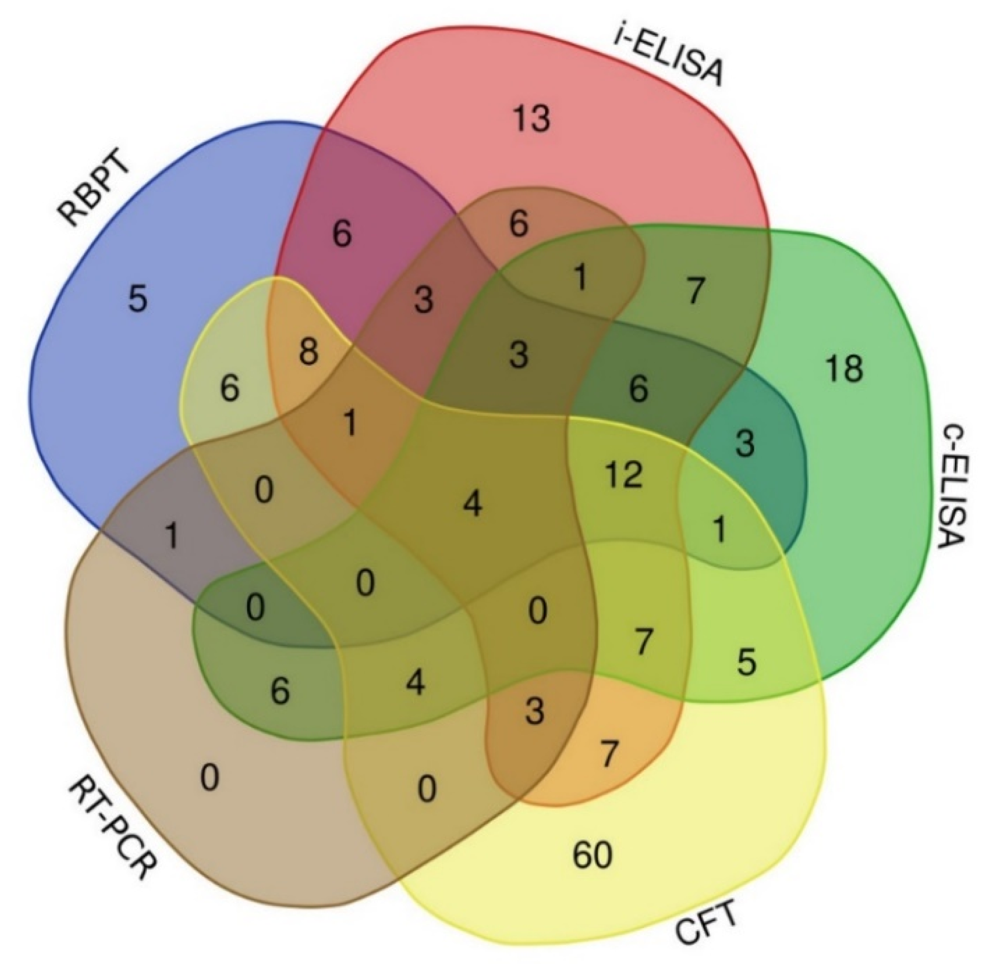
| Governorate | Sex | Number of Samples | Seroprevalence n (%) | Molecular Identification | |||||
|---|---|---|---|---|---|---|---|---|---|
| RBPT * | i-ELISA * | c-ELISA * | CFT * | Real-Time-PCR n (%) | Brucella spp. DNA Identification | Cq/Ct-Values ** | |||
| Giza | male | 55 | 11 (20.0) | 17 (30.9) | 7 (12.7) | 28 (50.9) | 3 (5.5) | 3 B. abortus | 37, 36, 35 |
| female | 51 | 4 (7.8) | 7 (13.7) | 8 (15.7) | 5 (9.8) | 6 (11.8) | 6 B. abortus | 30, 32, 33, 36, 37 | |
| Sub-total | 106 | 15 (14.2) | 24 (22.6) | 15 (14.2) | 33 (31.1) | 9 (8.5) | |||
| Aswan | male | 161 | 30 (18.6) | 41 (25.5) | 39 (24.2) | 51 (31.7) | 7 (4.4) | 6 B. abortus | 37, 37, 36, 36, 36, 36 |
| 1 B. melitensis | 34 | ||||||||
| female | 25 | 3 (12.0) | 7 (28.0) | 2 (8.0) | 8 (32.0) | 1 (4.0) | 1 B. abortus | 36 | |
| Sub-total | 186 | 33 (17.7) | 48 (25.8) | 41 (22.0) | 59 (31.7) | 8 (4.3) | |||
| Al-Bahr Al-Ahmar (the Red Sea) | male | 79 | 11(13.9) | 13 (16.5) | 20 (25.3) | 24 (30.4) | 14 (17.7) | 8 B. abortus | 37, 35, 31, 37, 35, 32, 36, 35 |
| 1 B. melitensis | 36 | ||||||||
| 5 B. suis | 37, 36, 33, 28, 37 | ||||||||
| female | 10 | 0 (0.0) | 2 (20.0) | 1 (10.0) | 2 (20.0) | 1 (10.0) | 1 B. abortus | 37 | |
| Sub-total | 89 | 11 (12.4) | 15 (16.9) | 21 (23.6) | 26 (29.2) | 15 (16.9) | |||
| Grand-total | 381 | 59 (15.5) | 87 (22.8) | 77 (20.2) | 118 (31.0) | 32 (8.4) | |||
| Variable | Seroprevalence n (%) | Molecular Identification | ||||
|---|---|---|---|---|---|---|
| RBPT * | i-ELISA * | c-ELISA * | CFT * | Real-Time PCR n (%) | Brucella DNA Identification | |
| Geographical location | ||||||
| Aswan (n = 186) | 33 (17.7) | 48 (25.8) | 41 (22.0) | 59 (31.7) | 8 (4.3) | 7 B. abortus 1 B. melitensis |
| Giza (n =106) | 15 (14.2) | 24 (22.6) | 15 (14.2) | 33 (31.1) | 9 (8.5) | 9 B. abortus |
| Al-Bahr Al-Ahmar (the Red Sea) (n = 89) | 11 (12.4) | 15 (16.9) | 21 (23.6) | 26 (29.2) | 15 (16.9) | 9 B. abortus 5 B. suis 1 B. melitensis |
| p-value ** | 0.4688 | 0.3205 | 0.4171 | NA | NA | |
| X2 | 1.5153 | 2.2757 | 1.7489 | |||
| Df | 2 | |||||
| 95% CI | - | - | - | |||
| OR | - | - | - | |||
| Breed | ||||||
| Al-Beshary (n = 89) | 11 (12.4) | 15 (16.9) | 21 (23.6) | 26 (29.2) | 15 (16.9) | 9 B. abortus 5 B. suis 1 B. melitensis |
| Al-Ebadi (n = 93) | 16 (17.2) | 26 (28.0) | 22 (23.7) | 30 (32.3) | 4 (4.3) | 3 B. abortus 1 B. melitensis |
| Al-Zemkly (n = 106) | 15 (14.2) | 24 (22.6) | 15 (14.2) | 33 (31.1) | 9 (8.5) | 9 B. abortus |
| Al-Zubaidi (n = 93) | 17 (18.3) | 22 (23.7) | 19 (20.4) | 29 (31.2) | 4 (4.3) | 4 B. abortus |
| p-value ** | 0.6775 | 0.4658 | 0.5823 | NA | NA | |
| X2 | 1.5205 | 2.5532 | 1.9524 | |||
| Df | 3 | |||||
| 95% CI | - | - | - | |||
| OR | - | - | - | |||
| Sex | ||||||
| Females (n = 86) | 7 (8.1) | 16 (18.6) | 11 (12.8) | 15 (17.4) | 8 (9.3) | 8 B. abortus |
| Males (n = 295) | 52 (17.6) | 71 (24.1) | 66 (23.4) | 103 (34.9) | 24 (8.3) | 17 B. abortus 5 B. suis 2 B. melitensis |
| p-value ** | 0.7177 | 0.3515 | 0.9164 | NA | NA | |
| X2 | 0.13075 | 0.86806 | 0.011028 | |||
| Df | 1 | |||||
| 95% CI | 0.2819–2.2582 | 0.6333–3.1044 | 0.4215–2.4729 | |||
| OR | 0.8438063 | 1.4091 | 1.0436 | |||
| Age | ||||||
| <8 years (n = 227) | 39 (17.2) | 52 (22.9) | 46 (20.2) | 81 (35.7) | 21 (9.3) | 15 B. abortus 5 B. suis 1 B. melitensis |
| ≥8–11 years (n = 68) | 13 (19.1) | 19 (27.9) | 20 (29.4) | 22 (32.4) | 3 (4.4) | 2 B. abortus 1 B. melitensis |
| 11–13 years (n = 51) | 4 (7.8) | 7 (13.7) | 8 (15.7) | 5 (9.8) | 6 (11.8) | 6 B. abortus |
| >13–15 years (n = 35) | 3 (8.6) | 9 (25.7) | 3 (8.6) | 10 (28.6) | 2 (5.7) | 2 B. abortus |
| p-value ** | 0.7844 | 0.5792 | 0.1672 | NA | NA | |
| X2 | 1.0699 | 1.9674 | 5.0641 | |||
| Df | 3 | |||||
| 95% CI | - | - | - | |||
| OR | - | - | - | |||
© 2020 by the authors. Licensee MDPI, Basel, Switzerland. This article is an open access article distributed under the terms and conditions of the Creative Commons Attribution (CC BY) license (http://creativecommons.org/licenses/by/4.0/).
Share and Cite
Khan, A.U.; Sayour, A.E.; Melzer, F.; El-Soally, S.A.G.E.; Elschner, M.C.; Shell, W.S.; Moawad, A.A.; Mohamed, S.A.; Hendam, A.; Roesler, U.; et al. Seroprevalence and Molecular Identification of Brucella spp. in Camels in Egypt. Microorganisms 2020, 8, 1035. https://0-doi-org.brum.beds.ac.uk/10.3390/microorganisms8071035
Khan AU, Sayour AE, Melzer F, El-Soally SAGE, Elschner MC, Shell WS, Moawad AA, Mohamed SA, Hendam A, Roesler U, et al. Seroprevalence and Molecular Identification of Brucella spp. in Camels in Egypt. Microorganisms. 2020; 8(7):1035. https://0-doi-org.brum.beds.ac.uk/10.3390/microorganisms8071035
Chicago/Turabian StyleKhan, Aman Ullah, Ashraf E. Sayour, Falk Melzer, Sherif Abdel Ghafar Elsayed El-Soally, Mandy C. Elschner, Waleed S. Shell, Amira A. Moawad, Shereen Aziz Mohamed, Ashraf Hendam, Uwe Roesler, and et al. 2020. "Seroprevalence and Molecular Identification of Brucella spp. in Camels in Egypt" Microorganisms 8, no. 7: 1035. https://0-doi-org.brum.beds.ac.uk/10.3390/microorganisms8071035