Article

First Record of a Suspected Human-Pathogenic Borrelia Species in Populations of the Bat Tick Carios vespertilionis in Sweden

by
Thomas G. T. Jaenson
1 and
Peter Wilhelmsson
2,3,*
1
Evolutionary Biology Centre, Department of Organismal Biology, Uppsala University, 752 36 Uppsala, Sweden
2
Department of Biomedical and Clinical Sciences, Division of Inflammation and Infection, Linköping University, 581 83 Linköping, Sweden
3
Department of Clinical Microbiology, Region Jönköping County, 553 05 Jönköping, Sweden
*
Author to whom correspondence should be addressed.
Submission received: 28 April 2021 / Revised: 17 May 2021 / Accepted: 18 May 2021 / Published: 20 May 2021
(This article belongs to the Section Systems Microbiology)

Abstract

:
The bat tick Carios vespertilionis has been reported from Sweden to occasionally feed on humans resulting in disease symptoms. The aim of this study was to investigate C. vespertilionis as a potential vector and reservoir of Borrelia species. In 2015 and 2018 in south-central Sweden, C. vespertilionis ticks were collected from a wooden bat box harboring Soprano pipistrelle bats, Pipistrellus pygmaeus. In addition, one C. vespertilionis tick found inside a house in southern Sweden in 2019 was collected. Ticks were screened for Borrelia spp. using a genus-specific quantitative PCR assay. The Borrelia species of the positive specimens were determined by conventional PCR followed by DNA sequencing and phylogenetic analyses. A total of 24% (22 of 92) of the analyzed C. vespertilionis ticks were Borrelia-positive. Phylogenetic analyses indicate that the bacteria belong to the relapsing fever group of borreliae; some of them appear to be identical with Borrelia sp. CPB1, a spirochete only found twice before—in the United Kingdom and in France. Our results also indicate a temporal and spatial distribution of this Borrelia species. Since C. vespertilionis occasionally bites humans, and since it exhibits a high prevalence of Borrelia bacteria, it is possible that it presents a risk of human disease. Further studies are needed to characterize Borrelia sp. CPB1 to determine if it is human-pathogenic and to determine if C. vespertilionis is a vector and/or reservoir of this agent.

1. Introduction

Many different species of putative or proven vertebrate-pathogenic parasites, bacteria, and viruses have been detected in bats and bat-associated ectoparasites, particularly ticks (Ixodida, Ixodidae, and Argasidae). Investigations of bat-associated microorganisms and viruses have drastically increased during the last two decades [1]. The main reasons for this are the severe outbreaks in humans by viruses related to or identical with bat-associated viruses, i.e., coronavirus disease 2019 (COVID-19), severe acute respiratory syndrome (SARS), Middle East respiratory syndrome (MERS), Ebola, bat lyssavirus rabies and Nipah [2,3,4,5,6,7].
There are more than 1400 species of bats [8], order Chiroptera, of which 19 species occur in Sweden [9]. Many bat species have synanthropic behavioral traits and benefit from using human habitations. For instance, nursery roosts of the Soprano pipistrelle bat, Pipistrellus pygmaeus, are in wall cavities and under roof coverings of houses, in tree holes, and in rock crevices [9,10,11]. Pipistrellus pygmaeus is distributed in western, central, eastern and southern Europe, including southern and south-central Sweden. According to Ahlén (2011), it is very common in southern Sweden northwards to River Dalälven and the coastal parts of the province of Gästrikland [11]. In the southern part of its Swedish range, this bat species occurs in all kinds of forests, gardens and parks, while to the north of Lake Mälaren it is mostly found in deciduous forests. Migration studies have shown that in recent years there have been many Soprano pipistrelle bats moving to wintering sites on the continent [11]. In Europe, populations of P. pygmaeus are often parasitized by the Round bat argas, also called the Short-legged bat tick, Carios vespertilionis (Ixodida, Argasidae) [12,13,14,15]. The resting places of these bats may be where humans are bitten by blood-feeding bat ectoparasites, such as the potentially pathogen-infected bat tick C. vespertilionis. This provides the opportunity for transmission of zoonotic pathogens [12,16,17].
Carios vespertilionis has a wide distribution range and is present in Africa, Asia and Europe [12,13,14,18], and it is usually host-specific to bats, particularly pipistrelles, but almost any bat species will serve as a host [13,19]. This tick species has been reported to be ‘highly aggressive’ [14] and may occasionally attack humans, dogs, and birds [12,16,17,18,19,20]. Hoogstraal (1956) writes: “Nymphs and adults on several occasions have attacked us in caves and we easily induce it to bite ourselves in the laboratory…On man mild itching resulting from a bite may persist for several weeks…It has been stated that this tick is a vector of a spirochaete of bats but reports of conclusive supporting evidence have thus far not been located” [19] (p. 106, p. 109). Furthermore, in the early summer of 1993 at Färentuna, province of Uppland, near Stockholm, Sweden, two persons experienced severe skin reactions with fever, ulceration, erythema, some swollen lymph glands, and oedema on their legs and arms after having been bitten by ticks in a bedroom. Shortly before that incident, unidentified bats had been roosting in the attic above the bedroom. One of us (TGTJ) morphologically identified the ticks as C. vespertilionis [16]. One of the two persons was eventually treated with penicillin, which cured the infection; the other person’s symptoms disappeared spontaneously without any pharmacological treatment.
Only a few studies have investigated the presence of tick-associated microorganisms in C. vespertilionis. A total of 86% (29 of 34) of museum specimens of C. vespertilionis ticks collected between 1896 and 1994 in the United Kingdom contained DNA of Borrelia burgdorferi sensu lato as revealed by PCR [21]. B. burgdorferi s.l., the causative agent of the most common tick-borne human disease, Lyme borreliosis in the Holarctic Region, has also been detected in C. vespertilionis in southern Russia [22]. In France, C. vespertilionis ticks harbored a new genotype of the spotted fever group of rickettsiae called Rickettsia sp. AvBat, a new species of the Ehrlichia canis group—Ehrlichia sp. AvBat—and Borrelia sp. CPB1 [23], which is a member of the relapsing fever group of Borrelia and suspected to have caused a lethal Borrelia infection in a bat in the United Kingdom [24]. Other bacteria such as Coxiella burnetii, Rickettsia spp. and Ehrlichia spp., as well as the protozoan Babesia vesperuginis, and Issyk-Kul virus and Sokuluk virus, have been detected in C. vespertilionis ticks in Europe or Asia [21,25,26,27,28,29].
To our knowledge, the species of pathogens associated with bats and bat ticks in Sweden have not been previously investigated. The potential roles of C. vespertilionis as a reservoir and vector of bacteria, protozoan parasites and viruses are therefore worth investigating. A deeper knowledge about the biology of C. vespertilionis and its potential role as a reservoir and vector of harmful microorganisms is a prerequisite to better understand the biology of many bat species and to mitigate and potentially control certain zoonoses associated with this tick species.
The aim of this study was to investigate the role of C. vespertilionis as a potential reservoir and vector of species in the B. burgdorferi s.l. complex and in the relapsing fever Borrelia complex.

2. Materials and Methods

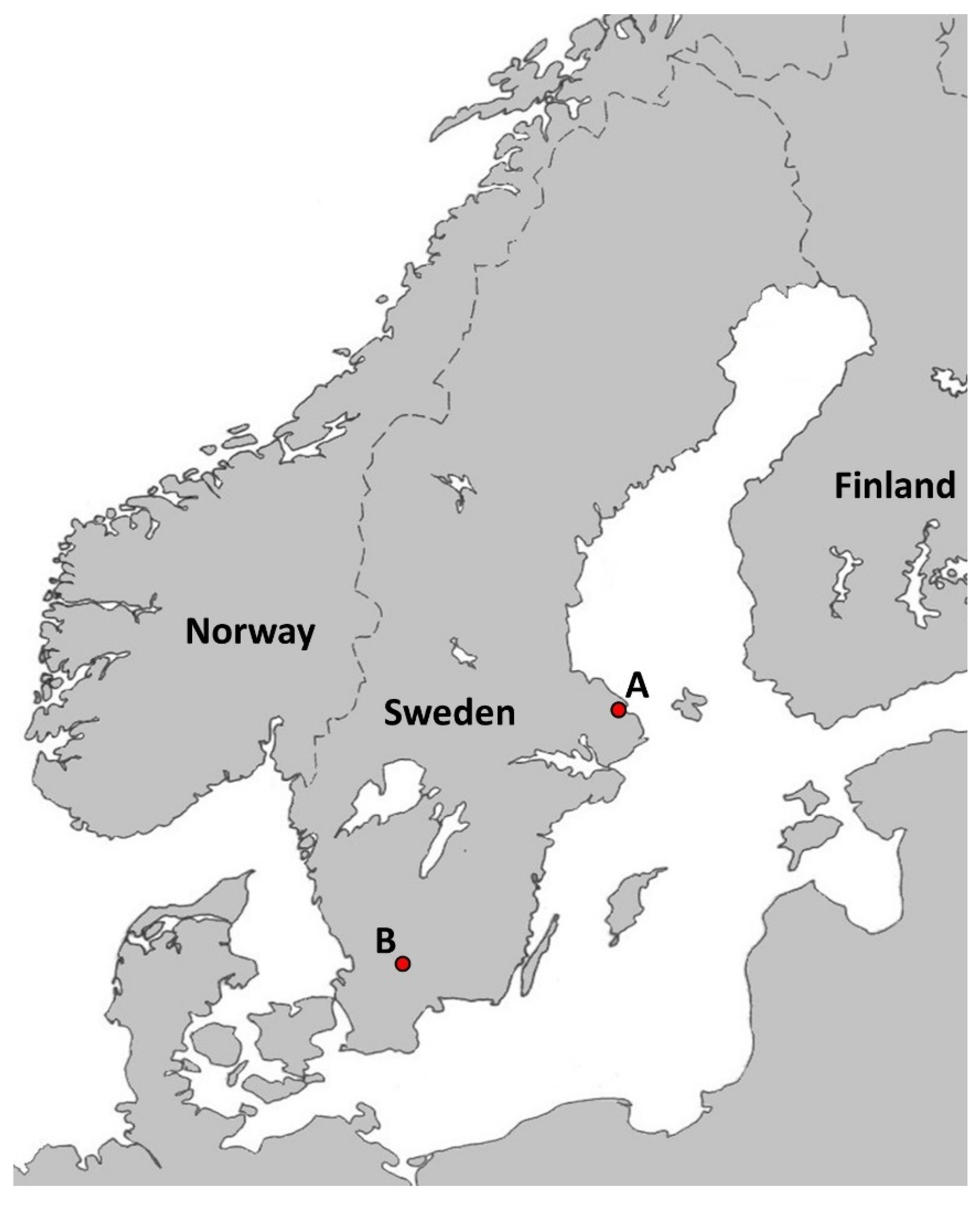
2.1. Tick Collection

In the summers of 2015 and 2018, at Snesslinge (60°19.567 N, 18°15.067 E, Figure 1), province of Uppland, south-central Sweden, 91 ticks were collected from a water-filled tray placed below a wooden bat box harboring an estimated number of 250–500 adult females and young, <2 months-old, Soprano pipistrelle bats, P. pygmaeus. All sampling occasions took place each morning from mid-June to mid-August in 2015 and in 2018. All ticks detected were put in numbered vials containing 80% ethanol. In addition, one tick specimen detected in July 2019 inside a house at Älmhult (56°32.720 N, 13°52.667 E, Figure 1), province of Småland, southern Sweden was also examined. The attic of this house was inhabited by bats of unknown species. The distance between Älmhult and Snesslinge is 483 km.

2.2. Tick Identification

All tick specimens were identified to developmental stage and species by morphological characteristics. A Leica Wild M10 stereomicroscope was used together with keys and illustrations in [13,19,30,31,32,33,34]. All ticks were also photographed dorsally and ventrally, and the length and width of each tick were measured, using a USB-microscope (Dino-Lite Long AM4013TL, AnMo Electronics Corp., Taiwan). Each tick was categorized into one of two categories, where + corresponds to “contains host blood” and—corresponds to “empty/no visible blood in the gut”.
Regarding the family Argasidae, we are aware that there is widespread disagreement concerning the genus-level classification and names of the genera in this family. Regarding the tick species, Carios vespertilionis (also known as Argas vespertilionis), found in this study, we followed the recent study of Mans et al. [35]. They showed that this species should be placed in the genus Carios, subfamily Ornithodorinae, family Argasidae.

2.3. Nucleic Acid Extraction and cDNA Synthesis from Ticks

Collected ticks were homogenized individually by bead-beating in 2 mL safe-lock microcentrifuge tubes (Eppendorf AG, Hamburg, Germany) with a 5-mm stainless steel bead (Qiagen, Hilden, Germany) in 350 µL RLT buffer (Qiagen), supplemented with 1% 2-mercaptoethanol (Sigma-Aldrich, Stockholm, Sweden), using a TissueLyser II (Qiagen) for 2 min at 25 Hz. After centrifugation at 20,000× g for 3 min, 300 µL supernatant was transferred to new microcentrifuge tubes for total nucleic acid (NA) extraction, using MagAttract® Viral RNA M48 kit (Qiagen) in a BioRobot M48 workstation (Qiagen), using a 65-µL elution volume. Each batch of 24 samples consisted of 22 ticks, one positive control (5 µL of B. burgdorferi sensu stricto B31 ATCC 35210 [108 cells/mL]) and one negative control (H2O) that were extracted simultaneously.
The eluted NA was reverse-transcribed to cDNA using illustra™ Ready-to-Go RT-PCR Beads kit (GE Healthcare, Amersham Place, UK). Twenty microliters NA and 10 µL pd(N)6 random hexamer primers (0.25 µg/µL) were incubated for 5 min at 97 °C and then mixed with one RT-PCR bead dissolved in 20 µL RNase-free water. The mixture was incubated for 30 min at 42 °C, followed by 5 min at 97 °C, producing 50 µL cDNA.

2.4. Detection of Borrelia Bacteria and Determination of Species

Detection of Borrelia bacteria was done using a genus-specific TaqMan real-time PCR assay, as previously described [36]. The primers Borrelia-F and Borrelia-R and the probe Borrelia-P are designed to target the Borrelia spp. 16S rRNA gene to amplify a 116-bp long amplicon (Table 1).
To determine Borrelia species of the samples positive in the TaqMan real-time PCR assay, a nested, conventional PCR assay using primers targeting the intergenic spacer region (IGS) between 5S and 23S rRNA genes (Table 1) was applied as previously described [37,38]. Samples that failed to produce PCR products with this assay were instead analyzed with primers targeting the IGS between 16S and 23S rRNA genes [39,40]. Tick samples, positive for Borrelia spp. in the TaqMan real-time PCR assay, which failed to produce PCR products with the 5S-23S IGS assay and the 16S-23S IGS assay, were denoted as ‘’untypeable’’. Samples positive for 16S-23S rRNA IGS were further analyzed with primers targeting the 16S rRNA gene [41], and the flaB gene (this study, see below) (Table 1).
To further determine the Borrelia species of the samples positive with the 16S-23S IGS assay, another nested, conventional PCR assay using primers targeting the flagellin B gene (flaB), was developed. The primers flaB-F and flaB-R were designed to target the Borrelia flaB gene to amplify a 699-bp long amplicon of the species in the relapsing fever Borrelia complex (Table 1). A 50-µL reaction consisted of 10 µL 5× Phusion HF Buffer (Thermo Fisher Scientific, Waltham, MA, USA), 1 µL dNTP (10 mM), 2.5 µL of each primer (10 µM; Invitrogen; Table 1), 28.5 µL RNase-free water, 0.5 µL Phusion™ High-Fidelity DNA Polymerase (2 U/µL) (Thermo Fisher Scientific) and 5 µL cDNA template. The PCR reactions were performed on a MyCycler™ Thermal Cycler (Bio-Rad Laboratories, Inc., Hercules, CA, USA) using an activation step at 94 °C for 5 min, and 40 cycles of 94 °C for 1 min, 60 °C for 1 min, and 72 °C for 1 min, and finally one cycle of 72 °C for 10 min. An aliquot (5 µL) of the PCR product obtained in this assay was added to a second PCR mixture, which was prepared using the same volumes, concentrations, and amplification program as those for the first mixture, except with a different primer pair (flaB-Fn and flaB-Rn, Table 1).

2.5. Detection of Borrelia miyamotoi

Detection of B. miyamotoi was done using a species-specific TaqMan real-time PCR assay, as previously described [42]. The primers Bm_F and Bm_R, and the probe Bm_P, are designed to target the B. miyamotoi flagellin B gene (flaB) to amplify a 156-bp long amplicon [43] (Table 1). As a positive control, a synthetic plasmid containing the target sequence of the TaqMan real-time PCR assay was used. The plasmid contained the target sequence, spanning the nucleotides 510–665 of the B. miyamotoi flagellin (flaB) gene (GenBank: KT932823), synthesized and cloned into Eurofins standard vector carrying the ampicillin selection marker (Eurofins Genomics, Ebersberg, Germany).

2.6. Nucleotide Sequencing of PCR-Products and Phylogenetic Analysis

Nucleotide sequencing of the PCR products amplified by conventional PCR assays to determine species of Borrelia was performed by Macrogen Inc. (Amsterdam, The Netherlands). All sequences were confirmed by sequencing both strands. The obtained chromatograms were initially edited and analyzed using BioEdit Software v7.0 (Tom Hall, Ibis Therapeutics, Carlsbad, CA, USA), and the sequences were examined using Basic Local Alignment Tool (BLAST). Phylogenetic trees were constructed with MEGA7 by neighbor-joining using Kimura 2-parameter and pairwise deletion with bootstrap value of 500 replicates. The scale bar of each phylogenetic tree corresponds to the number of substitutions per nucleotide site, and only values greater that 50% are shown in the trees. Sequences obtained have been deposited in GenBank with accession number MZ215741 for the 16S-23S IGS, MZ210080 for the 16S and MZ217187 for the flaB. One additional file shows all the aligned Borrelia sequences (see Document S1 in Supplementary Materials).

2.7. Statistical Analyses

Data were presented as percentages for categorical variables and as medians with interquartile range (IQR) for numerical variables. The categorical variables (e.g., developmental stage of the tick; season of tick collection, etc.) were analyzed using Yate’s corrected chi-square test, but when the expected frequency was <5 in at least one of the cells of the contingency table, Fisher’s exact test with a confidence interval (CI) of 95% was used instead. The numerical variables (i.e., the cycle threshold [Cq]-values obtained by the real-time PCR assay for the Borrelia-positive samples) were analyzed using Mann–Whitney test to compare Cq-values for the Borrelia-positive samples that could be determined to species with the Borrelia-positive samples that were denoted as ‘’untypeable’’. Statistical analyses were performed using GraphPad Prism version 8.0.0 for Windows (GraphPad Software, San Diego, CA, USA). p-Values ≤ 0.05 were considered statistically significant.

3. Results

3.1. Tick Collection and Tick Blood Engorgement

Between June and August in the summers of 2015 and 2018 in the province of Uppland, a total of 91 ticks (31 larvae, 48 nymphs and 12 adults) were collected from a water-filled tray placed below a wooden bat box. In 2015, 28 ticks (3 larvae, 19 nymphs and 6 adult ticks) were collected; n June: 5 nymphs and 3 adults; in July: 3 larvae, 6 nymphs and 3 adults; and in August: 8 nymphs. In 2018, 63 ticks (28 larvae, 29 nymphs and 6 adult ticks) were collected; in July: 10 larvae, 11 nymphs and 4 adults; and in August: 18 larvae, 18 nymphs and 2 adults. All ticks, except three nymphs, had visible blood in their guts. The tick collected in the province of Småland was an adult tick with visible blood in its gut. All ticks were microscopically identified as C. vespertilionis.

3.2. Prevalence of Borrelia Bacteria in the Ticks

Of all ticks collected from below the wooden bat box, 23.1% (21/91) were Borrelia-positive using the genus-specific real-time PCR assay. All Borrelia-positive ticks had visible blood in their guts. A significantly higher proportion of larvae (38.7%, 12/31) than nymphs (14.6%, 7/48) [χ2 = 4.75, df = 1, P = 0.029] was positive for Borrelia sp. There was no significant difference between the proportions of Borrelia-positive nymphs and Borrelia-positive adult ticks (16.7%, 2/12) and no significant difference between the proportion of Borrelia-positive larvae and that of Borrelia-positive adult ticks. No significant difference was detected between the proportion of Borrelia-positive ticks collected in 2015 (14.3%, 4/28) and that in 2018 (27.0%, 17/63). The adult tick collected in the house in Småland was Borrelia-positive based on the genus-specific real-time PCR assay. The prevalence of Borrelia bacteria in each tick developmental stage is shown in Table 2.

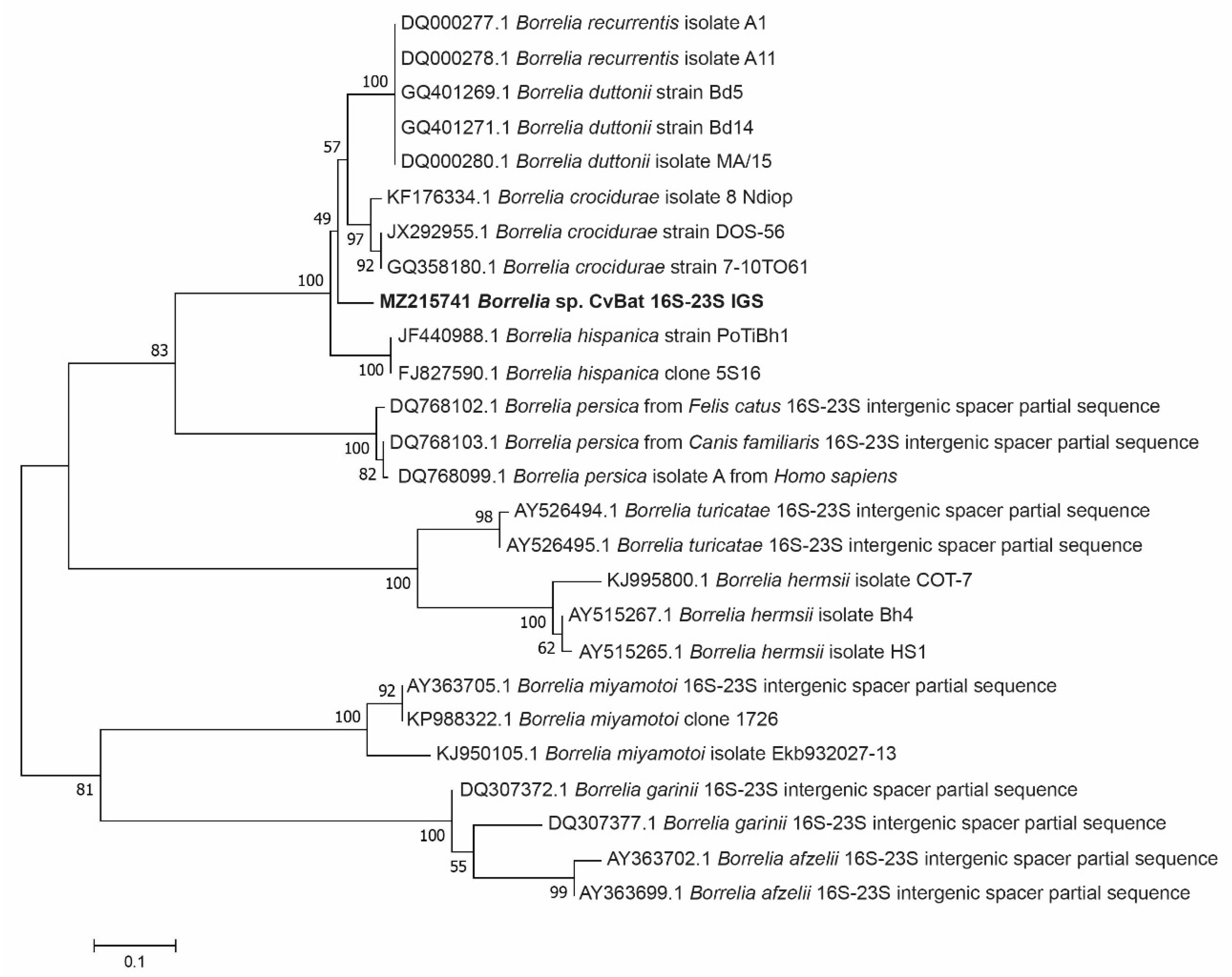
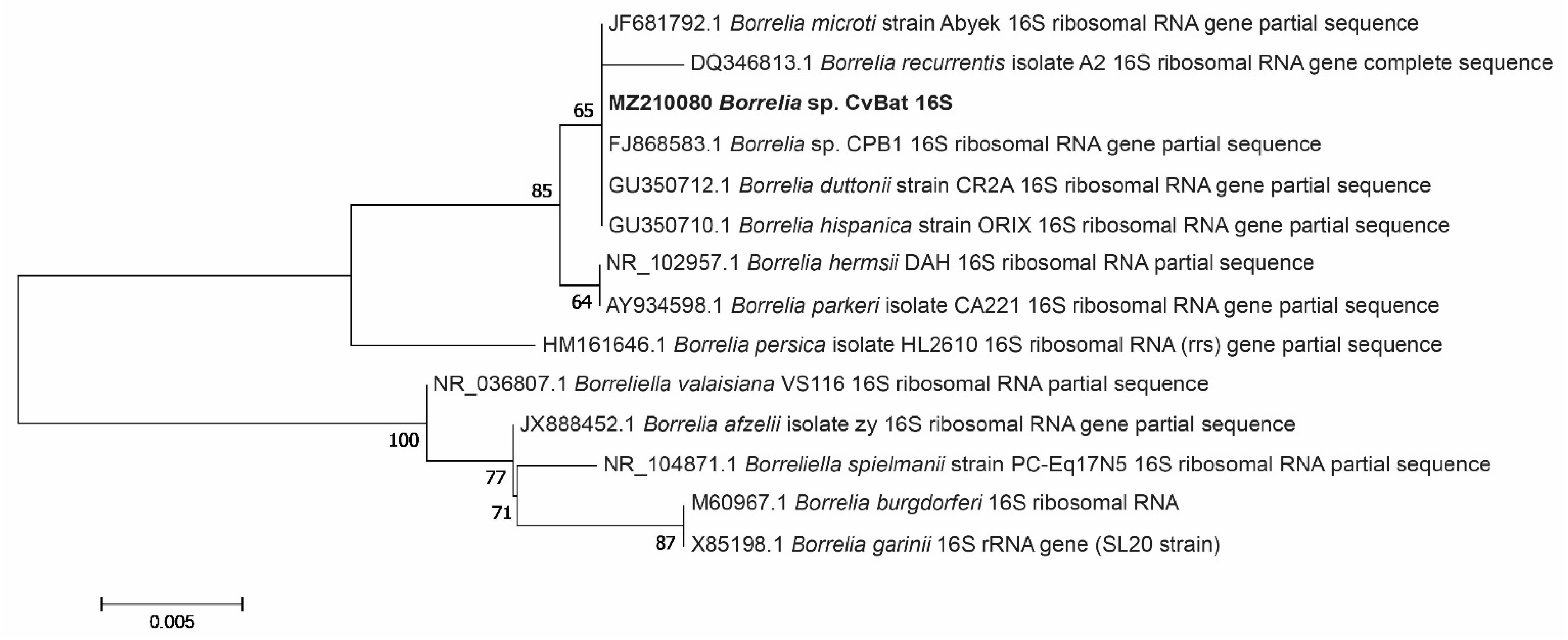
3.3. Phylogenetic Analysis of the Borrelia bacteria Detected in Ticks

Of all ticks tested, including the tick from Småland, 22 ticks were positive for genus-specific Borrelia DNA by real-time PCR analysis. Attempts to amplify the 5S-23S rRNA IGS from the positive samples were unsuccessful (Table 2).
Amplification of the 16S-23S rRNA IGS, on the other hand, was successful in 11 samples, including the adult tick from Småland (Table 2). Subsequent sequencing of these amplicons showed that they were 100% identical to each other and will henceforth be collectively referred to as ‘’MZ215741 Borrelia sp. CvBat 16S-23S IGS’’ in Figure 2. The closest sequence to ‘’MZ215741 Borrelia sp. CvBat 16S-23S IGS’’, available in GenBank, was that of Borrelia crocidurae (accession no. CP003426), which showed 90.4% (482/533 bp) sequence identity.
Amplification and sequencing of the flaB gene amplicon was successful in 6 samples, including the adult tick from Småland (Table 2). All sequences, referred to as ‘‘MZ217187 Borrelia sp. CvBat flaB’’ in Figure 3, were identical to each other and showed a 100% sequence identity (649/649) with the sequence of Borrelia sp. CPB1 (accession no. FJ868584).
Amplification and sequencing of the 16S rRNA gene amplicon was successful in 4 samples (analysis of the adult tick from Småland was, however, not successful) (Table 2). All sequences, referred to as ‘‘MZ210080 Borrelia sp. CvBat 16S’’ in Figure 4, were identical to each other and showed 100% sequence identity (353/353) with those of Borrelia sp. CPB1 (accession no. FJ868583), B. hispanica (accession no. GU350710), B. duttonii (accession no. GU350712) and B. microti (accession no. JF681792).
Samples that resulted in a successful sequencing of the 16S-23S rRNA IGS had significantly lower Cq-values (n = 11, median 25.9, IQR 23.4–29.4) in the real-time PCR assay compared to samples that failed to produce PCR products with the 16S-23S IGS assay (n = 11, median 32.4, IQR 31.1–39.7, p = 0.003).

3.4. Prevalence of B. miyamotoi in the Ticks

All ticks, including the adult tick from Småland, were negative for B. miyamotoi.

4. Discussion

To our knowledge, this is the first study identifying Borrelia bacteria in ticks that have fed on blood from bats in Sweden. We recorded a high prevalence of Borrelia bacteria in populations of C. vespertilionis ticks. The phylogenetic analyses of ribosomal RNA genes and flagellin gene indicate that the bacteria belong to the relapsing fever group of borreliae; some of them appear to be identical to Borrelia sp. CPB1. Borrelia sp. CPB1 has been suspected to have caused a lethal Borrelia infection in a bat specimen in the United Kingdom [24]. The same Borrelia species was later identified in C. vespertilionis collected in a bat-infested house in France [23]. There is, to the best of our knowledge, no previous Swedish record of any Borrelia species from this tick species or from any bat species in Sweden. Our results also indicate a temporal and spatial distribution of this Borrelia species. It was detected in ticks collected at two locations separated by a distance of 480 km, and in ticks collected both in 2015 and 2018 and in 2019.
Except for one record of a specimen of Otobius megnini, the bat tick C. vespertilionis is the only argasid tick species present in Sweden and also the only bat-associated tick species recorded in Sweden [16]. Carios vespertilionis is widely distributed in Europe, southern Asia and North Africa, and all active stages are blood-feeding ectoparasites of bats of many different genera [34]. It inhabits locations where contact with humans may occur. Occasionally birds are hosts and sometimes even humans and dogs may be bitten [12,16,17,19,34]. The Borrelia spirochaetes are considered to have evolved as symbionts of ticks, particularly Argasidae, and are now endoparasites in mammals and birds, which act as amplifiers after having been bitten by infectious ticks [12].
All bat ticks investigated by us were negative for B. miyamotoi. However, B. miyamotoi, which is a member of the relapsing fever complex of Borrelia species, is present in Sweden [44,45]. B. miyamotoi was detected first in ixodid ticks in Japan [46]. Later it was revealed by Platonov et al. (2011) in Russia that this species is a human pathogen [47]. It is also present as a human pathogen in the United States and in Europe, including Sweden [48]. Contrary to the relapsing fever group of borreliae, which are vectored by argasid ticks, B. miyamotoi is transmitted by Ixodes ticks—in Sweden by I. ricinus and presumably by I. persulcatus [42,49]. Transovarial (vertical) transmission from female tick to her offspring and transstadial survival of the spirochaetes from larva to nymph to adult are common traits for B. miyamotoi in the Ixodes populations. It is possible that this new Borrelia sp. CPB1 is transovarially transmitted. There are at least two reasons for this: first, many other relapsing fever group borreliae are transovarially transmitted; second, the presence of a relatively high infection rate in all active stages—larvae, nymphs and adults—may indicate transstadial transmission. In contrast, the B. burgdorferi s.l. species are rarely if ever transovarially transmitted. Seven species in the B. burgdorferi s.l complex (B. afzelii, B. burgdorferi sensu stricto, B. garinii, B. lusitaniae, B. spielmanii, B. turdi and B. valaisiana) are present in the Swedish population of I. ricinus [37,45,50]. None of these species were detected in any of the C. vespertilionis ticks investigated in the present study.
Borrelia, Rickettsia, Ehrlichia, Bartonella, Coxiella and Babesia species have previously been molecularly identified in C. vespertilionis from Europe [18,22,23,26,27,29]. It is often suggested that these putatively zoonotic microbes are vector-borne, i.e., tick-borne pathogens, presumably causing disease in bats and/or humans. However, this cannot be taken for granted. That a microorganism is tick-transmitted is doubtful if the tick specimen from which the DNA of the microbe was identified contained host blood. Nearly all of the ticks investigated by us contained visible host blood. Thus, our investigation neither shows that the Borrelia species found by us is tick-transmitted nor that it is pathogenic to bats or humans. While B. miyamotoi spirochaetes are transmitted to vertebrate hosts by the tick’s salivary fluid, other Borrelia relapsing fever spirochaetes are usually transmitted either in salivary fluid during blood ingestion by infectious larvae, nymphs and adult argasid ticks or in coxal fluid during and after feeding by nymphs and adult ticks. Since argasid larvae do not have any functioning coxal glands they can only transmit their relapsing fever spirochaetes via salivary fluid during blood feeding.
In the present study, a relatively large number of our Borrelia-positive specimens were not possible to identify by species; if they had been possible to identify, it is likely that some of them might have been Borrelia sp. CPB1. In 50% of the Borrelia-positive specimens as detected by the genus-specific quantitative PCR assay, attempts to amplify genes by conventional PCR assays were unsuccessful. These ticks contained, in general, a lower amount of Borrelia bacteria as indicated by a significantly higher Cq-value, compared to ticks containing a typeable Borrelia species. This may, at least partly, explain why PCR products, used to determine Borrelia species, were not amplified in the conventional PCR assays. There are two reasons why we used total nucleic acid (cDNA) and not DNA as template for PCR amplification: first, the use of cDNA in 16S rRNA based PCR-assays is more sensitive for detecting Borrelia than assays using DNA [45,51,52]; second, it allows us to investigate the presence of potential tick and/or bat associated RNA-viruses in the same material, which is planned to be done in subsequent work.
Deeper knowledge about the biology of C. vespertilionis and its role as a reservoir and vector of potentially harmful microorganisms is necessary to better understand the biology of many bat species and to mitigate and control potential zoonoses associated with this tick species. It is important to investigate the biology of the Borrelia species CPB1, its pathogenic potential to bats and humans, its geographic distribution, its host associations and the possibility of it being transmitted horizontally and/or vertically by C. vespertilionis and by other tick species such as I. ricinus and/or I. vespertilionis.

5. Conclusions

The examined tick populations had a high prevalence of species belonging to the relapsing fever Borrelia complex, but no species in the B. burgdorferi sensu lato complex were detected. The potential of C. vespertilionis as a reservoir and vector of Borrelia bacteria and other microorganisms needs to be further investigated.

Supplementary Materials

The following are available online at https://0-www-mdpi-com.brum.beds.ac.uk/article/10.3390/microorganisms9051100/s1, Document S1: Aligned Borrelia nucleotide sequences based on PCR-products.

Author Contributions

T.G.T.J. and P.W. planned the study. T.G.T.J. morphologically determined species and developmental stage of the ticks. P.W. developed the flagellin B gene (flaB) conventional PCR assay, photographed and measured the ticks, performed the laboratory analyses and processed the data. P.W. and T.G.T.J. interpreted the data and wrote the paper. All authors have read and agreed to the published version of the manuscript.

Funding

This study was supported by Carl Tryggers stiftelse; Helge Ax:son Johnsons stiftelse; Längmanska kulturfonden; Magnus Bergvalls stiftelse; the Medical Research Council of Southeast Sweden (FORSS, 931010); and the Division of Laboratory Medicine, Region Jönköping County. The work was carried out under the auspices of ESGBOR (the European Study Group on Lyme Borrelioses) and VectorNet, a European network for sharing data on the geographic distribution of arthropod vectors and transmitting human and animal disease agents (framework contract OC/EFSA/AHAW/2013/02-FWC1), funded by the European Food Safety Authority (EFSA) and the European Centre for Disease prevention and Control (ECDC).

Institutional Review Board Statement

Not applicable.

Informed Consent Statement

Not applicable.

Data Availability Statement

The data supporting the conclusions of this article are included within the article. Raw data can be shared with researchers upon a specific request. One additional file shows the aligned Borrelia sequences (Document S1).

Acknowledgments

We are most grateful to Lars-Eric Roxin, who constructed the bat box and collected the ticks at Snesslinge, Uppland in 2015 and 2018; to Elina Nordin, for skilful laboratory assistance; and to Håkan Lundkvist, for collecting the tick at Älmhult, Småland.

Conflicts of Interest

The authors declare no conflict of interest.

References

  1. Brook, C.E.; Dobson, A.P. Bats as ‘special’ reservoirs for emerging zoonotic pathogens. Trends Microbiol. 2015, 23, 172–180. [Google Scholar] [CrossRef]
  2. Leroy, E.M.; Kumulungui, B.; Pourrut, X.; Rouquet, P.; Hassanin, A.; Yaba, P.; Délicat, A.; Paweska, J.T.; Gonzalez, J.-P.; Swanepoel, R. Fruit bats as reservoirs of Ebola virus. Nature 2005, 438, 575–576. [Google Scholar] [CrossRef] [PubMed]
  3. Li, W.; Shi, Z.; Yu, M.; Ren, W.; Smith, C.; Epstein, J.H.; Wang, H.; Crameri, G.; Hu, Z.; Zhang, H.; et al. Bats are natural reservoirs of SARS-like coronaviruses. Science 2005, 310, 676–679. [Google Scholar] [CrossRef] [PubMed]
  4. Dobson, A.P. Virology: What links bats to emerging infectious diseases? Science 2005, 310, 628–629. [Google Scholar] [CrossRef] [PubMed]
  5. Williams, C.J. Bats as the reservoir for outbreaks of emerging infectious diseases. Eurosurveillance 2005, 10, E051110.4. [Google Scholar] [CrossRef] [PubMed]
  6. Shi, J.; Hu, Z.; Deng, F.; Shen, S. Tick-Borne Viruses. Virol. Sin. 2018, 33, 21–43. [Google Scholar] [CrossRef] [Green Version]
  7. Streicker, D.G.; Gilbert, A.T. Contextualizing bats as viral reservoirs. Science 2020, 370, 172–173. [Google Scholar] [CrossRef]
  8. Wilson, D.E.; Mittermeier, R.A. Handbook of Mammals of the World, Volume 9—Bats; Lynx Edicions: Barcelona, Spain, 2019; 1008p, ISBN 9788416728190. [Google Scholar]
  9. Swedish Environmental Protection Agency. Fladdermusarter i Sverige [Bat Species in Sweden]. Available online: http://www.naturvardsverket.se/Var-natur/Djur-och-vaxter/Rad/Fladdermossen-i-Sverige/Fladdermusarter-i-Sverige/ (accessed on 28 April 2021).
  10. Lourenço, S.I.; Palmeirim, J.M. Influence of temperature in roost selection by Pipistrellus pygmaeus (Chiroptera): Relevance for the design of bat boxes. Biol. Conserv. 2004, 119, 237–243. [Google Scholar] [CrossRef]
  11. Ahlén, I. Fladdermusfaunan i Sverige—Arternas utbredning och Status. Kunskapsläget 2011. [Summary: The Bat fauna in Sweden. Present knowledge on distribution and status]. Fauna Flora 2011, 106, 2–19. [Google Scholar]
  12. Hoogstraal, H. Argasid and Nuttalliellid ticks as parasites and vectors. Adv. Parasitol. 1985, 24, 135–238. [Google Scholar] [CrossRef]
  13. Arthur, D.A. British Ticks; Butterworths: London, UK, 1963. [Google Scholar]
  14. Estrada-Peña, A.; Mihalca, A.; Petney, T. Ticks of Europe and North Africa: A Guide to Species Identification, 1st ed.; Springer: Cham, Switzerland, 2017. [Google Scholar]
  15. Hornok, S.; Szőke, K.; Tu, V.T.; Kontschán, J.; Takács, N.; Sándor, A.D.; Halajian, A.; Földvári, G.; Estók, P.; Plantard, O.; et al. Mitochondrial gene heterogeneity of the bat soft tick Argas vespertilionis (Ixodida: Argasidae) in the Palaearctic. Parasites Vectors 2017, 10, 109. [Google Scholar] [CrossRef] [PubMed] [Green Version]
  16. Jaenson, T.G.; Tälleklint, L.; Lundqvist, L.; Olsen, B.; Chirico, J.; Mejlon, H. Geographical distribution, host associations, and vector roles of ticks (Acari: Ixodidae, Argasidae) in Sweden. J. Med. Èntomol. 1994, 31, 240–256. [Google Scholar] [CrossRef] [PubMed]
  17. Péter, Á.; Barti, L.; Corduneanu, A.; Hornok, S.; Mihalca, A.D.; Sándor, A.D. First record of Ixodes simplex found on a human host, with a review of cases of human infestation by bat tick species occurring in Europe. Ticks Tick-Borne Dis. 2021, 12, 101722. [Google Scholar] [CrossRef] [PubMed]
  18. Hornok, S.; Szőke, K.; Görföl, T.; Foldvari, G.; Tu, V.T.; Takács, N.; Kontschán, J.; Sándor, A.D.; Estók, P.; Epis, S.; et al. Molecular investigations of the bat tick Argas vespertilionis (Ixodida: Argasidae) and Babesia vesperuginis (Apicomplexa: Piroplasmida) reflect “bat connection” between Central Europe and Central Asia. Exp. Appl. Acarol. 2017, 72, 69–77. [Google Scholar] [CrossRef] [PubMed]
  19. Hoogstraal, H. African Ixodoidea. Ticks of the Sudan (with special reference to Equatoria Province and with preliminary reviews of the genera Boophilus, Margaropus, and Hyalomma). In African Ixodoidea; United States Naval Medical Research Unit no. 3: Cairo, Egypt, 1956; Volume 1, pp. 1–1105. [Google Scholar]
  20. Shah, A.; Khan, M.; Iqbal, Z.; Sajid, M.; Akhtar, M. Some epidemiological aspects and vector role of tick infestation on layers in the Faisalabad district (Pakistan). World’s Poult. Sci. J. 2006, 62, 145–157. [Google Scholar] [CrossRef]
  21. Hubbard, M.J.; Baker, A.S.; Cann, K.J. Distribution of Borrelia burgdorferi s.l. spirochaete DNA in British ticks (Argasidae and Ixodidae) since the 19th Century, assessed by PCR. Med. Vet. Èntomol. 1998, 12, 89–97. [Google Scholar] [CrossRef]
  22. Zabashta, M.V.; Orlova, M.V.; Pichurina, N.L.; Khametova, A.P.; Romanova, L.V.; Borodina, T.N.; Zabashta, A.V. Participation of bats (Chiroptera, Mammalia) and their ectoparasites in circulation of pathogens of natural focal infections in the south of Russia. Èntomol. Rev. 2019, 99, 513–521. [Google Scholar] [CrossRef]
  23. Socolovschi, C.; Kernif, T.; Raoult, D.; Parola, P. Borrelia, Rickettsia, and Ehrlichia species in bat ticks, France, 2010. Emerg. Infect. Dis. 2012, 18, 1966–1975. [Google Scholar] [CrossRef]
  24. Evans, N.J.; Bown, K.; Timofte, D.; Simpson, V.R.; Birtles, R. Fatal borreliosis in bat caused by relapsing fever spirochete, United Kingdom. Emerg. Infect. Dis. 2009, 15, 1331–1333. [Google Scholar] [CrossRef]
  25. Lvov, D.K.; Karas, F.R.; Timofeev, E.M.; Tsyrkin, Y.M.; Vargina, S.G.; Veselovskaya, O.V.; Osipova, N.Z.; Grebenyuk, Y.I.; Gromashevski, V.L.; Steblyanko, S.N.; et al. “Issyk-Kul” virus, a new arbovirus isolated from bats and argas (Carios) vespertilionis (Latr., 1802) in the Kirghiz S.S.R. Arch. Gesamte Virusforsch. 1973, 42, 207–209. [Google Scholar] [CrossRef]
  26. Zhmaeva, Z.M.; Pchelkina, A.A.; Belashova, V.S. Spontaneous infection of Argas vespertilionis with Rickettsia burnetii in the south of Kazakhstan. Med. Parazitol. Mosk. 1966, 35, 595–596. [Google Scholar] [PubMed]
  27. Lv, J.; Fernández de Marco, M.D.M.; Goharriz, H.; Phipps, L.P.; McElhinney, L.M.; Hernández-Triana, L.M.; Wu, S.; Lin, X.; Fooks, A.R.; Johnson, N. Detection of tick-borne bacteria and babesia with zoonotic potential in Argas (Carios) vespertilionis (Latreille, 1802) ticks from British bats. Sci. Rep. 2018, 8, 1865. [Google Scholar] [CrossRef] [PubMed] [Green Version]
  28. Liu, X.; Yan, B.; Wang, Q.; Jiang, M.; Tu, C.; Chen, C.; Hornok, S.; Wang, Y. Babesia vesperuginis in Common Pipistrelle (Pipistrellus pipistrellus) and the bat soft tick Argas vespertilionis in the People’s Republic of China. J. Wildl. Dis. 2018, 54, 419–421. [Google Scholar] [CrossRef]
  29. Hornok, S.; Szőke, K.; Meli, M.L.; Sándor, A.D.; Görföl, T.; Estók, P.; Wang, Y.; Tu, V.T.; Kováts, D.; Boldogh, S.A.; et al. Molecular detection of vector-borne bacteria in bat ticks (Acari: Ixodidae, Argasidae) from eight countries of the Old and New Worlds. Parasites Vectors 2019, 12, 1–7. [Google Scholar] [CrossRef] [Green Version]
  30. Arthur, D.R.; Nuttall, G.H.F.; Robinson, L.E.; Warburton, C. Ticks, a Monograph of the Ixodoidea; Cambridge [England] University Press: Cambridge, UK, 1908. [Google Scholar]
  31. Hoogstraal, H. Bat ticks of the genus Argas (Ixodoidea, Argasidae), 3. The Subgenus Carios, A Redescription of A. (C.) vespertilionis (Latreille, 1802), and variation within an Egyptian population. Ann. Èntomol. Soc. Am. 1958, 51, 19–26. [Google Scholar] [CrossRef]
  32. Filippova, N.A. Argasid ticks (Argasidae). Fauna of the USSR; Paukoobraznye, Nauka: Moscow/Leningrad, Russia, 1966; Volume 4. [Google Scholar]
  33. Yamaguti, N. Ticks of Japan, Korea, and the Ryukyu Islands; Brigham Young University: Provo, UT, USA, 1971. [Google Scholar]
  34. Hillyard, P.D. Ticks of North-West Europe; Field Studies Council: Shrewsbury, UK, 1996. [Google Scholar]
  35. Mans, B.J.; Kelava, S.; Pienaar, R.; Featherston, J.; de Castro, M.H.; Quetglas, J.; Reeves, W.K.; Durden, L.A.; Miller, M.M.; Laverty, T.M.; et al. Nuclear (18S-28S rRNA) and mitochondrial genome markers of Carios (Carios) vespertilionis (Argasidae) support Carios Latreille, 1796 as a lineage embedded in the Ornithodorinae: Re-classification of the Carios sensu Klompen and Oliver (1993) clade into its respective subgenera. Ticks Tick-borne Dis. 2021, 12, 101688. [Google Scholar] [CrossRef]
  36. Gyllemark, P.; Wilhelmsson, P.; Elm, C.; Hoornstra, D.; Hovius, J.W.; Johansson, M.; Tjernberg, I.; Lindgren, P.E.; Henningsson, J.H.; Sjöwall, J. Are other tick-borne infections overlooked in patients investigated for Lyme neuroborreliosis? A large retrospective study from south-eastern Sweden. Ticks Tick Borne Dis. 2021. under review. [Google Scholar]
  37. Wilhelmsson, P.; Fryland, L.; Börjesson, S.; Nordgren, J.; Bergström, S.; Ernerudh, J.; Forsberg, P.; Lindgren, P.-E. Prevalence and diversity of Borrelia species in ticks that have bitten humans in Sweden. J. Clin. Microbiol. 2010, 48, 4169–4176. [Google Scholar] [CrossRef] [Green Version]
  38. Postic, D.; Assous, M.V.; Grimont, P.A.D.; Baranton, G. Diversity of Borrelia burgdorferi sensu lato evidenced by restriction fragment length polymorphism of rrf (5S)-rrl (23S) intergenic spacer amplicons. Int. J. Syst. Bacteriol. 1994, 44, 743–752. [Google Scholar] [CrossRef] [Green Version]
  39. Bunikis, J.; Garpmo, U.; Tsao, J.; Berglund, J.; Fish, D.; Barbour, A.G. Sequence typing reveals extensive strain diversity of the Lyme borreliosis agents Borrelia burgdorferi in North America and Borrelia afzelii in Europe. Microbiology 2004, 150, 1741–1755. [Google Scholar] [CrossRef] [PubMed] [Green Version]
  40. Bunikis, J.; Tsao, J.; Garpmo, U.; Berglund, J.; Fish, D.; Barbour, A.G. Typing of Borrelia relapsing fever group strains. Emerg. Infect. Dis. 2004, 10, 1661–1664. [Google Scholar] [CrossRef]
  41. Lambert, J.S.; Cook, M.J.; Healy, J.E.; Murtagh, R.; Avramovic, G.; Lee, S.H. Metagenomic 16S rRNA gene sequencing survey of Borrelia species in Irish samples of Ixodes ricinus ticks. PLoS ONE 2019, 14, e0209881. [Google Scholar] [CrossRef] [PubMed] [Green Version]
  42. Jaenson, T.G.T.; Wilhelmsson, P. First records of tick-borne pathogens in populations of the taiga tick Ixodes persulcatus in Sweden. Parasites Vectors 2019, 12, 559. [Google Scholar] [CrossRef] [Green Version]
  43. Hovius, J.W.; de Wever, B.; Sohne, M.; Brouwer, M.C.; Coumou, J.; Wagemakers, A.; Oei, A.; Knol, H.; Narasimhan, S.; Hodiamont, C.J.; et al. A case of meningoencephalitis by the relapsing fever spirochaete Borrelia miyamotoi in Europe. Lancet 2013, 382, 658. [Google Scholar] [CrossRef] [Green Version]
  44. Fraenkel, C.-J.; Garpmo, U.; Berglund, J. Determination of novel Borrelia genospecies in Swedish Ixodes ricinus ticks. J. Clin. Microbiol. 2002, 40, 3308–3312. [Google Scholar] [CrossRef] [PubMed] [Green Version]
  45. Wilhelmsson, P.; Lindblom, P.; Fryland, L.; Ernerudh, J.; Forsberg, P.; Lindgren, P.-E. Prevalence, diversity, and load of Borrelia species in ticks that have fed on humans in regions of Sweden and Åland Islands, Finland with different Lyme borreliosis incidences. PLoS ONE 2013, 8, e81433. [Google Scholar] [CrossRef] [Green Version]
  46. Fukunaga, M.; Koreki, Y. The flagellin gene of Borrelia miyamotoi sp. nov. and its phylogenetic relationship among Borrelia species. FEMS Microbiol. Lett. 1995, 134, 255–258. [Google Scholar] [CrossRef]
  47. Platonov, A.E.; Karan, L.S.; Kolyasnikova, N.M.; Makhneva, N.A.; Toporkova, M.G.; Maleev, V.V.; Fish, D.; Krause, P.J. Humans infected with relapsing fever spirochete Borrelia miyamotoi, Russia. Emerg. Infect. Dis. 2011, 17, 1816–1823. [Google Scholar] [CrossRef] [PubMed]
  48. Henningsson, A.J.; Asgeirsson, H.; Hammas, B.; Karlsson, E.; Parke, Å.; Hoornstra, D.; Wilhelmsson, P.; Hovius, J.W. Two cases of Borrelia miyamotoi meningitis, Sweden, 2018. Emerg. Infect. Dis. 2019, 25, 1965–1968. [Google Scholar] [CrossRef] [Green Version]
  49. Cutler, S.; Vayssier-Taussat, M.; Estrada-Peña, A.; Potkonjak, A.; Mihalca, A.D.; Zeller, H. A new Borrelia on the block: Borrelia miyamotoi—A human health risk? Eurosurveillance 2019, 24, 1800170. [Google Scholar] [CrossRef] [Green Version]
  50. Wilhelmsson, P.; Jaenson, T.G.T.; Olsen, B.; Waldenström, J.; Lindgren, P.-E. Migratory birds as disseminators of ticks and the tick-borne pathogens Borrelia bacteria and tick-borne encephalitis (TBE) virus: A seasonal study at Ottenby Bird Observatory in South-eastern Sweden. Parasites Vectors 2020, 13, 1–17. [Google Scholar] [CrossRef] [PubMed]
  51. Ornstein, K.; Barbour, A.G. A reverse transcriptase–polymerase chain reaction assay of Borrelia burgdorferi 16S rRNA for highly sensitive quantification of pathogen load in a vector. Vector-Borne Zoonotic Dis. 2006, 6, 103–112. [Google Scholar] [CrossRef] [PubMed]
  52. Lager, M.; Faller, M.; Wilhelmsson, P.; Kjelland, V.; Andreassen, Å.; Dargis, R.; Quarsten, H.; Dessau, R.; Fingerle, V.; Margos, G.; et al. Molecular detection of Borrelia burgdorferi sensu lato—An analytical comparison of real-time PCR protocols from five different Scandinavian laboratories. PLoS ONE 2017, 12, e0185434. [Google Scholar] [CrossRef] [PubMed] [Green Version]
Figure 1. Map showing the localities where ticks were collected. (A) 91 ticks were collected from a water-filled tray placed below a wooden bat box located at Snesslinge (60°19.567 N, 18°15.067 E) in the province of Uppland, south-central Sweden. (B) One tick was collected inside a house located at Älmhult (56°32.720 N, 13°52.667 E) in the province of Småland, southern Sweden. The distance between A and B is 483 km.
Microorganisms 09 01100 g001
Figure 2. Phylogenetic tree based on 16S-23S intergenic spacer region sequences of Borrelia species. Sequences detected in our study (‘MZ215741 Borrelia sp. CvBat 16S-23S IGS’, n = 11) are highlighted in bold.
Microorganisms 09 01100 g002
Figure 3. Phylogenetic tree based on flaB gene sequences of Borrelia species. Sequences detected in our study (‘‘MZ217187 Borrelia sp. CvBat flaB’’, n = 6) are highlighted in bold.
Microorganisms 09 01100 g003
Figure 4. Phylogenetic tree based on 16S rRNA gene sequences of Borrelia species. Sequences detected in our study (‘‘MZ210080 Borrelia sp. CvBat 16S’’, n = 4) are highlighted in bold.
Microorganisms 09 01100 g004
Table 1. Primers and probes used for molecular analysis of Borrelia bacteria.
OrganismTargetOligo NameSequence (5’→3’)Amplicon Length (bp)Reference
Borrelia spp.16S rRNABorrelia-FGCTGAGTCACGAAAGCGTAG116[36]
Borrelia-RCACTTAACACGTTAGCTTCGGTA
Borrelia-PFAM-CGCTGTAAACGATGCACACTTGGT-MGB
5S-23S rRNA IGSB5S-23S_FCTGCGAGTTCGCGGGAGA225–266 a[38]
B5S-23S_RTCCTAGGCATTCACCATA
B5S-23S_FnGAGTTCGCGGGAGAGTAA[37]
B5S-23S_RnTAGGCATTCACCATAGACTCTT
16S-23S rRNA IGSB16S-23S_FGTATGTTTAGTGAGGGGGGTG388–685 a[39,40]
B16S-23S_RGGATCATAGCTCAGGTGGTTAG
B16S-23S_FnAGGGGGGTGAAGTCGTAACAAG
B16S-23S_RnGTCTGATAAACCTGAGGTCGGA
flaBflaB-FCATCTGATGATGCTGCTGGT699This study
flaB-RTGTTTTGGAAAGCACCAAGA
flaB-FnGGGTGTTGCTGGGAAAATTA672
flaB-RnTGGAAAGCACCAAGATTTGC
16S rRNAM1ACGATGCACACTTGGTGTTAA357–358 a[41]
M2TCCGACTTATCACCGGCAGTC
B. miyamotoiflaBBm_FAGAAGGTGCTCAAGCAG156[42]
Bm_RTCGATCTTTGAAAGTGACATAT
Bm_PFAM-AGCACAACAGGAGGGAGTTCAAGC-BHQ1
a Amplicon length varies with the species. Abbreviations: FAM, 6-carboxy-fluorescine; MGB, minor groove binder; BHQ, black hole quencher; IGS, intergenic spacer.
Table 2. Number and prevalence of Borrelia bacteria detected in different stages of Carios vespertilionis.
Tick Developmental StageNo. of Ticks ExaminedNo. (%) of Borrelia-Positive Ticks by Real-Time PCRNo. of Specimens with Successful Amplification of Gene Targets by Conventional PCR
5S-23S IGS16S-23S IGSflaB16S rRNA
Larva3112 (38.7)0 432
Nymph487 (14.6)0 411
Adult13 a3 (23.1) a0 3 a2 a1
Total9222 (23.9)0 1164
a One adult C. vespertilionis tick in this group was collected in the province of Småland.
Publisher’s Note: MDPI stays neutral with regard to jurisdictional claims in published maps and institutional affiliations.

