Article

The Rhizobacterium Pseudomonas alcaligenes AVO110 Induces the Expression of Biofilm-Related Genes in Response to Rosellinia necatrix Exudates

by
Adrián Pintado
1,2,
Isabel Pérez-Martínez
1,2,
Isabel M. Aragón
1,2,
José Antonio Gutiérrez-Barranquero
2,3,
Antonio de Vicente
2,3,
Francisco M. Cazorla
2,3,* and
Cayo Ramos
1,2,*
1
Área de Genética, Facultad de Ciencias, Campus Teatinos, Universidad de Málaga, E-29010 Málaga, Spain
2
Departamento de Microbiología y Protección de Cultivos, Instituto de Hortofruticultura Subtropical y Mediterránea «La Mayora», Extensión Campus de Teatinos, Universidad de Málaga-Consejo Superior de Investigaciones Científicas (IHSM-UMA-CSIC), E-29010 Málaga, Spain
3
Departamento de Microbiología, Campus Teatinos, Universidad de Málaga, E-29010 Málaga, Spain
*
Authors to whom correspondence should be addressed.
Submission received: 25 May 2021 / Revised: 15 June 2021 / Accepted: 23 June 2021 / Published: 25 June 2021
(This article belongs to the Special Issue Plant-Associated Pseudomonads)

Abstract

:
The rhizobacterium Pseudomonas alcaligenes AVO110 exhibits antagonism toward the phytopathogenic fungus Rosellinia necatrix. This strain efficiently colonizes R. necatrix hyphae and is able to feed on their exudates. Here, we report the complete genome sequence of P. alcaligenes AVO110. The phylogeny of all available P. alcaligenes genomes separates environmental isolates, including AVO110, from those obtained from infected human blood and oyster tissues, which cluster together with Pseudomonas otitidis. Core and pan-genome analyses showed that P. alcaligenes strains encode highly heterogenic gene pools, with the AVO110 genome encoding the largest and most exclusive variable region (~1.6 Mb, 1795 genes). The AVO110 singletons include a wide repertoire of genes related to biofilm formation, several of which are transcriptionally modulated by R. necatrix exudates. One of these genes (cmpA) encodes a GGDEF/EAL domain protein specific to Pseudomonas spp. strains isolated primarily from the rhizosphere of diverse plants, but also from soil and water samples. We also show that CmpA has a role in biofilm formation and that the integrity of its EAL domain is involved in this function. This study contributes to a better understanding of the niche-specific adaptations and lifestyles of P. alcaligenes, including the mycophagous behavior of strain AVO110.

1. Introduction

In recent years, multitrophic interactions have gained the attention of many research groups because of their importance in plant disease development and the assembly of root-associated microbiomes [1,2,3,4,5]. These interactions modulate the colonization and establishment of microorganisms in different environments and ecological niches [6], including the plant rhizosphere [7]. The rhizosphere is considered to be the portion of soil intimately associated with roots and has been described as one of the most diverse and complex soil environments on the Earth [7,8]. The rhizosphere is strongly influenced by plant root exudates [9], and root exudates are considered to be the major nutrient source for microorganisms in this environment, influencing the assembly of specific fungal and bacterial communities [7,10,11]. Additionally, certain microorganisms can obtain nutrients from living fungi, a process that has been defined as mycophagy [12]. In particular, bacterial mycophagy was first demonstrated in a soil bacterium of the Collimonas genus [13], which became the model bacterium for disentangling the mechanisms governing this feature [14,15,16,17,18,19,20]. Although only a few soil bacteria present this feeding ability [12], a more recent study reported that a substantial number of rhizobacteria are able to feed on fungal exudates [21].
Pseudomonas alcaligenes (formerly Pseudomonas pseudoalcaligenes) AVO110, an efficient avocado root tip colonizer, exhibits in vitro and in planta antagonistic activity toward the soil-borne phytopathogenic fungus Rosellinia necatrix [22]. P. alcaligenes AVO110 is able to feed on R. necatrix exudates and profusely colonize its hyphae without lysing host cells [1], providing evidence for the mycophagous ability of this bacterium as an extracellular biotroph. In addition, P. alcaligenes AVO110 forms biofilms on R. necatrix mycelia and avocado root surfaces [1,22]. Prior to biofilm development on plant roots, competitive rhizosphere colonization takes place, a process involving bacterial motility [23,24]. However, only a few studies have addressed the identification of bacterial genes involved in interactions with fungal phytopathogens [16,20,25,26,27].
In this sense, a recent study identified several molecular mechanisms linked to the mycophagous behavior of P. alcaligenes AVO110 toward R. necatrix exudates [28]. Using signature-tagged mutagenesis (STM) [29], these authors identified a collection of AVO110 genes required for growth and survival in R. necatrix exudates, several of which are biofilm related. For example, the algQ gene, which encodes a regulator of alginate production, was identified as being necessary for efficient colonization of the avocado rhizosphere. Production of the exopolysaccharide (EPS) alginate has been shown to influence biofilm architecture in Pseudomonas aeruginosa [30]. In addition, another gene identified in that study encoded a GGDEF/EAL domain protein, named CmpA, from cyclic-di-GMP (c-di-GMP)-metabolizing protein. This gene was shown to be involved in the colonization of both the avocado rhizosphere and fungal hyphae. Among other genes, transcription of cmpA has been shown to be induced in response to R. necatrix exudates [28]. GGDEF and EAL (or HY-GYP) protein domains are involved in the turnover of c-di-GMP, a universal intracellular second messenger mediating phenotypic changes related to the transition between sessility and motility, such as the production of extracellular matrix components (EPSs and proteins), biofilm formation, and bacterial motility. Cellular c-di-GMP levels have been related to the regulatory network that controls root colonization in plant-associated rhizobacteria [31,32] and virulence traits in bacterial phytopathogens [33,34,35,36]. The GGGDEF and EAL domains are encoded in enzymes responsible for the synthesis (diguanylate cyclases, DGCs) and breakdown (phosphodiesterases, PDEs) of c-di-GMP, respectively [37,38,39].
In this study, we obtained the complete genome sequence of P. alcaligenes AVO110 and performed a comparative analysis with the genomes of related pseudomonads of the P. aeruginosa and P. oleovorans groups. The genome of P. alcaligenes AVO110 encodes a variable region of approximately 1.6 Mb (1795 genes), which is absent in all other sequenced P. alcaligenes strains. Remarkably, this exclusive region encodes a broad collection of genes related to bacterial lifestyle transitions, such as flagellar motility, chemotaxis, adhesion, biofilm formation, and colonization of host surfaces, 25 of which encode GDEEF and/or EAL domain proteins. Annotation of the P. alcaligenes AVO110 genome, followed by transcriptional analyses, allowed us to identify several genes transcriptionally modulated by R. necatrix exudates, some of which are encoded in its variable genomic region and are related to cell adhesion and biofilm formation. We also showed that CmpA had a role in biofilm formation and that the integrity of its EAL domain was required for this function.

2. Materials and Methods

2.1. Bacterial Strains, Media, and Growth Conditions

The bacterial strains used in this study are listed in Table 1. Pseudomonas and Escherichia coli strains were grown in lysogeny broth (LB) medium [40] or super optimal broth (SOB) [41] at 28 and 37 °C, respectively. BM minimal medium [42] was used for growth of R. necatrix Rn400 mycelia to obtain fungal exudate-containing medium (BM-RE, pH 7.1), as previously described [1,28]. When necessary, solid and liquid media were supplemented with antibiotics as follows: for E. coli, ampicillin (Ap) 100 µg mL−1, kanamycin (Km) 50 µg mL−1, and gentamicin (Gm) 50 µg mL−1 final concentration; for P. alcaligenes AVO110, Ap 300 µg mL−1, Km 25 µg mL−1, and Gm 10 µg mL−1; and for P. putida KT2440, Km 50 µg mL−1 and Gm 50 µg mL−1.

2.2. Construction of Bacterial Strains and Plasmids

All recombinant DNA techniques, including restriction digestion, agarose gel electrophoresis, purification of DNA fragments, and ligation with T4 DNA ligase, were performed as previously described [47]. Plasmids were purified using the GenEluteTM Plasmid Miniprep kit (Sigma-Aldrich, Burlington, VT, USA) and, when necessary, sequenced by STAB VIDA Lda. (Caparica, Portugal). DNA amplifications were performed by polymerase chain reaction (PCR) with a standard enzyme (GoTaq® Flexi DNA Polymerase, Promega, Madison, WI, USA) or, for cloning, with Expand High Fidelity polymerase (Roche Applied Science, Mannheim, Germany).
The plasmids and oligonucleotides used for plasmid construction and mutagenesis are listed in Table 1 and Supplementary Materials Table S1, respectively. Construction of the P. alcaligenes AVO110 ΔcmpA mutant was performed by marker exchange mutagenesis as follows: First, DNA fragments of approximately 1 kb corresponding to the 5′ and 3′ flanking regions of cmpA were amplified by PCR using appropriate primers and P. alcaligenes AVO110 genomic DNA as a template. The PCR products were joined by the BamHI restriction sites included in the primer sequence, and after cloning the resulting fragment into pBluescript SK II using EcoRI/NotI sites, the fragment was sequenced to verify the absence of mutations. Then, the plasmid was labeled with the nptII Km resistance gene obtained from pGEM-T-KmFRT-BamHI, yielding pBSKII-ΔcmpA-Km. For marker exchange mutagenesis, plasmid pBSKII-ΔcmpA-Km was transformed by electroporation into AVO110, as previously described [48]. Transformants were selected on LB medium containing Km, and replica plates of the resulting colonies were generated on LB-Ap plates to determine whether each transconjugant underwent plasmid integration (Ap-resistant, ApR) or allelic exchange (Ap-sensitive, ApS). Southern blot analysis was used to confirm that the allelic exchange occurred at a single site and at the correct position within the genome.
To overexpress the P. alcaligenes AVO110 cmpA gene, a 3.6 kb PCR fragment encoding the complete cmpA ORF and its ribosome binding site was cloned into pGEM-T Easy and sequenced. The resulting plasmid (pGEM-T-cmpA) was digested with EcoRI and SacI, and the EcoRI/SacI fragment encoding cmpA was subcloned into the broad host range vector pAMEX, yielding pAMEX-cmpA. Site-directed mutagenesis of the GGDEF (pAMEX-cmpA-GGAAF) and EAL (pAMEX-cmpA-AAL) sites of cmpA was performed on pAMEX-cmpA using the QuickChange II Site-Directed Mutagenesis kit (Stratagene, San Diego, CA, USA), following the supplier’s instructions.

2.3. Complete Sequencing of the P. alcaligenes AVO110 Genome

P. alcaligenes AVO110 was grown overnight in LB medium and genomic DNA was extracted using a JetFlex genomic DNA purification kit (Genomed GmbH, Löhne, Germany). The sample was further purified by extraction with phenol–chloroform. Genome sequencing and assembly was performed at BGI Tech Solutions Co., Ltd. (Hong Kong). A library of randomly sheared DNA fragments (0.5–2.0 kb) was subjected to Illumina GA II (Solexa) sequencing. Reads (coverage 100×) were qualitatively assessed before assembling with SOAPdenovo [49]. Primer walking and PCR amplification were used to fill the remaining gaps and solve misassembled regions. The genome (accession number LZEU00000000) was automatically annotated upon submission to GenBank at the National Center for Biotechnology Information (NCBI).

2.4. Bioinformatics Methods

Core and pan-genome analyses were performed using BPGA v1.3 [50] with assemblies downloaded from the NCBI. Orthologous genes were identified using the USEARCH algorithm [51] with a threshold of 0.9 (90% Blastp identity). To determine whether pan-genomes were open or closed, we used the medians of the total number of genes found, and then the curves were fitted to Heaps’ law model [52]. The core and pan-genomes of these assemblies were also estimated using Roary v3.12.0 [53] with ≥90% Blastp identity. Phylogenetic relationships were predicted using the estimated core genomes with ≥90% Blastp. Sequences were aligned using MUSCLE and a tree was built using the maximum likelihood method with 100 bootstraps within MEGA 7 [54]. The tree was rooted using the genome of P. putida KT2440 as an outgroup.
Variable genomic regions in P. alcaligenes strains were identified with the GView Pangenome Analysis tool [55] using P. alcaligenes NEB 858 as the seed and Blastn with e-value ≤ 1 × 10⁻¹⁰ and ≥90% identity. Singleton coding sequences (CDSs) of P. alcaligenes AVO110 were extracted and annotated using Sma3s.v2 software [56].
CmpA homologs were identified by Blastp analysis using the P. alcaligenes AVO110 amino acid sequence as a query. Identification of conserved protein domains was performed with the Pfam database (Pfam 34.0, http://pfam.xfam.org/, accessed on 30 March 2021).

2.5. RNA Techniques

For the RNA-seq analysis, P. alcaligenes AVO110 was grown overnight in LB. The next day, the cells were diluted in 100 mL of LB medium to an OD600 of 0.05 and grown to an OD600 of 0.5. Cells were washed with NaCl 0.9% 3 times and inoculated in 100 mL of BM-RE medium [1] to start the induction. Two samples of 25 mL each were extracted from this volume, and RNA extraction was carried out for one sample at time zero and the other after 4 h of incubation in BM-RE medium. Total RNA was extracted using TriPure isolation reagent (Roche Applied Science) according to the manufacturer’s instructions, except that the TriPure was preheated to 65 °C and the lysis step was performed at 65 °C. Total RNA was cleaned up using an RNAeasy kit (Qiagen GmbH, Hilden, Germany), as detailed by the manufacturer. RNA concentration was determined spectrophotometrically, and its integrity was assessed by agarose gel electrophoresis. Total RNA was treated with a Turbo DNA-free kit (Applied Biosystems, Foster City, CA, USA), as detailed by the manufacturer’s instructions. rRNA depletion using an Illumina RiboZero RNA removal kit, library construction using an Illumina TrueSeq Stranded Sample Preparation kit, Illumina HiSeq 2500 sequencing, and bioinformatics analysis were performed by ChunLab Inc. (Seoul, Korea). Data of differentially expressed genes, provided as CLT format files, were viewed and visualized using Chunlab’s CLRNAseq software (Seoul, Korea).
For the quantitative real-time PCR (qRT-PCR), the induction of P. alcaligenes AVO110 cells in BM-RE medium and RNA extraction and purification were performed, as described above for RNA-seq analysis. RNA was extracted from three 25 mL samples at time zero and after 4 h of incubation in BM-RE medium. DNA-free total RNA was reverse transcribed using the cDNA Reverse Transcription kit (Applied Biosystems, Foster City, CA, USA) and random hexamers. The primer efficiency tests, qRT-PCR, and confirmation of the specificity of amplification reactions were performed, as previously described [57]. The relative transcript abundance was calculated using the ΔΔ cycle-threshold (Ct) method [58]. Target cDNAs from the experimental samples were PCR-amplified using 0.3 μM of each primer (Table S1). Transcriptional data were normalized to the housekeeping gene rpoD. The qRT-PCR values are the means from 3 biological replicates with 3 technical replicates ± standard deviation.

2.6. Biofilm Assays

Biofilm formation by P. putida KT2440 overexpressing the wild-type cmpA gene from P. alcaligenes AVO110 or its mutated alleles, cmpA-GGAAF and cmpA-AAL, was carried out as previously described [59,60]. P. putida KT2440 was transformed by electroporation [48] with pAMEX (empty vector), pAMEX-cmpA, pAMEX-cmpA-GGAAF, or pAMEX-cmpA-AAL (Table 1). Transformants were grown overnight in LB-Km medium at 25 °C. The cultures were prediluted in LB to an OD600 of 0.1 and subjected to six 10-fold serial dilutions. Samples (150 µL) of the diluted suspensions were deposited in the wells of a microtiter dish, and the plates were incubated for 20 h at 25 °C with moderate shaking (150 rpm). Wells were subsequently emptied by inverting the plates onto filter paper and washed 3 times by soaking the plates in water. The plates were dried on filter paper and the attached biofilms were stained for 15 min with 200 μL of 0.1% crystal violet. The wells were emptied and again washed 3 times to remove unbound dye. Finally, the wells were filled with 200 μL of 96% ethanol and the plates were incubated for 20 min at room temperature with vigorous shaking (600 rpm). Biofilm growth was determined from the A630 reading using a plate reader (FL600 Fluorescence Microplate Reader; Bio-Tek, Winooski, VT, USA).

3. Results

3.1. Sequencing and General Features of P. alcaligenes AVO110 Genome

The sequencing of the P. alcaligenes AVO110 genome yielded a large 4.95 Mb contig encoding 4474 genes and two additional small contigs of 15.6 and 5.8 kb encoding only 13 and 5 genes, respectively. No plasmid-related genes were found among the annotated products of these two small contigs.
The total sequence length of the genome (4.97 Mb) and its number of coding sequences (4406) are within the range of the other 10 P. alcaligenes and two P. pseudoalcaligenes genomes currently available at NCBI, two of which correspond to the P. alcaligenes type strain deposited in two collections (NCTC 10367 and NBRC 14159). However, the 11 P. alcaligenes genomes showed a wide range of sizes ranging from 3.68 Mb (strain Bin_52_1) to 7.03 Mb (strain OT 69). Noticeably, the genomes of P. alcaligenes strains isolated from infected human blood (MRY13-0052) or oyster tissues (OT 69) were, on average, approximately 2.5 Mb larger than those of strains isolated from water (six strains from wastewater, swimming pools, and aquatic environment of duckweeds) or rhizosphere soil (strain AVO110). Nevertheless, the total G+C content of the P. alcaligenes AVO 110 genome (64.9%) was similar to that of the other 10 P. alcaligenes genomes (64.1–66.5%) but differed from the approximately 62% G+C characteristics of P. pseudoalcaligenes (Table 2).

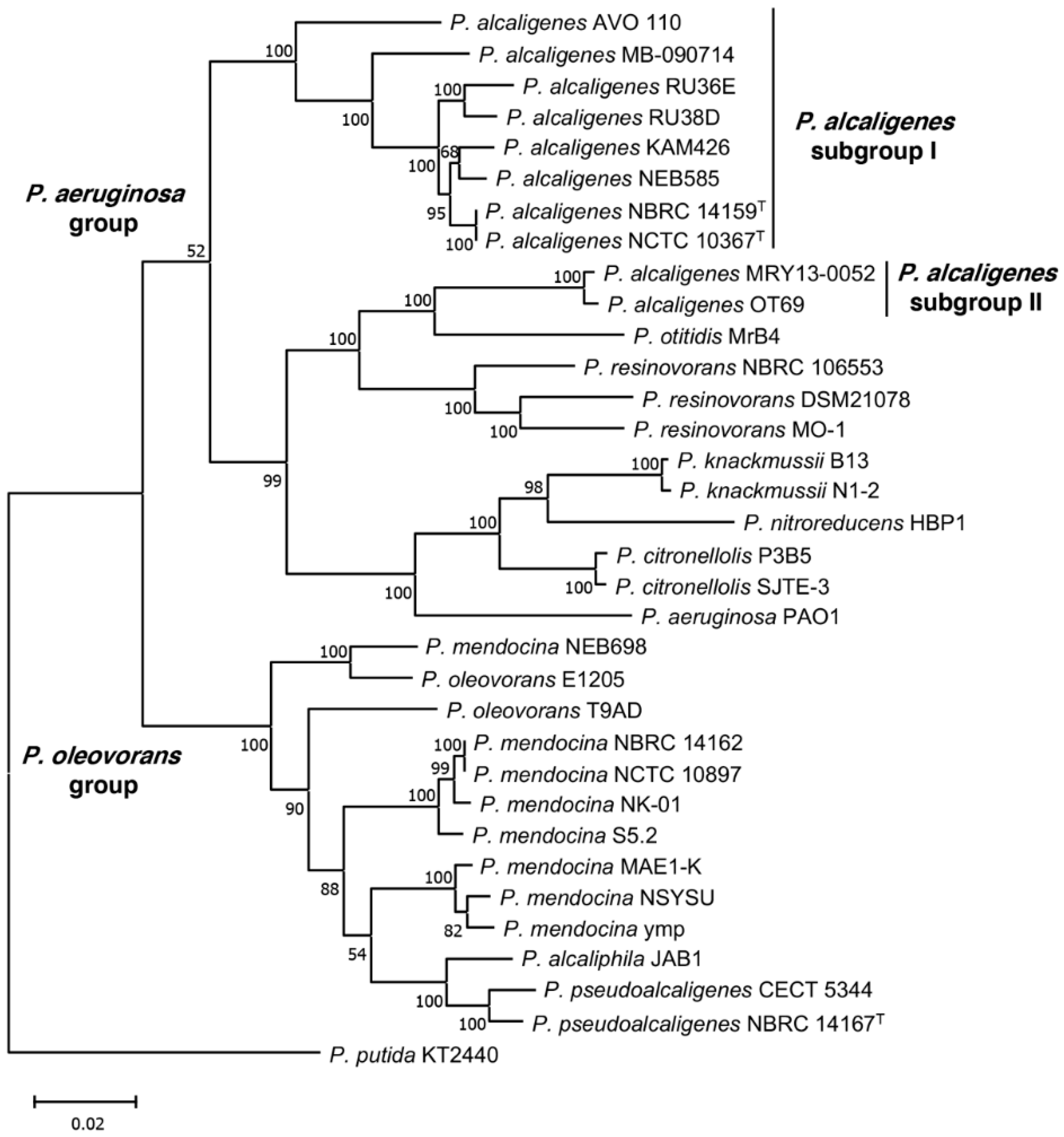
3.2. Phylogenetic Analysis of P. alcaligenes and P. pseudoalcaligenes Strains

To further study the inclusion of AVO110 in the species P. alcaligenes, we analyzed the core genome phylogeny of this strain and 34 other pseudomonads belonging to the P. aeruginosa and P. oleovorans groups, including, among others, P. alcaligenes and P. pseudoalcaligenes, respectively [61,62]. The tree was rooted using the genome of P. putida KT2440 as an outgroup. The genome sequence of P. alcaligenes Bin_52_1, which is fragmented into a large number of scaffolds (Table 2), was not included in this analysis or in the comparative genomic analysis described below. The phylogeny showed separation of the strains into two monophyletic branches corresponding to the P. aeruginosa and P. oleovorans groups. All P. alcaligenes strains were included in the P. aeruginosa group. However, while P. alcaligenes AVO110 grouped in a monophyletic branch with the P. alcaligenes strain NBRC 14159 (NCTC 10367) and all other P. alcaligenes strains isolated from water samples (P. alcaligenes subgroup I), P. alcaligenes strains isolated from blood and oyster infections were included in a different sub-branch of the tree (P. alcaligenes subgroup II) together with P. otitidis and P. resinovorans (Table 2). The P. pseudoalcaligenes strains analyzed were both included in the P. oleovorans group, which also included P. mendocina strains and P. alcaliphila JAB1 (Figure 1).
Table 2. General genome features of Pseudomonas strains analyzed in this work.
Strain aSubgroup bGenome Size (Mb)G+C
Content (%)
Coding SequencesScaffolds (Number)CoverageSource of
Isolation
Year of IsolationAccession Number cReference or Sequence
Source/Year
Pseudomonas alcaligenes
NCTC 10367 TI5.0264.545942100×Swimming pool water1961UGUP00000000[63]
NBRC 14159 TI4.8264.84445122131×Swimming pool water1961BATI00000000[63]
NEB 585I4.4165.540721382×Water sample1989CP014784R. D. Morgan/2016
AVO110I4.9764.944063100×Avocado rhizosphere2005LZEU00000000This study
MB-090714I4.0166.538151692.7×Water (lake)2009QJRX00000000M. Batrich/2018
Bin_52_1I3.6864.63700325978.2×Water purification facility2017SSFO00000000B.W. Stamps/2018
KAM 426I4.6864.84363193×Wastewater2020AP024354M. Suzuki/2021
RU38DI4.2565.1392010262×Water (duckweeds)NAFTNW00000000S. Lebeis /2016
RU36EI4.6364.1424623221×Water (duckweeds)NAFTMP00000000S. Lebeis /2016
OT 69II7.0366.06379223200×Oyster tissue2013ATCP00000000[64]
MRY13-0052II6.8865.8612923716.1×Blood infection2013BATO00000000[65]
Pseudomonas pseudoalcaligenes
NBRC 14167 TNAP4.7062.24200204131×Sinus drainage1982BDAJ00000000D. Wibberg/2013
CECT 5344NAP4.6962.34082140×Water (river)2005HG916826A. Hosoyama/2016
a Superscript T indicates type strains; NCTC 10367 and NBRC 14159 are the same strain deposited in two different collections; b P. alcaligenes subgroup according to the phylogeny (see Figure 1); c National Centre for Biotechnology Information.

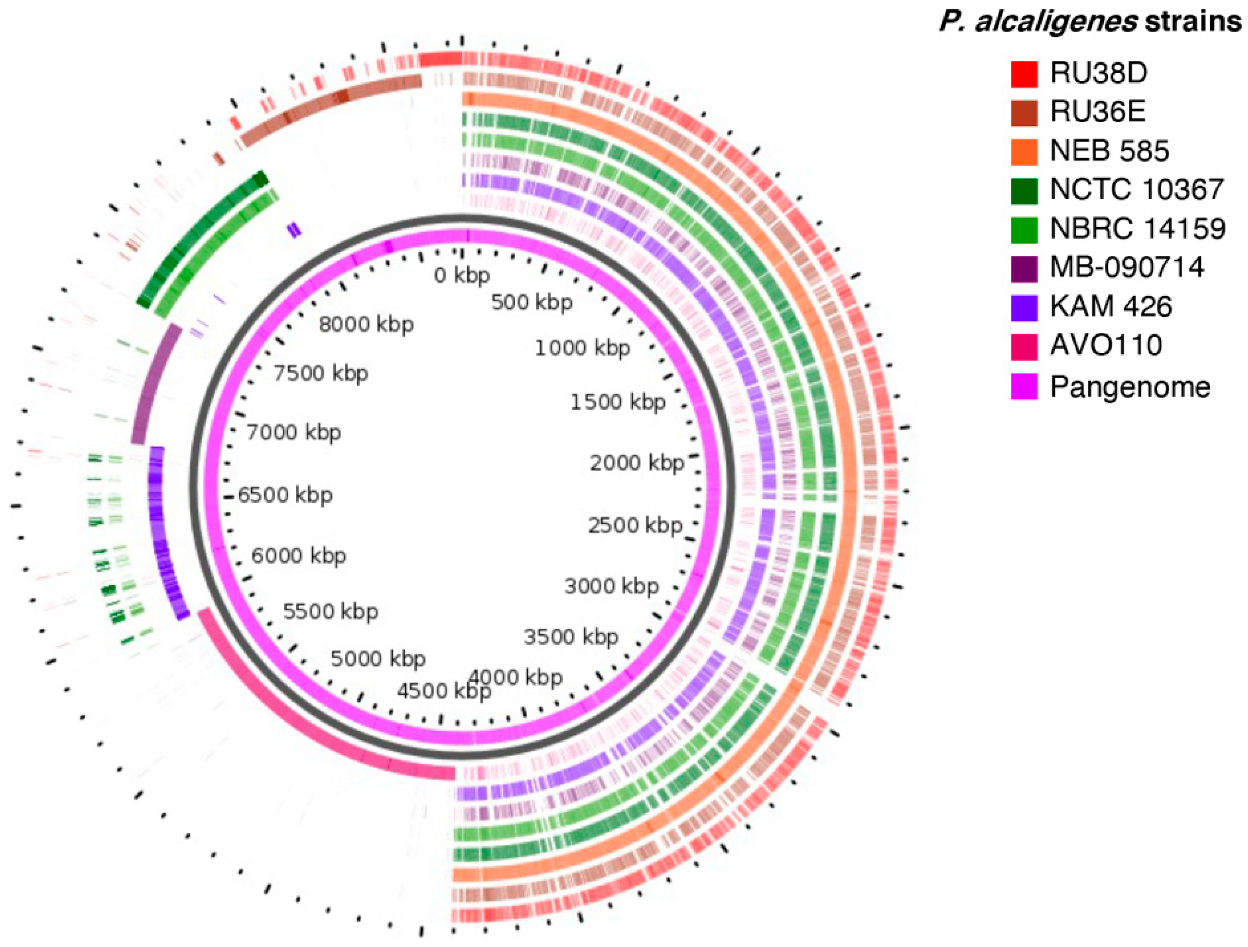
3.3. Pan-Genome and Core Genome of P. alcaligenes

To further analyze the genomic diversity of P. alcaligenes, we performed core and pan-genome analyses of P. alcaligenes subgroups I and II. The eight strains of P. alcaligenes subgroup I yielded a hard pan-genome of 11,778 ortholog groups, of which 974 (8%) constitute the hardcore genome, 4176 (36%) are accessory (encoded in 2–7 genomes), and the remaining 6629 (56%) are singletons (Table 3, Figure S1). These results suggest high genomic diversity in this bacterial subgroup. In fact, using Heaps’ law model [52], we estimated a fitting parameter (γ) of 0.50 for P. alcaligenes subgroup I, which is well above the critical threshold of γ = 0, distinguishing closed (γ < 0) from open (γ > 0) genomes.
We also investigated the core and pan-genome profiles for the 10 P. alcaligenes strains included in subgroups I and II. As expected for higher genomic diversity, the species hard pan-genome (17,284 genes) and hardcore genome (402 genes, 2%) substantially increased and decreased, respectively as compared with the values obtained for subgroup I alone. Furthermore, the number of singletons increased up to 69% (12,004 genes) (Table 3, Figure S1).
Singleton CDSs in P. alcaligenes genomes included in subgroup I were identified using the GView Pangenome Analysis tool. The complete sequence of P. alcaligenes NEB 858 was used as a seed in this analysis. With the exception of the synonymous strains P. alcaligenes NCTC 10367 and NBRC 14159, which share the same variable region, all other genomes encode variable regions (singletons) not shared with any of the other strains.
Remarkably, P. alcaligenes AVO 110, the only rhizobacterium from this species that was analyzed, encodes the largest and most exclusive variable region, which covers approximately 1.6 Mb and encodes 1795 CDSs (Figure 2). The clusters of orthologous groups (COG) analysis of the P. alcaligenes AVO110 singletons assigned functions to 1483 (82%) of their predicted proteins (Figure S2). The largest COG categories were those related to biosynthetic and metabolic processes, comprising 47% of the proteins assigned to biological processes followed by transport-related proteins (9.0%). Manual curing of COG categories related to cell motility, locomotion, cellular component assembly, and signal transduction (6.8% of the total number of annotated products) revealed that P. alcaligenes AVO110 singletons encode 115 genes related to bacterial lifestyle transitions, such as those involved in flagellar motility, chemotaxis, adhesion, biofilm formation, and colonization of host surfaces, 25 of which encode GDEEF and/or EAL domain proteins (Table S2). We also found that 3 of the 21 P. alcaligenes AVO110 genes previously identified as required for growth in fungal exudates [28] were singletons, i.e., the GGDEF-EAL domain gene cmpA and two genes potentially involved in the metabolism of fatty acids, fadE and fadD.

3.4. Identification of P. alcaligenes AVO110 Genes Transcriptionally Regulated by R. necatrix Exudates

We previously reported the identification of 21 P. alcaligenes AVO110 genes required for growth and survival in R. necatrix exudates. Transcriptional analysis of five of these genes showed that four of them, including cmpA, modulate their expression after transfer to fungal exudates [28]. To further characterize the transcriptional response of this bacterium toward fungal exudates, P. alcaligenes AVO110 mid-log phase cells grown in LB were transferred to minimal medium containing R. necatrix exudates (BM-RE). As a first approach, we performed RNAseq analysis of single biological replicates taken immediately after the transfer (time zero) and after 4 h of incubation in BM-RE. Differentially expressed genes after 4 h in BM-RE relative to time zero (relative fold change (RFC)) were identified. In agreement with our previous results, the transcript levels of the cmpA gene increased after transfer to BM-RE (RFC = 3.1). Twelve potentially upregulated genes (RFC = 44.5 to 5.7) and 10 downregulated genes (RFC = 0.08 to 0.5) were selected based on their high and low RFC, respectively. Singleton P. alcaligenes AVO110 genes, identified as described above, and genes previously reported to be essential for growth of this bacterium in BM-RE [28], were preferentially selected. The transcript levels of these 22 genes were quantified by qRT-PCR at time zero and after 4 h in BM-RE. Three biological replicates with three technical replicates were used for these assays. Results from qRT-PCR assays confirmed that all 12 upregulated genes, eight of which were P. alcaligenes AVO110 singletons, showed increased transcript levels after transfer to the BM-RE medium. However, downregulation of transcription was demonstrated for only 5 of the 10 genes selected, none of which were singletons. The upregulated genes include four genes involved in fatty acid degradation, one of which (A9179_RS08450) was a P. alcaligenes AVO110 singleton, plus a contiguous regulatory gene, the chemotaxis gene cheB, and three genes related to the assembly of the type IVb pili. Downregulated genes encode two glutamine synthetases, a potassium transporter, a metallohydrolase, and an alanine racemase (Table 4).

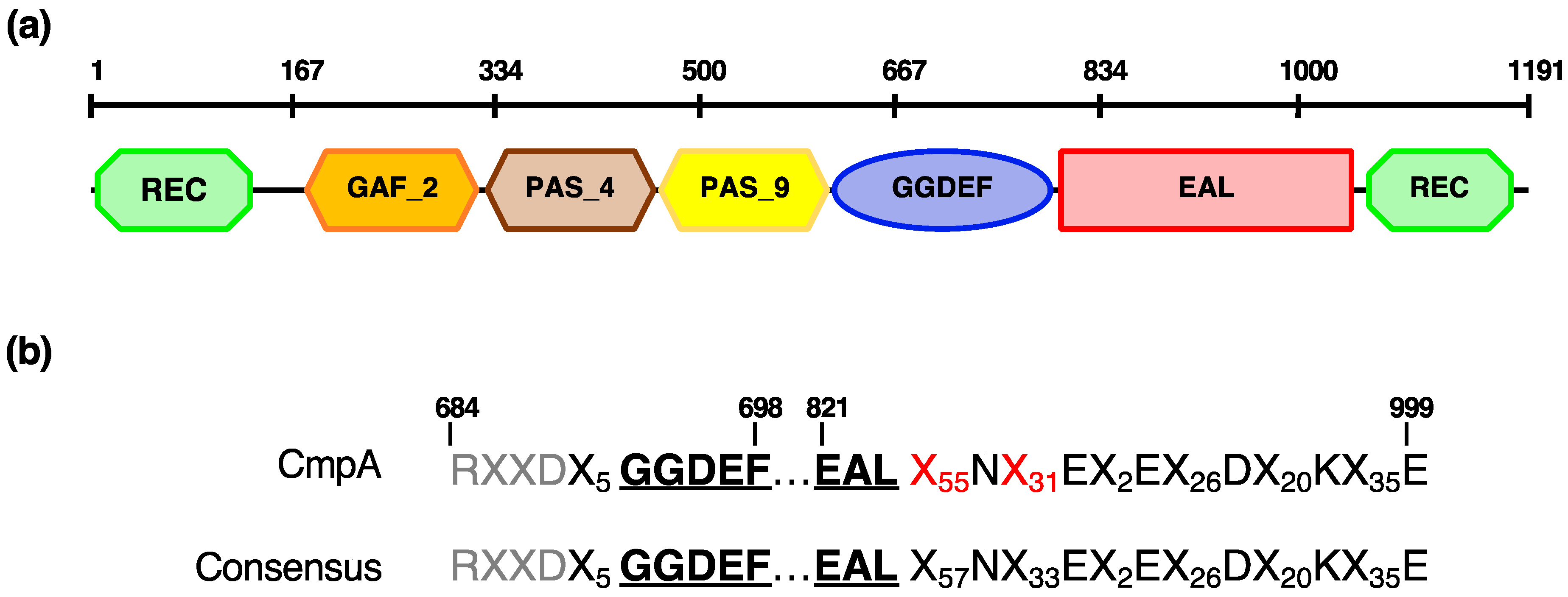
3.5. CmpA, a Pseudomonas spp.-Specific Multidomain Protein

Previously, we reported that the P. alcaligenes AVO110 cmpA gene encodes a GGDEF/EAL domain protein [28]. Here, we conducted a detailed domain analysis of CmpA using the Pfam database. In addition to the GGDEF and EAL domains, five additional domains were found in this protein: a C-terminal and an N-terminal REC domain, a GAF2 domain, and two tandem PAS domains (PAS4 and PAS9) (Figure 3). All of these domains, which are frequently combined in DGCs and PDEs, are predicted to play a role in small-molecule recognition and in signal transduction via phosphorylation [66,67].
Blastp searches with the sequence of this protein revealed that homologs (>60% identity) exist only within the Pseudomonas genus and include those encoded by 10 strains isolated from nonspecified soil, rhizosphere soil (Pseudomonas indica PIC105, Pseudomonas stutzeri NT0128, and Pseudomonas spp. strains LFM046 and 30_B), hyperthermal compost material (Pseudomonas azotifigens DSM 17556), and water. CmpA homologs conserve the same multidomain structure as that found in P. alcaligenes AVO110, with the exception of that in P. stutzeri NT0128, which lacks the PAS4 domain. In addition, we found that the PAS9 and GAF2 domains are truncated in the homologs encoded by Pseudomonas sp. ML96 (isolated from lake water) and the soil bacterium Pseudomonas sp. LAM-KW06, respectively (Table 5).

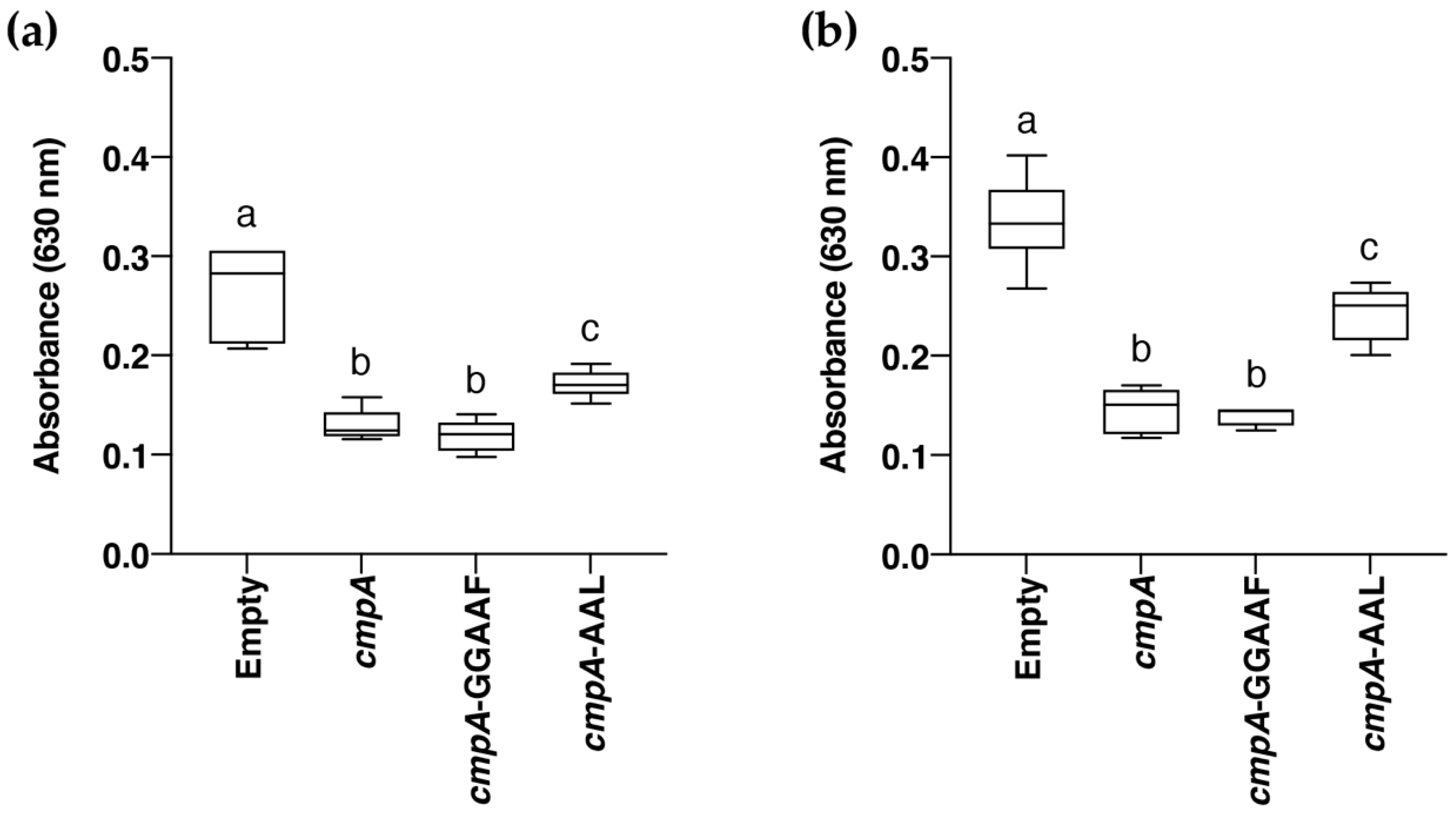
3.6. Role of CmpA in Biofilm Formation

Previous studies have demonstrated the key role of c-di-GMP in the transition between sessile and motile lifestyles in different bacterial species [34], including Pseudomonas spp. strains isolated from the rhizosphere [31,32]. To decipher the role of CmpA in motility and biofilm formation, a P. alcaligenes AVO110 knockout ∆cmpA mutant was constructed (Table 1).
However, this mutant showed no differences in swimming, swarming, or biofilm formation in relation to the wild-type strain (data not shown). Thus, we decided to construct a broad host-range plasmid (pAMEX-cmpA) overexpressing the cmpA gene from a constitutive promoter (Table 1). After several attempts, no P. alcaligenes AVO110 transformants carrying the plasmid were obtained. Then, we decided to use Pseudomonas putida KT2440 as a chassis host for heterologous expression of the cmpA gene. In addition, we performed site-directed mutagenesis of the GGDEF (pAMEX-cmpA-GGAAF) and EAL (pAMEX-cmpA-AAL) motifs of cmpA on plasmid pAMEX-cmpA (Table 1). Independent P. putida KT2440 transformants carrying these three plasmids or the empty vector were obtained, and the biofilm formation ability of the strains was assessed using a serial dilution-based method [59]. Overexpression of all three cmpA alleles in P. putida KT2440 negatively impacted biofilm formation in relation to the transformant carrying the empty vector, with the sharpest effect being observed using dilutions of 10−5 and 10−6 as starting inoculum. Expression of wild-type cmpA or cmpA-GGAAF decreased biofilm formation by approximately 2.0-fold (dilution 10−5) and by 2.3-fold (dilution 10−6) as compared with the wild-type strain transformed with empty vector. However, the ability of P. putida KT2440 overexpressing cmpA-AAL to form biofilm was higher than that of the strains expressing the other two alleles (Figure 4). These results reveal a role for CmpA in biofilm formation and determining the involvement of the EAL domain of this protein in this c-di-GMP-related phenotype.

4. Discussion

P. alcaligenes, a useful bacterium for the biodegradation of toxic polycyclic aromatic hydrocarbons, which is frequently isolated from soil and water [71], has also been known to be a rare opportunistic human pathogen [65]. Recently, several genome sequences of P. alcaligenes strains isolated from water or infected human and oyster tissues have been made available (Table 2). However, the P. alcaligenes AVO 110 genome obtained in this study represents the first genome of a P. alcaligenes strain isolated from the rhizosphere, allowing comparative analysis of niche-specific adaptation and lifestyles in this species, including its mycophagous behavior. Metabolic profiling and partial 16S rRNA gene sequencing initially classified AVO110 as belonging to the species P. pseudoalcaligenes [22]. However, results from our phylogenetic analysis (Figure 1) and comparative genome analysis (Table 2 and Table 3, Figure 2) unequivocally assign this strain to the species P. alcaligenes. Furthermore, P. alcaligenes AVO110 colonies grown on LB medium exhibit a characteristic yellowish-orange pigment, which is not the case with P. pseudoalcaligenes strains [72].
In agreement with previous data [61], all sequenced P. alcaligenes strains were included in a discrete genomic branch in the core genome phylogenetic tree, which corresponds to the P. aeruginosa group. However, separation of P. alcaligenes strains into two distinct branches (Figure 1) evidences a different evolutionary history for the strains of subgroup I, isolated from environmental samples (water and rhizosphere), and subgroup II. Subgroup II strains, isolated from infected blood (MRY13-0052) and minced oyster tissue (OT69), clustered together with P. otitidis strain MrB4. Although P. otitidis was initially isolated from a patient with an ear infection, this species is also widespread in nonclinical environments, as was MrB4, isolated from the near-shore area of Lake Biwa in Japan [73]. In agreement with these results, a recent study showed that a pair-wise comparison of P. alcaligenes OT69 against several type-strain genomes indicated P. otitidis DSM 17224 as its closest relative, rather than the type P. alcaligenes strain NBRC 14159, suggesting that OT69 should be included in a potentially new species [74]. In addition, these authors reported that P. alcaligenes OT69 and the other two oyster-associated Pseudomonas spp. strains share conserved gene pools, encoding unique metabolic traits that might be recruited via bacteriophage-mediated horizontal gene transfer. Our rarefaction analysis of 10 P. alcaligenes genomes belonging to subgroups I and II (Table 3, Figure S1) also showed that both, the large size of the pan-genome and the restricted group of core genes, are likely reflective of the varied lifestyles and niches of members of this species. Thus, the inventory of genes of the P. alcaligenes pan-genome will likely continue to increase as new genome sequences become available.
GView identified variable genomic regions in all analyzed P. alcaligenes strains of subgroup I, the largest and most exclusive of which is AVO 110 (Figure 2). Functional annotation of the P. alcaligenes AVO 110 variable region (singletons) revealed that the largest COG categories were those related to biosynthetic and metabolic processes. One of these genes (A9179_RS08450) was found in a cluster of five genes involved in the ß-oxidation of fatty acids, all of which are regulated by R. necatrix exudates (Table 4). This cluster also encodes fadD, a gene previously identified as essential for the growth and survival of P. alcaligenes AVO110 in BM-RE medium [28], suggesting that this bacterium utilizes fatty acid compounds released in R. necatrix exudates. In E. coli, fatty acid degradation requires the activation of acyl-CoA esters, a process performed by FadD and coupled to transport from the periplasm to the cytosol. Afterwards, degradation of acyl-CoA compounds results in the release of acetyl-CoA, which is oxidized by FadE to enoyl-CoA. In E. coli, fadD and fadE mutants are unable to grow in oleate and oleic acid, respectively [75]. A fadE homolog was also found in the variable region of P. alcaligenes AVO 110. Disruption of this gene also disables the ability of this strain to grow in R. necatrix exudates [28].
Our COG analysis also revealed more than 100 P. alcaligenes AVO 110 singletons related to bacterial lifestyle transitions, such as those involved in flagellar motility, chemotaxis, adhesion, biofilm formation, and colonization of host surfaces (Table S2 and Figure S2). Several studies have reported the relevance of the molecular mechanisms responsible for these phenotypes which underpin the ecological success of bacteria in soil [76], the plant rhizosphere [31,77,78], and fungal surfaces or their exudates [16,26,27,28]. Thus, our analysis of P. alcaligenes AVO110 genes transcriptionally modulated by R. necatrix exudates particularly focused on a selection of genes potentially relevant to bacterial–fungal interactions and preferentially encoded in the variable genomic region of this strain. Three of the upregulated genes were found to be related to the assembly of type IVb pili (Table 4). Type IVb pili has been shown to be essential for motility, biofilm formation, adherence to eukaryotic cells, colonization, and pathogenesis [79]. In addition, one of the downregulated genes encodes an alanine racemase (Table 4). Alanine racemases catalyze the interconversion of L- and D-alanine, a process essential for peptidoglycan formation and also related to biofilm formation [80].
In a previous study, we reported that the GGDEF/EAL domain-encoding gene cmpA was also induced (RFC = 32.27) by R. necatrix exudates, and that this gene is involved in the colonization of fungal hyphae and the avocado rhizosphere [28]. Bioinformatics analysis of bacterial genome sequences has led to the identification of GGDEF, EAL, and HY-GYP domains in proteins of all major bacterial phyla. The number of these enzymes greatly varies among bacterial genomes. However, plant-associated bacteria generally encode a high number of proteins with c-di-GMP-related domains. In particular, plant-associated Pseudomonas spp. strains encode approximately 30 to 65 proteins that encode one or several of these domains [34]. The P. alcaligenes AVO110 genome encodes 58 GGDEF and/or EAL domain-containing proteins, of which 25 (including CmpA) were found in the variable genomic region of this rhizobacterium (Table S2), suggesting that they might have a role in the interaction of this strain with the plant rhizosphere. In fact, CmpA homologs were only found in environmental Pseudomonas spp. isolates, most of which were isolated from rhizosphere samples (Table 5).
In addition to the GGDEF and EAL domains, CmpA encodes five additional domains: two PAS, two REC, and a GAF domain (Figure 3). The PAS and GAF domains, commonly found in signaling proteins such as c-di-GMP synthases/hydrolases, are characterized by a ligand-binding pocket interacting with a variety of small-molecule ligands [66]. The REC (receiver) domains are common modules found in a variety of response regulators of bacterial signal transduction systems [67]. Thus, the fusion between a REC domain and various output domains found in CmpA suggests a role for this protein in c-di-GMP-mediated regulation of gene expression or protein–protein interactions in response to environmental challenges. A role for CmpA in c-di-GMP-related phenotypes could not be determined by either construction of a P. alcaligenes AVO110 ∆cmpA mutant or overexpression of the cmpA gene in this strain. Considering that cmpA is involved in the colonization of R. necatrix hyphae and is overexpressed in response to fungal exudates [28], it may be possible that cmpA is not required for motility and biofilm formation in synthetic media or under the conditions used in these assays. Complementation of CmpA by functional overlapping of other GGDEF/EAL/PAS enzymes could also explain these results. In this sense, we found 11 GGDEF/EAL/PAS domain proteins in the genome of P. alcaligenes AVO110.
Heterologous expression of cmpA in the soil bacterium P. putida KT2240 allowed us to establish a role for this gene in biofilm formation. On the one hand, the serial dilution-based method used for this purpose [59] successfully differentiated biofilm formation-defective mutants of P. putida KT2440 from the wild-type strain [81]. On the other hand, unlike P. alcaligenes AVO110, this strain stability maintains pBBR-MCS derivatives [48], such as pAMEX-cmpA (Table 1). In addition, P. putida KT2440 does not encode a cmpA homolog and contains a sole GGDEF/EAL response regulator [31], a useful characteristic to avoid functional overlapping with similar c-di-GMP signaling circuits. Although a negative impact on biofilm formation was observed by overexpression of wild-type cmpA and its mutant alleles cmpA-GGAAF and cmpA-AAL, the observed decrease was higher for the transformants overexpressing cmpA and cmpA-GGAAF. Considering that a decrease in biofilm formation usually correlates with a decrease in global c-di-GMP levels in P. putida KT2440 and other bacteria [31,34], these results suggests that CmpA exhibits PDE activity, and that the integrity of its EAL motif is involved in this activity. This hypothesis also correlates with the higher ability to colonize the avocado rhizosphere and R. necatrix hyphae of a P. alcaligenes AVO110 cmpA mutant generated by transposon mutagenesis as compared with the wild-type strain [28]. Inactivation of the EAL domain of other GGDEF/EAL/PAS domain-containing proteins also produces an increase in biofilm formation in relation to their wild-type alleles. This is the case with PdeB, a PDE from Shewanella oneidensis MR1, a strain that forms biofilms on mineral surfaces [82].
In summary, the sequencing and annotation of the mycophagous rhizobacterium P. alcaligenes AVO110 allowed us to identify a variable region in the genome of this strain not shared with other sequenced P. alcaligenes strains isolated from water. Our transcriptomics analysis showed that several genes encoded in this region and related to bacterial lifestyle transitions are induced under the influence of exudates from the phytopathogenic fungus R. necatrix. We also show that CmpA, a GGDEF/EAL/PAS domain-containing protein specific to Pseudomonas spp. strains isolated from environmental samples, has a role in biofilm formation, and the integrity of its EAL domain is involved in this function. Further studies are necessary to demonstrate the PDE activity of this enzyme and its role in c-di-GMP signaling in response to environmental challenges.

Supplementary Materials

The following are available online at https://0-www-mdpi-com.brum.beds.ac.uk/article/10.3390/microorganisms9071388/s1, Figure S1: Core and pan-genome analysis of P. alcaligenes strains present in (a) group I and (b) groups I and II. Boxes indicate changes in number of gene families relative to number of genes added sequentially, median values are denoted with a horizontal black line and standard deviation with vertical bars. Pie chart shows frequency distribution of ortholog groups of genes, Figure S2: Predicted biological functions of genes present in specific region of P. alcaligenes AVO110. Specific genes were classified by their predicted biological function using Sma3s_v2 software [56], Table S1: Primers used in this study, Table S2: Relevant genes encoded in exclusive genomic region identified with GView in P. alcaligenes AVO110.

Author Contributions

Conceptualization and funding acquisition, A.d.V., F.M.C. and C.R.; project administration, F.M.C.; supervision, I.P.-M., A.d.V., F.M.C. and C.R.; methodology and experiments, A.P., I.P.-M. and I.M.A.; bioinformatics analysis, A.P. and C.R.; data curation A.P., I.P.-M., I.M.A., J.A.G.-B. and C.R.; C.R. drafted the manuscript with contributions from I.P.-M., I.M.A., A.P. and J.A.G.-B.; preparation of figures, A.P. and C.R.; preparation of tables, A.P., J.A.G.-B. and C.R. All authors have read and agreed to the published version of the manuscript.

Funding

This research was funded by the Ministerio de Ciencia, Innovación y Universidades (Spain), co-financed by the Fondo Europeo de Desarrollo Regional (FEDER), grant number AGL2017-83368-C2-1-R, and the Programa Operativo FEDER Andalucía 2014–2020 (grant number UMA18-FEDERJA-046).

Data Availability Statement

The datasets presented in this study can be found in online repositories. The names of the repository/repositories and accession number(s) can be found in the paper and Supplementary Materials.

Acknowledgments

We thank E. Arrebola, J.A. Crespo-Gómez, and C. Pliego for the isolation of P. alcaligenes AVO110 genomic DNA of sufficient quality for DNA sequencing; L. Díaz for help with bioinformatics pipelines; and P. García-Vallejo for technical assistance.

Conflicts of Interest

The authors declare no conflict of interest. The funders had no role in the design of the study; in the collection, analysis, or interpretation of data; in the writing of the manuscript; or in the decision to publish the results.

References

  1. Pliego, C.; de Weert, S.; Lamers, G.; de Vicente, A.; Bloemberg, G.; Cazorla, F.M.; Ramos, C. Two similar enhanced root-colonizing Pseudomonas strains differ largely in their colonization strategies of avocado roots and Rosellinia necatrix hyphae. Environ. Microbiol. 2008, 10, 3295–3304. [Google Scholar] [CrossRef]
  2. Calderón, C.E.; de Vicente, A.; Cazorla, F.M. Role of 2-hexyl, 5-propyl resorcinol production by Pseudomonas chlororaphis PCL1606 in the multitrophic interactions in the avocado rhizosphere during the biocontrol process. FEMS Microbiol. Ecol. 2014, 89, 20–31. [Google Scholar] [CrossRef] [PubMed] [Green Version]
  3. Dubey, R.K.; Tripathi, V.; Dubey, P.K.; Singh, H.B.; Abhilash, P.C. Exploring rhizospheric interactions for agricultural sustainability: The need of integrative research on multi-trophic interactions. J. Clean. Prod. 2016, 115, 362–365. [Google Scholar] [CrossRef]
  4. Tamborindeguy, C.; Huot, O.B.; Ibanez, F.; Levy, J. The influence of bacteria on multitrophic interactions among plants, psyllids, and pathogen. Insect Sci. 2017, 24, 961–974. [Google Scholar] [CrossRef] [PubMed]
  5. Rossmann, M.; Pérez-Jaramillo, J.E.; Kavamura, V.N.; Chiaramonte, J.B.; Dumack, K.; Fiore-Donno, A.M.; Mendes, L.W.; Ferreira, M.M.C.; Bonkowski, M.; Raaijmakers, J.M.; et al. Multitrophic interactions in the rhizosphere microbiome of wheat: From bacteria and fungi to protists. FEMS Microbiol. Ecol. 2020, 96. [Google Scholar] [CrossRef] [PubMed]
  6. Braga, R.M.; Dourado, M.N.; Araújo, W.L. Microbial interactions: Ecology in a molecular perspective. Braz. J. Microbiol. 2016, 47, 86–98. [Google Scholar] [CrossRef] [Green Version]
  7. Raaijmakers, J.M.; Paulitz, T.C.; Steinberg, C.; Alabouvette, C.; Moënne-Loccoz, Y. The rhizosphere: A playground and battlefield for soilborne pathogens and beneficial microorganisms. Plant Soil 2009, 321, 341–361. [Google Scholar] [CrossRef] [Green Version]
  8. Mendes, R.; Garbeva, P.; Raaijmakers, J.M. The rhizosphere microbiome: Significance of plant beneficial, plant pathogenic, and human pathogenic microorganisms. FEMS Microbiol. Rev. 2013, 37, 634–663. [Google Scholar] [CrossRef]
  9. Berendsen, R.L.; Pieterse, C.M.J.; Bakker, P.A.H.M. The rhizosphere microbiome and plant health. Trends Plant Sci. 2012, 17, 478–486. [Google Scholar] [CrossRef]
  10. Hu, L.; Robert, C.A.M.; Cadot, S.; Zhang, X.; Ye, M.; Li, B.; Manzo, D.; Chervet, N.; Steinger, T.; Van Der Heijden, M.G.A.; et al. Root exudate metabolites drive plant-soil feedbacks on growth and defense by shaping the rhizosphere microbiota. Nat. Commun. 2018, 9. [Google Scholar] [CrossRef] [Green Version]
  11. Zhalnina, K.; Louie, K.B.; Hao, Z.; Mansoori, N.; Da Rocha, U.N.; Shi, S.; Cho, H.; Karaoz, U.; Loqué, D.; Bowen, B.P.; et al. Dynamic root exudate chemistry and microbial substrate preferences drive patterns in rhizosphere microbial community assembly. Nat. Microbiol. 2018, 3, 470–480. [Google Scholar] [CrossRef] [Green Version]
  12. Leveau, J.H.J.; Preston, G.M. Bacterial mycophagy: Definition and diagnosis of a unique bacterial–fungal interaction. New Phytol. 2008, 177, 859–876. [Google Scholar] [CrossRef]
  13. De Boer, W.; Folman, L.B.; Summerbell, R.C.; Boddy, L. Living in a fungal world: Impact of fungi on soil bacterial niche development. FEMS Microbiol. Rev. 2005, 29, 795–811. [Google Scholar] [CrossRef] [Green Version]
  14. Höppener-Ogawa, S.; Leveau, J.H.J.; Van Veen, J.A.; De Boer, W. Mycophagous growth of Collimonas bacteria in natural soils, impact on fungal biomass turnover and interactions with mycophagous Trichoderma Fungi. ISME J. 2009, 3, 190–198. [Google Scholar] [CrossRef] [Green Version]
  15. Leveau, J.H.J.; Uroz, S.; de Boer, W. The bacterial genus Collimonas: Mycophagy, weathering and other adaptive solutions to life in oligotrophic soil environments. Environ. Microbiol. 2010, 12, 281–292. [Google Scholar] [CrossRef] [PubMed]
  16. Mela, F.; Fritsche, K.; De Boer, W.; Van Veen, J.A.; De Graaff, L.H.; Van Den Berg, M.; Leveau, J.H.J. Dual transcriptional profiling of a bacterial/fungal confrontation: Collimonas fungivorans versus Aspergillus niger. ISME J. 2011, 5, 1494–1504. [Google Scholar] [CrossRef] [PubMed] [Green Version]
  17. Mela, F.; Fritsche, K.; De Boer, W.; Van den Berg, M.; Van Veen, J.A.; Maharaj, N.N.; Leveau, J.H.J. Comparative genomics of bacteria from the genus Collimonas: Linking (dis)similarities in gene content to phenotypic variation and conservation. Environ. Microbiol. Rep. 2012, 4, 424–432. [Google Scholar] [CrossRef] [Green Version]
  18. Garbeva, P.; Hordijk, C.; Gerards, S.; de Boer, W. Volatiles produced by the mycophagous soil bacterium Collimonas. FEMS Microbiol. Ecol. 2014, 87, 639–649. [Google Scholar] [CrossRef] [Green Version]
  19. Ballhausen, M.B.; Van Veen, J.A.; Hundscheid, M.P.J.; De Boer, W. Methods for baiting and enriching fungus-feeding (Mycophagous) rhizosphere bacteria. Front. Microbiol. 2015, 6, 1–11. [Google Scholar] [CrossRef] [PubMed] [Green Version]
  20. Song, C.; Schmidt, R.; de Jager, V.; Krzyzanowska, D.; Jongedijk, E.; Cankar, K.; Beekwilder, J.; van Veen, A.; de Boer, W.; van Veen, J.A.; et al. Exploring the genomic traits of fungus-feeding bacterial genus Collimonas. BMC Genom. 2015, 16, 1103. [Google Scholar] [CrossRef] [PubMed]
  21. Rudnick, M.B.; van Veen, J.A.; de Boer, W. Baiting of rhizosphere bacteria with hyphae of common soil fungi reveals a diverse group of potentially mycophagous secondary consumers. Soil Biol. Biochem. 2015, 88, 73–82. [Google Scholar] [CrossRef] [Green Version]
  22. Pliego, C.; Cazorla, F.M.; González-Sánchez, M.Á.; Pérez-Jiménez, R.M.; de Vicente, A.; Ramos, C. Selection for biocontrol bacteria antagonistic toward Rosellinia necatrix by enrichment of competitive avocado root tip colonizers. Res. Microbiol. 2007, 158, 463–470. [Google Scholar] [CrossRef] [PubMed]
  23. De Weert, S.; Vermeiren, H.; Mulders, I.H.M.; Kuiper, I.; Hendrickx, N.; Bloemberg, G.V.; Vanderleyden, J.; De Mot, R.; Lugtenberg, B.J.J. Flagella-driven chemotaxis towards exudate components is an important trait for tomato root colonization by Pseudomonas fluorescens. Mol. Plant-Microbe Interact. 2002, 15, 1173–1180. [Google Scholar] [CrossRef] [PubMed] [Green Version]
  24. Martínez-Granero, F.; Rivilla, R.; Martín, M. Rhizosphere selection of highly motile phenotypic variants of Pseudomonas fluorescens with enhanced competitive colonization ability. Appl. Environ. Microbiol. 2006, 72, 3429–3434. [Google Scholar] [CrossRef] [Green Version]
  25. Lee, S.W.; Cooksey, D.A. Genes expressed in Pseudomonas putida during colonization of a plant-pathogenic fungus. Appl. Environ. Microbiol. 2000, 66, 2764–2772. [Google Scholar] [CrossRef] [PubMed] [Green Version]
  26. Ahn, S.J.; Yang, C.H.; Cooksey, D.A. Pseudomonas putida 06909 genes expressed during colonization on mycelial surfaces and phenotypic characterization of mutants. J. Appl. Microbiol. 2007, 103, 120–132. [Google Scholar] [CrossRef]
  27. Barret, M.; Frey-Klett, P.; Boutin, M.; Guillerm-Erckelboudt, A.Y.; Martin, F.; Guillot, L.; Sarniguet, A. The plant pathogenic fungus Gaeumannomyces graminis var. tritici improves bacterial growth and triggers early gene regulations in the biocontrol strain Pseudomonas fluorescens Pf29Arp. New Phytol. 2009, 181, 435–447. [Google Scholar] [CrossRef]
  28. Pliego, C.; Crespo-Gómez, J.I.; Pintado, A.; Pérez-Martínez, I.; de Vicente, A.; Cazorla, F.M.; Ramos, C. Response of the biocontrol agent Pseudomonas pseudoalcaligenes AVO110 to Rosellinia necatrix exudate. Appl. Environ. Microbiol. 2019, 85. [Google Scholar] [CrossRef] [Green Version]
  29. Holden, D.W.; Hensel, M. 7.3 Signature Tagged Mutagenesis. Methods Microbiol. 1998, 27, 359–370. [Google Scholar] [CrossRef]
  30. Stapper, A.P.; Narasimhan, G.; Ohman, D.E.; Barakat, J.; Hentzer, M.; Molin, S.; Kharazmi, A.; Høiby, N.; Mathee, K. Alginate production affects Pseudomonas aeruginosa biofilm development and architecture, but is not essential for biofilm formation. J. Med. Microbiol. 2004, 53, 679–690. [Google Scholar] [CrossRef]
  31. Matilla, M.A.; Travieso, M.L.; Ramos, J.L.; Ramos-González, M.I. Cyclic diguanylate turnover mediated by the sole GGDEF/EAL response regulator in Pseudomonas putida: Its role in the rhizosphere and an analysis of its target processes. Environ. Microbiol. 2011, 13, 1745–1766. [Google Scholar] [CrossRef]
  32. Muriel, C.; Arrebola, E.; Redondo-Nieto, M.; Martínez-Granero, F.; Jalvo, B.; Pfeilmeier, S.; Blanco-Romero, E.; Baena, I.; Malone, J.G.; Rivilla, R.; et al. AmrZ is a major determinant of c-di-GMP levels in Pseudomonas fluorescens F113. Sci. Rep. 2018, 8, 1979. [Google Scholar] [CrossRef] [Green Version]
  33. Simm, R.; Morr, M.; Kader, A.; Nimtz, M.; Römling, U. GGDEF and EAL domains inversely regulate cyclic di-GMP levels and transition from sessibility to motility. Mol. Microbiol. 2004, 53, 1123–1134. [Google Scholar] [CrossRef]
  34. Romling, U.; Galperin, M.Y.; Gomelsky, M. Cyclic di-GMP: The First 25 Years of a Universal Bacterial Second Messenger. Microbiol. Mol. Biol. Rev. 2013, 77, 1–52. [Google Scholar] [CrossRef] [Green Version]
  35. Jenal, U.; Reinders, A.; Lori, C. Cyclic di-GMP: Second messenger extraordinaire. Nat. Rev. Microbiol. 2017, 15, 271–284. [Google Scholar] [CrossRef] [Green Version]
  36. Martínez-Gil, M.; Ramos, C. Role of cyclic di-GMP in the bacterial virulence and evasion of the plant immunity. Curr. Issues Mol. Biol. 2018, 25, 199–221. [Google Scholar] [CrossRef]
  37. Ross, P.; Weinhouse, H.; Aloni, Y.; Michaeli, D.; Weinberger-Ohana, P.; Mayer, R.; Braun, S.; De Vroom, E.; Van Der Marel, G.A.; Van Boom, J.H.; et al. Regulation of cellulose synthesis in Acetobacter xylinum by cyclic diguanylic acid. Nature 1987, 325, 279–281. [Google Scholar] [CrossRef] [PubMed]
  38. Ryan, R.P.; Fouhy, Y.; Lucey, J.F.; Crossman, L.C.; Spiro, S.; He, Y.W.; Zhang, L.H.; Heeb, S.; Cámara, M.; Williams, P.; et al. Cell-cell signaling in Xanthomonas campestris involves an HD-GYP domain protein that functions in cyclic di-GMP turnover. Proc. Natl. Acad. Sci. USA 2006, 103, 6712–6717. [Google Scholar] [CrossRef] [PubMed] [Green Version]
  39. Sondermann, H.; Shikuma, N.J.; Yildiz, F.H. You’ve come a long way: c-di-GMP signaling. Curr. Opin. Microbiol. 2012, 15, 140–146. [Google Scholar] [CrossRef] [PubMed] [Green Version]
  40. Bertani, G. Studies on lysogenesis I. J. Bacteriol. 1951, 62, 293–300. [Google Scholar] [CrossRef] [PubMed] [Green Version]
  41. Hanahan, D. Studies on transformation of Escherichia coli with plasmids. J. Mol. Biol. 1983, 166, 557–580. [Google Scholar] [CrossRef]
  42. Lugtenberg, B.J.J.; Dekkers, L.C. What makes Pseudomonas bacteria rhizosphere competent? Environ. Microbiol. 1999, 1, 9–13. [Google Scholar] [CrossRef] [PubMed]
  43. Franklin, F.C.H.; Bagdasarian, M.; Bagdasarian, M.M.; Timmis, K.N. Molecular and functional analysis of the TOL plasmid pWWO from Pseudomonas putida and cloning of genes for the entire regulated aromatic ring meta cleavage pathway. Proc. Natl. Acad. Sci. USA 1981, 78, 7458–7462. [Google Scholar] [CrossRef] [PubMed] [Green Version]
  44. Palmer, B.R.; Marinus, M.G. The dam and dcm strains of Escherichia coli—A review. Gene 1994, 143, 1–12. [Google Scholar] [CrossRef]
  45. Macho, A.P.; Ruiz-Albert, J.; Tornero, P.; Beuzón, C.R. Identification of new type III effectors and analysis of the plant response by competitive index. Mol. Plant Pathol. 2009, 10, 69–80. [Google Scholar] [CrossRef] [PubMed]
  46. Ortiz-Martin, I.; Thwaites, R.; Mansfield, J.W.; Beuzón, C.R. Negative regulation of the Hrp type III secretion system in Pseudomonas syringae pv. phaseolicola. Mol. Plant-Microbe Interact. 2010, 23, 682–701. [Google Scholar] [CrossRef] [Green Version]
  47. Russell, D.W.; Sambrook, J. Molecular Cloning: A Laboratory Manual; Cold Spring Harbor Laboratory Press: New York, NY, USA, 2001. [Google Scholar]
  48. Pérez-Martínez, I.; Rodriguez-Moreno, L.; Matas, I.M.; Ramos, C. Strain selection and improvement of gene transfer for genetic manipulation of Pseudomonas savastanoi isolated from olive knots. Res. Microbiol. 2007, 158, 60–69. [Google Scholar] [CrossRef]
  49. Li, R.; Li, Y.; Kristiansen, K.; Wang, J. SOAP: Short oligonucleotide alignment program. Bioinformatics 2008, 24, 713–714. [Google Scholar] [CrossRef] [Green Version]
  50. Chaudhari, N.M.; Gupta, V.K.; Dutta, C. BPGA- an ultra-fast pan-genome analysis pipeline. Nat. Publ. Gr. 2016, 1–10. [Google Scholar] [CrossRef] [Green Version]
  51. Edgar, R.C. Search and clustering orders of magnitude faster than BLAST. Bioinformatics 2010, 26, 2460–2461. [Google Scholar] [CrossRef] [Green Version]
  52. Tettelin, H.; Riley, D.; Cattuto, C.; Medini, D. Comparative genomics: The bacterial pan-genome. Curr. Opin. Microbiol. 2008, 11, 472–477. [Google Scholar] [CrossRef]
  53. Page, A.J.; Cummins, C.A.; Hunt, M.; Wong, V.K.; Reuter, S.; Holden, M.T.G.; Fookes, M.; Falush, D.; Keane, J.A.; Parkhill, J. Roary: Rapid large-scale prokaryote pan genome analysis. Bioinformatics 2015, 31, 3691–3693. [Google Scholar] [CrossRef]
  54. Kumar, S.; Stecher, G.; Tamura, K. MEGA7: Molecular Evolutionary Genetics Analysis Version 7.0 for Bigger Datasets. Mol. Biol. Evol. 2016, 33, 1870–1874. [Google Scholar] [CrossRef] [Green Version]
  55. Petkau, A.; Stuart-Edwards, M.; Stothard, P.; van Domselaar, G. Interactive microbial genome visualization with GView. Bioinformatics 2010, 26, 3125–3126. [Google Scholar] [CrossRef]
  56. Casimiro-Soriguer, C.S.; Muñoz-Mérida, A.; Pérez-Pulido, A.J. Sma3s: A universal tool for easy functional annotation of proteomes and transcriptomes. Proteomics 2017, 17. [Google Scholar] [CrossRef] [PubMed] [Green Version]
  57. Vargas, P.; Felipe, A.; Michán, C.; Gallegos, M.-T. Induction of Pseudomonas syringae pv. tomato DC3000 MexAB-OprM multidrug efflux pump by flavonoids is mediated by the repressor PmeR. Mol. Plant. Microbe. Interact. 2011, 24, 1207–1219. [Google Scholar] [CrossRef] [Green Version]
  58. Livak, K.J.; Schmittgen, T.D. Analysis of Relative Gene Expression Data Using Real-Time Quantitative PCR and the 2−ΔΔCT Method. Methods 2001, 25, 402–408. [Google Scholar] [CrossRef] [PubMed]
  59. López-Sánchez, A.; Jiménez-Fernández, A.; Calero, P.; Gallego, L.D.; Govantes, F. New methods for the isolation and characterization of biofilm-persistent mutants in Pseudomonas putida. Environ. Microbiol. Rep. 2013, 5, 679–685. [Google Scholar] [CrossRef] [PubMed]
  60. Belda, E.; van Heck, R.G.A.; José Lopez-Sanchez, M.; Cruveiller, S.; Barbe, V.; Fraser, C.; Klenk, H.-P.; Petersen, J.; Morgat, A.; Nikel, P.I.; et al. The revisited genome of Pseudomonas putida KT2440 enlightens its value as a robust metabolic chassis. Environ. Microbiol. 2016, 18, 3403–3424. [Google Scholar] [CrossRef]
  61. Mulet, M.; Lalucat, J.; García-Valdés, E. DNA sequence-based analysis of the Pseudomonas species. Environ. Microbiol. 2010, 12, 1513–1530. [Google Scholar] [CrossRef] [Green Version]
  62. Lalucat, J.; Mulet, M.; Gomila, M.; García-Valdés, E. Genomics in Bacterial Taxonomy: Impact on the Genus Pseudomonas. Genes 2020, 11, 139. [Google Scholar] [CrossRef] [Green Version]
  63. Hugh, R.; Ikari, P. The proposed neotype strain of Pseudomonas alcaligenes Monias 1928: Request for an Opinion. Int. Bull. Bacteriol. Nomencl. Taxon. 1964, 14, 103–107. [Google Scholar] [CrossRef]
  64. Chauhan, A.; Green, S.; Pathak, A.; Thomas, J.; Venkatramanan, R. Whole-genome sequences of five oyster-associated bacteria show potential for crude oil hydrocarbon degradation. Genome Announc. 2013, 1, 61–62. [Google Scholar] [CrossRef] [Green Version]
  65. Suzuki, M.; Suzuki, S.; Matsui, M.; Hiraki, Y.; Kawano, F.; Shibayama, K. Genome Sequence of a Strain of the Human Pathogenic Bacterium Pseudomonas alcaligenes That Caused Bloodstream Infection. Genome Announc. 2013, 1, e00919-13. [Google Scholar] [CrossRef] [Green Version]
  66. Galperin, M.Y. Bacterial signal transduction network in a genomic perspective. Environ. Microbiol. 2004, 6, 552–567. [Google Scholar] [CrossRef]
  67. Galperin, M.Y. Structural classification of bacterial response regulators: Diversity of output domains and domain combinations. J. Bacteriol. 2006, 188, 4169–4182. [Google Scholar] [CrossRef] [Green Version]
  68. Cardinali-Rezende, J.; Alexandrino, P.M.R.; Nahat, R.A.T.P.D.S.; Sant’Ana, D.P.V.; Silva, L.F.; Gomez, J.G.C.; Taciro, M.K. Draft Genome Sequence of Pseudomonas sp. Strain LFM046, a Producer of Medium-Chain-Length Polyhydroxyalkanoate. Genome Announc. 2015, 3. [Google Scholar] [CrossRef] [Green Version]
  69. Tsang, H.L.; Huang, J.L.; Lin, Y.H.; Huang, K.F.; Lu, P.L.; Lin, G.H.; Khine, A.A.; Hu, A.; Chen, H.P. Borneol dehydrogenase from Pseudomonas sp. strain TCU-HL1 catalyzes the oxidation of (+)-borneol and its isomers to camphor. Appl. Environ. Microbiol. 2016, 82, 6378–6385. [Google Scholar] [CrossRef] [PubMed] [Green Version]
  70. Busquets, A.; Peña, A.; Gomila, M.; Mulet, M.; Mayol, J.; García-Valdés, E.; Bennasar, A.; Huntemann, M.; Han, J.; Chen, I.-M.; et al. Draft Genome Sequence of Pseudomonas azotifigens Strain DSM 17556T (6H33bT), a Nitrogen Fixer Strain Isolated from a Compost Pile. Genome Announc. 2013, 1, e00893-1. [Google Scholar] [CrossRef] [Green Version]
  71. O’Mahony, M.M.; Dobson, A.D.W.; Barnes, J.D.; Singleton, I. The use of ozone in the remediation of polycyclic aromatic hydrocarbon contaminated soil. Chemosphere 2006, 63, 307–314. [Google Scholar] [CrossRef] [PubMed]
  72. Garrity, G.M.; Bell, J.A.; Lilburn, T. Pseudomonadales Orla-Jensen 1921, 270AL. In Bergey’s Manual® of Systematic Bacteriology; Springer: Boston, MA, USA, 2005; pp. 323–442. [Google Scholar]
  73. Miyazaki, K.; Hase, E.; Maruya, T. Complete Genome Sequence of Pseudomonas otitidis Strain MrB4, Isolated from Lake Biwa in Japan. Microbiol. Resour. Announc. 2020, 9, e00148-20. [Google Scholar] [CrossRef] [Green Version]
  74. Pathak, A.; Stothard, P.; Chauhan, A. Comparative Genomic Analysis of Three Pseudomonas Species Isolated from the Eastern Oyster (Crassostrea virginica) Tissues, Mantle Fluid, and the Overlying Estuarine Water Column. Microorganisms 2021, 9, 490. [Google Scholar] [CrossRef]
  75. Janßen, H.J.; Steinbüchel, A. Fatty acid synthesis in Escherichia coli and its applications towards the production of fatty acid based biofuels. Biotechnol. Biofuels 2014, 7, 1–26. [Google Scholar] [CrossRef] [PubMed] [Green Version]
  76. Varivarn, K.; Champa, L.A.; Silby, M.W.; Robleto, E.A. Colonization strategies of Pseudomonas fluorescens Pf0-1: Activation of soil-specific genes important for diverse and specific environments. BMC Microbiol. 2013, 13, 1–11. [Google Scholar] [CrossRef] [Green Version]
  77. Giddens, S.R.; Jackson, R.W.; Moon, C.D.; Jacobs, M.A.; Zhang, X.X.; Gehrig, S.M.; Rainey, P.B. Mutational activation of niche-specific genes provides insight into regulatory networks and bacterial function in a complex environment. Proc. Natl. Acad. Sci. USA 2007, 104, 18247–18252. [Google Scholar] [CrossRef] [Green Version]
  78. Blanco-Romero, E.; Redondo-Nieto, M.; Martínez-Granero, F.; Garrido-Sanz, D.; Ramos-González, M.I.; Martín, M.; Rivilla, R. Genome-wide analysis of the FleQ direct regulon in Pseudomonas fluorescens F113 and Pseudomonas putida KT2440. Sci. Rep. 2018, 8, 13145. [Google Scholar] [CrossRef]
  79. Craig, L.; Forest, K.T.; Maier, B. Type IV pili: Dynamics, biophysics and functional consequences. Nat. Rev. Microbiol. 2019, 17, 429–440. [Google Scholar] [CrossRef]
  80. Hernández, S.B.; Cava, F. Environmental roles of microbial amino acid racemases. Environ. Microbiol. 2016, 18, 1673–1685. [Google Scholar] [CrossRef]
  81. López-Sánchez, A.; Leal-Morales, A.; Jiménez-Díaz, L.; Platero, A.I.; Bardallo-Pérez, J.; Díaz-Romero, A.; Acemel, R.D.; Illán, J.M.; Jiménez-López, J.; Govantes, F. Biofilm formation-defective mutants in Pseudomonas putida. FEMS Microbiol. Lett. 2016, 363. [Google Scholar] [CrossRef]
  82. Chao, L.; Rakshe, S.; Leff, M.; Spormanna, A.M. PdeB, a cyclic di-GMP-specific phosphodiesterase that regulates Shewanella oneidensis MR-1 motility and biofilm formation. J. Bacteriol. 2013, 195, 3827–3833. [Google Scholar] [CrossRef] [Green Version]
Figure 1. Phylogenetic analysis based on core genome of 34 species belonging to P. aeruginosa and P. oleovorans groups. The tree was built using MEGA7 [54] and maximum likelihood method with 100 bootstraps. Sequences were downloaded from NCBI and P. putida KT2440 was used as an external group. Values in nodes indicate percentages of bootstrap. Superscript T indicates type strains; P. alcaligenes NCTC 10367 and P. alcaligenes NBRC 14159 are the same strain deposited in two different collections.
Microorganisms 09 01388 g001
Figure 2. Circular plots of P. alcaligenes subgroup I genomes. Genome sequences were subjected to pan-genome analysis using the GView server. Innermost slot (purple) shows constructed pan-genome. White space indicates a region missing in the specified genome.
Microorganisms 09 01388 g002
Figure 3. (a) Distribution of functional domains present in CmpA protein; (b) conservation of GGDEF and EAL domains. Numbers indicate positions of amino acid residues from first methionine. Residues corresponding to RXXD motif, responsible for allosteric control and present in other proteins that contain GGDEF domains, are indicated in gray.
Microorganisms 09 01388 g003
Figure 4. Biofilm formation of P. putida KT2440 overexpressing cmpA gene from P. alcaligenes AVO 110 and its mutated alleles cmpA-GGAAF and cmpA-AAL. P. putida KT2440 LB cultures were subjected to six 10-fold serial dilutions and biofilm assays were conducted on these dilutions. (a,b) Dilutions of 10−5 and 10−6, respectively. Biofilm growth was determined from absorbance at 630 nm after crystal violet staining.
Microorganisms 09 01388 g004
Table 1. Bacterial strains and plasmids used in this study.
Strain/PlasmidRelevant CharacteristicsReference or Source
Pseudomonas alcaligenes
 AVO110Wild-type (NfR)[1,22]
 AVO110 ∆cmpAcmpA deletion mutant (NfR KmR)This work
Pseudomonas putida
 KT2440Wild-type (NfR)[43]
Escherichia coli
 DH5αF, ϕ80dlacZΔM15, Δ(lacZYA-argF)U169, deoR, recA1 endA1, hsdR17(rK mK+), phoA, supE44, λ, thi-1, gyrA96, relA1[41]
 GM2929F, ara-14, leuB6, thi-1, tonA31, lacY1, tsx-78, galK2, galT22, glnV44, hisG4, rpsL136, xyl-5, mtl-1, dam13::Tn9, dcm-6, mcrB1, hsdR2, mcrA, recF143 (SpR CmR)[44]
Plasmids
 pGEM-T Easy vectorCloning vector, orif1(-) and lacZ (ApR) Promega, Madison, WI, USA
 pBluescript SK IICloning vector, orif1(-), oripUC, Plac, lacZ’ (ApR)Agilent Technologies, USA
 pAMEXExpression vector, PnptII (KmR)[45]
 pGEM-T-KmFRT-BamHIContains nptII gene from pKD4 (ApR KmR)[46]
 pGEM-T-cmpAContains P. alcaligenes AVO110 cmpA ORF and its ribosomal binding siteThis work
 pBSKII-ΔcmpA-KmpBluescript SK II derivative used for marker exchange mutagenesis of P. alcaligenes AVO110 cmpA gene (ApR KmR)This work
 pAMEX-cmpApAMEX derivative carrying wild-type cmpA gene from P. alcaligenes AVO110 (KmR)This work
 pAMEX-cmpA-GGAAFpAMEX-cmpA derivative carrying mutated cmpA-GGAAF allele (KmR)This work
 pAMEX-cmpA-AALpAMEX-cmpA derivative carrying mutated cmpA-AAL allele (KmR)This work
Nf, nitrofurantoin; Km, kanamycin; Ap, ampicillin.
Table 3. Comparison of core and pan-genomes of Pseudomonas alcaligenes and Pseudomonas pseudoalcaligenes strains.
SpeciesSubgroup aStrainsHard Core Genome bSoft Core Genome bHard
Pan-Genome b
Soft
Pan-
Genome b
Heaps’ Law
(Gamma)
P. alcaligenesI897498411,77811,5530.5030
II25503631968906319NAP
I + II1040240517,28417,0640.5817
P. pseudoalcaligenesNAP23301400948724009NAP
aP. alcaligenes subgroups I and II (Figure 1, Table 2); b hard- and softcore genomes, genes present in 100 and 95% of genomes, respectively; NAP, not applicable.
Table 4. Expression (qRT-PCR) of selected genes in P. alcaligenes AVO110 after transfer to R. necatrix exudates-containing medium (BM-RE medium).
TranscriptionAccession a (A9179_No.)ProductFold Change b
UpregulatedRS00870Chemotaxis-specific protein-glutamate methyltransferase CheB149.92 ± 55.77
RS08440Long-chain acyl-CoA synthetase/AMP-binding protein FadD389.19 ± 46.96
RS08445Long-chain fatty acid-CoA ligase/iron-containing redox enzyme family protein373.48 ± 68.63
RS08450Short-chain dehydrogenase/
SDR family oxidoreductase
317.69 ± 94.41
RS08455Tetratricopeptide motif repeat protein107.12 ± 22.51
RS08460DNA-binding response regulator38.27 ± 10.57
RS09465Pilus assembly protein PilA (type IVb pili)50.76 ± 11.58
RS09470Prepilin peptidase (type IVb pili)29.27 ± 6.44
RS09515Flp pilus assembly complex ATPase component TadA (type IVb pili)39.55 ± 4.21
RS09580 *cmpA (GGDEF/EAL domain-containing protein)32.27 ± 14,61
RS10675DUF411 domain-containing protein94.81 ± 28.25
RS10680OprD family outer membrane porin 935.76 ± 253.68
RS21175Hypothetical protein192.12 ± 85.68
DownregulatedRS00240Glutamine synthetase0.17 ± 0.02
RS10035Potassium-transporting ATPase subunit KdpA0.50 ± 0.04
RS19970MBL fold metallohydrolase (hydrolysis of beta-lactam antibiotics)0.39 ± 0.01
RS21875Alanine racemase (peptidoglycan biosynthesis)0.17 ± 0.02
RS22500Glutamine synthetase0.50 ± 0.06
a Bold accession numbers (National Center for Biotechnology Information (NCBI)) indicate singleton P. alcaligenes AVO110 genes (see Figure 2), dotted lines separate gene clusters, * data previously reported [28]; b relative fold changes (normalized to housekeeping gene rpoD) after 4 h in BM-RE relative to time zero. Values are means from three biological replicates with three technical replicates ± standard deviations.
Table 5. Protein homologs to CmpA from Pseudomonas alcaligenes AVO110 in other bacteria.
StrainQuery CoverIdentityAccession Number aSourceReference or Sequence Source/Year
Pseudomonas sp. LFM046100%78.34%WP_044875132Sugarcane soil[68]
Pseudomonas sp. F(2018)100%76.91%WP_171016323Alpine spring waterO. Jousson/2018
Pseudomonas sp. PDM15100%76.24%WP_192398170SoilK. Gowda/2020
Pseudomonas sp. 30_B100%75.99%WP_207883400Rice rhizosphereV. Venturi/2021
Pseudomonas sp. TCU-HL199%75.97%WP_069081885Soil[69]
Pseudomonas sp. ML96 b99%75.32%WP_043307322Lake waterX. Li/2014
Pseudomonas indica PIC10599%71.52%WP_084333623Olea europaea rhizosphereC. Gomez-Lama/2017
Pseudomonas sp. LAM-KW06 b99%70.94%WP_172149757SoilD. Kong/2020
Pseudomonas azotifigens DSM 1755699%63.30%WP_028239826Hyperthermal compost material[70]
Pseudomonas stutzeri NT0128 b99%62.51%WP_052679321Wheat rhizosphereN. Tovi/2015
a National Center for Biotechnology Information (NCBI); b Pseudomonas spp. ML96 and LAM-KW06 CmpA homolog encode truncated PAS9 and GAF2 domains, respectively, CmpA homolog from P. stutzeri NT0128 does not encode a PAS4 domain (see Figure 3).
Publisher’s Note: MDPI stays neutral with regard to jurisdictional claims in published maps and institutional affiliations.

