Transformation of S-Bearing Minerals in Organic Matter-Rich Sediments from a Saline Lake with Hydrothermal Inputs


Abstract
:1. Introduction
2. Materials and Methods
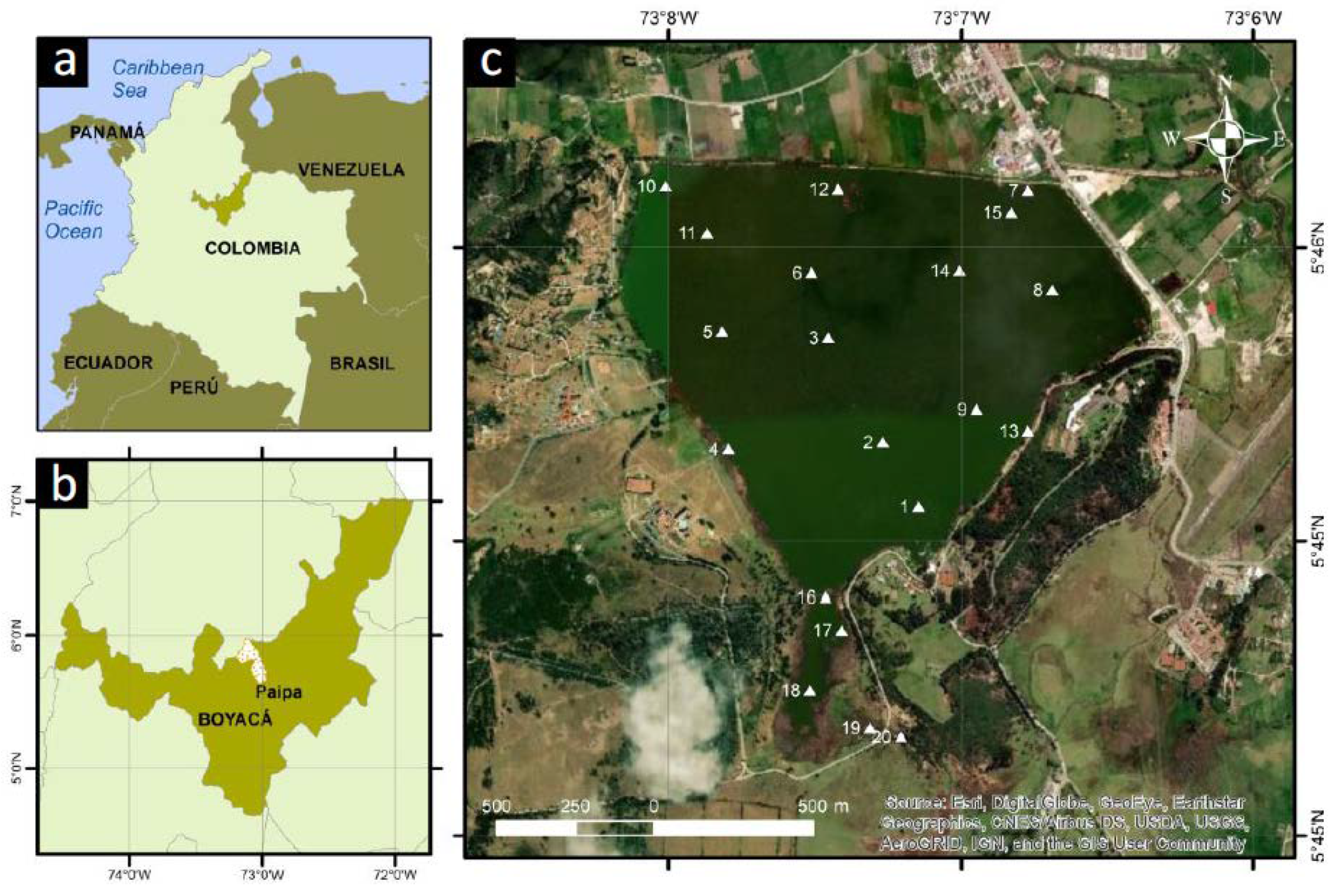
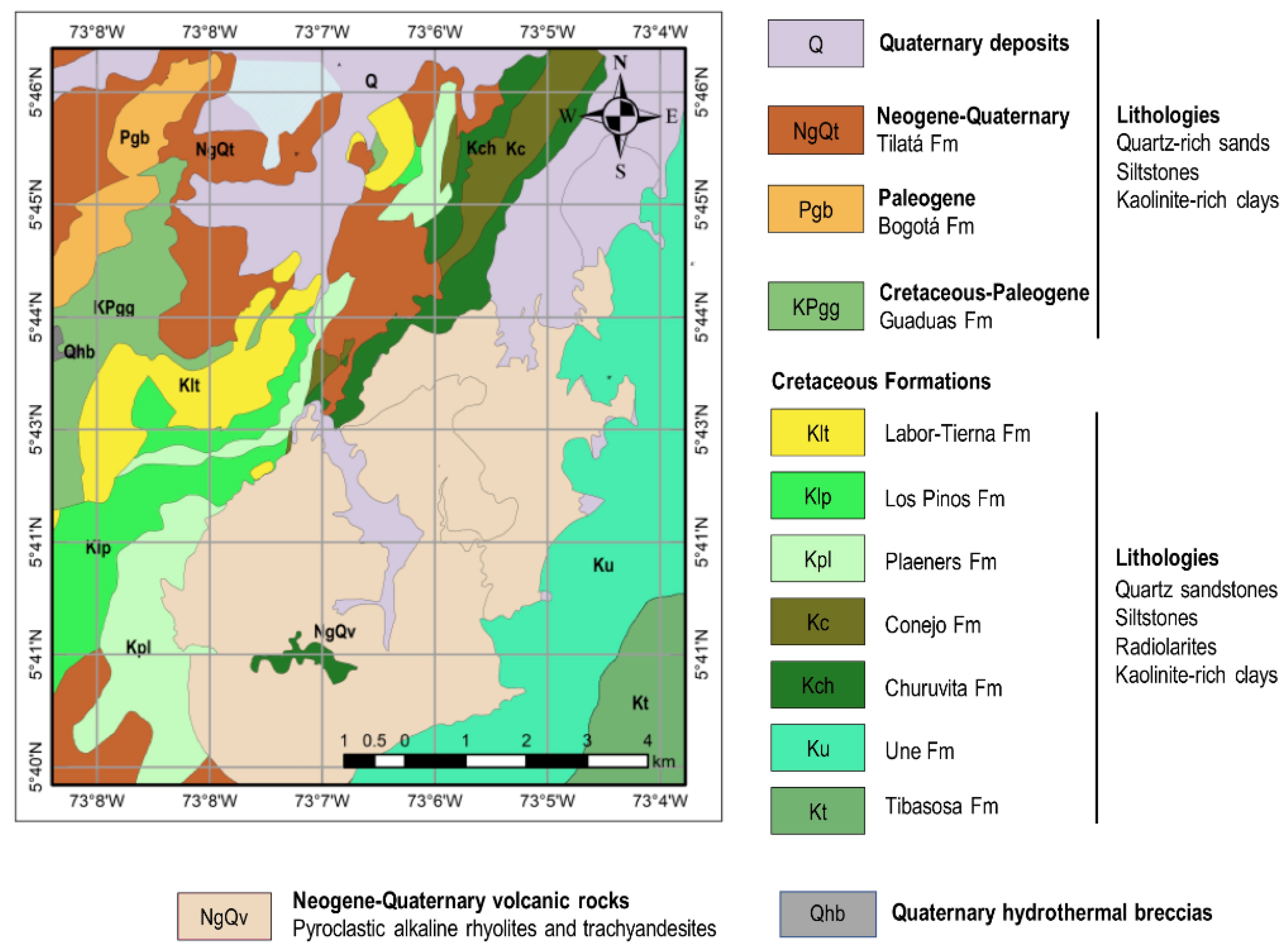
2.1. Study Site
2.2. Methods
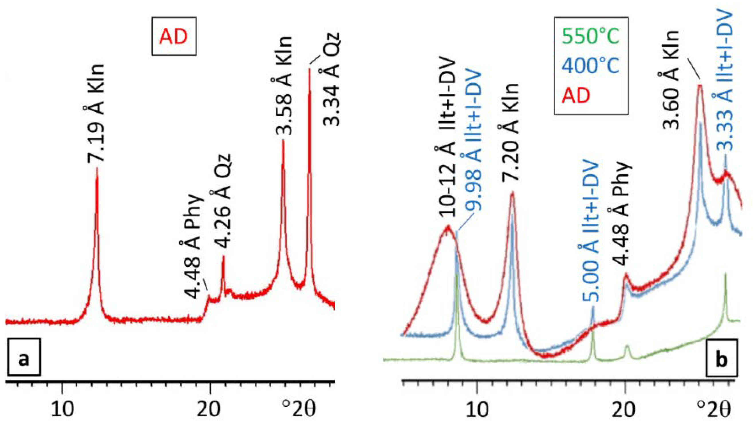
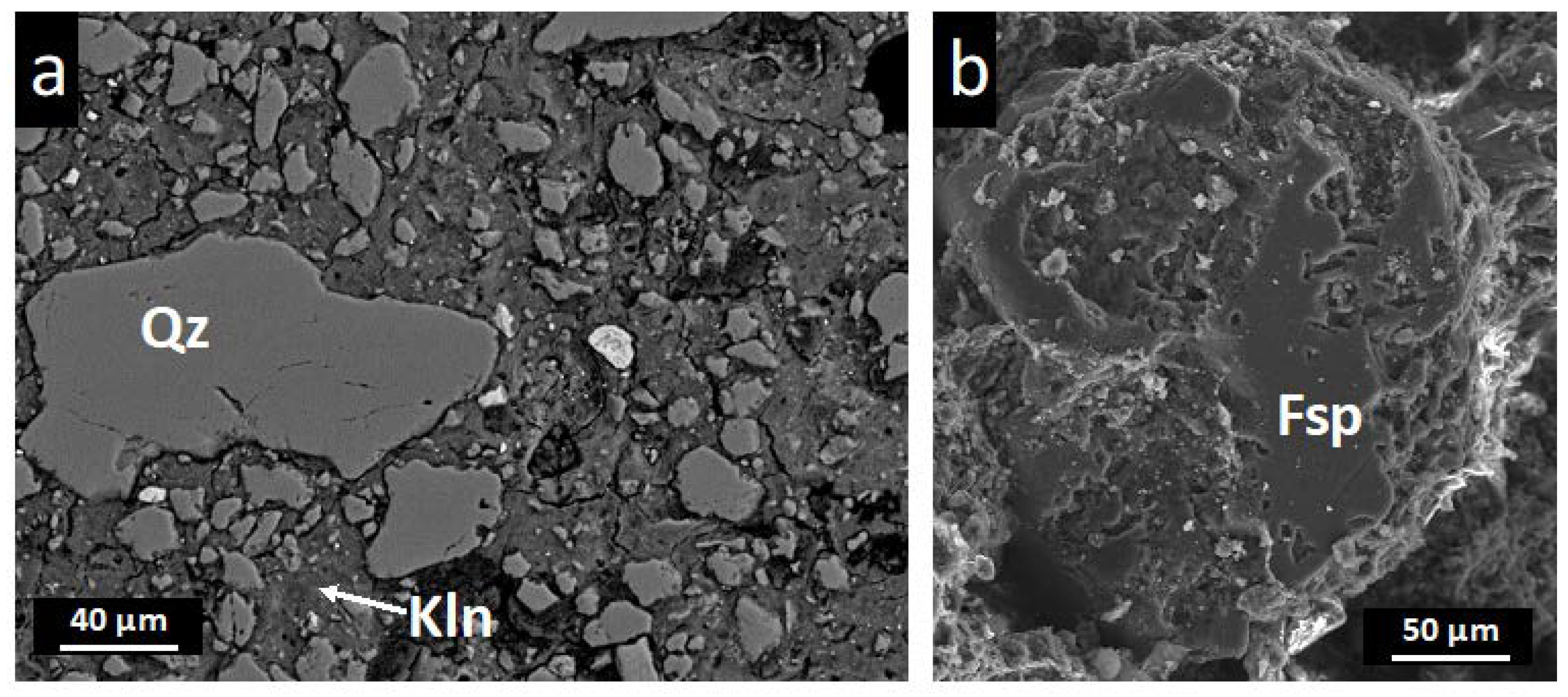
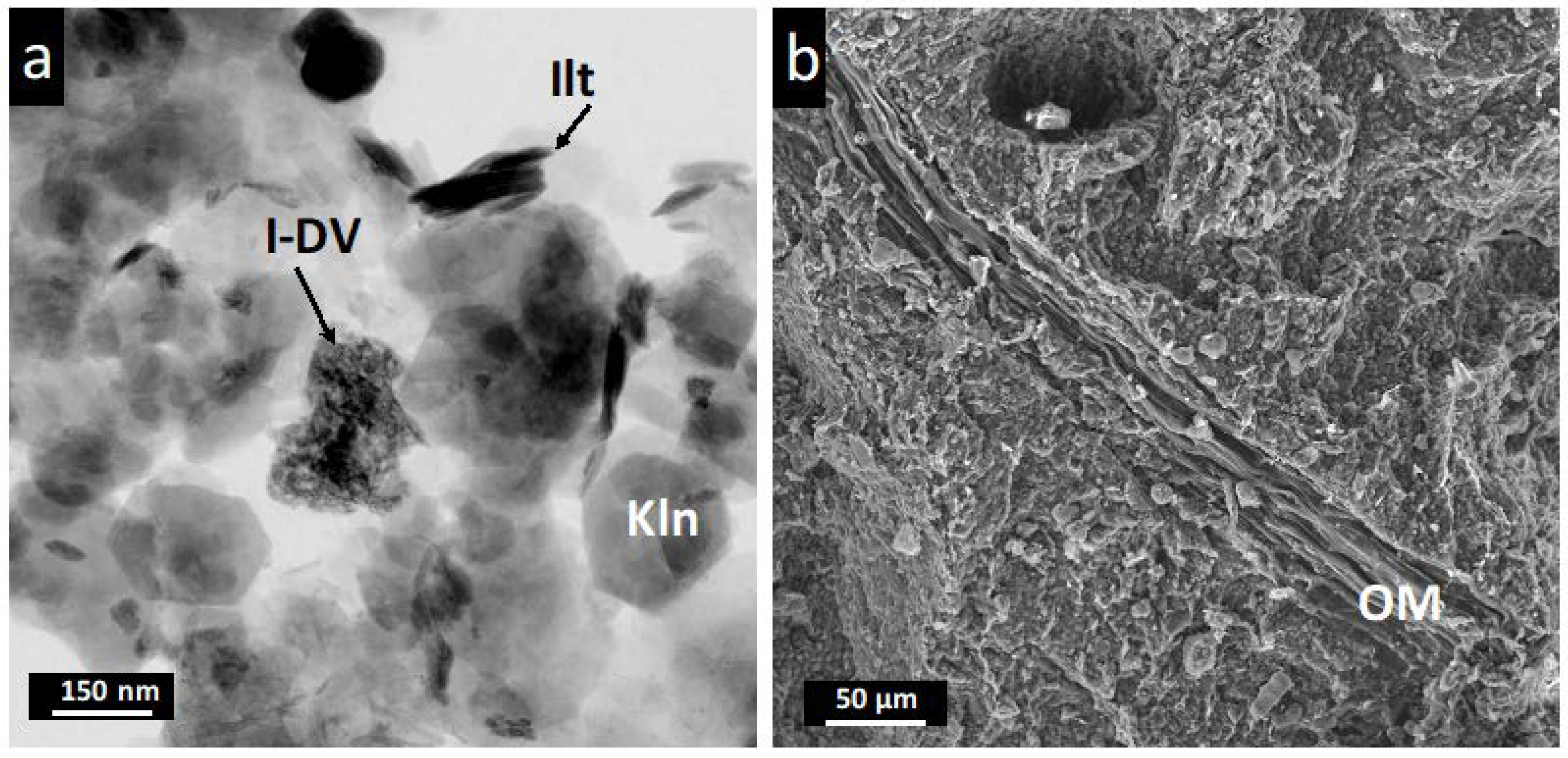
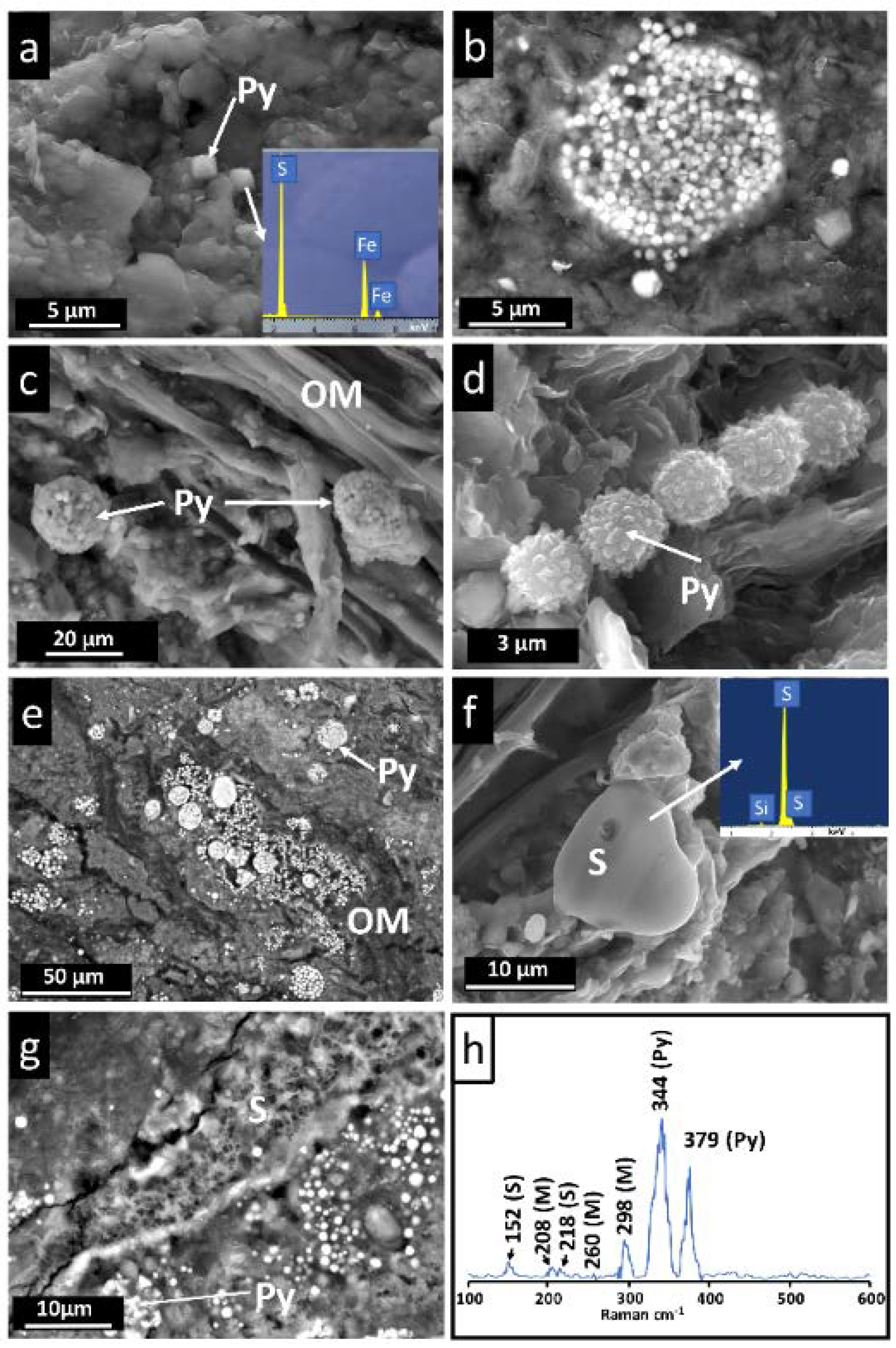
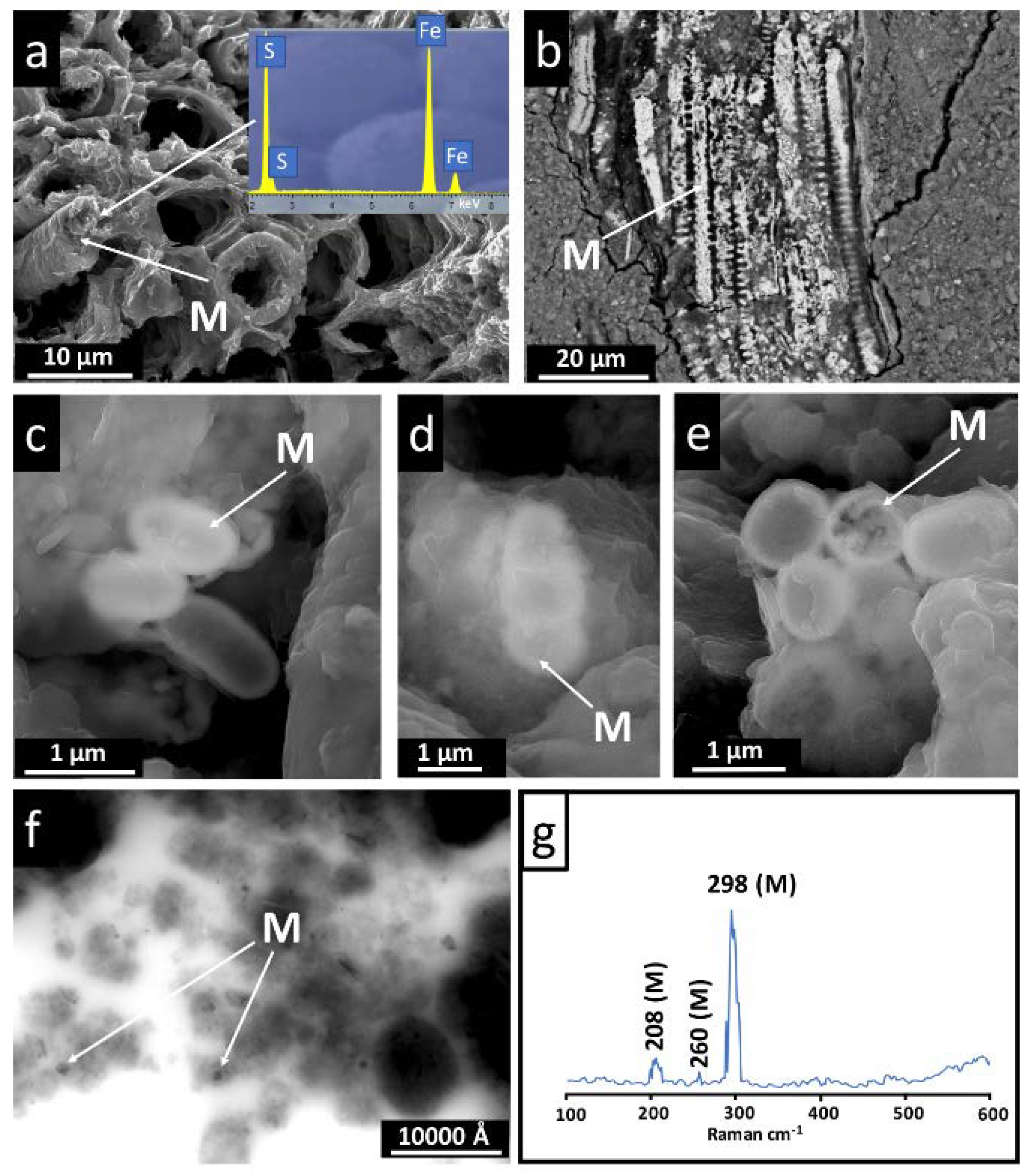
3. Results
4. Discussion
4.1. Detrital Minerals and Neoformed Clay Minerals
4.2. Hydrothermal Inputs and Reduced Microenvironments Favored by Organic Matter
4.3. The S Cycle in the Materials of the Sochagota Lake
5. Conclusions
Author Contributions
Funding
Acknowledgments
Conflicts of Interest
References
- Kaasalainen, H.; Stefánsson, A. Sulfur speciation in natural hydrothermal waters, Iceland. Geochim. Cosmochim. Acta 2011, 75, 2777–2791. [Google Scholar] [CrossRef]
- Kaasalainen, H.; Stefánsson, A.; Giroud, N.; Arnórsson, S. The geochemistry of trace elements in geothermal fluids, Iceland. App. Geochem. 2015, 62, 207–223. [Google Scholar] [CrossRef]
- Björke, J.K.; Stefánsson, A.; Arnórsson, S. Surface water chemistry at Torfajökull, Iceland—Quantification of boiling, mixing, oxidation and water-rock interaction and reconstruction of reservoir fluid composition. Geothermics 2015, 58, 75–86. [Google Scholar] [CrossRef]
- Nordstrom, D.K.; McCleskey, R.B.; Ball, J.W. Sulfur geochemistry of hydrothermal waters in Yellowstone National Park: IV Acid-sulfate waters. App. Geochem. 2009, 24, 191–207. [Google Scholar] [CrossRef]
- Aghasian, K.; Moridi, A.; Mirbagheri, A.; Abbaspour, M. Selective withdrawal optimization in a multipurpose water use reservoir. Int. J. Environ. Sci. Technol. 2019, 16, 5559–5568. [Google Scholar] [CrossRef]
- Zhang, Y.; Liao, J.; Pei, Z.; Lu, X.; Xu, S.; Wang, X. Effect of dam construction on nutrient deposition from a small agricultural karst catchment. Ecol. Indic. 2019, 107. [Google Scholar] [CrossRef]
- Cuadros, J.; Andrade, G.; Ferreira, T.O.; Partiti, C.S.M.; Cohen, R.; Vidal-Torrado, P. The mangrove reactor: Fast clay transformation and potassium sink. Appl. Clay Sci. 2017, 140, 50–58. [Google Scholar] [CrossRef]
- Ferreira, T.O.; Vidal-Torrado, P.; Otero, X.L.; Macías, F. Are mangrove forest substrates sediments or soils? A case study in southeastern Brazil. Catena 2007, 70, 79–91. [Google Scholar] [CrossRef]
- Noël, V.; Boye, K.; Kukkadapu, R.K.; Bone, S.; Pacheco, J.S.L.; Cardarelli, E.; Janot, N.; Fendorf, S.; Williams, K.H.; Bargar, J.R. Understanding controls on redox processes in floodplain sediments of the Upper Colorado River Basin. Sci. Total Environ. 2017, 603–604, 663–675. [Google Scholar] [CrossRef] [Green Version]
- Andrade, G.R.P.; Cuadros, J.; Partiti, C.M.S.; Cohen, R.; Vidal-Torrado, P. Sequential mineral transformation from kaolinite to Fe-illite in two Brazilian mangrove soils. Geoderma 2018, 309, 84–99. [Google Scholar] [CrossRef]
- Rickard, D.; Luther, G.W. Chemistry of Iron Sulfides. Chem. Rev. 2007, 107, 514–562. [Google Scholar] [CrossRef]
- Kraal, P.; Burton, E.D.; Bush, R.T. Iron monosulfide accumulation and pyrite formation in eutrophic estuarine sediments. Geochim. Cosmochim. Acta 2013, 122, 75–88. [Google Scholar] [CrossRef]
- Rickard, D.T. Microbial sulfate reduction in sediments. In Developments in Sedimentology: Sulfidic Sediments and Sedimentary Rocks; van Loon, A.J., Ed.; Elsevier: Oxford, UK, 2012; pp. 319–351. [Google Scholar] [CrossRef]
- Picard, A.; Gartman, A.; Clarke, D.R.; Girguis, P.R. Sulfate-reducing bacteria influence the nucleation and growth of mackinawite and greigite. Geochim. Cosmochim. Acta 2018, 220, 367–384. [Google Scholar] [CrossRef]
- Cosmidis, J.; Nims, C.; Diercks, D.; Templeton, A.S. Formation and stabilization of elemental sulfur through organomineralization. Geochim. Cosmochim. Acta 2019, 247, 59–82. [Google Scholar] [CrossRef]
- Pardo, N.; Cepeda, H.; Jaramillo, J.M. The Paipa volcano, Eastern Cordillera of Colombia, South America: Volcanic stratigraphy. Earth Sci. Res. J. 2005, 9, 3–18. [Google Scholar]
- Alfaro, C.; Velandia, F.; Cepeda, H. Colombian Geothermal Resources. In Proceedings of the World Geothermal Congress, Antalya, Turkey, 24–29 April 2005; pp. 1–11. [Google Scholar]
- Cifuentes, G.R.; Jiménez-Espinosa, R.; Quevedo, C.P.; Jiménez-Millán, J. El ciclo del azufre en sedimentos de lagos con aportes hidrotermales y antrópicos: El Lago Sochagota (Boyacá-Colombia). Macla 2017, 22, 27–28. (In Spanish) [Google Scholar]
- Cifuentes, G.R.; Jiménez-Millán, J.; Quevedo, C.P.; Jiménez-Espinosa, R. Isotopic evidence of the hydrothermal origin of the waters of the Sochagota Lake (Colombia). In Proceedings of the AGU Fall Meeting, San Francisco, CA, USA, 7–11 December 2020. [Google Scholar]
- Rye, R.O.; Bethke, P.M.; Wasserman, M.D. The stable isotope geochemistry of acid sulfate alteration. Econ. Geol. 1992, 87, 225–262. [Google Scholar] [CrossRef]
- John, D.A.; Lee, R.G.; Breit, G.N.; Dilles, J.H.; Calvert, A.T.; Muffler, L.J.P.; Clynne, M.A. Pleistocene hydrothermal activity on Brokeoff volcano and in the Maidu volcanic center, Lassen Peak area, northeast California: Evolution of magmatic-hydrothermal systems on stratovolcanoes. Geosphere 2019, 15, 946–982. [Google Scholar] [CrossRef]
- Sánchez-Roa, C.; Jiménez-Millán, J.; Abad, I.; Faulkner, D.R.; Nieto, F.; García-Tortosa, F.J. Fibrous clay mineral authigenesis induced by fluid-rock interaction in the Galera fault zone (Betic Cordillera, SE Spain) and its influence on fault gouge frictional properties. Appl. Clay Sci. 2016, 134, 275–288. [Google Scholar] [CrossRef]
- Sánchez-Roa, C.; Faulkner, D.R.; Boulton, C.; Jimenez-Millan, J.; Nieto, F. How phyllosilicate mineral structure affects fault strength in Mg-rich fault systems. Geophys. Res. Lett. 2017, 44, 5457–5467. [Google Scholar] [CrossRef] [Green Version]
- Jiménez-Millán, J.; Abad, I.; García-Tortosa, F.J.; Nieto, F.; Jiménez-Espinosa, R. Clay saline diagenesis in lake Plio-Pleistocene sediments rich in organic matter from the Guadix-Baza Basin (Betic Cordillera, SE Spain). Appl. Clay Sci. 2020, in press. [Google Scholar]
- Nieto, F.; Ortega-Huertas, M.; Peacor, D.R.; Arostegui, J. Evolution of illite/smectite from early diagenesis through incipient metamorphism in sediments of the Basque-Cantabrian Basin. Clay Clay Miner. 1996, 44, 304–323. [Google Scholar] [CrossRef]
- Guerra, I.; Cardell, C. Optimizing use of the structural chemical analyser (variable pressure FESEM-EDX Raman spectroscopy) on micro-size complex historical paintings characterization. J. Microsc. 2015, 260, 47–61. [Google Scholar] [CrossRef]
- Benner, R.; Strom, M. A critical evaluation of the analytical blank associated with DOC measurements by high-temperature catalytic oxidation. Mar. Chem. 1993, 41, 153–160. [Google Scholar] [CrossRef]
- Cifuentes, G.R.; Jiménez-Millán, J.; Quevedo, C.P.; Jiménez-Espinosa, R.; Nieto, F. Fijación de K hidrotermal a través de transformaciones secuenciales de formación de illita en ambiente lacustre hipersalino reductor (Lago Sochagota, Colombia). In Sociedad Española de Arcillas 2018; Sociedad Española de Arcillas: Granada, Spain, 2018. (In Spanish) [Google Scholar]
- Kleppe, A.K.; Jephcoat, A.P. High-pressure Raman spectroscopic studies of FeS2 pyrite. Mineral. Mag. 2004, 68, 433–441. [Google Scholar] [CrossRef]
- Meyer, B. Elemental Sulfur. Chem. Rev. 1976, 76, 367–388. [Google Scholar] [CrossRef]
- Bourdoiseau, J.A.; Jeannin, M.; Sabot, R.; Rémazeilles, C.; Refait, P. Characterisation of mackinawite by Raman spectroscopy: Effects of crystallisation, drying and oxidation. Corrosion Sci. 2008, 50, 3247–3255. [Google Scholar] [CrossRef]
- Cifuentes, G.R.; Jiménez-Millán, J.; Quevedo, C.P.; Jiménez-Espinosa, R. Low temperature illitization through illite-dioctahedral vermiculite mixed layers in a tropical latitude hypersaline lake rich in hydrothermal fluids (Lago Sochagota, Colombia). In Proceedings of the AGU Fall Meeting, San Francisco, CA, USA, 7–11 December 2020. [Google Scholar]
- Smieja-Król, B.; Janeczek, J.; Bauerek, A.; Thorseth, I.H. The role of authigenic sulfides in immobilization of potentially toxic metals in the Bagno Bory wetland, southern Poland. Environ. Sci. Pollut. Res. 2015, 22, 15495–15505. [Google Scholar] [CrossRef] [Green Version]
- Love, L.G.; Al-Kaisy, A.T.H.; Brockley, H. Mineral and organic material in matrices and coatings of framboidal pyrite from Pennsylvanian sediments, England. J. Sediment. Res. 1984, 54. [Google Scholar] [CrossRef]
- Folk, R.L. Nannobacteria and the formation of framboidal pyrite: Textural evidence. J. Earth Syst. Sci. 2005, 114, 369–374. [Google Scholar] [CrossRef]
- MacLean, L.C.W.; Tyliszczak, T.; Gilbert, P.U.P.A.; Zhou, D.; Pray, T.J.; Onstott, T.C.; Southam, G. A high-resolution chemical and structural study of framboidal pyrite formed within a low temperature bacterial biofilm. Geobiology 2008, 6, 471–480. [Google Scholar] [CrossRef] [PubMed]
- Wilkin, R.; Barnes, H. Formation processes of framboidal pyrite. Geochim. Cosmochim. Acta 1997, 61, 323–339. [Google Scholar] [CrossRef]
- Schoonen, M.A.A. Mechanisms of sedimentary pyrite formation. Geol. Soc. Am. Spec. Pap. 2004, 379, 117–134. [Google Scholar]
- Ohfuji, H.; Rickard, D. High resolution transmission electron microscopic study of synthetic nanocrystalline mackinawite. Earth Planet. Sci. Lett. 2006, 241, 227–233. [Google Scholar] [CrossRef]
- Hu, S.Y.; Evans, K.; Fisher, L.; Rempel, K.; Craw, D.; Evans, N.J.; Cumberland, S.; Robert, A.; Grice, K. Associations between sulfides, carbonaceous material, gold and other trace elements in polyframboids: Implications for the source of orogenic gold deposits, Otago Schist, New Zealand. Geochim. Cosmochim. Acta 2016, 180, 197–213. [Google Scholar] [CrossRef]
- Cosmidis, J.; Benzerara, K.; Menguy, N.; Arning, E. Microscopy evidence of bacterial microfossils in phosphorite crusts of the Peruvian shelf: Implications for phosphogenesis mechanisms. Chem. Geol. 2013, 359, 10–22. [Google Scholar] [CrossRef]
- Xu, J.; Murayama, M.; Roco, C.M.; Veeramani, H.; Michel, F.M.; Rimstidt, J.D.; Winkler, C.; Hochella, M.F.J. Highly-defective nanocrystals of ZnS formed via dissimilatory bacterial sulfate reduction: A comparative study with their abiogenic analogues. Geochim. Cosmochim. Acta 2016, 180, 1–14. [Google Scholar] [CrossRef] [Green Version]
- Rickard, D.; Grimes, S.T.; Butler, I.; Oldroyd, A.; Davies, K.L. Botanical constraints on pyrite formation. Chem. Geol. 2007, 236, 228–246. [Google Scholar] [CrossRef]
- Nabbefeld, B.; Grice, K.; Schimmelmann, A.; Sauer, P.E.; Böttcher, M.E.; Twitchett, R. Significance of δD kerogen, δ13C, δ 13C kerogen and δ34S pyrite from several Permian/Triassic (P/Tr) sections. Earth Planet. Sci. Lett. 2010, 295, 21–29. [Google Scholar] [CrossRef]
- Jaraula, C.M.; Grice, K.; Twitchett, R.J.; Böttcher, M.E.; LeMetayer, P.; Dastidar, A.G.; Opazo, L.F. Elevated pCO2 leading to Late Triassic extinction, persistent photic zone euxinia, and rising sea levels. Geology 2013, 41, 955–958. [Google Scholar] [CrossRef]
- Melendez, I.; Grice, K.; Schwark, L. Exceptional preservation of Palaeozoic steroids in a diagenetic continuum. Sci. Rep. 2013, 3. [Google Scholar] [CrossRef] [PubMed] [Green Version]
- Luther, G.W.; Findlay, A.; MacDonald, D.J.; Owings, S.M.; Hanson, T.E.; Beinart, R.A.; Girguis, P.R. Thermodynamics and kinetics of sulfide oxidation by oxygen: A look at inorganically controlled reactions and biologically mediated processes in the environment. Front. Microbiol. 2011, 2, 62. [Google Scholar] [CrossRef] [PubMed] [Green Version]
- Cosmidis, J.; Templeton, A.S. Self-assembly of biomorphic carbon/sulfur microstructures in sulfidic environments. Nat. Commun. 2016, 7, 12812. [Google Scholar] [CrossRef] [PubMed]


| Physical-Chemical Parameters and Units | Average Values |
|---|---|
| Cl¯ (mg/L) | 672.3 |
| SO2¯4 (mg/L) | 2165.0 |
| HCO¯3 (mg/L) | 204.2 |
| CO2¯3 (mg/L) | 1.5 |
| NO¯3 (mg/L) | 314.9 |
| Na+ (mg/L) | 1493.1 |
| Mg2+ (mg/L) | 9.1 |
| Ca2+ (mg/L) | 55.5 |
| K+ (mg/L) | 280.7 |
| pH | 9.27 |
| E.C. (µS/cm) | 2440.0 |
| Temperature (°C) | 22.0 |
| Sample | IC (%) | TOC (%) | Total C (%) |
|---|---|---|---|
| 1p ** | 0.149 (0.01) | 9.15 (0.04) | 9.30 |
| 1s * | 0.142 (0.01) | 4.11 (0.02) | 4.25 |
| 2p ** | 0.269 (0.03) | 1.01 (0.01) | 1.28 |
| 2s * | 0.176 (0.01) | 1.17 (0.01) | 1.34 |
| 3p ** | 0.290 (0.02) | 1.88 (0.02) | 2.17 |
| 3s * | 0.188 (0.01) | 1.74 (0.01) | 1.93 |
| 4p ** | 0.161 (0.01) | 0.996 (0.01) | 1.16 |
| 4s * | 0.090 (0.01) | 2.01 (0.01) | 2.10 |
| 5p ** | 0.131 (0.01) | 3.04 (0.03) | 3.18 |
| 5s * | 0.121 (0.01) | 2.51 (0.03) | 2.63 |
| 6p ** | 0.123 (0.02) | 11.1 (0.01) | 11.20 |
| 6s * | 0.142 (0.01) | 3.59 (0.04) | 3.73 |
| M7 *** | <0.100 | 1.16 (0.01) | 1.24 |
| 7p ** | 0.253 (0.01) | 1.41 (0.01) | 1.66 |
| 7s * | 0.229 (0.01) | 1.21 (0.01) | 1.43 |
| M8 *** | <0.10 (0.01) | 1.25 (0.03) | 1.33 |
| 8p ** | 0.181 (0.01) | 2.12 (0.01) | 2.30 |
| 8s * | 0.264 (0.01) | 2.26 (0.01) | 2.52 |
| M9 *** | <0.100 | 2.10 (0.01) | 2.11 |
| 9p ** | 1.26 (0.03) | 0.66 (0.01) | 1.93 |
| 9s * | 0.683 (0.02) | 0.62 (0.01) | 1.31 |
| 10p ** | 0.199 (0.01) | 3.64 (0.04) | 3.84 |
| 10s * | 0.194 (0.01) | 3.18 (0.02) | 3.38 |
| 11p ** | 0.195 (0.01) | 1.52 (0.01) | 1.72 |
| 11s * | 0.200 (0.01) | 1.57 (0.01) | 1.77 |
| 12p ** | 0.206 (0.01) | 1.24 (0.01) | 1.45 |
| 12s * | 0.141 (0.01) | 1.80 (0.01) | 1.94 |
| 13p ** | 0.216 (0.01) | 1.27 (0.01) | 1.48 |
| 13s * | 0.957 (0.01) | 0.37 (0.01) | 1.33 |
| M14 *** | <0.10 | 0.96 (0.01) | 0.96 |
| 14p ** | 0.218 (0.01) | 3.45 (0.04) | 3.67 |
| 14s * | 0.149 (0.01) | 2.24 (0.01) | 2.38 |
| 15p ** | 0.218 (0.01) | 1.73 (0.01) | 1.94 |
| 15s * | 0.188 (0.01) | 1.59 (0.01) | 1.78 |
| 16p ** | 0.408 (0.01) | 0.48 (0.01) | 0.89 |
| 17p ** | 0.156 (0.01) | 0.64 (0.01) | 0.80 |
| 17s * | 0.309 (0.01) | 0.31 (0.01) | 0.62 |
| 18p ** | 0.212 (0.01) | 4.35 (0.05) | 4.56 |
| 18s * | 1.180 (0.02) | 0.29 (0.01) | 1.48 |
| 19p ** | 0.257 (0.01) | 1.64 (0.02) | 1.89 |
| 19s * | 0.137 (0.01) | 1.78 (0.01) | 1.89 |
| M20 *** | <0.100 | 0.69 (0.01) | 0.70 |
| 20p ** | 0.158 (0.01) | 1.79 (0.02) | 1.95 |
| 20s * | 0.189 (0.01) | 2.59 (0.03) | 2.78 |
© 2020 by the authors. Licensee MDPI, Basel, Switzerland. This article is an open access article distributed under the terms and conditions of the Creative Commons Attribution (CC BY) license (http://creativecommons.org/licenses/by/4.0/).
Share and Cite
Cifuentes, G.R.; Jiménez-Millán, J.; Quevedo, C.P.; Jiménez-Espinosa, R. Transformation of S-Bearing Minerals in Organic Matter-Rich Sediments from a Saline Lake with Hydrothermal Inputs. Minerals 2020, 10, 525. https://0-doi-org.brum.beds.ac.uk/10.3390/min10060525
Cifuentes GR, Jiménez-Millán J, Quevedo CP, Jiménez-Espinosa R. Transformation of S-Bearing Minerals in Organic Matter-Rich Sediments from a Saline Lake with Hydrothermal Inputs. Minerals. 2020; 10(6):525. https://0-doi-org.brum.beds.ac.uk/10.3390/min10060525
Chicago/Turabian StyleCifuentes, Gabriel Ricardo, Juan Jiménez-Millán, Claudia Patricia Quevedo, and Rosario Jiménez-Espinosa. 2020. "Transformation of S-Bearing Minerals in Organic Matter-Rich Sediments from a Saline Lake with Hydrothermal Inputs" Minerals 10, no. 6: 525. https://0-doi-org.brum.beds.ac.uk/10.3390/min10060525