
Design and Characterization of a Novel p1025 Peptide-Loaded Liquid Crystalline System for the Treatment of Dental Caries

 , and
, and
Abstract
:1. Introduction
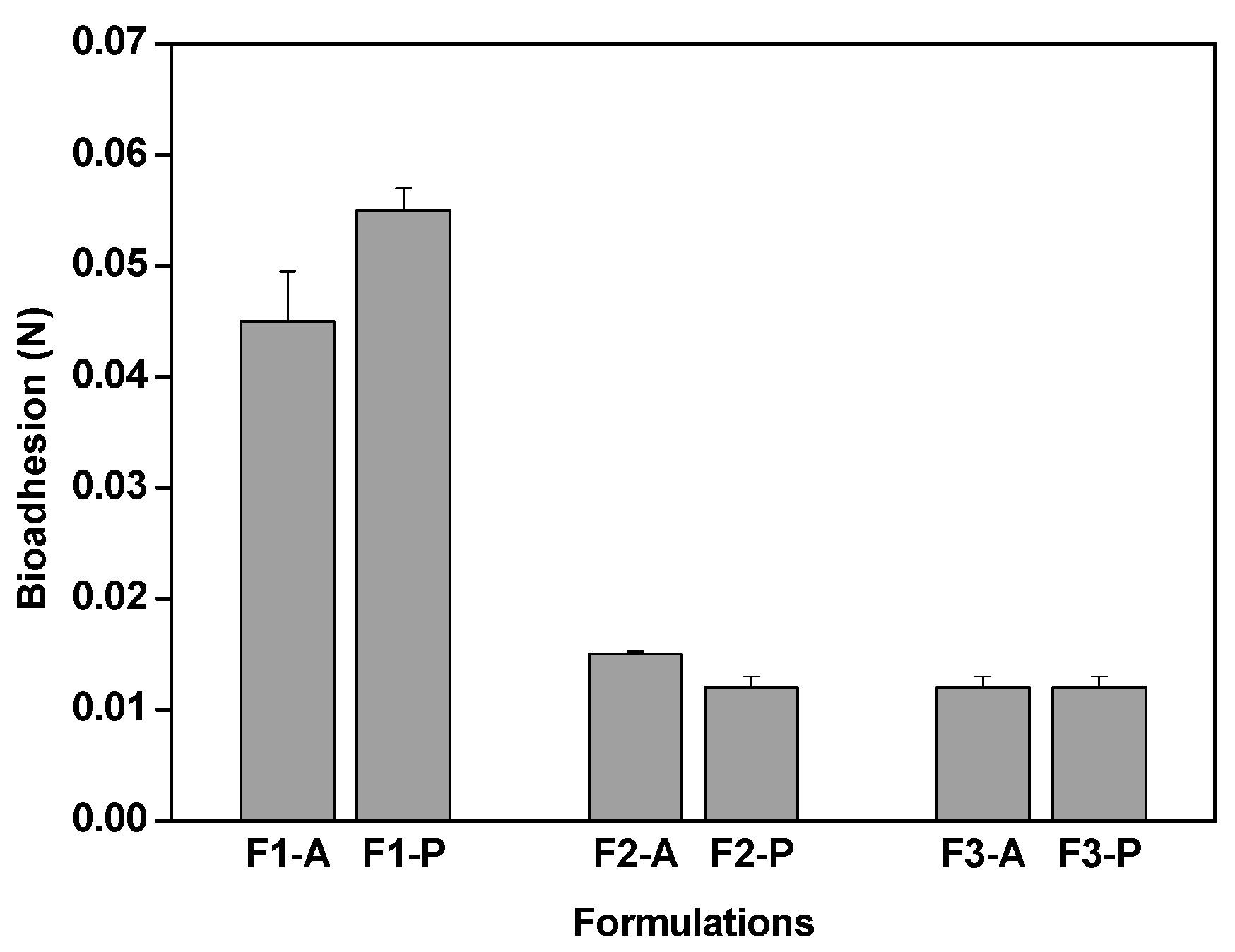
2. Results and Discussion


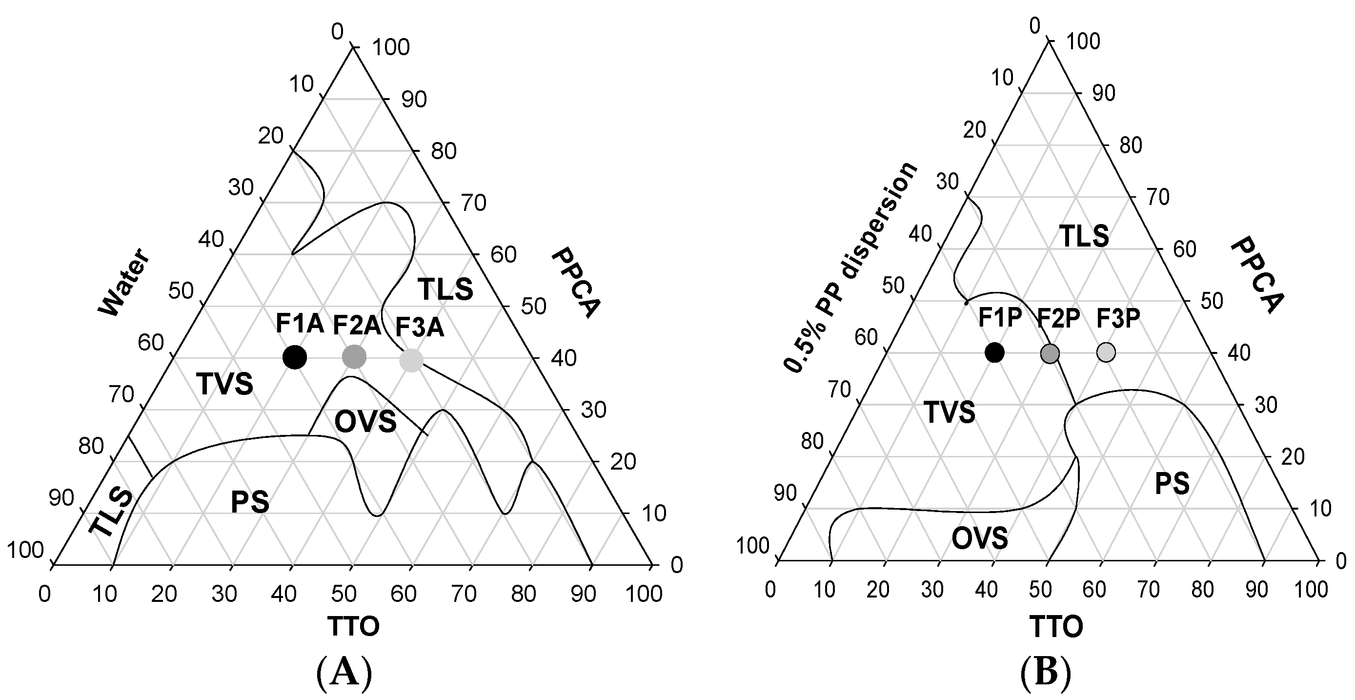
| Formulations | TTO (%) | PPCA (%) | Water (%) | 5% PP (%) |
|---|---|---|---|---|
| F1-A | 20 | 40 | 40 | - |
| F2-A | 30 | 40 | 30 | - |
| F3-A | 40 | 40 | 20 | - |
| F1-P | 20 | 40 | 30 | 10 |
| F2-P | 30 | 40 | 20 | 10 |
| F3-P | 40 | 40 | 10 | 10 |


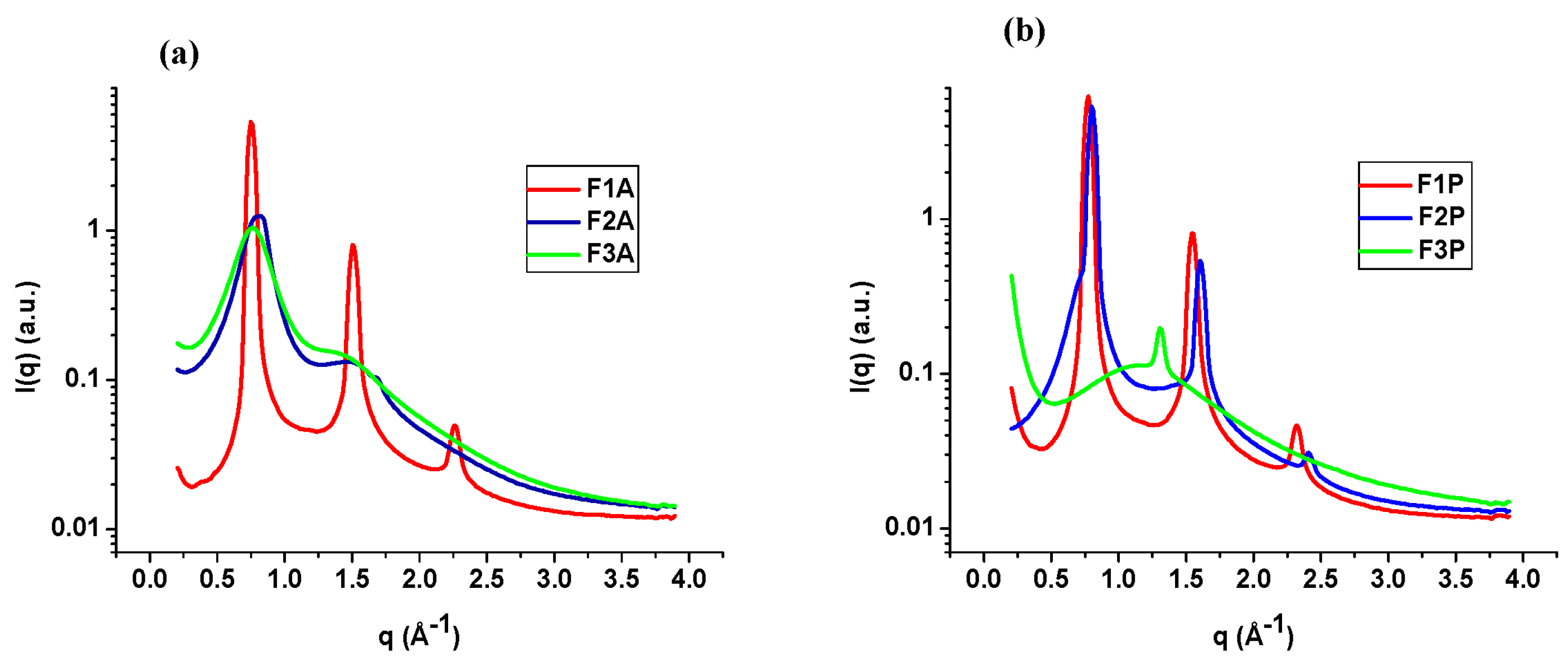
| Formulation | Qmax peak 1 | Qmax peak2 | Qmax peak3 | D-Spacing | d/dh |
|---|---|---|---|---|---|
| F1-A | 0.75 | 1.50 | 2.26 | 8.37 | 1.00 |
| F2-A | 0.79 | 1.52 | - | 7.95 | 1.00 |
| F3-A | 0.76 | 1.46 | - | 8.26 | - |
| F1-P | 0.78 | 1.53 | 2.32 | 8.05 | 1.00 |
| F2-P | 0.80 | 1.60 | 2.41 | 7.85 | 1.00 |
| F3-P | 1.30 | - | - | 4.79 | - |


| Formulations | Hardness (N) | Compressibility (N.s) | Adhesiveness (N.s) | Cohesiveness |
|---|---|---|---|---|
| F1-A | 0.031 ± 0.002 | 0.398 ± 0.01 | 0.152 ± 0.02 | 0.860 ± 0.04 |
| F1-P | 0.022 ± 0.003 | 0.297 ± 0.03 | 0.299 ± 0.01 | ± 0.05 |
3. Experimental Section
3.1. Materials
3.2. Methods
3.2.1. Synthesis, Purification, and Preparation of the Peptide
3.2.2. Construction of the Pseudoternary Phase Diagram
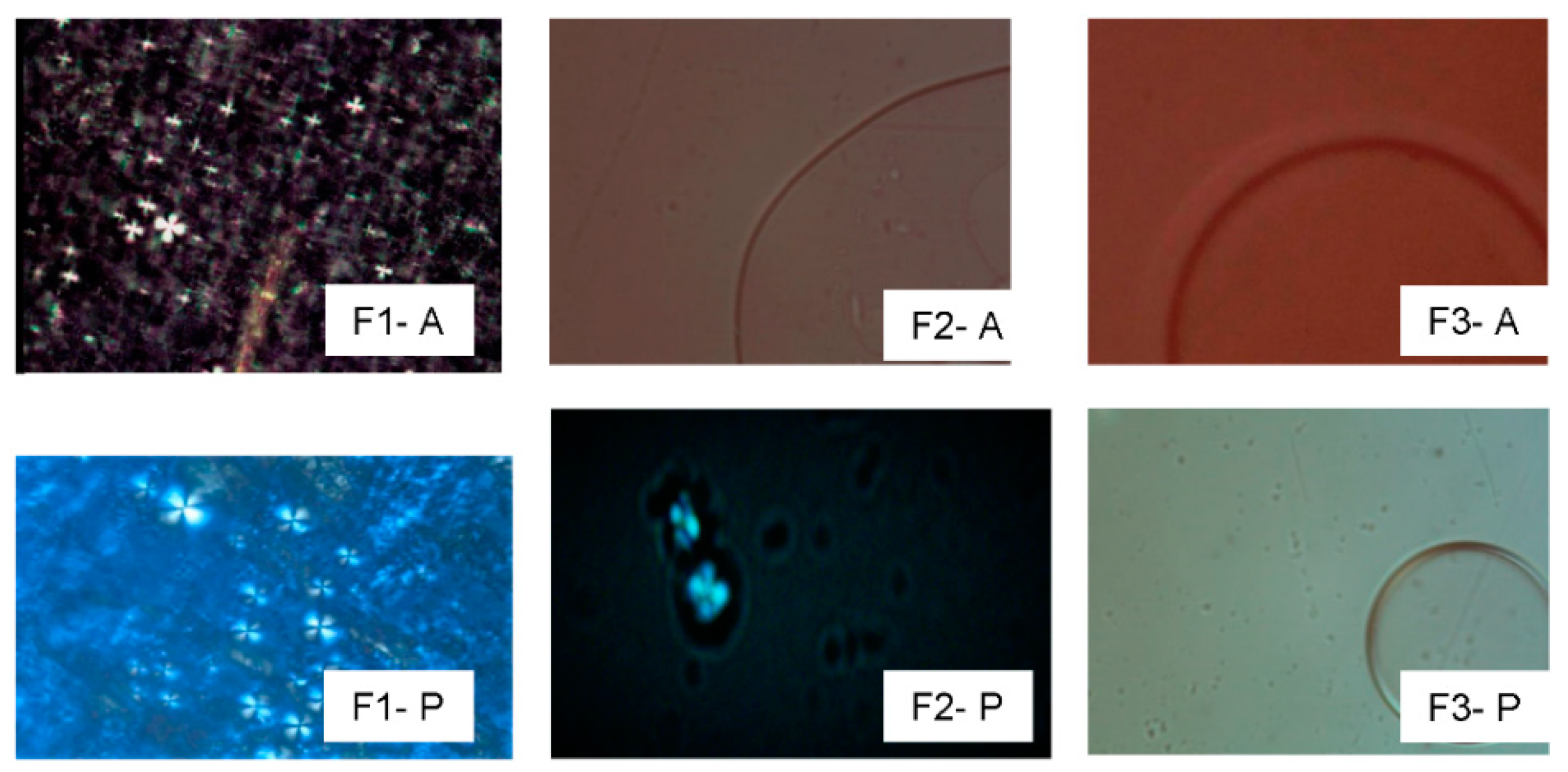
3.2.3. Polarized Light Microscope
3.2.4. Small-Angle X-ray Scattering
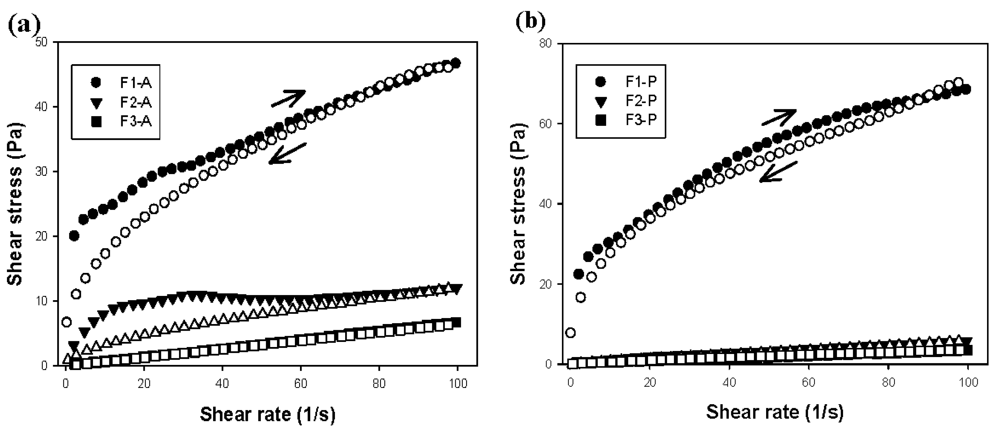
3.2.5. Rheological Measurements
3.2.6. Texture Profile Analysis
3.2.7. In Vitro Bioadhesion Strength
Saliva Collection
Preparation of Discs
Bioadhesion Measurement by the Tensile Strength Method
4. Conclusions
Acknowledgments
Author Contributions
Conflicts of Interest
References
- Saunders, R.H.; Meyerowitz, C. Dental caries in older adults. Dent. Clin. N. Am. 2005, 49, 293–308. [Google Scholar] [CrossRef] [PubMed]
- Young, D.A.; Nový, B.B.; Zeller, G.G.; Hale, R.; Hart, T.C.; Truelove, E.L. The American Dental Association Caries Classification System for Clinical Practice: A report of the American Dental Association Council on Scientific Affairs. J. Am. Dent. Assoc. 2015, 146, 79–86. [Google Scholar] [CrossRef] [PubMed]
- Bernegossi, J.; Calixto, G.; Fonseca-Santos, B.; Limi Aida, K.; de Cássia Negrini, T.; Duque, C.; Gremião, M.P.D.; Chorilli, M. Highlights in peptide nanoparticle carriers intended to oral diseases. Curr. Top. Med. Chem. 2015, 15, 345–355. [Google Scholar] [CrossRef]
- Da Silva, B.R.; de Freitas, V.A.A.; Carneiro, V.A.; Arruda, F.V.; Lorenzon, E.N.; de Anguiar, A.S.; Cilli, E.M.; Cavada, B.S.; Teixeira, E.H. Antimicrobial activity of the synthetic peptide Lys-a1 against oral streptococci. Peptides 2013, 42, 78–83. [Google Scholar] [CrossRef] [PubMed]
- Kelly, C.G.; Younson, J.S.; Hikmat, B.Y.; Todryk, S.M.; Czisch, M.; Haris, P.I.; Flindall, I.R.; Newby, C.; Mallet, A.I.; Ma, J.K.; et al. A synthetic peptide adhesion epitope as a novel antimicrobial agent. Nat. Biotechnol. 1999, 17, 42–47. [Google Scholar] [CrossRef] [PubMed]
- Li, M.Y.; Wang, J.; Lai, G.Y. Effect of a dentifrice containing the peptide of streptococcal antigen I/II on the adherence of mutans streptococcus. Arch. Oral Biol. 2009, 54, 1068–1073. [Google Scholar] [CrossRef] [PubMed]
- Ichikawa, H.; Peppas, N.A. Novel complexation hydrogels for oral peptide delivery: In vitro evaluation of their cytocompatibility and insulin transport enhancing effects using Caco-2 cell monolayers. J. Biomed. Mater. Res. A 2003, 67, 609–617. [Google Scholar] [CrossRef] [PubMed]
- Formariz, T.P.; Urban, M.C.C.; Júnior, A.A.S.; Gremiao, M.P.D.; Oliveira, A.G. Microemulsões e fases líquidas cristalinas como sistemas de liberação de fármacos. Rev. Bras. Cienc. Farm. 2005, 41. [Google Scholar] [CrossRef]
- Souza, A.L.R.; Kiill, C.P.; Santos, F.K.; Luz, G.M.; Chorilli, M.; Gremião, M.P.D. Nanotechnology-based drug delivery systems for dermatomycosis treatment. Curr. Nanosc. 2012, 8, 512–519. [Google Scholar] [CrossRef]
- Santos, F.K.; Oyafuso, M.H.; Kiill, C.P.; Gremião, M.P.D.; Chorilli, M. Nanotechnology-based drug delivery systems for treatment of hyperproliferative skin diseases-A review. Curr. Nanosc. 2013, 9, 159–167. [Google Scholar]
- Carson, C.F.; Hammer, K.A.; Riley, T. Melaleuca alternifolia (Tea Tree) oil: A review of antimicrobial and other medicinal properties. Am. Soc. Microbiol. 2006, 19, 50–62. [Google Scholar] [CrossRef] [PubMed]
- Prabhakar, A.R.; Vipin, A.; Basappa, N. Effect of curry leaves, garlic and tea tree Oil on Streptococcus mutans and Lactobacilli in children: A clinical and microbiological study. Braz. Res. Pediatr. Dent. Integr. Clin. 2009, 9, 259–263. [Google Scholar]
- Hearnden, V.; Sankar, V.; Hull, K.; Juras, D.V.; Greenberg, M.; Kerr, A.R.; Lockhart, P.B.; Patton, L.L.; Porter, S.; Thornhill, M.H. New developments and opportunities in oral mucosal drug delivery for local and systemic disease. Adv. Drug Deliv. Rev. 2012, 64, 16–28. [Google Scholar] [CrossRef] [PubMed]
- Calixto, G.; Yoshii, A.C.; Rocha e Silva, H.; Stringhetti Ferreira Cury, B.; Chorilli, M. Polyacrylic acid polymers hydrogels intended to topical drug delivery: Preparation and characterization. Pharm. Dev. Technol. 2015, 20, 490–496. [Google Scholar] [CrossRef] [PubMed]
- Salmazi, R.; Calixto, G.; Bernegossi, J.; Ramos, M.A.; Bauab, T.M.; Chorilli, M. A curcumin-loaded liquid crystal precursor mucoadhesive system for the treatment of vaginal candidiasis. Int. J. Nanomed. 2015, 10, 4815. [Google Scholar]
- Silva, S.A.M.; Valarini, M.F.C.; Chorilli, M.; Friberg, S.T.; Leonardi, G.R. Minimum evaporation model of dermatological delivery systems. Lamellar liquid crystal formulations containing Brazilian nut (Bertholletia excelsa HBK) vegetable oil and guarana glycolic extract. J. Dispers. Sci. Technol. 2014, 35, 1191–1199. [Google Scholar] [CrossRef]
- Oyafuso, M.H.; Carvalho, F.; Chiavacci, L.A.; Gremiao, M.P.; Chorilli, M. Design and characterization of silicone and surfactant based systems for topical drug delivery. J. Nanosci. Nanotechnol. 2015, 15, 817–826. [Google Scholar] [CrossRef] [PubMed]
- Beaucage, G.; Ulibarri, T.A.; Black, E.P.; Schaefer, D.W. Multiple size scale structures in silica-siloxane composites studied by small-angle scattering. ACS Symp. Ser. 1995, 585, 97–111. [Google Scholar]
- Jones, D.S.; Bruschi, M.L.; de Freitas, O.; Gremiao, M.P.; Lara, E.H.; Andrews, G.P. Rheological, mechanical and mucoadhesive properties of thermoresponsive, bioadhesive binary mixtures composed of poloxamer 407 and Carbopol® 974P designed as platforms for implantable drug delivery systems for use in the oral cavity. Int. J. Pharm. 2009, 373, 49–58. [Google Scholar] [CrossRef]
- Oliveira, M.B.; Calixto, G.; Graminha, M.; Cerecetto, H.; Gonzalez, M.; Chorilli, M. Development, characterization, and in vitro biological performance of fluconazole-loaded microemulsions for the topical treatment of cutaneous leishmaniasis. Biomed. Res. Int. 2015, 2015, 1–8. [Google Scholar]
- Prestes, P.S.; Chorilli, M.; Chiavacci, L.A.; Scarpa, M.V.; Leonardi, G.R. Physicochemical characterization and rheological behavior evaluation of the liquid crystalline mesophases developed with different silicones. J. Dispers. Sci. Technol. 2009, 31, 117–123. [Google Scholar] [CrossRef]
- Carvalho, F.C.; Calixto, G.; Hatakeyama, I.N.; Luz, G.M.; Gremiao, M.P.; Chorilli, M. Rheological, mechanical, and bioadhesive behavior of hydrogels to optimize skin delivery systems. Drug Dev. Ind. Pharm. 2013, 39, 1750–1757. [Google Scholar] [CrossRef] [PubMed]
- Gonçalez, M.L.; Marcussi, D.G.; Calixto, G.; Correa, A.C.; Chorilli, M. Structural characterization and in vitro antioxidant activity of kojic dipalmitate loaded W/O/W multiple emulsions intended to skin disorders. Biomed. Res. Int. 2015, 1–8. [Google Scholar] [CrossRef] [PubMed]
- Woodley, J. Bioadhesion, new possibilities for drug administration? Clin. Pharmacokinet. 2001, 40, 77–84. [Google Scholar] [CrossRef] [PubMed]
- Fini, A.; Bergamante, V.; Ceschel, G.C. Mucoadhesive gels designed for the controlled release of chlorhexidine in the oral cavity. Pharmaceutics 2011, 3, 665–679. [Google Scholar] [CrossRef] [PubMed]
- Carvalho, F.C.; Bruschi, M.L.; Evangelista, R.C.; Gremiao, M.P.D. Mucoadhesive drug delivery systems. Braz. J. Pharm. Sci. 2010, 46, 1–17. [Google Scholar] [CrossRef]
- Jones, D.S.; Woolfson, A.; David, B.; Brown, A.F. Textural analysis and flow rheometry of novel, bioadhesive antimicrobial oral gels. Pharm. Res. 1997, 14, 450–457. [Google Scholar] [CrossRef] [PubMed]
- Hurler, J.; Engesland, A.; Poorahmary Kermany, B.; Skalko-Basnet, N. Improved texture analysis for hydrogel characterization: Gel cohesiveness, adhesiveness, and hardness. J. Appl. Polym. Sci. 2012, 125, 180–188. [Google Scholar] [CrossRef]
- Carvalho, F.C.; Rocha e Silva, H.; da Luz, G.M.; Barbi Mda, S.; Landgraf, D.S.; Chiavacci, L.A.; Sarmento, V.H.; Gremiao, M.P. Rheological, mechanical and adhesive properties of surfactant-containing systems designed as a potential platform for topical drug delivery. J. Biomed. Nanotechnol. 2012, 8, 280–289. [Google Scholar] [CrossRef] [PubMed]
- Sample Availability: Samples of the p1025 peptide-loaded or -unloaded liquid crystalline systems are not available from the authors
© 2016 by the authors. Licensee MDPI, Basel, Switzerland. This article is an open access article distributed under the terms and conditions of the Creative Commons by Attribution (CC-BY) license ( http://creativecommons.org/licenses/by/4.0/).
Share and Cite
Calixto, G.M.F.; Garcia, M.H.; Cilli, E.M.; Chiavacci, L.A.; Chorilli, M. Design and Characterization of a Novel p1025 Peptide-Loaded Liquid Crystalline System for the Treatment of Dental Caries. Molecules 2016, 21, 158. https://0-doi-org.brum.beds.ac.uk/10.3390/molecules21020158
Calixto GMF, Garcia MH, Cilli EM, Chiavacci LA, Chorilli M. Design and Characterization of a Novel p1025 Peptide-Loaded Liquid Crystalline System for the Treatment of Dental Caries. Molecules. 2016; 21(2):158. https://0-doi-org.brum.beds.ac.uk/10.3390/molecules21020158
Chicago/Turabian StyleCalixto, Giovana Maria Fioramonti, Matheus Henrique Garcia, Eduardo Maffud Cilli, Leila Aparecida Chiavacci, and Marlus Chorilli. 2016. "Design and Characterization of a Novel p1025 Peptide-Loaded Liquid Crystalline System for the Treatment of Dental Caries" Molecules 21, no. 2: 158. https://0-doi-org.brum.beds.ac.uk/10.3390/molecules21020158