Labdane and Abietane Diterpenoids from Juniperus oblonga and Their Cytotoxic Activity


1
School of Pharmaceutical Science and Technology, Health Sciences Platform, Tianjin University, Tianjin 30072, China
2
National Herbarium of Georgia, Ilia State University, Tbilisi 100995, Georgia
3
Institute of Botany, Azerbaijan National Academy of Sciences, Baku AZ1102, Azerbaijan
4
New York Botanical Garden, Bronx, NY 10041, USA
*
Author to whom correspondence should be addressed.
Molecules 2019, 24(8), 1561; https://0-doi-org.brum.beds.ac.uk/10.3390/molecules24081561
Submission received: 18 March 2019
/
Revised: 9 April 2019
/
Accepted: 18 April 2019
/
Published: 19 April 2019
(This article belongs to the Section Natural Products Chemistry)
Abstract
:A phytochemical investigation of the whole plant of Juniperus oblonga led to the isolation of one previously undescribed labdane diterpenoid, (4R,5S,9S,10R)-13-des-ethyl-13-oxolabda-8(17),11E-dien-19-oic acid (1), together with nine known diterpenoids (2–3, 6–12), two lignans (4, 5), and a coumarin (13). The structures of all the compounds were elucidated on the basis of spectrometric data, primarily one-dimensional (1D)- and two-dimensional (2D)-NMR and mass spectrometry. Electronic circular dichroism (ECD) calculations determined the absolute configuration of 1. In addition, the isolated compounds were evaluated for their cytotoxic activity against three human tumor cell lines (HepG2, MCF-7, and HeLa). 6,12-Dihydroxyabieta-5,8,11,13-tetraen-7-one (6) showed moderate cytotoxicity against all three cell lines with IC50 values ranging from 24.41 μM to 58.39 μM and trilobinone (10) showed weaker activity with IC50 values ranging from 56.93 μM to 79.98 μM. None of the isolated diterpenoids have been previously reported from Juniperus oblonga, and five compounds are here reported from the genus Juniperus for the first time.
1. Introduction
Juniperus oblonga M. Bieb. belongs to the family Cupressaceae (Cypress family). The genus Juniperus is one of the largest conifer genera and it is widely distributed in the temperate regions of the Northern Hemisphere [1]. Juniperus is a well-known source of folk medicines in several parts of the world [2]. For traditional medicine, some species represent drugs with several properties, such as antitussive and haemostatic activities [3], antifertility effect [4], and antitumor activity [5]. The berries also have antimicrobial activity and anti-hypercholesterolemic activity [6,7]. The Juniperus species are used as an insect repellent and in the treatment of fever and dysuria in Bhutan [8]. Juniperus oblonga belongs to the subgenus Oxycedrus of the genus Juniperus. Its ripe berries have been found to exert diuretic and antiscorbutic effects [9], and the essential oils that were obtained from the fruits and branchlets of this plant possess antioxidant and anti-glycation properties [10].
Diterpenoids that were extracted from Juniperus species are mainly based on abietane and labdane skeleta. Many abietane diterpenoids function as ecophysiological mediators, especially defense chemicals, and they exhibit broad biological activities, including anticancer [11], antimicrobial [12], anti-tumor-promoting activity [13], and antiulcerogenic effects [14]. The labdane diterpenes have also been shown to possess cardiovascular effects [15], anti-fungal activity [16], and anti-inflammatory and cytotoxic effects [17].
Our continuing phytochemical investigation on Juniperus oblonga has led to the discovery of one undescribed labdane diterpene, nine known diterpenes, two known ligans, and a coumarin. Herein we report the isolation and structural elucidation of the undescribed diterpenoid by extensive spectroscopic techniques and chemical means. The CD exciton chirality method and calculated ECD spectra determined the absolute configuration of the new compound. X-ray diffraction analysis determined the crystal structures of compound 3 and 6. This is the initial report of the crystal structure of 6. The cytotoxicity of the isolated compounds was evaluated against three human tumor cell lines—HepG2, MCF-7, and HeLa.
2. Results and Discussion
Compound 1 was isolated as a white amorphous powder from the dichloromethane extract of the roots, stems, leaves, and fruits of Juniperus oblonga. The UV spectrum showed absorption maxima at 246 and 231 nm. Its optical rotation was determined as [α]20D = +8.0 (c 0.2 DMSO). The high-resolution mass spectrum (Figure S1, Supplementary Material) of 1 displayed a deprotonated molecular ion [M − H]− at m/z 289.1809 (calcd. 289.1804), which corresponded to formula C18H26O3, accounting for six degrees of unsaturation. The 1H-NMR, 13C-NMR, and DEPT135 data of 1 (Table 1) suggested a labdane diterpene skeleton (Figure 1). In the 13C-NMR spectrum, two carbonyl carbons, C-13 (δC 200.9), C-19 (δC 181.2), are observed, along with four olefinic carbon signals at C-8 (δC 149.9), C-11 (δC 148.7), C-12 (δC 134.6), and C-17 (δC 108.9). The chemical shifts of C-17 and C-8 are typical of an exocyclic methylene group in a labdane skeleton. Three methyl singlets at δH 1.21, 2.27, and 0.83 ppm showed HSQC correlations with the carbons at δC 29.4, 27.1, and 14.2 ppm.
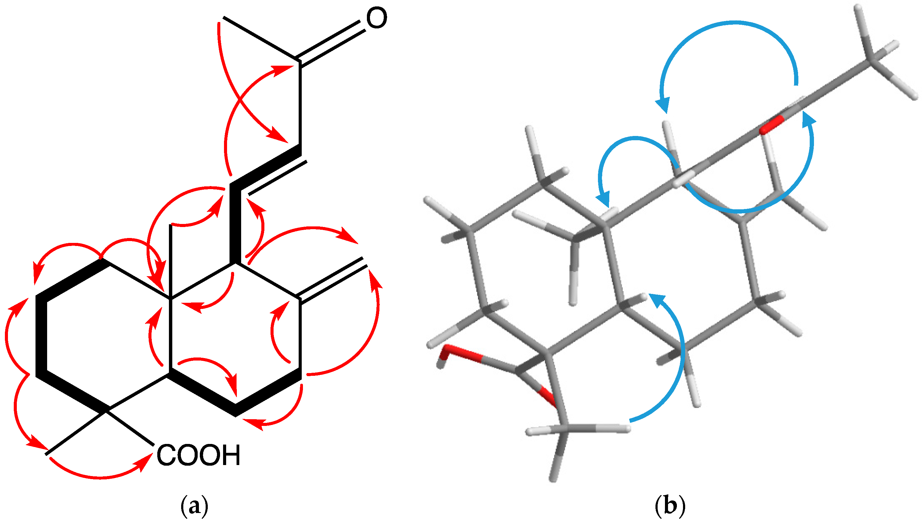
From the COSY spectrum (Figure 2), correlations between H2-1 (δH 1.42, 1.14), H2-2 (δH 1.88, 1.43), and H2-3 (δH 1.08, 2.14) suggested a typical A-ring skeleton of a diterpene without further substitution. HMBC correlations were observed between H2-3 and the methyl group, C-18 (δC 29.4), and a carboxylic acid, C-19, (Figure 2). Additionally, C-19 showed HMBC correlations, with H-5 (δH 1.44) and H-18 (δH 1.21) establishing the location of both the carboxylic acid and methyl group at C-4. COSY correlations of H2-6 (δH 2.00, 1.93) to H-5 and H2-7 (δH 2.45, 2.06) were observed, as well as HMBC correlations between H2-7 and both C-8 and C-17, establishing the position of the exocyclic double bond. Observation of HMBC correlations from H-9 (δH 2.57) to C-5 (δC 56.4) and C-17 further supported this assignment. COSY correlations from H-11 (δH 6.95) to H-12 (δH 6.08) and H-9 established that the second double bond was attached at C-9. The ketone carbonyl carbon, C-13, displayed HMBC correlations, with H-11, H-12, and a methyl group, H3-16, while H-12 showed correlations with C-13 and C-16, establishing the structure of the side chain. Correlations in the NOESY spectrum from H-11 to H3-20 (-CH3) and H3-16 (-CH3) (Figure 2) showed the spatial proximity of these groups, while correlations between H-12 and H-9, H-5, and H-18 supported a chair conformation for both rings. Based on these observations, the structure of 1 was determined to be 13-des-ethyl-13-oxolabda-8(17),11E-dien-19-oic acid.
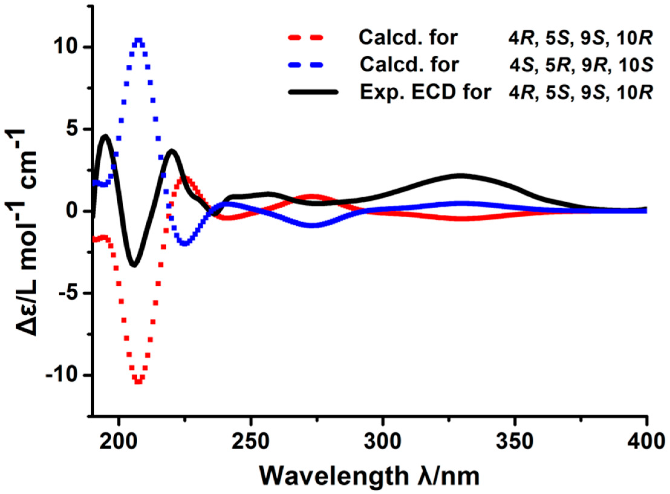
Applying the CD exciton chirality method determined the absolute configuration of compound 1. The experimental ECD spectrum of 1 showed Cotton effects (CEs) at 200–250 nm, including a positive CE at 220 nm, which is indicative of a butene moiety and a negative CE at 205 nm due to a n → π transition. The calculated ECD spectrum for the 4R, 5S, 9S, and 10R configuration matched the experimental data of compound 1. Therefore, the absolute configuration of 1 is (4R,5S,9S,10R)-13-des-ethyl-13-oxolabda-8(17),11E-dien-19-oic acid. (Figure 3)
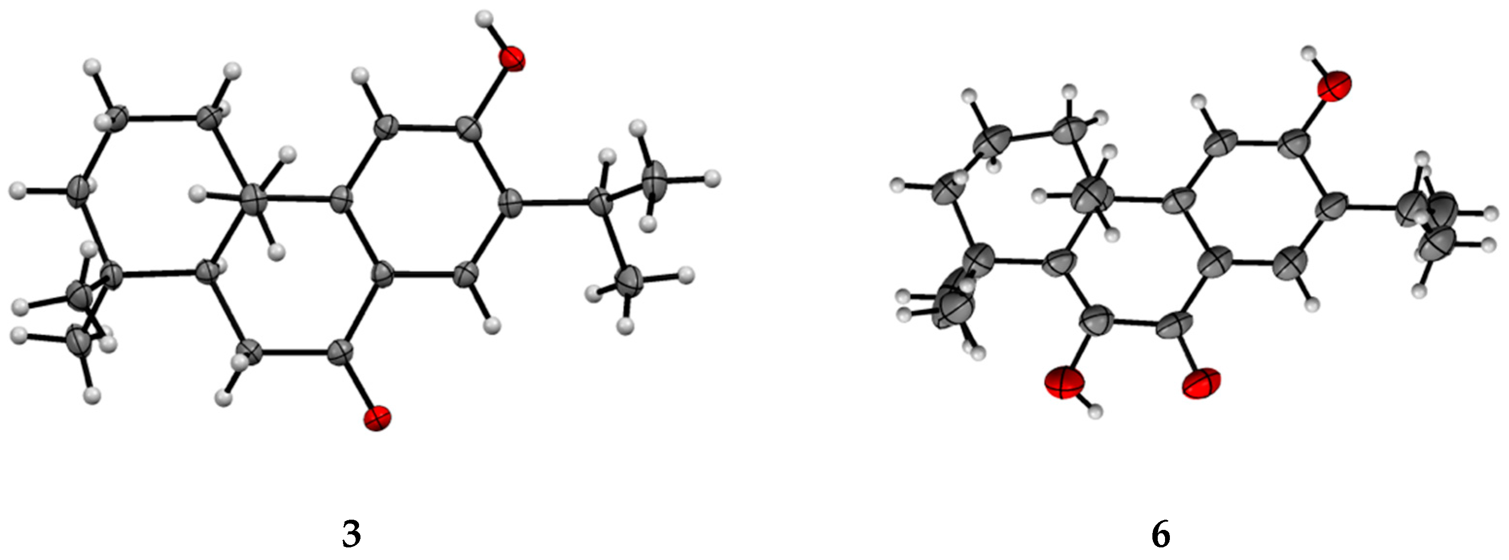
Single crystal X-ray diffraction studies allowed for the elucidation of the structures of 3 and 6 as sugiol (3) [18], and 6,12-dihydroxyabieta-5,8,11,13-tetraen-7-one (6) [19] (Figure 4). While the crystal structure of sugiol has been previously reported [18], this is the first report of the crystal structure of 6, which has a CCDC (Cambridge Crystallographic Data Centre) code: 1900758. Supplementary Information (Tables S1–S7) presents the X-ray diffraction data, parameters, bond lengths, and bond angles.
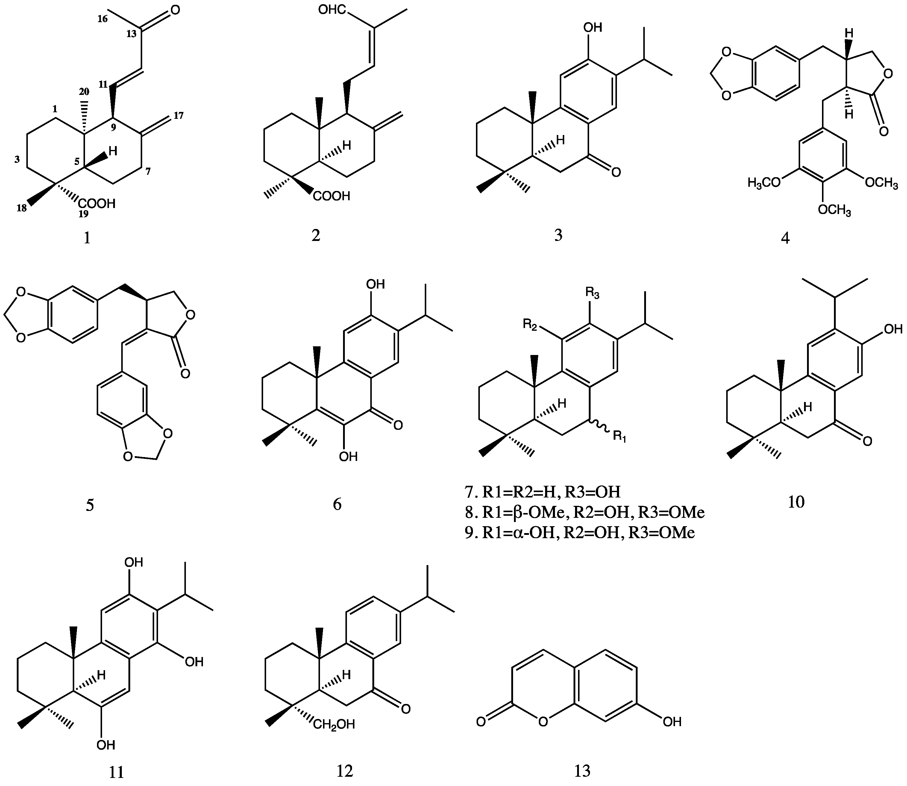
The remaining compounds were identified by comparison of their spectroscopic data with those that were reported in the literature. The other seven diterpenoids were identified as 15-nor-14-oxolabda-8(17),12Z-dien-19-oic acid (2) [20], ferruginol (7) [21], 7α-methoxydeoxocryptojaponol (8) [22], 7β-hydroxydeoxocryptojaponol (9) [23], sempervirol (10) [24], trilobinol (11) [25], and 7-oxodehydroabietinol (12) [26]. Two lignans were identified as (−)-yatein (4) [27] and helioxanthin (5) [28]. One coumarin was determined as umbelliferone (13) [29]. To date, none of these diterpenoids have been reported from Juniperus oblonga. (4R,5S,9S,10R)-13-des-ethyl-13-oxolabda-8(17),11E-dien-19-oic acid (1), 15-nor-14-oxolabda-8(17),12Z-dien-19-oic acid (2), sempervirol (10), trilobinol (11), and 7-oxodehydroabietinol (12) are first reported here from the genus Juniperus.
When isolated quantities permitted, the isolated compounds were evaluated for cytotoxicity against three cancer cell lines, including a human hepatocellular carcinoma cell line (HepG2),a human breast cancer cell line (MCF-7) and a human cervical carcinoma cancer cell line (HeLa), as well as a normal human liver cell line (LO2), at an initial concentration of 4 mg/mL (Table 2) using a standard MTT assay [30]. The IC50 values were determined for compounds that showed greater than 50% inhibition at this concentration. Compound 6 exhibited moderate cytotoxicity against all three cell lines, with IC50 values ranging from 24.41 to 58.39 μM, while 10 showed weaker activity, with IC50 values that ranged from 56.93 to 79.98 μM (Table 3). The remaining compounds did not exhibit cytotoxicity. The absence of cytotoxicity against the normal human liver cell line (LO2) may have some importance. While none of the compounds that were isolated in this study have the potential for development into clinically useful anticancer agents, other labdane and abietane diterpenoids have shown antibacterial, antifungal, and anti-inflammatory activities [17,31,32,33]. Members of these diterpene families not being cytotoxic to normal human cells may enhance their potential for development for these other bioactivities.
3. Materials and Methods
3.1. General Experimental Procedures
ECD measurements were determined on a BioLogic SAS MOS-500 spectropolarimeter (Bio-Logic SAS, Claix, France). Analytical and semipreparative HPLC separations were performed using an Agilent 1260VL instrument (G1311C pump, G1329B autosampler, G1316A thermostatted column compartment and G1315D photodiode array detector, Agilent Technologies, Santa Clara, CA, USA). Analytical HPLC separations were carried out using a SunFire C18 column (3.5 μm particles, 2.1 × 150 mm, Waters Corp., Milford, MA, USA). Semi-preparative and preparative HPLC separations were performed on a Hypersil Gold C18 column (5 μm particles, 10 × 250 mm, Thermo Scientific, Waltham, MA, USA) and a Hypersil Gold C18 column (5 μm particles, 21.2 × 250 mm, Thermo Scientific). The UV spectra were recorded using a Hitachi U-3900 spectrophotometer (Hitachi Limited, Tokyo, Japan). The samples were routinely evaporated while using a Thermo Scientific SC210A SpeedVac concentrator. NMR spectra were recorded on Bruker Avance III 600MHz and 400MHz spectrometers (Bruker-Biospin Corp., Billerica, MA, USA). Residual solvent resonances were used as the internal reference, and chemical shifts are reported in δ (parts per million). Mass spectra were determined on an Agilent Technologies 6230 TOF LC-MS or an Agilent Technologies 6420 triple quadrupole LC-MS using electrospray ionization in the positive and negative modes. Column chromatography was performed on silica gel (60 Å, 40–63 μm) that was purchased from Sorbent Technologies (Norcross, GA, USA). All of the solvents used for isolation were of HPLC grade (Concord Technology, Tianjin, China). Dulbecco’s modified Eagle’s medium (DMEM), Roswell Park Memorial Institute 1640 (RPMI-1640), Phosphate buffered saline (PBS), and fetal bovine serum (FBS) were purchased from Gibco Laboratories (Gaithersburg, MD, USA). Penicillin-Streptomycin solution (100X) was purchased from Beijing Solarbio Science and Technology (Beijing, China). 3-(4,5-Dimethylthiazol-2-yl)-2,5-diphenyl tetrazolium bromide (MTT) was purchased from Biotopped Technology (Beijing, China).
3.2. X-ray Diffraction Analyses
The single crystal X-ray diffraction data were collected on a ROD, Synergy Custom system, HyPix diffractometer (Rigaku, Japan). The crystal was maintained at 159.99(10) K during data collection. The structures were solved using Olex2 with the ShelXT structure solution program using intrinsic phasing and refined with the ShelXL refinement package while using least squares minimization.
3.3. Plant Material
Samples of the roots, stems, leaves, and fruits of Juniperus oblonga M. Bieb. (Cupressaceae) were individually collected near Kish Village in the Sheki District of Azerbaijan in September 2006. Herbarium specimens documenting the collection (Kerimov 57) have been deposited in the herbaria of the Institute of Botany, Azerbaijan National Academy of Sciences (BAK) and the New York Botanical Garden (NY).
3.4. Extraction and Isolation
Fresh plant samples were freed of extraneous matter, air-dried, and then milled to a coarse powder. A 1 kg portion of each dried sample was extracted with methanol (3 × 4 L). After the removal of solvent, the resulting viscous oil was dispersed in 1 L of methanol: water (9:1) and extracted with n-hexane (3 × 1 L). The hexane-depleted hydroalcoholic phase was freed of methanol, dispersed in distilled water (1 L), and then sequentially extracted with dichloromethane and water saturated n-butanol (each 3 × 1 L). The resulting solvent-soluble fractions were each evaporated to dryness in vacuo, while the residual aqueous fraction was freed of solvent and lyophilized. The dichloromethane part (7.46 g) was fractionated by column chromatography on silica gel, eluting with a step-gradient of CH2Cl2/MeOH (100:0, 99:1, 98:2, 97:3, 95:5, 90:10, 80:20, 50:50, 100:0) to give twenty-seven fractions (Fr. 1–Fr. 27).
Compound 3 was crystallized by the slow evaporation of the CH2Cl2/MeOH mixture from Fr. 5.
Fr. a (1.1 g), formed by combining fraction 5 (after depletion of 3 by crystallization) with fractions 6–13, was subjected to silica gel column chromatography eluting with a step- gradient of n-hexane/ethyl acetate (8:2, 7:3, 6:4, 5:5, 0:10) to yield five fractions (Fr. a-1 to Fr. a-5). Fr. a-3 (290 mg) was separated by preparative HPLC eluting with a linear gradient of ACN/H2O (20–100% ACN, 10–50 min) that contained 0.1% formic acid at 10 mL/min to afford four subfractions (Fr. a-3-1 to Fr. a-3-4).
Fr. a-3-2 (78 mg) was purified by preparative HPLC eluting with ACN/H2O (40:60) containing 0.1% formic acid at 10 mL/min, affording 2 (1 mg). Further fractionation by semi-preparative HPLC eluting with ACN/H2O (45:55) containing 0.1% formic acid at 4 mL/min afforded compound 13 (1.1 mg).
Fr. a-3-3 (100 mg) was purified by preparative HPLC eluting with ACN/H2O (35:65) containing 0.1% formic acid at 10 mL/min, to afford compound 1 (2.3 mg), 4 (1.4 mg), and 5 (1.1 mg).
Fractions 2-4 were pooled to form Fr. b (240 mg), which was subjected to silica gel column chromatography, eluting with a step-gradient of n-hexane/ethyl acetate (95:5, 92.5:7.5, 90:10, 80:20, 50:50, 0:100) to give eighteen fractions (Fr. b-1 to Fr. b-18).
Fr. b-1 (57 mg) was purified by semi-preparative HPLC eluting with ACN/H2O (10:90) containing 0.1% formic acid at 4 mL/min, affording compound 12 (2.3 mg).
Fr. b-3 (35 mg) was purified by semi-preparative HPLC eluting with ACN/H2O (65:35) containing 0.1% formic acid at 4 mL/min, affording compound 7 (1.5 mg) and 8 (2 mg).
Fr. b-5 (48 mg) was purified by semi-preparative HPLC eluting with ACN/H2O (60:40) containing 0.1% formic acid at 4 mL/min, affording compound 6, 9 (1.3 mg), and 10 (2.5 mg). Compound 6 (2.1 mg) was crystallized by the slow evaporation of the ACN/H2O mixture.
Fr. b-9 (10 mg) was subjected to semi-preparative HPLC eluting with ACN/H2O (55:45) containing 0.1% formic acid at 4 mL/min, to afford compound 11 (1 mg).
(4R,5S,9S,10R)-13-Des-ethyl-13-oxolabda-8(17),11E-dien-19-oic acid (1): Colorless amorphous powder; UV (DMSO) λmax (log ε) 246 (3.05) and 231 (2.52) nm; HR-TOF-MS m/z 289.1809 [M − H]−, (calcd. for C18H25O3−, m/z 289.1804). ECD (DMSO) λ (Δε) 205 (−3.14), 220 nm (+3.52). 1H-NMR and 13C-NMR spectral assignments, see Table 1.
3.5. Single Crystal X-ray Diffraction Analysis
3.5.1. Crystallographic Data for 3
The crystal data for 3 (C20H28O2) is clear light colorless needle crystal, crystal size 0.2 × 0.2 × 0.2 mm3, orthorhombic, space group P212121 (no. 19), a = 9.54890(10) Å, b = 12.6943(2) Å, c = 14.1587(2) Å, V = 1716.27(4) Å3, Z = 4, T = 100.0(3) K, μ (Cu Kα) = 0.565 mm−1, Dcalc = 1.163 g/cm3, 9031 reflections measured (9.356° ≤ 2θ ≤ 148.94°), 3362 unique (Rint = 0.0317, Rsigma = 0.0323), which were used in all of the calculations. The final R1 was 0.0331 (I > 2σ(I)) and wR2 was 0.0867 (all data).
3.5.2. Crystallographic Data for 6
The crystal data for 6 (C20H26O3) is colorless needle crystal, crystal size 0.15 × 0.15 × 0.2 mm3, orthorhombic, space group P212121 (no. 19), a = 10.4261(4) Å, b = 14.6852(6) Å, c = 23.2228(12) Å, V = 3555.6(3) Å3, Z = 8, T = 159.99(10) K, μ (Cu Kα) = 0.614 mm−1, Dcalc = 1.175 g/cm3, 12,897 reflections measured (7.122° ≤ 2θ ≤ 154.618°), 6523 unique (Rint = 0.1032, Rsigma = 0.1098), which were used in all calculations. The final R1 was 0.0945 (I > 2σ(I)) and wR2 was 0.2949 (all data).
CCDC 1900758 contains the supplementary crystallographic data for this paper. These data can be obtained free of charge via http://www.ccdc.cam.ac.uk/conts/retrieving.html (or from the CCDC, 12 Union Road, Cambridge CB2 1EZ, UK; Fax: +44 1223 336033; E-mail: [email protected]).
3.6. Cytotoxicity Assay
MTT assay was used to measure the in vitro cytotoxicity of the isolated compounds. Human breast adenocarcinoma cell line (MCF-7), human liver hepatocellular carcinoma cell line (HepG2), and human cervical cancer cell line (HeLa) were cultured in Dulbecco’s modified Eagle’s medium (DMEM) and Roswell Park Memorial Institute medium (RPMI 1640), which was supplemented with 10% fetal bovine serum (FBS) at 37 °C in a humidified atmosphere of 5% CO2. The cells were cultured in 96-well plates for 24 h and then treated with test compounds at various concentrations (8–125 μM) for 72 h. After incubation for another 4 h with a 20 µL aliquot of the MTT [3-(4,5-dimethylthiazol-2-yl)-2,5-diphenyl tetrazolium bromide] solution (5 mg/mL in PBS), the medium was discarded, and 150 µL of DMSO was added to dissolve the produced formazan. The absorbance was measured at 490 nm and 570 nm using a microplate reader. Doxorubicin was used as a positive control. Each experiment was carried out in triplicate. The IC50 values were calculated using Graphpad Prism 5 software.
3.7. Calculations of the CD Spectra
The theoretical calculations of the model molecules were carried out using Gaussian 09. MMFF94 was used to initially perform conformational analysis. The conformers were optimized at the B3LYP/6-31 G (d) level. Room temperature equilibrium populations were calculated according to the Boltzmann distribution law. Table S7 (Supplementary Material) show the optimized conformation geometries, thermodynamic parameters, and populations.
4. Conclusions
Thirteen compounds, including one previously undescribed labdane diterpene, nine known diterpenoids, two lignans, and a coumarin were isolated from Juniperus oblonga, and their structures were primarily elucidated on the basis of NMR and MS studies. The absolute configuration of the new compound was determined by CD exciton chirality and calculated ECD methods. The crystal structures were determined for two of the diterpenes (3 and 6) by single crystal X-ray diffraction analyses. The isolated compounds were tested for cytotoxicity against three cell lines, with 6 showing moderate cytotoxicity against all three cell lines and 10 exhibiting weaker activity.
Supplementary Materials
The following are available online: NMR, MS data of compound 1, data of ECD calculation for compound 1, single crystal X-ray diffraction data and refinement parameters with bond lengths and bond angles of compound 3 and 6. IC50 valves of selected compounds.
Author Contributions
M.K., V.A. and D.A. collected and identified the plant material, prepared and submitted voucher specimens and reviewed this manuscript. Y.Q. and R.P.B. conceived and performed the isolation, structure determination and cytotoxicity testing of the isolated compounds, prepared and reviewed this manuscript.
Funding
This research was funded in part by the National Basic Research Program of China, grant number 2015CB856500.
Acknowledgments
The authors kindly acknowledge Matthias Bureik at Tianjin University for providing the MCF-7 and HepG2 cell lines and Haixia Chen, also of Tianjin University, for providing the HeLa cell line.
Conflicts of Interest
The authors declare no conflict of interest.
References
- Adams, R.P. The leaf essential oils and chemotaxonomy of Juniperus sect. Juniperus. Biochem. Syst. Ecol. 1998, 26, 637–645. [Google Scholar] [CrossRef]
- Emami, S.A.; Asili, J.; Mohagheghi, Z.; Hassanzadeh, M.K. Antioxidant activity of leaves and fruits of Iranian conifers. Evid.-Based Complement. Altern. 2007, 4, 313–319. [Google Scholar] [CrossRef] [PubMed]
- San, F.A.; Gordaliza, M.; Salinero, M.A.; Jm, M.D.C. Abietane acids: Sources, biological activities, and therapeutic uses. Planta Med. 1993, 59, 485–490. [Google Scholar]
- Agrawal, O.P.; Bharadwaj, S.; Mathur, R. Antifertility effects of fruits of Juniperus communis. Planta Med. 1980, 40, 98–101. [Google Scholar] [CrossRef] [PubMed]
- Wang, W.S.; Li, E.W.; Jia, Z.J. Terpenes from Juniperus przewalskii and their antitumor activities. Pharmazie 2002, 57, 343–345. [Google Scholar] [PubMed]
- Pepeljnjak, S.; Kosalec, I.; Kalodera, Z.; Blazević, N. Antimicrobial activity of juniper berry essential oil (Juniperus communis L. Cupressaceae). Acta Pharm. 2005, 55, 417–422. [Google Scholar]
- Akdogan, M.; Koyu, A.; Ciris, M.; Yildiz, K. Anti-hypercholesterolemic activity of J. communis Oil in rats: A biochemical and histopathological investigation. Biomed. Res. 2012, 23, 321–328. [Google Scholar]
- Kagawa, K.; Tokura, K.; Uchida, K.; Kakushi, H.; Shike, T.; Kikuchi, J.; Nakai, H.; Dorji, P.; Subedi, L. Platelet aggregation inhibitors in a Bhutanese medicinal plant, shug chher. Chem. Pharm. Bull. 1993, 41, 1604–1607. [Google Scholar] [CrossRef]
- Asili, J.; Emami, S.A.; Rahimizadeh, M.; Fazly-Bazzaz, B.S.; Hassanzadeh, M.K. Chemical and Antimicrobial Studies of Juniperus communis subsp. hemisphaerica and Juniperus oblonga Essential Oils. J. Essent. Oil Bear. Plants 2008, 11, 96–105. [Google Scholar] [CrossRef]
- Emami, S.A.; Asgary, S.; Naderi, G.A.; Ardekani, M.R.S.; Aslani, S.; Airin, A.; Kasher, T.; Sahebkar, A. Investigation of antioxidant and anti-glycation properties of essential oils from fruits and branchlets of Juniperus oblonga. Rev. Bras. Farmacogn. 2012, 22, 985–993. [Google Scholar] [CrossRef]
- Zhang, F.G.; Ren-Jie, H.U.; Zhang, S.Y.; Zhang, Y.W.; Gao, W.Y.; Duan, H.Q. Anticancer activity of diterpenes from Veronica sibirica in vitro. Chin. Tradit. Herbal Drugs 2005, 39, 967–970. [Google Scholar]
- Lin, F.M.; Tsai, C.H.; Yang, Y.C.; Tu, W.C.; Chen, L.R.; Liang, Y.S.; Wang, S.Y.; Shyur, L.F.; Chien, S.C.; Cha, T.L. A novel diterpene suppresses CWR22Rv1 tumor growth in vivo through antiproliferation and proapoptosis. Cancer Res. 2008, 68, 6634–6642. [Google Scholar] [CrossRef]
- Konoshima, T.; Konishi, T.; Takasaki, M.; Yamazoe, K.; Tokuda, H. Anti-tumor-promoting activity of the diterpene from Excoecaria agallocha. II. Biol. Pharm. Bull. 2001, 24, 1440–1442. [Google Scholar] [CrossRef]
- Hiruma-Lima, C.A.; Toma, W.; Gracioso, J.S.; de Almeida, A.B.A.; Batista, L.M.; Magri, L.; de Paula, A.C.B.; Soares, F.R.; Nunes, D.S.; Bruto, A.R.M.S. Natural trans-crotonin: The antiulcerogenic effect of another diterpene isolated from the bark of Croton cajucara Benth. Biol. Pharm. Bull. 2002, 25, 452–456. [Google Scholar] [CrossRef] [PubMed]
- Lahlou, S.; Correia, C.A.B.; Vasconcelos dos Santos, M.; David, J.M.; David, J.P.; Duarte, G.P.; Magalhaes, P.J.C. Mechanisms underlying the cardiovascular effects of a labdenic diterpene isolated from Moldenhawera nutans in normotensive rats. Vascul. Pharmacol. 2007, 46, 60–66. [Google Scholar] [CrossRef]
- Singh, M.; Pal, M.; Sharma, R.P. Biological activity of the labdane diterpenes. Planta Med. 1999, 65, 2–8. [Google Scholar] [CrossRef] [PubMed]
- Demetzos, C.; Dimas, K.; Hatziantoniou, S.; Anastasaki, T.; Angelopoulou, D. Cytotoxic and anti-inflammatory activity of labdane and cis-clerodane type diterpenes. Planta Med. 2001, 67, 614–618. [Google Scholar] [CrossRef] [PubMed]
- Rajouani, N.; Ait Itto, M.Y.; Benharref, A.; Auhmani, A.; Daran, J.C. 6-Hydroxy-7-isopropyl-1,1,4a-trimethyl-2,3,4,4a,10,10a-hexahydrophenanthren-9(1H)-one. Acta Crystallogr. E 2008, 64, 762. [Google Scholar] [CrossRef]
- Wenchiung, S.U.; Fang, J.; Cheng, Y. Abietanes and kauranes from leaves of Cryptomeria japonica. Phytochemistry 1994, 35, 1279–1284. [Google Scholar]
- Kobayashi, M.; Ishida, K.; Terabayashi, S.; Mitsuhashi, H. 10-Hydroxypheophytins and norlabdane diterpene from the leaves of Cupressus funebris Endl. Chem. Pharm. Bull. 1991, 39, 3348–3349. [Google Scholar] [CrossRef]
- Mirzaei, H.H.; Firuzi, O.; Schneider, B.; Baldwin, I.T.; Jassbi, A.R. Cytotoxic diterpenoids from the roots of Salvia lachnocalyx. Rev. Bras. Farmacogn. 2017, 27, 475–479. [Google Scholar] [CrossRef]
- Kuo, Y.H.; Wu, T.R.; Cheng, M.C.; Wang, Y. Five new compounds from the heartwood of Juniperus formosana Hayata. Chem. Pharm. Bull. 1990, 38, 3195–3201. [Google Scholar] [CrossRef]
- Kuo, Y.H.; Lin, N.H.; Lin, Y.T. Two New Diterpens Phenols-7α-Methoxydeoxocryptojaponol and 7β-Hydroxydeoxocryptojaponol. J. Chin. Chem. Soc. 2013, 27, 19–22. [Google Scholar] [CrossRef]
- Mangoni, L.; Caputo, R. Sempervirol, a novel type of diterpene phenol. Tetrahedron Lett. 1967, 8, 673–675. [Google Scholar] [CrossRef]
- Ulubelen, A. New Diterpenoids from the Roots of Salvia triloba. Planta Med. 1990, 56, 82–83. [Google Scholar] [CrossRef]
- Tanaka, R.; Ohtsu, H.; Matsunaga, S. Abietane diterpene acids and other constituents from the leaves of Larix kaempferi. Phytochemistry 1997, 46, 1051–1057. [Google Scholar] [CrossRef]
- Harmatha, J.; Budesinsky, M.; Trka, A. The structure of yatein, determination of the positions, and configurations of benzyl groups in lignans of the 2,3-dibenzylbutyrolactone type. Coll. Czech. Chem. Commun. 1982, 47, 644–663. [Google Scholar] [CrossRef]
- Lee, S.; Yoo, H.H.; Piao, X.L.; Kim, J.S.; Kang, S.S.; Shin, K.H. Anti-estrogenic activity of lignans from Acanthopanax chiisanensis root. Arch. Pharm. Res. 2005, 28, 186–189. [Google Scholar] [CrossRef]
- Sankar, S.S.; Gilbert, R.D.; Fornes, R.E. 13C-NMR studies of some hydroxycoumarins and related compounds. Magn. Reson. Chem. 1982, 19, 222–224. [Google Scholar] [CrossRef]
- Gerlier, D.; Thomasset, N. Use of MTT colorimetric assay to measure cell activation. J. Immunol. Methods 1986, 9, 457–463. [Google Scholar] [CrossRef]
- Echeverría, J.; Gonzálezteuber, M.; Urzúa, A. Antifungal activity against Botrytis cinerea of labdane-type diterpenoids isolated from the resinous exudate of Haplopappus velutinus Remy (Asteraceae). Nat. Prod. Res. 2018, 1–5. [Google Scholar] [CrossRef] [PubMed]
- Chinou, I.; Demetzos, C.; Harvala, C.; Verbist, J. Cytotoxic and antibacterial labdane-type diterpenes from the aerial parts of Cistus incanus subsp. creticus. Planta Med. 1994, 60, 34–36. [Google Scholar] [CrossRef] [PubMed]
- Dellar, J.E.; Cole, M.D.; Waterman, P.G. Antimicrobial abietane diterpenoids from Plectranthus elegans. Phytochemistry 1996, 41, 735–738. [Google Scholar] [CrossRef]
Sample Availability: Samples of the compounds are available from the authors. |
Figure 1.
Structures of compounds 1–13.

Figure 2.
Some of the key correlations observed for compound 1. (a) 1H-1H COSY (bold) and key HMBC (red single headed) correlations of compound 1. (b) NOESY (blue single headed) correlations of compound 1.
Figure 2.
Some of the key correlations observed for compound 1. (a) 1H-1H COSY (bold) and key HMBC (red single headed) correlations of compound 1. (b) NOESY (blue single headed) correlations of compound 1.

Figure 3.
Electronic circular dichroism (ECD) spectrum of compound 1 (The black line is the experimental ECD spectrum, red and blue dashed lines are calculated ECD spectra.).
Figure 3.
Electronic circular dichroism (ECD) spectrum of compound 1 (The black line is the experimental ECD spectrum, red and blue dashed lines are calculated ECD spectra.).

Figure 4.
Crystal structure representations of compound 3 and 6. The structure of 6 was deposited with the Cambridge Crystallographic Data Centre (CCDC) code 1900758.
Figure 4.
Crystal structure representations of compound 3 and 6. The structure of 6 was deposited with the Cambridge Crystallographic Data Centre (CCDC) code 1900758.


Table 1.
NMR spectroscopic data for compound 1 acquired in CD3OD.
| Position | δC | δH, J (Hz) | 1H-1H-COSY | HMBC | NOESY |
|---|---|---|---|---|---|
| 1 | 42.2 | 1.42 m, 1.14 ddd 4.0, 13.0 | H-2 | C-2, C-10, C-20 | |
| 2 | 20.9 | 1.88 td 3.0,13.0, 1.43 m | H-1, H-3 | C-3, C-5 | |
| 3 | 39.3 | 2.14m, 1.08 ddd 3.4, 13.0 | H-2 | C-2, C-4, C-5, C-18, C-19 | |
| 4 | 45.1 | ||||
| 5 | 56.4 | 1.44 m | H-6 | C-6, C-10, C-19, C-20 | H-18 |
| 6 | 26.4 | 2.00m, 1.93 ddd 3.8, 13.0 | H-7 | C-5, C-7 | |
| 7 | 38.3 | 2.45 dt 3, 12, 2.06 ddd 5.0,13.0 | H-6 | C-5, C-8, C-6, C-17 | |
| 8 | 149.9 | ||||
| 9 | 61.4 | 2.57 d 10 | H-11 | C-5, C-10, C-11, C-12, C-17, C-20 | H-12 |
| 10 | 41.0 | ||||
| 11 | 148.7 | 6.95 dd 5.0 10.0 | H-9, H-12 | C-8, C-9, C-10, C-13 | H-12, H-14, H-20 |
| 12 | 134.6 | 6.08 d 5.0 | H-11 | C-8, C-9, C-13, C-16 | H-11 |
| 13 | 200.9 | ||||
| 16 | 27.1 | 2.27 s | C-11, C-12, C-13 | H-11, | |
| 17 | 108.9 | 4.80 d 1.5, 4.42 d 1.5 | C-7, C-8, C-9 | ||
| 18 | 29.4 | 1.21 s | C-2, C-3, C-4, C-5, C-19 | H-5 | |
| 19 | 181.2 | ||||
| 20 | 14.2 | 0.83 s | C-5, C-9, C-10 | H-11 |
Table 2.
Cytotoxicity (%I @ 4 mg/mL)) of compounds from Juniperus oblonga against various cell lines.
Table 2.
Cytotoxicity (%I @ 4 mg/mL)) of compounds from Juniperus oblonga against various cell lines.
| Compound | HepG2 | HeLa | MCF-7 | LO2 |
|---|---|---|---|---|
| 1 | 18.39 | 26.36 | 28.45 | 20.57 |
| 2 | 16.06 | 23.95 | 21.08 | 10.61 |
| 3 | 35.05 | 12.29 | 9.2 | 26.62 |
| 5 | 0.11 | 0.22 | 10.09 | 9.05 |
| 6 | 86.57 | 83.83 | 83.99 | 27.76 |
| 8 | 7.84 | 0 | 1.35 | 17.07 |
| 9 | 24.33 | 0 | 10.96 | 22.21 |
| 10 | 48.67 | 63.4 | 43.4 | 19.08 |
| 11 | 67.12 | 22.01 | 29.88 | 23.29 |
| 13 | 32.71 | 3.6 | 17.96 | 26.59 |
| Doxorubicin a | 42.21 | 49.51 | 46.46 | 46.7 |
a: Doxorubicin was tested as a positive control.
Table 3.
Cytotoxicity of compounds 6 and 10 against three human cancer cell lines.
| Compounds | Cytotoxicity (IC50: μM) a | ||
|---|---|---|---|
| HepG2 | MCF-7 | Hela | |
| 6 | 48.73 ± 1.31 | 58.39 ± 2.45 | 24.41 ± 2.05 |
| 10 | 64.94 ± 2.64 | 79.98 ± 1.20 | 56.93 ± 2.39 |
| Doxorubicin b | 3.18 ± 1.19 | 3.44 ± 1.59 | 3.64 ± 1.37 |
a IC50 values are expressed as the mean values of three experiments ± SD; b Doxorubicin was tested as a positive control.
© 2019 by the authors. Licensee MDPI, Basel, Switzerland. This article is an open access article distributed under the terms and conditions of the Creative Commons Attribution (CC BY) license (http://creativecommons.org/licenses/by/4.0/).
Share and Cite
MDPI and ACS Style
Qiao, Y.; Khutsishvili, M.; Alizade, V.; Atha, D.; Borris, R.P. Labdane and Abietane Diterpenoids from Juniperus oblonga and Their Cytotoxic Activity. Molecules 2019, 24, 1561. https://0-doi-org.brum.beds.ac.uk/10.3390/molecules24081561
AMA Style
Qiao Y, Khutsishvili M, Alizade V, Atha D, Borris RP. Labdane and Abietane Diterpenoids from Juniperus oblonga and Their Cytotoxic Activity. Molecules. 2019; 24(8):1561. https://0-doi-org.brum.beds.ac.uk/10.3390/molecules24081561
Chicago/Turabian StyleQiao, Yilin, Manana Khutsishvili, Valida Alizade, Daniel Atha, and Robert P. Borris. 2019. "Labdane and Abietane Diterpenoids from Juniperus oblonga and Their Cytotoxic Activity" Molecules 24, no. 8: 1561. https://0-doi-org.brum.beds.ac.uk/10.3390/molecules24081561