
Chemical Characterization and Evaluation of the Antibacterial Activity of Essential Oils from Fibre-Type Cannabis sativa L. (Hemp)

 ,
,  , , and
Abstract
:
1. Introduction
2. Results and Discussion
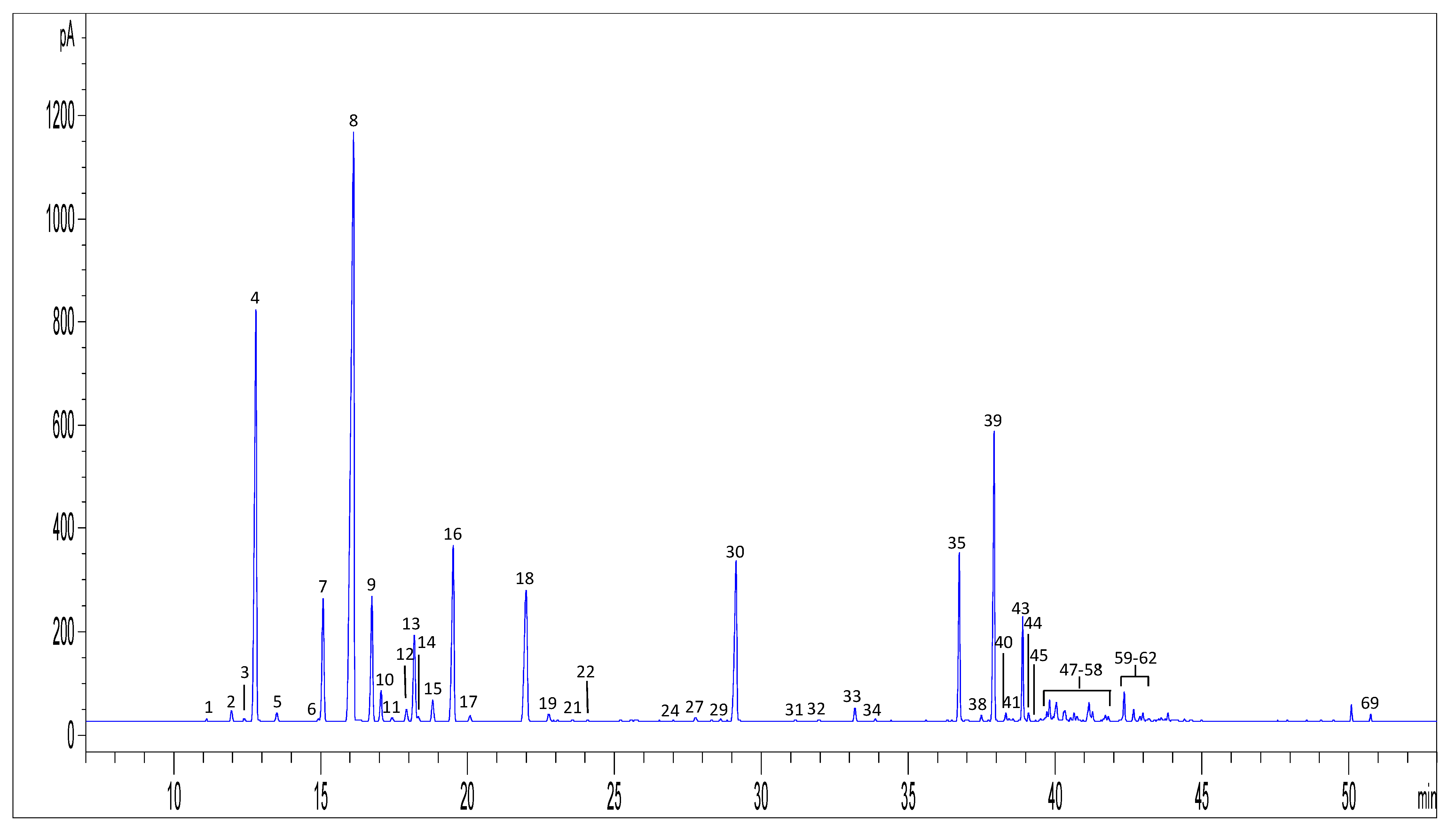
2.1. Qualitative and Semi-Quantitative Analysis of Hemp EOs
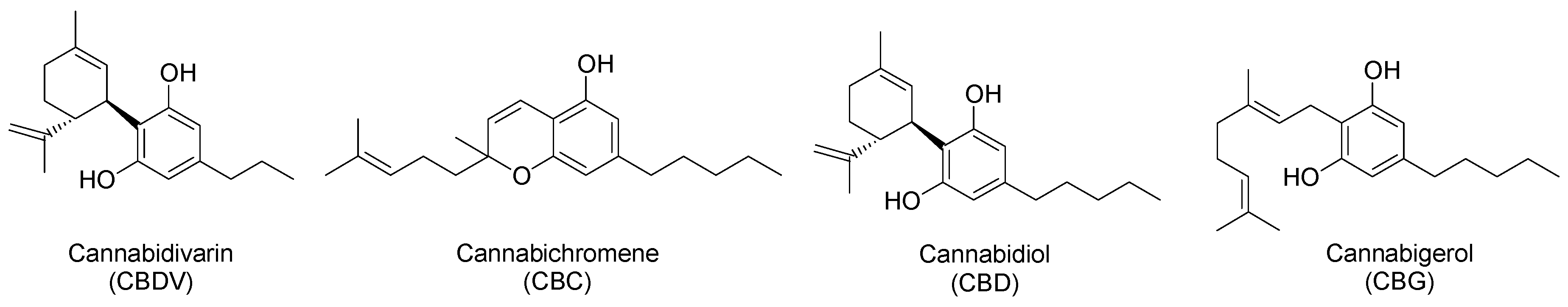
2.2. Analysis of Cannabinoids in Hemp EOs
2.3. Preliminary Screening by Means of the Agar Well Disk Diffusion Assay
2.4. Minimum Inhibitory Concentration (MIC)
2.5. Chemical Composition–Bioactivity Relationships
3. Materials and Methods
3.1. Chemicals and Solvents
3.2. Hemp EO Samples
3.3. GC-MS Analysis of Volatile Compounds
3.4. GC-FID Analysis of Volatile Compounds
3.5. Qualitative and Semi-Quantitative Analysis of Volatile Compounds
3.6. Quantitative GC-MS Analysis of Cannabinoids
3.7. Bacterial Strains
3.8. Agar Well Disk Diffusion Assay
3.9. Minimum Inhibitory Concentration (MIC) Assay
4. Conclusions
Supplementary Materials
Author Contributions
Funding
Acknowledgments
Conflicts of Interest
References
- Leghissa, A.; Hildenbrand, Z.L.; Schug, K.A. The imperatives and challenges of analyzing Cannabis edibles. Curr. Opin. Food Sci. 2018, 28, 18–24. [Google Scholar] [CrossRef]
- Leghissa, A.; Hildenbrand, Z.L.; Schug, K.A. A review of methods for the chemical characterization of Cannabis natural products. J. Sep. Sci. 2018, 41, 398–415. [Google Scholar] [CrossRef] [PubMed]
- Marchetti, L.; Brighenti, V.; Rossi, M.C.; Sperlea, J.; Pellati, F.; Bertelli, D. Use of 13C-qNMR Spectroscopy for the analysis of non-psychoactive cannabinoids in fibre-type Cannabis sativa L. (hemp). Molecules 2019, 24, 1138. [Google Scholar] [CrossRef]
- Protti, M.; Brighenti, V.; Battaglia, M.R.; Anceschi, L.; Pellati, F.; Mercolini, L. Cannabinoids from Cannabis sativa L.: A new tool based on HPLC-DAD-MS/MS for a rational use in medicinal chemistry. ACS Med. Chem. Lett. 2019, 10, 539–544. [Google Scholar] [CrossRef] [PubMed]
- Pellati, F.; Brighenti, V.; Sperlea, J.; Marchetti, L.; Bertelli, D.; Benvenuti, S. New methods for the comprehensive analysis of bioactive compounds in Cannabis sativa L. (hemp). Molecules 2018, 23, 2639. [Google Scholar] [CrossRef] [PubMed]
- Citti, C.; Braghiroli, D.; Vandelli, M.A.; Cannazza, G. Pharmaceutical and biomedical analysis of cannabinoids: A critical review. J. Pharm. Biomed. Anal. 2018, 147, 565–579. [Google Scholar] [CrossRef]
- Brighenti, V.; Pellati, F.; Steinbach, M.; Maran, D.; Benvenuti, S. Development of a new extraction technique and HPLC method for the analysis of non-psychoactive cannabinoids in fibre-type Cannabis sativa L. (hemp). J. Pharm. Biomed. Anal. 2017, 143, 228–236. [Google Scholar] [CrossRef]
- Corsi, L.; Pellati, F.; Brighenti, V.; Plessi, N.; Benvenuti, S. Chemical composition and in vitro neuroprotective activity of fibre-type Cannabis sativa L. (hemp). Curr. Bioact. Compd. 2019, 15, 201–210. [Google Scholar] [CrossRef]
- Pellati, F.; Borgonetti, V.; Brighenti, V.; Biagi, M.; Benvenuti, S.; Corsi, L. Cannabis sativa L. and nonpsychoactive cannabinoids: Their chemistry and role against oxidative stress, inflammation and cancer. BioMed Res. Int. 2018, 2018, 1691428. [Google Scholar] [CrossRef]
- Borrelli, F.; Fasolino, I.; Romano, B.; Capasso, R.; Maiello, F.; Coppola, D.; Orlando, P.; Battista, G.; Pagano, E.; Di Marzo, V.; et al. Beneficial effect of the non-psychotropic plant cannabinoid cannabigerol on experimentalinflammatory bowel disease. Biochem. Pharmacol. 2013, 85, 1306–1316. [Google Scholar] [CrossRef]
- Borgonetti, V.; Governa, P.; Montopoli, M.; Biagi, M. Cannabis sativa L. constituents and their role in neuroinflammation. Curr. Bioact. Compd. 2019, 15, 147–158. [Google Scholar] [CrossRef]
- Appendino, G.; Gibbons, S.; Giana, A.; Pagani, A.; Grassi, G.; Stavri, M.; Smith, E.; Rahman, M.M. Antibacterial cannabinoids from Cannabis sativa: A structure-activity study. J. Nat. Prod. 2008, 71, 1427–1430. [Google Scholar] [CrossRef] [PubMed]
- Morales, P.; Reggio, P.H.; Jagerovich, N. An overview of medicinal chemistry vof synthetic and natural derivatives of cannabidiol. Front Pharmacol. 2017, 8, 422. [Google Scholar] [CrossRef] [PubMed]
- Fiorini, D.; Molle, A.; Nabissi, M.; Santini, G.; Benelli, G.; Maggi, F. Valorizing industrial hemp (Cannabis sativa L.) by-products: Cannabidiol enrichment in the inflorescence essential oil optimizing sample pretreatment prior to distillation. Ind. Crops Prod. 2019, 128, 581–589. [Google Scholar] [CrossRef]
- Hazekamp, A.; Fischedick, J.T.; Llano Dìez, M.; Lubbe, A.; Ruhaak, R.L. Chemistry of Cannabis. In Comprehensive Natural Products II Chemistry and Biology, 1st ed.; Mander, L., Liu, H.-W., Eds.; Elsevier: Kidlington, UK, 2010; Volume 3, pp. 1034–1077. [Google Scholar]
- Wang, C.-T.; Wiedinmyer, C.; Ashworth, K.; Harley, P.C.; Ortega, J.; Vizuete, W. Leaf enclosure measurements for determining volatile organic compound nemission capacity from Cannabis spp. Atmos. Environ. 2019, 199, 80–87. [Google Scholar] [CrossRef]
- Ibrahim, E.A.; Wang, M.; Radwan, M.M.; Wanas, A.S.; Majumdar, C.G.; Avula, B.; Wang, Y.H.; Khan, I.A.; Chandra, S.; Lata, H.; et al. Analysis of terpenes in Cannabis sativa L. using GC/MS: Method development, validation and application. Planta Med. 2019, 85, 431–438. [Google Scholar] [CrossRef]
- Nagy, D.U.; Cianfaglione, K.; Maggi, F.; Sut, S.; Dall’Acqua, S. Chemical characterization of leaves, male and female flowers from spontaneous Cannabis (Cannabis sativa L.) growing in Hungary. Chem. Biodiversity 2019, 16, e1800562. [Google Scholar] [CrossRef]
- Zengin, G.; Menghini, L.; Di Sotto, A.; Mancinelli, R.; Sisto, F.; Carradori, S.; Cesa, S.; Fraschetti, C.; Filippi, A.; Angiolella, L.; et al. Chromaotgrphic analysis, in vitro biological activities and cytotoxicity of Cannabis sativa L. essential oil: A multidisciplinary study. Molecules 2018, 23, 3266. [Google Scholar] [CrossRef]
- Nissen, L.; Zatta, A.; Stefanini, I.; Grandi, S.; Sgorbati, S.; Biavati, B.; Monti, A. Characterization and antimicrobial activity of essential oils of industrial hemp varieties (Cannabis sativa L.). Fitoterapia 2010, 81, 413–419. [Google Scholar] [CrossRef]
- Tognolini, M.; Barocelli, E.; Ballabeni, V.; Bruni, R.; Bianchi, A.; Chiavarini, M.; Impicciatore, M. Comparative screening of plant essential oils: Phenylpropanoid moiety as basic core of antiplatelet activity. Life Sci. 2006, 78, 1419–1432. [Google Scholar] [CrossRef]
- Andre, C.M.; Hausman, J.F.; Guerriero, G. Cannabis sativa: The plant of the thousand and one molecule. Front. Plant. Sci. 2016, 7, 1–17. [Google Scholar] [CrossRef]
- Russo, E.B. Taming THC: Potential cannabis synergy and phytocannabinoid-terpenoid entourage effects. Br. J. Pharmacol. 2011, 163, 1344–1364. [Google Scholar] [CrossRef] [PubMed]
- Marini, E.; Magi, G.; Ferretti, G.; Bacchetti, T.; Giuliani, A.; Pugnaloni, A.; Rippo, M.R.; Facinelli, B. Attenuation of Listeria monocytogenes virulence by Cannabis sativa L. essential oil. Front. Cell. Infect. Microbiol. 2018, 8, 293. [Google Scholar] [CrossRef]
- Da Porto, C.; Decorti, D.; Natolino, A. Separation of aroma compounds from industrial hemp inflorescences (Cannabis sativa L.) by supercritical CO2 extraction and on-line fractionation. Ind. Crops Prod. 2014, 58, 99–103. [Google Scholar] [CrossRef]
- Bertoli, A.; Tozzi, S.; Pistelli, L.; Angelini, L.G. Fibre hemp inflorescences: From crop-residues to essential oil production. Ind. Crops Prod. 2010, 32, 329–337. [Google Scholar] [CrossRef]
- Calvi, L.; Pentimalli, D.; Panseri, S.; Giupponi, L.; Gelmini, F.; Beretta, G.; Vitali, D.; Bruno, M.; Zilio, E.; Pavlovic, R.; et al. Comprehensive quality evaluation of medical Cannabis sativa L. inflorescence andmacerated oils based on HS-SPME coupled to GC–MS and LC-HRMS (q-exactive orbitrap®) approach. J. Pharm. Biomed. Anal. 2018, 150, 208–219. [Google Scholar] [CrossRef]
- Marchini, M.; Charvoz, C.; Dujourdy, L.; Baldovini, N.; Filippi, J.-J. Multidimensional analysis of cannabis volatile constituents: Identification of 5,5-dimethyl-1-vinylbicyclo[2.1.1]hexane as a volatile marker of hashish, the resin of Cannabis sativa L. J. Chromatogr. A 2014, 1370, 200–215. [Google Scholar] [CrossRef]
- Ross, S.A.; ElSohly, M.A. The volatile oil composition of fresh and air-dried buds of Cannabis sativa. J. Nat. Prod. 1996, 59, 49–51. [Google Scholar] [CrossRef]
- Plant Variety Catalogues, Databases & Information Systems. Available online: https://ec.europa.eu/food/plant/plant_propagation_material/plant_variety_catalogues_databases_en (accessed on 10 May 2019).
- CLSI. Performance Standards for Antimicrobial Disk Susceptibility Tests, 11th ed.; Approved Standard. CLSI Document M02-A11; Clinical and Laboratory Standards Institute: Wayne, PA, USA, 2012. [Google Scholar]
- Klancnik, A.; Piskernik, S.; Jersek, B.; Mozina, S.S. Evaluation of diffusion and dilution methods to determine the antibacterial activity of plant extracts. J. Microbiol. Methods 2010, 81, 121–126. [Google Scholar] [CrossRef]
- Şahin, F.; Güllüce, M.; Daferera, D.; Sökmen, A.; Sökmen, M.; Polissiou, M.; Agar, G.; Özer, H. Biological activities of the essential oils and methanol extract of Origanum vulgare ssp. vulgare in the Eastern Anatolia region of Turkey. Food Control 2004, 15, 549–557. [Google Scholar]
Sample Availability: Samples of hemp EOs are available from the authors. |


| Peak n. | Compound | LRI | LRI lit b | EO1 | EO2 | EO3 | EO4 | EO5 | EO6 | EO7 | EO8 |
|---|---|---|---|---|---|---|---|---|---|---|---|
| 1 | Heptanal | 903 | 901 | - | - | 0.2 a | - | - | - | 0.3 a | - |
| 2 | 5,5-Dimethyl-1-vinylbicyclo[2.1.1]-hexane | 918 | 921 | 0.1 a | - | 0.5 a | 0.5 a | 0.4 a | 0.3 a | 0.4 a | 0.4 a |
| 3 | α-Thujene | 926 | 924 | - | - | - | 0.2 a | 0.1 a | 0.1 a | 0.1 a | - |
| 4 | α-Pinene | 934 | 931 | 7.4 ± 0.3 | 19.4 ± 0.1 | 14.6 ± 0.4 | 18.6 ± 0.2 | 20.4 ± 0.2 | 4.8 ± 0.1 | 16.0 ± 0.1 | 11.0 ± 0.1 |
| 5 | Camphene | 947 | 947 | 0.2 a | - | 0.3 a | 0.4 a | 0.4 a | 0.1 a | 0.3 a | 0.4 a |
| 6 | β-Thujene | 973 | 966 | 0.1 a | - | 0.1 a | - | 0.1 a | 0.1 a | - | |
| 7 | β-Pinene | 976 | 978 | 3.4 ± 0.1 | 7.8 a | 5.5 ± 0.1 | 6.1 ± 0.2 | 8.2 ± 0.1 | 2.1 ± 0.1 | 6.5 a | 4.1 a |
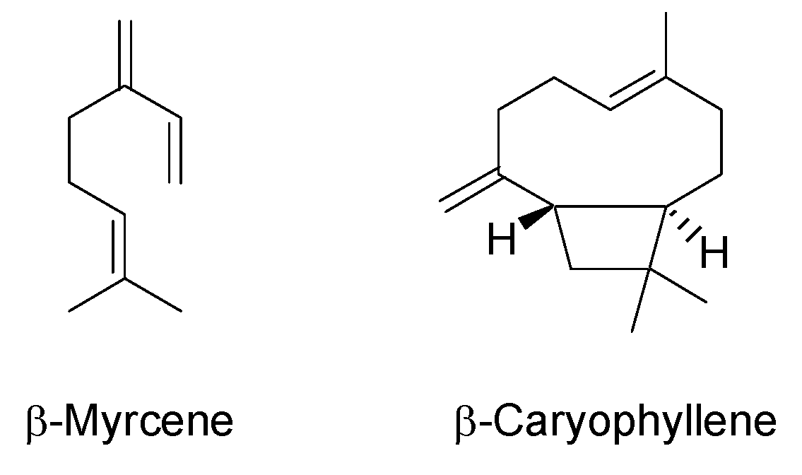
| 8 | β-Myrcene | 994 | 992 | 8.3 a | 29.5 ± 0.3 | 34.4 ± 1.3 | 25.9 ± 1.3 | 30.4 ± 0.5 | 12.5 ± 0.8 | 33.5 ± 0.3 | 4.5 ± 0.1 |
| 9 | α-Phellandrene | 1006 | 1007 | 0.1 a | - | 1.6 ± 0.1 | 0.4 ± 0.2 | 0.3 a | 0.2 a | 0.3 a | - |
| 10 | Δ3-Carene | 1010 | 1010 | 0.2 a | - | 0.6 a | 0.8 a | 0.7 a | 0.9 ± 0.1 | 0.5 a | - |
| 11 | α-Terpinene | 1016 | 1017 | 0.1 a | - | 0.3 a | 0.2 a | 0.3 a | 0.2 a | 0.3 a | 0.3 a |
| 12 | p-Cymene | 1024 | 1026 | - | - | 0.2 a | 0.2 a | 0.1 a | 0.3 a | - | |
| 13 | Limonene | 1028 | 1035 | 0.1 a | 5.7 ± 0.2 | 4.3 ± 0.2 | 4.2 ± 0.4 | 5.5 a | 1.2 ± 0.1 | 4.2 a | 1.9 a |
| 14 | 1,8-Cineole | 1030 | 1032 | 1.8 a | - | 0.3 a | 0.9 ± 0.1 a | 0.2 a | 0.1 a | 0.2 a | 1.8 a |
| 15 | cis-Ocimene | 1038 | 1040 | 0.3 a | - | 0.4 a | 0.6 a | 0.5 a | 0.8 a | 0.5 a | 0.5 a |
| 16 | trans-Ocimene | 1049 | 1050 | 5.0 ± 0.1 | 4.9 ± 0.1 | 4.2 ± 0.2 | 4.2 ± 0.1 | 4.0 ± 0.1 | 5.0 a | 4.6 a | 2.2 a |
| 17 | γ-Terpinene | 1058 | 1062 | 0.1 a | - | 0.3 a | 0.3 a | 0.2 a | 0.2 a | 0.3 a | 0.2 a |
| 18 | α-Terpinolene | 1089 | 1088 | 3.4 ± 0.1 | 7.5 ± 0.1 | 8.3 ± 0.5 | 5.4 ± 0.3 | 8.2 ± 0.2 | 6.6 ± 0.2 | 8.4 ± 0.1 | 1.9 ± 0.1 |
| 19 | Linalool | 1101 | 1104 | - | - | 0.4 a | 0.4 a | 0.2 a | 0.1 a | 0.3 a | 3.7 a |
| 20 | 6-Camphenol | 1105 | 1110 | 0.1 a | - | - | 0.1 a | - | - | 0.1 a | - |
| 21 | Fenchol | 1113 | 1114 | 0.1 a | - | 0.3 ± 0.1 | 0.2 a | 0.2 a | - | 0.2 a | - |
| 22 | neo-Alloocimene | 1121 | 1143 | - | - | - | 0.1 a | 0.1 a | - | 0.2 a | - |
| 23 | trans-Pinocarveol | 1138 | 1139 | - | - | - | 0.1 a | - | - | 0.1 a | 0.2 a |
| 24 | Camphor | 1144 | 1145 | - | - | - | - | - | - | 0.2 a | 0.9 a |
| 25 | Isopulegol | 1146 | 1156 | - | - | - | - | - | - | - | |
| 26 | Borneol | 1166 | 1166 | - | - | - | - | - | - | 0.1 a | 0.4 a |
| 27 | Terpinen-4-ol | 1178 | 1177 | - | - | 0.6 a | 0.2 a | 0.1 a | 0.1 a | 0.2 a | 1.1 a |
| 28 | p-Cymen-8-ol | 1190 | 1183 | - | - | - | - | - | 0.2 a | - | - |
| 29 | α-Terpineol | 1191 | 1193 | - | - | 0.2 a | 0.1 a | 0.1 a | - | 0.2 a | 0.5 ± 0.1 |
| 30 | Methyl chavicol | 1199 | 1196 | 0.1 a | - | 1.8 a | - | - | 0.1 a | - | - |
| 31 | Neral | 1243 | 1249 | - | - | - | - | - | - | - | - |
| 32 | Linalyl acetate | 1261 | 1253 | 0.1 a | - | - | 0.1 a | - | - | 4.3 a | |
| 33 | Bornyl acetate | 1289 | 1287 | - | - | - | - | - | - | - | - |
| 34 | Thymol | 1301 | 1297 | 0.1 a | - | - | 0.1 a | 0.1 a | 0.4 a | ||
| 35 | Geranyl acetate | 1388 | 1392 | - | - | 1.6 ± 0.1 | 0.5 a | 0.1 a | 0.3 a | 0.1 a | 0.3 ± 0.1 |
| 36 | β-Bourbonene | 1389 | 1386 | 0.1 a | - | - | 0.1 a | - | 0.1 a | - | - |
| 37 | α-Gurjunene | 1411 | 1408 | 0.3 a | - | - | 0.3 a | 0.2 a | 0.6 a | 0.2 a | 0.8 a |
| 38 | Isocaryophyllene | 1417 | 1411 | 0.2 a | - | 0.2 a | - | - | - | - | 0.4 a |
| 39 | β-Caryophyllene | 1427 | 1428 | 21.6 ± 0.2 | 20.0 ± 0.6 | 8.5 ± 0.3 | 14.3 ± 0.5 | 9.3 ± 0.2 | 21.1 ± 0.3 | 10.3 ± 0.4 | 29.8 ± 0.2 |
| 40 | α-Bergamotene | 1441 | 1432 | 1.6 ± 0.1 | - | 0.2 a | 0.3 a | 0.2 a | 2.4 ± 0.1 | 0.3 a | 1.9 a |
| 41 | Aromadendrene | 1445 | 1449 | 0.1 a | - | - | 0.2 a | 0.1 a | 0.3 a | 0.1 a | 0.9 a |
| 42 | Aristolene | 1453 | 1450 | - | - | - | 0.1 a | - | - | - | 0.3 a |
| 43 | α-Humulene | 1461 | 1455 | 10.1 ± 0.1 | 5.3 ± 0.3 | 2.8 ± 0.1 | 4.6 ± 0.2 | 2.9 ± 0.1 | 9.7 ± 0.2 | 3.3 ± 0.1 | 10.1 |
| 44 | Alloaromadendrene | 1468 | 1467 | 1.8 ± 0.1 | - | 0.3 a | 0.6 ± 0.1 | 0.2 a | 1.0 ± 0.2 | 0.3 a | 0.7 a |
| 45 | γ-Muurolene | 1482 | 1477 | 0.3 a | - | 0.1 a | 0.2 a | - | 0.5 a | 0.1 a | 0.9 a |
| 46 | Germacrene D | 1487 | 1485 | 0.2 a | - | - | 0.1 a | - | 0.3 a | 0.1 a | - |
| 47 | β-Selinene | 1490 | 1489 | 0.2 a | - | 0.2 a | 0.4 a | 0.2 a | - | 0.3 a | 2.9 ± 0.6 |
| 48 | α-Selinene | 1493 | 1494 | 1.0 ± 0.3 | - | 0.5 a | 0.9 ± 0.1 | 0.5 a | 2.1 ± 0.2 | 0.6 a | - |
| 49 | Valencene | 1497 | 1496 | 3.0 ± 0.4 | - | 0.1 a | 0.1 a | 0.1 a | 0.1 a | 0.1 a | - |
| 50 | β-Bisabolene | 1501 | 1505 | 2.7 ± 0.4 | - | 0.6 ± 0.1 | 0.9 a | 0.5 a | 1.8 ± 0.1 | 0.6 a | 2.2 ± 0.2 |
| 51 | α-Muurolene | 1512 | 1497 | 0.7 ± 0.2 | - | 0.3 ± 0.1 | 0.6 | 0.4 | 0.7 ± 0.1 | 0.4 | 1.6 ± 0.1 |
| 52 | α-Patchoulene | 1521 | 1445 | 0.2 a | - | 0.1 a | 0.1 a | - | 0.3 a | 0.3 a | 0.4 a |
| 53 | γ-Cadinene | 1525 | 1512 | 0.5 ± 0.1 | - | 0.3 a | 0.2 a | 0.2 a | 0.3 a | 0.2 a | - |
| 54 | δ-Cadinene | 1529 | 1519 | 0.5 ± 0.2 | - | 0.2 a | 0.2 a | 0.2 a | 0.7 ± 0.1 | - | 0.8 a |
| 55 | δ-Selinene | 1545 | 1540 | 1.1 ± 0.3 | - | 0.7 ± 0.1 | 0.7 ± 0.1 | 0.8 a | 1.3 a | 0.9 a | 0.4 ± 0.1 |
| 56 | Selina-3,7(11)-diene | 1550 | 1545 | 0.4 a | - | 0.3 ± 0.1 | 0.3 a | 0.2 a | 1.3 ± 0.2 | 0.3 a | - |
| 57 | trans-Nerolidol | 1568 | 1562 | 1.0 ± 0.2 | - | - | 0.2 a | 0.2 a | 0.7 a | 0.2 a | 0.2 a |
| 58 | Spathulenol | 1585 | 1577 | 0.1 a | - | - | - | - | 0.2 a | - | - |
| 59 | Caryophyllene oxide | 1593 | 1583 | 9.5 ± 0.5 | - | 0.8 ± 0.1 | 1.3 ± 0.1 | 0.8 ± 0.1 | 6.6 ± 0.2 | 0.8 | 2.5 ± 0.1 |
| 60 | Viridiflorol | 1607 | 1600 | 0.4 a | - | 0.1 a | - | - | 0.3 a | - | - |
| 61 | Guaiol | 1619 | 1602 | 2.6 ± 0.3 | - | - | 0.3 a | 0.2 a | 1.8 ± 0.1 | 0.2 a | 0.5 a |
| 62 | Humulene-1,2-epoxide | 1621 | 1615 | - | - | 0.2 a | - | - | - | - | - |
| 63 | Caryophylla-4(12),8(13)-dien-5-ol | 1628 | 1634 | 0.5 a | - | - | - | 0.2 a | 0.3 a | 0.2 a | - |
| 64 | epi-γ-Eudesmol | 1636 | 1625 | 0.2 a | - | 0.1 a | - | 0.1 a | 0.2 a | 0.1 a | - |
| 65 | Cubenol | 1643 | 1643 | - | - | 0.1 a | - | 0.1 a | - | 0.1 a | - |
| 66 | (Z)-14-Hydroxycaryophyllene | 1667 | 1666 | 0.2 a | - | - | - | - | 0.5 a | - | - |
| 67 | epi-α-Bisabolol | 1681 | 1683 | 0.8 ± 0.2 | - | 0.1 a | - | - | 0.7 ± 0.1 | - | - |
| 68 | β-Bisabolol | 2041 | - | 0.1 a | - | - | - | - | 0.2 a | - | - |
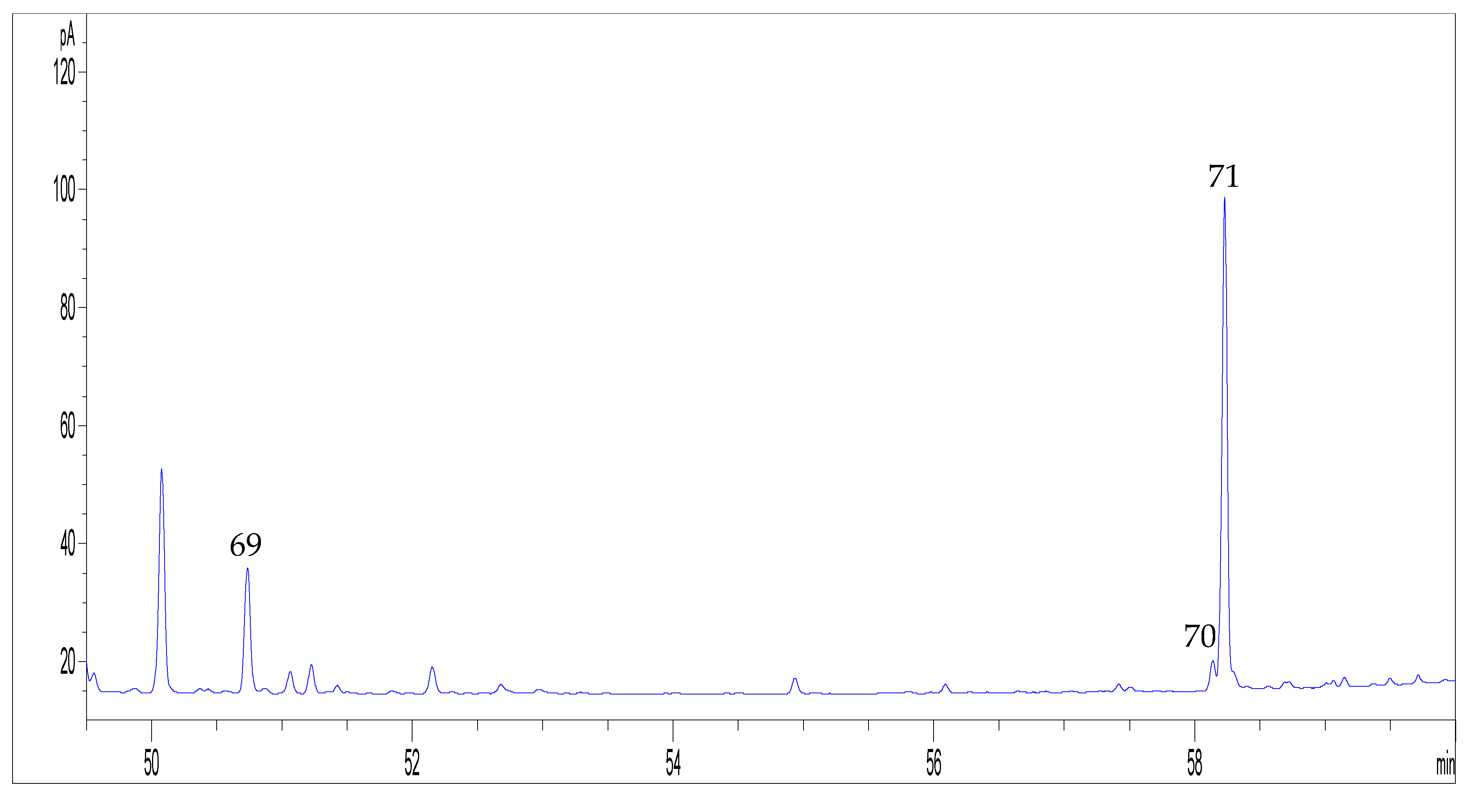
| 69 | Cannabidivarin (CBDV) | 2193 | 2208 | - | - | 0.2 a | 0.1 a | 0.2 a | 0.1 a | 0.2 a | - |
| 70 | Cannabichromene (CBC) | 2772 | - | - | - | - | - | - | - | - | - |
| 71 | Cannabidiol (CBD) | 2779 | - | 0.1 a | - | - | - | - | 0.3 a | - | 0.1 a |
| Total area | 92.9 ± 1.4 | 100.0 | 97.5 ± 1.4 | 98.1 ± 0.7 | 98.7 ± 0.2 | 92.1 ± 0.8 | 98.0 ± 0.5 | 98.4 ± 1.0 |
| Peak n. | Compound | LRI | LRI lit b | EO9 | EO10 | EO11 | EO12 | EO13 | EO14 | EO15 | EO16 | EO17 |
|---|---|---|---|---|---|---|---|---|---|---|---|---|
| 1 | Heptanal | 903 | 901 | - | - | - | 0.3 a | 0.1 a | 0.3 a | 0.1 a | 0.1 a | - |
| 2 | 5,5-Dimethyl-1-vinylbicyclo[2.1.1]-hexane | 918 | 921 | 0.3 a | 0.5 a | - | 0.3 a | 0.4 a | 1.4 a | 0.3 ± 0.1 | 0.4 a | 0.3 a |
| 3 | α-Thujene | 926 | 924 | 0.1 a | 0.1 a | - | 0.2 a | 0.1 a | 0.1 a | 0.1 a | 0.1 a | 0.1 a |
| 4 | α-Pinene | 934 | 931 | 8.4 ± 0.3 | 25.4 ± 0.3 | 20.7 ± 1.6 | 14.6 ± 0.4 | 15.5 ± 0.1 | 11.1 | 16.6 ± 0.3 | 20.3 ± 0.5 | 14.7 ± 0.4 |
| 5 | Camphene | 947 | 947 | 0.2 a | 0.5 a | 0.4 ± 0.1 | 0.3 a | 0.3 a | 0.2 a | 0.3 a | 0.4 a | 0.3 ± 0.1 |
| 6 | β-Thujene | 973 | 966 | 0.1 a | - | - | 0.1 a | 0.1 a | - | - | - | - |
| 7 | β-Pinene | 976 | 978 | 3.2 ± 0.1 | 9.2 ± 0.1 | 6.9 ± 0.5 | 4.2 ± 0.1 | 5.3 ± 0.1 | 5.1 ± 0.1 | 5.3 ± 0.1 | 8.0 ± 0.1 | 4.1 ± 0.1 |
| 8 | β-Myrcene | 994 | 992 | 19.8 ± 0.5 | 19.8 ± 0.2 | 14.5 ± 1.0 | 29.0 ± 0.4 | 33.4 ± 0.1 | 29.9 ± 0.2 | 39.2 ± 2.1 | 33.4 ± 0.5 | 28.9 ± 2.4 |
| 9 | α-Phellandrene | 1006 | 1007 | 0.3 a | 0.2 a | 0.3 a | 3.8 ± 0.1 | 2.7 a | 0.2 a | 0.3 a | 0.3 a | 3.9 ± 0.3 |
| 10 | Δ3-Carene | 1010 | 1010 | 0.7 a | 0.7 a | 0.6 a | 1.02 a | 0.9 a | 0.5 a | 1.1 a | 0.8 a | 1.0 a |
| 11 | α-Terpinene | 1016 | 1017 | 0.3 a | 0.2 a | 0.3 a | 0.1 a | 0.2 a | 0.2 a | 0.2 a | 0.3 a | 0.1 a |
| 12 | p-Cymene | 1024 | 1026 | 0.1 a | 0.1 a | - | 0.6 a | 0.2 a | 0.2 a | - | 0.1 a | 0.4 a |
| 13 | Limonene | 1028 | 1035 | 2.1 a | 5.3 ± 0.1 | 3.4 ± 0.2 | 3.0 a | 4.1 a | 2.1 a | 2.8 ± 0.1 | 5.6 ± 0.1 | 2.9 ± 0.2 |
| 14 | 1,8-Cineole | 1030 | 1032 | 0.1 a | 0.7 a | 0.2 a | 0.2 a | 0.2 a | 0.3 a | - | 0.2 a | 0.2 a |
| 15 | cis-Ocimene | 1038 | 1040 | 0.6 a | 0.5 a | 0.6 a | 0.7 a | 0.6 a | 1.3 a | 0.5 a | 0.3 a | 0.7 ± 0.1 |
| 16 | trans-Ocimene | 1049 | 1050 | 4.9 ± 0.2 | 3.1 a | 6.3 ± 0.4 | 6.3 ± 0.1 | 5.6 a | 7.1 ± 0.1 | 6.4 ± 0.7 | 3.2 ± 0.1 | 6.2 ± 0.6 |
| 17 | γ-Terpinene | 1058 | 1062 | 0.2 a | 0.3 a | 0.3 a | - | 0.2 a | 0.3 a | 2.8 ± 0.6 | 0.2 a | 0.2 a |
| 18 | α-Terpinolene | 1089 | 1088 | 9.6 ± 0.4 | 4.0 ± 0.1 | 6.3 ± 0.3 | 6.4 a | 7.5 a | 3.5 a | 5.2 a | 7.4 ± 0.2 | 6.2 ± 0.3 |
| 19 | Linalool | 1101 | 1104 | 0.2 a | 0.6 a | - | 0.3 a | 0.4 a | - | 0.3 a | 0.3 a | 0.3 ± 0.1 |
| 20 | 6-Camphenol | 1105 | 1110 | 0.1 a | - | - | - | 0.1 a | 0.6 a | - | 0.1 a | - |
| 21 | Fenchol | 1113 | 1114 | - | 0.1 a | - | - | 0.2 a | - | 0.1 a | 0.3 ± 0.1 | 0.1 a |
| 22 | neo-Alloocimene | 1121 | 1143 | - | 0.1 a | - | - | 0.1 a | - | 0.1 a | 0.2 a | 0.1 a |
| 23 | trans-Pinocarveol | 1138 | 1139 | - | 0.1 a | - | - | - | - | 0.1 a | - | |
| 24 | Camphor | 1144 | 1145 | 0.1 a | 0.1 a | - | 0.1 a | - | - | - | 0.1 a | |
| 25 | Isopulegol | 1146 | 1156 | - | - | - | - | - | 0.2 a | - | ||
| 26 | Borneol | 1166 | 1166 | - | 0.2 a | - | - | - | - | 0.1 a | - | |
| 27 | Terpinen-4-ol | 1178 | 1177 | 0.1 a | 0.4 a | 0.3 a | 0.2 a | 0.3 a | 0.2 a | 0.1 a | 0.2 a | 0.2 a |
| 28 | p-Cymen-8-ol | 1190 | 1183 | 0.1 a | - | - | - | - | - | 0.2 a | - | |
| 29 | α-Terpineol | 1191 | 1193 | - | 0.2 a | - | - | 0.2 a | - | 0.1 a | 0.1 a | 0.1 a |
| 30 | Methyl chavicol | 1199 | 1196 | 0.1 a | - | 6.2 a | 3.6 ± 0.1 | - | 0.1 a | - | 5.9 ± 0.6 | |
| 31 | Neral | 1243 | 1249 | - | 1.4 ± 0.1 | - | - | - | - | - | 0.1 a | |
| 32 | Linalyl acetate | 1261 | 1253 | 0.1 a | 0.1 a | - | - | - | - | 0.1 a | 0.1 a | |
| 33 | Bornyl acetate | 1289 | 1287 | - | - | - | 0.4 a | - | - | 0.1 a | 0.4 a | |
| 34 | Thymol | 1301 | 1297 | - | 0.3 a | - | - | - | - | 0.1 a | ||
| 35 | Geranyl acetate | 1388 | 1392 | 0.2 a | 0.1 a | - | 4.3 ± 0.3 | 0.5 a | 0.2 a | 0.1 a | 0.4 a | 4.3 ± 0.4 |
| 36 | β-Bourbonene | 1389 | 1386 | 0.1 a | - | - | - | - | - | - | 0.1 a | - |
| 37 | α-Gurjunene | 1411 | 1408 | 0.4 a | 0.4 a | - | - | 0.2 a | 0.5 a | 0.2 ± 0.1 | - | - |
| 38 | Isocaryophyllene | 1417 | 1411 | - | - | 0.4 a | 0.1 a | - | - | 0.2 a | 0.2 a | |
| 39 | β-Caryophyllene | 1427 | 1428 | 14.8 ± 0.5 | 13.1 ± 0.4 | 22.3 ± 0.5 | 7.6 ± 0.6 | 8.2 ± 0.3 | 14.6 ± 0.3 | 9.2 ± 0.4 | 7.6 ± 0.5 | 7.7 ± 0.6 |
| 40 | α-Bergamotene | 1441 | 1432 | 1.4 ± 0.2 | 0.3 a | 1.3 a | 0.2 a | 0.2 a | 1.0 a | 0.2 ± 0.1 | 0.2 a | 0.2 a |
| 41 | Aromadendrene | 1445 | 1449 | 0.2 a | 0.2 a | 0.6 ± 0.1 | 0.1 a | 0.1 a | - | 0.1 a | 0.1 a | 0.1 a |
| 42 | Aristolene | 1453 | 1450 | - | - | - | - | - | - | - | - | |
| 43 | α-Humulene | 1461 | 1455 | 6.6 ± 0.1 | 3.8 ± 0.2 | 7.5 ± 0.2 | 2.5 ± 0.3 | 2.5 ± 0.1 | 5.3 ± 0.1 | 2.7 ± 0.2 | 2.2 ± 0.2 | 2.5 ± 0.2 |
| 44 | Alloaromadendrene | 1468 | 1467 | 0.7 a | 0.5 a | 0.4 a | 0.2 a | 0.2 a | 0.9 a | 0.2 ± 0.1 | 0.2 a | 0.2 a |
| 45 | γ-Muurolene | 1482 | 1477 | 0.4 a | 0.1 a | 0.6 a | - | 0.1 a | - | 0.1 a | 0.1 a | 0.1 a |
| 46 | Germacrene D | 1487 | 1485 | 0.7 a | 0.1 a | - | 0.1 a | 0.1 a | - | - | 0.1 a | - |
| 47 | β-Selinene | 1490 | 1489 | 0.4 a | 0.3 a | 0.5 a | 0.5 a | - | 0.4 a | 0.2 a | 0.2 a | 0.3 a |
| 48 | α-Selinene | 1493 | 1494 | 1.3 ± 0.2 | 0.6 a | 1.5 ± 0.1 | 0.1 a | 0.5 ± 0.1 | 1.0 a | 0.4 a | 0.4 a | 0.5 ± 0.1 |
| 49 | Valencene | 1497 | 1496 | 0.2 a | 0.1 a | - | - | 0.1 a | 0.2 a | 0.1 a | 0.1 a | 0.1 a |
| 50 | β-Bisabolene | 1501 | 1505 | 1.3 ± 0.1 | 0.7 a | 1.5 ± 0.1 | 0.7 a | 0.5 a | 0.9 a | 0.5 a | 0.5 a | 0.7 ± 0.1 |
| 51 | α-Muurolene | 1512 | 1497 | 0.5 ± 0.1 | 0.5 | 1.3 ± 0.1 | 0.4 | 0.4 | 0.7 | 0.3 ± 0.1 | 0.2 | 0.5 ± 0.1 |
| 52 | α-Patchoulene | 1521 | 1445 | 0.2 a | 0.1 a | - | 0.1 a | - | 0.2 a | - | 0.1 a | |
| 53 | γ-Cadinene | 1525 | 1512 | 0.3 a | 0.2 a | - | 0.2 a | 0.2 a | 0.3 a | - | 0.2 a | 0.2 a |
| 54 | δ-Cadinene | 1529 | 1519 | 0.5 ± 0.1 | 0.2 a | 0.6 a | 0.2 a | 0.1 a | 0.3 a | 0.1 ± 0.1 | 0.1 a | 0.1 a |
| 55 | δ-Selinene | 1545 | 1540 | 1.3 ± 0.4 | 0.8 ± 0.1 | - | 0.6 ± 0.1 | 0.7 ± 0.1 | 0.8 ± 0.2 | 0.6 ± 0.1 | 0.6 a | 0.7 ± 0.1 |
| 56 | Selina-3,7(11)-diene | 1550 | 1545 | 1.1 ± 0.1 | 0.3 ± 0.1 | - | 0.2 a | 0.4 a | 0.2 a | 0.2 a | 0.3 a | |
| 57 | trans-Nerolidol | 1568 | 1562 | 0.7 a | 0.2 a | 0.2 a | 0.2 a | 0.1 a | 0.3 a | 0.1 a | - | 0.2 a |
| 58 | Spathulenol | 1585 | 1577 | 0.1 a | - | - | - | - | - | - | - | - |
| 59 | Caryophyllene oxide | 1593 | 1583 | 5.2 ± 0.2 | 1.1 ± 0.1 | 1.1 a | 0.9 ± 0.2 | 3.6 a | 0.5 ± 0.2 | 0.8 ± 0.1 | 0.8 ± 0.1 | |
| 60 | Viridiflorol | 1607 | 1600 | 0.2 a | - | - | 0.3 a | 0.1 a | - | - | 0.3 a | |
| 61 | Guaiol | 1619 | 1602 | 1.4 ± 0.1 | 0.2 a | 0.3 a | 0.1 a | 0.9 a | - | 0.2 a | 0.2 a | |
| 62 | Humulene-1,2-epoxide | 1621 | 1615 | - | - | - | 0.2 a | 0.1 a | - | - | - | 0.2 a |
| 63 | Caryophylla-4(12),8(13)-dien-5-ol | 1628 | 1634 | 0.5 a | 0.1 a | - | - | 0.2 a | - | 0.1 a | - | |
| 64 | epi-γ-Eudesmol | 1636 | 1625 | 0.2 a | - | - | - | - | 0.1 a | - | - | - |
| 65 | Cubenol | 1643 | 1643 | 0.5 a | 0.1 a | - | 0.1 a | - | 0.2 a | - | 0.1 a | - |
| 66 | (Z)-14-Hydroxycaryophyllene | 1667 | 1666 | 0.4 a | - | - | - | 0.1 a | 0.1 a | - | 0.1 a | - |
| 67 | epi-α-Bisabolol | 1681 | 1683 | 0.5 ± 0.1 | - | - | 0.1 a | - | 0.2 a | - | - | - |
| 68 | β-Bisabolol | 2041 | - | 0.1 a | - | - | - | - | 0.1 a | - | - | - |
| 69 | Cannabidivarin (CBDV) | 2193 | 2208 | 0.1 a | 0.1 a | - | 0.2 a | - | 0.2 a | 0.3 a | 0.2 a | 0.2 a |
| 70 | Cannabichromene (CBC) | 2772 | - | - | - | - | - | - | - | - | - | - |
| 71 | Cannabidiol (CBD) | 2779 | - | 0.6 a | - | 0.1 a | - | - | 0.1 a | - | - | - |
| Total area | 94.7 ± 0.4 | 98.1 ± 0.9 | 97.4 ± 2.1 | 97.7 ± 0.7 | 98.0 ± 0.1 | 97.6 ± 0.3 | 98.4 ± 1.3 | 98.0 ± 0.1 | 98.2 ± 2.1 |
| Bacterial strain | EO1 | EO2 | EO3 | EO4 | EO5 | EO6 | EO7 | EO8 | EO9 | EO10 | EO11 | EO13 | EO14 | EO15 | EO16 | EO17 | Ampicillin | Ciprofloxacin |
|---|---|---|---|---|---|---|---|---|---|---|---|---|---|---|---|---|---|---|
| Staphylococcus aureus ATCC 6538 | 2 | - | - | - | - | - | - | - | 16 | 8 | 2 | - | - | - | - | - | - | 0.5 |
| Staphylococcus aureus 18As * | - | - | - | 16 | 32 | - | - | - | - | 16 | - | - | - | - | - | - | - | 16 |
| Staphylococcus epidermidis 18Bs * | 4 | - | - | 16 | - | 16 | - | - | 1 | 8 | - | 16 | - | - | - | - | - | 0.5 |
| Staphylococcus aureus 386 * | - | - | - | - | - | - | - | 32 | - | - | - | 16 | 16 | - | - | - | - | 16 |
| Listeria monocytogenes NCTC 10888 | 16 | 8 | - | - | 4 | 32 | - | - | 8 | 8 | - | 16 | - | 16 | - | - | 0.25 | - |
| Listeria monocytogenes ATCC 13932 | 4 | - | - | 32 | - | - | - | 32 | 8 | 16 | 4 | 16 | - | - | - | - | 0.25 | - |
| Listeria monocytogenes ATCC 5008 | 8 | - | 2 | - | - | - | 8 | 4 | - | 8 | 2 | 16 | - | - | 4 | 4 | 0.25 | - |
| Listeria monocytogenes 70 * | 4 | - | - | - | 32 | 32 | 2 | 16 | - | - | 4 | - | - | - | - | - | 2 | - |
| Listeria monocytogenes 139 * | - | - | - | - | 32 | - | 8 | 32 | - | - | 2 | - | - | 1 | - | - | 0.5 | - |
| Enterococcus faecalis ATCC 29212 | 2 | 2 | - | 16 | 0.5 | 4 | - | 1 | 8 | 2 | 2 | 16 | 1 | 1 | 4 | 4 | 4 | |
| Enterococcus hirae ATCC 10541 | 2 | - | - | 32 | - | 16 | 4 | - | 4 | 16 | 16 | 16 | 32 | - | - | - | - | 8 |
| Enterococcus faecalis V3 * | 1 | - | 2 | 32 | 32 | 4 | - | 4 | - | - | 4 | 16 | 32 | - | 4 | - | - | 0.25 |
| Enterococcus faecalis V4 * | 2 | - | - | - | - | 32 | 8 | 16 | 16 | 16 | 2 | - | - | - | - | - | - | 16 |
| Enterococcus faecium V5 * | - | - | 1 | - | 16 | 4 | 8 | 8 | 1 | 16 | 2 | 2 | 32 | 16 | - | - | - | 8 |
| Enterococcus faecalis V6 * | 2 | - | 0.5 | - | 16 | 16 | - | 8 | 1 | 16 | 16 | - | - | 8 | - | - | - | 16 |
| Enterococcus faecium EQ19 * | 2 | - | - | - | 8 | 4 | 4 | 8 | 8 | 16 | 16 | - | 2 | - | - | - | - | 4 |
| Bacillus subtilis ATCC 6633 | 2 | 8 | 2 | 8 | 8 | 16 | - | 16 | 4 | - | 8 | - | 1 | 2 | - | 4 | 2 | - |
| Bacillus cereus EB 362 | 1 | - | 1 | - | - | 1 | - | - | 1 | - | - | 16 | - | - | - | - | 2 | - |
| Bacillus 1 ˣ | 2 | 8 | - | - | 8 | 1 | - | - | 4 | 8 | 16 | 16 | - | 4 | - | - | 1 | - |
| Bacillus 2 ˣ | 0.5 | - | - | - | 16 | 16 | 8 | 16 | - | 16 | - | 16 | 32 | 16 | - | 4 | 0.25 | - |
| Bacillus 3 ˣ | - | - | 2 | 8 | - | 16 | 8 | 32 | - | - | 16 | - | 16 | 16 | 4 | - | 1 | - |
| Bacillus 4 ˣ | 2 | - | 2 | 32 | 16 | 1 | - | - | 4 | - | 16 | - | - | 4 | - | 4 | 0.25 | - |
| Bacillus 5 ˣ | 2 | - | 2 | - | - | 4 | - | 32 | 4 | - | - | 16 | - | 4 | - | - | 2 | - |
| Bacillus 6 ˣ | 2 | - | 1 | 16 | - | 4 | 8 | - | 4 | 16 | - | 16 | - | 8 | 4 | - | 1 | - |
| Bacillus 9 ˣ | - | - | 2 | 8 | 8 | 32 | - | - | 8 | - | - | - | - | 4 | - | - | 1 | - |
| Bacillus 10988 ˣ | 2 | - | - | - | - | 4 | 2 | 32 | 4 | - | 16 | 16 | - | 8 | - | 4 | 2 | - |
| Bacillus 18100 ˣ | 0.5 | - | - | - | - | 16 | - | - | 4 | - | - | - | - | - | - | - | 0.5 | - |
| Bacillus 18102 ˣ | 2 | - | 2 | - | - | 16 | - | - | 4 | - | 16 | 16 | - | 16 | - | - | 0.25 | - |
| Bacterial Strain | CBD | α-Pinene | β-Pinene | β-Myrcene | α-Terpinolene | β-Caryophyllene | Ampicillin | Ciprofloxacin |
|---|---|---|---|---|---|---|---|---|
| Staphylococcus aureus ATCC 6538 | 8 | 4 | 4 | 8 | 8 | 16 | - | 0.5 |
| Staphylococcus aureus 18As * | 32 | 16 | 32 | 8 | 32 | 32 | - | 16 |
| Staphylococcus epidermidis 18Bs * | 16 | 8 | 4 | 16 | 16 | 32 | - | 0.5 |
| Staphylococcus aureus 386 * | 32 | 16 | 8 | 32 | 32 | 32 | - | 16 |
| Listeria monocytogenes NCTC 10888 | 1 | 1 | 2 | 2 | 2 | 1 | 0.25 | - |
| Listeria monocytogenes ATCC 13932 | 2 | 2 | 2 | 1 | 1 | 1 | 0.25 | - |
| Listeria monocytogenes ATCC 5008 | 1 | 1 | 0.5 | 2 | 1 | 2 | 0.25 | - |
| Listeria monocytogenes 70 * | 4 | 2 | 2 | 2 | 4 | 4 | 2 | - |
| Listeria monocytogenes 139 * | 4 | 2 | 2 | 2 | 4 | 1 | 0.5 | - |
| Enterococcus faecalis ATCC 29212 | 1 | 2 | 0.5 | 1 | 2 | 1 | 4 | |
| Enterococcus hirae ATCC10541 | 2 | 1 | 2 | 8 | 4 | 8 | - | 8 |
| Enterococcus faecalis V3 * | 1 | 1 | 1 | 4 | 1 | 2 | - | 0.25 |
| Enterococcus faecalis V4 * | 2 | 4 | 1 | 4 | 8 | 4 | - | 16 |
| Enterococcus faecium V5 * | 4 | 1 | 4 | 4 | 8 | 16 | - | 8 |
| Enterococcus faecalis V6 * | 4 | 1 | 2 | 8 | 16 | 1 | - | 16 |
| Enterococcus faecium EQ19 * | 1 | 4 | 2 | 1 | 2 | 4 | - | 4 |
| Bacillus subtilis ATCC 6633 | 8 | 8 | 4 | 32 | 16 | 1 | 2 | - |
| Bacillus cereus EB 362 | 8 | 2 | 1 | 2 | 4 | 8 | 2 | - |
| Bacillus 1 ˣ | 4 | 4 | 2 | 4 | 8 | 4 | 1 | - |
| Bacillus 2 ˣ | 8 | 8 | 4 | 4 | 16 | 16 | 0.25 | - |
| Bacillus 3 ˣ | 16 | 8 | 16 | 8 | 8 | 16 | 1 | - |
| Bacillus 4 ˣ | 8 | 16 | 16 | 16 | 16 | 8 | 0.25 | - |
| Bacillus 5 ˣ | 2 | 4 | 1 | 2 | 4 | 4 | 2 | - |
| Bacillus 6 ˣ | 4 | 2 | 1 | 4 | 4 | 8 | 1 | - |
| Bacillus 9 ˣ | 4 | 8 | 4 | 8 | 16 | 16 | 1 | - |
| Bacillus 10988 ˣ | 4 | 4 | 4 | 4 | 8 | 16 | 2 | - |
| Bacillus 18100 ˣ | 4 | 8 | 4 | 8 | 16 | 8 | 0.5 | - |
| Bacillus 18102 ˣ | 8 | 8 | 4 | 2 | 16 | 8 | 0.25 | - |
© 2019 by the authors. Licensee MDPI, Basel, Switzerland. This article is an open access article distributed under the terms and conditions of the Creative Commons Attribution (CC BY) license (http://creativecommons.org/licenses/by/4.0/).
Share and Cite
Iseppi, R.; Brighenti, V.; Licata, M.; Lambertini, A.; Sabia, C.; Messi, P.; Pellati, F.; Benvenuti, S. Chemical Characterization and Evaluation of the Antibacterial Activity of Essential Oils from Fibre-Type Cannabis sativa L. (Hemp). Molecules 2019, 24, 2302. https://0-doi-org.brum.beds.ac.uk/10.3390/molecules24122302
Iseppi R, Brighenti V, Licata M, Lambertini A, Sabia C, Messi P, Pellati F, Benvenuti S. Chemical Characterization and Evaluation of the Antibacterial Activity of Essential Oils from Fibre-Type Cannabis sativa L. (Hemp). Molecules. 2019; 24(12):2302. https://0-doi-org.brum.beds.ac.uk/10.3390/molecules24122302
Chicago/Turabian StyleIseppi, Ramona, Virginia Brighenti, Manuela Licata, Antonella Lambertini, Carla Sabia, Patrizia Messi, Federica Pellati, and Stefania Benvenuti. 2019. "Chemical Characterization and Evaluation of the Antibacterial Activity of Essential Oils from Fibre-Type Cannabis sativa L. (Hemp)" Molecules 24, no. 12: 2302. https://0-doi-org.brum.beds.ac.uk/10.3390/molecules24122302