Article

Chiral Pyridine-3,5-bis- (L-phenylalaninyl-L-leucinyl) Schiff Base Peptides as Potential Anticancer Agents: Design, Synthesis, and Molecular Docking Studies Targeting Lactate Dehydrogenase-A

by
Abd El-Galil E. Amr
1,2,
Randa E. Abdel Mageid
3,
Mohamed El-Naggar
4,
Ahmed M. Naglah
1,5,
Eman S. Nossier
6 and
Elsayed A. Elsayed
7,8,*
1
Drug Exploration & Development Chair (DEDC), Pharmaceutical Chemistry Department, College of Pharmacy, King Saud University, Riyadh 11451, Saudi Arabia
2
Applied Organic Chemistry Department, National Research Center, Cairo, Dokki 12622, Egypt
3
Photochemistry Department, National Research Center, Cairo, Dokki 12622, Egypt
4
Chemistry Department, Faculty of Sciences, University of Sharjah, Sharjah 27272, UAE
5
Department of Peptide Chemistry, National Research Centre, Cairo 12622, Egypt
6
Pharmaceutical Medicinal Chemistry Department, Faculty of Pharmacy (Girls), Al-Azhar University, Cairo 12622, Egypt
7
Zoology Department, Bioproducts Research Chair, Faculty of Science, King Saud University, Riyadh 11451, Saudi Arabia
8
Chemistry of Natural and Microbial Products Department, National Research Centre, Cairo, Dokki 12622, Egypt
*
Author to whom correspondence should be addressed.
Submission received: 3 February 2020 / Revised: 26 February 2020 / Accepted: 27 February 2020 / Published: 29 February 2020
(This article belongs to the Special Issue Recent Advances in Anticancer Drugs II)

Abstract

:
A series of branched tetrapeptide Schiff bases 36 were designed and synthesized from corresponding tetrapeptide hydrazide 2 as a starting material.In vitroevaluation of the synthesized compounds 46 against breast MCF-7 carcinoma cells identified their excellent anticancer potency, with IC50 ranging from 8.12 ± 0.14 to 17.55 ± 0.27 μM in comparison with the references, cisplatin and milaplatin (IC50= 13.34 ± 0.11and 18.43 ± 0.13 μM, respectively). Furthermore, all derivatives demonstrated promising activity upon evaluation of theirin vitroandin vivosuppression of p53 ubiquitination and inhibition assessment for LDHA kinase. Finally, molecular docking studies were performed to predict the possible binding features of the potent derivatives within the ATP pocket of LDHA in an attempt to get a lead for developing a more potent LDHA inhibitor with anti-proliferative potency.

1. Introduction

Peptides are short linear chains of amino acids (usually less than 50 AA in length) and are often stabilized by disulfide bonds [1]. Recently, there has been a noticeable increase in the number of publications covering the potential effects of peptides on lipid metabolism and blood pressure (BP), in addition to their antimicrobial, anti-inflammatory, analgesic, antioxidant, immunomodulatory, and anticancer activities [2,3,4].
Still, cancer is one of the most prominent causes of death in developed countries and there is a great demand for the discovery of new chemotherapies with more selectivity for cancer cells and less side effects [4,5]. The anticancer effects of peptides have been extensively explored with a raised number of approved peptide-based drugs [6,7,8]. The great potential of their antitumor activity has been exhibited through the action on multiple molecular pathways like enzymes involved in carcinogenesis with no genotoxicity [9].
Moreover, tumor-targeting peptides are effective alternative entities for the selective delivery of high doses of chemotherapeutic drugs or diagnostic agents to tumor sites while sparing normal tissues. Several peptide hormones have already been applied for tumor targeting, for example, the cyclic octapeptide analogue of somatostatin, octreotide, has been utilized for radiotargeting in neuroendocrine tumor [10]. Additionally, the linear peptide analogue of Luteinizing hormone-releasing hormone (LHRH), AN-152, has been used to target the LHRH receptor of breast cancer, ovarian cancer, and prostate cancer [11]. Recently, most peptidopyridine derivatives that have been synthesized consist of a wide range of pharmaceutical activities, including anticancer, analgesic, anti-convulsant, antiparkinsonian, analgesic, anti-inflammatory, and antimicrobial activities [12,13,14,15].
Schiff bases have been important candidates owing to their coordination chemistry, and they can be synthesized and used as ligands in building complex with different metal ions. On the other hand, Schiff bases have been interested with a broad spectrum of biological and pharmacological activities, such as antibacterial, DNA binding, anti HIV, Largicidal, antifertility, anticancer, and anti-inflammatory activities [16,17,18,19,20,21,22,23]. The broad-spectrum biological activities of Schiff bases have been attributed to the -C = N- imine bond, where the electrophilic carbon and nucleophilic nitrogen provide excellent binding characters with different nucleophiles and electrophiles, resulting in the inhibition of targeted diseases, enzymes, or DNA replication [24].
The lactate dehydrogenase (LDH) is one of the most prominent proteins and is responsible for the interconversion of lactate and pyruvate, parallel with the interconversion of NAD+ and NADH. LDH is present in humans as three subunits—LDHA, LDHB, and LDHC [25]. It is clear that cancer cells mainly produce energy through glycolysis and LDHA is the final enzyme involved in this pathway [26]. As LDHA is a vital supporter of glucose metabolism in cancer cells and its increased level is a marker for many tumors, LDHA can be defined as an anticancer drug target [27]. Previous literatures reported that V-shape structures illustrate good LDHA inhibition, so molecular modification of the potentially identified LDHA inhibitors AC (Figure 1) might provide more effective hits [28,29,30].
In the present investigation and as a continuation of our previous work [31,32,33,34,35,36,37,38] in peptide and heterocyclic chemistry, chiral N3,N5-bis[(l-phenylalaninyl-l-leucinyl) substituted aryl or heterocyclic Schiff base]pyridine candidates were successfully designed, synthesized, and plausibly characterized by analytical techniques including IR, nuclear magnetic resonance (NMR), and mass spectroscopy evidences. In addition, their cytotoxicity effects on MCF-7 cancer cell lines, in vivo and in vitro inhibition of p53 ubiquitination, and lactate dehydrogenase-A were demonstrated in detail. Furthermore, molecular docking illustrates the binding affinity to LDHA kinase, which could facilitate the discovery of novel anticancer and LDHA inhibitory agents.

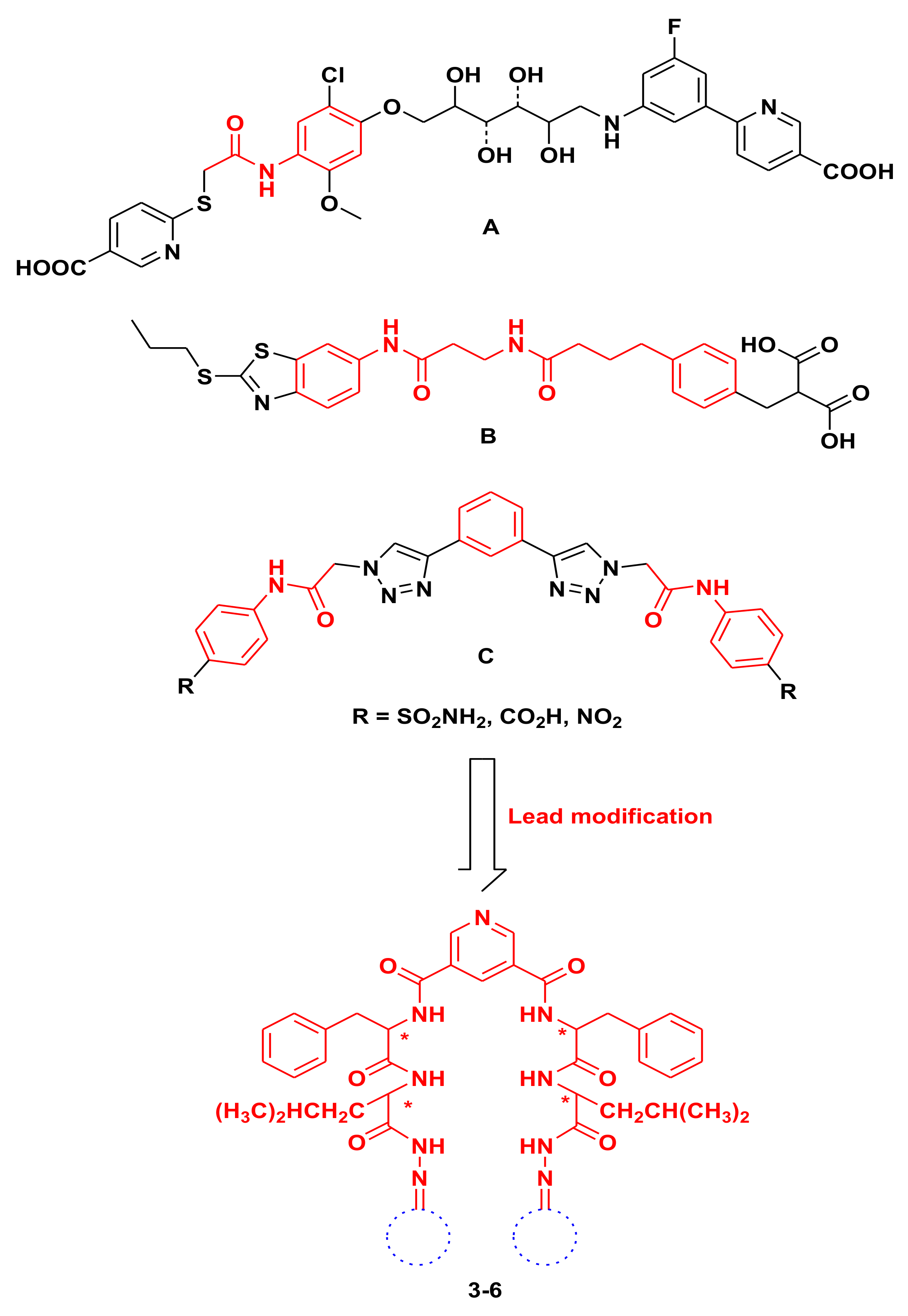
2. Results and Discussion
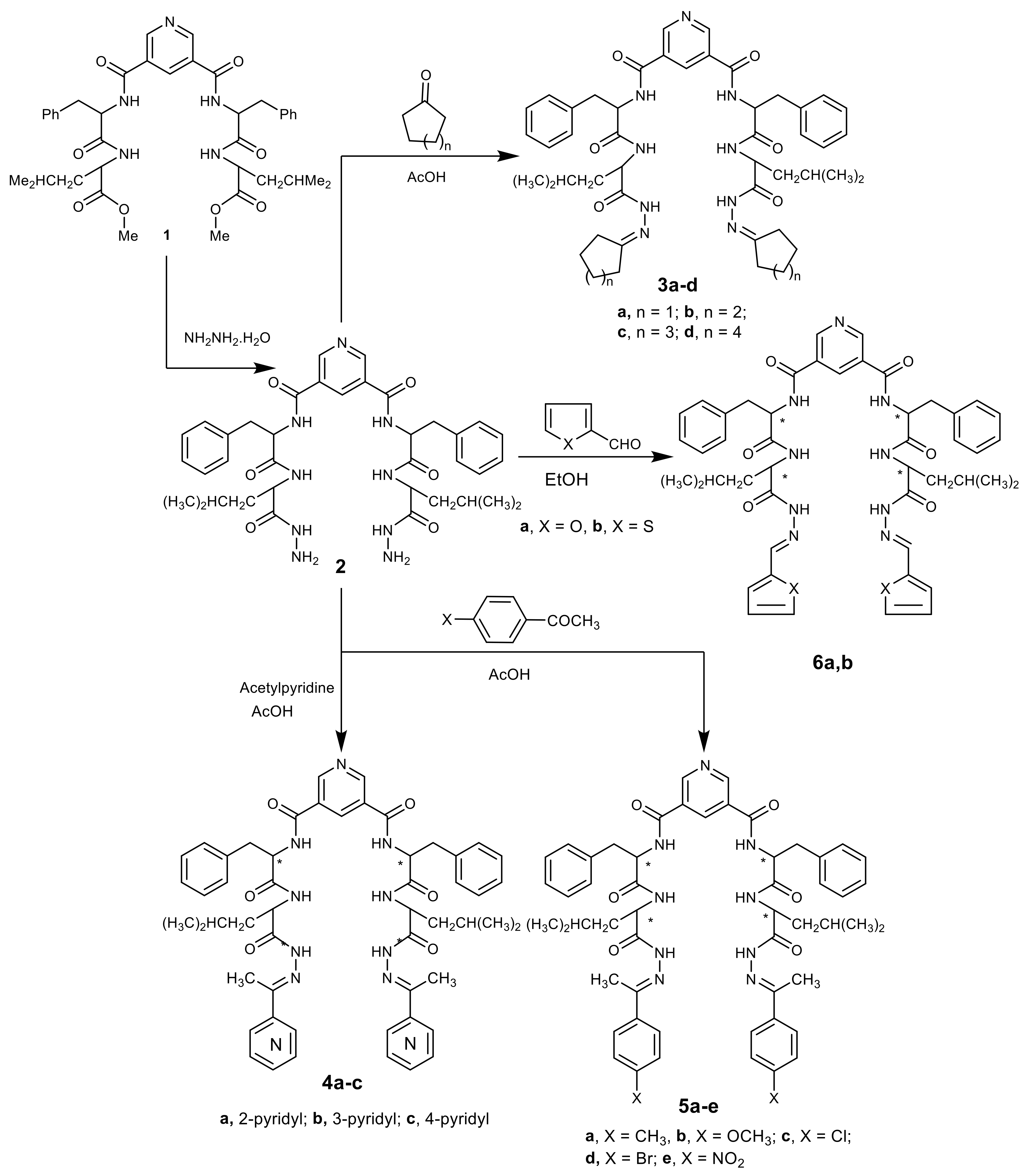
2.1. Chemistry

In the present work, some of the prepared tetrapeptide derivatives 36 were obtained from Nα-dipicolinoyl-bis[(l-phenylalanyl-l-leucine)hydrazide] 2, which was obtained from corresponding ester 1 [39]. Reaction of hydrazide 2 with cycloalkanone derivatives in refluxing glacial acetic acid afforded the corresponding Nα-dipicolinoyl-bis[(l-phenylalanyl-l-leucine)- N`-cycloalkylidenehydrazide] 3ad. Condensation of 2 with active carbonyl derivatives, namely, acetylpyridine or substituted acetophenone derivatives in refluxing acetic acid, gave the corresponding Nα-dipicolinoyl-bis[(l-phenylalanyl-l-leucine)-N`-(1-(pyridyl)ethylidene)benzo- hydrazide] 4ac and Nα-dipicolinoyl-bis-[(l-phenylalanyl-l-leucine)-N`-(1-(substituted phenyl) ethylidene)benzohydrazide] 5ae, respectively. Finally, treatment of heterocyclic aldehydes in refluxing ethanol gave the corresponding Nα-dipicolinoylbis-[(l-phenylalanyl-l-leucine)-N`-(1- (furyl- 6a and thienyl)ethylidene)benzohydrazide] 6b, respectvely (Scheme 1).

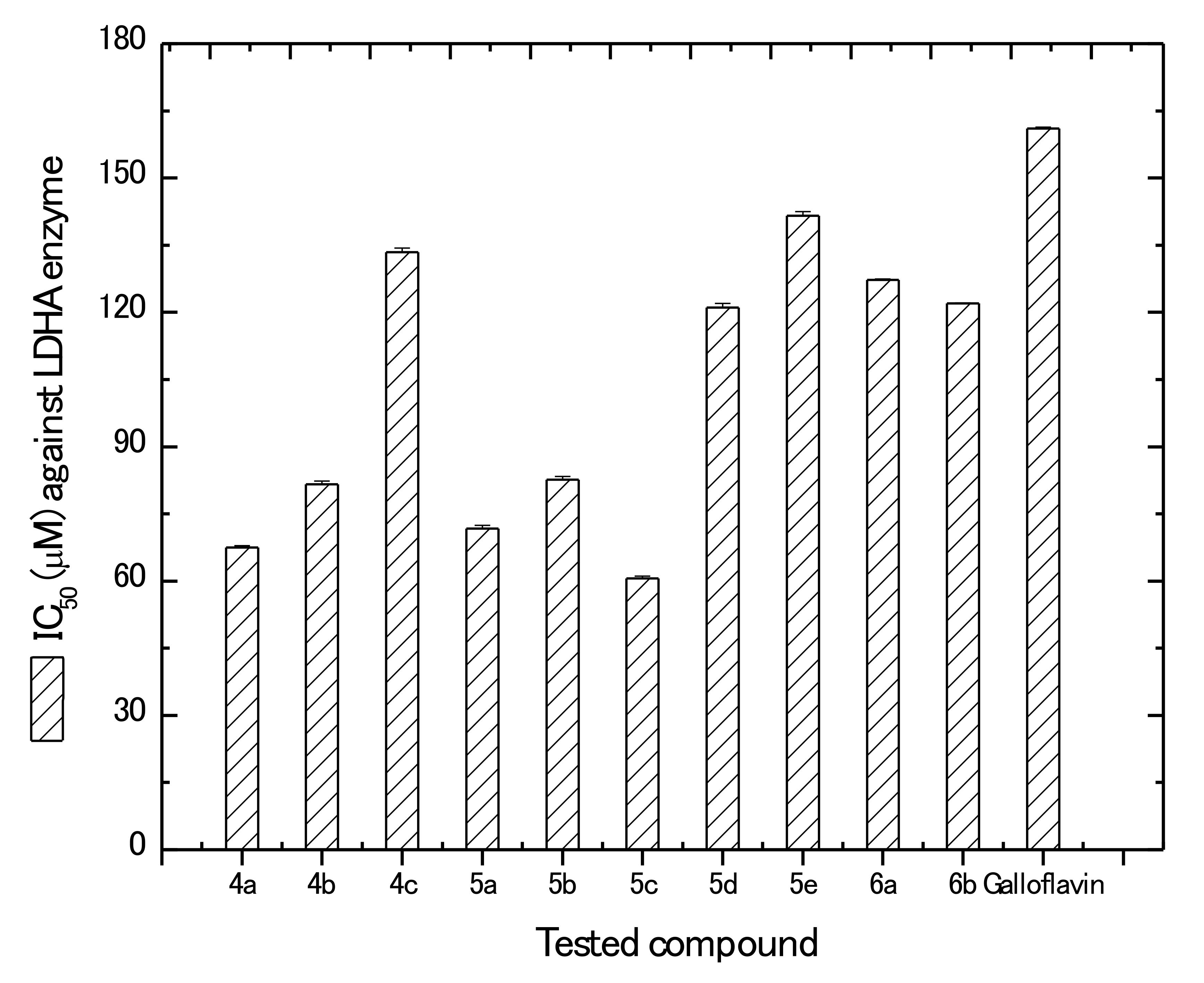
2.2. Biological Evaluation

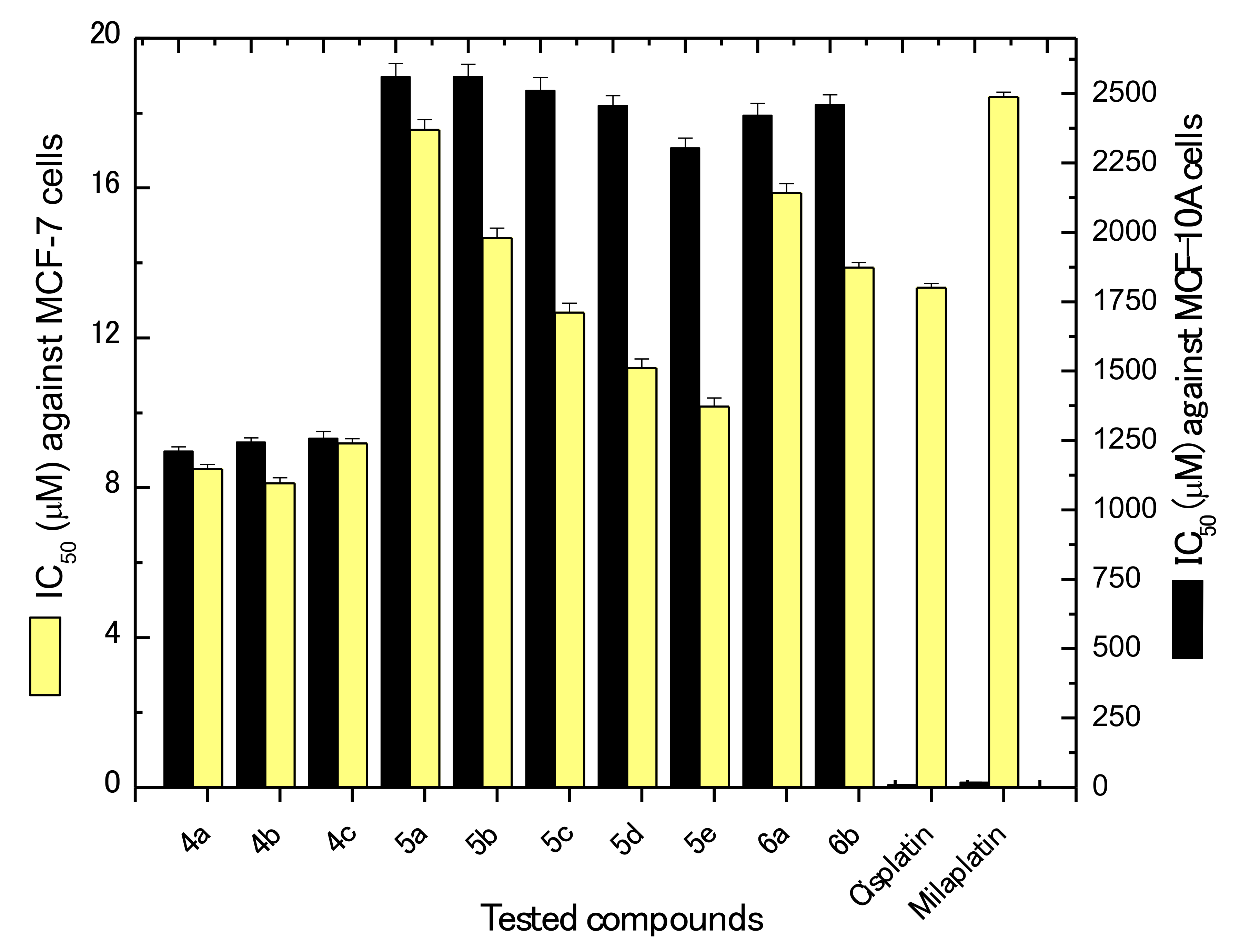
2.2.1. Evaluation of In vitro Anticancer Potentials Against Breast Cancer

Following the synthesis of the new bis-dipeptide derivatives, we evaluated the anticancer potentials of these derivatives in vitro against breast cancer cells (MCF-7) as well as normal non tumorigenic MCF-10A cells. The obtained results were expressed as IC50 values in µM and were also compared for both cell lines with positive reference controls; that is cisplatin and milaplatin. The obtained results are presented in Figure 2. Generally, it can be seen that our newly synthesized bis-dipeptides have potential anticancer activities against MCF-7 cells. Furthermore, the obtained IC50 values ranged from 8.12 ± 0.14 to 17.55 ± 0.27 µM for compounds 4b and 5a, respectively. Additionally, all compounds were much active than milaplatin (IC50 = 18.43 ± 0.13 µM). Similarly, except for 5a, 5b, and 6a, all other compounds were more active than cisplatin (IC50 = 13.34 ± 0.11 µM). The most active compound (4b) was more active by about 39.1% and 55.9% than cisplatin and milaplatin, respectively.
Generally, the anticancer activities of the prepared compounds can be arranged in descending order as follows: 4b > 4a> 4c > 5e > 5d > 5c > 6b > 5b>6a > 5a. This arrangement can be related with the structure function relationship. Pyridine containing derivatives 4ac were more active than benzene ring-containing ones (5ad), which were in turn less active than the five-membered ring compounds (6a,b). Furthermore, the nitrogen-position in pyridine nucleus affected the activities where the 3-pyridyl >2-pyridyl >4-pyridyl ones. This may be attributed to the aromaticity and the location of the nitrogen atom in the pyridine ring. On the other hand, substitution on the benzene ring affected the anticancer activity (nitro 5e > bromo 5d > chloro 5c > methoxyl 5b > methyl 5a), owing to the difference in the electron withdrawing or donation capacity of the substituting group. Finally, the substitution with five-membered rings afforded notably increased activity in the thiophene derivative 6b than the furan one 6a, owing to the difference in electronegativity between the oxygen and sulfur atoms. Moreover, the larger size of the sulfur atom makes the thiophene ring more similar in size to a phenyl ring and gives it a more pronounced aromatic character, compared with the furan ring, thus more closely resembling the benzene ring of the active compounds. Bis-dipeptide Schiff bases have been previously reported to exhibit anticancer activities owing to their ionophoric properties, which may contribute to apoptosis induction [40,41].
It is worth mentioning that comparing IC50results between both MCF-7 and non-tumorigenic MCF-10A cells revealed that our newly synthesized compounds are far less toxic against normal cells. The IC50 values obtained for the most and least potent derivatives (4b and 5a, respectively) were higher by about 142.7- and 145.9-fold, respectively, against MCF-10A. It is generally preferable that pharmaceutical candidates with potential anticancer properties should exhibit less toxicity against normal cells. The fact that normal cells are usually less affected by possible anticancer candidates may be attributed to the fact that, upon cancer initiation and development, cancer cells overcome normal growth regulation pathways present in normal cells, and thusthey tend to be affected by other different regulatory growth pathways, which are not evident in the case of normal cells.

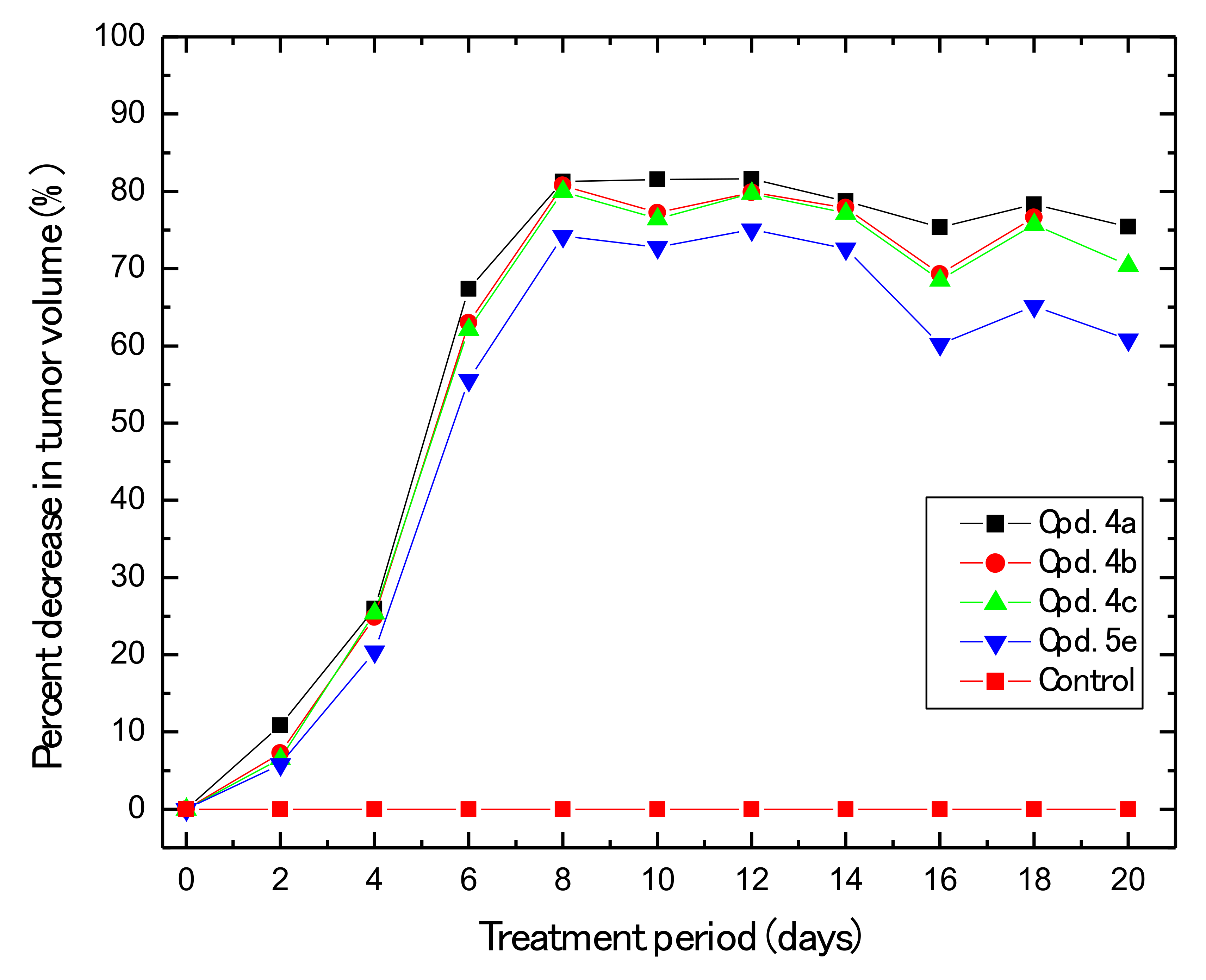
2.2.2. In vivo Anti-Breast Cancer

In this section, we developed a breast cancer animal xenograft model for the evaluation of thein vivo anticancer potential of our newly prepared compounds. We chose the derivatives that showed the lowest IC50 values in the previous in vitro investigation; namely, 4a, 4b, 4c, and 5e. The results obtained (Figure 3) showed that all four tested compounds have a great potential to decrease tumor growth in animal model, where the decrease in tumor growth effect increased gradually with the treatment period up to eight days of treatment. At that point, the highest decrease in tumor growth obtained ranged from 81.3% to 74.2% for compounds 4a and 5e, respectively. After wards, the noticeable effects were more or less constant up to 14 days of treatment. However, by the end of the treatment period (20 days), the percentage of tumor growth inhibition decreased to about 75.4%, 73.1%, 70.5%, and 60.8% for compounds 4a, 4a, 4c, and 5e, respectively. Our results are in good agreement with those previously published on the in vivo anticancer potential of Schiff bases owing to apoptosis induction [42].

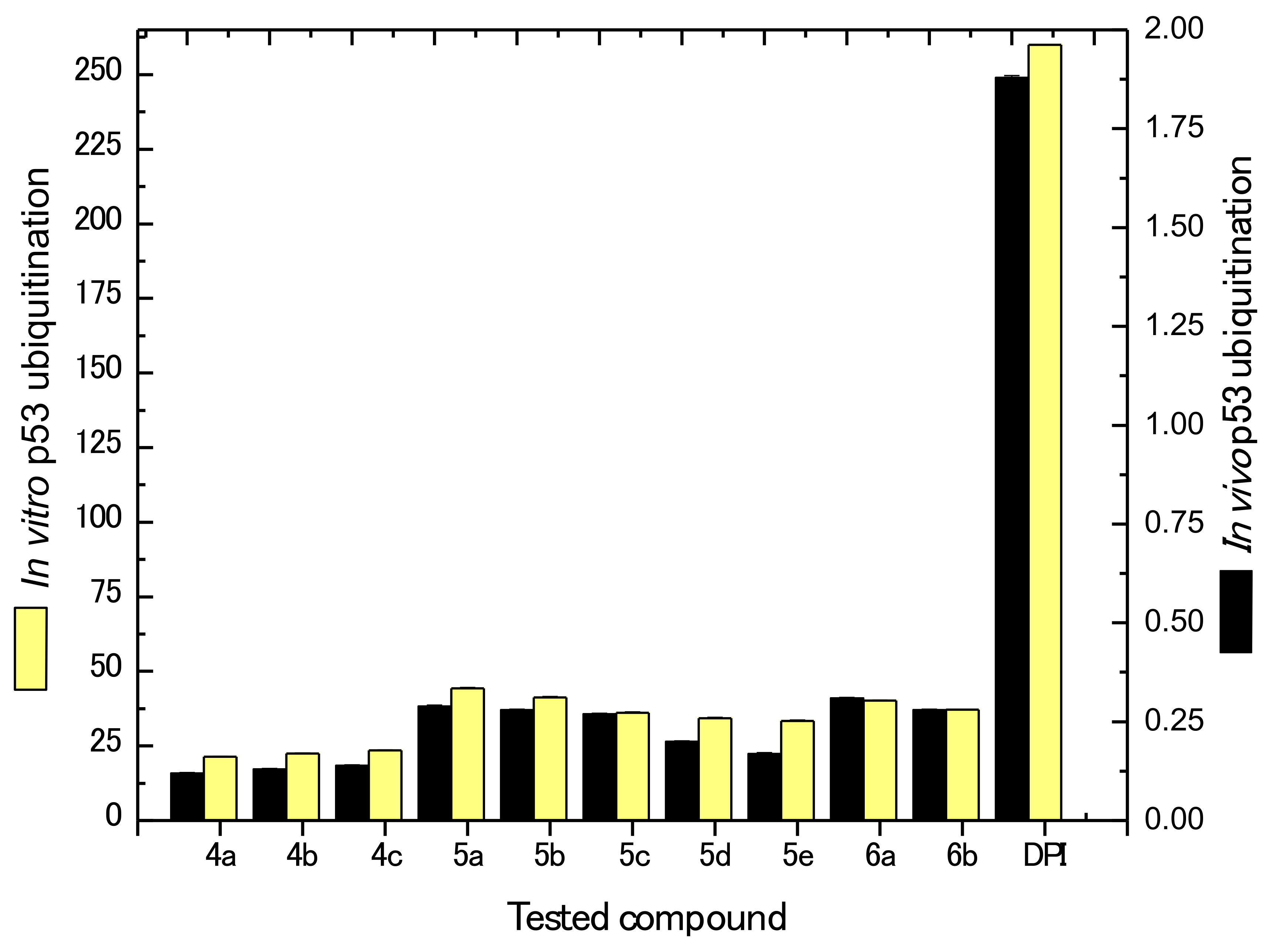
2.2.3. In vitro and In vivo Inhibition of p53

Several cancer prevention drugs depend mainly on the ubiquitination activities against p53 as a suppressor protein. When E3 ubiquitin protein ligase HDM2 binds to p53, the resulting mechanism is the inhibition of its ability as a transcription activator. Accordingly, the regulative mechanism is acting negatively. Therefore, blocking the p53 binding site on HDM2 provides for a better possible antitumorigenic molecule. Currently, murine double minute 2 (MDM2) is considered as the regulator of choice when it comes to investigatingp53 inhibition [43].
The results obtained (Figure 4) showed that all prepared derivatives exhibited potential in vitro as well as in vivo suppression of p53 ubiquitination when compared with the positive control (diphenyl imidazole). The obtained in vitro IC50values ranged from 21.34 ± 0.13 to 44.33 ± 0.34 µM for compounds 4a and 5a, respectively. For thein vivo results, the IC50 values ranged from 0.12 ± 0.0011 to 0.31 ± 0.0010 µM for compounds 4a and 6a, respectively. Furthermore, it can be seen that inhibitory effects of 4a were higher by about 12.2- and 15.7-fold compared with the positive control for in vitro and in vivo p53 ubiquitination, respectively.

2.2.4. Kinase Inhibition Studies

The newly synthesized compounds 46 were evaluated for their ability to inhibit LDHA in comparison with galloflavin as a known isoform nonselective LDHA inhibitor [44,45]. The choice of galloflavin as a reference was based on the fact that it is potent anticancer agent and commercially available [46,47,48,49,50]. The results presented in Figure 5 show that all investigated derivatives exhibited better LDHA inhibition compared with galloflalvin, where the IC50 values ranged from 60.54 ± 0.56 to 141.56 ± 0.98 µM for compounds 5c and 5e, respectively. These values correspond to about 2.66- and 1.14-fold in increase in the inhibitory activity compared with that recorded for galloflavin IC50 = 161.05 ± 0.32 µM).

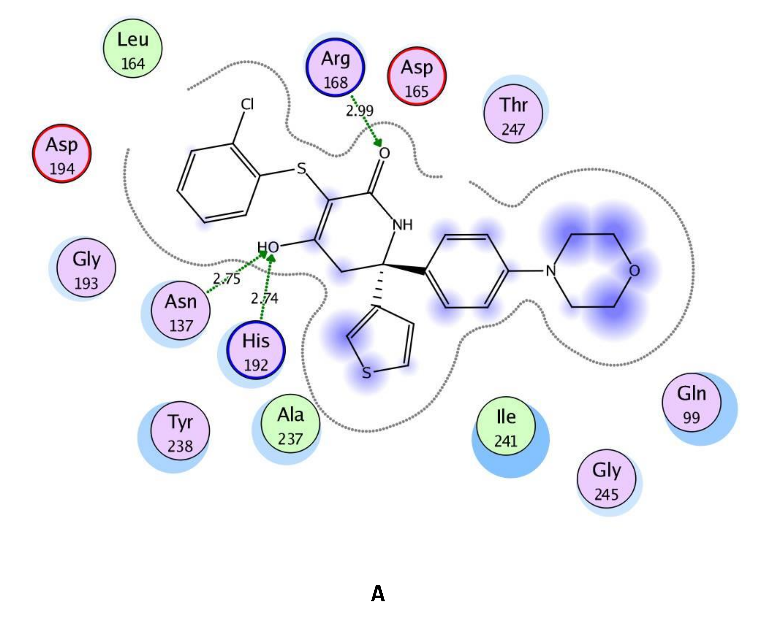
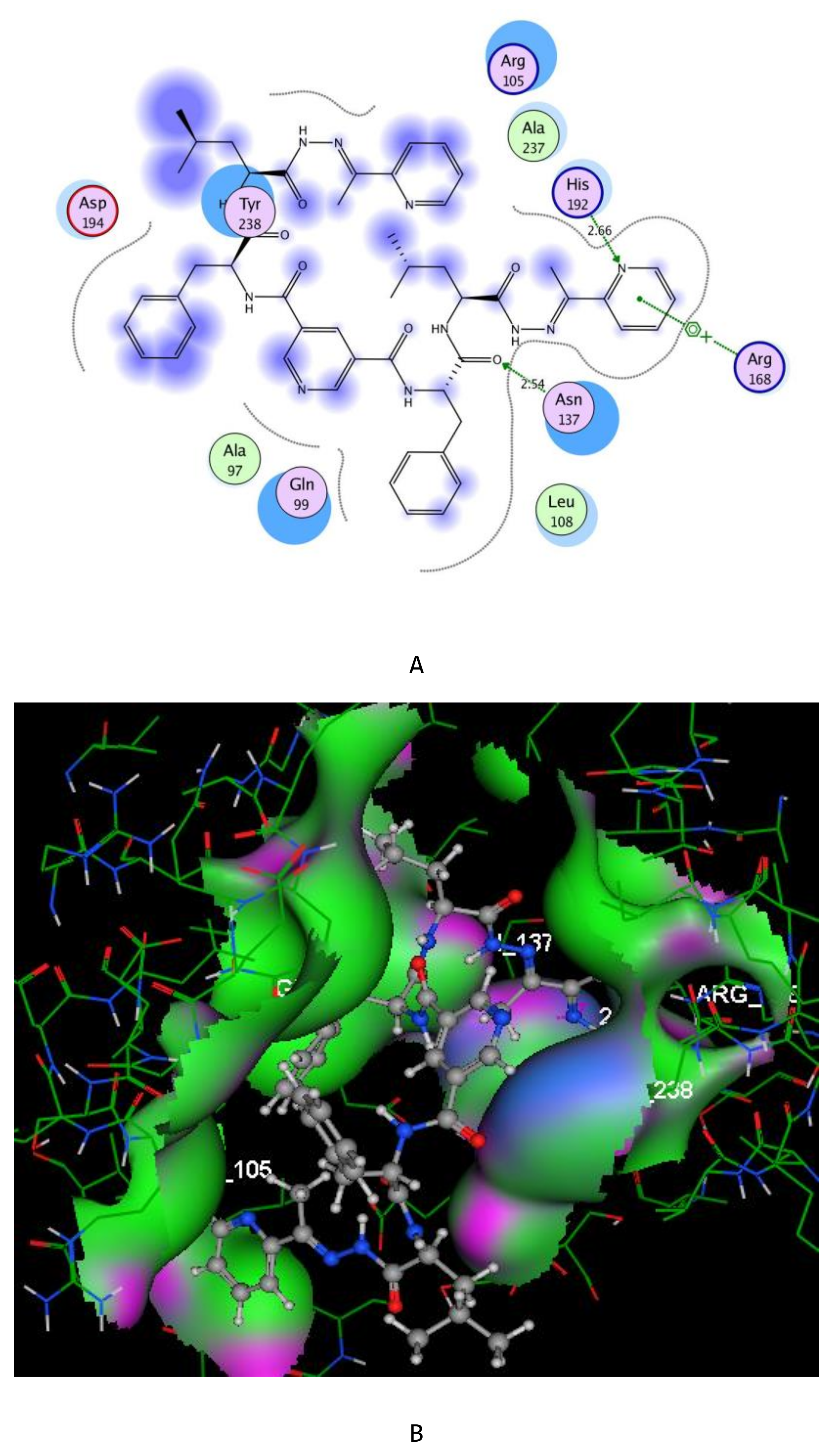
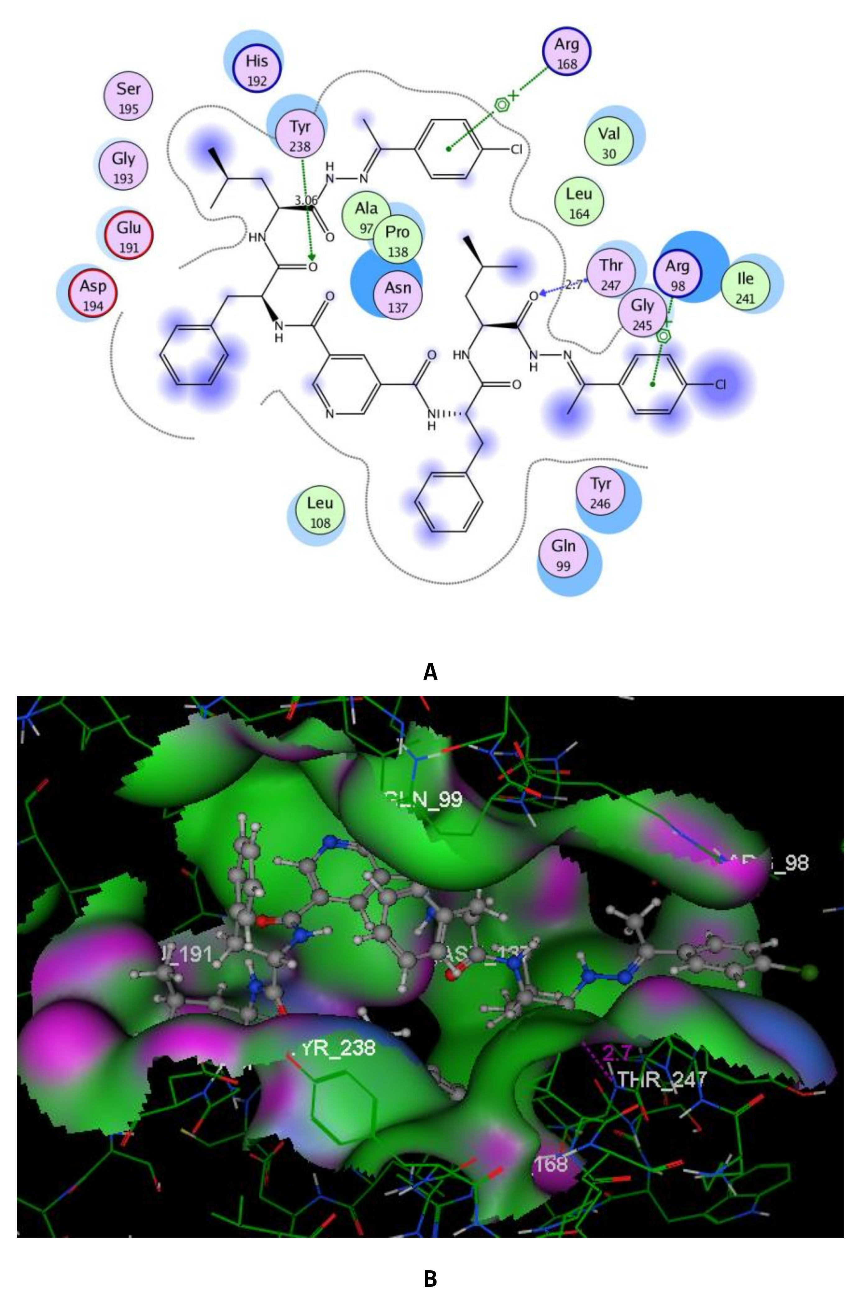
2.3. Molecular Modeling Studies

In order to further understand and illustrate the interaction of compounds 46 with the LDHA active site, the docking modeling studies were done in an open loop conformation of the enzyme. The docking studies were developed using Molecular Operating Environment software, MOE 10.2008 software [51,52] with X-ray crystallographic structure of LDHA (PDB ID: 4ZVV) [53]. The root mean square deviation (RMSD) value of 8.93 was calculated via redocking of the co-crystallized ligand, GNE-140, into the pocket site of LDH-A with docking score energy of −9.73 kcal/mol. The docking scores of the screened compounds 46 were all in the range of −15.60 to −11.29 kcal/mol. Representations of the docking results of these compounds with LDHA are depicted in Table 1and Figure 6, Figure 7 and Figure 8.
As the reported binding mode of GNE-140 with LDHA [53], the oxygen of the hydroxyl group binds to the active site via two hydrogen bond acceptors with the sidechains of Asn137 and His192 (distance: 2.75 and 2.74 Å), respectively. Additionally, the carbonyl oxygen forms H-bonding with the sidechain of Arg168 (distance: 2.99 Å) (Figure 6).
The increased inhibitory activity of the newly synthesized derivatives 4–6 against LDHA enzyme could be partially explained by the evidence obtained from the docking studies (Table 1). By focusing on the highly potent inhibitors 4a and 5c (Figure 7 and Figure 8), the essential residue Arg168 connected to the newly formed 2-pyridyl (in 4a) and 4-chlorophenyl moieties (in 5c) by arene–cation interactions. Moreover, the compound 4a was fitted nicely in the active pocket through two hydrogen bond interactions between nitrogen of 2-pyridyl moiety and oxygen of the amide group with the key amino acids His192 and Asn137 (distance: 2.66 and 2.54 Å), respectively.
The enhanced inhibitory potency of 5c may be attributed to its stabilization within the ATP binding pocket via further arene–cation interaction between the other 4-chlorophenyl and Arg98, and two H-bond acceptors between the oxygens of the two amide groups located at opposite sites around the parent pyridine ring with the sidechain of Tyr238 and the backbone of Thr247 (distance: 3.06 and 2.70 Å), respectively. Additionally, the essential amino acids Ala97, Asn137, and Pro138 were located between the two wings of the symmetrical side chains facing the central core (pyridine moiety) (Figure 8).

3. Materials and Methods

3.1. Chemistry

Melting points were determined in open glass capillary tubes with an electro thermal digital melting point apparatus (model: IA9100), and are uncorrected. Elemental microanalysis for carbon, hydrogen, and nitrogen (Microanalytical Unit, NRC) was found within the acceptable limits of the calculated values. Infrared spectra (KBr) were recorded on a Nexus 670 FTIR Nicolet, Fourier Transform infrared spectrometer. Proton and carbon nuclear magnetic resonance (1H- and 13C NMR) spectra were run in (DMSO-d6) on Jeol 500 MHz instruments. Mass spectra were run on a MAT Finnigan SSQ 7000 spectrometer, using the electron impact technique (EI). Analytical thin layer chromatography (TLC) was performed on silica gel aluminum sheets, 60 F254 (E. Merck). Compound 2 was prepared according to our previous reported procedure [39].
Synthesis of Nα-dinicotinoyl-bis[L-phenylalaninyl-L-leucyl]cycloalkanonehydrazones 3ad. A mixture of hydrazide 2 (1 mmol) and cycloalkanone, namely, cyclopentanone, cyclohexanone, cycloheptanone, or cyclooctanone (2 mmol), in glacial acetic acid (30 mL) was refluxed for 6 h. The reaction mixture was poured onto ice water; the formed precipitate was filtered off, dried, and crystallized from AcOH/H2O to give compounds 3ad, respectively.
N3,N5-Bis{1-[(1-(2-cyclopentylidenehydrazinyl)-4-methyl-1-oxopentan-2-yl]amino)-1-oxo-3-phenylpropan-2-yl}pyridine-3,5-dicarboxamide (3a): Yield 72%, mp 232–234 °C. [α] D 25 = –98 (c = 0.5, DMF). IR ν, cm–1: 3518-3334 (NH), 3090 (CH-Ar), 2985 (CH-aliph.), 1654, 1532, 1254 (C=O, amide I, II and III). 1H NMR spectrum (DMSO-d6), δH, ppm:0.90–0.95 (m, 12H, 4CH3),1.24–1.35 (m, 16H, 8 CH2), 1.58–1.70 (t, 4H, 2CH2), 2.18–2.25 (m, 2H, 2CH), 3.42 (d, 4H, 2CH2), 4.35 (t, 2H, 2CH), 4.55 (t, 2H, 2CH), 6.95–7.65 (m, 10H, 2Ph-H), 8.38, 9.05 (2s, 3H, pyr-H), 8.65, 8.75, 9.15 (3s, 6H, 6NH, exchangeable with D2O). 13C NMR spectrum (DMSO-d6), δC, ppm: 17.32, 17.54 (4C, 4CH3), 24.15, 29.42, 34.32, 186.86 (10C, cyclopentyl ring), 23.98 (2C, 2CH), 40.12 (2C, 2CH2), 42.26 (2C, 2CH2), 52.48, 53.12 (4C, 4 CH), 124.18, 128.26, 129.35, 138.64 (12C, 2Ph-C), 131.55, 140.13, 152.12 (5C, pyr-C), 163.72, 169.15 (4C, 4CO-amide), 174.40 (2C, 2CO-hydrazone). MS (EI, 70 eV): m/z (%) = 848 (45) [M]+. Found, %: C 66.45; H 7.18; N 14.76. C47H61N9O6 (848.04). Calculated, %: C 66.57; H 7.25; N 14.86.
N3,N5-Bis{1-[(1-(2-cyclohexylidenehydrazinyl)-4-methyl-1-oxopentan-2-yl)amino]-1-oxo-3-phenylpropan-2-yl}pyridine-3,5-dicarboxamide (3b): Yield 65%, mp 198–200 °C. [α] D 25 = –118 (c = 0.5, DMF). IR ν, cm−1: 3510–3387 (NH), 3088 (CH-Ar), 2989 (CH-aliph.), 1655, 1533, 1254 (C=O, amide I, II and III). 1H NMR spectrum (DMSO-d6), δH, ppm:0.86–0.94 (m, 12H, 4CH3),1.50–1.65 (m, 16H, 8CH2), 2.22–2.26 (m, 2H, 2CH), 2.10–2.14 (m, 8H, 4CH2), 3.46 (d, 4H, 2CH2), 4.34 (t, 2H, 2CH), 4.54 (t, 2H, 2CH), 6.98–7.68 (m, 10H, 2Ph-H), 8.42, 9.09 (2s, 3H, pyr-H), 8.64, 8.78, 9.16 (3s, 6H, 6NH, exchangeable with D2O). 13C NMR spectrum (DMSO-d6), δC, ppm: 17.30, 17.42 (4C, 4CH3), 24.76, 27.02, 28.32, 165.85 (12C, cyclohexyl ring), 23.90 (2C, 2CH), 40.18 (2C, 2CH2), 42.34 (2C, 2CH2), 52.45, 53.16 (4C, 4 CH), 124.24, 128.28, 129.38, 138.65 (12C, 2Ph-C), 131.58, 140.15, 152.16 (5C, pyr-C), 163.76, 169.18, 174.42 (6C, 6 CO-amide). MS (EI, 70 eV): m/z (%) = 876 (32) [M]+. Found, %: C 67.00; H 7.42; N 14.30. C49H65N9O6 (876.10). Calculated, %: C 67.18; H 7.48; N 14.39.
N3,N5-Bis{1-[(1-(2-cycloheptylidenehydrazinyl)-4-methyl-1-oxopentan-2-yl)amino]-1-oxo-3-phenylpropan-2-yl}pyridine-3,5-dicarboxamide (3c): Yield 56%, mp 221–223 °C. [α] D 25 = –96 (c = 0.5, DMF). IR ν, cm−1: 3412–3375 (NH), 3092 (CH-Ar), 2995 (CH-aliph.), 1655, 1535, 1255 (C=O, amide I, II and III). 1H NMR spectrum (DMSO-d6), δH, ppm: 1H NMR spectrum (DMSO-d6), δH, ppm: 0.88–0.96 (m, 12H, 4CH3), 1.50–1.70 (m, 20H, 10CH2), 2.20–2.24 (m, 2H, 2CH), 2.02–2.10 (m, 8H, 4CH2), 3.45 (d, 4H, 2CH2), 4.44 (t, 2H, 2CH), 4.58 (t, 2H, 2CH), 6.92-7.60 (m, 10H, 2Ph-H), 8.54, 9.00 (2s, 3H, pyr-H), 8.65, 8.76, 9.15 (3s, 6H, 6NH, exchangeable with D2O). 13C NMR spectrum (DMSO-d6), δC, ppm: 17.40, 17.44 (4C, 4CH3), 27.12, 28.82, 30.28, 36.12, 183.90 (14C, cycloheptyl ring), 24.00 (2C, 2CH), 40.24 (2C, 2CH2), 42.46 (2C, 2CH2), 52.40, 53.24 (4C, 4 CH), 124.28, 128.30, 129.42, 138.64 (12C, 2Ph-C), 131.60, 140.18, 152.18 (5C, pyr-C), 163.72, 169.24, 174.40 (6C, 6 CO-amide). MS (EI, 70 eV): m/z (%) = 902 (14) [M-2]+. Found, %: C 67.64; H 7.61; N 13.86. C51H69N9O6 (904.15). Calculated, %: C 67.75; H 7.69; N 13.94.
N3,N5-Bis{1-[(1-(2-cyclooctylidenehydrazinyl)-4-methyl-1-oxopentan-2-yl)amino]-1-oxo-3-phenylpropan-2-yl}pyridine-3,5-dicarboxamide (3d): Yield 56%, mp 242–244 °C. [α] D 25 = –104 (c = 0.5, DMF). IR ν, cm–1: 3418–3360 (NH), 3086 (CH-Ar), 2990 (CH-aliph.), 1654, 1533, 1255 (C=O, amide I, II and III). 1H NMR spectrum (DMSO-d6), δH, ppm:0.84–0.90 (m, 12H, 4CH3), 1.22–1.60 (m, 20H, 10CH2), 1.65–1.70 (m, 4H, 2CH2), 2.00–2.08 (m, 8H, 4CH2), 2.18–2.22 (m, 2H, 2CH), 3.52 (d, 4H, 2CH2), 4.20–4.35 (m, 2H, 2CH), 4.60–4.68 (m, 2H, 2CH), 6.96–7.54 (m, 10H, 2Ph-H), 8.46, 9.05 (2s, 3H, pyr-H), 8.68, 8.74, 9.12 (3s, 6H, 6NH, exchangeable with D2O). 13C NMR spectrum (DMSO-d6), δC, ppm: 17.30, 17.54 (4C, 4 CH3), 22.92, 24.80, 25.28, 27.02, 28.79, 154.94 (16C, cyclooctyl ring), 23.68 (2C, 2CH), 40.10 (2C, 2CH2), 42.56 (2C, CH2), 52.62, 53.12 (4C, 4 CH), 124.24, 128.25, 129.40, 138.65 (12C, 2Ph-C), 131.70, 140.34, 152.25 (5C, pyr-C), 163.68, 169.16, 174.45 (6C, 6 CO-amide). MS (EI, 70 eV): m/z (%) = 932 (32) [M]+. Found, %: C 68.18; H 7.80; N 13.45. C53H73N9O6 (932.22). Calculated, %: C 68.29; H 7.89; N 13.52.
Synthesis of Nα-dinicotinoyl-bis[L-phenylalaninyl-L-leucyl] Schiff bases 4a–c, 5a–e, and 6a,b. A mixture of 2 (1 mmol) and acetylpyridines or substituted acetophenones or heterocyclic aldehyde, namely, 2-, 3-, 4-acetylpyridine,4-methyl-, 4-methoxy-, 4-chloro-, 4-bromo-, 4-nitroacetophenone or 2-furane, 2-thiophene aldeydes (2 mmol) in AcOH or ethanol (30 mL) was refluxed for 4–6 h, and then evaporated to dryness. The formed residue was solidified with n-hexane, filtered off, and crystallized from the proper solvents to give Schiff base derivatives 4a–c, 5a–e,and 6a,b, respectively.
N3,N5-Bis{1-[(4-methyl-1-oxo-1-(2-(1-(pyridin-2-yl)ethylidene)hydrazinyl)pentan-2-yl)amino]-1-oxo-3-phenylpropan-2-yl}pyridine-3,5-dicarboxamide (4a): Yield 60%, mp 216–218 °C (DMF/H2O). [α] D 25 = −112 (c = 0.5, DMF). IR ν, cm−1: 3450–3354 (NH), 3092 (CH-Ar), 2986 (CH-aliph.), 1654, 1534, 1252 (C=O, amide I, II and III). 1H NMR spectrum (DMSO-d6), δH, ppm: 0.85–0.95 (m, 12H, 4CH3), 1.60–1.70 (m, 4H, 2CH2), 1.82 (s, 6H, 2CH3), 2.20-2.30 (m, 2H, 2CH), 3.48 (d, 4H, 2CH2), 4.25–4.34 (m, 2H, 2CH), 4.60–4.70 (m, 2H, 2CH), 6.92–7.65 (m, 10H, 2Ph-H), 7.68–8.50 (m, 10H, pyr-H), 9.04 (s, 1H, pyr-H-4), 8.65, 8.85, 9.15 (3s, 6H, 6NH, exchangeable with D2O). 13C NMR spectrum (DMSO-d6), δC, ppm: 13.45, 17.85, 18.00 (6C, 6 CH3), 23.96 (2C, 2CH), 40.56 (2C, 2CH2), 42.44 (2C, CH2), 52.56, 53.12 (4C, 4 CH), 124.24, 128.26, 129.35, 138.68 (12C, 2Ph-C), 145.30 (2C, 2C=N), 122.90, 126.00, 131.60, 135.80, 140.20, 148.62, 152.22, 154.48 (15C, 3 pyr-C), 163.82, 169.33 (4C, 4 CO-amide), 174.55 (2C, 2CO-hydrazone). MS (EI, 70 eV): m/z (%) = 922 (54) [M]+. Found, %: C 66.32; H 6.36; N 16.60. C51H59N11O6 (922.08). Calculated, %: C 66.43; H 6.45; N 16.71.
N3,N5-Bis{1-[(4-methyl-1-oxo-1-(2-(1-(pyridin-3-yl)ethylidene)hydrazinyl)pentan-2-yl)-amino]-1-oxo-3-phenylpropan-2-yl}pyridine-3,5-dicarboxamide (4b): Yield 58 %, mp 208–210 °C (AcOH/H2O). [α] D 25 = −132 (c = 0.5, DMF). IR ν, cm−1: 3467–3350 (NH), 3075 (CH-Ar), 2990 (CH-aliph.), 1652, 1536, 1252 (C=O, amide I, II and III). 1H NMR spectrum (DMSO-d6), δH, ppm: 0.88-0.93 (m, 12H, 4CH3), 1.62–1.74 (m, 4H, 2CH2), 1.80 (s, 6H, 2CH3), 2.16–2.24 (m, 2H, 2CH), 3.45 (d, 4H, 2CH2), 4.24–4.35 (m, 2H, 2CH), 4.62-4.70 (m, 2H, 2CH), 6.96-7.60 (m, 10H, 2Ph-H), 7.72-8.65 (m, 8H, pyr-H), 9.04, 9.08 (3s, 3H, pyr-H), 8.70, 8.86, 9.18 (3s, 6H, 6NH, exchangeable with D2O). 13C NMR spectrum (DMSO-d6), δC, ppm: 15.25, 17.88, 18.02 (6C, 6 CH3), 23.94 (2C, 2CH), 40.55 (2C, 2CH2), 42.45 (2C, CH2), 52.55, 53.15 (4C, 4 CH), 124.25, 128.25, 129.35, 138.65 (12C, 2Ph-C), 145.35 (2C, 2C=N), 122.96, 126.14, 131.65, 137.56, 140.45, 147.82, 151.32, 154.80 (15C, 3 pyr-C), 163.80, 169.14 (4C, 4CO-amide), 168.12 (2C, 2CO-hydrazone). MS (EI, 70 eV): m/z (%) = 921 (18) [M-1]+. Found, %: C 66.33; H 6.34; N 16.64. C51H59N11O6 (922.08). Calculated, %: C 66.43; H 6.45; N 16.71.
N3,N5-Bis{1-[(4-methyl-1-oxo-1-(2-(1-(pyridin-4-yl)ethylidene)hydrazinyl)pentan-2-yl)-amino]-1-oxo-3-phenylpropan-2-yl}pyridine-3,5-dicarboxamide (4c): Yield 75%, mp 196–198 °C (EtOH/H2O). [α] D 25 = −140 (c = 0.5, DMF). IR ν, cm–1: 3466–3348 (NH), 3080 (CH-Ar), 2986 (CH-aliph.), 1655, 1535, 1255 (C=O, amide I, II and III). 1H NMR spectrum (DMSO-d6), δH, ppm: 0.86-0.90 (m, 12H, 4CH3), 1.65–1.70 (m, 4H, 2CH2), 1.96 (s, 6H, 2CH3), 2.18–2.20 (m, 2H, 2CH), 3.46 (d, 4H, 2CH2), 4.25–4.32 (m, 2H, 2CH), 4.60–4.68 (m, 2H, 2CH), 6.98–7.64 (m, 10H, 2Ph-H), 7.70–8.65 (m, 10H, pyr-H), 9.06 (s, 1H, pyr-H), 8.75, 8.85, 9.14 (3s, 6H, 6NH, exchangeable with D2O). 13C NMR spectrum (DMSO-d6), δC, ppm: 16.02, 17.86, 18.12 (6C, 6 CH3), 23.86 (2C, 2CH), 40.50 (2C, 2CH2), 42.60 (2C, CH2), 52.54, 53.18 (4C, 4 CH), 124.60, 128.42, 129.38, 138.658 (12C, 2Ph-C), 146.30 (2C, 2C=N), 124.14, 131.60, 138.50, 140.40, 147.50, 149.54 (15C, 3 pyr-C), 164.15, 169.46 (4C, 4CO-amide), 174.10 (2C, 2CO-hydrazone). MS (EI, 70 eV): m/z (%) = 920 (8) [M-2]+. Found, %: C 66.37; H 6.35; N 16.65. C51H59N11O6 (922.08). Calculated, %: C 66.43; H 6.45; N 16.71.
N3,N5-Bis{1-[(1-(2-(1-(4-methoxyphenyl)ethylidene)hydrazinyl)-4-methyl-1-oxopentan-2-yl)-amino]-1-oxo-3-phenylpropan-2-yl}pyridine-3,5-dicarboxamide (5a): Yield 55%, mp 202–204 °C (AcOH/H2O). [α] D 25 = −110 (c = 0.5, DMF). IR ν, cm−1: 3478–3348 (NH), 3085 (CH-Ar), 2976 (CH-aliph.), 1655, 1534, 1253 (C=O, amide I, II and III). 1H NMR spectrum (DMSO-d6), δH, ppm:0.86–0.94 (m, 12H, 4CH3), 1.62–1.74 (m, 4H, 2CH2), 1.98 (s, 6H, 2CH3), 2.18–2.25 (m, 2H, 2CH), 3.45 (d, 4H, 2CH2), 3.68 (s, 6H, 2OCH3), 4.20–4.35 (m, 2H, 2CH), 4.65–4.70 (m, 2H, 2CH), 6.95–7.65 (m, 18H, 4Ph-H), 8.62, 9.00 (2s, 3H, pyr-H), 8.66, 8.76, 9.20 (3s, 6H, 6NH, exchangeable with D2O). 13C NMR spectrum (DMSO-d6), δC, ppm: 15.50, 17.85, 18.24 (6C, 6 CH3), 23.78 (2C, 2CH), 40.26 (2C, 2CH2), 42.48 (2C, CH2), 52.76, 53.15 (4C, 4 CH), 55.72 (2C, 2OCH3), 114.32, 125.65, 127.28, 128.32, 128.65, 129.75, 138.68, 162.84 (24C, 4Ph-C), 131.60, 140.12, 152.20 (5C, pyr-C), 145.15 (2C, 2C=N), 164.14, 169.65 (4C, 4 CO-amide), 174.06 (2C, 2CO-hydrazone). MS (EI, 70 eV): m/z (%) = 980 (16) [M]+. Found, %: C 67.28; H 6.62; N 12.76. C55H65N9O8 (980.16). Calculated, %: C 67.40; H 6.68; N 12.86.
N3,N5-Bis{1-[(1-(2-(1-(4-chlorophenyl)ethylidene)hydrazinyl)-4-methyl-1-oxopentan-2-yl)-amino]-1-oxo-3-phenylpropan-2-yl}pyridine-3,5-dicarboxamide (5b): Yield 60 %, mp 184–186 °C (DMF/H2O). [α] D 25 = −99 (c = 0.5, DMF). IR ν, cm−1: 3467–3345 (NH), 3086 (CH-Ar), 2979 (CH-aliph.), 1654, 1535, 1254 (C=O, amide I, II and III). 1H NMR spectrum (DMSO-d6), δH, ppm: 0.88–0.92 (m, 12H, 4CH3), 1.64–1.70 (m, 4H, 2CH2), 2.05 (s, 6H, 2CH3), 2.20–2.24 (m, 2H, 2CH), 3.46 (d, 4H, 2CH2), 4.26–4.34 (m, 2H, 2CH), 4.68–4.72 (m, 2H, 2CH), 7.10–7.90 (m, 18H, 4Ph-H), 8.65, 9.04 (2s, 3H, pyr-H), 8.70, 8.78, 9.18 (3s, 6H, 6NH, exchangeable with D2O). 13C NMR spectrum (DMSO-d6), δC, ppm: 15.85, 17.80, 18.12 (6C, 6 CH3), 23.84 (2C, 2CH), 40.38 (2C, 2CH2), 42.50 (2C, CH2), 52.74, 53.10 (4C, 4 CH), 125.65, 127.28, 128.32, 128.78, 129.05, 134.95, 135.68, 136.84 (24C, 4Ph-C), 131.70, 140.18, 152.26 (5C, pyr-C), 144.98 (2C, 2C=N), 164.34, 169.65 (4C, 4 CO-amide), 173.96 (2C, 2CO-hydrazone). MS (EI, 70 eV): m/z (%) = 988 (24) [M-1]+. Found, %: C 64.24; H 5.90; Cl 7.08; N 12.65. C53H59Cl2N9O6 (988.99). Calculated, %: C 64.36; H 6.01; Cl 7.17; N 12.75.
N3,N5-Bis{1-[(1-(2-(1-(4-bromophenyl)ethylidene)hydrazinyl)-4-methyl-1-oxopentan-2-yl)-amino]-1-oxo-3-phenylpropan-2-yl}pyridine-3,5-dicarboxamide (5c): Yield 72 %, mp 210–212 °C (DMF/H2O). [α] D 25 = −124 (c = 0.5, DMF). IR ν, cm−1: 3462–3345 (NH), 3090 (CH-Ar), 2988 (CH-aliph.), 1655, 1532, 1250 (C=O, amide I, II and III). 1H NMR spectrum (DMSO-d6), δH, ppm: 0.84–0.90 (m, 12H, 4CH3), 1.60–1.65 (m, 4H, 2CH2), 2.02 (s, 6H, 2CH3), 2.18–2.22 (m, 2H, 2CH), 3.48 (d, 4H, 2CH2), 4.25–4.35 (m, 2H, 2CH), 4.66–4.70 (m, 2H, 2CH), 7.14–7.82 (m, 18H, 4Ph-H), 8.62, 9.08 (2s, 3H, pyr-H), 8.72, 8.76, 9.16 (3s, 6H, 6NH, exchangeable with D2O). 13C NMR spectrum (DMSO-d6), δC, ppm: 15.90, 17.60, 17.86 (6C, 6 CH3), 23.80 (2C, 2CH), 40.42 (2C, 2CH2), 42.56 (2C, CH2), 52.78, 53.15 (4C, 4 CH), 125.55, 126.04, 127.56, 128.24, 128.70, 130.75, 135.98, 136.34 (24C, 4Ph-C), 131.62, 140.26, 152.28 (5C, pyr-C), 145.08 (2C, 2C=N), 164.65, 168.98 (4C, 4 CO-amide), 174.16 (2C, 2CO-hydrazone). MS (EI, 70 eV): m/z (%) = 1078 (14) [M]+. Found, %: C 58.88; H 5.45; N 11.62. C53H59Br2N9O6 (1077.90). Calculated, %: C 59.06; H 5.52; N 11.69.
N3,N5-Bis{1-[(4-methyl-1-oxo-1-(2-(1-(p-tolyl)ethylidene)hydrazinyl)pentan-2-yl)amino]-1-oxo-3-phenylpropan-2-yl}pyridine-3,5-dicarboxamide (5d): Yield 55%, mp 235–237 °C (DMF/H2O). [α] D 25 = −78 (c = 0.5, DMF). IR ν, cm−1: 3465–3340 (NH), 3088 (CH-Ar), 2980 (CH-aliph.), 1656, 1536, 1256 (C=O, amide I, II, and III). 1H NMR spectrum (DMSO-d6), δH, ppm:0.88–0.92 (m, 12H, 4CH3), 1.66–1.70 (m, 4H, 2CH2), 2.00 (s, 6H, 2CH3), 2.18–2.24 (m, 2H, 2CH), 2.34 (s, 6H, 2CH3), 3.43 (d, 4H, 2CH2), 4.23–4.30 (m, 2H, 2CH), 4.66–4.72 (m, 2H, 2CH), 7.12–7.68 (m, 18H, 4Ph-H), 8.65, 9.05 (2s, 3H, pyr-H), 8.68, 8.75, 9.18 (3s, 6H, 6NH, exchangeable with D2O). 13C NMR spectrum (DMSO-d6), δC, ppm: 16.80, 17.90, 18.15, 21.12 (8C, 8 CH3), 23.86 (2C, 2CH), 40.35 (2C, 2CH2), 42.52 (2C, CH2), 52.75, 53.18 (4C, 4 CH), 125.72, 126.45, 127.35, 128.40, 128.92, 134.08, 136.10, 139.56 (24C, 4Ph-C), 131.68, 140.25, 152.25 (5C, pyr-C), 145.60 (2C, 2C=N), 164.12, 169.68 (4C, 4 CO-amide), 174.18 (2C, 2CO-hydrazone). MS (EI, 70 eV): m/z (%) = 948 (42) [M]+. Found, %: C, 69.60; H, 6.86; N, 13.22. C55H65N9O6 (948.18). Calculated, %: C, 69.67; H, 6.91; N, 13.30.
N3,N5-Bis{1-[(4-methyl-1-(2-(1-(4-nitrophenyl)ethylidene)hydrazinyl)-1-oxopentan-2-yl)-amino]-1-oxo-3-phenylpropan-2-yl}pyridine-3,5-dicarboxamide (5e). Yield 72 %, mp 256–258 °C (DMF/H2O). [α] D 25 = −104 (c = 0.5, DMF). IR ν, cm−1: 3485–3354 (NH), 3092 (CH-Ar), 2986 (CH-aliph.), 1655, 1533, 1255 (C=O, amide I, II, and III). 1H NMR spectrum (DMSO-d6), δH, ppm: 0.86–0.90 (m, 12H, 4CH3), 1.66–1.74 (m, 4H, 2CH2), 2.10 (s, 6H, 2CH3), 2.24–2.26 (m, 2H, 2CH), 3.45 (d, 4H, 2CH2), 4.25–4.32 (m, 2H, 2CH), 4.65–4.68 (m, 2H, 2CH), 7.08–7.26 (m, 10H, 2Ph-H), 8.00–8.28 (m, 8H, 2Ph-H), 8.58, 9.03 (2s, 3H, pyr-H), 8.65, 8.75, 9.15 (3s, 6H, 6NH, exchangeable with D2O). 13C NMR spectrum (DMSO-d6), δC, ppm: 16.04, 17.88, 18.06 (6C, 6 CH3), 23.95 (2C, 2CH), 40.45 (2C, 2CH2), 42.54 (2C, CH2), 52.73, 53.13 (4C, 4 CH), 125.63, 126.86, 127.32, 127.90, 128.24, 136.70, 143.06, 149.85 (24C, 4Ph-C), 131.55, 140.25, 152.20 (5C, pyr-C), 145.00 (2C, 2C=N), 163.98, 169.40 (4C, 4 CO-amide), 174.34 (2C, 2CO-hydrazone). MS (EI, 70 eV): m/z (%) = 1010 (8) [M-1]+. Found, %: C, 62.94; H, 5.80; N, 15.20. C53H59N11O10 (1010.12). Calculated, %: C, 63.02; H, 5.89; N, 15.25.
N3,N5-bis{1-[(1-(2-(furan-2-ylmethylene)hydrazinyl)-4-methyl-1-oxopentan-2-yl)amino]-1-oxo-3-phenylpropan-2-yl}pyridine-3,5-dicarboxamide (6a):Yield 75%, mp 205–207 °C (DMF/Ether). [α] D 25 = −105 (c = 0.5, DMF). - IR (film): ν = 3495–3350 (NH), 3095 (CH-Ar), 2985 (CH-aliph.), 1654, 1532, 1250 (C=O, amide I, II, and III) cm−1.1H NMR spectrum (DMSO-d6), δH, ppm: 0.85–0.92 (m, 12H, 4CH3), 1.68–1.74 (m, 4H, 2CH2), 2.22–2.34 (m, 2H, 2CH), 3.42 (d, 4H, 2CH2), 4.16–4.26 (m, 2H, 2CH), 4.45–4.58 (m, 2H, 2CH), 6.90–6.90 (m, 6H, 2 furyl ring), 7.12–7.38 (m, 10H, 2 Ph-H), 7.98 (s, 2H, 2CH=N), 8.32, 9.10 (2s, 3H, pyr-H), 8.78, 8.80, 9.16 (3s, 6H, 6NH, exchangeable with D2O). 13C NMR spectrum (DMSO-d6), δC, ppm: 18.60, 19.15 (4C, 4 CH3), 22.96 (2C, 2CH), 40.25 (2C, 2CH2), 42.48 (2C, CH2), 52.56, 53.34 (4C, 4 CH), 112.05, 117.95, 143.45, 148.16 (8C, 2 furyl-C), 125.84, 127.42, 127.98, 137.85 (12C, 2Ph-C), 131.62, 140.34, 152.40 (5C, pyr-C), 137.12 (2C, 2C=N), 163.95, 169.48 (4C, 4 CO-amide), 178.46 (2C, 2CO-hydrazone). MS (EI, 70 eV): m/z (%) = 872 (16) [M]+. Found, %: C, 64.65; H, 6.10; N, 14.39. C47H53N9O8 (872.00). Calculated, %: C, 64.74; H, 6.13; N, 14.46.
N3,N5-bis{1-[(4-methyl-1-oxo-1-(2-(thiophen-2-ylmethylene)hydrazinyl)pentan-2-yl)amino]-1-oxo-3-phenylpropan-2-yl}pyridine-3,5-dicarboxamide (6b). Yield 70%, mp 218–220 °C (DMF/Ether). [α] D 25 = −110 (c = 0.5, DMF). IR (film): ν = 3482–3365 (NH), 3084 (CH-Ar), 2978 (CH-aliph.), 1655, 1535, 1254 (C=O, amide I, II, and III) cm−1.1H NMR spectrum (DMSO-d6), δH, ppm: 0.82–0.90 (m, 12H, 4CH3), 1.65–1.70 (m, 4H, 2CH2), 2.20–2.30 (m, 2H, 2CH), 3.44 (d, 4H, 2CH2), 4.18–4.30 (m, 2H, 2CH), 4.46–4.55 (m, 2H, 2CH), 7.18–7.56 (m, 16H, 2 Ph-H + 2 furyl ring), 7.86 (s, 2H, 2CH=N), 8.36, 9.08 (2s, 3H, pyr-H), 8.75, 8.84, 9.18 (3s, 6H, 6NH, exchangeable with D2O). 13C NMR spectrum (DMSO-d6), δC, ppm: 18.32, 19.10 (4C, 4 CH3), 22.78 (2C, 2CH), 40.55 (2C, 2CH2), 42.68 (2C, CH2), 52.54, 53.32 (4C, 4 CH), 126.45, 127.90, 130.45, 144.06 (8C, 2 thienyl-C), 125.80, 127.52, 128.14, 137.80 (12C, 2Ph-C), 131.65, 140.36, 152.42 (5C, pyr-C), 125.18 (2C, 2C=N), 163.86, 169.45 (4C, 4 CO-amide), 178.42 (2C, 2CO-hydrazone). MS (EI, 70 eV): m/z (%) = 904 (24) [M]+. Found, %: C, 62.32; H, 5.86; N, 13.88; S, 7.00. C47H53N9O6S2 (904.12). Calculated, %: C, 62.44; H, 5.91; N, 13.94; S, 7.09.

3.2. Biological Evaluation

3.2.1. In Vitro Cytotoxic Activity against MCF-7 Cancer Cells

Human breast cancer (MCF-7) cells, obtained from Sigma-Aldrich Chemie GmbH, Taufkirchen, Germany, were propagated in RPMI-1640 supplemented with 10% heat inactivated FBS, 2 mM L-glutamine, and 1% standard antibiotic solution. Cells were incubated in a 5% CO2humidified incubator at 37 °C and passaged bi-weekly. The in vitro anti-proliferative activity of the newly synthesized derivatives was assayed using the standard MTT technique [54,55,56].
The results were expressed as IC50. Experiments were repeated at least in triplicate, to obtain good reproducibility between replicate wells with standard errors below 10%. Furthermore, the cytotoxic effects of the prepared derivatives were evaluated against normal nonmalignant cells, non-tumorigenic MCF-10A, in order to find out if the synthesized derivatives have toxicity against normal cells. Additionally, the results were compared with reference compounds (cisplatin and milaplatin) as positive controls.

3.2.2. In Vivo Human Breast Cancer Xenograft

The breast cancer xenograft model protocol was approved by the Institutional Animal Use and Care Committee of the University of Alabama at Birmingham (50-01-05-08B). Female athymic pathogen-free nude mice (nu/nu, 4–6 weeks) were used. Firstly, MCF-7 xenografts were initiated by implanting pellets slowly releasing estrogen for two months (1.7mg 17β-estradiol/pellet) subcutaneously in the female nude mice. After 24 h, confluent MCF-7 cells were harvested, washed two times with serum-free medium, re-suspended, and injected subcutaneously (s.c.) (5 × 10⁶ cells, total volume 0.2 mL) into the left inguinal area of the mice. Caliper measurement was used to measure tumor growth in two perpendicular diameters of the implant after 48 h, and its volume was determined. Mice grafted with MCF-7 were divided into different groups (7–10 mice/group). Untreated mice received the solvent only. Treated groups received different prepared derivatives as previously described [57].

3.2.3. In Vitro and In Vivo p53 Ubiquitination

Different prepared derivatives 46 were evaluated for their inhibitory potential against p53 ubiquitination according to the previously reported procedure [57].

3.2.4. LDHA Inhibition Assay

Lactate dehydrogenase assay kit (Abcam, ab102526) and recombinant human LDHA protein (Abcam, ab93699) were used to assess the inhibitory effect of the tested compounds. First, we generated an NADH standard curve for colorimetric detection by measuring the OD (450 nm) at different molar amounts of NADH. The linear regression equation of the curve was derivatized in Graphpad Prism. Tested compounds were dissolved in DMSO and their inhibitory activity was assessed at 140 and 300 μM strength. Galloflavin (Sigma, SML0776) at the same molar concentration was used as a known LDH inhibitor. The master reaction mix composed of LDH assay buffer and 10 ng human LDHA substrate per reaction was prepared. Tested compounds were added and the final reaction volume was adjusted to 50 μL. Absorbance at 450 nm was taken after 2–3 min (T-initial) and continued every minute for 118 min (T-final). The change in measurement over time (delta A450) was calculated as T-final minus T-initial. The amount of NADH generated by the assay between T-initial and T-final was deduced by comparing the A450 of each sample to the standard NADH standard curve. LDHA activity was calculated in milliunits per mL as the amount of NADH generated by the assay / (reaction time in minutes (118) x reaction volume in mL (0.005) [45].

3.3. Molecular Docking Studies

The three-dimensional X-ray structure of LDHA (PDB code: 4ZVV) [53] was chosen as the template for the modeling study of the screened compounds 46. The crystal structure was derived from the RCSB Protein Data Bank and the molecular docking procedure was performed using MOE, 10.2008 software following the reported procedure [51,52].

4. Conclusions

In conclusion, a series of new tetrapeptide Schiff bases 36were designed, synthesized, and evaluated for their antitumor activities against breast MCF-7 cell lines. These compounds exhibited potent antiproliferative activities in comparison with the standards, cisplatin and milaplatin. All of these derivatives were subjected to evaluation ofin vitroandin vivosuppression of p53 ubiquitination and inhibition assay for LDHA enzyme. Molecular docking study was further performed to illustrate the possible binding interactions with LDHA that might be a promising base for the further development of novel anticancer agents with excellent LDH inhibitory activity.

Author Contributions

The listed authors contributed to this work as described in the following: A.E.-G.E.A. and R.E.A.M. gave the concepts of the work, interpreted the results, did the experimental part, and prepared the manuscript; M.E.-N. analyzed the data; E.S.N. and E.A.E. contributed to the anticancer activity and the modeling studies. All authors read and approved the final manuscript.

Acknowledgments

Authors are grateful to King Saud University for funding the work through Researchers Supporting Project (Project No. RSP-2019/66)”.

Conflicts of Interest

The authors declare no conflict of interest.

References

  1. Marqus, S.; Pirogova, E.; Piva, T.J. Evaluation of the use of therapeutic peptides for cancer treatment. J. Biomed. Sci. 2017, 24, 21. [Google Scholar] [CrossRef] [Green Version]
  2. Chakrabarti, S.; Jahandideh, F.; Wu, J. Food-derived bioactive peptides on inflammation and oxidative stress. Biomed. Res. Int. 2014, 2014, 608979. [Google Scholar] [CrossRef] [PubMed] [Green Version]
  3. Yang, R.Y.; Zhang, Z.F.; Pei, X.R.; Han, X.L.; Wang, J.B.; Wang, L.L.; Long, Z.; Shen, X.Y.; Li, Y. Immunomodulatory effects of marine oligopeptide preparation from Chum Salmon (Oncorhynchus keta) in mice. Food Chem. 2009, 113, 464–470. [Google Scholar] [CrossRef]
  4. Cicero, A.F.G.; Fogacci, F.; Colletti, A. Potential role of bioactive peptides in prevention and treatment of chronic diseases: A narrative review. Br. J. Pharmacol. 2017, 174, 1378–1394. [Google Scholar] [CrossRef] [PubMed]
  5. Holohan, C.; Van Schaeybroeck, S.; Longley, D.B.; Johnston, P.G. Cancer drug resistance: an evolving paradigm. Nat. Rev. Cancer 2013, 13, 714–726. [Google Scholar] [CrossRef] [PubMed]
  6. Boohaker, R.J.; Lee, M.W.; Vishnubhotla, P.; Perez, J.M.; Khaled, A.R. The use of therapeutic peptides to target and to kill cancer cells. Curr. Med. Chem. 2012, 19, 3794–3804. [Google Scholar] [CrossRef]
  7. Gautam, A.; Kapoor, P.; Chaudhary, K.; Kumar, R.; Raghava, G.P. Tumor homing peptides as molecular probes for cancer therapeutics, diagnostics and theranostics. Curr. Med. Chem. 2014, 21, 2367–2391. [Google Scholar] [CrossRef]
  8. Vlieghe, P.; Lisowski, V.; Martinez, J.; Khrestchatisky, M. Synthetic therapeutic peptides: science and market. Drug Discov. Today 2010, 15, 40–56. [Google Scholar] [CrossRef]
  9. Blanco-Míguez, A.; Gutiérrez-Jácome, A.; Pérez-Pérez, M.; Pérez-Rodríguez, G.; Catalán-García, S.; Fdez-Riverola, F.; Lourenço, A.; Sánchez, B. From amino acid sequence to bioactivity: scientific evidence on antitumor peptides. Protein Sci. 2016, 25, 1084–1095. [Google Scholar] [CrossRef]
  10. Lopci, E.; Nanni, C.; Rampin, L.; Rubello, D.; Fanti, S. Clinical applications of 68Ga-1239 DOTANOC in neuroendocrine tumours. Minerva Endocrinol. 2008, 33, 277–281. [Google Scholar]
  11. Emons, G.; Sindermann, H.; Engel, J.; Schally, A.V.; Grundker, C. Luteinizing hormone-releasing hormone receptor-targeted chemotherapy using AN-152. Neuroendocrinology 2009, 90, 15–18. [Google Scholar] [CrossRef] [PubMed]
  12. Amr, A.E.; Naglah, A.M.; Sabry, N.M.; Ibrahim, A.A.; Elsayed, E.A.; Attar, A. Synthesis and investigation of 3,5-bis-linear and macrocyclic tripeptidopyridine candidates by using l-valine, N,N′-(3,5-pyridinediyldicarbonyl)bis-dimethyl ester as synthon. Z. Naturforsch. 2019, 74, 473–478. [Google Scholar] [CrossRef]
  13. Khalifa, N.M.; Naglah, A.M.; Al-Omar, M.A.; Abo-Ghalia, M.H.; Amr, A.E. Synthesis and reactions of new chiral linear carboxamides with an incorporated peptide linkage using nalidixic acid and amino acids as starting materials. Z. Naturforsch. 2014, 69, 351–361. [Google Scholar] [CrossRef] [Green Version]
  14. Khayyat, S.; Amr, A.E. Synthesis and biological activities of some new (Nα-dinicotinoyl)-bis- L-leucyllnear and macrocyclic peptides. Molecules 2014, 19, 10698–10716. [Google Scholar] [CrossRef] [PubMed] [Green Version]
  15. Amr, A.E.; Abu Ghalia, M.H.; Al-Omar, M.A.; Abdalla, M.M. Anticancer activities of some macrocyclic peptidocalix[4]arene and peptidopyridine candidates. Lat. Am. J. Pharm. 2016, 35, 734–739. [Google Scholar]
  16. Fahmi, N.; Shrivastava, S.; Meena, R.; Joshi, S.; Singh, R. Microwave assisted synthesis, spectroscopic characterization and biological aspects of some new chromium (III) complexes derived from NΑ O donor Schiff bases. N. J. Chem. 2013, 37, 1445–1453. [Google Scholar] [CrossRef]
  17. Baseer, M.A.; Jadhav, V.D.; Phule, R.M.; Archana, Y.V.; Vibhute, Y.B. Synthesis and antibacterial activity of some new Schiff bases. Orient J. Chem. 2000, 16, 553–556. [Google Scholar]
  18. Singh, W.M.; Dash, B.C. Synthesis of some new Schiff bases containing thiazole and oxazole nuclei and their fungicidal activity. Pesticides 1988, 22, 33–37. [Google Scholar]
  19. Sridhar, S.; Pandeya, S.; De Clercq, E. Synthesis and anti-HIV activity of some isatin derivatives. Boll. Chim. Farm. 2000, 140, 302–305. [Google Scholar]
  20. Das, B.P.; Choudhury, T.R.; Das, G.K.; Chowdhury, D.N.; Choudhury, B. Comparative studies on largicidal activity of some Schiff bases with correspondian amines. Chem. Environ. Res. 1994, 3, 19–23. [Google Scholar]
  21. Sparatore, F.; Pirisino, G.; Alamanni, M.; Manca-Dimich, P.; Satta, M. Azomethine derivatives with anti-inflammatory activity. Boll. Chim. Farm. 1978, 117, 638–651. [Google Scholar] [PubMed]
  22. Chaviara, A.T.; Christidis, P.C.; Papageorgiou, A.; Chrysogelou, E.; Hadjipavlou-Litina, D.J.; Bolos, C.A. In vivo anticancer, anti-inflammatory, and toxicity studies of mixed-ligand Cu(II) complexes of dien and its Schiff dibases with heterocyclic aldehydes and 2-amino-2-thiazoline. Crystal structure of [Cu (dien)(Br)(2a-2tzn)](Br)(H(2)O). J. Inorg. Biochem. 2005, 99, 2102–2109. [Google Scholar] [CrossRef] [PubMed]
  23. Jamshidvand, A.; Sahihi, M.; Mirkhani, V.; Moghadam, M.; Mohammadpoor-Baltork, I.; Tangestaninejad, S.; Rudbari, H.A.; Kargar, H.; Keshavarzi, R.; Gharaghani, S. Studies on DNA binding properties of new Schiff base ligands using spectroscopic, electrochemical and computationalmethods: Influence of substitutions on DNA-binding. J. Mol. Liq. 2018, 253, 61–71. [Google Scholar] [CrossRef]
  24. Hameeda, A.; Al-Rashida, M.; Uroosc, M.; Ali, S.A.; Khan, K.M. Schiff bases in medicinal chemistry: A patent review (2010–2015). Expert. Opin. Ther. Pat. 2017, 27, 63–79. [Google Scholar] [CrossRef] [PubMed]
  25. Di Stefano, G.; Manerba, M.; Di Ianni, L.; Fiume, L. Lactate dehydrogenase inhibition: Exploring possible applications beyond cancer treatment. Future Med. Chem. 2016, 8, 713–725. [Google Scholar] [CrossRef] [PubMed]
  26. Warburg, O. On the origin of cancer cells. Science 1956, 123, 309–314. [Google Scholar] [CrossRef] [PubMed]
  27. Talaiezadeh, A.; Shahriari, A.; Tabandeh, M.R.; Fathizadeh, P.; Mansouri, S. Kinetic characterization of lactate dehydrogenase in normal and malignant human breast tissues. Cancer Cell Int. 2015, 15, 19. [Google Scholar] [CrossRef] [Green Version]
  28. Ward, R.A.; Brassington, C.; Breeze, A.L.; Caputo, A.; Critchlow, S.; Davies, G.; Goodwin, L.; Hassall, G.; Greenwood, R.; Holdgate, G.A.; et al. Design and synthesis of novel lactate dehydrogenase A inhibitors by fragment-based lead generation. J. Med. Chem. 2012, 55, 3285–3306. [Google Scholar] [CrossRef]
  29. Kohlmann, A.; Zech, S.G.; Li, F.; Zhou, T.; Squillace, R.M.; Commodore, L.; Greenfield, M.T.; Lu, X.; Miller, D.P.; Huang, W.S.; et al. Fragment growing and linking lead to novel nanomolar lactate dehydrogenase inhibitors. J. Med. Chem. 2013, 56, 1023–1040. [Google Scholar] [CrossRef]
  30. Altamimi, A.S.; Alafeefy, A.M.; Balode, A.; Vozny, I.; Pustenko, A.; El Shikh, M.E.; Alasmary, F.A.S.; Abdel-Gawad, S.A.; Žalubovskis, R. Symmetric molecules with 1,4-triazole moieties as potent inhibitors of tumour-associated lactate dehydrogenase-A. J. Enzy. Inh. Med. Chem. 2018, 33, 147–150. [Google Scholar] [CrossRef] [Green Version]
  31. Amr, A.E.; Abo-Ghalia, M.H.; Abdalla, M.M. Synthesis of novel macrocyclic peptidocalix[4]arenes and peptido-pyridines as precursors for potential molecular metallacages, chemo-sensors and biologically active candidates. Z. Naturforsch. 2006, 61, 1335–1345. [Google Scholar]
  32. Amr, A.E.; Abo-Ghalia, M.H.; Moustafa, G.O.; Al-Omar, M.A.; Nossier, E.S.; Elsayed, E.A. Synthesis and characterization of some newly macrocyclic pentapeptide derivatives as anticancer activity. Molecules 2018, 23, 2416. [Google Scholar] [CrossRef] [PubMed] [Green Version]
  33. Amr, A.E.; Al-Omar, M.A.; Abdalla, M.M. Analgesic, anti-convulsant and antiparkinsonian activities of some synthesized 2,6-bis(tetracarbox-amide)-pyridine and macrocyclic tripeptide derivatives. Int. J. Pharmacol. 2016, 12, 74–80. [Google Scholar]
  34. Khayyat, S.; Amr, A.E.; Abd El-Salam, O.I.; Al-Omar, M.A.; Abdalla, M.M. Analgesic and anti-inflammatory activities of some newly synthesized 3,5-bis[(peptidohydrazinyl)pyridine Schiff bases. Int. J. of Pharmacol. 2015, 11, 423–431. [Google Scholar] [CrossRef]
  35. Flefel, E.M.; Alsafi, M.A.; Alahmadi, S.M.; Amr, A.E.; Fayed, A.A. Antimicrobial activities of some synthesized macrocyclic pentaazapyridine and dipeptide pyridine derivatives. Biomed. Res. 2018, 29, 1407–1413. [Google Scholar] [CrossRef] [Green Version]
  36. Khalifa, N.M.; Amr, A.E.; Al-Omar, M.A.; Nossier, E.S. Synthesis, characterization, and antimicrobial activity of some chiral linear carboxamides with incorporated peptide linkage. Russ. J. General Chem. 2016, 86, 2785–2790. [Google Scholar] [CrossRef]
  37. Azab, M.E.; Flefel, E.M.; Sabry, N.M.; Amr, A.E. Synthesis and antimicrobial activity of some linear dipeptide pyridine and macrocyclic pentaazapyridine candidates. Z. Naturforsch. 2016, 71, 803–810. [Google Scholar] [CrossRef]
  38. Amr, A.E.; Ali, K.A.; Abdalla, M.M. Cytotoxic, antioxidant activities and structure activity relationship of some newly synthesized terpenoidaloxaliplatin analogs. Eur. J. Med. Chem. 2009, 44, 901–907. [Google Scholar] [CrossRef]
  39. Naglah, A.M.; Amr, A.E.; Abdel Mageid, R.E.; Al-Omar, M.A.; Abd El-Salam, O.I. Synthesis of chiral 3,5-bis(L-phenylalaninyl-L-leucinyl)pyridine Schiff base and their macrocyclic carboxaimide derivatives using 3,5-bis(L-phenylalaninyl)pyridine methyl ester. Z. Naturforsch. 2019. [Google Scholar] [CrossRef] [Green Version]
  40. Abo-Ghalia, M.; Amr, A. Synthesis and investigation of a new cyclo (Nα-dipicolinoyl) pentapeptide of a breast and CNS cytotoxic activity and an ionophoric specificity. Amino Acids 2004, 26, 283–289. [Google Scholar] [CrossRef]
  41. AbouMelha, K.S.A.; Al-Hazmi, G.A.A.; Refat, M.S. Synthesis of nano-metric gold complexes with new Schiff bases derived from 4-aminoantipyrene, their structures and anticancer activity. Russ. J. Gen. Chem. 2017, 87, 3043–3051. [Google Scholar] [CrossRef]
  42. Padhye, S.; Yang, H.; Jamadar, A.; Cui, Q.C.; Chavan, D.; Dominiak, K.; McKinney, J.; Banerjee, S.; Dou, Q.P.; Sarkar, F.H. New difluoroKnoevenagel condensates of curcumin, their Schiff bases and copper complexes as proteasome inhibitors and apoptosis inducers in cancer cells. Pharm. Res. 2009, 26, 1874–1880. [Google Scholar] [CrossRef] [PubMed]
  43. Dai, M.-S.; Lu, H. Inhibition of MDM2-mediated p53 Ubiquitination and Degradation by Ribosomal Protein L5. J. Biol. Chem. 2004, 279, 44475–44482. [Google Scholar] [CrossRef] [PubMed] [Green Version]
  44. Manerba, M.; Vettraino, M.; Fiume, L.; Di Stefano, G.; Sartini, A.; Giacomini, E.; Buonfiglio, R.; Roberti, M.; Recanatini, M. Galloflavin (CAS 568-80-9): A novel inhibitor of lactate dehydrogenase. Chem. Med. Chem. 2012, 7, 311–317. [Google Scholar] [CrossRef] [PubMed]
  45. Amr, A.E.; El-Shehry, M.F.; Ibrahim, A.A.; Hosni, H.M.; Al-Omar, M.A.; Ghabbour, H.A. Synthesis and molecular docking of new thiophene derivatives as lactate dehydrogenase-A inhibitors. Mini Rev. in Med. Chem. 2019, 19, 833–841. [Google Scholar] [CrossRef] [PubMed]
  46. Farabegoli, F.; Vettraino, M.; Manerba, M.; Fiume, L.; Roberti, M.; Di Stefano, G. Galloflavin, a new lactate dehydrogenase inhibitor, induces the death of human breast cancer cells with different glycolytic attitude by affecting distinct signaling pathways. Eur. J. Pharm. Sci. 2012, 47, 729–738. [Google Scholar] [CrossRef] [PubMed]
  47. Vettraino, M.; Manerba, M.; Govoni, M.; Di Stefano, G. Galloflavin suppresses lactate dehydrogenase activity and causes MYC downregulation in Burkitt lymphoma cells through NAD/NADH-dependent inhibition of sirtuin-1. Anticancer Drugs 2013, 24, 862–870. [Google Scholar] [CrossRef]
  48. Manerba, M.; Di Ianni, L.; Fiume, L.; Roberti, M.; Recanatini, M.; Di Stefano, G. Lactate dehydrogenase inhibitors sensitize lymphoma cells to cisplatin without enhancing the drug effects on immortalized normal lymphocytes. Eur. J. Pharm. Sci. 2015, 74, 95–102. [Google Scholar] [CrossRef]
  49. Han, X.; Sheng, X.; Jones, H.M.; Jackson, A.L.; Kilgore, J.; Stine, J.E.; Schointuch, M.N.; Zhou, C.; Bae-Jump, V.L. Evaluation of the anti-tumor effects of lactate dehydrogenase inhibitor galloflavin in endometrial cancer cells. J. Hematol. Oncol. 2015, 8, 2–8. [Google Scholar] [CrossRef] [Green Version]
  50. Manerba, M.; Di Ianni, L.; Govoni, M. Lactate dehydrogenase inhibitors can reverse inflammation induced changes in colon cancer cells. Eur. J. Pharm. Sci. 2017, 96, 37–44. [Google Scholar] [CrossRef]
  51. Elzahabi, H.S.A.; Nossier, E.S.; Khalifa, N.M.; Alasfoury, R.A.; El-Manawat, M.A. Anticancer evaluation and molecular modeling of multi-targeted kinase inhibitors based pyrido[2–d]pyrimidine scaffold. J. Enzy. Inh. Med. Chem. 2018, 33, 546–557. [Google Scholar] [CrossRef] [PubMed] [Green Version]
  52. Othman, I.M.M.; Gad-Elkareem, M.A.M.; El-Naggar, M.; Nossier, E.S.; Amr, A.E. Novel phthalimide based analogues: Design, synthesis, biological evaluation, and molecular docking studies. J. Enzy. Inh. Med. Chem. 2019, 34, 1259–1270. [Google Scholar] [CrossRef] [PubMed] [Green Version]
  53. Boudreau, A.; Purkey, H.E.; Hitz, A.; Robarge, K.; Peterson, D.; Labadie, S.; Kwong, M.; Hong, R.; Gao, M.; Del Nagro, C.; et al. Metabolic plasticity underpins innate and acquired resistance to LDHA inhibition. Nat. Chem. Biol. 2016, 12, 779–786. [Google Scholar] [CrossRef] [PubMed]
  54. Elsayed, E.A.; Sharaf-Eldin, M.A.; El-Enshasy, H.A.; Wadaan, M. In vitroassessment of anticancer properties of Moringaperegrina essential seed oil on different cell lines. Pak. J. Zool. 2016, 48, 853–859. [Google Scholar]
  55. Elsayed, E.A.; Farooq, M.; Dailin, D.; El-Enshasy, H.A.; Othman, N.Z.; Malek, R.; Danial, E.; Wadaan, M. In vitro and in vivo biological screening of kefiran polysaccharide produced by Lactobacillus kefiranofaciens. Biomed. Res. 2017, 28, 594–600. [Google Scholar]
  56. Amr, A.E.; El-Naggar, M.; Al-Omar, M.A.; Elsayed, E.A.; Abdalla, M.M. In vitro and in vivo anti-breast cancer activities of some synthesized pyrazolinyl-estran-17-one candidates. Molecules 2018, 23, 1572. [Google Scholar] [CrossRef] [PubMed] [Green Version]
  57. Amr, A.E.; Elsayed, E.A.; Al-Omar, M.A.; BadrEldin, H.O.; Nossier, E.S.; Abdallah, M.M. Design, Synthesis, Anticancer Evaluation and Molecular Modeling of Novel Estrogen Derivatives. Molecules 2019, 24, 416. [Google Scholar] [CrossRef] [Green Version]
Sample Availability: Samples of the compounds are available from the authors.
Figure 1. Design of new target derivatives 36 via lead modification of known candidates AC having promising cytotoxic and LDHA inhibitory activities.
Molecules 25 01096 g001
Scheme 1. Synthetic routes for compounds 3ad, 4ac, 5ae, and 6a,b.
Molecules 25 01096 sch001
Figure 2. In vitro cytotoxic activities of the tested compounds 46 against MCF-7 and MCF-10A cell lines.
Molecules 25 01096 g002
Figure 3. Decreased percentage ofin vivotumor growth in response to different compound treatments.
Molecules 25 01096 g003
Figure 4. In vitro and in vivop53 ubiquitination of the newly synthesized compounds.
Molecules 25 01096 g004
Figure 5. In vitro kinase inhibition activity of the tested compounds 46 against human lactate dehydrogenase-A.
Molecules 25 01096 g005
Figure 6. Two-dimensional (A) and 3D (B) patternsillustrating the proposed binding mode of the co-crystallized ligand GNE-140 in the LDHA binding pocket (PDB code: 4ZVV). Hydrogen bonding interactions with the protein are shown as dashed lines. Green color reflects a hydrophobic area, pink color reflects a high polar area, and blue color reflects a mild polar area.
Molecules 25 01096 g006aMolecules 25 01096 g006b
Figure 7. Two-dimensional (A) and 3D (B) patternsillustrating the proposed binding mode of the compound 4a in the LDHA binding pocket (PDB code: 4ZVV). Hydrogen bonding interactions with the protein are shown as dashed lines. Green color reflects a hydrophobic area, pink color reflects a high polar area, and blue color reflects a mild polar area.
Molecules 25 01096 g007
Figure 8. Two-dimensional (A) and 3D (B) patternsillustrating the proposed binding mode of the compound 5c in the LDHA binding pocket (PDB code: 4ZVV). Hydrogen bonding interactions with the protein are shown as dashed lines. Green color reflects a hydrophobic area, pink color reflects a high polar area, and blue color reflects a mild polar area.
Molecules 25 01096 g008
Table 1. Molecular docking data of the synthesized compounds 46 in comparison with GNE-140 within the active site of LDHA kinase using MOE software version 2008.10.
Compd. NO.Docking Score
(Kcal/mol)
Amino Acid Residues
(Bond Length Ao)
Atoms of CompoundType of bond
GNE-140−9.73Asn137(2.75);
Arg168(2.99);
His192(2.74)
O(OH);
O(CO);
O(OH)
H- acc
H- acc
H-acc
4a−14.22Asn137(2.54);
Arg168;
His192(2.66)
O(CONH);
2-pridyl;
N(2-pridyl)
H- acc
Arene-cation
H-acc
4b−13.62Asn137(2.68);
Arg168;
His192(2.88)
O(CONH);
3-pridyl;
N(3-pridyl)
H- acc
Arene-cation
H-acc
4c−11.29Asn137(2.77);
Arg168
O(CONH);
3-pridyl
H- acc
Arene-cation
5a−13.01Arg98;
Arg168;
Tyr238(3.20);
Thr247(3.00)
C6H5-4-CH3;
C6H5-4-CH3;
O(CONH);
O(CONH)
Arene-cation
Arene-cation
H-acc
H- acc
5b−12.78Arg168;
His192(2.90)
Tyr238(3.00);
C6H5-4-OCH3;
O(C6H5-4-OCH3);
O(CONH)
Arene-cation
H-acc
H- acc
5c−15.60Arg98;
Arg168;
Tyr238(3.06);
Thr247(2.70)
C6H5-4-Cl;
C6H5-4-Cl;
O(CONH);O(CONH)
Arene-cation
Arene-cation
H-accH- acc
5d−11.89Arg98;
Arg168;
Asn137(2.88)
C6H5-4-Br;
C6H5-4-Br;
O(CONH)
Arene-cation
Arene-cation
H-acc
5e−11.37Arg168;
Asn137(3.05)
C6H5-4-NO2;
O(CONH)
Arene-cation
H-acc
6a−12.06Asn137(2.88);
Tyr238
O(CONH);
furan
H- acc
Arene-cation
6b−12.88Asn137(2.35);
Tyr238
O(CONH);
thiophene
H- acc
Arene-cation

