
Synthesis and Antimicrobial Properties of a Ciprofloxacin and PAMAM-dendrimer Conjugate


Abstract
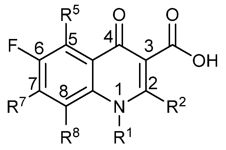
:1. Introduction
2. Results
3. Discussion
4. Experimental
4.1. Bacterial Cultures
4.2. MIC Determinations
4.3. Synthetic Procedures
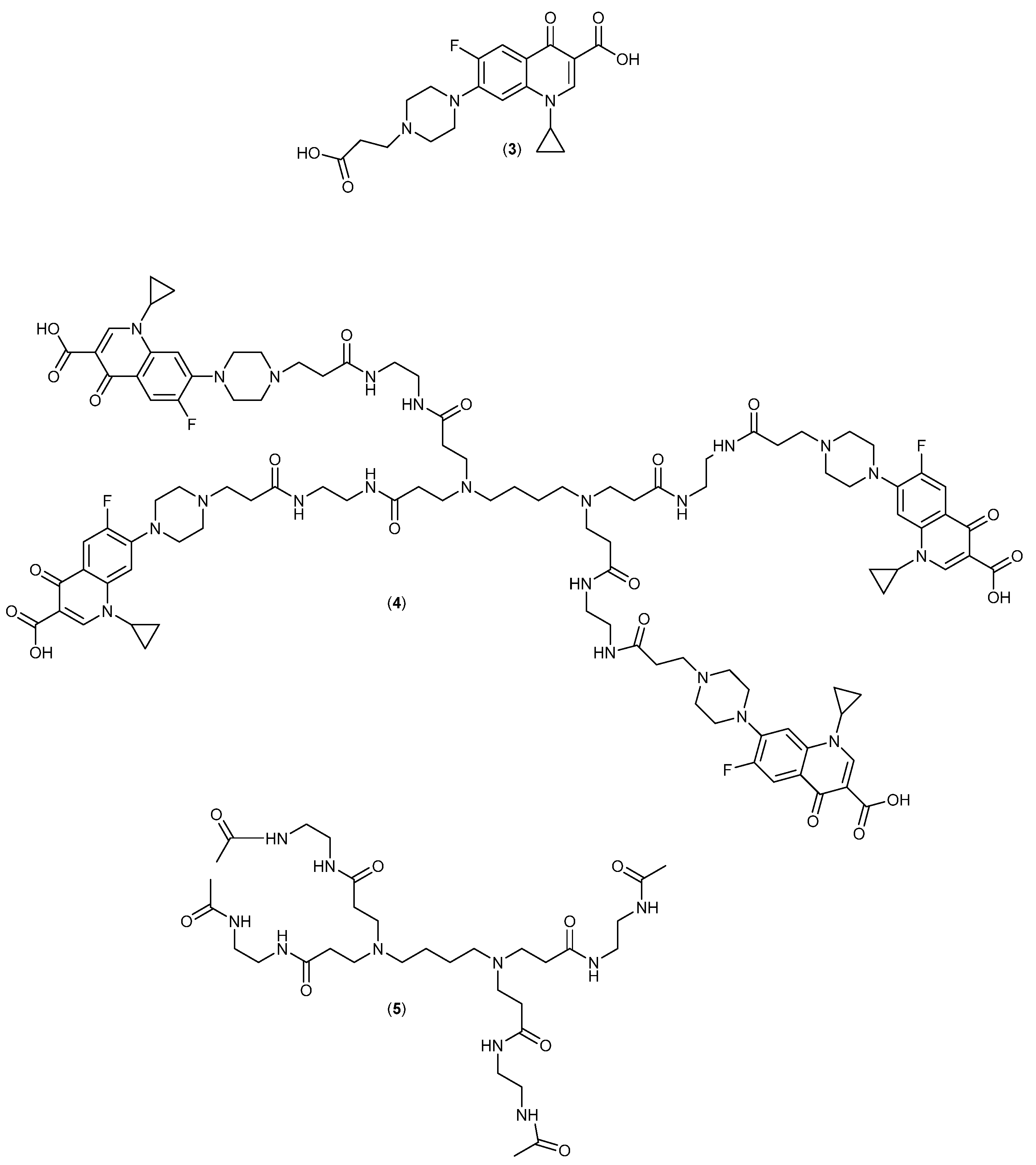
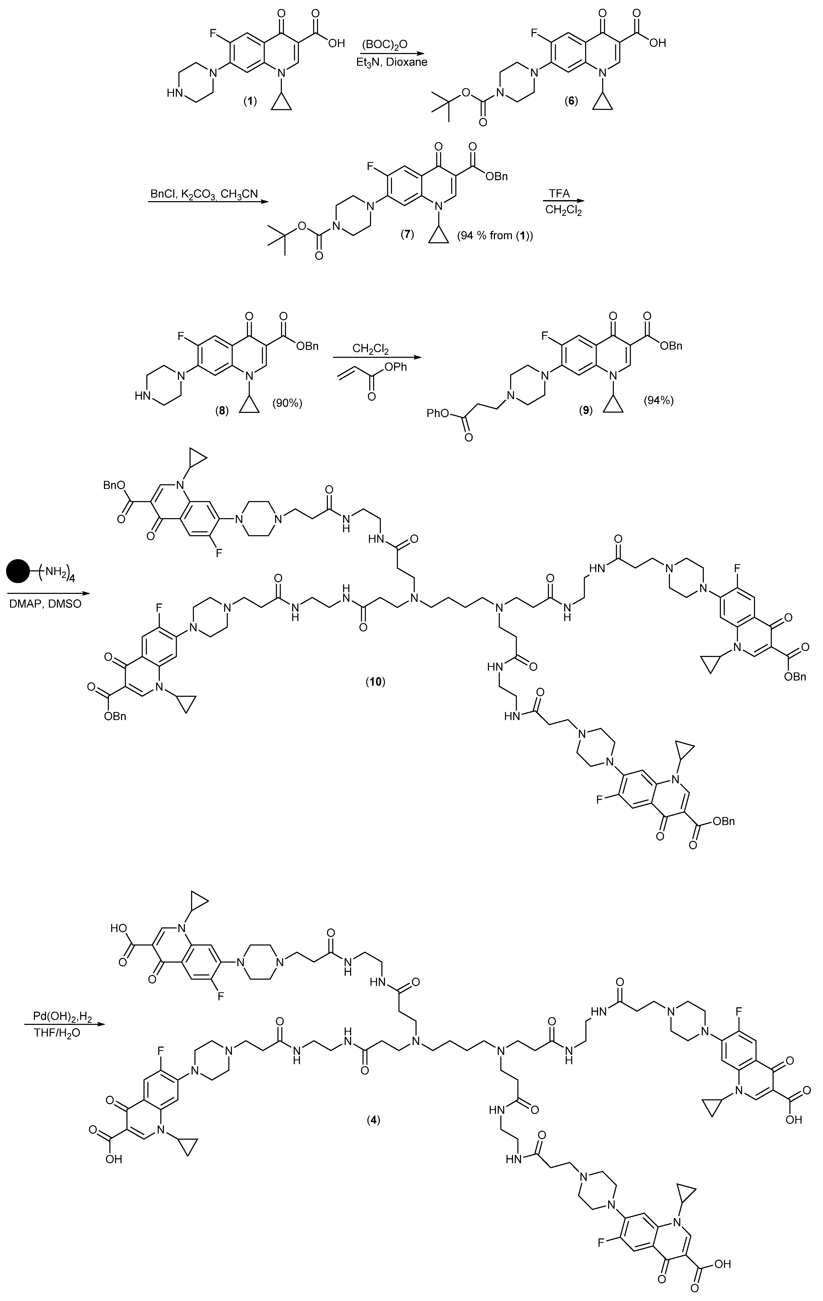
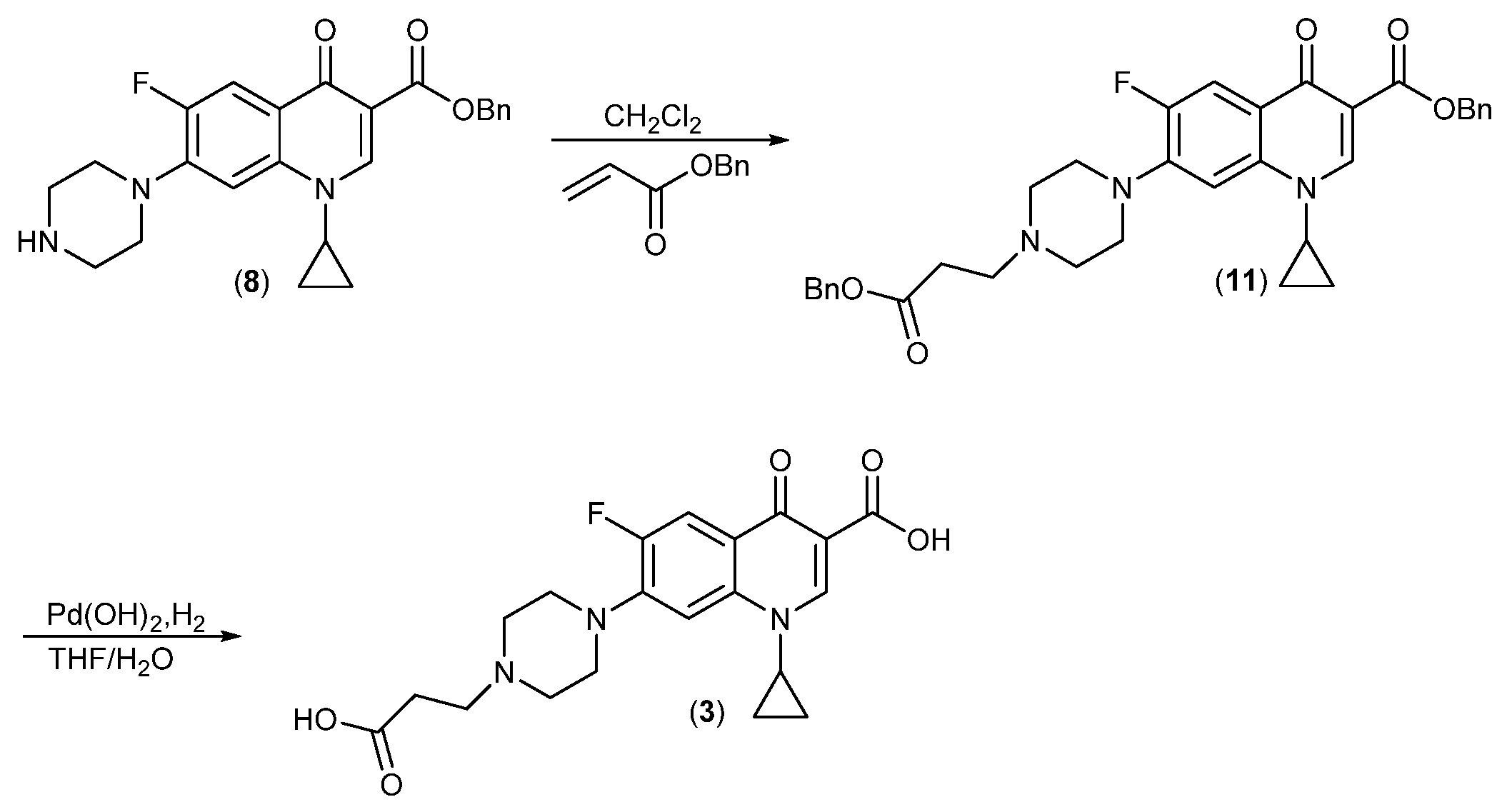
4.3.1. 7-(4-(2-Carboxyethyl)piperazin-1-yl)-1-cyclopropyl-6-fluoro-4-oxo-1,4-dihydroquinoline-3-carboxylic acid (3)
4.3.2. DAB-PAMAM-G0-(Cipro-COOH)4 ● 6HCl (4)
4.3.3. DAB-PAMAM-G0-(Acetamide)4 ● 2HCl (5)
4.3.4. Benzyl 7-(4-tert-butoxycarbonyl)piperazin-1-yl)-1-cyclopropyl-6-fluoro-4-oxo-1,4-dihydroquinoline-3-carboxylate (7)
4.3.5. Benzyl 1-cyclopropyl-6-fluoro-4-oxo-piperazin-1-yl)-1,4-dihydroquinoline-3-carboxylate (8)
4.3.6. Benzyl 1-cyclopropyl-6-fluoro-4-oxo-7-(4-(3-oxo-3-phenoxypropyl)piperazin-1-yl)-1,4-dihydroquinoline-3-carboxylate (9)
4.3.7. DAB-PAMAM-G0-(Cipro-Bn)4 (10)
5. Conclusions
Supplementary Materials
Author Contributions
Funding
Conflicts of Interest
References
- World Health Organization. Antimicrobial Resistance: Global Report on Surveillance 2014. Available online: https://www.who.int/health-topics/antimicrobial-resistance (accessed on 18 March 2020).
- Levy, S.B.; Marshall, B. Antibacterial resistance worldwide: Causes, challenges and responses. Nat. Med. 2004, 10, S122–S129. [Google Scholar] [CrossRef] [PubMed]
- Davies, J.; Davies, D. Origins and Evolution of Antibiotic Resistance. Microbiol. Mol. Biol. Rev. 2010, 74, 417–433. [Google Scholar] [CrossRef] [PubMed] [Green Version]
- Poole, K. Efflux pumps as antimicrobial resistance mechanisms. Ann. Med. 2007, 39, 162–176. [Google Scholar]
- Amaral, L.; Martins, A.; Spengler, G.; Molnar, J. Efflux pumps of Gram-negative bacteria: What they do, how they do it, with what and how to deal with them. Front. Pharmacol. 2015, 4, 168. [Google Scholar] [CrossRef] [PubMed] [Green Version]
- Poole, K. Outer membranes and efflux: The path to multidrug resistance in Gram-negative bacteria. Curr. Pharm. Biotechnol. 2002, 3, 77–98. [Google Scholar] [CrossRef]
- Schindler, B.D.; Kaatz, G.W. Multidrug efflux pumps of Gram positive bacteria. Drug Res. Updates 2016, 27, 1–13. [Google Scholar] [CrossRef]
- Van Bambeke, F.; Balzi, E.; Tulkens, P.M. Antibiotic efflux pumps—Commentary. Biochem. Pharmacol. 2000, 60, 457–470. [Google Scholar] [CrossRef]
- Sikri, N.; Dalal, S.; Taneja, R. Efflux Pumps: An Overview. Int. J. Pharm. Sci. Res. 2018, 9, 854–861. [Google Scholar]
- Wiese, M.; Pajeva, I.K. Structure-activity relationships of multidrug resistance reversers. Curr. Med. Chem. 2001, 8, 685–713. [Google Scholar] [CrossRef]
- Amaral, L.; Engi, H.; Viveiros, M.; Molnar, M. Comparison of multidrug resistant efflux pumps of cancer and bacterial cells with respect to the same inhibitory agents. In Vivo 2007, 21, 237–244. [Google Scholar]
- Martins, M.; Dastidar, S.G.; Fanning, S.; Kristiansen, J.E.; Molnar, J.; Pagès, J.-M.; Schelz, S.; Spengler, G.; Viveiros, M.; Amaral, L. Potential role of non-antibiotics (helper compounds) in the treatment of multidrug-resistant Gram-negative infections: Mechanisms for their direct and indirect activities. Int. J. Antimicrobial Agents 2008, 31, 198–208. [Google Scholar] [CrossRef]
- Kristiansen, J.E.; Hendricks, O.; Delvin, T.; Butterworth, T.S.; Aagaard, L.; Christensen, J.B.; Flores, V.C.; Keyzer, H. Reversal of resistance in microorganisms by help of non-antibiotics. J. Antimicrob. Chem. 2007, 59, 1271–1279. [Google Scholar] [CrossRef] [Green Version]
- Palmeira, A.; Sousa, E.; Vasconcelos, M.H.; Pinto, M.M. Three Decades of P-gp Inhibitors: Skimming through Several Generations and Scaffolds. Curr. Med. Chem. 2012, 19, 1946–2025. [Google Scholar] [CrossRef]
- Raschi, E.; Ceccarini, L.; De Ponti, F.; Recanatini, M. hERG-related drug toxicity and models for predicting hERG liability and QT prolongation. Expert Opin. Drug Metab. Tox. 2009, 5, 1005–1021. [Google Scholar] [CrossRef]
- Clancy, C.E.; Kurokawa, J.; Tateyama, M.; Wehrens, X.H.T.; Kass, R.S. K+ channel structure-activity relationships and mechanisms of drug-induced QT prolongation. Ann. Rev. Pharm. Toxicol. 2003, 43, 441–461. [Google Scholar] [CrossRef] [PubMed]
- Najlah, M.; Freeman, S.; Attwood, D.; D’Emanuele, A. Synthesis and assessment of first-generation polyamidoamine dendrimer prodrugs to enhance the cellular permeability of P-gp substrates. Bioconj. Chem. 2007, 18, 937–946. [Google Scholar] [CrossRef] [PubMed]
- Cheng, Y.; Cheng, Y.; Qu, H.; Ma, M.; Xu, Z.; Xu, P.; Fang, Y.; Xu, T. Polyamidoamine (PAMAM) dendrimers as biocompatible carriers of quinolone antimicrobials: An in vitro study. Eur. J. Med. Chem. 2007, 42, 1032–1038. [Google Scholar] [CrossRef] [PubMed]
- Ma, M.; Cheng, Y.; Xu, Z.; Xu, P.; Qu, H.; Fang, Y.; Xu, T.; Wen, L. Evaluation of polyamidoamine (PAMAM) dendrimers as drug carriers of anti-bacterial drugs using sulfamethoxazole (SMZ) as a model drug. Eur. J. Med. Chem. 2007, 42, 93–98. [Google Scholar] [CrossRef]
- Felczak, A.; Wrońska, N.; Janaszewska, A.; Klajnert, B.; Bryszewska, M.; Appelhans, D.; Voit, B.; Różalska, S.; Lisowska, K. Antimicrobial activity of poly(propylene imine) dendrimers. New J. Chem. 2012, 36, 2215–2222. [Google Scholar] [CrossRef]
- Ortega, P.; Copa-Patiño, J.L.; Muñoz-Fernandez, M.A.; Soliveri, J.; Gomez, R.; de la Mata, F.J. Amine and ammonium functionalization of chloromethylsilane-ended dendrimers. Antimicrobial activity studies. Org. Biomol. Chem. 2008, 6, 3264–3269. [Google Scholar] [CrossRef]
- Fuentes-Paniagua, E.; Hernández-Ros, J.M.; Sánchez-Milla, M.; Camero, M.A.; Maly, M.; Pérez-Serrano, J.; Copa-Patiño, J.L.; Sánchez-Nieves, J.; Soliveri, J.; Gómez, R.; et al. Carbosilane cationic dendrimers synthesized by thiol-ene click chemistry and their use as antibacterial agents. RSC Adv. 2014, 4, 1256–1265. [Google Scholar] [CrossRef]
- Fuentes-Paniagua, E.; Hernández-Ros, J.M.; Sánchez-Milla, M.; Camero, M.A.; Maly, M.; Pérez-Serrano, J.; Copa-Patiño, J.L.; Sánchez-Nieves, J.; Soliveri, J.; Gómez, R.; et al. Structure-activity relationship study of cationic carbosilane dendritic systems as antibacterial agents. RSC Adv. 2016, 6, 7022–7033. [Google Scholar] [CrossRef]
- Fernandez, J.; Acosta, G.; Pulido, D.; Malý, M.; Copa-Patiño, J.L.; Soliveri, J.; Royo, M.; Gómez, R.; Albericio, F.; Ortega, P.; et al. Carbosilane Dendron-Peptide Nanoconjugates as Antimicrobial Agents. Mol. Pharm. 2019, 16, 2661–2674. [Google Scholar] [CrossRef] [PubMed]
- Janiszewska, J.; Swieton, J.; Lipkowski, A.W.; Urbanczyk-Lipkowska, Z. Low molecular mass peptide dendrimers that express antimicrobial properties. Bioorg. Med. Chem. Lett. 2003, 13, 3711–3713. [Google Scholar] [CrossRef] [PubMed]
- Janiszewska, J.; Urbanczyk-Lipkowska, Z. Synthesis, antimicrobial activity and structural studies of low molecular mass lysine dendrimers. Acta Biochim. Polonica 2006, 53, 77–82. [Google Scholar] [CrossRef]
- Klajnert, B.; Janiszewska, J.; Urbanczyk-Lipkowska, Z.; Bryszewska, M.; Shcharbin, D.; Labieniec, M. Biological properties of low molecular mass peptide dendrimers. Int. J. Pharm. 2006, 309, 208–217. [Google Scholar] [CrossRef] [PubMed]
- Lind, T.K.; Zielińska, P.; Wacklin, H.P.; Urbańczyk-Lipkowska, Z.; Cárdenas, M. Continuous Flow Atomic Force Microscopy Imaging Reveals Fluidity and Time-Dependent Interactions of Antimicrobial Dendrimer with Model Lipid Membranes. ACS Nano 2014, 8, 396–408. [Google Scholar] [CrossRef] [PubMed]
- Stach, M.; Siriwardena, T.N.; Köhler, T.; van Delden, C.; Darbre, T.; Reymond, J.-L. Combining Topology and Sequence Design for the Discovery of Potent Antimicrobial Peptide Dendrimers against Multidrug-Resistant Pseudomonas aeruginosa. Ang. Chem. Int. Ed. 2014, 53, 12827–12831. [Google Scholar] [CrossRef]
- Michaud, G.; Visini, R.; Bergmann, M.; Salerno, G.; Bosco, R.; Gillon, E.; Richichi, B.; Nativi, C.; Imberty, A.; Stocker, A.; et al. Overcoming antibiotic resistance in Pseudomonas aeruginosa biofilms using glycopeptide dendrimers. Chem. Sci. 2016, 7, 166–182. [Google Scholar] [CrossRef] [Green Version]
- Bergmann, M.; Michaud, G.; Visini, R.; Jin, X.; Gillon, E.; Stocker, A.; Imberty, A.; Darbre, T.; Reymond, J.-L. Multivalency effects on Pseudomonas aeruginosa biofilm inhibition and dispersal by glycopeptide dendrimers targeting lectin LecA. Org. Biomol. Chem. 2016, 14, 138–148. [Google Scholar] [CrossRef] [Green Version]
- Siriwardena, T.N.; Stach, M.; He, R.; Gan, B.-H.; Javor, S.; Heitz, M.; Ma, L.; Cai, X.; Chen, P.; Wei, D.; et al. Lipidated Peptide Dendrimers Killing Multidrug-Resistant Bacteria. J. Am. Chem. Soc. 2018, 140, 423–432. [Google Scholar] [CrossRef] [PubMed] [Green Version]
- Mishra, M.K.; Kotta, K.; Hali, M.; Wykes, S.; Gerard, H.C.; Hudson, A.P.; Whittum-Hudson, J.A.; Kannan, R.M. PAMAM dendrimer-azithromycin conjugate nanodevices for the treatment of Chlamydia trachomatis infections. Nanomed.-Nanotech. Biol. Med. 2011, 7, 935–944. [Google Scholar] [CrossRef] [PubMed]
- Wong, P.T.; Tang, S.; Mukherjee, J.; Tang, K.; Gam, K.; Isham, D.; Murat, C.; Sun, R.; Baker, J.R., Jr.; Choi, S.K. Light-controlled active release of photocaged Ciprofloxacin for lipopolysaccharide-targeted drug delivery using dendrimer conjugates. Chem. Commun. (Cambridge UK) 2016, 52, 10357–10360. [Google Scholar] [CrossRef] [PubMed] [Green Version]
- Galdiero, S.; Falanga, A.; Cantisani, M.; Tarallo, R.; Elena Della Pepa, M.; D’Oriano, V.; Galdiero, M. Microbe-Host Interactions: Structure and Role of Gram-Negative Bacterial Porins. Curr. Protein Pept. Sci. 2012, 13, 843–854. [Google Scholar] [CrossRef] [Green Version]
- Aguilella, V.M.; Queralt-Martín, M.; Alcaraz, A. Bacterial porins. In Electrophysiology of Unconventional Channels and Pores; Delcour, H.A., Ed.; Springer International Publishing: Cham, Switzerland, 2015; pp. 101–121. [Google Scholar]
- Tillotson, G.S. Quinolones: Structure-activity relationships and future predictions. J. Med. Microbiol. 1996, 44, 320–324. [Google Scholar] [CrossRef] [Green Version]
- Ficker, M.; Paolucci, V.; Christensen, J.B. Improved large-scale synthesis and characterization of small and medium generation PAMAM dendrimers. Can. J. Chem. 2017, 95, 954–964. [Google Scholar] [CrossRef]
- Pang, Y.; Wan, L.; Huang, G.; Zhang, X.; Jin, X.; Xu, P.; Liu, Y.; Han, M.; Wu, G.-P.; Ji, S. Controlling Block Copolymer-Substrate Interactions by Homopolymer Brushes/Mats. Macromolecules 2017, 50, 6733–6741. [Google Scholar] [CrossRef]
- Pittelkow, M.; Lewinsky, R.; Christensen, J.B. Selective synthesis of carbamate protected polyamines using alkyl phenyl carbonates. Synthesis-Stuttgart 2002, 2195–2202. [Google Scholar]
- Pittelkow, M.; Lewinsky, R.; Christensen, J.B. Mono Carbamate Protection of Aliphatic Diamines Using Alkyl Phenyl Carbonates (2-Aminoethyl) carbamic acid tert-butyl ester. Org. Synth. 2007, 84, 209–214. [Google Scholar]
- Sølvhøj, A.B.; Tortzen, C.; Christensen, J.B. Monoprotection of Triamines with Alkyl Phenyl Carbonates. Org. Prep. Proc. Int. 2012, 44, 397–400. [Google Scholar] [CrossRef]
- Pittelkow, M.; Christensen, J.B. Convergent Synthesis of Internally Branched PAMAM Dendrimers. Org. Lett. 2005, 7, 1295–1298. [Google Scholar] [CrossRef]
- McNamara, P.J. Genetic manipulation of Staphylococcus aureus. In Staphylococcus Molecular Genetics; Lindsay, J.A., Ed.; Caister Academic Press: Norfolk, UK, 2008; pp. 89–129. [Google Scholar]
- O’Neill, A.J. Staphylococcus aureus SH1000 and 8325-4: Comparative genome sequences of key laboratory strains in staphylococcal research. Lett. Appl. Microbiol. 2010, 51, 358–361. [Google Scholar] [CrossRef] [PubMed]
- Sahm, D.F.; Kissinger, J.; Gilmore, M.S.; Murray, P.R.; Mulder, R.; Solliday, J.; Clarke, B. In vitro susceptibility studies of vancomycin-resistant Enterococcus faecalis. Antimicrob. Agents Chemother. 1989, 33, 1588–1591. [Google Scholar] [CrossRef] [PubMed] [Green Version]
- Paulsen, I.T.; Banerjei, L.; Myers, G.S.; Nelson, K.E.; Seshadri, R.; Read, T.D.; Fouts, D.E.; Eisen, J.A.; Gill, S.R.; Heidelberg, J.F.; et al. Role of mobile DNA in the evolution of vancomycin-resistant Enterococcus faecalis. Science 2003, 299, 2071–2074. [Google Scholar] [CrossRef] [PubMed] [Green Version]
- Richardson, E.J.; Limaye, B.; Inamdar, H.; Datta, A.; Manjari, K.S.; Pullinger, G.D.; Thomson, N.R.; Joshi, R.R.; Watson, M.; Stevens, M.P. Genome sequences of Salmonella enterica serovar Typhimurium, Choleraesuis, Dublin, and Gallinarum strains of well-defined virulence in food-producing animals. J. Bacteriol. 2011, 193, 3162–3163. [Google Scholar] [CrossRef] [PubMed] [Green Version]
- Cramariuc, O.; Rog, T.; Javanainen, M.; Monticelli, L.; Polishchuk, A.V.; Vattulainen, I. Mechanism for translocation of fluoroquinolones across lipid membranes. Biochim. Biophys. Acta-Biomem. 2012, 1818, 2563–2571. [Google Scholar] [CrossRef] [Green Version]
- Berlanga, M.; Montero, M.T.; Hernandez-Borrell, J.; Vinas, M. Influence of the cell wall on Ciprofloxacin susceptibility in selected wild-type Gram-negative and Gram-positive bacteria. Int. J. Antimicrobial Agents 2004, 23, 627–630. [Google Scholar] [CrossRef]
- Ricci, V.; Coldham, N.C.; Piddock, L.J.V.; Buckley, A.; Tzakas, P. Ciprofloxacin-Resistant Salmonella enterica Serovar Typhimurium Strains Are Difficult To Select in the Absence of AcrB and TolC. Antimicrob. Agents Chemother. 2006, 50, 38–42. [Google Scholar] [CrossRef] [Green Version]
- Ricci, V.; Paddock, L.V.J. Ciprofloxacin selects for multidrug resistance in Salmonella enterica serovar Typhimurium mediated by at least two different pathways. J. Antimicrob. Chemother. 2009, 63, 909–916. [Google Scholar] [CrossRef] [Green Version]
- Stover, C.K.; Pham, X.Q.; Erwin, A.L.; Mizoguchi, S.D.; Warrener, P.; Hickey, M.J.; Brinkman, F.S.L.; Hufnagle, W.O.; Kowalik, D.J.; Lagrou, M.; et al. Complete genome sequence of Pseudomonas aeruginosa PAO1, an opportunistic pathogen. Nature 2002, 406, 959–964. [Google Scholar] [CrossRef]
- Klockgether, J. Genome Diversity of Pseudomonas aeruginosa PAO1 Laboratory Strains. J. Bacteriol. 2010, 192, 1113–1121. [Google Scholar] [CrossRef] [Green Version]
- Chapman, J.S.; Georgopapadakou, N.H. Routes of Quinolone Permeation in Escherichia coli. Antimicrob. Agents Chemother. 1988, 32, 438–442. [Google Scholar] [CrossRef] [PubMed] [Green Version]
- Hirai, K.; Aoyama, H.; Irikura, T.; Iyobe, S.; Mitsuhashi, S. Differences in Susceptibility to Quinolones of Outer-Membrane Mutants of Salmonella Typhimurium and Escherichia coli. Antimicrob. Agents Chemother. 1986, 29, 535–538. [Google Scholar] [CrossRef] [PubMed] [Green Version]
- Nikaido, H. Porins and Specific Diffusion Channels in Bacterial Outer Membranes. J. Biol. Chem. 1994, 269, 3905–3908. [Google Scholar] [PubMed]
- Chevalier, S.; Bouffartigues, E.; Bodilis, J.; Maillot, O.; Lesouhaitier, O.; Feuilloley, M.G.J.; Orange, N.; Dufour, A.; Cornelis, P. Structure, function and regulation of Pseudomonas aeruginosa porins. FEMS Microbiol. Rev. 2017, 41, 698–722. [Google Scholar] [CrossRef] [PubMed]
- Fabrega, A.; Madurga, S.; Giralt, E.; Vila, J. Mechanisms of action and resistance to quinolones. Microb. Biotechnol. 2009, 2, 40–61. [Google Scholar] [CrossRef] [PubMed] [Green Version]
- Idowu, T.; Schweizer, F. Ubiquitous nature of fluoroquinolones: The oscillation between antibacterial and anticancer activities. Antibiotics 2017, 6, 26. [Google Scholar] [CrossRef] [PubMed] [Green Version]
- Calabretta, M.K.; Kumar, A.; McDermott, A.M.; Cai, C. Antibacterial activities of poly(amidoamine) dendrimers terminated with amino and poly(ethylene glycol) groups. Biomacromolecules 2007, 8, 1807–1811. [Google Scholar] [CrossRef] [Green Version]
- Lopez, A.I.; Reins, R.Y.; McDermott, A.M.; Trautner, B.W.; Cai, C. Antibacterial activity and cytotoxicity of PEGylated poly(amidoamine) dendrimers. Mol. Biosys. 2009, 5, 1148–1156. [Google Scholar] [CrossRef]
- Gholami, M.; Mohammadi, R.; Arzanlou, M.; Dourbash, F.A.; Kouhsari, E.; Majidi, G.; Mohseni, S.M.; Nazari, S. In vitro antibacterial activity of poly (amidoamine)-G7 dendrimer. BMC Infect. Dis. 2017, 17, 395. [Google Scholar] [CrossRef]
- Fox, L.J.; Richardson, R.M.; Briscoe, W.H. PAMAM dendrimer—Cell membrane interactions. Adv. Colloid Interface Sci. 2018, 257, 1–18. [Google Scholar] [CrossRef]
- Oki, A.; Seidman, D.; Lacina III, M.G.; Mishra, M.K.; Kannan, R.M.; Yang, H.; Carlyon, J.A. Dendrimer-enabled transformation of Anaplasma phagocytophilum. Microbes Infect. 2015, 17, 817–822. [Google Scholar] [CrossRef] [PubMed] [Green Version]
- Wrońska, N.; Majoral, J.P.; Appelhans, D.; Bryszewska, M.; Lisowska, K. Synergistic effects of anionic/cationic dendrimers and levofloxacin on antibacterial activities. Molecules 2019, 24, 2894. [Google Scholar] [CrossRef] [PubMed] [Green Version]
Sample Availability: Samples of the compounds are available from the authors. |


| Bacterial Strain | Compound | MIC mg/L | MIC µM |
|---|---|---|---|
| Gram-positive | |||
| S. aureus 8325-4 | 1 | 0.25 | 0.75 |
| 3 | 0.5 | 1.24 | |
| 4 | 16 | 0.0069 | |
| 5 | >256 | >0.47 | |
| E. faecalis V583 | 1 | 0.25 | 0.75 |
| 3 | 0.5 | 1.24 | |
| 4 | 16 | 0.0069 | |
| 5 | >256 | >0.47 | |
| Gram-negative | |||
| S. enterica serovar Typhimurium 4/74 | 1 | 0.5 | 1.5 |
| 3 | 0.25 | 0.62 | |
| 4 | 16 | 0.0069 | |
| 5 | >256 | >0.47 | |
| P. aeruginosa PAO1 | 1 | 0.25 | 0.75 |
| 3 | 0.5 | 1.24 | |
| 4 | 32 | 0.014 | |
| 5 | >256 | >0.47 | |
© 2020 by the authors. Licensee MDPI, Basel, Switzerland. This article is an open access article distributed under the terms and conditions of the Creative Commons Attribution (CC BY) license (http://creativecommons.org/licenses/by/4.0/).
Share and Cite
Svenningsen, S.W.; Frederiksen, R.F.; Counil, C.; Ficker, M.; Leisner, J.J.; Christensen, J.B. Synthesis and Antimicrobial Properties of a Ciprofloxacin and PAMAM-dendrimer Conjugate. Molecules 2020, 25, 1389. https://0-doi-org.brum.beds.ac.uk/10.3390/molecules25061389
Svenningsen SW, Frederiksen RF, Counil C, Ficker M, Leisner JJ, Christensen JB. Synthesis and Antimicrobial Properties of a Ciprofloxacin and PAMAM-dendrimer Conjugate. Molecules. 2020; 25(6):1389. https://0-doi-org.brum.beds.ac.uk/10.3390/molecules25061389
Chicago/Turabian StyleSvenningsen, Søren Wedel, Rikki Franklin Frederiksen, Claire Counil, Mario Ficker, Jørgen J. Leisner, and Jørn Bolstad Christensen. 2020. "Synthesis and Antimicrobial Properties of a Ciprofloxacin and PAMAM-dendrimer Conjugate" Molecules 25, no. 6: 1389. https://0-doi-org.brum.beds.ac.uk/10.3390/molecules25061389