Article

Preclinical Evaluation of NHS-Activated Gold Nanoparticles Functionalized with Bombesin or Neurotensin-Like Peptides for Targeting Colon and Prostate Tumours

1
Radiopharmaceutical Research Centre, Horia Hulubei National Institute for R&D in Physics and Nuclear Engineering, 30 Reactorului Street, Magurele, 077125 Ilfov, Romania
2
Faculty of Applied Chemistry and Materials Science, University Politehnica of Bucharest, 1–7 Polizu Street, 011061 Bucharest, Romania
3
National Institute for Research and Development of Isotopic and Molecular Technologies, 67–103 Donat Street, 400293 Cluj-Napoca, Romania
4
Faculty of Biology, University of Bucharest, 91–95 Splaiul Independentei Street, 050095 Bucharest, Romania
5
Victor Babes National Institute of Pathology, 99–101 Splaiul Independentei Street, 050096 Bucharest, Romania
*
Author to whom correspondence should be addressed.
Submission received: 1 June 2020 / Revised: 13 July 2020 / Accepted: 22 July 2020 / Published: 24 July 2020
(This article belongs to the Special Issue Functional Peptide-Based Nanomaterials)

Abstract

:
Recent advances and large-scale use of hybrid imaging modalities like PET-CT have led to the necessity of improving nano-drug carriers that can facilitate both functional and metabolic screening in nuclear medicine applications. In this study, we focused on the evaluation of four potential imaging nanoparticle structures labelled with the 68Ga positron emitter. For this purpose, we functionalized NHS-activated PEG-gold nanoparticles with 68Ga-DOTA-Neuromedin B, 68Ga-DOTA-PEG(4)-BBN(7-14), 68Ga-DOTA-NT and 68Ga-DOTA-Neuromedin N. In vitro binding kinetics and specific binding to human HT-29 colon carcinoma cells and DU-145 prostate carcinoma cells respectively were assessed, over 75% retention being obtained in the case of 68Ga-DOTA-PEG(4)-BBN(7-14)-AuNP in prostate tumour cells and over 50% in colon carcinoma cells. Biodistribution in NU/J mice highlighted a three-fold uptake increase in tumours at 30 min post-injection of 68Ga-DOTA-NT-AuNP and 68Ga-DOTA-PEG(4)-BBN(7-14)-AuNP compared to 68Ga-DOTA-NT and 68Ga-DOTA-PEG(4)-BBN(7-14) respectively, therewith fast distribution in prostate and colon tumours and minimum accumulation in non-targeted tissues.

1. Introduction

The fast development in recent years of diagnostic imaging techniques is traceable, on the one hand, to research seeking new tracers aimed for hybrid or multimodal imaging. This approach implies the integration of a structural imaging modality, such as computed tomography (CT) or magnetic resonance imaging (MRI), with a functional imaging modality, like positron emission tomography (PET) or single-photon emission computed tomography (SPECT). Among hybrid imaging combinations PET-CT is commonly preferred, as PET provides highly sensitive and quantitative information about tissue metabolic activity, while CT scanners highlight detailed and precise anatomical features. Over the last decade, special attention has been given to the development of nano-sized drug and contrast agent carriers, based on multifunctional nanoparticle (NP) systems that, due to their physicochemical properties and the large contact surface available for biomolecule functionalization, can easily improve the tumour targeting due to the enhanced permeability (EPR) effect of blood vessels that surround the tumour tissue and concentrate the amount of tracer at the tumour site [1]. These findings motivated the use of such NP systems to deliver already established radioisotopes for PET imaging, as recently reported: superparamagnetic iron oxide nanoparticles-based tracers: 18F-CLIO [1], 64Cu-DOTA-mSPIO [2,3], gold nanoparticles (AuNPs) functionalized with 64Cu-NODAGA [4], nitrone modified gold nanoparticles for 18F labelling [5], 89Zr-cRGDY-PEG-functionalized silica nanoparticles [6] or 68Ga-DOTA-TOC-AuNP [7]. A general problem encountered in the development of nano-drug structures for in vivo applications is the formation of a protein corona by non-specific absorption of opsonin proteins which facilitates the accumulation in reticuloendothelial system tissues (spleen, bone marrow, lungs, Kupffer cells of liver), instability of radiolabelled molecules or even determine the lack of targeting efficiency [8,9,10]. To overcome these problems, modification of NPs characteristics is necessary, such as size and surface chemistry but also suitable NPs’ coating with biocompatible structures, like polyethylene glycol (PEG), that improves the pharmacokinetic behaviour, changes plasma half-life of the tracer and tissue distribution. Sometimes, these difficulties can be avoided by choosing structures that meet all the above requirements, but it is still not enough and the optimization of the radiolabelled vector becomes necessary (radioisotope-labelled biological active molecule) as well as a full characterization of the targeting properties of the chosen biomolecule to the expected target tissue.
Among various radioisotopes used in nuclear medicine imaging, 68Ga has the characteristics of positron emission and low photon emission, which enable high detection resolution and sensitivity (T1/2 = 67.83 min, Eβ+ = 1.899 MeV). 68Ga can be obtained either by irradiation of 68Zn targets at cyclotrons [11], or, more often, by elution of a 68Ge/68Ga radionuclide generator [7]. Gallium can form stable complexes with a large number of ligands. Coupling of 68Ga to biological vectors demands proper chelating agents, capable of stably binding the radioisotope to the biologically active molecule. For the studies underlying this work, 1,4,7,10-tetraazacyclododecane-1,4,7,10-tetraacetic acid (DOTA) was used as the chelator for 68Ga.
The current study focuses on preclinical in vitro and ex vivo evaluation on colon and prostate carcinoma cells of four different peptides labelled with 68Ga, functionalized on gold nanoparticles, with potential to be used in multimodal imaging application. We labelled two groups of homologous peptides: PEG(4)-bombesin (7–14) (Gln-Trp-Ala-Val-Gly-His-Leu-Met-NH2) and its counterpart neuromedin B (Gly-Asn-Leu-Trp-Ala-Thr-Gly-His-Phe-Met-NH2), also neurotensin (Pyr-Leu-Tyr-Glu-Asn-Lys-Pro-Arg-Arg-Pro-Tyr-Ile-Leu-OH) and neuromedin N (Lys-Ile-Pro-Tyr-Ile-Leu). Bombesin receptors family consists of three subtypes of receptors: the neuromedin B (NMB) receptor (BBRS1 receptor), the gastrin-releasing peptide (GRP) receptor (BBRS2 receptor) and bombesin receptor subtype 3 (BBRS3). They are frequently over-expressed and released by breast tumours, prostate tumours, ovarian cancers, and small lung cancer. The bombesin-like peptide neuromedin B (NMB) is present in the central nervous system and gastrointestinal tract of mammals, stimulates smooth muscle contraction and is an autocrine growth factor in non-small cell lung cancer [12]. Neurotensin (NT) and neurotensin-like peptide neuromedin N (NMN) are derived from the same polypeptide precursor [13], neurotensin receptors (NTRS1, NTRS2 and NTRS3) being overexpressed by lung, breast cancer, colon or prostate cancer. To demonstrate the ability of gold nanoparticles to improve the in vivo uptake of tracers, we selected the stabilised sequence 7–14 of the bombesin peptide, with improved pharmacokinetics via the oligoethylene glycol spacer PEG4 [14] but also its homologous peptide neuromedin B for comparison. The unmodified neurotensin peptide has previously been shown to have faster in vivo kinetics due to degradation under the action of metalloendopeptidase, more stable analogues being recently developed [15,16]. Thus, we selected a peptide with already improved pharmacokinetics (and its counterpart), but also a peptide with a shorter biological time to demonstrate that in all cases gold NPs may be able to improve the tumour-targeting. In the present study we functionalized gold nanoparticles conjugated with PEG and activated with NHS groups aimed for better stability of the binding with selected peptides. We then assessed in vitro cell specific and non-specific uptake on HT-29 colon and DU-145 prostate carcinoma cells, which express bombesin and neurotensin receptors on their surface. Ex vivo biodistribution in NU/J mice was performed to assess the targeting potential of each of the investigated nanostructures.

2. Results

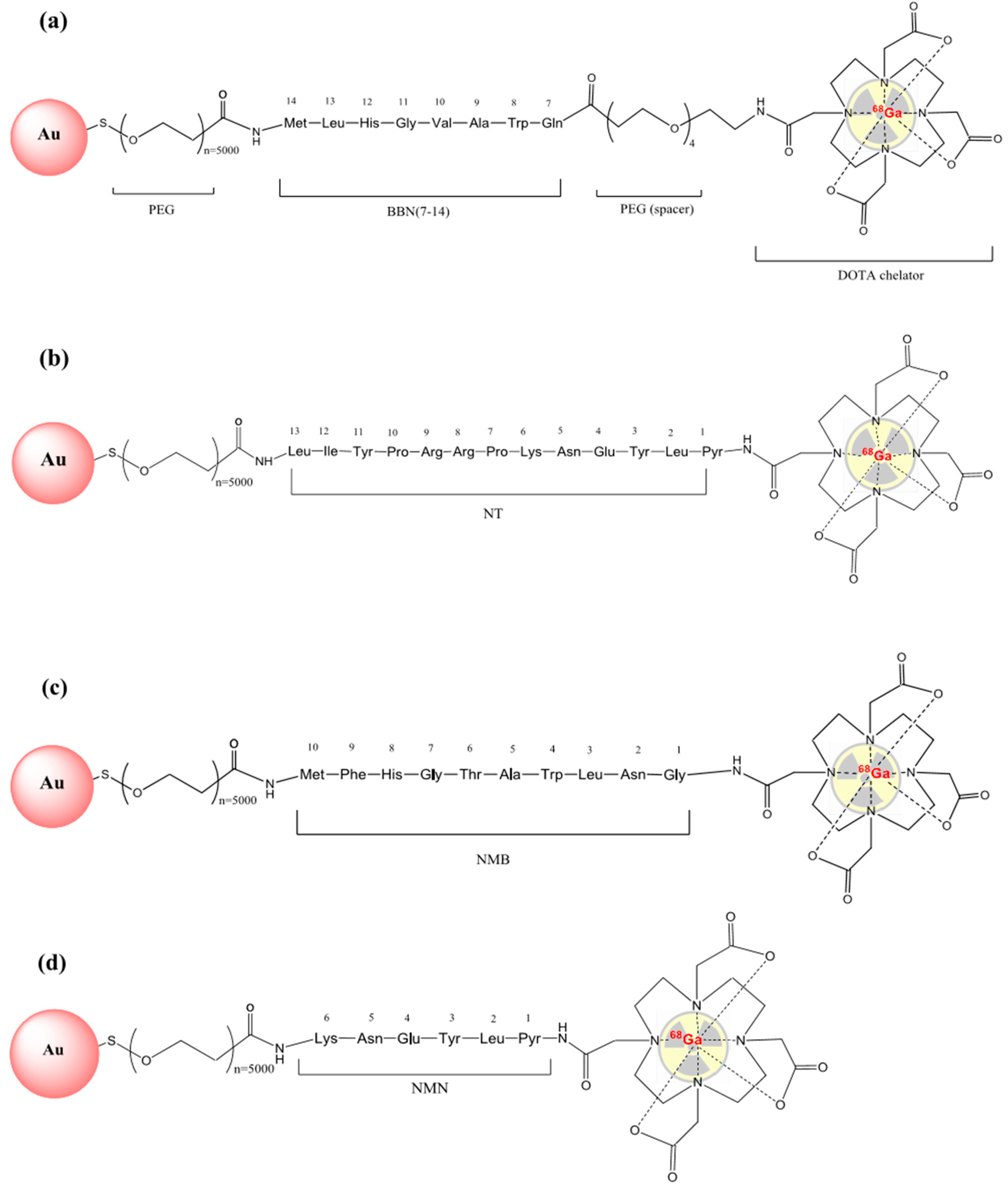
2.1. Peptides Labelling

The overall process, including elution, labelling and purification procedures, took less than 25 min to the final product, on an automated synthesis module. Irrespective of peptide, the radiolabelling processes were set to similar parameters (temperature, pH, reaction time, molar ratios) and purification steps. This led to slightly different results in terms of preparation yields, as presented in Table 1.

2.2. Quality Control of 68Ga-Labelled Peptides

The radiochemical purity (RCP) of the labelled biomolecules, assessed at the end of preparation and purification process, showed no unbound 68Ga and no degradation molecules resulted from preparation, in the final product. In case of 68Ga-DOTA-NT and 68Ga-DOTA-NMN, RCP is 100%. The radio-chromatogram profiles of 68Ga-DOTA-PEG(4)-BBN(7-14) and 68Ga-DOTA-NMB are similar to corresponding chromatograms of the cold peptides as per certificates of analysis provided by the producers, no other radiolabelled by-products being detected; in these cases, RCP was higher than 80% and 85%, respectively. Appendix A. Figure A1 presents radio-chromatograms of each studied compound at the end of synthesis (EOS).
RCP was checked every hour up to 4 h after the end of synthesis, with no degradation observed. All labelled molecules have qualified for biological studies. The pH of the final compound was neutral (pH = 7 ± 0.2), thus proper for direct injection.

2.3. Physicochemical Characterisation of Peptide-Functionalized Gold Nanoparticles

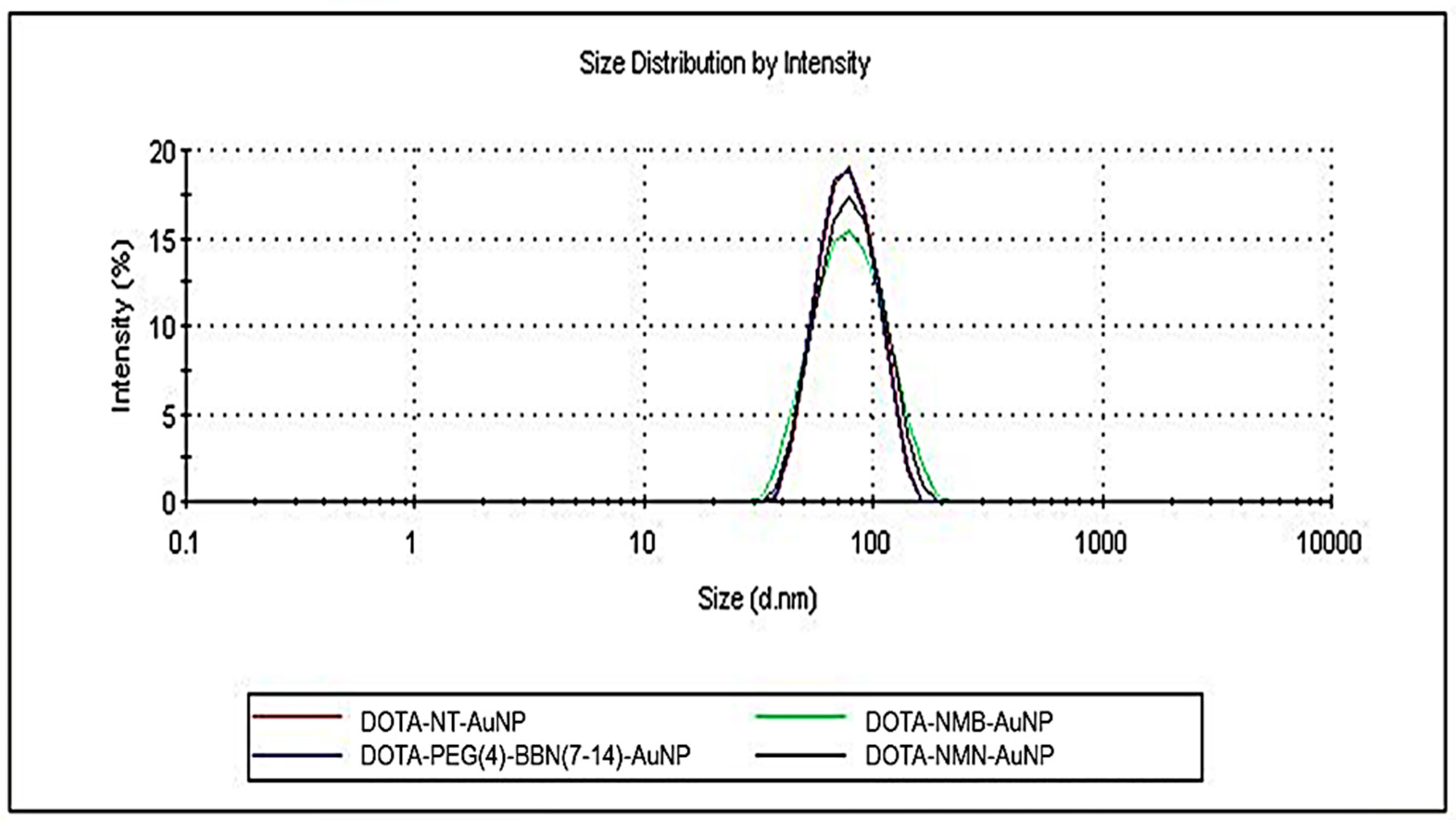
2.3.1. Dynamic Light Scattering (DLS)
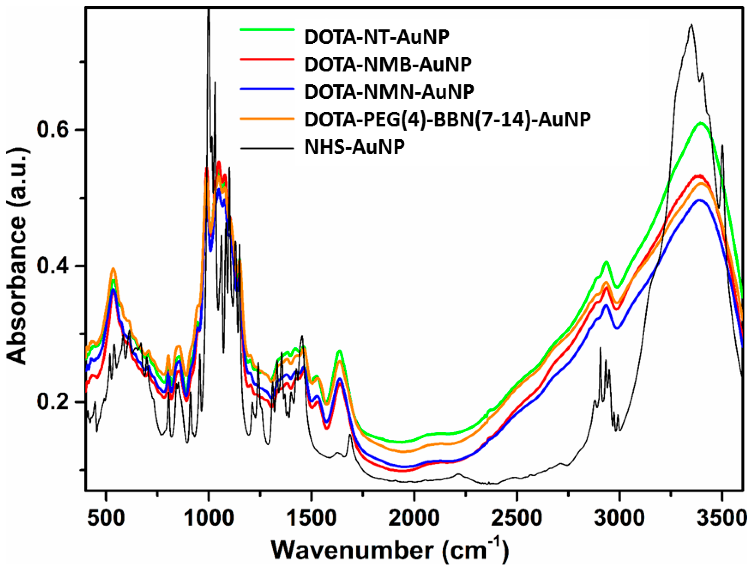
The reaction between DOTA-peptides and NHS-activated PEG-AuNPs was monitored by detecting changes in nanoparticles size as a peptide layer is formed around nanoparticles. The initial hydrodynamic diameter of bare PEG-AuNPs was 42.56 nm, and after functionalization increased to approximately 73 nm (Figure 1). Details of each nanostructures physical properties are listed in Table 2. The electric potential at the boundary of the double layer (counter ions and Stern layer) of nanoparticles, also known as zeta potential is higher than −44 mV. Thus, the peptide-functionalized nanoparticles have sufficient repulsive force to anticipate a good physical stability.

2.3.2. FTIR Spectra

In Figure 2 comparison between the FTIR spectra of NHS-AuNP DOTA-NT-AuNP, DOTA-NMB-AuNP, DOTA-NMN-AuNP and DOTA-PEG(4)-BBN(7-14)-AuNP is presented. As can be seen, the signals in the FTIR spectrum of the starting NHS-AuNP nanoparticles are distinct from those present in the spectra of DOTA peptide-functionalized AuNPs, due to the fact that the FTIR spectra were not measured for quantitative analysis. The signals in the NHS-AuNPs spectrum are more intense than those present in the DOTA peptide-functionalized AuNP spectra. The FTIR spectra of the gold nanoparticles samples contain the characteristic absorption bands of both constituents, namely DOTA-peptide and AuNPs. The relatively intense absorption band from AuNPs FTIR spectrum located around 605 cm−1 is typical for Au nanoparticles. In the FTIR spectra of the DOTA-peptide functionalized AuNPs is shifted to lower wavenumber (538 cm−1), due to the attachment of the DOTA-peptide complex on the AuNPs. The characteristic bands of the peptide conjugated with the chelating agent DOTA appear in the range 600–3600 cm−1. The intense band at 3380 cm−1 is attributed to the free NH stretching band and H-bonded NH groups from the peptide. Bands at 2914 cm−1 and 2855 cm−1 region are arising from C-H and CH2 stretching bonds of the organic layer found at the AuNPs surface. The band at 1640 cm−1 in the spectra corresponds to C=O stretching, highly indicating the presence of peptides on the nanoparticles surface. The same C=O group present in NHS ester molecule provides in the FTIR spectrum of NHS-AuNP two weak absorption bands with a peak at 1623 cm−1 and the other at 1690 cm−1; this fact indicates the covalent linkage of the DOTA-peptides on AuNPs surface. The absorption band situated at 1547 cm−1 is ascribed to the collective vibration of C=C/C-C of peptide chains. The band at 1455 cm−1 was assigned for N-H/C-N stretch vibration present in the amide linkages of the proteins.

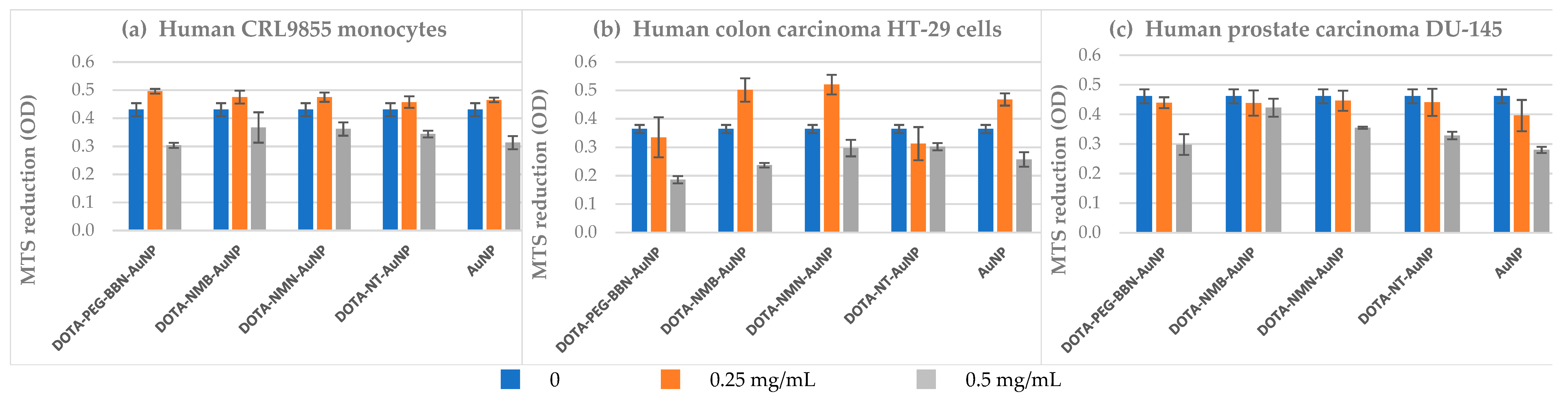
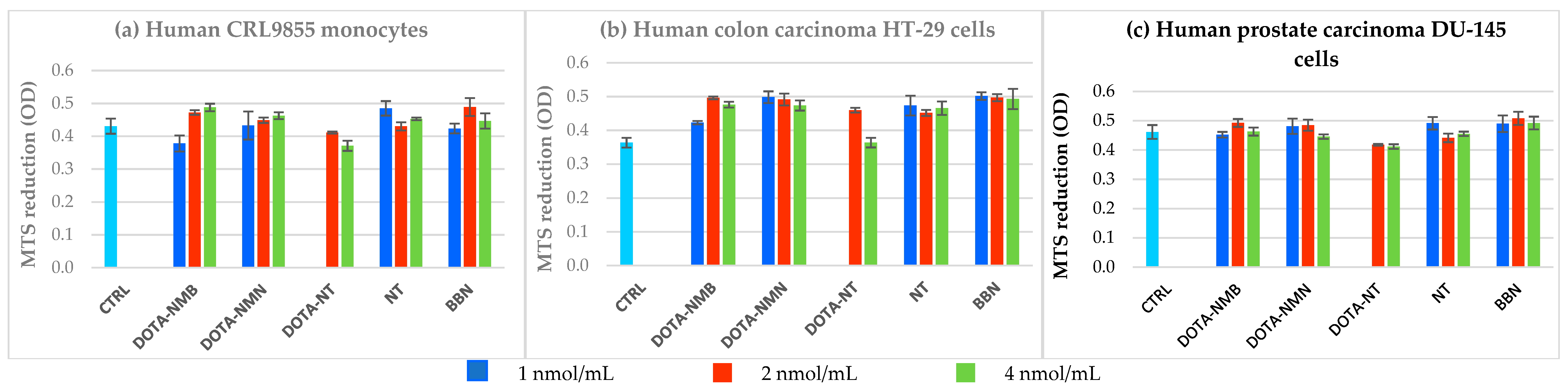
2.4. Cytotoxicity Evaluation

We performed a preliminary study for assessing the potential cytotoxic effects of the investigated types of NPs, both bare NPs (AuNP) and peptide-loaded NPs (DOTA-PEG(4)-BBN(7-14)-AuNP, DOTA-NMB-AuNP, DOTA-NMN-AuNP, DOTA-NT-AuNP), on normal cells (human proliferating CRL9855 monocytes) and tumour cells (human colon carcinoma HT-29 cells and human prostate carcinoma DU-145 cells). The MTS reduction test was used to evaluate the number of metabolically active cells in culture, under various experimental conditions. NPs were tested at 0.25 mg/mL and 0.50 mg/mL, that are higher than those used for the in vitro uptake and retention study (0.10–0.15 mg/mL). These concentrations are also considered high as compared to the concentration domain in which AuNPs are generally tested in vitro (0.005–0.025 mg/mL) [17].
At the concentration of 0.25 mg/mL, the investigated peptide-loaded NPs did not significantly change MTS reduction in normal or tumour cells, exerting almost the same effect as AuNP. In turn, higher concentrations (0.50 mg/mL) of peptide-loaded NPs and AuNP had an inhibitory action on MTS reduction (Appendix A, Figure A2). Results indicate that a concentration of 0.25 mg/mL is biocompatible with the investigated types of cells, and therefore lower concentrations might be safely used for functional studies on 68Ga-labelled NPs addressing cellular uptake and retention. In turn, higher NPs concentrations (0.50 mg/mL) can trigger a significant reduction in the number of metabolically active cells (Appendix A, Figure A2), indicating that accumulation of NPs in blood, after the intravenous inoculation, or in organs, might have deleterious effects. This might be instead a therapeutic benefit in tumour cells, whose viability and proliferation might be limited by the investigated NPs, even in the absence of the radioisotope.
Considering that peptides loaded on NPs might detach in the blood flow or when diffusing into organs, we have investigated their direct impact on MTS reduction (Appendix A, Figure A3). DOTA-NMB, DOTA-NMN, NT and BBN do not alter MTS reduction by the investigated normal and tumour cells in the concentration domain of 1–4 nmol/mL, whereas DOTA-NT has only a slightly inhibitory action at concentrations lower than 2 nmol/mL.
In conclusion, the tested peptide-loaded NPs appear to be biocompatible with normal proliferating cells and tumour cells, and do not raise any cytotoxicity issues in the absence of the radioisotope.

2.5. In Vitro Binding Assay

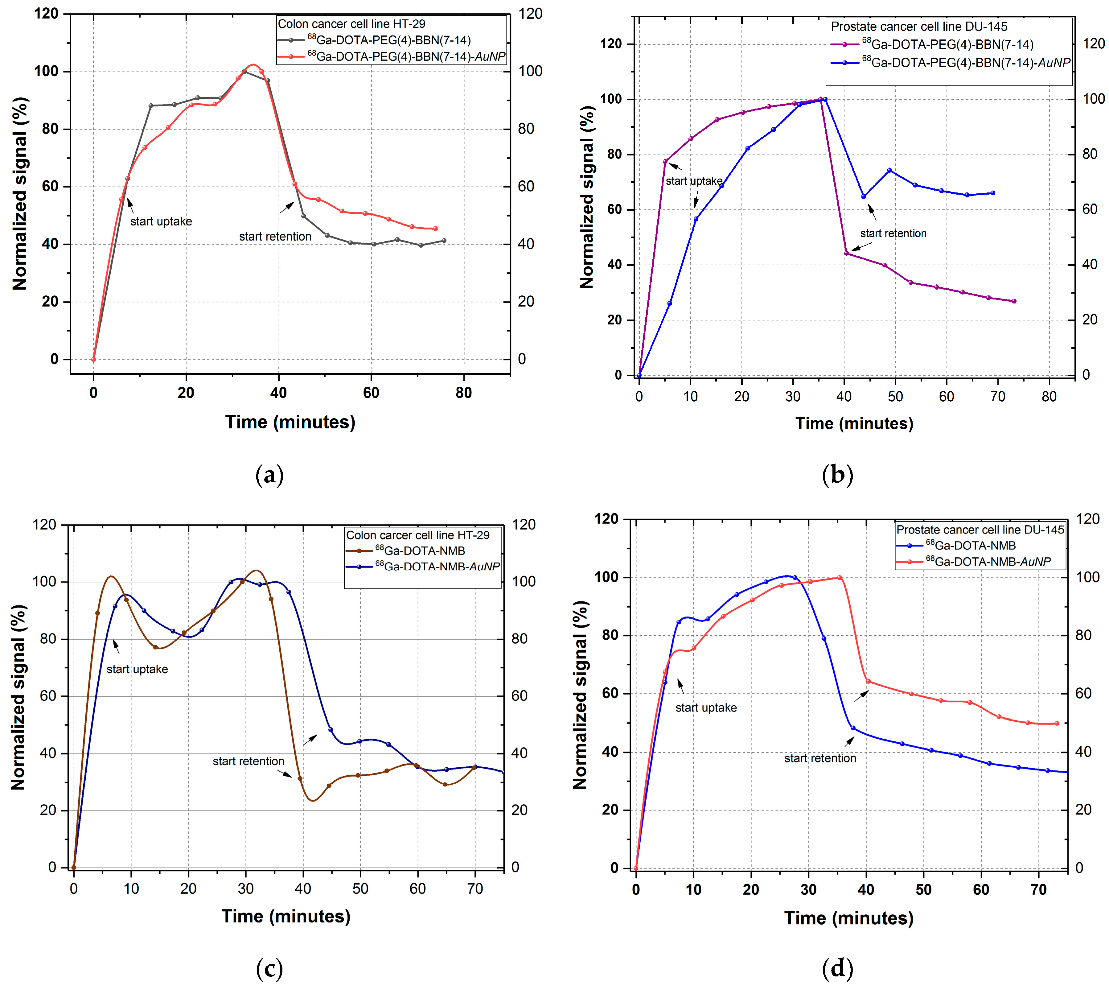
2.5.1. Uptake and Retention

The binding kinetic (uptake and retention) of the 68Ga-labelled peptides and peptide-functionalized gold nanoparticles we investigated using rotating radioimmunoassay. 2–3 nmol of each peptide labelled with 68Ga was incubated on DU-145 prostate and HT-29 colon cancer cells. After uptake measurement, the radioactive medium was removed and the cells were washed with fresh DMEM-F12 medium. The cell retention is measured after this process, during each rotation of the Petri dish support the detector is measuring only the signal coming from the tracers bound to cells. The results are reported as measured signal during the time of analysis (where t = 0 min is the time of incubation), as illustrated in Figure 3. Following the analysis of each peptide, we can easily observe that the uptake of both nanostructured and peptide-based pharmaceuticals is increasing almost exponentially with the incubation time, which indicates a good dynamic on the tumour cells. A lower outcome is obtained for 68Ga-DOTA-NMB and 68Ga-DOTA-NMB-AuNP on colon cancer cell line HT-29, for which the uptake profile follows an up and down trend, an indicator of unstable binding to neuromedin B receptors (NMB, BBRS2, BBRS3).
The retention level is illustrated after the indicator arrows and reflects the percent of activity retained by the cells from the maximum counted during uptake, after hot medium removal and cells washing step. This part of analysis is a screening that enable evaluation of peptide potential to form stable bonds with its specific receptors. The retention of all peptides exceeds 30%, for most the level being maintained until the end of the analysis (approx. 75 min). The only peptide for which retention level fluctuates is NMB incubated on HT-29 cells, which indicates an unstable or weak peptide-receptor bond. From the uptake-retention trend of functionalized AuNPs, we can observe that gold nanoparticles improve cell retention of 68Ga-labelled peptides without affecting the ability of peptides to bind to receptors. A significant increase was obtained for 68Ga-DOTA-PEG(4)-BBN(7-14)-AuNP on DU-145 prostate cancer cells, that reaches 75% compared to approximately 38% for 68Ga-DOTA-PEG(4)-BBN(7-14).

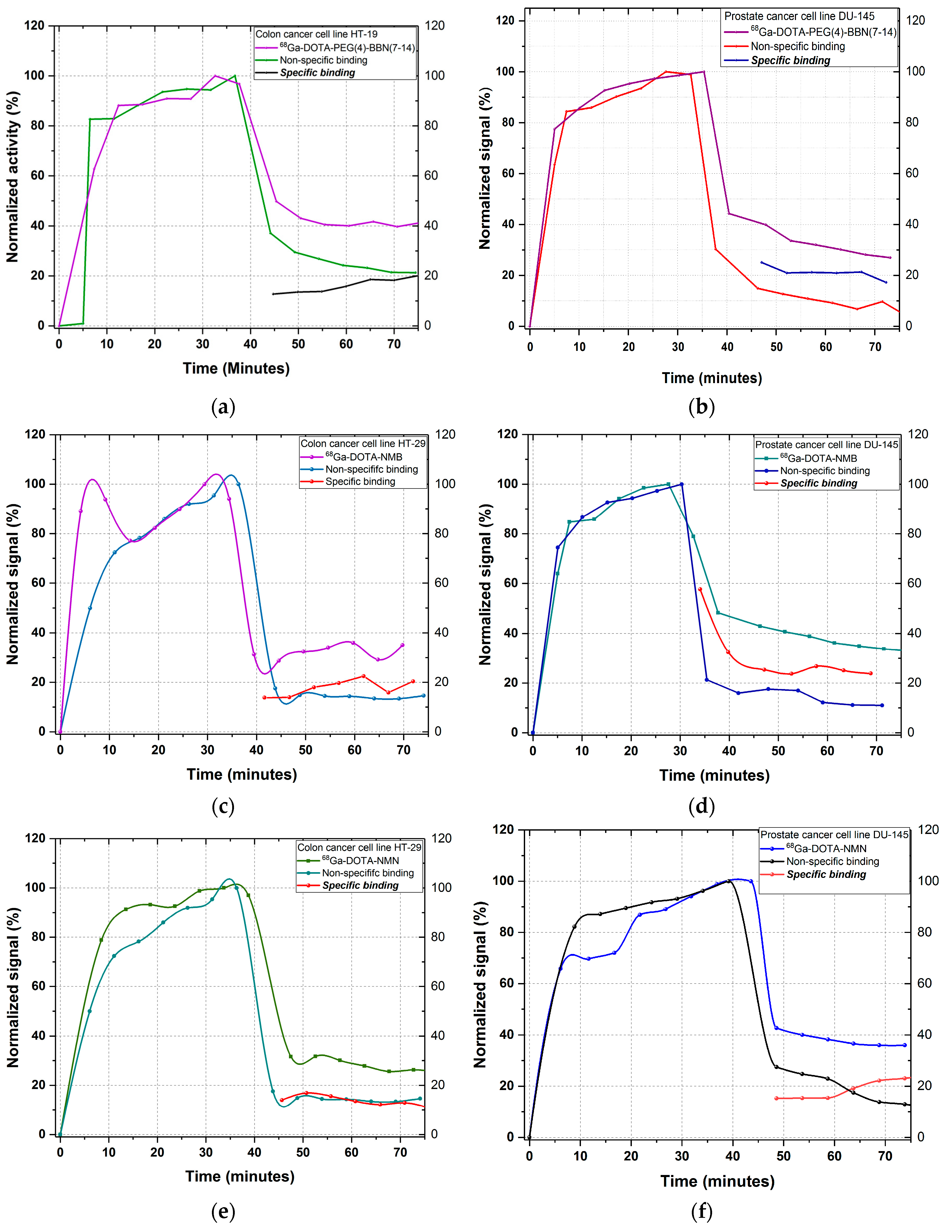
2.5.2. Specific Binding

The evaluation of specific binding of a peptide towards cell membrane receptors is strongly dependent upon the density of receptors, their conformation (active or inactive) and the concentration of inhibitor used to block the receptors prior to peptide incubation [18,19]. For specific binding determination, the receptors for each peptide were blocked with several antagonists: ML-18 and PD176252 (Kd = 1 nM) for bombesin BBRS1, BBRS2 and BBRS3 receptors; SR48692 (Kd = 15 nM), SR142948 (Kd = 10 nM) and NTRC 824 (Kd = 38 nM) for neurotensin NTRS1, NTRS2 and NTRS3 receptors. The incubation time of antagonists was 1–1.5 h, then the medium was removed and the cells were washed and supplemented with ~2 mL fresh culture medium. The non-specific uptake and retention of each 68Ga-labelled peptide was subsequently evaluated using the same protocol as the one used for total binding. The binding kinetics profiles for non-specific binding of each peptide and the total binding are presented in Figure 4. Specific binding is the difference between the total binding and non-specific binding. We can observe that the specific retention of the peptides is about 20%, which represents half of the total binding for each 68Ga-labelled peptide.

2.6. Ex Vivo Biodistribution

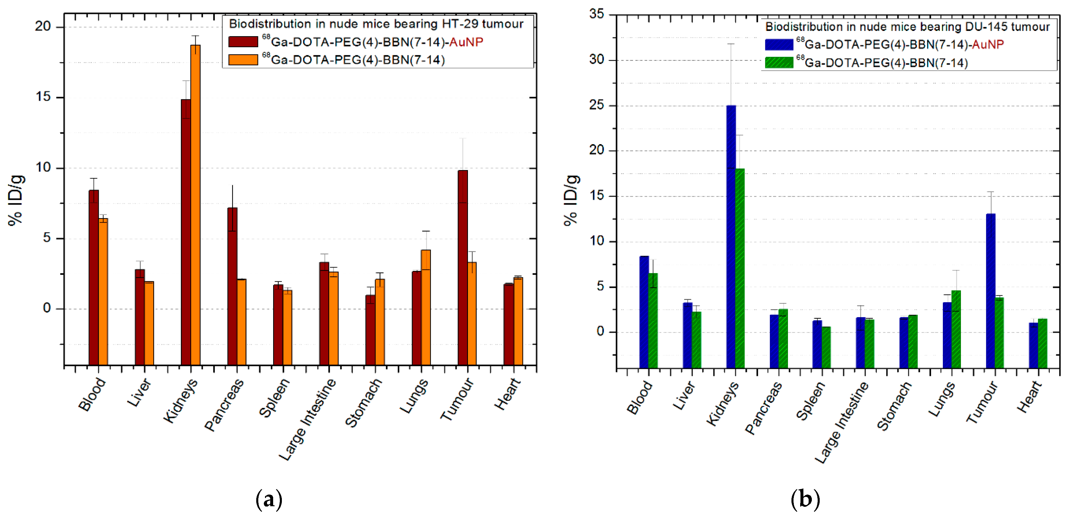
2.6.1. 68Ga-DOTA-PEG(4)-BBN(7-14)/68Ga-DOTA-PEG(4)-BBN(7-14)-AuNP

The comparative biodistribution of 68Ga-DOTA-PEG(4)-BBN(7-14)/68Ga-DOTA-PEG(4)- BBN(7-14)-AuNP is presented in Figure 5, all the experimental data being included in Appendix B, Table A1 and Table A2. After 30 min post-injection (p.i.) a fast clearance of 68Ga-DOTA-PEG(4)-BBN(7-14) was observed in the kidneys (18.74 ± 0.67% ID/g) and in the blood (6.42 ± 0.27% ID/g). Compared to that, 68Ga-DOTA-PEG(4)-BBN(7-14)-AuNP shows higher level in the blood (8.24 ± 0.86% ID/g) and lower in the kidneys (14.86 ± 1.32% ID/g), considered significant as p = 0.047. Additionally, 4.17 ± 1.37% ID/g of 68Ga-DOTA-PEG(4)-BBN(7-14) is found in the lungs. Instead, the activity in the pancreas is 3.37 times higher if BBN-functionalized gold nanoparticles are used instead of 68Ga-DOTA-PEG(4)-BBN(7-14) (p = 0.022). A slight increase was registered in the spleen (1.69 ± 0.26% ID/g for AuNP compared to 1.31 ± 0.22% ID/g for BBN, p = 0.025), liver (2.81 ± 0.57% ID/g for AuNP compared to 1.92 ± 0.07% ID/g for BBN, p = 0.014), and large intestine (3.31 ± 0.56% ID/g for AuNP compared to 2.63 ± 0.34 for BBN, p = 0.021).
A significant increase in activity (p = 0.007) is observed at the tumour site, where activity triples after 68Ga-DOTA-PEG(4)-BBN(7-14)-AuNP use, reaching 9.82 ± 2.28% ID/g from 3.31 ± 0.76% ID/g with 68Ga-DOTA-PEG(4)-BBN(7-14).
A similar behaviour was observed in mice inoculated with prostate tumour cells (Figure 5b), the lungs and heart having lower values after 68Ga-DOTA-PEG(4)-BBN(7-14)-AuNP injection compared to 68Ga-DOTA-PEG(4)-BBN(7-14) (p = 0.042), and increasing activity in the liver and large intestine. Prostate tumour retained 13.01 ± 2.49% ID/g of 68Ga-DOTA-PEG(4)-BBN(7-14)-AuNP, three times (>3.44) higher than 68Ga-DOTA-PEG(4)-BBN(7-14) uptake (3.78 ± 0.27% ID/g), the values being significant as the p-value is p = 0.013.

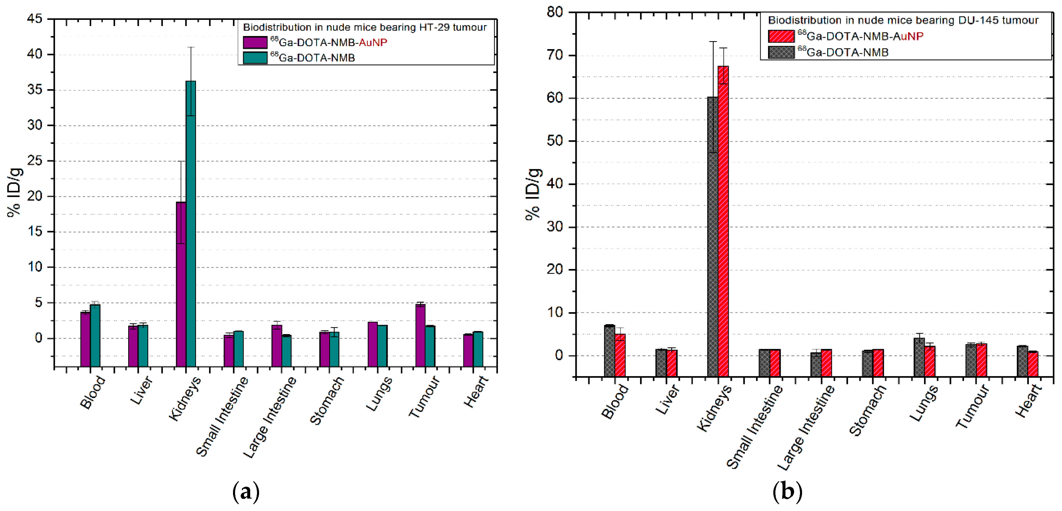
2.6.2. 68Ga-DOTA-NMB/68Ga-DOTA-NMB-AuNP

In both mice with colon tumours and those with prostate tumours, 68Ga-labelled neuromedin B peptide(-AuNP) is quickly removed from the circulatory system and after only 30 min p.i. both tracers are found in the kidneys (60.28 ± 13.01% ID/g of 68Ga-DOTA-NMB and 67.51 ± 4.16% ID/g for 68Ga-DOTA-NMB-AuNP, with p = 0.017) and only 7.00 ± 0.24% ID/g 68Ga-DOTA-NMB in the blood. This observation is also corroborated with the results obtained in the other tissues, being significantly lower compared to its counterparts 68Ga-DOTA-PEG(4)-BBN(7-14) and 68Ga-DOTA-PEG(4)-BBN(7-14)-AuNP (p = 0.005). The uptake of 68Ga-DOTA-NMB (Figure 6b) by the prostate tumour is 2.54 ± 0.42% ID/g and 2.79 ± 0.44% ID/g for 68Ga-DOTA-NMB-AuNP (p = 0.043). Similar values were recorded in colon tumours (Figure 6a), as 1.73 ± 0.08% ID/g 68Ga-DOTA-NMB and 4.75 ± 0.32% ID/g of 68Ga-DOTA-NMB-AuNP were reported (p = 0.026).

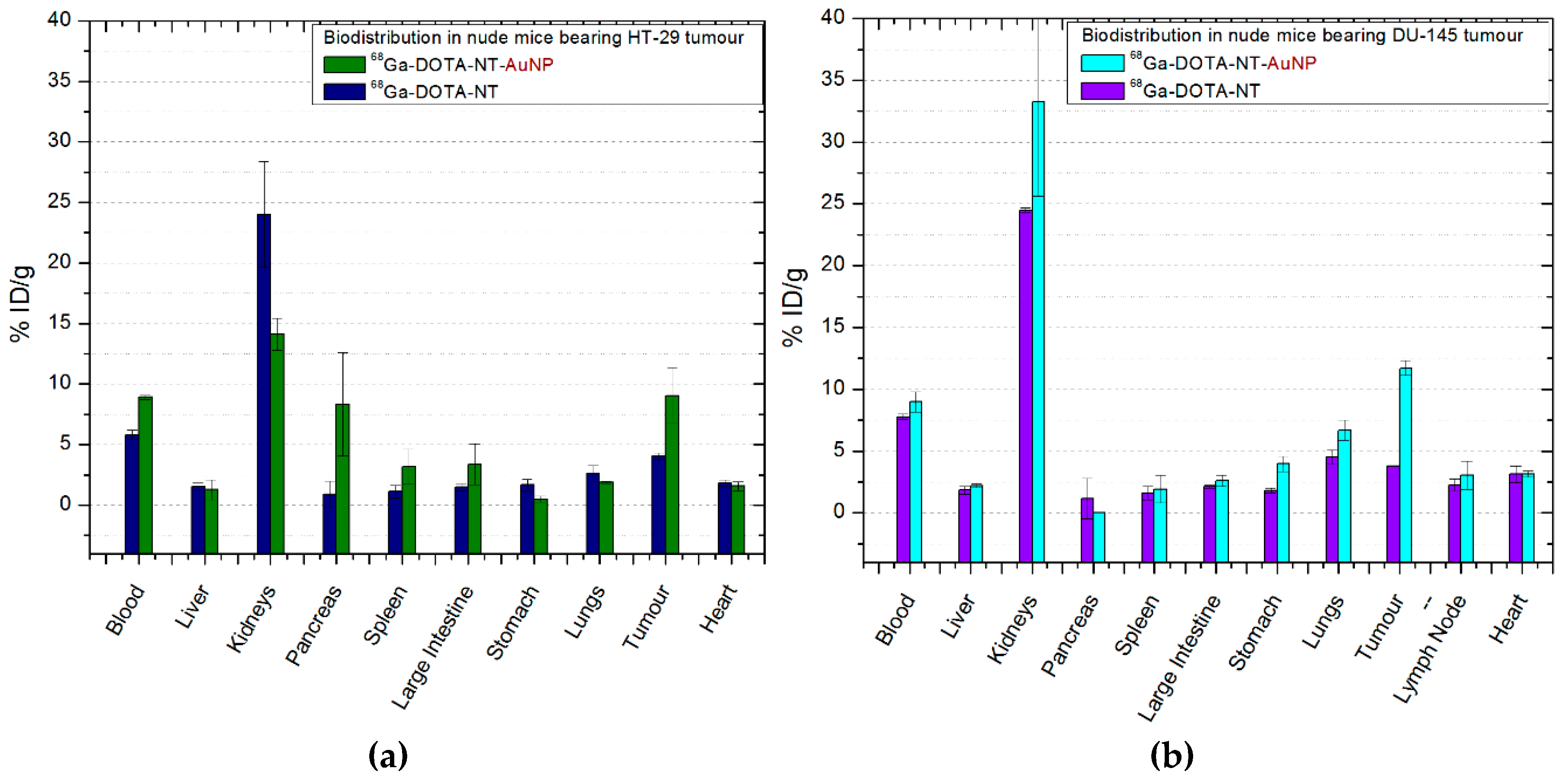
2.6.3. 68Ga-DOTA-NT/68Ga-DOTA-NT-AuNP

68Ga-DOTA-NT uptake in colon and prostate tumours (Figure 7a,b) is similar as 4.06 ± 0.27% ID/g of tracer was measured in the colon tumours of mice, and 3.78 ± 0.05% ID/g in prostate tumours. 68Ga-DOTA-NT-AuNP shows comparative results with 68Ga-DOTA-PEG(4)-BBN(7-14)-AuNP being a 3-fold increase of activity in prostate tumours (11.7 ± 0.59% ID/g) and two times higher in the colon tumours (9.04 ± 2.29% ID/g). In both mice with colon tumours and those with prostate tumours, the excretion of neurotensin is fast, after 30 min p.i. about 25% ID/g being found in the kidneys and 7–9% ID/g in the bloodstream.
Functionalized gold nanoparticles influenced the increase of the amount of tracer in the spleen (3.19 ± 1.39% ID/g in mice with colon cancer and 1.92 ± 0.11% ID/g in those with prostate tumours, p = 0.035) and large intestine (2.59 ± 0.44% ID/g in mice with colon tumours and 3.37 ± 1.6% ID/g in those with prostate cancer, p = 0.021). In addition, a significant uptake (p = 0.016) was measured in the pancreas to three of the mice group (n = 4) inoculated with HT-29 colon cancer cells. Also, in the inguinal lymph node of mice with prostate cancer there is a high percentage of 68Ga-DOTA-NT (2.23 ± 0.49% ID/g) as well as 68Ga-DOTA-NT-AuNP (3.04 ± 1.13% ID/g), although, the result is not statistically significant (p = 0.7).

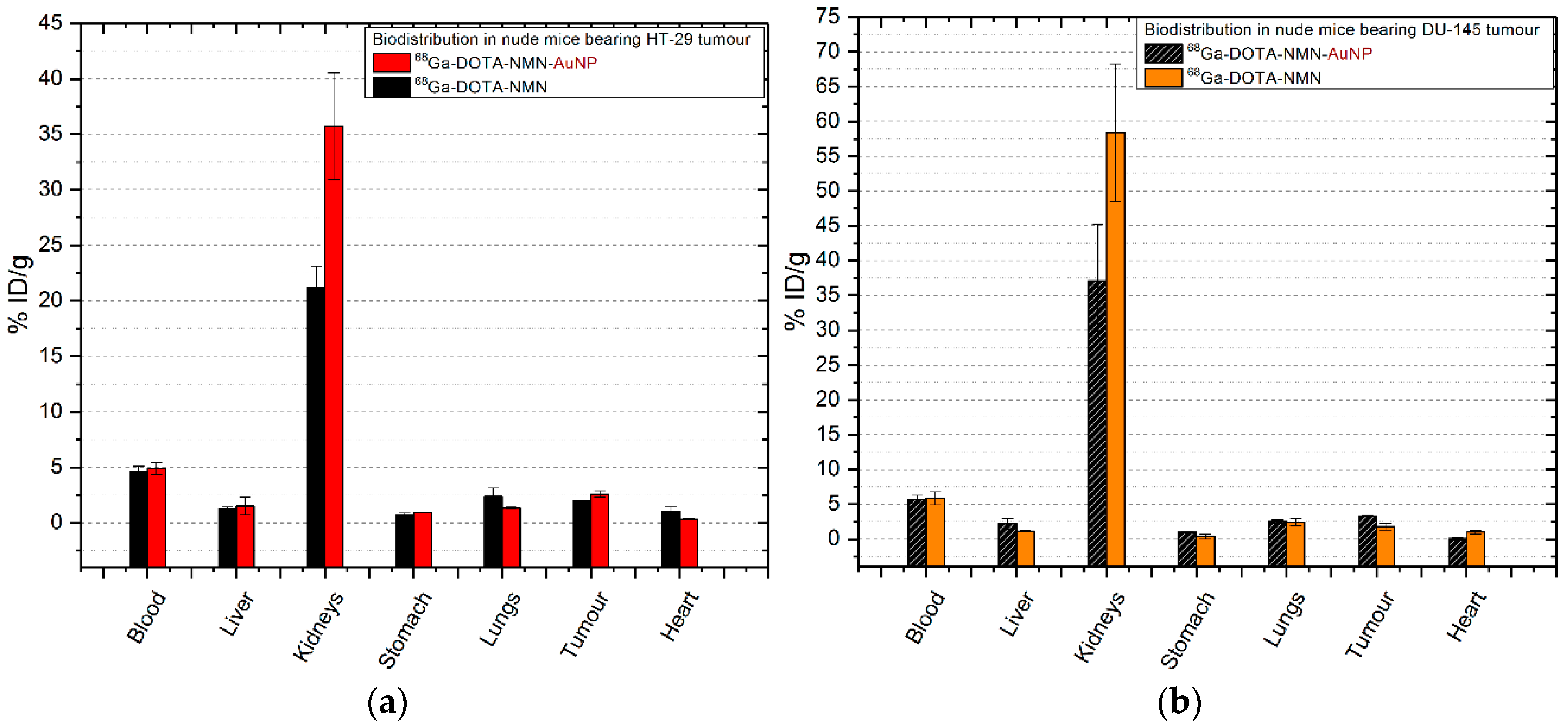
2.6.4. 68Ga-DOTA-NMN/68Ga-DOTA-NMN-AuNP

The results obtained for mice injected with neuromedin N and gold nanoparticles functionalized with neuromedin N are illustrated in Figure 8. The pharmacokinetic of neuromedin N peptide in mice inoculated with HT-29 colon cancer cells is predominated by the fast clearance of tracers from the bloodstream (4.59 ± 0.49% ID/g for 68Ga-DOTA-NMN and 4.89 ± 0.53% ID/g of 68Ga-DOTA-NMN-AuNP). There are no significant accumulation in organs (p > 0.19) other than the kidneys (21.14 ± 1.97% ID/g for 68Ga-DOTA-NMN; 35.72 ± 4.83% ID/g for 68Ga-DOTA-NMN-AuNP; p = 0.041), liver, lungs and tumour (2.02 ± 0.02% ID/g of 68Ga-DOTA-NMN and 2.57 ± 0.26% ID/g 68Ga-DOTA-NMN-AuNP, p = 0.035), which indicates receptors mediated uptake as they express neurotensin receptors [20].
In the case of mice with prostate cancer (Figure 8b) there is a slower renal excretion after 30 min p.i. as 58.34 ± 9.86% ID/g for 68Ga-DOTA-NMN is found in the kidneys. After the 68Ga-DOTA-NMN-AuNP injection, the renal excretion of the tracer is improved, decreasing to 37.07 ± 8.11% ID/g but keeping similar values in the blood (5.69 ± 0.59% ID/g for 68Ga-DOTA-NMN-AuNP compared to 5.85 ± 0.83% ID/g for 68Ga-DOTA-NMN; p = 0.028). Furthermore, tumour uptake is doubled from 1.77 ± 0.5% ID/g for 68Ga-DOTA-NMN to 3.29 ± 0.08% ID/g for 68Ga-DOTA-NMN-AuNP with statistical significance (p = 0.09). Nanoparticles also double the uptake in the liver, small intestine and stomach.

3. Discussion

In this study, we successfully labelled with a positron emitter (68Ga) DOTA-neuromedin B, DOTA-neuromedin N, bombesin analogue DOTA-PEG(4)-BBN(7-14) and neurotensin DOTA-NT peptide. The labelling of the four peptides with 68Ga radioisotope was performed in semi-automated synthesis module, the synthesis process being optimised to obtain a labelling yield higher than 80% and radiochemical purity higher than 80% for 68Ga-DOTA-PEG(4)-BBN(7-14), and up to 100% for 68Ga-DOTA-NT and 68Ga-DOTA-NMN. The final compounds contain no unbound 68Ga and are stable for at least 4 h post final purification, being suitable for biological tests.
After the functionalization process of gold nanoparticles with the above-mentioned peptides we evaluated the in vitro uptake, retention, and specific binding of each tracer on human colon cancer cells HT-29 and DU-145 prostate cancer cells. Almost all tracers formed stable bonds with their specific receptors, except for 68Ga-DOTA-NMB for which the uptake and cell retention profile varies during the analysis. The highest retention value was 75%, measured for 68Ga-DOTA-PEG(4)-BBN(7-14)-AuNP on DU-145 prostate cancer cells.
Ex vivo biodistribution studies in immunocompromised mice inoculated with HT-29 and DU-145 tumour cells confirmed the results obtained in vitro, showing that cell retention was significantly improved after the incubation of cells with peptide-functionalized gold nanoparticles. Due to the ability of NHS-activated PEG coating of gold nanoparticles to react with the primary amines of peptides, an external 68Ga-labelled peptide layer is formed [21], and the signal detected in the tumour tissue is doubled in prostate tumours for 68Ga-DOTA-NMN-AuNP, as well as in colon tumours for 68Ga-DOTA-NMB-AuNP and 68Ga-DOTA-NT-AuNP. In the case of 68Ga-DOTA-PEG(4)-BBN(7-14)-AuNP on both colon and prostate tumours, the activity of 68Ga is three times higher than that obtained for 68Ga-DOTA-PEG(4)-BBN(7-14). Peptide functionalized gold nanoparticles can be delivered to tumours by both passive targeting mechanism through EPR effect; and by active targeting mechanism, as NT/NMN and BBN/NMN peptides recognise specific receptors on colon and prostate cells. Thereby, tumour neo-vascularisation can influence the uptake of nanoparticles into tumour cells [8]. A slight increase in 68Ga-DOTA-NMB-AuNP uptake was observed also in prostate tumours as well as 68Ga-DOTA-NMN-AuNP uptake in colon tumours, but further studies are needed to assess whether this slightly lower outcome (compared to the other peptides-AuNPs systems) was influenced by poorly vascularised or necrotic tumour tissues.
Previous studies for nanoparticle-based drug development highlighted the importance of reticuloendothelial system (RES), a network of cells and tissues responsible for avid clearance of unmodified NPs from the blood circulation within seconds or minutes [8,9,22]. Among the essential organs of RES, spleen, kidneys, liver, pancreas, lungs, bone marrow and lymph nodes play an important role in the pharmacokinetics of nanoparticle-based pharmaceuticals, and for this reason special attention has been paid to the amount of tracer absorbed in these tissues. In the case of 68Ga-DOTA-NMB (-AuNP) and of 68Ga-DOTA-NMN injected into mice with prostate cancer, although the clearance of tracers from the bloodstream is fast, the renal excretion is slower compared with the other peptides, with large amounts of tracers found at 30 min p.i. in the kidneys (approx. 60% ID/g). As neurotensin-like receptors are also found in the kidneys [20], the uptake of 68Ga-DOTA-NMN is most likely due to the interaction of neuromedin N with its receptors, which still needs to be studied to fully understand its nature.
Liver and biliary system represents the primary route for excretion of nanoparticles that do not undergo renal clearance, of which Kupffer cells are a potential site for toxicity as they express receptors for selective endocytosis of opsonized nanoparticles and rely on particle degradation for clearance [9]. Thus, nanoparticles that cannot be degraded by intracellular processes will remain within the cells and will therefore be retained. The role of polyethylene glycol (PEG 5 kDa) is to prevent the opsonin proteins from passively absorbing on the nanoparticles surface, and thus to prevent unwanted accumulations of NPs in the liver. However, slightly increased 68Ga-DOTA-PEG(4)-BBN(7-14)-AuNP activities were measured in mice with both colon and prostate cancer, and higher levels of 68Ga-DOTA-NMN-AuNP (doubled compared to 68Ga-DOTA-NMN) in mice inoculated with DU-145 prostate cancer cells.
Previous studies revealed that NPs size strongly influence the route by which they are excreted/eliminated from the system; nanoparticles with hydrodynamic diameter less than 6 nm are primary filtered by the kidneys [23], NPs <200 nm undergo liver excretion mediated by Kupffer cells, while NPs >200 nm by splenic macrophages excretion. We observed 3.19 ± 1.39% ID/g 68Ga-DOTA-NT-AuNP (with hydrodynamic diameter of 72.36 nm) in the spleen, for mice inoculated with colon cancer cells, which suggests receptor mediated uptake [24].
Several tracers have been developed to target and facilitate the molecular imaging of colon or prostate cancer, such as: 68Ga-NGR with over 2.7 ± 0.65% ID/g in colon tumour at 30 min p.i. [25], [18F]FDG with 2.5 ± 0.5% ID/g [18F]FDG in colon tumours [26], [18F]fluorocholine (FCH) with 7.9 ± 5% ID/g at 60 min p.i. in prostate tumour bearing mice [27]. Compared to these, the values obtained for tumour uptake using the four studied nanotracers are between 2.53 ± 0.26% ID/g for 68Ga-DOTA-NMN-AuNP to 9.82 ± 2.28% ID/g for 68Ga-DOTA-PEG(4)-BBN(7-14)-AuNP in colon tumours and 2.79 ± 0.4% ID/g for 68Ga-DOTA-NMB-AuNP to 13.01 ± 2.49% ID/g for 68Ga-DOTA-PEG(4)-BBN(7-14)-AuNP. Despite the promising findings, the advantages of bombesin, neuromedin B, neurotensin and neuromedin N-functionalized gold nanoparticles have only been demonstrated in vitro and ex vivo. In vivo μPET/CT imaging using these radiolabelled nanocarriers will be investigated in our future studies along with quantitative evaluation by ICP-MS to confirm that the peptides do not detach from the gold nanoparticles in physiological fluids.

4. Materials and Methods

4.1. Peptides Labelling

The peptides used in this work are commercially available and were purchased already coupled with DOTA chelator from Phoenix Europe GmbH (Manheim, Germany; DOTA-NT and DOTA-NMB), AnaSpec Inc. (Fremont, CA, USA; DOTA-PEG(4)-BBN(7-14)) and PolyPeptide (Strasbourg, France; DOTA-NMN), respectively. 68Ga used for labelling was obtained from a GMP-grade 68Ge/68Ga generator (ITG Isotope Technologies Garching GmbH, Munich, Germany) eluted with 0.05 M HCl prepared with suprapure HCl (Merck KGaA, Darmstadt, Germany). Labelling pH was adjusted with ammonium acetate (Carl Roth GmbH, Karlsruhe, Germany), used as 1 M solution. Separation and purification of labelled peptides were performed on Strata™-X solid phase extraction cartridges (Phenomenex Inc., Torrance, CA, USA). Ethanol (Chimreactiv, Bucharest, Romania) was used to recover the compound retained on cartridge. After evaporation of ethanol, the labelled compounds were dissolved in 1 mL saline solution (0.9% NaCl, B. Braun Melsungen AG, Melsungen, Germany), then finally filtered through a 0.22 μm pore size filter (Merck KGaA). Ultrapure water used for solutions preparation and for rinsing was freshly prepared in-house on a Millipore Milli-Q Direct 8 (Merck KGaA). The 68Ge/68Ga generator was coupled to a programmable synthesis module Galigand GAL-102 (Veenstra Instruments, Joure, The Netherlands). The whole preparation process was implemented and remotely controlled on Galigand GAL-102 labelling module. The peptides were previously dissolved in ultrapure water. Aliquots of 20 nmol of peptide per 50 μL solution were used for each synthesis, except DOTA-PEG(4)-BBN(7-14) for which 40 nmol per 50 μL solution were used. Prior of being loaded into reaction vial, the peptide must be protected by adding a certain amount of buffer (ammonium acetate 1 M). The amount of buffer necessary to adjust pH in the range of 3.8–4.3, suitable for the labelling reaction, was previously determined [28]. In order to be used for biomolecules labelling, 68Ga was eluted from the generator using 0.05 M HCl. At this pH value, the gallium is introduced into the process as gallium (III) chloride. Exactly 2 mL of gallium eluate, containing the highest amount of eluted activity, was added over the buffered peptide already loaded into the reaction vial. To shorten the overall preparation time, the labelling temperature was increased to 95 °C; at this value, the duration required for the labelling step is 5 min. The reaction mixture is then passed on the separation cartridge which retains the peptide, while the impurities are evacuated to waste. After rinsing on cartridge by passing 1 mL water with 5% ethanol content, the peptide was recovered with 1 mL of ethanol and sent to the evaporation vial, where the ethanol was evaporated to dryness under vacuum and heating (95 °C). The peptide was dissolved in 1 mL saline solution and transferred to the final product vial by passing through a 0.22 μm pore size filter for sterilization purpose.

4.2. Quality Control of 68Ga-Labelled Peptides

Activity was measured using the Atomlab™ 500 dose calibrator (Biodex Medical Systems Inc., Shirley, NY, USA). RCP and stability of the radiolabelled peptides were assessed by radio-HPLC performed onn a HPLC Shimadzu Prominence 20A system (Shimadzu, Kyoto, Japan) equipped with FlowStar LB 513 bismuth germanate γ detector (Berthold Technologies GmbH & Co.KG, Bad Wildbad, Germany). The stationary phase used was Nucleosil® C18 column (5 μm, 250 mm × 4.6 mm—Merck KGaA) and the mobile phase consisted of water and acetonitrile, both acidified with 0.1% TFA. pH was evaluated with pH paper for 3.6–6.1 and 2.0–9.0 ranges (Macherey-Nagel, Duren, Germany). The activity loaded in the final vial was measured by dose calibrator and used for calculation of the specific activity and for yield evaluation. Due to the short half-life of 68Ga radioisotope, the value had to be decay-corrected for the moment of use, in order to keep a rigorous control over the activity added in biological tests. For accurate calculation of activity used in biological tests, the value of exact activity used was calculated by difference, measuring the activity of the syringe before and after adding the radiolabelled compound over the biological sample. In order to check the reproducibility of the preparation process, along with the main compound activity, the values of residual activity of the key components were measured and decay-corrected for the reference time. The preparation yields were then calculated based on these sets of values. Prior of using the final compound to biological tests, its pH was checked. A radio-HPLC method was used to assess the radiochemical purity of the final compounds, and the stability of the radiolabelled molecules. RCP was checked every hour, up to 4 h after the end of synthesis.

4.3. Nanoparticles Functionalization

Ready-to-use kits containing pre-made mixtures for peptides functionalization on 40 nm NHS-activated gold nanoparticles conjugated with 5 kDa PEG-spacer were purchased from Cytodiagnostics (Burlington, ON, Canada). The functionalization process was conducted according to the protocol provided by the manufacturer [29]. The conjugation kit consists of NHS-activated gold NPs, reaction buffer, protein resuspension buffer and quencher solution. One vial with ~1 mg NHS-activated gold nanoparticles was mixed with 12 nmol of each 68Ga-labelled peptide (68Ga-DOTA-PEG(4)-BBN(7-14), 68Ga-DOTA-NT, 68Ga-DOTA-NMB, 68Ga-DOTA-NMN) and 60 µL of reaction buffer. The peptide-AuNP solution was then stirred at room temperature for 30 min. At the end of the functionalization process 10 µL of quencher solution was added to stop the reaction. The structures of the obtained nanocarriers are presented in Figure 9.

4.4. Physicochemical Characterisation

4.4.1. DLS

The size distribution and zeta potential of cold peptide functionalized-gold nanoparticles were measured using a Nano ZS zetasizer system (Malvern Instruments, Malvern, UK). Measurement parameters were as follows: a laser wavelength of 633 nm (He-Ne), measurement angle of 90°, measurement temperature of 25 °C, and refractive index of medium n = 1.330.

4.4.2. FTIR Spectra

FTIR spectroscopy was performed using KBr (IR grade) pellets containing the sample over the 5000–350 cm−1 wavenumber range with a model 6100.4.5 FTIR spectrophotometer (Jasco, Easton, MD, USA).

4.5. Cell Culture and Petri Dishes Preparation

Biological investigations were performed on human normal cell lines (proliferating human CRL monocytes) and tumour cell lines (human colon carcinoma HT-29 cells and human prostate carcinoma DU-145 cells), purchased from the American Cell and Tissue Collection (ATCC, Manassas, VA, USA). Cells were maintained in culture according to the protocol provided by the depositor. Human CRL9855 cells were cultivated in IMDM culture medium (ThermoFisher Scientific, Wiltham, MA, USA) supplemented with 10% fetal bovine serum (FBS, Merck), HT solution (100x: 10 mM sodium hypoxanthine and 1.6 mM thymidine, Thermo Fisher Scientific) and 0.05 mM 2-mercaptoethanol (Sigma-Aldrich, Darmstadt, Germany). Adherent HT-29 and DU-145 cells were cultivated in DMEM-F12 culture medium (ThermoFisher Scientific) supplemented with 10% fetal bovine serum (FBS, Merck) and antibiotic-antimycotic solution (Sigma-Aldrich, Darmstadt, Germany). Passage of cells was done every 2–5 days for maintaining an appropriate density of cells in culture. For adherent cells split a 0.25%/0.02% trypsine/EDTA solution (Biochrom Ltd., Cambridge, UK) was used. Cells at passage 6–9 and were counted before seeding for experimentation by optical microscopy using a Burker-Turk counting chamber, and cell viability was assessed by the trypan blue exclusion test. Only cell suspensions with viability above 95% were used in experiments. 24 h prior to a binding experiment with LigandTracer Yellow® (Ridgeview Instruments AB, Uppsala, Sweden), Petri dishes (Nunc™ 150350, ThermoFisher Scientific) were prepared with approximately 4 × 10⁵ cells. The cells were seeded on a well-defined area of a tilted cell culture dish and incubated with 2 mL DMEM medium for 12 h. Before the experiment, the Petri dish is verified under microscope to check for cells migration outside the area of interest.

4.6. Cytotoxicity Investigation

The effect of AuNP NPs loaded with neuropeptides (DOTA-PEG(4)-BBN(7-14), DOTA-NMB, DOTA-NMN, DOTA-NT) on normal proliferating cells (human CRL9855 cells) or human tumour cells (human colon carcinoma HT-29 cells and human prostate carcinoma DU-145 cells) was investigated using the MTS reduction test (CellTiter 96® Aqueous One Solution Cell Proliferation Assay, Promega, Madison, WY, USA), as indirect measure of the number of metabolically active cells in culture. Briefly, cells were cultivated in 96-well plates (3 × 10⁴ CRL9855 cells/mL, 10 × 104 HT-29 cells/mL and 5 × 10⁴ DU-145 cells/mL) in a total volume of 100 μL. Adherent cells were left for 24 h to adhere to the culture well. Thereafter, NPs and peptides were added at various concentrations and cells were cultivated for 48 h. Non-treated cells were considered as control (CTRL). For background assessment culture medium, NPs and peptides were cultivated in absence of cells, considering that NPs might interfere with the spectrophotometric measurement. All samples were cultivated in triplicate. At the end of the incubation time, 20 μL of MTS reagent were added and samples were cultivated for another 1–3 h until the reaction color was developed. The OD at 490 nm was measured in each sample using a Tecan Sunrise ELISA reader with Magellan universal reader control and data analysis software (Tecan Group Ltd., Mannedorf, Switzerland). Final OD in cellular samples was calculated by subtracting the OD of the corresponding background samples. Results were presented as mean ± standard error of the mean (SEM) for triplicate samples.

4.7. In Vitro Binding Studies

4.7.1. Uptake and Retention Assay

The interaction between 68Ga-labelled tracers and living cells expressing a target receptor was measured in real-time using rotating radioimmunoassay (RIA) method implemented on the automatic LigandTracer® Yellow system [30], by calculating at each rotation the difference between the signals detected from 6 points in the background area of the Petri dish and 6 points from the area of interest. The binding curve is obtained after a two-phase process consisting of an incubation phase and a wash-out phase. In the first phase, a background point is acquired after which 2–3 nmol of peptide-containing tracer is incubated over the cells for a time equal to that required to reach the equilibrium, which is recognized on the graph by a plateau trend. After this phase, the radioactive medium was carefully extracted from the dish, ensuring that the liquid did not exceed the defined area, and the cells were washed twice with 1 mL of DMEM medium. At the end, 2 mL of fresh medium is added over the cells to ensure optimal survival conditions during the next phase. In the wash-out phase, the retention of the tracer to cancer cells is evaluated. By removing the radioactive medium, the uptake process is stopped, the signal detected coming from 68Ga radioisotope coupled to the receptor-bound peptides. This part is measured for a time long enough to permit to assess the stability of the interaction. Since kinetic parameters are dependent only on the amount of incubated peptide and the incubation time, signal values were normalized to 0% at baseline level and 100% at the end of the incubation phase, respectively, to enable easy visual comparison of interaction profiles between different peptides.

4.7.2. Specific Binding Assay

Specific uptake of 68Ga-labelled peptides at their receptors was assessed using non-peptide antagonists capable of blocking receptors prior to incubation of peptides. The antagonists selected to block the receptors were chosen based on their affinity for the three receptors of each peptide. For accuracy, a combination of several antagonists was used to block all receptors before incubating the tracers. Thus, ML 18 and PD 176252 antagonists were used to prevent 68Ga-DOTA-BBN(7-14) and 68Ga-DOTA-Neuromedin N from binding to bombesin receptors (BBRS1, BBRS2 and BBRS3) and SR 142948, SR 48692 and NTRC 824 to block the neuromedin N peptide binding to neurotensin receptors (NTRS1, NTRS2 and NTRS3). ML-18 is a non-peptide bombesin receptor subtype 3 (BBRS3) antagonist for which has been recently demonstrated its ability to inhibit the growth of lung cancer [31]. PD 176252 is a non-peptide gastrin-releasing peptide receptor (GRP-R) antagonist also known as bombesin receptor subtype 2 (BBRS2), and neuromedin B receptor (NMB-R) antagonist also known as BBRS1 [32]. NTRC 824 is selective for neurotensin receptor subtype 2 (NTRS2) over NTRS1, by contrast, SR 48692 is selective for NTRS1 over NTRS2 but not NTRS3. For this reason, a third neurotensin receptor antagonist was used, namely SR 14294.
Predefined amounts of each antagonist presented in Table 3 were incubated over the cells for 1–1.5 h followed by removal of the medium, washing the cells with DMEM medium and incubation of the radiolabeled peptides according to the protocol described above.

4.8. Ex Vivo Biodistribution

All experiments were conducted according to the guidelines of EU Directive 2010/63 approved by The Romanian National Sanitary Veterinary and Food Safety Authority (NSVFSA, approval no. 442/25.02.2019). Athymic nude male mice (NU/J, 8–10 weeks old) were obtained from The Jackson Laboratory (Bar Harbor, ME, USA). Animals were housed in ventilated cages with a 12-h light-dark cycle and receive food and water ad libitum. Colon/prostate tumours were induced 7–10 days prior the experiments by a subcutaneous injection of 1 × 10⁶ HT-29/DU-145 cancer cells. All injected animals were monitored daily until an ideal size of tumour for biodistribution was reached (60–100 mm3). 3.81 ± 0.51 MBq of 68Ga-labelled tracers were retro-orbital injected into the ophthalmic venous sinus after intraperitoneal anesthesia with 0.1 mL ketamine/xylazine/acepromazine cocktail: 7.5 mg/mL ketamine (Narkamon Bio, Bioveta, Cluj-Napoca, Romania), 1.5 mg/mL xylazine (Narcoxyl 2, MSD Animal Health, Madison, NJ, USA) and 0.25 mg/mL acepromazine (Sedam, SC Pasteur Filiala Filipesti SRL, Filipestii de Padure, Romania). All animals were sacrificed at 30 min p.i. and the organs were weighted and measured in a dose calibrator (Biodex Medical Systems Inc., Shirley, NY, USA). The biodistribution was calculated as percent of injected dose per gram of organ (% ID/g) ± standard deviation (SD, n = 4 for each injected tracer).

4.9. Statistical Analysis

All the data were expressed as means ± standard deviation (n = 4). Comparison of results among the groups (% ID/g vs. tracers, % ID/g vs. tracers and organs, % ID/g vs. tracers and tumoral cells), was carried out by one-way analysis of variance (ANOVA) and Tukey post-hoc analysis using SPSS v26 (IBM, Armonk, NY, USA) The results were considered significant for a p-value less than 0.05.

5. Conclusions

In conclusion, we synthetized and evaluate both in vitro and ex vivo in tumour-bearing immunocompromised mice four different gold nanocarriers for 68Ga-labelled bombesin and neurotensin analogues that exhibited high stability in physiological conditions, minimum accumulation in non-targeted organs and fast distribution in colon and prostate tumour tissue. Promising results were obtained for 68Ga-DOTA-PEG(4)-BBN(7-14)-AuNP and 68Ga-DOTA-NT-AuNP, which we propose for future multimodal imaging PET/CT studies.

Author Contributions

Conceptualization, D.N., peptides labelling, R.A.L.; in vitro and ex vivo studies, L.E.C., R.A.L., C.M., R.M.S. and G.M.; physicochemical characterization of nanostructures, R.T., A.N. and L.E.C.; writing—original draft preparation, L.E.C., R.A.L.; writing—review and editing, D.N. and V.L.; project administration, D.N., G.M.; funding acquisition, D.N., G.M. All authors have read and agreed to the published version of the manuscript.

Funding

This research was funded by Romanian Ministry of Research and Innovation, through UEFISCDI, grant numbers 64PCCDI/2018 and 7PFE/16.10.2018.

Conflicts of Interest

The authors declare no conflict of interest.

Appendix A

Figure A1. Radiochemical purity of 68Ga labelled peptides at EOS: (a) 68Ga-DOTA-NT; (b) 68Ga-DOTA-MN; (c) 68Ga-DOTA-PEG(4)-BBN(7-14); (d) 68Ga-DOTA-NMB.
Molecules 25 03363 g0a1
Figure A2. MTS reduction by human CRL955 monocytes (a), human colon carcinoma HT-29 cells (b) and human prostate DU-145 carcinoma cells (c) incubated for 48 h with bare NPs (AuNP) or with peptide-loaded NPs (DOTA-PEG-BBN-AuNP, DOTA-NMB-AuNP, DOTA-NMN-AuNP, DOTA-NT-AuNP). Results are presented as mean ± SEM (standard error of the mean) for triplicate samples.
Molecules 25 03363 g0a2
Figure A3. The effect exerted by neuropeptides on MTS reduction by human CRL955 monocytes (a), human colon carcinoma HT-29 cells (b) and human prostate DU-145 carcinoma cells (c) in 48 h cultures. Results are presented as mean ± SEM (standard error of the mean) for triplicate samples.
Molecules 25 03363 g0a3

Appendix B

Table A1. Ex vivo biodistribution data on NU/J mice inoculated with HT-29 colon cancer cells.
Tissue68Ga-DOTA-PEG(4)-BBN(7-14)68Ga-DOTA-PEG(4)-BBN(7-14)-AuNP68Ga-DOTA-NMB68Ga-DOTA-NMB-AuNP68Ga-DOTA-NT68Ga-DOTA-NT-AuNP68Ga-DOTA-NMN68Ga-DOTA-NMN-AuNP
Blood6.42 ± 0.278.24 ± 0.864.72 ± 0.433.64 ± 0.325.79 ± 0.398.92 ± 0.174.59 ± 0.494.89 ± 0.53
Liver1.92 ± 0.072.81 ± 0.571.82 ± 0.331.70 ± 0.351.55 ± 0.291.27 ± 0.831.23 ± 0.201.51 ± 0.80
Kidneys18.74 ± 0.6714.86 ± 1.3236.23 ± 4.8319.17 ± 5.7724.00 ± 4.3714.13 ± 1.3121.14 ± 1.9735.72 ± 4.83
Pancreas2.13 ± 0.077.18 ± 1.6301.99 ± 0.020.90 ± 1.058.34 ± 4.2400.69 ± 0.11
Spleen1.31 ± 0.221.69 ± 0.260.05 ± 0.010.56 ± 0.121.12 ± 0.523.19 ± 1.390.35 ± 0.020
Small intestine1.70 ± 0.440.93 ± 0.250.43 ± 0.300.99 ± 0.061.30 ± 0.860.76 ± 0.250.43 ± 0.190.75 ± 0.33
Large intestine2.63 ± 0.343.31 ± 0.560.38 ± 0.141.84 ± 0.511.48 ± 0.293.37 ± 1.660.84 ± 0.290.75 ± 0.24
Stomach2.10 ± 0.500.98 ± 0.580.86 ± 0.240.85 ± 0.651.67 ± 0.460.49 ± 0.230.71 ± 0.170.94 ± 0.04
Lungs4.17 ± 1.372.65 ± 0.111.85 ± 0.062.27 ± 0.042.63 ± 0.631.88 ± 0.112.37 ± 0.771.32 ± 0.10
Muscle0.79 ± 0.211.03 ± 0.410.34 ± 0.030.53 ± 0.060.87 ± 0.270.93 ± 0.740.48 ± 0.320.33 ± 0.16
Tumour3.31 ± 0.769.82 ± 2.281.73 ± 0.084.75 ± 0.324.06 ± 0.279.04 ± 2.292.02 ± 0.022.57 ± 0.26
Brain0.49 ± 0.020.15 ± 0.03000.66 ± 0.120.35 ± 0.130.04 ± 0.010.05 ± 0.01
Bone0.75 ± 0.062.21 ± 0.510.10 ± 0.0700.84 ± 0.222.71 ± 0.800.16 ± 0.020.13 ± 0.02
Lymph Node1.10 ± 0.452.03 ± 1.230.49 ± 0.1701.47 ± 0.2700.39 ± 0.050
Heart2.24 ± 0.121.75 ± 0.070.91 ± 0.050.51 ± 0.121.85 ± 0.241.56 ± 0.361.00 ± 0.420.32 ± 0.09
Table A2. Ex vivo biodistribution data on NU/J mice inoculated with DU-145 prostate cancer cells.
Tissue68Ga-DOTA-PEG(4)-BBN(7-14)68Ga-DOTA-PEG(4)-BBN(7-14)-AuNP68Ga-DOTA-NMB68Ga-DOTA-NMB-AuNP68Ga-DOTA-NT68Ga-DOTA-NT-AuNP68Ga-DOTA-NMN68Ga-DOTA-NMN-AuNP
Blood6.48 ± 1.538.34 ± 0.057.00 ± 0.245.06 ± 1.517.77 ± 0.238.97 ± 0.795.85 ± 0.935.69 ± 0.59
Liver2.22 ± 0.703.23 ± 0.391.53 ± 0.211.33 ± 0.511.82 ± 0.342.19 ± 0.151.10 ± 0.072.18 ± 0.76
Kidneys17.96 ± 3.7825.00 ± 6.8760.28 ± 13.0167.51 ± 4.1624.4 ± 0.1833.26 ± 7.6158.34 ± 9.8637.07 ± 8.11
Pancreas2.51 ± 0.721.89 ± 0.631.36 ± 0.0701.15 ± 0.6300.58 ± 0.020
Spleen0.57 ± 0.021.22 ± 0.340.73 ± 0.2901.60 ± 0.531.92 ± 0.1101.08 ± 0.36
Small intestine1.13 ± 0.690.67 ± 0.271.43 ± 0.081.40 ± 0.141.92 ± 0.321.24 ± 0.170.26 ± 0.010.89 ± 0.05
Large intestine1.30 ± 0.251.59 ± 1.370.64 ± 0.911.41 ± 0.102.14 ± 0.122.59 ± 0.440.13 ± 0.270.17 ± 0
Stomach1.87 ± 0.031.61 ± 0.161.05 ± 0.251.42 ± 0.021.80 ± 1.193.96 ± 0.610.40 ± 0.330.96 ± 0.12
Lungs4.59 ± 2.253.24 ± 0.914.08 ± 1.182.14 ± 0.794.51 ± 0.576.67 ± 0.822.37 ± 0.502.52 ± 0.24
Muscle0.94 ± 0.140.63 ± 0.330.84 ± 0.070.92 ± 0.161.02 ± 0.201.78 ± 0.130.57 ± 0.220.71 ± 0.23
Tumour3.78 ± 0.2713.01 ± 2.492.54 ± 0.422.79 ± 0.443.78 ± 0.0511.7 ± 0.591.77 ± 0.503.29 ± 0.08
Brain0.21 ± 0.040.57 ± 0.0900.11 ± 0.0300.64 ± 0.1900.03 ± 0.01
Bone0000.24 ± 0.021.03 ± 0.580.48 ± 0.3500.13 ± 0.02
Lymph Node001.25 ± 0.6902.23 ± 0.493.04 ± 1.1300.27 ± 0.03
Heart1.45 ± 0.131.04 ± 0.482.22 ± 0.120.93 ± 0.193.12 ± 0.643.13 ± 0.240.99 ± 0.260.17 ± 0.09

References

  1. Kong, F.Y.; Zhang, J.W.; Li, R.F.; Wang, Z.X.; Wang, W.J.; Wang, W. Unique Roles of Gold Nanoparticles in Drug Delivery, Targeting and Imaging Applications. Molecules 2017, 22, 1445. [Google Scholar] [CrossRef] [PubMed] [Green Version]
  2. Devaraj, N.K.; Keliher, E.J.; Thurber, G.M.; Nahrendorf, M.; Weissleder, R. 18F labeled nanoparticles for in vivo PET-CT imaging. Bioconjug. Chem. 2009, 20, 397–401. [Google Scholar] [CrossRef] [PubMed] [Green Version]
  3. Glaus, C.; Rossin, R.; Welch, M.J.; Bao, G. In vivo evaluation of (64)Cu-labeled magnetic nanoparticles as a dual-modality PET/MR imaging agent. Bioconjug. Chem. 2010, 21, 715–722. [Google Scholar] [CrossRef] [PubMed] [Green Version]
  4. Pretze, M.; van der Meulen, N.P.; Wangler, C.; Schibli, R.; Wangler, B. Targeted (64) Cu-labeled gold nanoparticles for dual imaging with positron emission tomography and optical imaging. J. Labelled Comp. Radiopharm. 2019, 62, 471–482. [Google Scholar] [CrossRef] [PubMed]
  5. Ghiassian, S.; Yu, L.; Gobbo, P.; Nazemi, A.; Romagnoli, T.; Luo, W.; Luyt, L.G.; Workentin, M.S. Nitrone-Modified Gold Nanoparticles: Synthesis, Characterization, and Their Potential as (18)F-Labeled Positron Emission Tomography Probes via I-SPANC. ACS Omega. 2019, 4, 19106–19115. [Google Scholar] [CrossRef]
  6. Chen, F.; Ma, K.; Zhang, L.; Madajewski, B.; Zanzonico, P.; Sequeira, S.; Gonen, M.; Wiesner, U.; Bradbury, M.S. Target-or-Clear Zirconium-89 Labeled Silica Nanoparticles for Enhanced Cancer-Directed Uptake in Melanoma: A Comparison of Radiolabeling Strategies. Chem Mater. 2017, 29, 8269–8281. [Google Scholar] [CrossRef] [Green Version]
  7. Chilug, L.E.; Leonte, R.A.; Patrascu, M.E.B.; Ion, A.C.; Tuta, C.S.; Raicu, A.; Manda, G.; Niculae, D. In vitro binding kinetics study of gold nanoparticles functionalized with 68Ga-DOTA conjugated peptides. J. Radioanal. Nucl. Chem. 2016, 311, 1485–1493. [Google Scholar] [CrossRef]
  8. Yu, M.; Zheng, J. Clearance Pathways and Tumor Targeting of Imaging Nanoparticles. ACS Nano 2015, 9, 6655–6674. [Google Scholar] [CrossRef] [Green Version]
  9. Longmire, M.; Choyke, P.L.; Kobayashi, H. Clearance properties of nano-sized particles and molecules as imaging agents: Considerations and caveats. Nanomedicine 2008, 3, 703–717. [Google Scholar] [CrossRef] [Green Version]
  10. Nie, S. Understanding and overcoming major barriers in cancer nanomedicine. Nanomedicine 2010, 5, 523–528. [Google Scholar] [CrossRef] [Green Version]
  11. Leonte, R.A.; Niculae, D.; Craciun, L.S.; Cata-Danil, G. Medical radioisotopes production at TR-19 cyclotron from IFIN-HH. U.P.B. Sci. Bull. Ser. A 2017, 79, 223–236. [Google Scholar]
  12. Moody, T.W.; Berna, M.J.; Mantey, S.; Sancho, V.; Ridnour, L.; Wink, D.A.; Chan, D.; Giaccone, G.; Jensen, R.T. Neuromedin B receptors regulate EGF receptor tyrosine phosphorylation in lung cancer cells. Eur. J. Pharmacol. 2010, 637, 38–45. [Google Scholar] [CrossRef] [PubMed] [Green Version]
  13. Kitabgi, P. Neurotensin and neuromedin N are differentially processed from a common precursor by prohormone convertases in tissues and cell lines. In Cellular Peptide Hormone Synthesis and Secretory Pathways; Springer: Berlin, Germany, 2009; pp. 85–96. [Google Scholar] [CrossRef]
  14. Jamous, M.; Tamma, M.L.; Gourni, E.; Waser, B.; Reubi, J.C.; Maecke, H.R.; Mansi, R. PEG spacers of different length influence the biological profile of bombesin-based radiolabeled antagonists. Nucl. Med. Biol. 2014, 41, 464–470. [Google Scholar] [CrossRef] [PubMed]
  15. Falciani, C.; Lelli, B.; Brunetti, J.; Pileri, S.; Cappelli, A.; Pini, A.; Pagliuca, C.; Ravenni, N.; Bencini, L.; Menichetti, S.; et al. Modular Branched Neurotensin Peptides for Tumor Target Tracing and Receptor-Mediated Therapy: A Proof-of-Concept. Curr. Cancer Drug Targets 2010, 10, 695–704. [Google Scholar] [CrossRef]
  16. Falciani, C.; Brunetti, J.; Lelli, B.; Accardo, A.; Tesauro, D.; Morelli, G.; Bracci, L. Nanoparticles exposing neurotensin tumor-specific drivers. J. Pept. Sci. 2013, 19, 198–204. [Google Scholar] [CrossRef] [PubMed]
  17. Huai, Y.; Zhang, Y.; Xiong, X.; Das, S.; Bhattacharya, R.; Mukherjee, P. Gold Nanoparticles sensitize pancreatic cancer cells to gemcitabine. Cell Stress 2019, 3, 267–279. [Google Scholar] [CrossRef] [Green Version]
  18. Hein, P.; Michel, M.C.; Leineweber, K.; Wieland, T.; Wettschureck, N.; Offermanns, S. Receptor and Binding Studies. In Practical Methods in Cardiovascular Research; Dhein, S., Mohr, F.W., Delmar, M., Eds.; Springer: Berlin, Germany, 2005; pp. 723–783. [Google Scholar]
  19. Schlegel, W. Peptide-Receptor Interaction: An Introduction; Elsevier: Ljubljana-Yugoslavia, Oxford, UK, 1980; p. 792. [Google Scholar] [CrossRef]
  20. Quirion, R.; Gaudreau, P.; St-Pierre, S.; Rioux, F. Localization of neurotensin binding sites in rat kidney. Peptides 1982, 3, 765–769. [Google Scholar] [CrossRef]
  21. Zong, J.; Cobb, S.L.; Cameron, N.R. Peptide-functionalized gold nanoparticles: Versatile biomaterials for diagnostic and therapeutic applications. Biomater. Sci. 2017, 5, 872–886. [Google Scholar] [CrossRef] [Green Version]
  22. Guo, S.; Huang, L. Nanoparticles Escaping RES and Endosome: Challenges for siRNA Delivery for Cancer Therapy. J. Nanomater. 2011, 2011, 1–12. [Google Scholar] [CrossRef] [Green Version]
  23. Cassano, D.; Mapanao, A.-K.; Summa, M.; Vlamidis, Y.; Giannone, G.; Santi, M.; Guzzolino, E.; Pitto, L.; Poliseno, L.; Bertorelli, R.; et al. Biosafety and Biokinetics of Noble Metals: The Impact of Their Chemical Nature. ACS Appl. Bio Mater. 2019, 2, 4464–4470. [Google Scholar] [CrossRef]
  24. García-Garayoa, E.; Bläuenstein, P.; Bruehlmeier, M.; Blanc, A.; Iterbeke, K.; Conrath, P.; Tourwé, D.; Schubiger, P.A. Preclinical Evaluation of a New Stabilized Neurotensin(8–13) Pseudopeptide Radiolabeled with 99mTc. J. Nucl. Med. 2002, 43, 374–383. [Google Scholar] [PubMed]
  25. Li, X.F.; Du, Y.; Ma, Y.; Postel, G.C.; Civelek, A.C. (18)F-fluorodeoxyglucose uptake and tumor hypoxia: Revisit (18)f-fluorodeoxyglucose in oncology application. Transl. Oncol. 2014, 7, 240–247. [Google Scholar] [CrossRef] [PubMed] [Green Version]
  26. Li, X.F.; Ma, Y.; Sun, X.; Humm, J.L.; Ling, C.C.; O’Donoghue, J.A. High 18F-FDG uptake in microscopic peritoneal tumors requires physiologic hypoxia. J. Nucl. Med. 2010, 51, 632–638. [Google Scholar] [CrossRef] [PubMed] [Green Version]
  27. DeGrado, T.R.; Reiman, R.E.; Price, D.T.; Wang, S.; Coleman, R.E. Pharmacokinetics and Radiation Dosimetry of 18F-Fluorocholine. J. Nucl. Med. 2002, 43, 92–96. [Google Scholar] [PubMed]
  28. Patrascu, I.; Niculae, D.; Lungu, V.; Ursu, I.; Iliescu, M.; Tuta, C.; Antohe, A. The purification and the quality control of 68Ga eluates from Ge/68Ga generator. Rom. Rep. Phys. 2011, 63, 988–996. [Google Scholar]
  29. Cytodiagnostic. Available online: http://portal.cytodocs.com:8008/documents/Product-Sheets/Product-Sheet-NHS-Activated-Gold-Nanoparticles.pdf (accessed on 27 May 2020).
  30. Bjorke, H.; Andersson, K. Automated, high-resolution cellular retention and uptake studies in vitro. Appl. Radiat. Isot. 2006, 64, 901–905. [Google Scholar] [CrossRef] [PubMed]
  31. Moody, T.W.; Mantey, S.A.; Moreno, P.; Nakamura, T.; Lacivita, E.; Leopoldo, M.; Jensen, R.T. ML-18 is a non-peptide bombesin receptor subtype-3 antagonist which inhibits lung cancer growth. Peptides 2015, 64, 55–61. [Google Scholar] [CrossRef] [Green Version]
  32. Schepetkin, I.A.; Kirpotina, L.N.; Khlebnikov, A.I.; Jutila, M.A.; Quinn, M.T. Gastrin-releasing peptide/neuromedin B receptor antagonists PD176252, PD168368, and related analogs are potent agonists of human formyl-peptide receptors. Mol. Pharmacol. 2011, 79, 77–90. [Google Scholar] [CrossRef] [Green Version]
Sample Availability: Samples of the compounds are not available from the authors.
Figure 1. DLS spectra-size distribution by intensity of gold nanoparticles functionalized with peptides: DOTA-NT-AuNP (red), DOTA-NMN-AuNP (black), DOTA-NMB-AuNP (green) and DOTA-PEG(4)-BBN(7-14)-AuNP (blue).
Molecules 25 03363 g001
Figure 2. FTIR absorption spectra of DOTA-NT-AuNP (green), DOTA-NMB-AuNP (red), DOTA-NMN-AuNP (blue) and DOTA-PEG(4)-BBN(7-14)-AuNP (orange) and NHS-AuNP (black).
Molecules 25 03363 g002
Figure 3. In vitro comparative uptake and retention of: (a) 68Ga-DOTA-PEG(4)-BBN(7-14) and 68Ga-DOTA-PEG(4)-BBN(7-14)-AuNP on HT-29 cell line; (b) 68Ga-DOTA-PEG(4)-BBN(7-14) and 68Ga-DOTA-PEG(4)-BBN(7-14)-AuNP on DU-145 cell line; (c) 68Ga-DOTA-NMB and 68Ga-DOTA-NMB-AuNP on HT-29 cell line; (d) 68Ga-DOTA-NMB and 68Ga-DOTA-NMB-AuNP on DU-145 cell line; (e) 68Ga-DOTA-NMN and 68Ga-DOTA-NMN-AuNP on HT-29 cell line; (f) 68Ga-DOTA-NMN and 68Ga-DOTA-NMN-AuNP on DU-145 cell line.
Molecules 25 03363 g003aMolecules 25 03363 g003b
Figure 4. In vitro specific binding evaluation of: (a) 68Ga-DOTA-PEG(4)-BBN(7-14) on HT-29 cell line; (b) 68Ga-DOTA-PEG(4)-BBN(7-14) on DU-145 cell line; (c) 68Ga-DOTA-NMB on HT-29 cell line; (d) 68Ga-DOTA-NMB on DU-145 cell line; (e) 68Ga-DOTA-NMN on HT-29 cell line; (f) 68Ga-DOTA-NMN on DU-145 cell line.
Molecules 25 03363 g004
Figure 5. Comparative ex vivo biodistribution after 30 min post-injection of 68Ga-DOTA-PEG(4)-BBN(7-14)/68Ga-DOTA-PEG(4)-BBN(7-14)-AuNP: (a) in nude mice bearing colon tumour; (b) in nude mice bearing prostate tumour. Data are presented as mean ± SD (n = 4).
Molecules 25 03363 g005
Figure 6. Comparative ex vivo biodistribution after 30 min post-injection of 68Ga-DOTA-NMB/68Ga-DOTA-NMB-AuNP: (a) in nude mice bearing colon tumour; (b) in nude mice bearing prostate tumour. Data are presented as mean ± SD (n = 4).
Molecules 25 03363 g006
Figure 7. Comparative ex vivo biodistribution after 30 min post-injection of 68Ga-DOTA-NT/68Ga-DOTA-NT-AuNP: (a) in nude mice bearing colon tumour; (b) in nude mice bearing prostate tumour. Data are presented as mean ±SD (n = 4).
Molecules 25 03363 g007
Figure 8. Comparative ex vivo biodistribution after 30 min post-injection of 68Ga-DOTA-NMN/68Ga-DOTA-NMN-AuNP: (a) in nude mice bearing colon tumour; (b) in nude mice bearing prostate tumour. Data are presented as mean ± SD (n = 4).
Molecules 25 03363 g008
Figure 9. The structure of peptide-AuNP nanocarriers labelled with 68Ga: (a) 68Ga-DOTA-PEG(4)-BBN(7-14)-AuNP; (b) 68Ga-DOTA-NT-AuNP; (c) 68Ga-DOTA-NMB-AuNP; (d) 68Ga-DOTA-NMN-AuNP.
Molecules 25 03363 g009
Table 1. The overall preparation yield of radiolabelled peptides.
PeptidePreparation Yield (%)
68Ga-DOTA-NT82.12 ± 3.43
68Ga-DOTA-NMN81.14 ± 3.35
68Ga-DOTA-PEG(4)-BBN(7-14)80.70 ± 3.10
68Ga-DOTA-NMB80.60 ± 0.46
Table 2. Nanocarriers physical properties as measured by DLS.
NanostructureHydrodynamic DiameterZeta Potential
DOTA-PEG(4)-BBN(7-14)-AuNP71.05 nm−44.7 mV
DOTA-NMB-AuNP72.18 nm−47.3 mV
DOTA-NT-AuNP72.36 nm−44.4 mV
DOTA-NMN-AuNP73.29 nm−49.6 mV
Table 3. The amounts of antagonists used to evaluate specific binding.
AntagonistTarget ReceptorsKdIncubation QuantityIncubation Time
PD 176252BBRS1, BBRS21 nM1 nM1 h
ML 18BBRS3-1 nM1 h
SR 48692NTRS1, NTRS215 nM30 nM1.5 h
NTRC 824NTRS1, NTRS238 nM70 nM1.5 h
SR 14294NTRS1, NTRS2, NTRS310 nM10 nM1.5 h

