
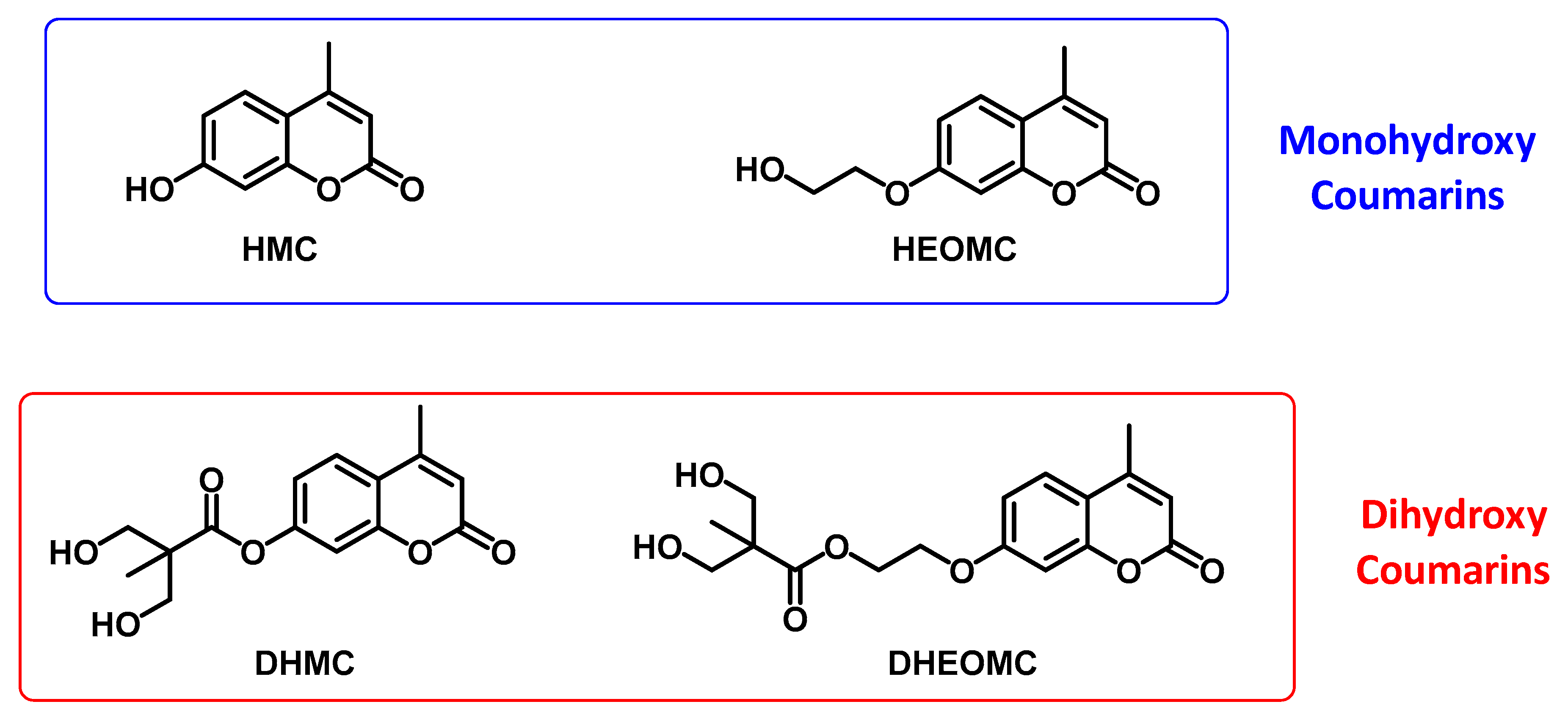
Structural Characterization of Mono and Dihydroxylated Umbelliferone Derivatives

 , ,
, ,  , and
, and
Abstract
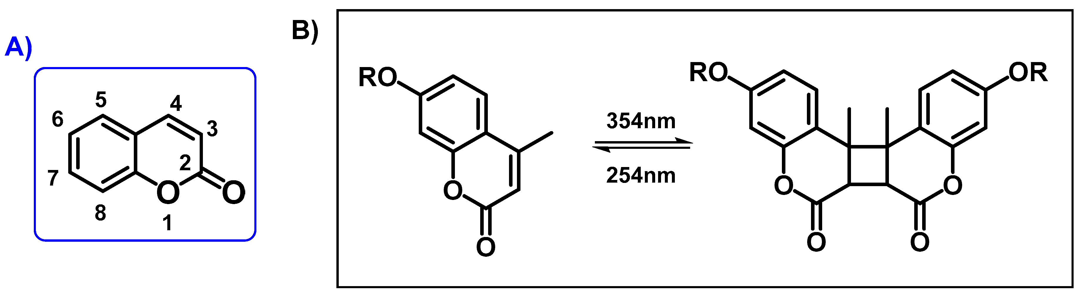
:1. Introduction
2. Results and Discussion
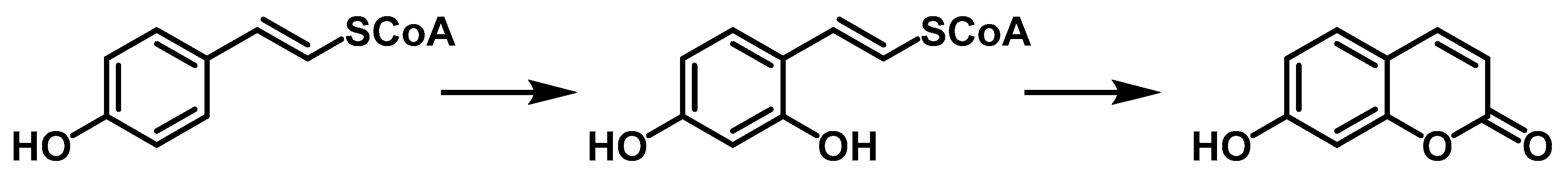
2.1. Synthetic Approach
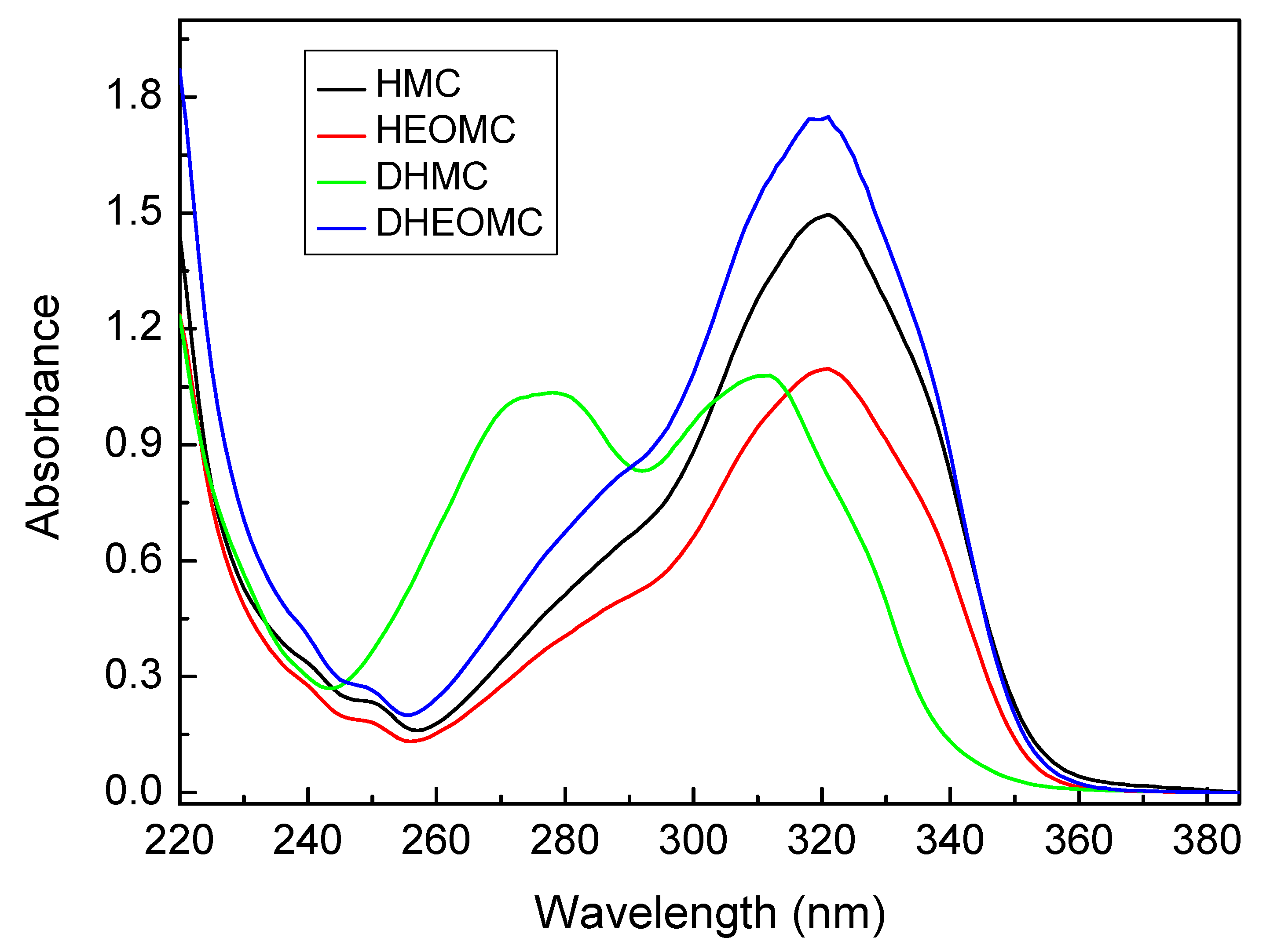
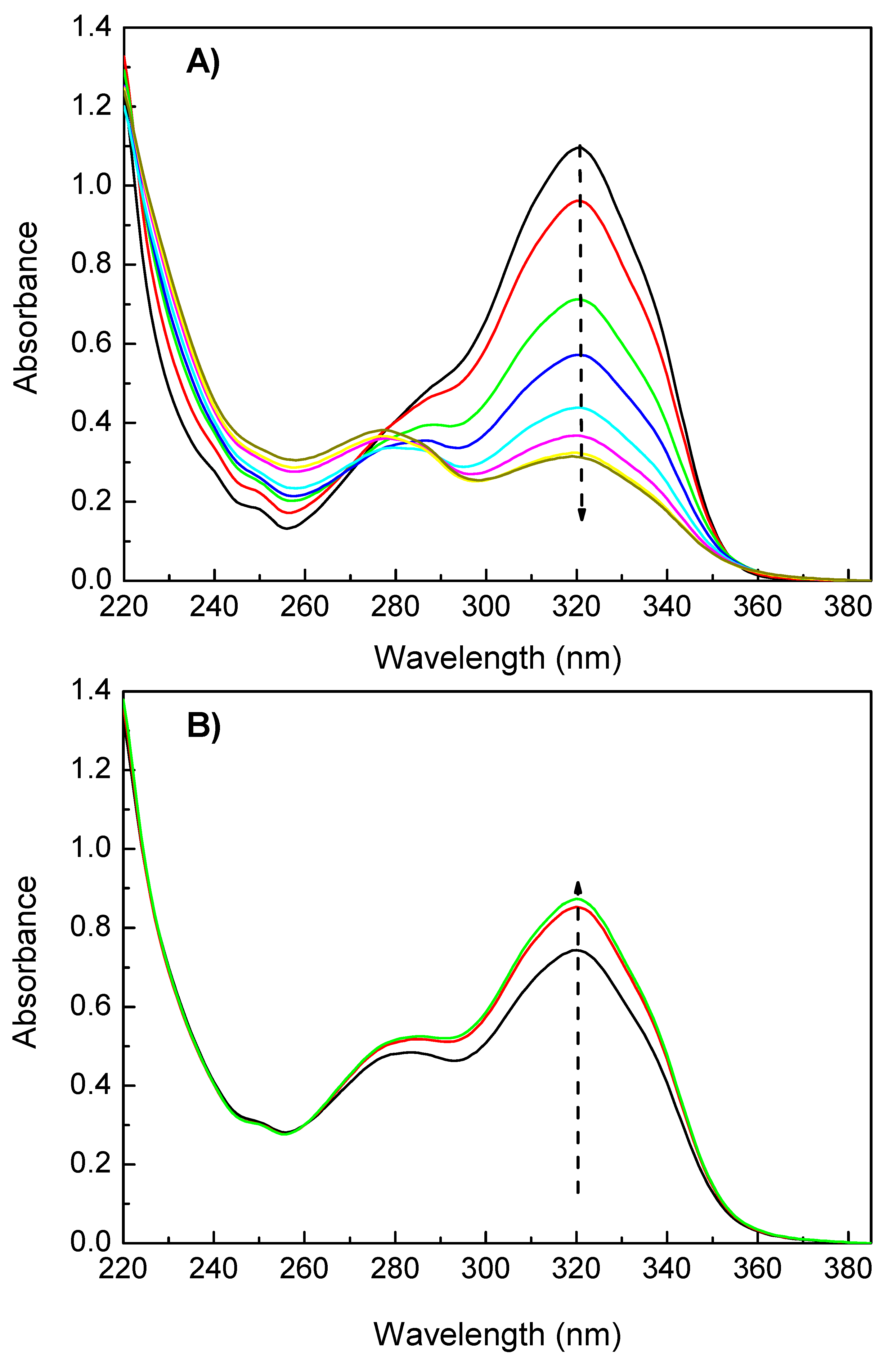
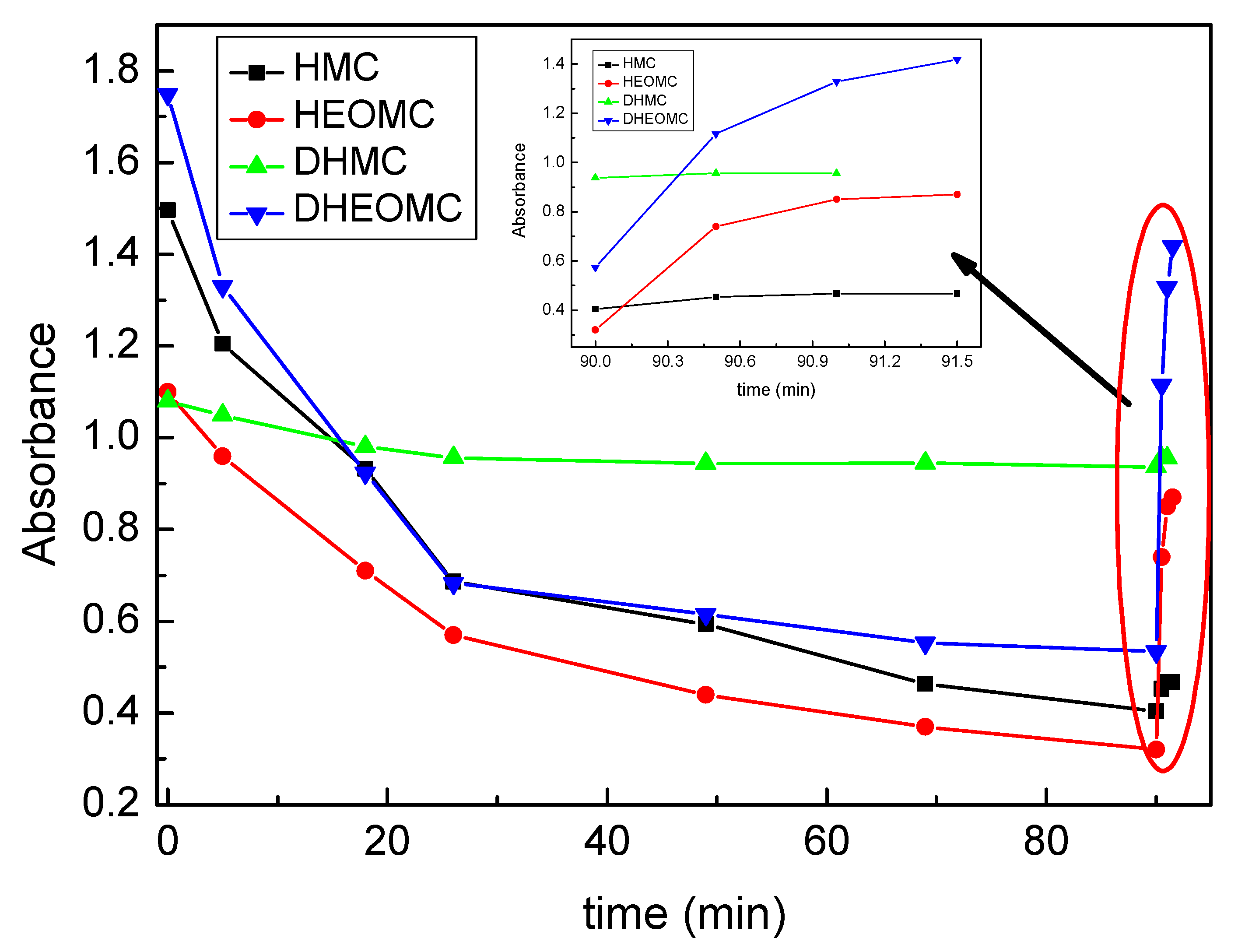
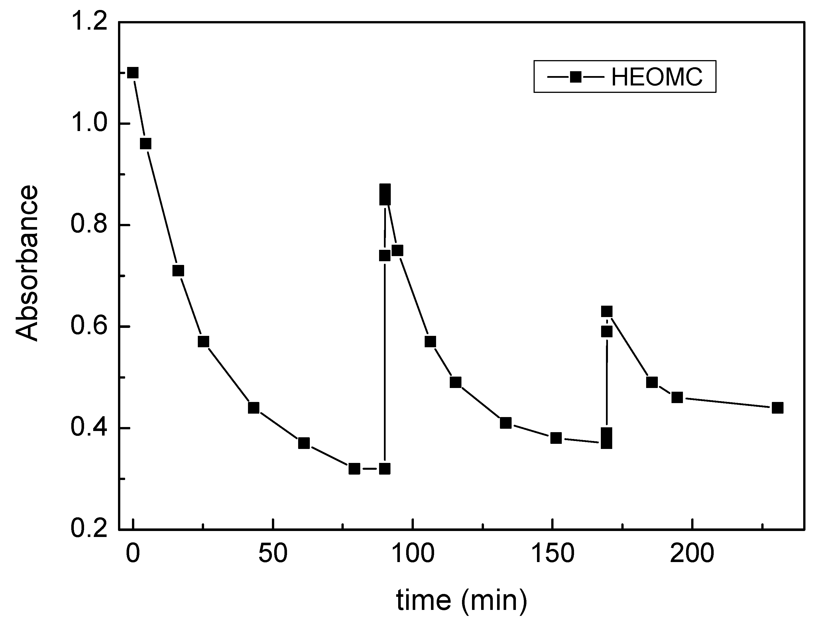
2.2. UV Experiments
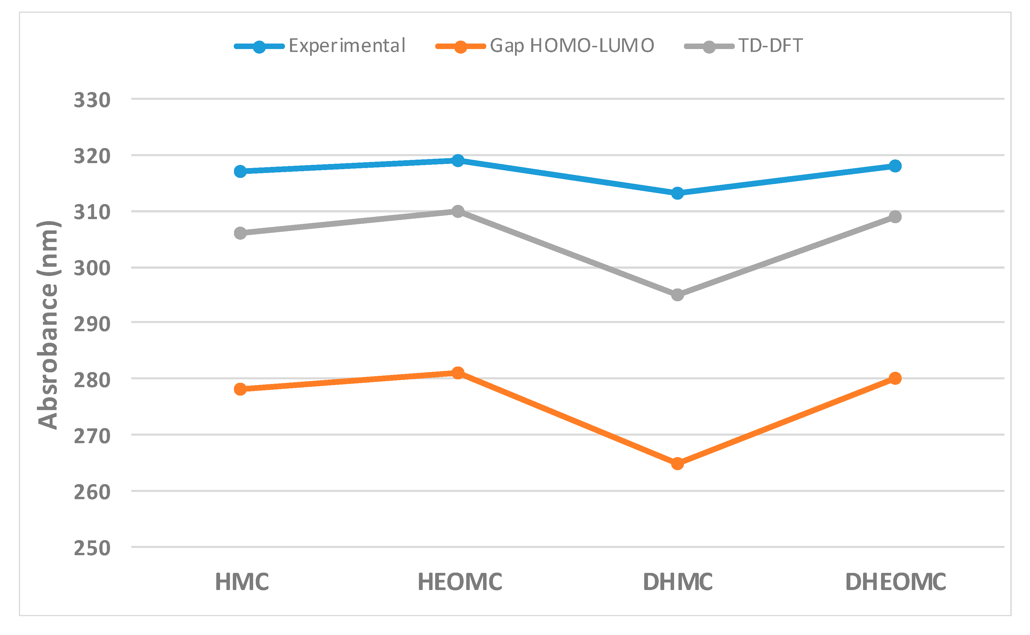
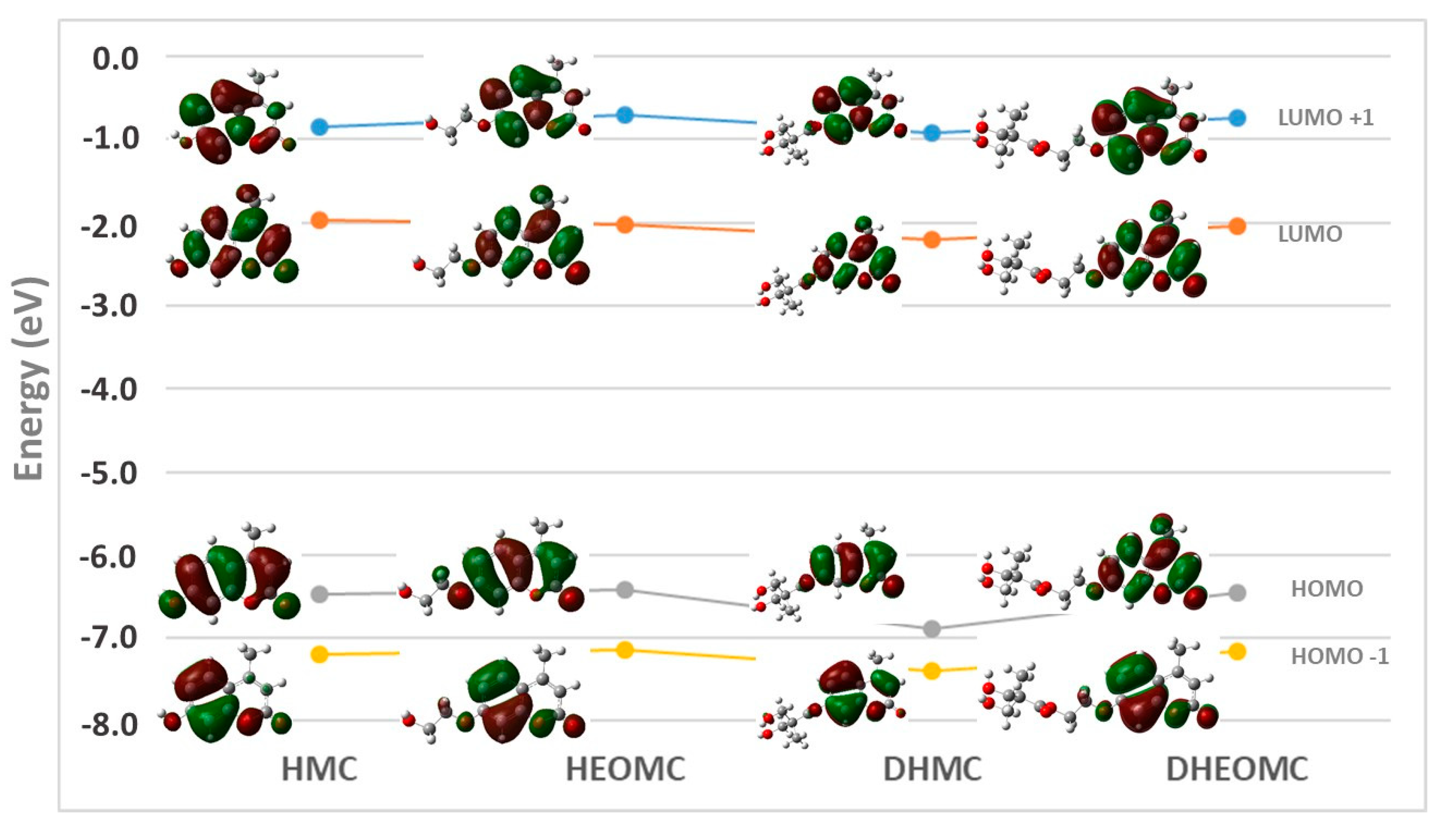
2.3. Theory: Absorption UV-Vis Spectra
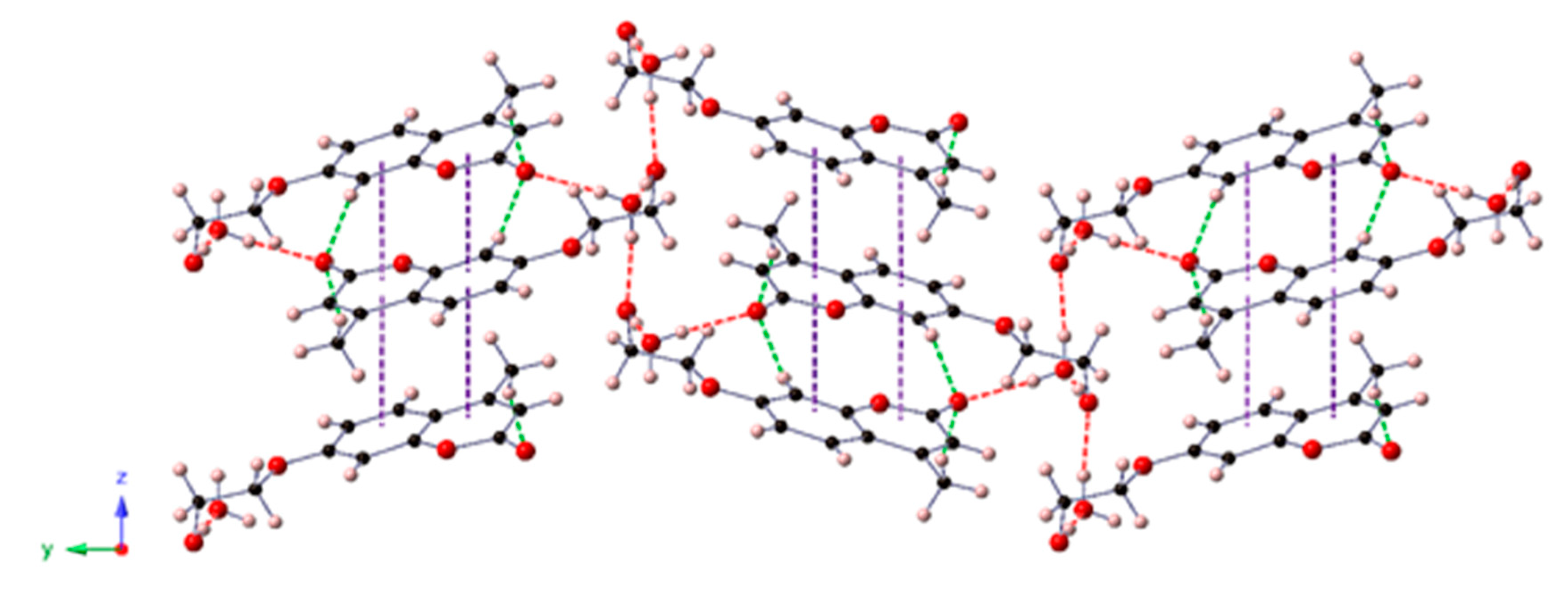
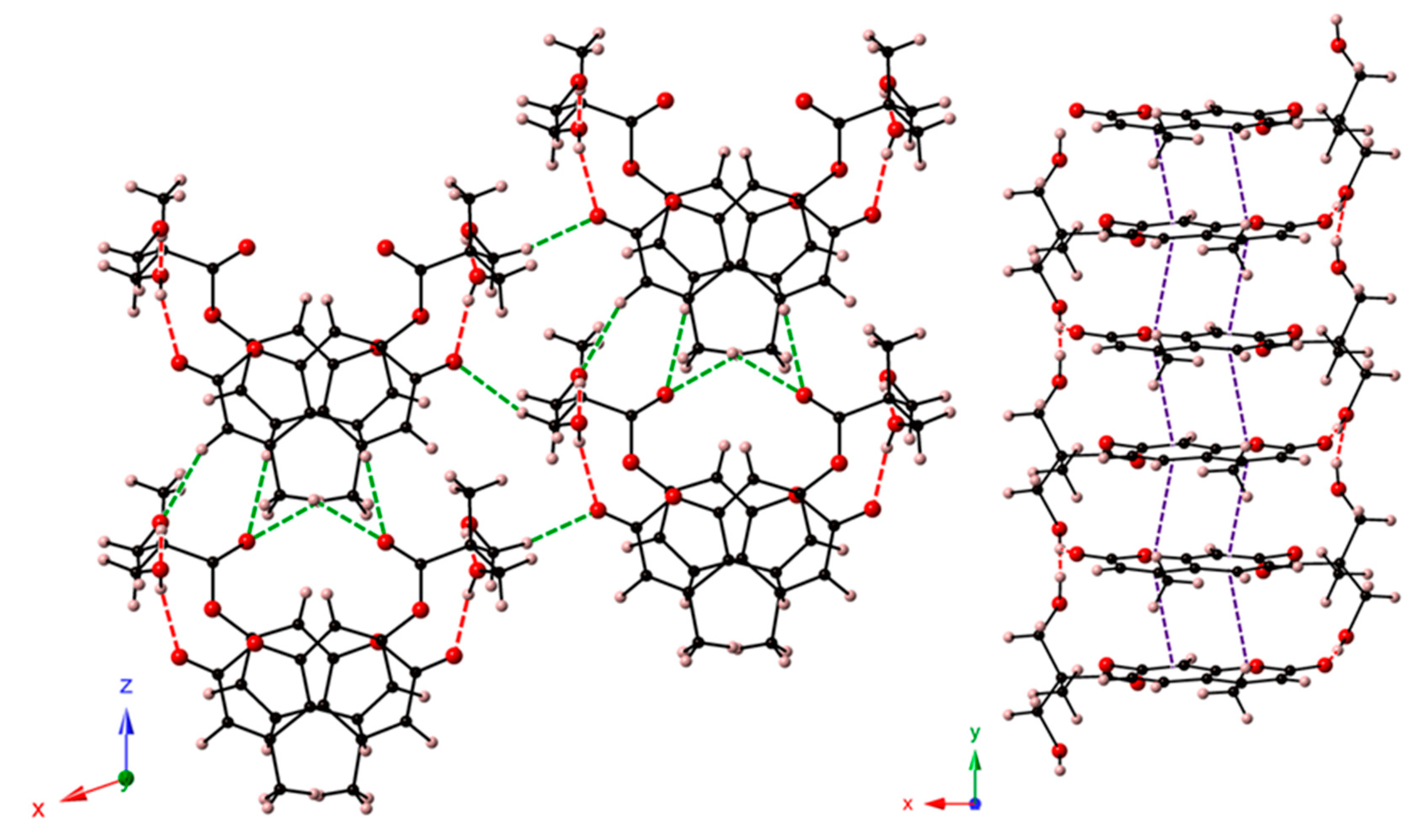
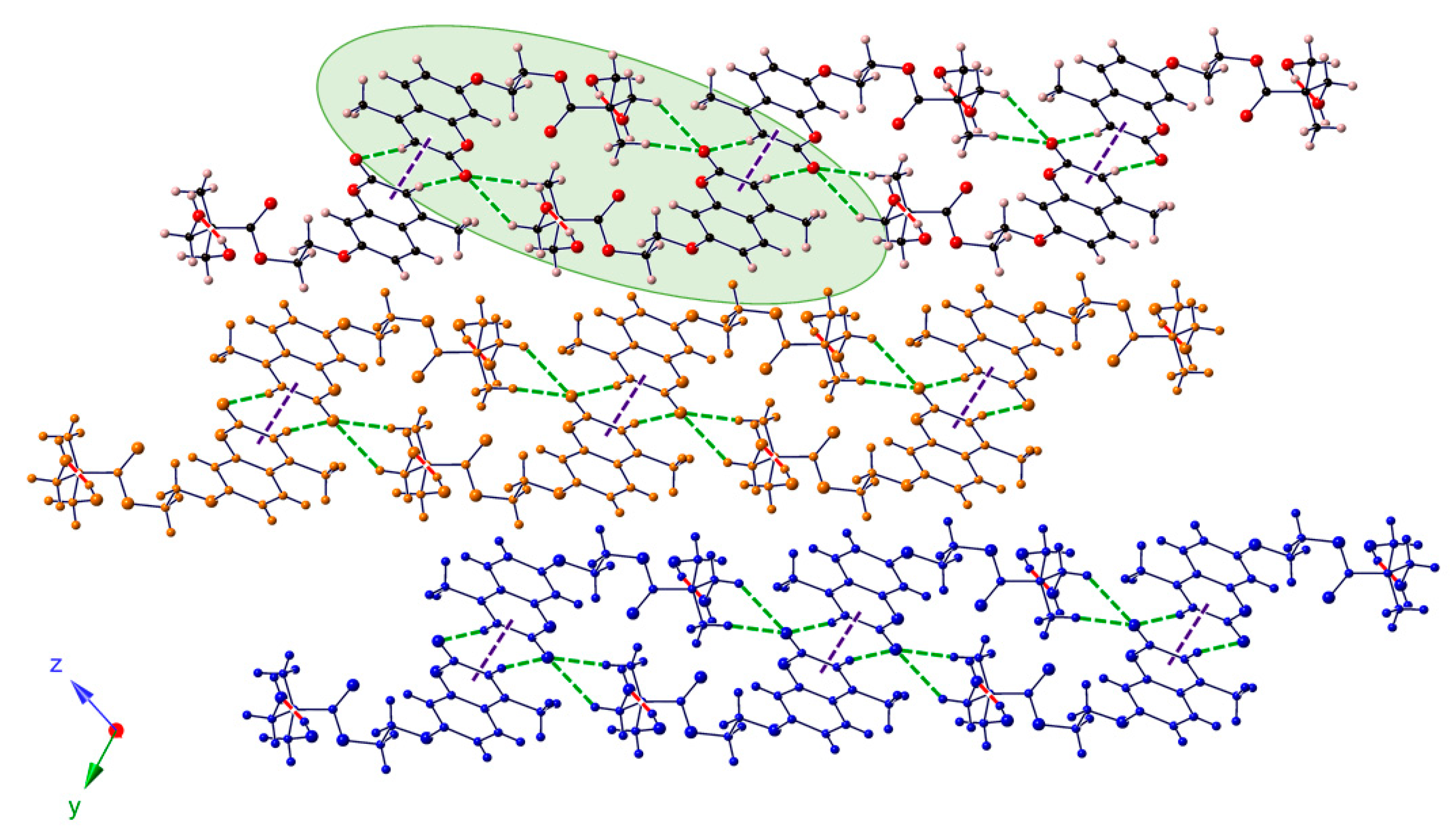
2.4. Crystal Structures
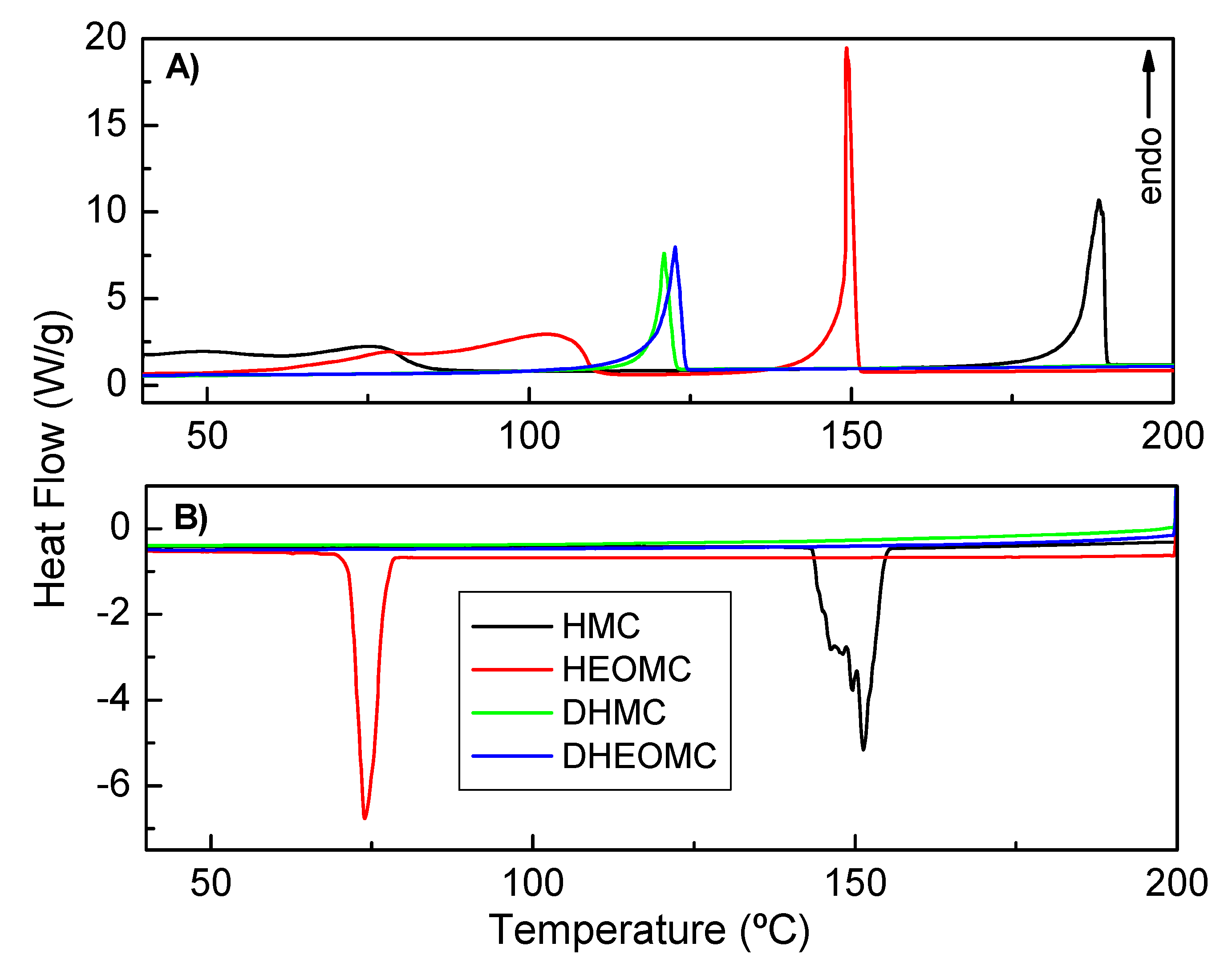
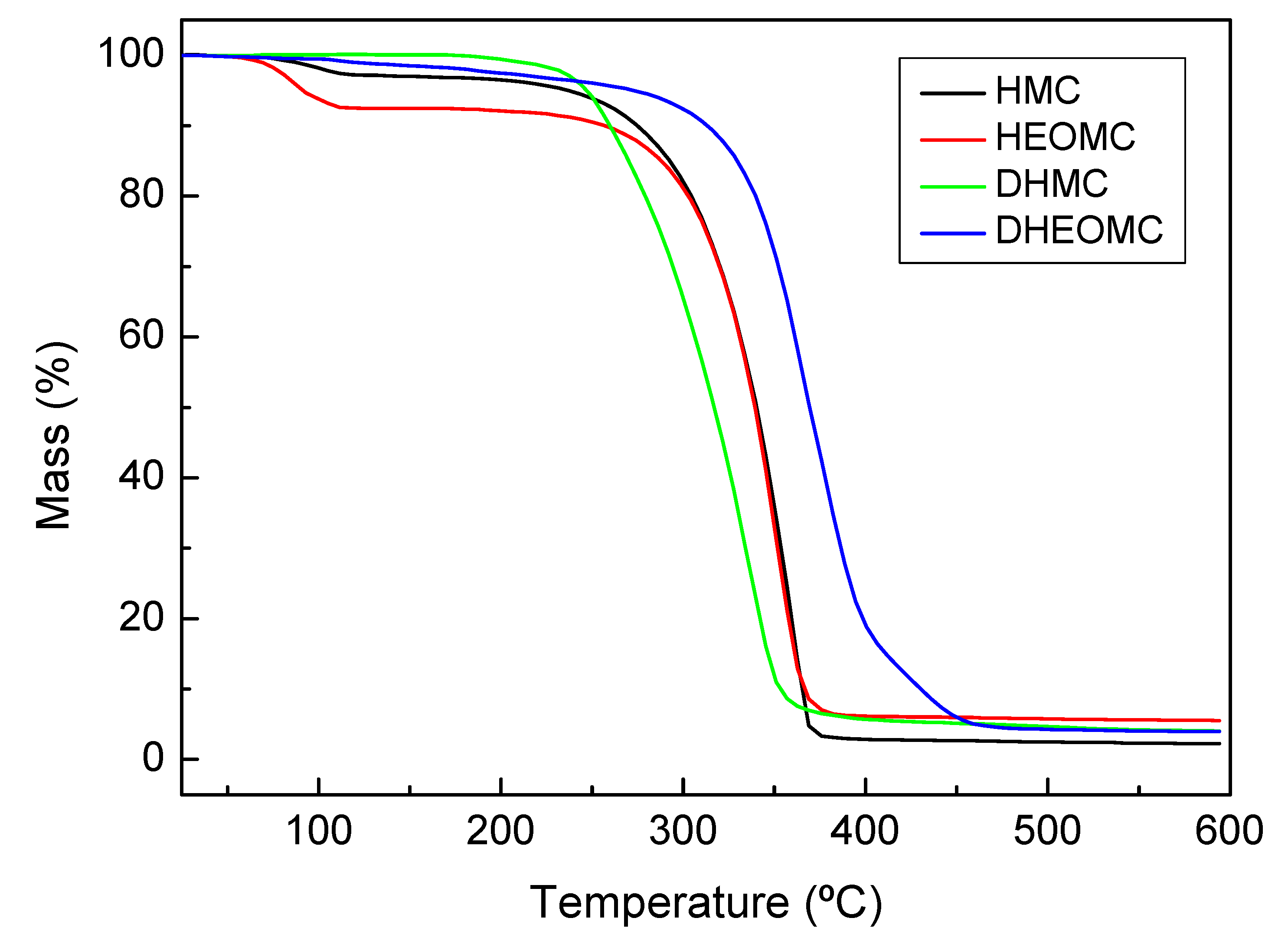
2.5. Thermal Analysis
3. Materials and Methods
3.1. Characterization
3.2. Single-Crystal X-Ray Diffraction
3.3. Computational Methods
4. Conclusions
Supplementary Materials
Author Contributions
Funding
Acknowledgments
Conflicts of Interest
References
- Zhang, D.; Li, D.; Li, X.; Jin, W. Post-assembly polymerization of discrete organoplatinum (II) metallacycles via dimerization of coumarin pendants. Dye. Pigment. 2018, 152, 43–48. [Google Scholar] [CrossRef]
- Seoane Rivero, R.; Bilbao Solaguren, P.; Gondra Zubieta, K.; Peponi, L.; Marcos-Fernández, A. Synthesis, kinetics of photo-dimerization/photo-cleavage and physical properties of coumarin-containing branched polyurethanes based on polycaprolactones. Express Polym. Lett. 2016, 10, 84. [Google Scholar] [CrossRef]
- Motoyanagi, J.; Fukushima, T.; Ishii, N.; Aida, T. Photochemical stitching of a tubularly assembled hexabenzocoronene amphiphile by dimerization of coumarin pendants. J. Am. Chem. Soc. 2006, 128, 4220–4221. [Google Scholar] [CrossRef] [PubMed]
- Mohamed, M.G.; Hsu, K.-C.; Kuo, S.-W. Bifunctional polybenzoxazine nanocomposites containing photo-crosslinkable coumarin units and pyrene units capable of dispersing single-walled carbon nanotubes. Polym. Chem. 2015, 6, 2423–2433. [Google Scholar] [CrossRef]
- Murray, R.D.H.; Mendez, J.; Brown, S.A. Chemistry and Biochemistry; John Wiely Sons: New York, NY, USA, 1982. [Google Scholar]
- Murray, R.D.H. Naturally occurring plant coumarins. In Progress in the Chemistry of Organic Natural Products; Springer: Berlin/Heidelberg, Germany, 1997; pp. 1–119. [Google Scholar]
- Trenor, S.R.; Shultz, A.R.; Love, B.J.; Long, T.E. Coumarins in polymers: from light harvesting to photo-cross-linkable tissue scaffolds. Chem. Rev. 2004, 104, 3059–3078. [Google Scholar] [CrossRef]
- Chen, Y.; Jean, C.-S. Polyethers containing coumarin dimer components in the main chain. II. Reversible photocleavage and photopolymerization. J. Appl. Polym. Sci. 1997, 64, 1759–1768. [Google Scholar] [CrossRef]
- Zhao, D.; Ren, B.; Liu, S.; Liu, X.; Tong, Z. A novel photoreversible poly (ferrocenylsilane) with coumarin side group: synthesis, characterization, and electrochemical activities. Chem. Commun. 2006, 779–781. [Google Scholar] [CrossRef]
- Sinkel, C.; Greiner, A.; Agarwal, S. A polymeric drug depot based on 7-(2′-methacryloyloxyethoxy)-4-methylcoumarin copolymers for photoinduced release of 5-fluorouracil designed for the treatment of secondary cataracts. Macromol. Chem. Phys. 2010, 211, 1857–1867. [Google Scholar] [CrossRef]
- Singh, D.; Rahman, M. Umbelliferone loaded nanocarriers for healthcare applications. Curr. Biochem. Eng. 2020, 6, 25–33. [Google Scholar] [CrossRef]
- Mazimba, O. Umbelliferone: Sources, chemistry and bioactivities review. Bull. Fac. Pharm. Cairo Univ. 2017, 55, 223–232. [Google Scholar] [CrossRef]
- Penta, S. Advances in Structure and Activity Relationship of Coumarin Derivatives; Academic Press: Cambridge, MA, USA, 2015; ISBN 978-0-12-803797-3. [Google Scholar]
- Morsy, S.A.; Farahat, A.A.; Nasr, M.N.A.; Tantawy, A.S. Synthesis, molecular modeling and anticancer activity of new coumarin containing compounds. Saudi Pharm. J. 2017, 25, 873–883. [Google Scholar] [CrossRef] [PubMed]
- Hassan, M.Z.; Osman, H.; Ali, M.A.; Ahsan, M.J. Therapeutic potential of coumarins as antiviral agents. Eur. J. Med. Chem. 2016, 123, 236–255. [Google Scholar] [CrossRef] [PubMed]
- Srinivasa, H.T.; Harishkumar, H.N.; Palakshamurthy, B.S. New coumarin carboxylates having trifluoromethyl, diethylamino and morpholino terminal groups: synthesis and mesomorphic characterisations. J. Mol. Struct. 2017, 1131, 97–102. [Google Scholar] [CrossRef]
- Dandriyal, J.; Singla, R.; Kumar, M.; Jaitak, V. Recent developments of C-4 substituted coumarin derivatives as anticancer agents. Eur. J. Med. Chem. 2016, 119, 141–168. [Google Scholar] [CrossRef]
- Sarkar, D.; Ghosh, P.; Gharami, S.; Mondal, T.K.; Murmu, N. A novel coumarin based molecular switch for the sequential detection of Al3+ and F-: Application in lung cancer live cell imaging and construction of logic gate. Sens. Actuators B Chem. 2017, 242, 338–346. [Google Scholar] [CrossRef]
- Wang, Z.; Wu, Q.; Li, J.; Qiu, S.; Cao, D.; Xu, Y.; Liu, Z.; Yu, X.; Sun, Y. Two benzoyl coumarin amide fluorescence chemosensors for cyanide anions. Spectrochim. Acta Part A Mol. Biomol. Spectrosc. 2017, 183, 1–6. [Google Scholar] [CrossRef]
- García, S.; Vázquez, J.L.; Rentería, M.; Aguilar-Garduño, I.G.; Delgado, F.; Trejo-Durán, M.; García-Revilla, M.A.; Alvarado-Méndez, E.; Vázquez, M.A. Synthesis and experimental-computational characterization of nonlinear optical properties of triazacyclopentafluorene-coumarin derivatives. Opt. Mater. (Amst.) 2016, 62, 231–239. [Google Scholar] [CrossRef]
- Li, C.; Qin, J.; Wang, B.; Bai, X.; Yang, Z. Fluorescence chemosensor properties of two coumarin-based compounds for environmentally and biologically important Al3+ ion. J. Photochem. Photobiol. A Chem. 2017, 332, 141–149. [Google Scholar] [CrossRef]
- Imit, M.; Imin, P.; Adronov, A. π-Conjugated polymers with pendant coumarins: design, synthesis, characterization, and interactions with carbon nanotubes. Can. J. Chem. 2016, 94, 759–768. [Google Scholar] [CrossRef]
- Gindre, D.; Iliopoulos, K.; Krupka, O.; Evrard, M.; Champigny, E.; Sallé, M. Coumarin-containing polymers for high density non-linear optical data storage. Molecules 2016, 21, 147. [Google Scholar] [CrossRef] [Green Version]
- He, J.; Tremblay, L.; Lacelle, S.; Zhao, Y. Preparation of polymer single chain nanoparticles using intramolecular photodimerization of coumarin. Soft Matter 2011, 7, 2380–2386. [Google Scholar] [CrossRef]
- Kiskan, B.; Yagci, Y. Self-healing of poly (propylene oxide)-polybenzoxazine thermosets by photoinduced coumarine dimerization. J. Polym. Sci. Part A Polym. Chem. 2014, 52, 2911–2918. [Google Scholar] [CrossRef]
- Fiore, G.L.; Rowan, S.J.; Weder, C. Optically healable polymers. Chem. Soc. Rev. 2013, 42, 7278–7288. [Google Scholar] [CrossRef] [PubMed]
- Binder, W.H. Self-Healing Polymers: From Principles to Applications; John Wiley & Sons: Hoboken, NJ, USA, 2013. [Google Scholar]
- Urban, M.W. Stimuli-Responsive Materials: From Molecules to Nature Mimicking Materials Design; Royal Society of Chemistry: London, UK, 2019. [Google Scholar]
- Ling, J.; Rong, M.Z.; Zhang, M.Q. Photo-stimulated self-healing polyurethane containing dihydroxyl coumarin derivatives. Polymer (Guildford) 2012, 53, 2691–2698. [Google Scholar] [CrossRef]
- Ling, J.; Rong, M.Z.; Zhang, M.Q. Coumarin imparts repeated photochemical remendability to polyurethane. J. Mater. Chem. 2011, 21, 18373–18380. [Google Scholar] [CrossRef]
- Aguirresarobe, R.H.; Irusta, L.; Fernández-Berridi, M.J. UV-light responsive waterborne polyurethane based on coumarin: synthesis and kinetics of reversible chain extension. J. Polym. Res. 2014, 21, 505. [Google Scholar] [CrossRef]
- Aguirresarobe, R.H.; Martin, L.; Aramburu, N.; Irusta, L.; Fernandez-Berridi, M.J. Coumarin based light responsive healable waterborne polyurethanes. Prog. Org. Coat. 2016, 99, 314–321. [Google Scholar] [CrossRef]
- Seoane Rivero, R.; Bilbao Solaguren, P.; Gondra Zubieta, K.; Gonzalez-Jimenez, A.; Valentin, J.L.; Marcos-Fernandez, A. Synthesis and characterization of a photo-crosslinkable polyurethane based on a coumarin-containing polycaprolactone diol. Eur. Polym. J. 2016, 76, 245–255. [Google Scholar] [CrossRef]
- Salgado, C.; Arrieta, M.P.; Peponi, L.; Fernández-García, M.; López, D. Influence of poly (epsilon-caprolactone) molecular weight and coumarin amount on photo-responsive polyurethane properties. Macromol. Mater. Eng. 2017, 302, 1600515. [Google Scholar] [CrossRef]
- Seoane Rivero, R.; Navarro, R.; Bilbao Solaguren, P.; Gondra Zubieta, K.; Cuevas, J.M.; Marcos-Fernández, A. Synthesis and characterization of photo-crosslinkable linear segmented polyurethanes based on coumarin. Eur. Polym. J. 2017, 92, 263–274. [Google Scholar] [CrossRef]
- Seth, S.K.; Sarkar, D.; Jana, A.D.; Kar, T. On the possibility of tuning molecular edges to direct supramolecular self-assembly in coumarin derivatives through cooperative weak forces: crystallographic and Hirshfeld surface analyses. Cryst. Growth Des. 2011, 11, 4837–4849. [Google Scholar] [CrossRef]
- Cuevas, J.M.; Seoane-Rivero, R.; Navarro, R.; Marcos-Fernández, Á. Coumarins into polyurethanes for smart and functional materials. Polymers 2020, 12, 630. [Google Scholar] [CrossRef] [Green Version]
- Jasinski, J.P.; Woudenberg, R.C. 7-Hydroxy-4-methylcoumarin monohydrate. Acta Crystallogr. Sect. C Cryst. Struct. Commun. 1994, 50, 1952–1953. [Google Scholar] [CrossRef]
- Panini, P.; Venugopala, K.N.; Odhav, B.; Chopra, D. Quantitative analysis of intermolecular interactions in 7-hydroxy-4-methyl-2H-chromen-2-one and its hydrate. Proc. Natl. Acad. Sci. India Sect. A Phys. Sci. 2014, 84, 281–295. [Google Scholar] [CrossRef]
- Gomes, L.R.; Low, J.N.; Fonseca, A.; Matos, M.J.; Borges, F. Crystal structures of three 6-substituted coumarin-3-carboxamide derivatives. Acta Crystallogr. Sect. E Crystallogr. Commun. 2016, 72, 926–932. [Google Scholar] [CrossRef] [PubMed]
- Ouédraogo, M.; Abou, A.; Djandé, A.; Ouari, O.; Zoueu, T.J. 2-Oxo-2H-chromen-7-yl 4-tert-butylbenzoate. Acta Crystallogr. Sect. E Crystallogr. Commun. 2018, 74, 530–534. [Google Scholar] [CrossRef]
- Abou, A.; Yoda, J.; Djandé, A.; Coussan, S.; Zoueu, T.J. Crystal structure of 2-oxo-2H-chromen-7-yl 4-fluorobenzoate. Acta Crystallogr. Sect. E Crystallogr. Commun. 2018, 74, 761–765. [Google Scholar] [CrossRef] [Green Version]
- Stachak, P.; Hebda, E.; Pielichowski, K. Foaming extrusion of thermoplastic polyurethane modified by POSS nanofillers. Compos. Theory Pract. 2019, 19, 23–29. [Google Scholar]
- Technologies, A. CrysAlisPro Software System; Version 1.171. 37.34; Rigaku Oxford Diffraction: Oxford, UK, 2014. [Google Scholar]
- Dolomanov, O.V.; Bourhis, L.J.; Gildea, R.J.; Howard, J.A.K.; Puschmann, H. OLEX2: a complete structure solution, refinement and analysis program. J. Appl. Crystallogr. 2009, 42, 339–341. [Google Scholar] [CrossRef]
- Sheldrick, G.M. A short history of SHELX. Acta Crystallogr. Sect. A Found. Crystallogr. 2008, 64, 112–122. [Google Scholar] [CrossRef] [Green Version]
- Spek, A.L. Structure validation in chemical crystallography. Acta Crystallogr. Sect. D Biol. Crystallogr. 2009, 65, 148–155. [Google Scholar] [CrossRef] [PubMed]
- Farrugia, L.J. WinGX suite for small-molecule single-crystal crystallography. J. Appl. Crystallogr. 1999, 32, 837–838. [Google Scholar] [CrossRef]
- Frisch, J.; Trucks, G.W.; Schlegel, H.B.; Scuseria, G.E.; Robb, M.A.; Cheeseman, J.R.; Scalmani, G.; Barone, V.; Mennucci, B.; Petersson, A.; et al. Gaussian 09 Program; Revision C. 01; Gaussian, Inc.: Wallingford, CT, USA, 2010. [Google Scholar]


| Coumarin Derivatives | Dimerization (1st Cycle) | Recovery (1st Cycle) | Dimerization (2nd Cycle) | Recovery (2nd Cycle) | Dimerization (3th Cycle) |
|---|---|---|---|---|---|
| HMC | 73 | - | - | - | - |
| HEOMC | 70 | 70 | 54 | 40 | 31 |
| DHMC | 13 | - | - | - | - |
| DHEOMC | 66 | 72 | 40 | 63 | 55 |
| HMC | HEOMC | DHMC | DHEOMC | |
|---|---|---|---|---|
| Formula | C10H10O4 | C12H14O5 | C15H16O6 | C17H20O7 |
| MW (g mol−1) | 194.18 | 238.23 | 292.31 | 336.33 |
| Crystal system | Monoclinic | Monoclinic | Monoclinic | Triclinic |
| Space group (N°) | P21/n (14) | P21/n (14) | Pc (7) | P-1 (2) |
| λ (Å) | 0.71073 | 0.71073 | 1.54184 | 1.54184 |
| a (Å) | 6.9524(2) | 7.1794(9) | 12.6290(4) | 6.5260(5) |
| b (Å) | 11.3058(4) | 21.811(3) | 6.9580(2) | 10.7751(8) |
| c (Å) | 11.7715(5) | 7.2972(12) | 16.7226(6) | 12.1148(7) |
| α (deg.) | 90 | 90 | 90 | 107.949(6) |
| β (deg.) | 105.674(4) | 105.052(15) | 109.292(4) | 93.774(5) |
| γ (deg.) | 90 | 90 | 90 | 102.412(6) |
| V (Å3) | 890.86(6) | 1103.5(3) | 1386.94(8) | 783.46(9) |
| Z | 4 | 4 | 4 | 2 |
| Dcalc (g cm-3) | 1.448 | 1.434 | 1.400 | 1.426 |
| μ (mm-1) | 0.113 | 0.112 | 0.918 | 0.937 |
| Collected reflns | 5797 | 8163 | 23764 | 4893 |
| Unique reflns (Rint) | 1844(0.0345) | 2274(0.084) | 4447(0.051) | 2742(0.025) |
| Observed reflns[I > 2σ(I)] | 1442 | 1363 | 4157 | 2376 |
| Parameters/Restrains | 132/2 | 159/2 | 681/304 | 297/0 |
| R(F)a [I > 2σ(I)] | 0.045 | 0.059 | 0.072 | 0.039 |
| wR(F2)b (all data) | 0.098 | 0.113 | 0.183 | 0.045 |
| GoF | 1.073 | 1.052 | 1.109 | 1.085 |
| Flack parameter | – | – | –0.06(12) | – |
| Hooft parameter | – | – | –0.01(10) | – |
| Coumarin | T0 (°C) | Weight Loss (wt %) | T0 (°C) | Weight Loss (wt %) |
|---|---|---|---|---|
| HMC | 63.8 | −2.7 | 279.4 | −86.0 |
| HEOMC | 65.8 | −7.6 | 272.7 | −85.9 |
| DHMC | - | - | 246.0 | −95.5 |
| DHEOMC | - | - | 323.2 | −95.2 |
© 2020 by the authors. Licensee MDPI, Basel, Switzerland. This article is an open access article distributed under the terms and conditions of the Creative Commons Attribution (CC BY) license (http://creativecommons.org/licenses/by/4.0/).
Share and Cite
Seoane-Rivero, R.; Ruiz-Bilbao, E.; Navarro, R.; Laza, J.M.; Cuevas, J.M.; Artetxe, B.; Gutiérrez-Zorrilla, J.M.; Vilas-Vilela, J.L.; Marcos-Fernandez, Á. Structural Characterization of Mono and Dihydroxylated Umbelliferone Derivatives. Molecules 2020, 25, 3497. https://0-doi-org.brum.beds.ac.uk/10.3390/molecules25153497
Seoane-Rivero R, Ruiz-Bilbao E, Navarro R, Laza JM, Cuevas JM, Artetxe B, Gutiérrez-Zorrilla JM, Vilas-Vilela JL, Marcos-Fernandez Á. Structural Characterization of Mono and Dihydroxylated Umbelliferone Derivatives. Molecules. 2020; 25(15):3497. https://0-doi-org.brum.beds.ac.uk/10.3390/molecules25153497
Chicago/Turabian StyleSeoane-Rivero, Rubén, Estibaliz Ruiz-Bilbao, Rodrigo Navarro, José Manuel Laza, José María Cuevas, Beñat Artetxe, Juan M. Gutiérrez-Zorrilla, José Luis Vilas-Vilela, and Ángel Marcos-Fernandez. 2020. "Structural Characterization of Mono and Dihydroxylated Umbelliferone Derivatives" Molecules 25, no. 15: 3497. https://0-doi-org.brum.beds.ac.uk/10.3390/molecules25153497