Article

Coumarin-Chalcone Hybrids as Inhibitors of MAO-B: Biological Activity and In Silico Studies

by
Guillermo Moya-Alvarado
1,†,
Osvaldo Yañez
2,3,†,
Nicole Morales
4,
Angélica González-González
5,
Carlos Areche
6,
Marco Tulio Núñez
7,
Angélica Fierro
8,* and
Olimpo García-Beltrán
9,10,*
1
Biology Department, Johns Hopkins University, Baltimore, MD 21218, USA
2
Center of New Drugs for Hypertension (CENDHY), Santiago 8330015, Chile
3
Computational and Theoretical Chemistry Group, Departamento de Ciencias Químicas, Facultad de Ciencias Exactas, Universidad Andres Bello, República 498, Santiago 7550196, Chile
4
Department of Physiology, Faculty of Biological Sciences, Pontificia Universidad Católica de Chile, Santiago 8331150, Chile
5
Laboratorio de Interacciones Insecto-Planta, Instituto de Ciencias Biológicas, Universidad de Talca, Casilla 747, Talca 3460000, Chile
6
Department of Chemistry, Faculty of Sciences, Universidad de Chile, Las Palmeras 3425, Nuñoa, Santiago 7800024, Chile
7
Biology Department, Faculty of Sciences, Universidad de Chile, Santiago 7800024, Chile
8
Department of Organic Chemistry, Faculty of Chemistry, Pontificia Universidad Católica de Chile, Casilla 306, Santiago 6094411, Chile
9
Centro Integrativo de Biología y Química Aplicada (CIBQA), Universidad Bernardo O’Higgins, General Gana 1702, Santiago 8370854, Chile
10
Facultad de Ciencias Naturales y Matemáticas, Universidad de Ibagué, Carrera 22 Calle 67, Ibagué 730002, Colombia
*
Authors to whom correspondence should be addressed.
These authors contributed equally to this work.
Submission received: 1 April 2021 / Revised: 17 April 2021 / Accepted: 18 April 2021 / Published: 22 April 2021
(This article belongs to the Special Issue Coumarin and Its Derivatives)

Abstract

:
Fourteen coumarin-derived compounds modified at the C3 carbon of coumarin with an α,β-unsaturated ketone were synthesized. These compounds may be designated as chalcocoumarins (3-cinnamoyl-2H-chromen-2-ones). Both chalcones and coumarins are recognized scaffolds in medicinal chemistry, showing diverse biological and pharmacological properties among which neuroprotective activities and multiple enzyme inhibition, including mitochondrial enzyme systems, stand out. The evaluation of monoamine oxidase B (MAO-B) inhibitors has aroused considerable interest as therapeutic agents for neurodegenerative diseases such as Parkinson’s. Of the fourteen chalcocumarins evaluated here against MAO-B, ChC4 showed the strongest activity in vitro, with IC50 = 0.76 ± 0.08 µM. Computational docking, molecular dynamics and MM/GBSA studies, confirm that ChC4 binds very stably to the active rMAO-B site, explaining the experimental inhibition data.

1. Introduction

Coumarins (α-benzopyrones, 2H-chromen-2-ones) are a large family of compounds, of natural and synthetic origin, that show numerous biological [1,2,3,4,5,6] and medicinal chemistry activities, such as anticoagulant, anticancer, antioxidant, antiviral, anti-diabetic, anti-inflammatory, antibacterial, antifungal and anti-neurodegerative properties [7,8,9], among which recent studies have paid special attention to enzyme inhibition. With regard to monoamine oxidase (MAO) inhibition, recent findings have revealed that MAO affinity and selectivity can be efficiently modulated by appropriate substitutions on the coumarin ring system [1,10,11,12,13].
MAOs (EC 1.4.3.4) are flavoproteins located in the outer mitochondrial membrane and involved in the oxidative deamination of endogenous and exogenous monoamines using oxygen (O2) as electron acceptor. In humans they exist in two isoforms called MAO-A and MAO-B. The high resolution crystal structures of both human isoforms A and B (hMAO) rat MAO-A (rMAO-A) have made it possible to analyze binding modes of ligands inside these macromolecules [14]. While the active site is formed by the common FAD cofactor and similar amino acid residues in the different forms, these are distinguished by their selectivity for substrates and inhibitors [15]. Thus, serotonin and noradrenaline are substrates of MAO-A which is selectively inhibited by clorgyline, while MAO-B oxidizes β-phenylethylamines and benzylamines and is selectively inhibited by l-deprenyl. MAO genes are expressed in various tissues. However, in the brain, although both isoforms are widely distributed, MAO-B is expressed in high concentrations in the hypothalamus, striatum, globus pallidus and thalamus, and mainly in serotonergic cells while the A isoform is rather evenly distributed, mainly in the cortex, and in nuclei containing preferably catecholaminergic and glial cells [16,17,18,19,20,21].
Although knowledge about MAO inhibition by compounds containing coumarin scaffolds is scarce, publications of articles describing new inhibitors of this class of compounds are increasing. The variety of substitutions on the coumarin ring provide insight into the influence on the activity-structure relationship. Among the most reported modifications of the coumarin ring with MAO activity are on C3 and the steric effect of the substituent appears to be important in modulating MAO-B inhibitory activity [11]. In addition, it has been reported that the introduction of various substituents at the para position of the 3-phenyl ring is a good strategy for improving the desired MAO-B inhibitory activity [22] and when the 3-phenyl skeleton is replaced by a 3-benzoyl group, the activity is strongly diminished [20]. It has also been observed that coumarins substituted with 3-indolyl and 3-thiophenyl shows greatest selective inhibition was against MAO-B [11,23,24].
In this work a merger of the coumarin scaffold and a 3-cinnamyl group led to new hybrid (chalcocoumarin) derivatives (Scheme 1) that preserve structural characteristics of compounds with the ability to interact with MAO. The synthetic strategy chosen allowed a large variety of substituents on the cinnamyl benzene ring to be accessed using different readily accessible benzaldehydes. Thus, the quantity and/or type of interactions with the enzyme were explored involving some bulky groups to determine their possible contribution to the biological activity as MAOIs. Our new compounds were screened versus both MAO isoforms, and in silico studies were carried out to rationalize their main interactions in the MAO active cavity. The computational biochemistry tools were used considering the geometrical restrictions and most probable positions in the formation of the ligand-receptor complex. The chalcocoumarin molecules were subjected to theoretical studies in which binding energies were estimated using docking and MM/GBSA analysis. In addition, physicochemical parameters that are responsible for governing the pharmacokinetic properties of drug molecules were determined.
In the present study, a series of coumarin-chalcone hybrid compounds were synthesized and tested on the 2 MAO isoforms. The activity shown was selective for MAO-B and in particular, compound ChC4 showed the highest inhibitory activity on rMAO-B at submicromolar concentrations. The results obtained will be useful to understand the mode of inhibition of chalcocoumarins against rMAO-B, and to help predict the activities of these new inhibitors that could be promising as therapeutics to treat neurodegenerative diseases such as Parkinson’s disease.

2. Results and Discussion
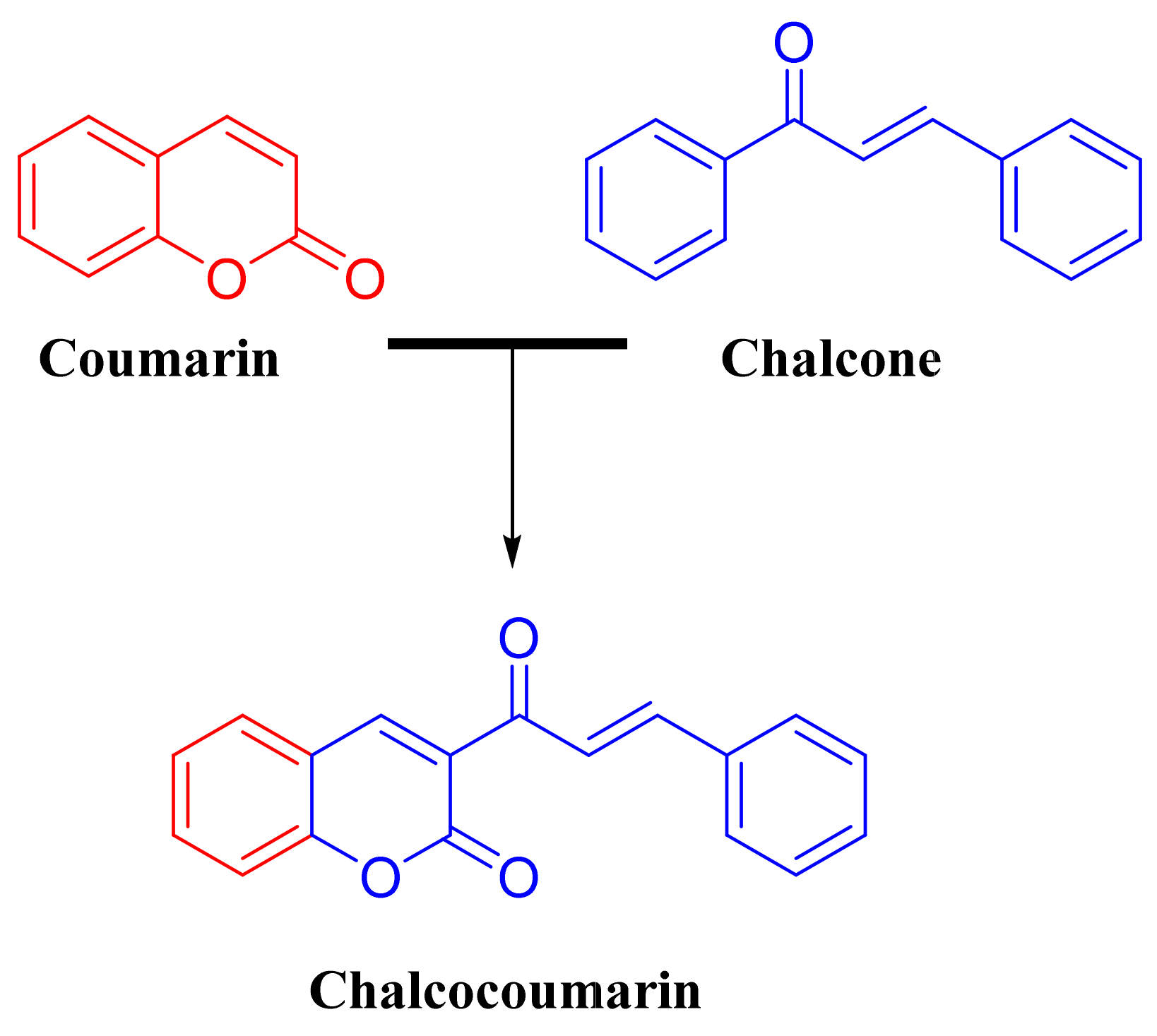
2.1. Chemistry

The route employed to synthesize the compounds is summarized in Scheme 1. The compounds were obtained starting from resorcinol (1), which was formylated using the Vilsmaier-Haack reaction [25]. Knoevenagel condensation of the aldehyde intermediate with ethyl acetoacetate afforded hydroxycoumarin 2. The 7-hydroxycoumarin obtained was methylated using a Williamson reaction using methyl sulfate as methylating agent, obtaining the compound 3, finally the compounds derived the 3-cinnamoyl-2H-chromen-2-one (ChC1ChC14) (Table 1) were prepared in moderate yields (25–47%, unoptimized) by Claisen-Schmidt condensation with the respective aldehyde (Supplementary Materials; Scheme S1) [26]. Coumarin-chalcone hybrids have been studied and are currently still being synthesized for various uses and their spectroscopy is well known, however, we will detail some signals that are key to their identification. The 1H-NMR spectra of the compounds ChC1ChC14 present very similar chemical shift patterns with a particular signal that identifies this type of molecules, the neighboring vinyl protons of the α,β-unsaturated ketone appear at very close low field from the aromatic proton region. These protons present signals corresponding to two doublets with variable δ between 8.5 and 7.0 and with Jab = 16 Hz on average. this high constant corresponds to a trans isomer [27,28,29]. As for the 13C-NMR spectrum, we will mention typical signals such as carbonyl shifts. first of all, we will detail that the carbon of the α,β-unsaturated ketone has a δ 190–180 ppm and carbonyl carbons of α-pyrone δ 165–155 pmm on average [27,28,29], the compounds were characterized by 1H and 13C NMR (Supplementary Materials; Figures S1–S15).

2.2. Biological Analysis in Rat MAO

Fourteen derivatives differing in the substitution pattern of the cinnamyl benzene ring were studied, these compounds were tested on rat MAO-A and B to determine their inhibitory activity MAO. A general screening was carried out at 10 µM finding moderate activity for some of the compounds against rMAO-B but none against rMAO-A. Thus, five molecules were identified as possibly selective IMAO-B.
ChC4, ChC5, ChC6, ChC9 and ChC11 in MAO-B exhibited micromolar or submicromolar in vitro potencies, all below 10 µM (Table 2). Out of these ChC4, substituted with a hydroxyl group on the meta position of the variable ring, displayed the highest rMAO-B inhibitory activity (IC50 = 0.76 µM). Interestingly, changing the position of the hydroxyl group from meta to ortho or para (ChC2 and ChC10 respectively) led to loss of the inhibitory activity. An approximately 12-fold lower IMAO activity was observed when the hydroxyl group (in ChC4) was methylated (ChC5). This might be attributed to steric hindrance and/or to the loss of hydrogen bonding donor quality which could be crucial for some interaction in the binding site. Moving the methoxyl group from the meta to the para position (ChC6 vs. ChC5) slightly increased potency.
Replacing the methoxy group of ChC6 with a bulkier, less electronegative and more polarisable methylthio group (ChC11) only produced ChC6 is less potent than ChC11. The second most potent molecule was ChC9, with a methylenedioxy group bridging the meta and para carbons. The methylenedioxy group increases the rigidity of the molecule, possibly stabilizing the complex protein-ligand interaction. The same effect, extending the rigidity, has been observed, on other derivatives as IMAO [30,31].

2.3. Molecular Docking and Ligand Efficiency Analysis

To analyze the changes in potency of the coumarin-chalcone hybrids, docking studies were carried out using the crystal structure of rMAO-A and the homology model of rMAO-B (Supplementary Materials; Figures S16 and S17), analyzing the possibility that each one of them has to form a stable complex with each of the 14 molecules synthesized by us. Table 3 shows that most of these molecules present better interactions with the rMAO-B binding site, since the corresponding energies are at least 2.5 kcal mol more negative in all but one of the cases. This difference could be due to the substitutes present in the molecules. The results of the molecular docking experiments showed more favorable interactions (more negative ∆Ebinding) for the complexes in rMAO-B than in rMAO-A, with average values around −9.26 kcal·mol−1 vs. −6.57 kcal·mol−1 respectively) which are in accord with the experimental data for the whole series. In the rMAO-B, although no major differences were observed in the binding modes of the active compounds, subtle energy differences were identified. The results of this molecular docking study point to strong interactions of the chalcocoumarins in the binding pocket of rMAO-B, but considerably weaker in rMAO-A.
When analyzing the docking results for rMAO-B from the conformational viewpoint, it is necessary to consider the residues that constitute the substrate-binding site of rMAO-B, which is composed of the FAD cofactor, two flanking residues, Tyr398 and Tyr435, that form an “aromatic box”, and a number of others, particularly Cys172, Tyr326, Met341, Ser200 Gln206 and Thr314 [32,33]. The results show that all the chalcocoumarins settle in the active site of rMAO-B (Supplementary Materials; Figure S18), with the benzene ring of the coumarin moiety close to the FAD, more specifically the central N-5, at a distance of about 4.0 Å. The benzene ring of the cinnamyl moiety extends into the generally hydrophobic entrance cavity adjoining the substrate-binding site. The mere length of the ChC molecules indicates that to bind in the active site of MAO-A the latter must undergo a rearrangement of the residues separating the entrance and the substrate cavities, which may explain their general preference for MAO-B.
ChC4 was located inside the cavity interacting with Tyr435, Tyr398, Tyr60, Phe343, Asn83, Arg307, Thr314, and Leu328. Two hydrogen bonds where generated with Asn83 and, via its C-3′ hydroxyl group, Thr316. ChC2 actually when interact with the amino acids into the pocket adopt a planar conformation because the hydrogen bond confirms our discussion that could be responsible for none activity of ChC2 in rMAO-B. A quantum geometric optimization of ChC2 and ChC4 at the M05-2X-D3/6-31G(d,p) level of theory, showed their C-2′ and C-3′ hydroxyl groups pointing in opposite directions, suggesting different preferred intramolecular interactions (Figure 1). Both the different electronic potential distribution and the resulting preferred intermolecular interaction might be responsible for the difference in in IC50 values.
The best three ligands obtained from the docking exhibit low Kd values, these ligands are ChC4, ChC9 and ChC13, which means that these ligand/rMAO-B complexes are the most stable in the series. These results are consistent with those obtained in the docking experiments in which these complexes were the most stable according to their ∆Ebinding values. The proposed tolerable values of LE for inhibitor candidates are LE > 0.3 kcal·mol−1 [34,35,36]. According to this reference value, ChC4 is a good prospect for development as an rMAO-B inhibitor with a LE value of 0.408. Although ChC2 and ChC4 have similar Kd, LE and ∆Ebinding values, the ChC2 molecule does not show in vitro activity against rMAO-B, on the other hand, ChC4 has a good inhibitory activity against rMAO-B, since micromolar concentrations are needed to inhibit it, which is consistent with the values obtained for the Kd. ChC1 would appear to be almost as good, with LE = 0.404, but again its activity, if any, is worse than our cutoff value. The low micromolar-active ChC5, ChC6, ChC9 and ChC11 have LE values of 0.384, 0.372, 0.380 and 0.356, respectively, and the less (or in-) active ChC13 and ChC14 have LE values of 0.380 and 0.391.

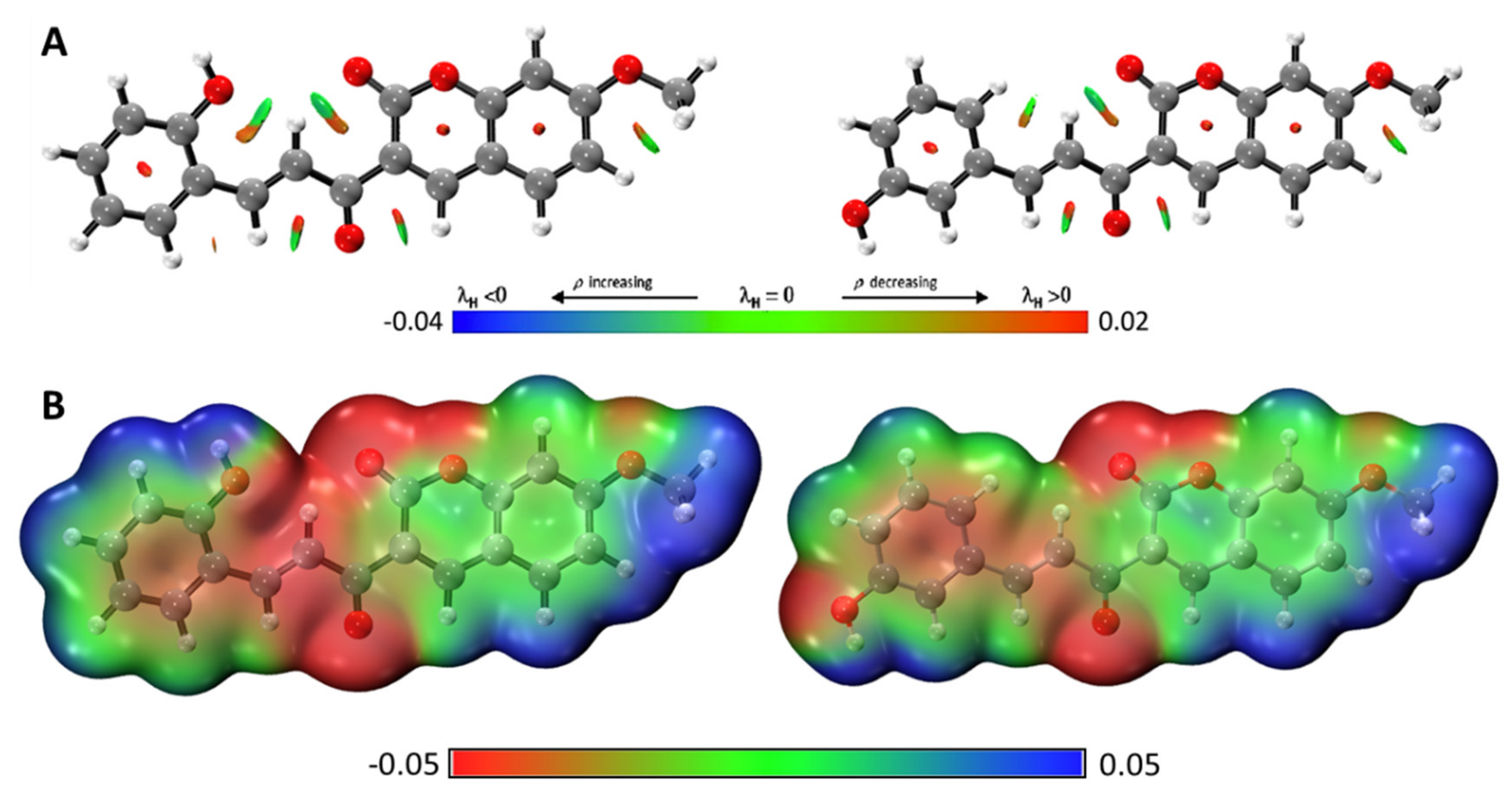
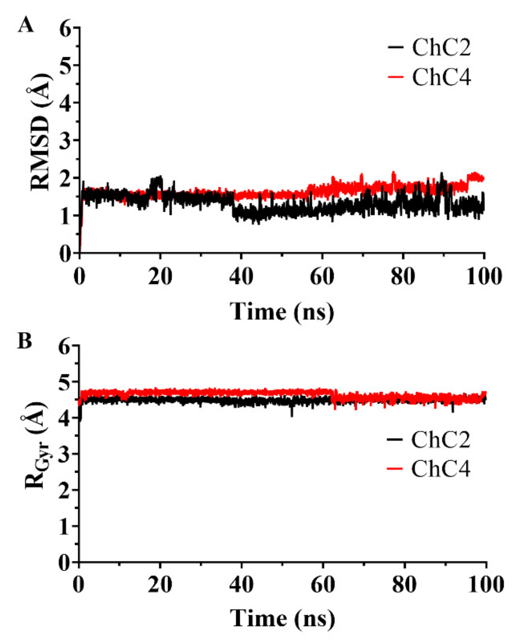
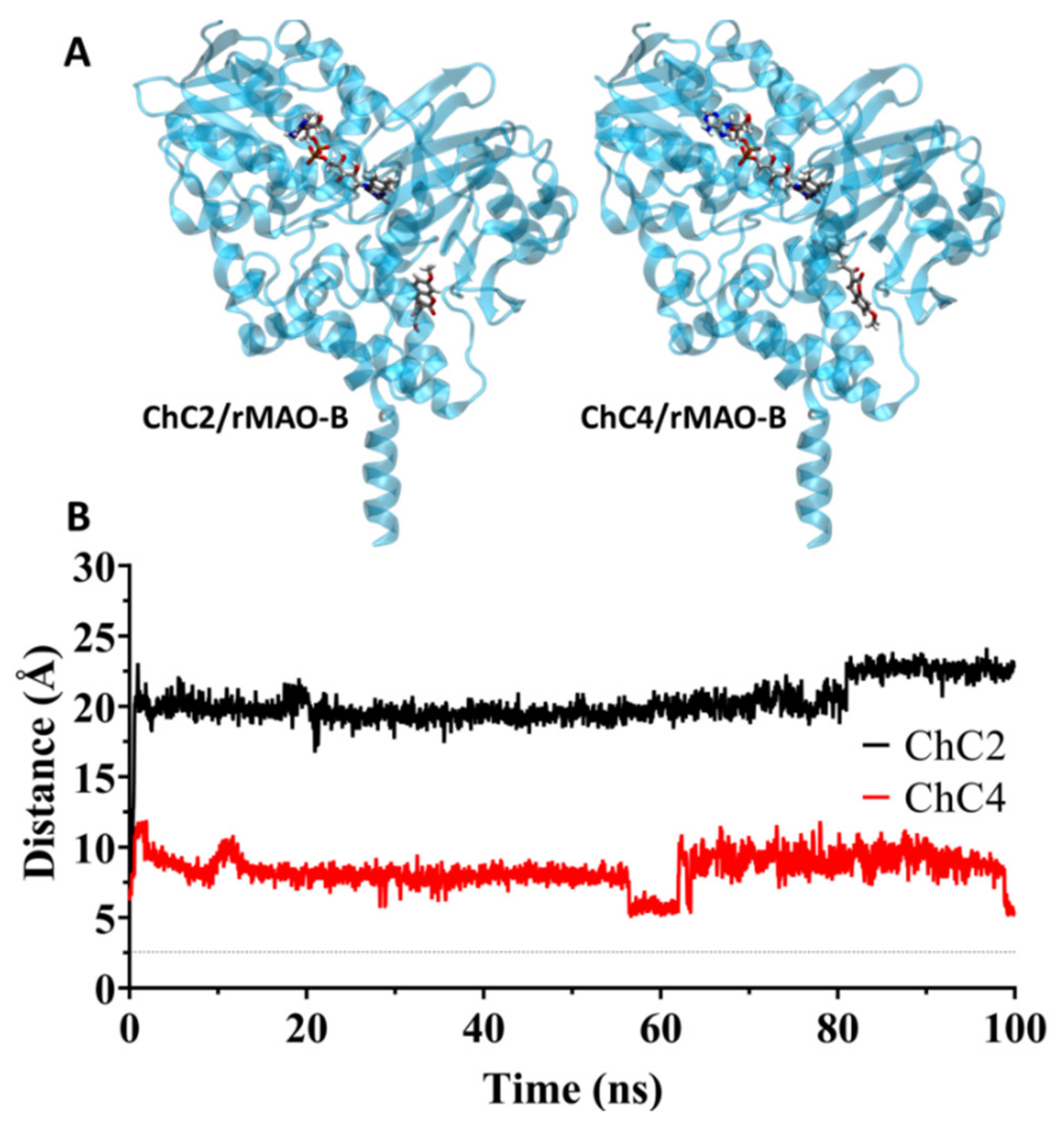
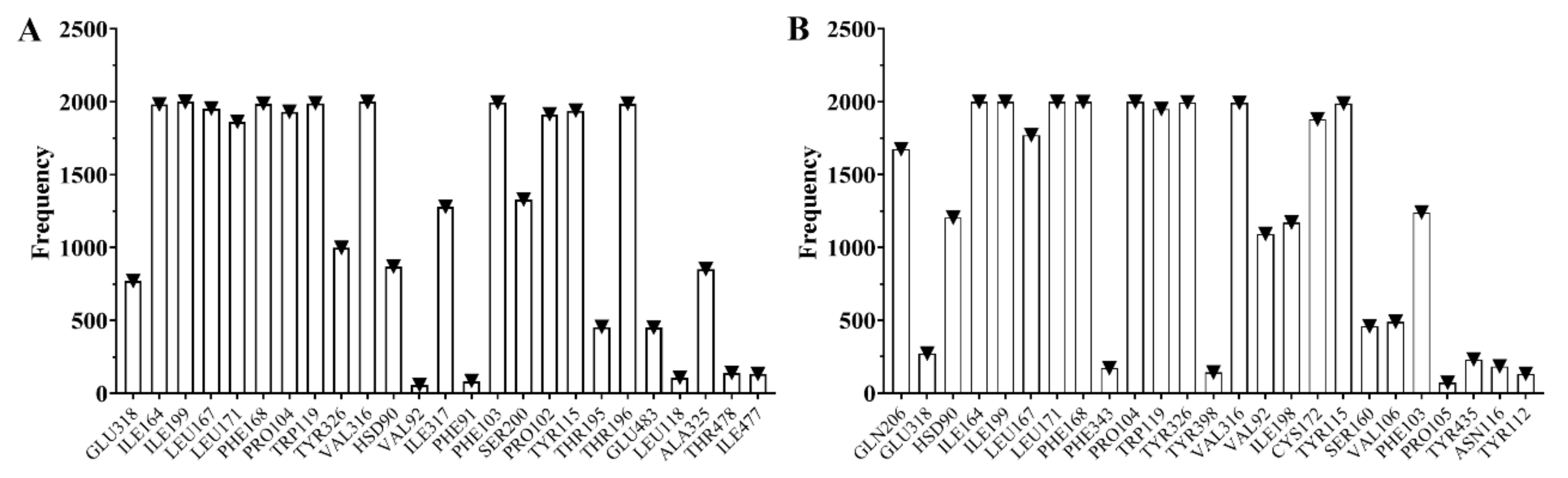
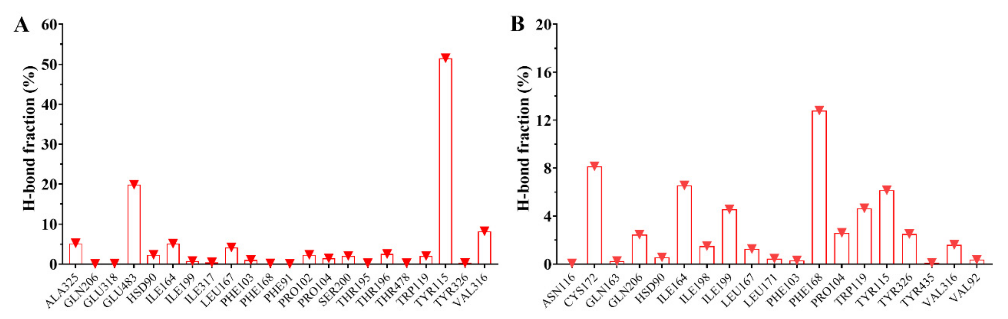
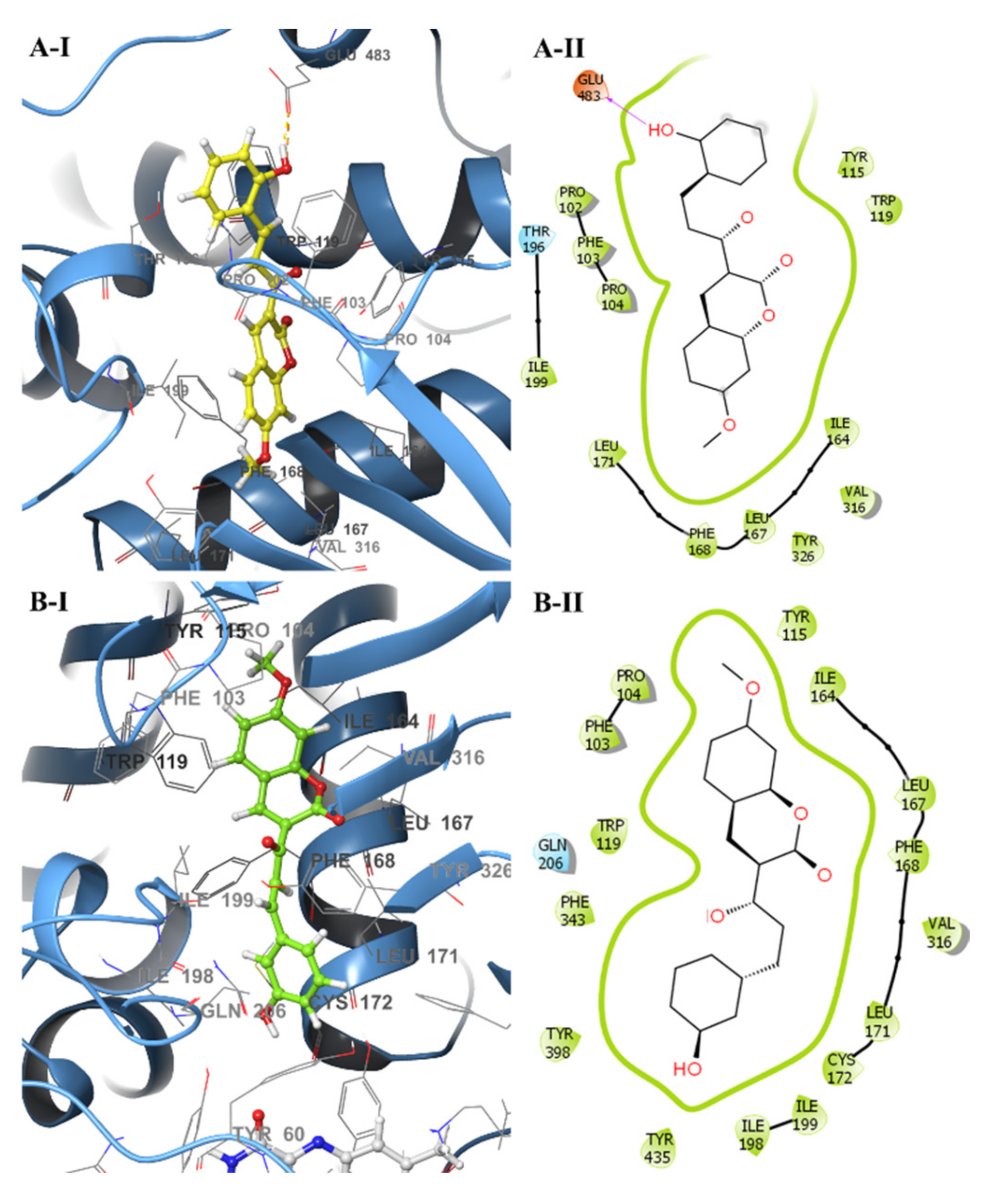
2.4. Analysis of Molecular Dynamics Simulations

Molecular dynamics simulations were performed for 100 ns to analyze the conformational stability of the rMAO-B/ChC2 and rMAO-B/ChC4 complexes. The RMSD, a quantitative parameter, was used to estimate the stability of the protein-ligand systems and the apoprotein. The RMSD in Figure 2A shows that the rMAO-B/ChC2 and rMAO-B/ChC4 complexes remain highly stable throughout the simulation time. We can see that the structures of the complexes does not change significantly. The RMSD values for the ChC4 complex are remarkably constant about 1.5 Å, with a very slight instability and increase near the end of the simulation. The ChC2 complex shows similar, somewhat less stable behavior for almost 40 ns, and then its RMSD value falls abruptly to about 1.0 Å and rises slowly with appreciable fluctuations to about 1.2 Å at 100 ns, indicative of weaker binding in the rMAO-B site. However, a maximum difference of 3.0 Å in the RMSD is taken to indicate that a system is in equilibrium [37], so this condition is fulfilled by both compounds. To complement the analysis carried out calculating the RMSD, the Radius of Gyration (RGyr) was analyzed for the same runs. From this analysis (Figure 2B), we can conclude that the RGyr of ChC2 and ChC4 oscillate in a narrow interval between 4.3–4.8 Å. These stable values during the 100 ns simulation indicate again that ligand binding does not induce major conformational changes in the protein structure.
Structural studies in MAO have shown that two residues Tyr398 and Tyr435 in MAO-B located in the active site approximately perpendicular to the FAD play a functional role in this enzyme, acting as a cofactor stabilizing the active site, forming an aromatic box whose function is to stabilize the ligand [13]. Molecular simulation results show a difference in the interaction of the compounds ChC2 and ChC4 with the FAD cofactor, see Figure 3A. Compound ChC2 shows a spacing that fluctuates between 17.0 Å and 20.0 Å from its original position, signifying a null interaction with the FAD cofactor. On the other hand, the compound ChC4 is within the range of interaction with the FAD cofactor. This distance was measured between the nitrogen atom of the alloxazine planar ring of FAD and the center of the benzaldehyde aromatic ring of compounds, Figure 3B.
Molecular dynamics simulations showed of rMAO-B that residues that interact with the ligands ChC2 and ChC4, see Figure 4. The most frequent residues in rMAO-B/ChC2 were Ile164, Ile199, Leu167, Leu171, Phe168, Pro104, Trp119, Val316, Phe103, Pro102, Tyr115 and Thr196. In contrast, the most frequent residues in rMAO-B/ChC4 were Ile164, Ile199, Leu171, Phe168, Pro104, Trp119, Tyr326, Val316, Cys172 and Tyr115 with van der Waals and hydrogen bonds interactions. Highlighting residues Cys172 and Tyr326, which are important for the active site of the rMAO-B flavoprotein. Tyr326 and Cys172 are key residues that determines substrate and inhibitor specificity, also exhibits conformational changes on the inhibitor binding and restricts the binding of certain inhibitors (e.g., harmine) to human MAO-B [38]. These results documents that ChC4 is a reversible inhibitor of rMAO-B.
The analyses of trajectories indicate that during most of the simulation the ligand ChC4 maintain hydrogen bonds with residues of the active site of rMAO-B. However, the number of hydrogen bonds formed was different for ChC2 and ChC4 (Figure 5). ChC2 formed two hydrogen bonds between the residues Glu483 and Tyr115, highlighting the participation of the residues Val316, Ala325, Ile164 and Leu167. Finally, ChC4 formed two hydrogen bonds with the Phe168, Cys172, Ile164 and Tyr115, highlighting the participation of the residues Ile199, Trp119 and Tyr326. These residues, see Figure 6, are consistent with previous theoretical-experimental studies carried out [39,40] where they detail the interaction that some of the synthesized compounds have with the active site of the rMAO-B. This difference in the formation of hydrogen bonds with key residues in rMAO-B could be explained the difference in experimental activity between the ChC2 and ChC4 compounds.
Finally, the binding free energy (MM/GBSA) was computed after the MD simulation; the last 70 ns for all the complexes and the results are given in Table 4. The compound ChC2 has a binding free energy of -29.06 kcal·mol−1 with rMAO-B enzyme, while the compound ChC4 showed relatively binding free energy of –25.87 kcal·mol−1. The results obtained from MM/GBSA show a slight difference in their binding free energy between ChC2 and ChC4 compounds bound to rMAO-B. This slight difference is due to the R1 to R2 position of the hydroxyl group in benzaldehyde aromatic ring. In particular, the ChC4 compound has a better activity at the experimental and in silico level.

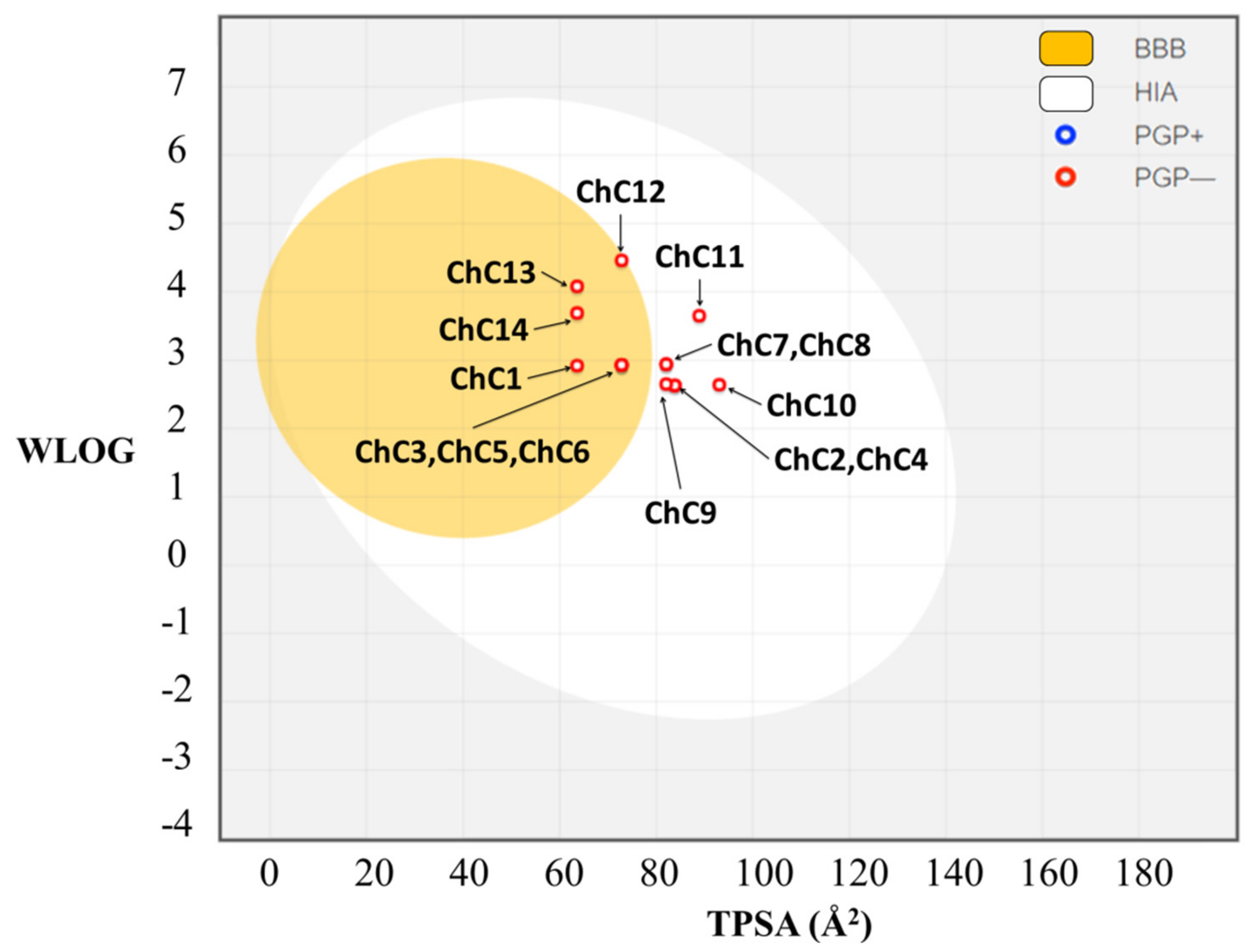
2.5. In Silico Pharmacokinetic Prediction

A good drug candidate is absorbed in required time and well distributed throughout the system for its effective metabolism and action. Toxicity is another very important factor that often overshadows the ADME behaviour. SwissADME explorer online was used for in silico prediction of drug likeness of the synthesized compounds (ChC1ChC14) based on various molecular descriptors and the results are presented in Table 5.
The most potent compound ChC4 in biological experiment data having logP value of 2.97, it’s clear that it doesn’t violate of five Lipinski rules, while the other molecules have logP values in the range of 2.90–4.48 and are expected to be orally active. In addition, the logS values for ChC4 have a value of −4.94 indicating proper solubility, which is an indication of favorable drug like property, makes compound ChC4 promising drug candidate for further research and development. Thirteen of fourteen synthesized molecules do not break the rules of Lipinski, Ghose, Veber, Egan and Muegge, since the molecule ChC12 breaks the rules of Ghose and Muegge.
The Boiled-egg model is proposed as an accurate predictive model that works by computing the lipophilicity and polarity of small molecules. The Boiled-egg analysis of the fourteen molecules (Figure 7) has shown that compounds ChC1, ChC3, ChC5, ChC6, ChC12, ChC13 and ChC14 are highly absorbable at the brain barrier, whereas compounds ChC2, ChC4, ChC7, ChC8, ChC9, ChC10 and ChC11 are highly absorbable in the gastrointestinal tract.
The ADMET properties showed much similarity among the thirteen molecules that can be used for advanced clinical trials.

3. Materials and Methods

3.1. Solvents and Reagents

Solvents and reagents (analytical grade and spectroscopic grade) were obtained from Sigma-Aldrich (St. Louis, MO, USA) and Merck (Darmstadt, Germany). Melting points were determined on a Galen III hot-plate microscope (Reichert-Jung, St. Louis, MO, USA) equipped with a thermocouple. 1H- and 13C-NMR spectra were recorded on a 400 MHz multidimensional spectrometer (Bruker Corporation, Billerica, MA, USA) using the solvent or the TMS signal as an internal standard.

3.2. Synthesis

3-Cinnamoyl-7-methoxy-2H-chromen-2-one (ChC1). 3-Acetyl-7-methoxy-2H-chromen-2-one (0.44 g, 2.0 mmol) and benzaldehyde (0.21 g, 2.0 mmol) were dissolved in 25 mL of DCM and to this solution 0.5 mL of piperidine were added. The mixture was kept at reflux temperature, monitoring the reaction by TLC for 10 h. The solution was concentrated under reduced pressure and dissolved in a small aliquot of DCM and then MeOH was added in excess to induce precipitation. This procedure was performed twice. The precipitate was finally purified by column chromatography on silica gel eluting with DCM: 0.25 g yellow solid, 40.8%, m.p. 190–192 ºC; 1H NMR (CDCl3): δ 8.59 (s, 1H, Ar-H), 8.01 (d, 1H, J = 15.8 Hz, Ar-CH), 7.85 (d, 1H, J = 15.8 Hz, CO-CH=), 7.67 (s, 2H, Ar-H), 7.56 (d, 1H, J = 8.6 Hz, Ar-H), 7.40 (s, 3H, Ar-H,), 6.90 (dd, 1H, J = 8.6, 1.0 Hz, Ar-H), 6.85 (s, 1H, Ar-H), 3.91 (s, 3H, OCH3). 13C-NMR (CDCl3): δ 56.1, 100.2, 112.4, 113.8, 124.0, 129.0, 130.6, 131.5, 135.3, 144.3, 148.5, 157.8, 160.0, 165.0, 186.3.
(E)-3-(3-(2-Hydroxyphenyl)acryloyl)-7-methoxy-2H-chromen-2-one (ChC2). 3-Acetyl-7-methoxy-2H-chromen-2-one (0.44 g, 2.0 mmol) and o-hydroxybenzaldehyde (0.24 g, 2.0 mmol) were reacted and worked up according to the previous procedure: 1.75 g, pale white solid, 95.6%, m.p.; 188–190 °C. 1H-NMR (DMSO-d6), δ 10.43 (s, 1H, OH), 8.71 (s, 1H, Ar-H), 8.04 (d, 1H, J = 15.9 Hz, Ar-CH=), 7.92 (d, 1H, J = 8.8 Hz, Ar-H), 7.89 (d, 1H, J = 15.9 Hz), 7.69 (dd, 1H, J = 7.7, 1.0 Hz, Ar-H), 7.35 (t, 1H, J = 7.0 Hz, Ar-H), 7.13 (d, 1H, J = 2.2 Hz, Ar-H), 7.08 (dd, 1H, J = 8.6, 2.2 Hz, Ar-H), 7.00 (d, 1H, J = 8.0 Hz, Ar-H), 6.94 (t, 1H, J = 7.5 Hz, Ar-H), 3.96 (s, 3H, OCH3), 13C-NMR (DMSO-d6): δ 56.7, 100.8, 112.5, 113.9, 116.8, 120.0, 121.8, 121.9, 124.3, 129.2, 132.3, 132.6, 139.5, 148.2, 157.4, 157.8, 159.4, 165.1, 187.1.
(E)-7-methoxy-3-(3-(2-methoxyphenyl)acryloyl)-2H-chromen-2-one (ChC3). 3-Acetyl-7-methoxy-2H-chromen-2-one (0.44 g, 2.0 mmol) and o-methoxybenzaldehide (0.27 g, 2.0 mmol), were reacted and worked up according to the previous procedure: 0.330 g, pale white solid, 49%, m.p.. 184–186 °C; 1H-NMR (CDCl3): δ 8.55 (s, 1H, =C-H), 8.21 (d, 1H, Ar-CH=, J = 15.8 Hz), 8.04 (d, 1H, CO-CH=, J = 15.8 Hz), 7.71 (d, 1H, Ar-H, J = 7.6 Hz), 7.56 (d, 1H, Ar-H, J = 8.4 Hz), 7.37 (t, 1H, Ar-H, J = 7.9), 6.98 (t, 1H, Ar-H, J = 7.7 Hz), 6.92 (d, 1H, Ar-H, J = 8.2 Hz), 6.90 (d, 1H, Ar-H, J = 8.5 Hz), 6.85 (s, 1H, Ar-H), 3.91 (s, 6H, 2 OCH3); 13C-NMR (DMSO-d6): δ 55.6, 56.0, 100.4, 111.2, 112.5, 113.7, 120.8, 121.9, 124.1, 124.6, 129.3, 131.3, 132.0, 139.9, 148.2, 157.6, 159.0, 159.8, 165.0, 186.8.
(E)-3-(3-(3-hydroxyphenyl)acryloyl)-7-methoxy-2H-chromen-2-one (ChC4). 3-Acetyl-7-methoxy-2H-chromen-2-one (0.44 g, 2.0 mmol) and m-hydroxybenzaldehyde (0.24 g, 2.0 mmol) were reacted and worked up according to the previous procedure: 0.195 g, white solid, 30%, m.p. 184–186 °C; 1H NMR (DMSO-d6): δ 9.77 (sbr, 1H), 8.76 (s, 1H, =C-H), 7.94 (d, 1H, Ar-H, J = 8.0 Hz), 7.82 (d, 1H, Ar-CH=, J = 15.9 Hz), 7.71 (d, 1H, CO-CH=, J = 15.9 Hz), 7.62 (d, 1H, Ar-H, J = 8.6 Hz), 7.33 (m, 1H, Ar-H), 7.25-7.17 (m, 2H, Ar-H), 7.15 (s, 1H, Ar-H), 7.09 (dd, 1H, Ar-H, J = 8.0, 1.0 Hz), 6.94 (dd, 1H, Ar-H, J = 8.0, 1.0 Hz), 3.97 (s, 3H, OCH3); 13C NMR (DMSO-d6): 56.7, 100.9, 112.5, 114.0, 114.8, 118.5, 120.5, 121.6, 124.9, 130.6, 132.5, 136.3, 143.9, 148.6, 157.5, 158.2, 159.4, 165.3, 186.7.
(E)-7-methoxy-3-(3-(3-methoxyphenyl)acryloyl)-2H-chromen-2-one (ChC5). 3-Acetyl-7-methoxy-2H-chromen-2-one (0.44 g, 2.0 mmol) and m-methoxybenzaldehide (0,27 g, 2.0 mmol), were reacted and worked up according to the previous procedure: 0,280 g, pale white solid, 42%, m.p. 164–166 °C; 1H-NMR (CDCl3): δ 8.58 (s, 1H, =C-H), 7.99 (d, 1H, Ar-CH=, J = 15.9 Hz), 7.82 (d, 1H, CO-CH=, J = 15.9 Hz), 7.57 (d, 1H, Ar-H, J = 8.0 Hz), 7.35-7.25 (m, 2H, Ar-H), 7.18 (s, 1H, Ar-H) 6.96 (dd, 1H, Ar-H, J = 8.0, 2.0 Hz), 6.91 (dd, 1H, Ar-H, J = 8.8, 2.0 Hz), 6.85 (d, 1H, Ar-H, J = 2.0 Hz), 3.83 (s, 3H, OCH3), 3.76 (s, 3H, OCH3); 13C-NMR (CDCl3): δ 55.8, 56.5, 100.8, 112.8, 113.9, 114.3, 117.1, 121.7, 122.0, 124.9, 130.3, 131.8, 136.8, 144.8, 149.0, 158.1, 160.2, 160.3, 165.8, 186.8.
(E)-7-methoxy-3-(3-(4-methoxyphenyl)acryloyl)-2H-chromen-2-one (ChC6). 3-Acetyl-7-methoxy-2H-chromen-2-one (0.44 g, 2.0 mmol) and p-methoxybenzaldehide (0.27 g, 2.0 mmol), were reacted and worked up according to the previous procedure: 0.26 g, pale white solid, 39%, m.p. 158–160 °C; 1H-NMR (CDCl3): δ 8.58 (s, 1H, =C-H), 7.91 (d, 1H, Ar-CH=, J = 15.9 Hz), 7.84 (d, 1H, CO-CH=, J = 15.9 Hz), 7.64 (d, 2H, Ar-H, J = 8.0 Hz), 7.56 (d, 1H, Ar-H, J = 8.8 Hz), 6.93 (d, 2H, Ar-H, J = 8.0 Hz), 6.91 (dd, 1H, Ar-H, J = 8.8, 2.0 Hz), 6.85 (d, 1H, Ar-H, J = 2.0 Hz), 3.92 (s, 3H, OCH3), 3.86 (s, 3H, OCH3); 13C-NMR (CDCl3): δ 57.2, 57.8, 102.0, 114.2, 115.5, 116.1, 123.4, 123.6, 129.6, 132.5, 133.0, 146.3, 150.0, 159.4, 161.6, 163.5, 166.8, 188.0.
(E)-3-(3-(3,4-dimethoxyphenyl)acryloyl)-7-methoxy-2H-chromen-2-one (ChC7). 3-Acetyl-7-methoxy-2H-chromen-2-one (0.44 g, 2.0 mmol) and 3,4-dimethoxybenzaldehide (0.33 g, 2.0 mmol) were reacted and worked up according to the previous procedure: 0.42 g, bright yellow solid, 57%, m.p. 182–184 °C; 1H-NMR (CDCl3): δ 8.55 (s, 1H, =C-H), 8.16 (d, 1H, Ar-CH=, J = 15.9 Hz), 8.00 (d, 1H, CO-CH=, J = 15.9 Hz), 7.55 (d, 1H, Ar-H, J = 8.6 Hz), 7.34 (d, 1H, Ar-H, J = 1.0 Hz), 7.19 (dd, 1H, AR-H, J = 8.0, 8.0 Hz) 7.07 (d, 1H, Ar-H, J = 8.0 Hz), 7.00 (dd, 1H, Ar-H, J = 8.0, 2.0 Hz), 6.94(d, 1H, Ar-H, J = 2.0 Hz), 3.90 (s, 3H, OCH3), 3.89 (s, 3H, OCH3), 3.87 (s, 3H, OCH3); 13C-NMR (CDCl3): δ 55.9, 56.0, 61.5, 100.4, 112.4, 113.8, 114.4, 119.9, 121.6, 124.2, 125.5, 129.2, 131.3, 139.1, 148.4, 149.2, 153.2, 157.7, 159.7, 165.1, 186.7.
(E)-3-(3-(2,5-dimethoxyphenyl)acryloyl)-7-methoxy-2H-chromen-2-one (ChC8). 3-Acetyl-7-methoxy-2H-chromen-2-one (0.44 g, 2.0 mmol) and 2,5-dimethoxybenzaldehide (0.33 g, 2.0 mmol), were reacted and worked up according to the previous procedure: 0,45 g, bright yellow solid, 62%, m.p. 174–176 °C; 1H-NMR (CDCl3): δ 8.55 (s, 1H, =C-H), 8.17 (d, 1H, Ar-CH=, J = 15.6 Hz), 8.0 (d, 1H, CO-CH=, J = 15.6 Hz), 7.6 (dd, 1H, Ar-H, J = 8.0, 9.0 Hz), 7.2 (d, 1H, Ar-H, J = 2.0 Hz), 6.92-6.97 (m, 4H, Ar-H), 3.91 (s, 3H, OCH3), 3.87 (s, 3H, OCH3), 3.82 (s, 3H, OCH3); 13C-NMR (CDCl3): δ 56.2, 56.4, 56.6, 100.7, 100.7, 112.8, 112.9, 113.7, 114.1, 114.3, 118.3, 125.1, 131.7, 131.9, 139.9, 148.6, 153.9, 154.0, 158.0, 165.4, 187.1.
(E)-3-(3-(benzo[d][1,3]dioxol-5-yl)acryloyl)-7-methoxy-2H-chromen-2-one (ChC9). 3-Acetyl-7-methoxy-2H-chromen-2-one (0.44 g, 2.0 mmol) and piperonal (0.30 g, 2.0 mmol) were reacted and worked up according to the previous procedure: 0.29 g, yellow solid, 41%, m.p. 178–180 °C; 1H-NMR (DMSO-d6): δ 8.62 (s, 1H, =C-H), 7.84 (d, 1H, J = 12.0 Hz), 7.63 (d, 1H, Ar-CH=, J = 15.7 Hz), 7.56 (d, 1H, CO-CH= J = 15.7 Hz), 7.68 (d, 1H, Ar-H, J = 8.0 Hz), 7.33 (s, 1H, Ar-H), 7.25 (d, 1H, Ar-H, J = 8.0 Hz), 7.06 (s, 1H, Ar-H), 7.03-6.94 (m, 3H, Ar-H), 6.07 (s, 2H, OCH2O), 3.87 (s, 3H, OCH3); 13C-NMR (CDCl3): δ 55.8, 100.6, 101.2, 106.7, 108.4, 111.5, 122.8, 125.7, 128.1, 130.8, 134.6, 142.2, 147.3, 147.9, 153.6, 160.9, 163.5, 187.1.
(E)-3-(3-(4-hydroxy-3-methoxyphenylacryloyl)-7-methoxy-2H-chromen-2-one (ChC10). 3-Acetyl-7-methoxy-2H-chromen-2-one (0.44 g, 2.0 mmol) and vainillin (0.30 g, 2.0 mmol), were reacted and worked up according to the previous procedure: 0,340 g, yellow solid, 48.3%, m.p. 210–212 °C; 1H-NMR (CDCl3): δ 8.65 (s, 1H, =C-H), 8.27 (d, 1H, Ar-CH=, J = 16 Hz), 8.10 (d, 1H, CO-CH=, J = 16 Hz), 7.65 (d, 1H, Ar-H, J = 8.0 Hz), 7.32 (d, 1H, Ar-H, J = 2.0 Hz), 7.45 (dd, 1H, Ar-H, J = 8.8, 2.0 Hz) 7.00 (dd, 1H, Ar-H, J = 8.8, 2.0 Hz), 6.91 (d, 1H, Ar-H, J = 8.8 Hz), 6.93 (d, 1H, Ar-H, J = 2.0 Hz), 3.97 (s, 3H, OCH3), 3.92 (s, 3H, OCH3); 13C-NMR (CDCl3): δ 55.9, 56.0, 100.4, 112.4, 112.5, 113.3, 113.7, 117.9, 121.7, 124.6, 124.8, 131.3, 139.6, 148.2, 153.5, 153.6, 157.6, 159.7, 165.7, 186.7.
(E)-3-(3-(4-mercaptophenyl)acryloyl)-7-methoxy-2H-chromen-2-one (ChC11). 3-Acetyl-7-methoxy-2H-chromen-2-one (0.44 g, 2.0 mmol) and methyl(4-vinylphenyl)sulfane (0.27g, 2.0 mmol), were reacted and worked up according to the previous procedure: 0,305 g, yellow solid, 43%.m.p. 196–198 °C; 1H-NMR (CDCl3): δ 8.58 (s, 1H, =C-H), 7.99 (d, 1H, Ar-CH=, J = 15.7 Hz), 7.82 (d, 1H, CO-CH=, J = 15.7 Hz), 7.58 (m, 3H, Ar-H), 7.26 (m, 3H, Ar-H 6.91 (dd, 1H, Ar-H, J = (8.0, 2.0), 6.86 (d, 1H, Ar-H, J = 2.0 Hz), 3.91 (s, 3H, OCH3), 2.50 (s, 3H, SCH3); 13C-NMR (CDCl3): δ 15.6, 56.4, 100.8, 112.9, 114.3, 121.9, 123.6, 126.3, 129.6, 131.7, 131.9, 142.9, 144.4, 148.8, 158.1, 160.2, 165.5, 165.6, 186.7.
(E)-3-(3-(3,5-dibromo-4-methoxyphenyl)acryloyl)-7-methoxy-2H-chromen-2-one (ChC12). 3-Acetyl-7-methoxy-2H-chromen-2-one (0,44 g, 2.0 mmol) and 3,5-dibromo-4-methoxybenzaldehyde (0.58 g, 2.0 mmol), were reacted and worked up according to the previous procedure: 0.21 g, yellow solid, 21.4%, m.p. 228–230 °C; 1H-NMR (CDCl3): δ 8.57 (s, 1H, =C-H), 8.18. (s, 2H, Ar-H), 7.95 (d, 1H, Ar-CH=, J = 16 Hz), 7.81 (d, 1H, CO-CH=, J = 16 Hz), 7.63 (d, 1H, Ar-H, J = 8.0 Hz), 7.22 (dd, 1H, Ar-H, J = 8.0, 2.0 Hz), 6.98 (d, 1H, Ar-H, J = 2.0 Hz) 4.01 (s, 3H, OCH3), 3.95 (s, 3H, OCH3); 13C-NMR (CDCl3): δ 60.9, 56.5, 100.5, 110.8, 111.7, 117.4, 123.7, 126.9, 132.3, 133.6, 136.1, 141.9, 147.7, 154.0, 154.5, 157.6, 159.4, 163.2, 188.1.
(E)-3-(3-(4-(dimethylamino)phenyl)acryloyl)-7-methoxy-2H-chromen-2-one (ChC13). 3-Acetyl-7-methoxy-2H-chromen-2-one (0.44 g, 2.0 mmol) and 4-(dimethylamino)benzaldehyde (0.27 g, 2.0 mmol), were reacted and worked up according to the previous procedure: 0.15 g, red solid, 22%, m.p. 220–222 °C; 1H-NMR (CDCl3): δ 8.56 (s, 1H, =C-H), 7.97 (d, 1H, Ar-CH=, J = 15.7 Hz), 7.81 (d, 1H, CO-CH=, J = 15.7 Hz), 7.58 (d, 1H, Ar-H, J = 8.8 Hz), 7.54 (d, 2H, Ar-H, J = 8.6 Hz), 6.88 (d, 1H, Ar-H, J = 8.0, 2.0 Hz), 6.85 (d, 1H, Ar-H, J = 2.0 Hz), 6.68 (d, 2H, Ar-H, J = 8.8 Hz), 3.90 (s, 3H, OCH3), 3.04 (s, 6H, N(CH3)2); 13C-NMR (CDCl3): δ 40.5, 56.4, 64.1, 100.7, 112.2, 113.0, 114.0, 119.3, 122.6, 123.3, 131.4, 131.5, 146.4, 148.2, 152.6, 158.0, 160.3, 165.2, 186.4.
(E)-3-(3-(4-bromophenyl)acryloyl)-7-methoxy-2H-chromen-2-one (ChC14). 3-Acetyl-7-methoxy-2H-chromen-2-one (0.44 g, 2.0 mmol) and p-methoxybenzaldehide (0.27 g, 2.0 mmol), were reacted and worked up according to the previous procedure: 0.27 g, pale white solid, 35%, m.p. 158–160 °C; 1H-NMR (CDCl3): δ 8.76 (s, 1H, =C-H), 7.94 (d, 1H, Ar-H, J = 8.0, 8.0 Hz), 7.82 (d, 1H, Ar-CH=, J = 16 Hz), 7.71 (d, 1H, Ar-CH=, J = 16 Hz), 7.33 (d, 1, Ar-H, J = 8.0, 8.0 Hz), 7.25-7.18 (m, 2H, Ar-H), 7.15 (s, 1H, Ar-H), 7.09 (dd, 1H Ar-H, J = 8.0, 2.0 Hz), 6.94 (dd, 1H Ar-H, J = 8.0, 2.0 Hz), 3.97 (s, 3H, OCH3); 13C-NMR (CDCl3): δ 57.2, 60.8, 110.2, 112.4, 118.5, 126.1, 129.6, 133.0, 134.2, 136.5, 142.0, 147.3, 154.1, 154.4, 159.6, 160.5, 186.7.

3.3. Biological Assessment

The effect of coumarin derivatives on MAO-A and MAO-B were measured using a suspension of crude rat brain mitochondria as enzyme source. 4-Dimethylaminophenethylamine (4-DMAPEA, 2.5 µM) and 5-hydroxytryptamine (5-HT, 100 mM) were used as substrates selective of MAO-B or MAO-A, respectively. Evaluation of the test compounds on rMAO activity was executed by measuring their effects on the production of 4-dimethylaminophenylacetic acid (DMAPAA) by rMAO-B and 5-hydroxyindoleacetic acid (5-HIAA) by rMAO-A with O2 using HPLC-ED (L-7110 LaChrom and amperometric detector L-3500 LaChrom Recipe, Hitachi, (Tokyo, Japan) (for more detail see methodological references [41,42]). The IC50 values (average ± SD was measured in two independent experiments each in triplicate) were assessed representing percentage of inhibition in function of the negative logarithm of different inhibitor concentrations (10−4 to 10−8) using the GraphPad Prism software [43].

3.4. Computational Analysis

3.4.1. Homology Modeling

Human monoamine oxidase B (hMAO-B) at 1.6Å resolution was used as template (PDB code 1OJ9) to obtain a 3D structure of rat MAO-B (rMAO-B) using homology modeling. The amino acid sequence and crystal structure of the protein was extracted from NCBI and PDB databases [44,45] considering the high level of amino acid identity (around 90%) the target protein and template were aligned through a single alignment using MultAlin interface [46]. MODELLER9v6 program [47] was used and 100 structures were prepared using standard parameters and the outcomes were ranked on the basis of the internal scoring function of the program (DOPE score). The best model was chosen as the target model. The cofactor FAD was placed inside of MAO using the corresponding crystal coordinates. To analyze the rMAO-B model, VMD program [48] was used to evaluate the 3D distribution and general physical chemistry characteristics. Then, stereochemical and energetic quality of the homology models was evaluated using PROSAII server [49], ANOLEA server [50] and Procheck program [51]. The crystal structure of rMAO-A (PDB code 1O5W [52]) and model of rMAO-B isoform were submited to H++ server [53,54] to computes pK values of ionizable groups and adds missing hydrogen atoms according to the specified pH of the environment as is described in H++ server.

3.4.2. Molecular Docking

Coumarin-chalcone hybrids were docked in the binding cavity of rMAO-A (PDB code 1O5W) and homology model for rMAO-B using AutoDock 4.012 suite. In general, the grid maps were calculated using the AutoGrid 4.0 option and were centered on the sites described before. The volume chosen for the grid maps were made up of 60 × 60 × 60 points, with a grid-point spacing of 0.375 Å. The author’s option was used to define the rotating bond in the ligand. In the Lamarckian genetic algorithm (LGA) dockings, an initial population of random individuals with a population size of 150 individuals, a maximum number of 2.5 × 10⁷ energy evaluations, a maximum number generation of 27,000, a mutation rate of 0.02 and crossover rate of 0.80 were employed. Each complex was built using the lowest docked-energy binding positions. Van der Waals interaction cutoff distances were set at 12 Å and dielectric constant was 10. The partial charges of each ligand were determined with PM6-D3H4 semi-empirical method [55,56] implemented in the MOPAC2016 [57] software. PM6-D3H4 [56] introduces dispersion and hydrogen-bonded corrections to the PM6 method.

3.4.3. Ligand Efficiency Approach

Ligand efficiency (LE) calculations were performed using one parameter Kd. The Kd parameter corresponds to the dissociation constant between a ligand/protein, and their value indicates the bond strength between the ligand/protein [34,35,36]. Low values indicate strong binding of the molecule to the protein. Kd calculations were done using the following Equations (1) and (2):
Δ G 0 = 2.303 R T l o g K d
K d = 10 Δ G 0 2.303 R T
where ∆G0 is the binding energy (kcal·mol−1) obtained from docking experiments, R is the gas constant, and T is the temperature in Kelvin. In standard conditions of aqueous solution at 298.15 K, neutral pH and remaining concentrations of 1 M. The LE allows us to compare molecules according to their average binding energy [36,58]. Thus, it determined as the ratio of binding energy per non-hydrogen atom, as follows (Equation (3)) [34,35,36,59]:
L L E = 2.303 R T H A C log K d
where Kd is obtained from Equation (2) and HAC denotes the heavy atom count (i.e., number of non-hydrogen atoms) in a ligand.

3.4.4. Molecular Dynamic Simulations

Two complexes were built for each modeled ChC2/rMAO-B and ChC4/rMAO-B, and each model was confined inside a periodic simulation box. Water model TIP3P [60] with 20.459 molecules was used as solvent. Furthermore, Na+ and Cl ions were added to neutralize the systems and maintain an ionic concentration of 0.15 mol·L−1.The full geometry optimizations of the two molecules were carried out with the density functional theory method by a M05-2X [61]-D3 [62] in conjunction with the 6-31G(d,p) basis set. ChC2, ChC4 and FAD compounds were parametrized using LigParGen web server and implementing the OPLS-AA/1.14*CM1A(-LBCC) force field parameters for organic ligands [63,64,65]. The partial charges of each ligand were determined with generated by the restrained electrostatic potential (RESP) model [66]. MD simulations were carried out using the modeled CHARMM22 and CHARMM36 force fields [67,68] within the NAMD software [69]. First, each system included 20,000 steps of conjugate-gradient energy minimization followed by 10 ns of simulation with the protein backbone atoms fixed and gradually releasing the backbone over 50,000 ps with 10 to 0.001 kcal·mol−1Å−2 restraints. The total duration of simulation was approximately 100 ns for each system. During the MD simulations, motion equations were integrated with a 1 fs time step in the NPT ensemble at a pressure of 1 atm. The SHAKE algorithm was applied to all hydrogen atoms, and the van der Waals cutoff was set to 12 Å. The temperature was maintained at 310 K, employing the Nosée-Hoover thermostat method with a relaxation time of 1 ps. The Nosée-Hoover-Langevin piston was used to control the pressure at 1 atm. Long-range electrostatic forces were taken into account by means of the particle-mesh Ewald approach. Data were collected every 1 ps during the MD runs. Molecular visualization of the systems and MD trajectory analysis were carried out with the VMD software package [48].

3.4.5. Free Energy Calculation

The molecular MM/GBSA method was employed to estimate the binding free energy of the rMAO-B/ligand complexes. For calculations from a total of 100 ns of MD, the last 70 ns were extracted for analysis, and the explicit water molecules and ions were removed. The MM/GBSA analysis was performed on three subsets of each system: the protein alone, the ligand alone, and the complex (protein-ligand). For each of these subsets, the total free energy (ΔGtot) was calculated as follows (Equation (4)):
Δ G t o t = E M M + G s o l v T Δ S c o n f
where EMM is the bonded and Lennard–Jones energy terms; Gsolv is the polar contribution of solvation energy and non-polar contribution to the solvation energy; T is the temperature; and ΔSconf corresponds to the conformational entropy [70]. Both EMM and Gsolv were calculated using NAMD software with the generalized Born implicit solvent model [71,72]. ΔGtot was calculated as a linear function of the solvent-accessible surface area, which was calculated with a probe radius of 1.4 Å [73]. The binding free energy of rMAO-B and ligand complexes ( Δ G b i n d ) were calculated by the difference where ΔSconf values are the averages over the simulation (Equation (5)):
Δ G b i n d = Δ G t o t c o m p l e x Δ G t o t p r o t e i n Δ G t o t l i g a n d

3.4.6. ADMET Prediction

The ADMET properties of a compound deal with its absorption, distribution, metabolism, excretion, and toxicity in and through the human body. ADMET, which constitutes the pharmacokinetic profile of a drug molecule, is very essential in evaluating its pharmacodynamic activities. In this study for all molecules, we have used the SwissADME [74] prediction tool, for in silico physicochemical properties such as molecular hydrogen bond acceptor (HBA), hydrogen bond donor (HBD), weight (MW), topological polar surface area (TPSA), rotatable bond count (RB), octanol/water partition coefficient (LogP), water solubility (LogS) and skin permeation (logKp). Further the ligands were analyzed for Bioavailability property using Boiled Egg analysis [75].

4. Conclusions

Fourteen compounds derived from chalcocoumarins were synthesized and evaluated against monoamine oxidase enzyme isoforms. The experimental results obtained against MAO-A and MAO-B show that the compounds ChC4, ChC5, ChC6, ChC9 and ChC11 exhibit MAO-B affinity at micro and sub-micromolar concentrations, in particular ChC4 which shows an IC50 value of 0 0.76 ± 0.08 µM. Where compound ChC4 is highlighted in molecular modeling, ADMET predictions, docking and MM/GBSA calculations, these results suggest that compound ChC4 has the appropriate interactions with the active site of rMAO-B. Furthermore, the ADMETox values obtained for the compound ChC4 indicate adequate solubility in the gastrointestinal tract, which is a favourable indication for it to be a promising drug candidate for further research and development. This compound complies with the interactions described for the active site of rMAO-, fitting into a distance close enough to the nitrogen atom of the aloxazine planar ring of FAD to form an interaction necessary for the inhibition of rMAO-B. These analyses may be important initial steps towards the development of new drugs in the fight against depressive disorder and Parkinson’s disease.

Supplementary Materials

The following are available online. Scheme S1. Synthetic route to compounds ChC1ChC14; Figure S1S15: 1H and 13C NMR spectrum of ChC1ChC14: Figure S16. Homology modeling analysis; Figure S17. Ramachandran plot generated via PROCHECK for the rMAO-B model; Figure S18. Alignment of all the ChC ligands docked in complex with rMAO-B.

Author Contributions

O.Y., M.T.N., C.A., A.F. and O.G.-B. contributed to the conception and design of the study; O.Y., A.F. and A.G.-G. preformed the theoretical calculations; O.Y., A.F., and O.G.-B. organized the database; A.F., M.T.N., G.M.-A. and N.M. design and performance of biological assay; O.Y., A.F. and O.G.-B. wrote the first draft of the manuscript. All authors contributed to manuscript revision, read and approved the submitted version. All authors have read and agreed to the published version of the manuscript.

Funding

Ministry of Science, Technology and Innovation, the Ministry of Education, the Ministry of Industry, Commerce and Tourism, and ICETEX, Programme Ecosistema Científico-Colombia Científica, from the Francisco José de Caldas Fund, Grand RC-FP44842-212-2018.

Institutional Review Board Statement

Not applicable.

Informed Consent Statement

Not applicable.

Conflicts of Interest

The authors declare that there is no conflict of interest.

Sample Availability

Samples of compounds ChC1ChC14 are available from the authors.

References

  1. Rodríguez-Enríquez, F.; Costas-Lago, M.C.; Besada, P.; Alonso-Pena, M.; Torres-Terán, I.; Viña, D.; Fontenla, J.Á.; Sturlese, M.; Moro, S.; Quezada, E.; et al. Novel coumarin-pyridazine hybrids as selective MAO-B inhibitors for the Parkinson’s disease therapy. Bioorg. Chem. 2020, 104, 104203. [Google Scholar] [CrossRef] [PubMed]
  2. Garćia-Beltran, O.; Mena, N.P.; Aguirre, P.; Barriga-Gonzalez, G.; Galdamez, A.; Nagles, E.; Adasme, T.; Hidalgo, C.; Nuñez, M.T. Development of an iron-selective antioxidant probe with protective effects on neuronal function. PLoS ONE 2017, 12, e0189043. [Google Scholar] [CrossRef] [Green Version]
  3. Aguirre, P.; García-Beltrán, O.; Tapia, V.; Muñoz, Y.; Cassels, B.K.; Núñez, M.T. Neuroprotective Effect of a New 7,8-Dihydroxycoumarin-Based Fe2+/Cu2+ Chelator in Cell and Animal Models of Parkinson’s Disease. ACS Chem. Neurosci. 2017, 8, 178–185. [Google Scholar] [CrossRef]
  4. Al-Warhi, T.; Sabt, A.; Elkaeed, E.B.; Eldehna, W.M. Recent advancements of coumarin-based anticancer agents: An up-to-date review. Bioorg. Chem. 2020, 103, 104163. [Google Scholar] [CrossRef] [PubMed]
  5. Liu, G.L.; Liu, L.; Hu, Y.; Wang, G.X. Evaluation of the antiparasitic activity of coumarin derivatives against Dactylogyrus intermedius in goldfish (Carassius auratus). Aquaculture 2021, 533, 736069. [Google Scholar] [CrossRef]
  6. Sahoo, C.R.; Sahoo, J.; Mahapatra, M.; Lenka, D.; Kumar Sahu, P.; Dehury, B.; Nath Padhy, R.; Kumar Paidesetty, S. Coumarin derivatives as promising antibacterial agent(s). Arab. J. Chem. 2021, 14, 102922. [Google Scholar] [CrossRef]
  7. Cione, E.; La Torre, C.; Cannataro, R.; Caroleo, M.C.; Plastina, P.; Gallelli, L. Quercetin, Epigallocatechin Gallate, Curcumin, and Resveratrol: From Dietary Sources to Human MicroRNA Modulation. Molecules 2020, 25, 63. [Google Scholar] [CrossRef] [Green Version]
  8. Oliver, D.M.A.; Reddy, P.H. Small molecules as therapeutic drugs for Alzheimer’s disease. Mol. Cell. Neurosci. 2019, 96, 47–62. [Google Scholar] [CrossRef] [PubMed]
  9. Lv, D.; Hu, Z.; Lu, L.; Lu, H.; Xu, X. Three-dimensional cell culture: A powerful tool in tumor research and drug discovery. Oncol. Lett. 2017, 14, 6999–7010. [Google Scholar] [CrossRef] [Green Version]
  10. Rodríguez-Enríquez, F.; Viña, D.; Uriarte, E.; Fontenla, J.A.; Matos, M.J. Discovery and optimization of 3-thiophenylcoumarins as novel agents against Parkinson’s disease: Synthesis, in vitro and in vivo studies. Bioorg. Chem. 2020, 101, 103986. [Google Scholar] [CrossRef]
  11. Patil, P.O.; Bari, S.B.; Firke, S.D.; Deshmukh, P.K.; Donda, S.T.; Patil, D.A. A comprehensive review on synthesis and designing aspects of coumarin derivatives as monoamine oxidase inhibitors for depression and Alzheimer’s disease. Bioorg. Med. Chem. 2013, 21, 2434–2450. [Google Scholar] [CrossRef]
  12. Carotti, A.; Altomare, C.; Catto, M.; Gnerre, C.; Summo, L.; De Marco, A.; Rose, S.; Jenner, P.; Testa, B. Lipophilicity plays a major role in modulating the inhibition on monoamine oxidase B by 7-substituted coumarins. Chem. Biodivers. 2006, 3, 134–149. [Google Scholar] [CrossRef] [PubMed]
  13. Akyüz, M.A.; Erdem, S.S.; Edmondson, D.E. The aromatic cage in the active site of monoamine oxidase B: Effect on the structural and electronic properties of bound benzylamine and p-nitrobenzylamine. J. Neural Transm. 2007, 114, 693. [Google Scholar] [CrossRef] [PubMed]
  14. Can, N.O.; Osmaniye, D.; Levent, S.; Sa Saǧlik, B.N.; Inci, B.; Ilgin, S.; Özkay, Y.; Kaplancikli, Z.A. Synthesis of new hydrazone derivatives for MAO enzymes inhibitory activity. Molecules 2017, 22, 1381. [Google Scholar] [CrossRef] [PubMed]
  15. Billett, E.E. Monoamine Oxidase (MAO) in Human Peripheral Tissues. Neurotoxicology 2004, 25, 139–148. [Google Scholar] [CrossRef]
  16. Binda, C.; Wang, J.; Pisani, L.; Caccia, C.; Carotti, A.; Salvati, P.; Edmondson, D.E.; Mattevi, A. Structures of human monoamine oxidase B complexes with selective noncovalent inhibitors: Safinamide and coumarin analogs. J. Med. Chem. 2007, 50, 5848–5852. [Google Scholar] [CrossRef]
  17. Novaroli, L.; Daina, A.; Favre, E.; Bravo, J.; Carotti, A.; Leonetti, F.; Catto, M.; Carrupt, P.A.; Reist, M. Impact of species-dependent differences on screening, design, and development of MAO B inhibitors. J. Med. Chem. 2006, 49, 6264–6272. [Google Scholar] [CrossRef] [PubMed]
  18. Reyes-Parada, M.; Fierro, A.; Iturriaga-Vasquez, P.; Cassels, B. Monoamine Oxidase Inhibition in the Light of New Structural Data. Curr. Enzym. Inhib. 2006, 1, 85–95. [Google Scholar] [CrossRef]
  19. Shih, J.C.; Chen, K.; Ridd, M.J. Monoamine oxidase: From genes to behavior. Annu. Rev. Neurosci. 1999, 22, 197–217. [Google Scholar] [CrossRef] [Green Version]
  20. Matos, M.J.; Vazquez-Rodriguez, S.; Uriarte, E.; Santana, L.; Viña, D. MAO inhibitory activity modulation: 3-Phenylcoumarins versus 3-benzoylcoumarins. Bioorg. Med. Chem. Lett. 2011, 21, 4224–4227. [Google Scholar] [CrossRef]
  21. Tong, J.; Meyer, J.H.; Furukawa, Y.; Boileau, I.; Chang, L.J.; Wilson, A.A.; Houle, S.; Kish, S.J. Distribution of monoamine oxidase proteins in human brain: Implications for brain imaging studies. J. Cereb. Blood Flow Metab. 2013, 33, 863–871. [Google Scholar] [CrossRef] [Green Version]
  22. Viña, D.; Matos, M.J.; Ferino, G.; Cadoni, E.; Laguna, R.; Borges, F.; Uriarte, E.; Santana, L. 8-Substituted 3-Arylcoumarins as Potent and Selective MAO-B Inhibitors: Synthesis, Pharmacological Evaluation, and Docking Studies. ChemMedChem 2012, 7, 464–470. [Google Scholar] [CrossRef] [PubMed]
  23. Sahoo, A.; Yabanoglu, S.; Sinha, B.N.; Ucar, G.; Basu, A.; Jayaprakash, V. Towards development of selective and reversible pyrazoline based MAO-inhibitors: Synthesis, biological evaluation and docking studies. Bioorg. Med. Chem. Lett. 2010, 20, 132–136. [Google Scholar] [CrossRef]
  24. Chimenti, F.; Secci, D.; Bolasco, A.; Chimenti, P.; Granese, A.; Carradori, S.; Befani, O.; Turini, P.; Alcaro, S.; Ortuso, F. Synthesis, molecular modeling studies, and selective inhibitory activity against monoamine oxidase of N,N′-bis[2-oxo-2H-benzopyran]-3-carboxamides. Bioorg. Med. Chem. Lett. 2006, 16, 4135–4140. [Google Scholar] [CrossRef] [PubMed]
  25. García-Beltrán, O.; Cassels, B.K.; Mena, N.; Nuñez, M.T.; Yañez, O.; Caballero, J. A coumarinylaldoxime as a specific sensor for Cu2+ and its biological application. Tetrahedron Lett. 2014, 55, 873–976. [Google Scholar] [CrossRef]
  26. García-Beltrán, O.; Mena, N.; Pérez, E.G.; Cassels, B.K.; Nuñez, M.T.; Werlinger, F.; Zavala, D.; Aliaga, M.E.; Pavez, P. The development of a fluorescence turn-on sensor for cysteine, glutathione and other biothiols. A kinetic study. Tetrahedron Lett. 2011, 52, 6606–6609. [Google Scholar] [CrossRef]
  27. Konidala, S.K.; Kotra, V.; Danduga, R.C.S.R.; Kola, P.K. Coumarin-chalcone hybrids targeting insulin receptor: Design, synthesis, anti-diabetic activity, and molecular docking. Bioorg. Chem. 2020, 104, 104207. [Google Scholar] [CrossRef]
  28. Emam, S.H.; Sonousi, A.; Osman, E.O.; Hwang, D.; Do Kim, G.; Hassan, R.A. Design and synthesis of methoxyphenyl- and coumarin-based chalcone derivatives as anti-inflammatory agents by inhibition of NO production and down-regulation of NF-κB in LPS-induced RAW264.7 macrophage cells. Bioorg. Chem. 2021, 107, 104630. [Google Scholar] [CrossRef]
  29. Kurt, B.Z.; Ozten Kandas, N.; Dag, A.; Sonmez, F.; Kucukislamoglu, M. Synthesis and biological evaluation of novel coumarin-chalcone derivatives containing urea moiety as potential anticancer agents. Arab. J. Chem. 2020, 13, 1120–1129. [Google Scholar] [CrossRef]
  30. Vilches-Herrera, M.; Miranda-Sepúlveda, J.; Rebolledo-Fuentes, M.; Fierro, A.; Lühr, S.; Iturriaga-Vasquez, P.; Cassels, B.K.; Reyes-Parada, M. Naphthylisopropylamine and N-benzylamphetamine derivatives as monoamine oxidase inhibitors. Bioorg. Med. Chem. 2009, 17, 2452–2460. [Google Scholar] [CrossRef] [PubMed]
  31. Lühr, S.; Vilches-Herrera, M.; Fierro, A.; Ramsay, R.R.; Edmondson, D.E.; Reyes-Parada, M.; Cassels, B.K.; Iturriaga-Vásquez, P. 2-Arylthiomorpholine derivatives as potent and selective monoamine oxidase B inhibitors. Bioorg. Med. Chem. 2010, 18, 1388–1395. [Google Scholar] [CrossRef] [PubMed]
  32. Binda, C.; Aldeco, M.; Mattevi, A.; Edmondson, D.E. Interactions of Monoamine Oxidases with the Antiepileptic Drug Zonisamide: Specificity of Inhibition and Structure of the Human Monoamine Oxidase B Complex. J. Med. Chem. 2011, 54, 909–912. [Google Scholar] [CrossRef] [PubMed] [Green Version]
  33. Binda, C.; Newton-Vinson, P.; Hubálek, F.; Edmondson, D.E.; Mattevi, A. Structure of human monoamine oxidase B, a drug target for the treatment of neurological disorders. Nat. Struct. Biol. 2002, 9, 22–26. [Google Scholar] [CrossRef] [PubMed]
  34. Abad-Zapatero, C. Ligand efficiency indices for effective drug discovery. Expert Opin. Drug Discov. 2007, 2, 469–488. [Google Scholar] [CrossRef]
  35. Abad-Zapatero, C.; Perišić, O.; Wass, J.; Bento, A.P.; Overington, J.; Al-Lazikani, B.; Johnson, M.E. Ligand efficiency indices for an effective mapping of chemico-biological space: The concept of an atlas-like representation. Drug Discov. Today 2010, 15, 804–811. [Google Scholar] [CrossRef]
  36. Abad-Zapatero, C. Ligand Efficiency Indices for Drug Discovery: Towards an Atlas-Guided Paradigm; Elsevier: Amsterdam, The Netherlands, 2013; ISBN 978-0-12-404635-1. [Google Scholar]
  37. Carugo, O. How root-mean-square distance (r.m.s.d.) values depend on the resolution of protein structures that are compared. J. Appl. Crystallogr. 2003, 36, 125–128. [Google Scholar] [CrossRef]
  38. Shalaby, R.; Petzer, J.P.; Petzer, A.; Ashraf, U.M.; Atari, E.; Alasmari, F.; Kumarasamy, S.; Sari, Y.; Khalil, A. SAR and molecular mechanism studies of monoamine oxidase inhibition by selected chalcone analogs. J. Enzyme Inhib. Med. Chem. 2019, 34, 863–876. [Google Scholar] [CrossRef] [Green Version]
  39. Binda, C.; Hubálek, F.; Li, M.; Edmondson, D.E.; Mattevi, A. Crystal structure of human monoamine oxidase B, a drug target enzyme monotopically inserted into the mitochondrial outer membrane. FEBS Lett. 2004, 564, 225–228. [Google Scholar] [CrossRef] [Green Version]
  40. Milczek, E.M.; Binda, C.; Rovida, S.; Mattevi, A.; Edmondson, D.E. The ‘gating’ residues Ile199 and Tyr326 in human monoamine oxidase B function in substrate and inhibitor recognition. FEBS J. 2011, 278, 4860–4869. [Google Scholar] [CrossRef] [Green Version]
  41. Fierro, A.; Osorio-Olivares, M.; Cassels, B.K.; Edmondson, D.E.; Sepúlveda-Boza, S.; Reyes-Parada, M. Human and rat monoamine oxidase-A are differentially inhibited by (S)-4-alkylthioamphetamine derivatives: Insights from molecular modeling studies. Bioorg. Med. Chem. 2007, 15, 5198–5206. [Google Scholar] [CrossRef] [Green Version]
  42. Morales-Camilo, N.; Salas, C.O.; Sanhueza, C.; Espinosa-Bustos, C.; Sepúlveda-Boza, S.; Reyes-Parada, M.; Gonzalez-Nilo, F.; Caroli-Rezende, M.; Fierro, A. Synthesis, biological evaluation, and molecular simulation of chalcones and aurones as selective MAO-B inhibitors. Chem. Biol. Drug Des. 2015, 85, 685–695. [Google Scholar] [CrossRef]
  43. GraphPad One-Way ANOVA with Dunnett’s Post Test was Performed Using GraphPad Prism, version 9.00 for Windows 2021; GraphPad: San Diego, CA, USA.
  44. Berman, H.M.; Westbrook, J.; Feng, Z.; Gilliland, G.; Bhat, T.N.; Weissig, H.; Shindyalov, I.N.; Bourne, P.E. The Protein Data Bank. Nucleic Acids Res. 2000, 28, 235–242. [Google Scholar] [CrossRef] [PubMed] [Green Version]
  45. Geer, L.Y.; Marchler-Bauer, A.; Geer, R.C.; Han, L.; He, J.; He, S.; Liu, C.; Shi, W.; Bryant, S.H. The NCBI BioSystems database. Nucleic Acids Res. 2009, 38, D492–D496. [Google Scholar] [CrossRef] [PubMed] [Green Version]
  46. Corpet, F. Multiple sequence alignment with hierarchical clustering. Nucleic Acids Res. 1988, 16, 10881–10890. [Google Scholar] [CrossRef]
  47. Sali, A.; Blundell, T.L. Comparative protein modelling by satisfaction of spatial restraints. J. Mol. Biol. 1993, 234, 779–815. [Google Scholar] [CrossRef] [PubMed]
  48. Humphrey, W.; Dalke, A.; Schulten, K. VMD: Visual molecular dynamics. J. Mol. Graph. 1996, 14, 33–38. [Google Scholar] [CrossRef]
  49. Wiederstein, M.; Sippl, M.J. ProSA-web: Interactive web service for the recognition of errors in three-dimensional structures of proteins. Nucleic Acids Res. 2007, 35, W407–W410. [Google Scholar] [CrossRef] [Green Version]
  50. Melo, F.; Devos, D.; Depiereux, E.; Feytmans, E. ANOLEA: A www server to assess protein structures. Proc. Int. Conf. Intell. Syst. Mol. Biol. 1997, 5, 187–190. [Google Scholar]
  51. Laskowski, R.A.; MacArthur, M.W.; Moss, D.S.; Thornton, J.M. PROCHECK: A program to check the stereochemical quality of protein structures. J. Appl. Crystallogr. 1993, 26, 283–291. [Google Scholar] [CrossRef]
  52. Ma, J.; Yoshimura, M.; Yamashita, E.; Nakagawa, A.; Ito, A.; Tsukihara, T. Structure of Rat Monoamine Oxidase A and Its Specific Recognitions for Substrates and Inhibitors. J. Mol. Biol. 2004, 338, 103–114. [Google Scholar] [CrossRef]
  53. Gordon, J.C.; Myers, J.B.; Folta, T.; Shoja, V.; Heath, L.S.; Onufriev, A. H++: A server for estimating p Ka s and adding missing hydrogens to macromolecules. Nucleic Acids Res. 2005, 33, W368–W371. [Google Scholar] [CrossRef] [PubMed]
  54. Anandakrishnan, R.; Onufriev, A. Analysis of Basic Clustering Algorithms for Numerical Estimation of Statistical Averages in Biomolecules. J. Comput. Biol. 2008, 15, 165–184. [Google Scholar] [CrossRef] [PubMed]
  55. Stewart, J.J.P. Optimization of parameters for semiempirical methods V: Modification of NDDO approximations and application to 70 elements. J. Mol. Model. 2007, 13, 1173–1213. [Google Scholar] [CrossRef] [PubMed] [Green Version]
  56. Řezáč, J.; Hobza, P. Advanced corrections of hydrogen bonding and dispersion for semiempirical quantum mechanical methods. J. Chem. Theory Comput. 2012, 8, 141–151. [Google Scholar] [CrossRef] [PubMed]
  57. Stewart, J.J.P. MOPAC 2016, Stewart Computational Chemistry: Colorado Springs, CO, USA.
  58. Reynolds, C.H.; Tounge, B.A.; Bembenek, S.D. Ligand Binding Efficiency: Trends, Physical Basis, and Implications. J. Med. Chem. 2008, 51, 2432–2438. [Google Scholar] [CrossRef]
  59. Cavalluzzi, M.M.; Mangiatordi, G.F.; Nicolotti, O.; Lentini, G. Ligand efficiency metrics in drug discovery: The pros and cons from a practical perspective. Expert Opin. Drug Discov. 2017, 12, 1087–1104. [Google Scholar] [CrossRef]
  60. Neria, E.; Fisher, S.; Karplus, M. Simulation of activation free energies in molecular systems. J. Chem. Phys. 1996, 105, 1902–1921. [Google Scholar] [CrossRef]
  61. Zhao, Y.; Schultz, N.E.; Truhlar, D.G. Design of Density Functionals by Combining the Method of Constraint Satisfaction with Parametrization for Thermochemistry, Thermochemical Kinetics, and Noncovalent Interactions. J. Chem. Theory Comput. 2006, 2, 364–382. [Google Scholar] [CrossRef]
  62. Grimme, S.; Ehrlich, S.; Goerigk, L. Effect of the damping function in dispersion corrected density functional theory. J. Comput. Chem. 2011, 32, 1456–1465. [Google Scholar] [CrossRef]
  63. Dodda, L.S.; Vilseck, J.Z.; Tirado-Rives, J.; Jorgensen, W.L. 1.14*CM1A-LBCC: Localized Bond-Charge Corrected CM1A Charges for Condensed-Phase Simulations. J. Phys. Chem. B 2017, 121, 3864–3870. [Google Scholar] [CrossRef] [Green Version]
  64. Dodda, L.S.; Cabeza de Vaca, I.; Tirado-Rives, J.; Jorgensen, W.L. LigParGen web server: An automatic OPLS-AA parameter generator for organic ligands. Nucleic Acids Res. 2017, 45, W331–W336. [Google Scholar] [CrossRef] [Green Version]
  65. Jorgensen, W.L.; Tirado-Rives, J. Potential energy functions for atomic-level simulations of water and organic and biomolecular systems. Proc. Natl. Acad. Sci. USA 2005, 102, 6665–6670. [Google Scholar] [CrossRef] [Green Version]
  66. Bayly, C.I.; Cieplak, P.; Cornell, W.D.; Kollman, P.A. A well-behaved electrostatic potential based method using charge restraints for deriving atomic charges: The RESP model. J. Phys. Chem. 1993, 97, 10269–10280. [Google Scholar] [CrossRef]
  67. MacKerell, A.D., Jr.; Bashford, D.; Bellott, M.L.D.R.; Dunbrack, R.L., Jr.; Evanseck, J.D.; Field, M.J.; Fischer, S.; Gao, J.; Guo, H.; Ha, S.; et al. All-Atom Empirical Potential for Molecular Modeling and Dynamics Studies of Proteins. J. Phys. Chem. B 1998, 102, 3586–3616. [Google Scholar] [CrossRef]
  68. Huang, J.; MacKerell, A.D., Jr. CHARMM36 all-atom additive protein force field: Validation based on comparison to NMR data. J. Comput. Chem. 2013, 34, 2135–2145. [Google Scholar] [CrossRef] [PubMed] [Green Version]
  69. Kalé, L.; Skeel, R.; Bhandarkar, M.; Brunner, R.; Gursoy, A.; Krawetz, N.; Phillips, J.; Shinozaki, A.; Varadarajan, K.; Schulten, K. NAMD2: Greater scalability for parallel molecular dynamics. J. Comput. Phys. 1999, 151, 283–312. [Google Scholar] [CrossRef]
  70. Hayes, J.M.; Archontis, G. MM-GB(PB)SA Calculations of Protein-Ligand Binding Free Energies. In Molecular Dynamics; Wang, L., Ed.; IntechOpen: Rijeka, Croatia, 2012. [Google Scholar]
  71. Song, L.; Lee, T.-S.; Zhu, C.; York, D.M.; Merz, K.M., Jr. Using AMBER18 for Relative Free Energy Calculations. J. Chem. Inf. Model. 2019, 59, 3128–3135. [Google Scholar] [CrossRef] [PubMed]
  72. Götz, A.W.; Williamson, M.J.; Xu, D.; Poole, D.; Le Grand, S.; Walker, R.C. Routine Microsecond Molecular Dynamics Simulations with AMBER on GPUs. 1. Generalized Born. J. Chem. Theory Comput. 2012, 8, 1542–1555. [Google Scholar] [CrossRef] [PubMed]
  73. Abroshan, H.; Akbarzadeh, H.; Parsafar, G.A. Molecular dynamics simulation and MM-PBSA calculations of sickle cell hemoglobin in dimer form with Val, Trp, or Phe at the lateral contact. J. Phys. Org. Chem. 2010, 23, 866–877. [Google Scholar] [CrossRef]
  74. Daina, A.; Michielin, O.; Zoete, V. SwissADME: A free web tool to evaluate pharmacokinetics, drug-likeness and medicinal chemistry friendliness of small molecules. Sci. Rep. 2017, 7, 1–13. [Google Scholar] [CrossRef] [Green Version]
  75. Daina, A.; Zoete, V. A BOILED-Egg To Predict Gastrointestinal Absorption and Brain Penetration of Small Molecules. ChemMedChem 2016, 11, 1117–1121. [Google Scholar] [CrossRef] [Green Version]
Scheme 1. Hybrids of chalcocoumarin.
Molecules 26 02430 sch001
Figure 1. (A) NCIplot of the non-covalent interaction regions with isosurface gradient (0.6 au) for ChC2 (left) and ChC4 (right). (B) Electrostatic potential (in a.u.) of ChC2 (left) and ChC4 (right) mapped on the 0.001 a.u. isodensity surface for the selected structure computed at the M05-2X-D3/6-31G(d,p) level of theory.
Molecules 26 02430 g001
Figure 2. (A) Root Mean Square Deviation (RMSD) and (B) Radius of gyration (RGyr) as a function of simulation times for the complexes formed between rMAO-B and ChC2 and ChC4.
Molecules 26 02430 g002
Figure 3. (A) Last frame of the molecular simulation showing the positions between the FAD molecule and ChC2ChC4 compounds interacting with rMAO-B. (B) Distance as a function of simulation time, between the nitrogen atom of the aloxazine planar ring of FAD and the center of the benzaldehyde aromatic ring, for compounds ChC2 and ChC4. Dashed lines represent the position of the nitrogen atom of the aloxazine planar ring.
Molecules 26 02430 g003
Figure 4. Frequency of the appearance of residues at a distance of 3.0 Å or closer from ligands (A) ChC2 and (B) ChC4 calculated using MD procedures.
Molecules 26 02430 g004
Figure 5. Fraction (in %) of intermolecular hydrogen bonds for rMAO-B interacting with (A) ChC2 and (B) ChC4. The graph bar shows the most common hydrogen bonds formed between the residues on the pocket and the inhibitors.
Molecules 26 02430 g005
Figure 6. Schematic representations at the end (100 ns) of their respective production runs for ligands (A) ChC2 and (B) ChC4 bound to rMAO-B. (I) The surrounding amino acid residues in the binding pocket of rMAO-B within 4Å from ligands. (II) Two-dimensional interaction map of ChC2 and ChC4 and rMAO-B. The arrows indicate potential interactions between amino acid residues and the ligands.
Molecules 26 02430 g006
Figure 7. Predictive human intestinal absorption (HIA) model and blood-brain barrier permeation (BBB) method (boiled-egg plot) of the 14 compounds.
Molecules 26 02430 g007
Table 1. Structures of the synthetized chalcocoumarin hybrids.
Molecules 26 02430 i001
CompoundsR1R2R3R4
ChC1HHHH
ChC2OHHHH
ChC3OCH3HHH
ChC4HOHHH
ChC5HOCH3HH
ChC6HHOCH3H
ChC7OCH3OCH3HH
ChC8OCH3HHOCH3
ChC10HHOHOCH3
ChC11HHSCH3H
ChC12HBrOCH3Br
ChC13HHN(CH3)2H
ChC14HHBrH
Molecules 26 02430 i002
Table 2. IC50 of the compounds in rMAO-A and rMAO-B.
CompoundsIC50 (µM) rMAO AIC50 (µM) rMAO B
ChC1>10>10
ChC2>10>10
ChC3>10>10
ChC4>100.76 ± 0.08
ChC5>109.63 ± 0.90
ChC6>106.96 ± 0.07
ChC7>10>10
ChC8>10>10
ChC9>103.71 ± 0.68
ChC10>10>10
ChC11>105.88 ± 0.57
ChC12>10>10
ChC13>10>10
ChC14>10>10
Each IC50 value was obtained from an average of three evaluations (n = 3).
Table 3. Molecular docking results for ChC1ChC14 in the rMAO-A/rMAO-B models. Intermolecular docking energy values (∆Ebinding), Kd values and calculated Ligand Efficiency (LE) for the rMAO-A and rMAO-B complexes.
CompoundDocking Results aLigand Efficiency
rMAO-ArMAO-BrMAO-ArMAO-B
Ebinding (kcal·mol−1)Ebinding (kcal·mol−1)KdLE (kcal·mol−1)KdLE (kcal·mol−1)
ChC1−6.4−9.32.03 × 10−50.2781.52 × 10−70.404
ChC2−6.7−9.81.22 × 10−50.2796.57 × 10−80.408
ChC3−6.7−9.11.22 × 10−50.2682.14 × 10−70.364
ChC4−6.3−9.82.41 × 10−50.2626.57 × 10−80.408
ChC5−6.5−9.61.72 × 10−50.2609.21 × 10−80.384
ChC6−6.6−9.31.45 × 10−50.2641.52 × 10−70.372
ChC7−6.2−8.72.85 × 10−50.2294.20 × 10−70.322
ChC8−6.7−9.61.22 × 10−50.2489.21 × 10−80.355
ChC9−6.8−9.91.03 × 10−50.2615.55 × 10−80.380
ChC10−6.4−9.22.03 × 10−50.2461.80 × 10−70.353
ChC11−6.4−8.92.03 × 10−50.2563.00 × 10−70.356
ChC12−6.7−7.21.22 × 10−50.2485.28 × 10−60.266
ChC13−7.0−9.97.41 × 10−60.2695.55 × 10−80.380
ChC14−6.6−9.41.45 × 10−50.2751.29 × 10−70.391
a In each site, the energy was calculated to see which site bound more strongly to the ligand. In bold ChC4 displayed the highest rMAO-B inhibitory activity.
Table 4. Predicted binding free energies (kcal·mol−1) and individual energy terms calculated from molecular dynamics simulation through the MM-GBSA protocol for rMAO-B complexes.
CompoundsCalculated Free Energy Descomposition (kcal·mol−1)
ΔGbindingΔEvdWΔEelectΔEpot
ChC2−29.06 ± 0.10−39.69 ± 0.1315.03 ± 0.17−24.65 ± 0.10
ChC4−25.87 ± 0.09−44.94 ± 0.0924.90 ± 0.04−20.03 ± 0.09
Table 5. In silico predicted physicochemical properties of all compounds ChC1ChC14.
CompoundsLog PMW
(g/mol)
TPSA
2)
HBAHBDRBLog SLog Kp
(cm/s)

Violations
ChC13.38307.3263.6414−4.89−5.430
ChC22.9323.3283.83524−4.94−5.790
ChC33.36337.3572.83515−5.06−5.640
ChC42.97323.3283.83524−4.94−5.790
ChC53.27337.3572.83515−5.06−5.640
ChC63.27337.3572.83515−5.06−5.640
ChC73.28367.3782.06616−5.22−5.840
ChC83.27367.3782.06616−5.22−5.840
ChC93.23351.3382.06614−5.08−5.840
ChC102.92353.3593.06625−5.11−5.990
ChC113.82353.4188.9415−5.95−5.350
ChC124.48495.1472.83515−6.49−5.620,1 *
ChC134.24351.4263.6416−5.86−5.040
ChC143.91386.2263.6414−5.61−5.430
MW = 150–500 g/mol; TPSA = 20 Å2–130 Å2; HBA = Nº of H-bond acceptors ≤ 10; HBD = Nº of H-bond donor ≤ 5; RB = 0–9; Log S = Insoluble < −10 < Poorly < −6 < Moderately < −4 < Soluble < −2; Log P ≤ 5; log Kp ≥ −2.5 considered to be permeable; Nº Violations of Lipinski, Ghose, Veber, Egan and Muegge rules. * Violation of Ghose and Muegge rules.
Publisher’s Note: MDPI stays neutral with regard to jurisdictional claims in published maps and institutional affiliations.

