Article

Bioactivities of Phenolic Compounds from Kiwifruit and Persimmon

1
Industry Academic Collaboration Foundation, Kwangju Women’s University, Gwangsan-gu, Gwangju 62396, Korea
2
Department of Food Science, Faculty of Food Science and Technology, University Putra Malaysia, Serdang 43400, Selangor, Malaysia
3
Laboratory of Natural Products, Institute of Bioscience, University Putra Malaysia, Serdang 43400, Selangor, Malaysia
4
Department of Horticultural Science, Mokpo National University, Muan 534-729, Jeonnam, Korea
5
Department of Food Engineering, Mokpo National University, Muan 534-729, Jeonnam, Korea
6
Department of Analytical Chemistry, Faculty of Chemistry, Gdansk University of Technology, 80-233 Gdansk, Poland
7
Faculty of Medicine, Institute for Drug Research, School of Pharmacy, The Hebrew University of Jerusalem, Jerusalem 9112001, Israel
*
Author to whom correspondence should be addressed.
Submission received: 25 June 2021 / Revised: 13 July 2021 / Accepted: 15 July 2021 / Published: 21 July 2021
(This article belongs to the Special Issue Measurement of Antioxidant Activity: Advances and Perspectives)

Abstract

:
Fruit used in the common human diet in general, and kiwifruit and persimmon particularly, displays health properties in the prevention of heart disease. This study describes a combination of bioactivity, multivariate data analyses and fluorescence measurements for the differentiating of kiwifruit and persimmon, their quenching and antioxidant properties. The metabolic differences are shown, as well in the results of bioactivities and antioxidant capacities determined by ABTS, FRAP, CUPRAC and DPPH assays. To complement the bioactivity of these fruits, the quenching properties between extracted polyphenols and human serum proteins were determined by 3D-fluorescence spectroscopy studies. These properties of the extracted polyphenols in interaction with the main serum proteins in the human metabolism (human serum albumin (HSA), α-β-globulin (α-β G) and fibrinogen (Fgn)), showed that kiwifruit was more reactive than persimmon. There was a direct correlation between the quenching properties of the polyphenols of the investigated fruits with serum human proteins, their relative quantification and bioactivity. The results of metabolites and fluorescence quenching show that these fruits possess multiple properties that have a great potential to be used in industry with emphasis on the formulation of functional foods and in the pharmaceutical industry. Based on the quenching properties of human serum proteins with polyphenols and recent reports in vivo on human studies, we hypothesize that HSA, α-β G and Fgn will be predictors of coronary artery disease (CAD).

Graphical Abstract

1. Introduction

Antioxidant activities of bioactive compounds for human nutrition and health are directly connected with different fruits, such as exotic and traditional, which are commonly used in daily consumption [1,2]. Many tropical fruits are known, but only a small number is widely consumed [3]. Persimmons and kiwifruits are on the list of the most-used tropical fruits, together with banana, mango, and avocado. Consumption of fruit and its biomarkers of intake are the main indices of a healthy diet [4]. Persimmons (Diospyros kaki) are recognized as an outstanding source of biologically active components that exhibit many health benefits, such as antioxidant behavior, radical scavenging activity, antihypertensive and antiatherosclerosis activities [5]. The metabolite profiles related to triglyceride/cholesterol metabolism treated with persimmon were studied [6]. The persistent calyx of Diospyros kaki is reported to contain phenolic compounds, including condensed tannins together with phenolic components, estimated in 70% acetone extract [7]. Kiwifruits also exhibit antioxidative, antiproliferative, anti-inflammatory, antimicrobial, antihypertensive, antihypercholesterolemic, neuroprotective and antiobese properties and promote gut health [8]. Nuclear magnetic resonance (NMR) has increasingly become an attractive tool in metabolomics analysis and has been combined with multivariate data analysis such as principal component analysis (PCA) and partial least-squares discriminate analysis (PLS-DA) in order to evaluate the properties of the bioactivity of compounds, especially phenolic ones with antioxidant properties. This approach has been used to identify differences among varieties of foods, the quality of cultivar selection, and taste evaluation [9,10,11,12]. The metabolic profiling of three kiwifruit varieties, including the most consumed Hayward cultivar (Actinidia deliciosa), the mini kiwi (Actinidia arguta), and the less-known Bidan (Actinidia eriantha), and their nutritional component analyses in different development stages were investigated in recent studies [13,14]. In spite of the frequent consumption of kiwifruit (Actinidia) and persimmon (Diospyros kaki), and also of their belonging to the same category as tropical fruits [3] such as mango (Mangifera indica), avocado (Persea Americana), dragon fruit (Hylocereus undatus), and others, information about metabolites in human studies is unavailable [4,15]. The bioactivity of fruit polyphenols in interaction with human serum proteins is an important factor to characterize their health and nutritional properties [16]. Recent reports conducted preclinical and clinical studies on the polyphenol bioavailability and health benefits of aronia berry consumption [17]. While reports on polyphenol bioavailability have increased, there is still limited knowledge about the dynamics of polyphenol metabolism [18,19,20].
Polyphenols and flavonoids are plant metabolites that interfere with different biological processes in the human metabolism. After absorption, in our case through fruits, they bind to human serum albumin (HSA), the most abundant carrier protein in the blood, which also binds various drugs. The binding of flavonoids to HSA may impact their distribution, influencing the active concentration in the blood. Other human proteins in addition to HSA, such as globulins and fibrinogen, also react with polyphenols. Human proteins, including albumin, α-acid glycoprotein, lipoproteins, fibrinogen and α, β, and γ globulins play an important role in the pharmacokinetic and pharmacodynamic properties of food diets. The polyphenols–protein interaction is reversible in that the polyphenols–protein complex can dissociate and release free polyphenols [21,22]. For the first time, our findings indicated that one of the positive benefits of fruit consumption in patients with coronary artery disease (CAD) was diminishing the production of fibrinogen and its stability, which reduces the potential risk exerted by this protein [23]. Serum albumin is a powerful prognostic marker in patients with cardiovascular diseases [24]. The albumin-to-globulin ratio (AGR) is used as a prognostic marker in acute ischemic cardiovascular events. Investigations were directed to determine whether serum AGR, fibrinogen, and the fibrinogen-to-albumin ratio (FAR) are related to the presence and severity of coronary artery disease [25,26].
Using animal models and humans in vitro and in vivo, it was estimated that various fruits, including persimmon and kiwifruit, contained a high number of potential antioxidants and improved the lipid and antioxidant status. Kiwifruit consumption changes plasma lipids, fibrinogen and insulin resistance in the context of a normal diet [27,28]. Pretreatment of platelets with tannic acid led to significant reductions in soluble fibrinogen binding, supporting the inhibition of diverse platelet activation pathways [29]. Some phenolic acid derivatives displayed anticoagulant activities [30].
In spite of wide information on the properties of fruits, the additional information provided in this study on the in vitro interaction with some metabolites is important to understand the health properties of fruit consumption. Based on our recent results and information, in vitro studies will be performed on persimmons and kiwifruits. The contents of the bioactive compounds in these fruits and their influence on the quenching properties of the main proteins in human metabolism such as HSA, globulins and fibrinogen will be studied using advanced analytical methods, including NMR, fluorescence, and different antioxidant assays.

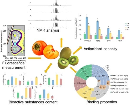
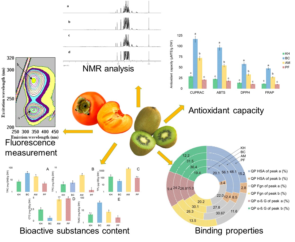
2. Results and Discussion

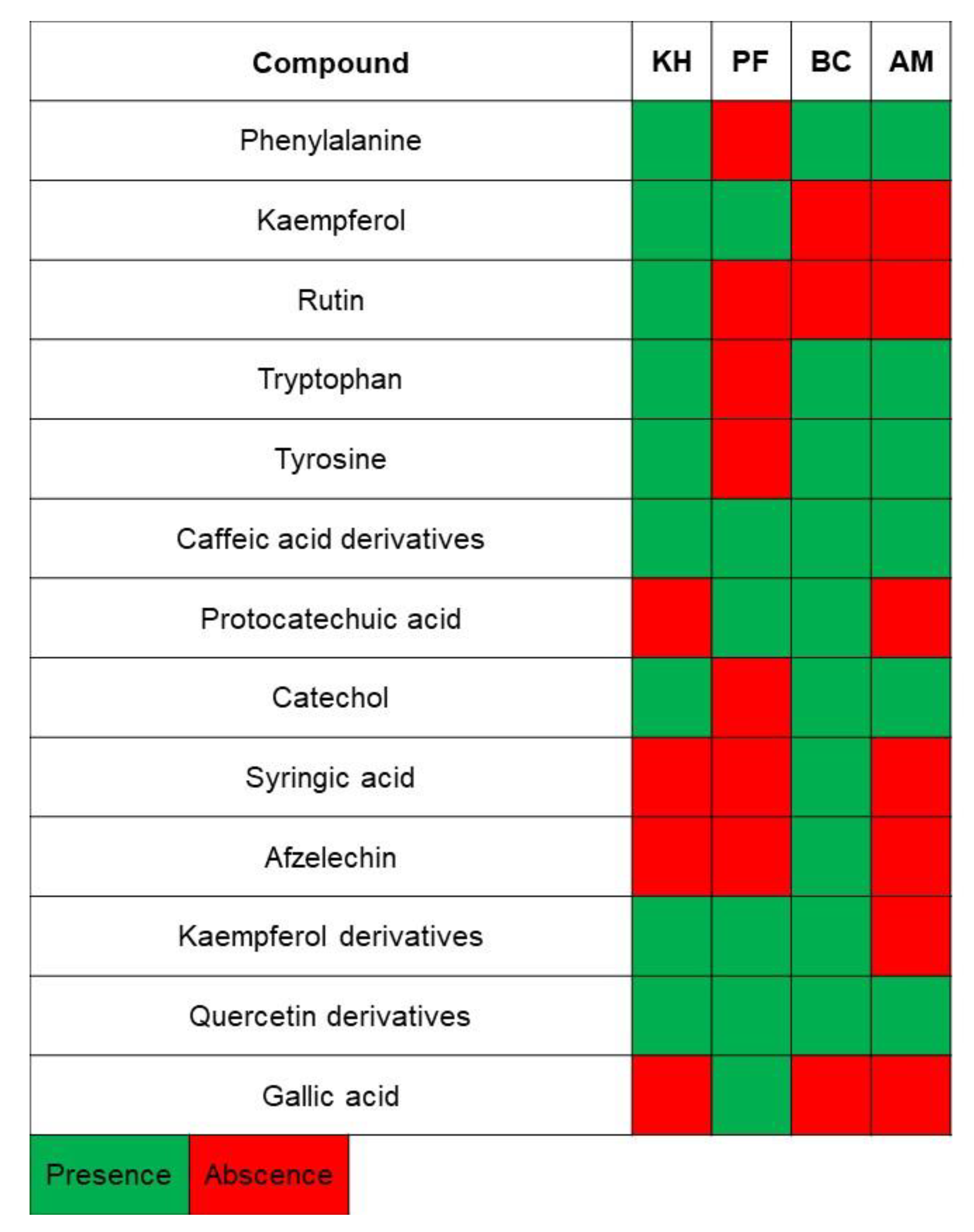
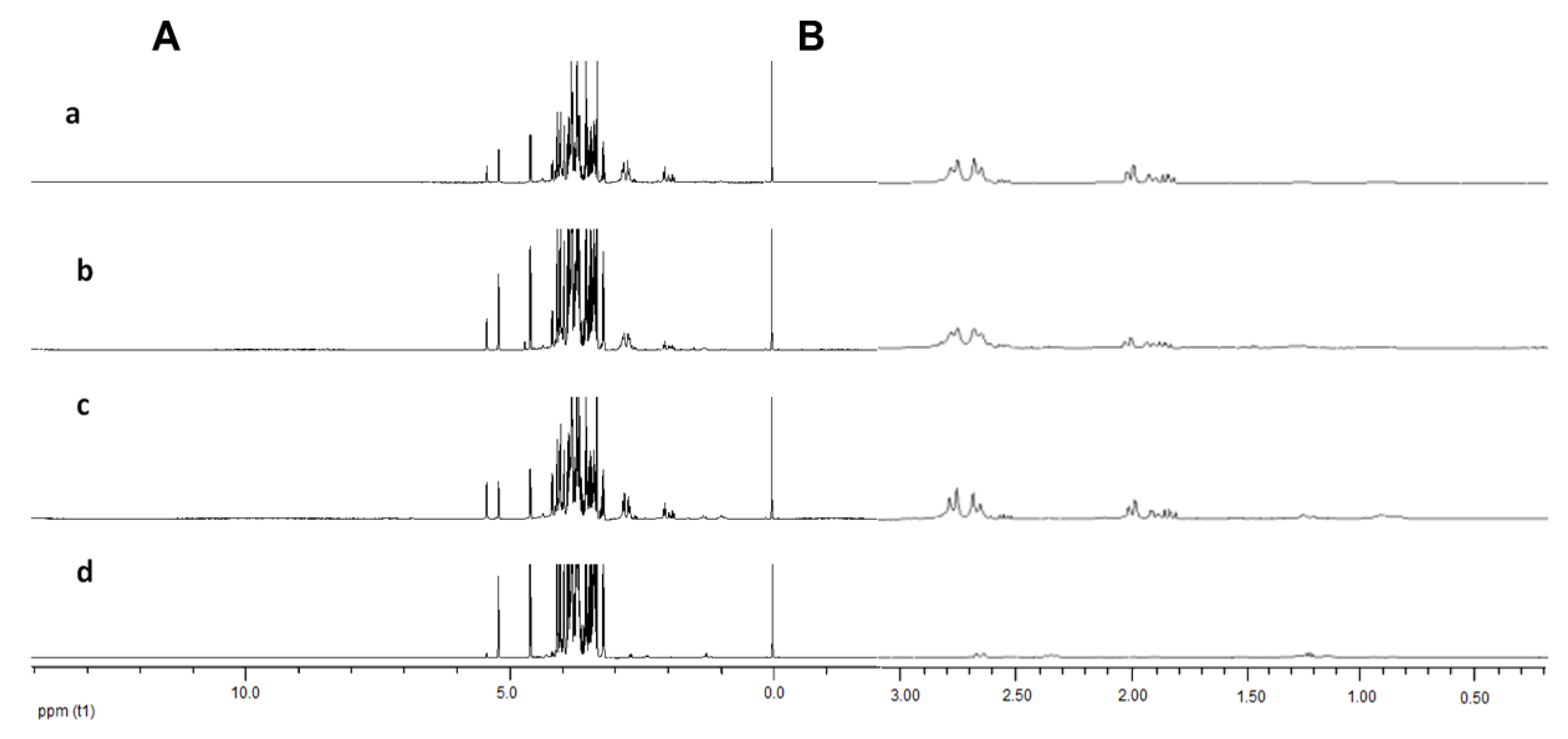
2.1. Identification of Bioactive Compounds in Fruit Extracts

In this study, three different varieties of kiwifruits, such as Actinidia (A.) deliciosa cv. Hayward (KH), A. eriantha cv. Bidan (BC), A. arguta Cheongsan (AM) and one cultivar of Diospyros kaki Thunb. cv. Fuyu (PF), were subjected to NMR analysis, and the obtained spectra were further evaluated using multivariate data analysis (MVDA). The identified metabolites and their NMR characteristics, as well as their presence in each sample, are listed in Table 1 and Figure 1.
There are differences in some metabolites of the different fruit samples, which can be detected from the 1H-NMR spectra. The principal peak areas of the spectra impart, as an immediate measure of metabolite concentration, permitting whole metabolites to be quantified based on a single internal standard. Different peak intensities can be observed at δ 5.20 (α glucose) and δ 5.40 (sucrose), where persimmon Fuyu showed the lowest concentration of sucrose as compared with the other kiwifruit samples (Figure 2A). It is also worth noting that different levels of peaks can be seen in the aliphatic region (δ 0.50–δ 3.00, Figure 2B). The same trend of intensities was observed in persimmons with the lowest signal compared with the tested kiwifruits. However, visual inspection of 1D-NMR does not permit any detection of compounds in the aromatic region (δ 6.00–δ 8.50). The 1D-NMR spectra of three different kiwifruits and persimmon, focusing on the region of δ 0.02 to 3.00 and different levels of signal intensities, can be observed in the aliphatic region (Figure 2B). Persimmons demonstrated the lowest intensities compared with kiwifruits.

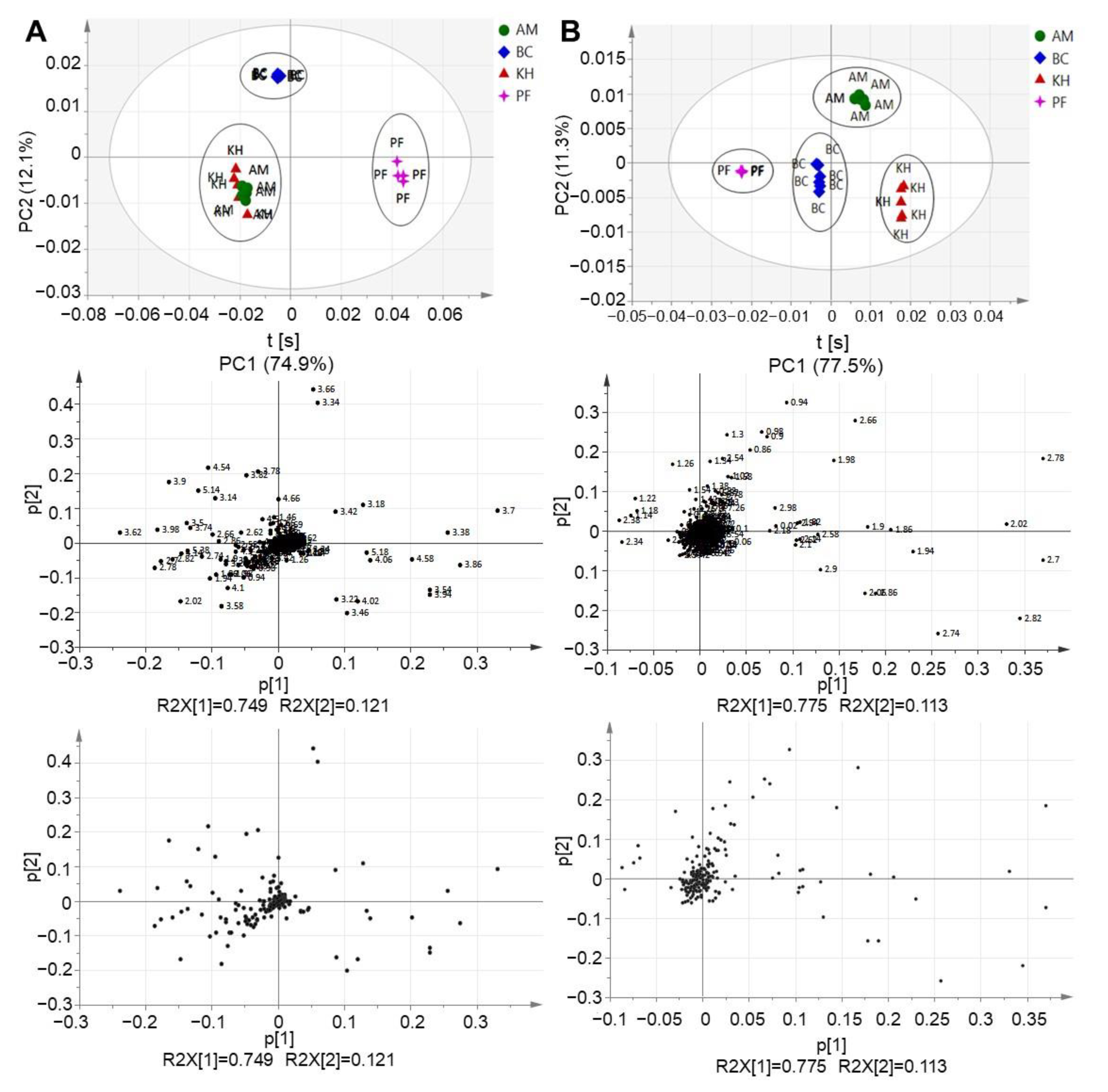
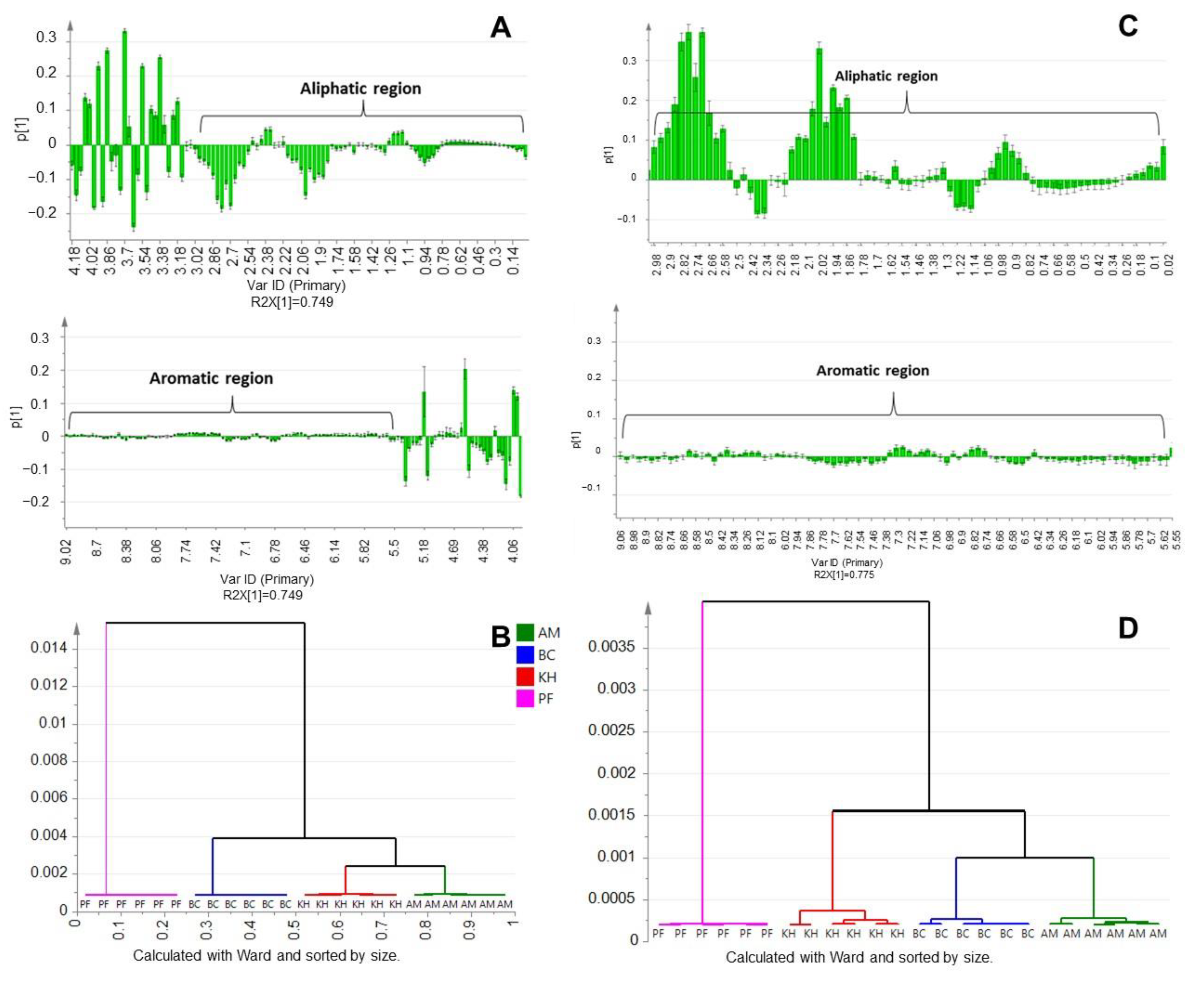
2.2. Principal Component Analysis (PCA) and Multivariate Data Analysis (MVDA)

PCA, as an unsupervised classification method, can be performed without prior knowledge of the data set, simplifying the dimensionality of the numerous variables while virtually sustaining the variances. The outcome of the PCA examination comprised of score plots, which signified the variation of the classes based on the metabolomics similarity, and loading plots, which offers information as to which NMR spectral regions were contributing with respect to the grouping attained in the score plots. Three different clusters were formed between the studied fruit samples (Figure 3A). The persimmon can be discriminated from kiwifruit samples by PC1. Meanwhile, BC can be distinguished from the other two kiwifruit varieties by PC2. However, no separation can be observed between KH and AM. The results (Figure 2A) prompted us to proceed with MVDA by excluding the sugar regions from the 1D-NMR spectrum (Figure 3B).
Therefore, as the next step, the sugar region (δ 3.00–δ 5.50) was removed from the binned excel file, and the unsupervised MVDA analysis was then repeated. This was carried out to observe if there was any clustering that could be formed without the interference of sugars in the samples. It is interesting to note that the removal of the sugar region managed to distinguish the KH from AM. Four distinct clusters can be observed where PF and BC are discriminated from AM and KH by PC1. Meanwhile, AM can be separated from the rest of the samples by PC2. Thus, it can be suggested that KH and AM are discriminated from each other without the masking effects of high sugar components (Figure 3B).
The upper regions of the PCA loading column plot, which corresponded to the persimmon, showed a lower intensity for most of the signals in the aliphatic regions, δ 0.50 to δ 3.00, compared with the lower regions of the model (Figure 4A). This was consistent with the trend demonstrated in the 1D-NMR spectra (Figure 2B). The relationship between the three varieties of kiwifruits and the persimmon was further evaluated in the first level of our hierarchical model (Figure 4B). PF was discernible from the other kiwifruit samples, based on different metabolite constitutions. Different results appeared after removing the sugar region (Figure 4C,D).
The obtained results can be compared with our recent data [13], where, in the same three varieties of kiwifruit, phenylalanine, tyrosine, arginine, citric acid, glutamine-hydroxy-L-proline, 4-aminobutyrate (GABA), glutamate, glutamine, quinic acid, actinic acid, shikimate, mannose, syringic acid and afzelechin were detected. The present results on kiwifruit metabolites can be compared with other reports, but there aliphatic part was most prominent. Some investigations were also carried out on parts of fresh kiwifruit or on fruit juices obtained with specific food processing procedures [31]. Similar results for the metabolites in persimmons were obtained in our report [32]. The differences in metabolites among five major Japanese persimmon cultivars were investigated using a nuclear magnetic resonance (NMR)-based metabolomics approach. Among the non-astringent cultivars analyzed, the Matsumotowase-Fuyu cultivar contains ethyl-beta-glycosides, as characteristic components, which may relate to fruit softening. The quantitative metabolomics approach based on broadband WET NMR spectra was mostly discussed in the aliphatic region as well [11,12].

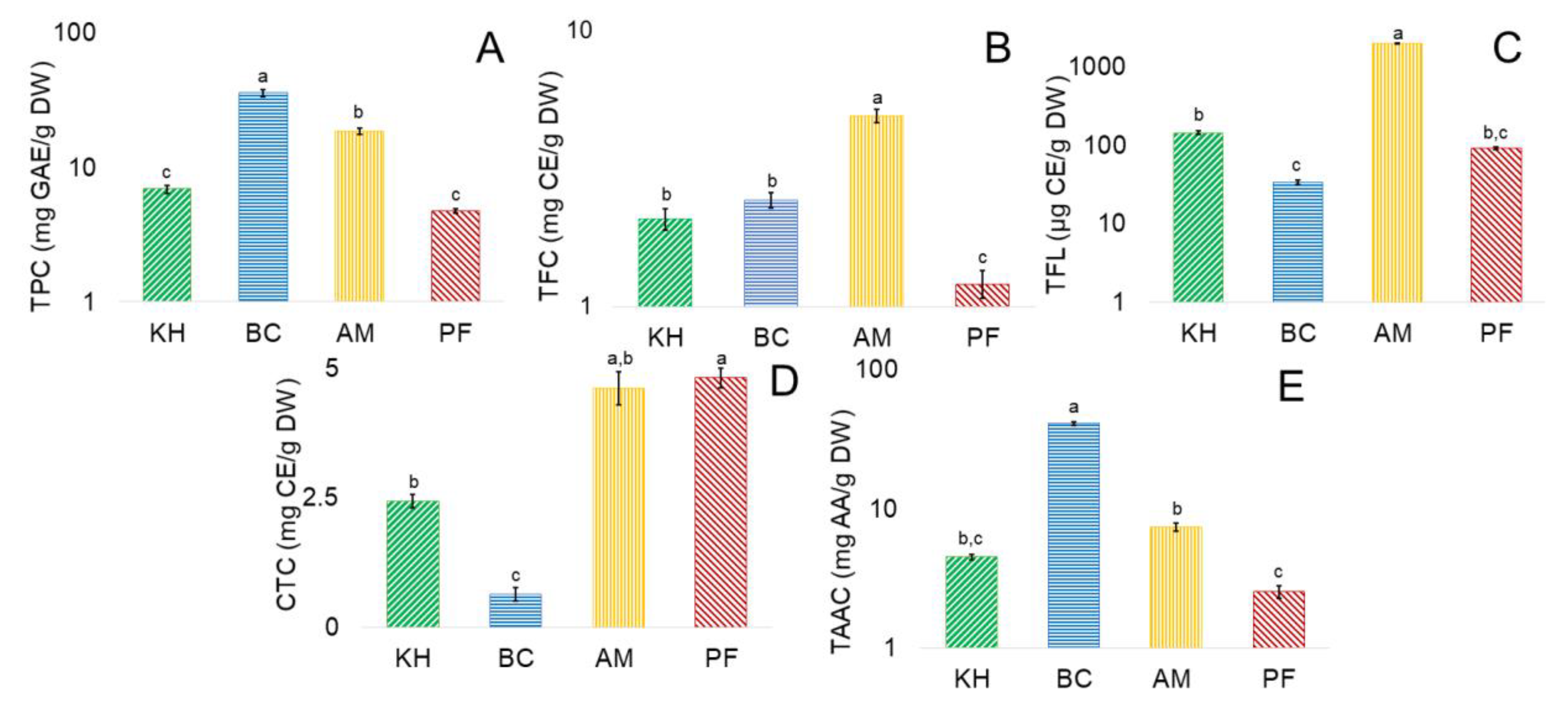
2.3. Determination of Bioactive Compounds

The number of bioactive compounds in kiwifruits and persimmon showed the following ranges (Figure 5).
From the presented results, nearly all bioactive compounds were the highest in BC, following by AM, and similar for KH and PF. Total phenolic content (TPC, mg GAE/g DW) from PF to BC was in the range of 4.74 ± 0.18 to 36.61 ± 2.15, respectively (Figure 5A). Total flavonoid content (TFC, mg CE/g DW) showed the lowest value of 1.21 ± 0.14 for PF and the highest for AM of 4.91 ± 0.28 (Figure 5B). Estimation of total flavanols content (TFL, µg CE/g DW) showed the highest for AM of 1969.81 ± 12.41 and the lowest for BC of 33.63 ± 2.43 (Figure 5C). Condensed tannin content (CTC, mg CE/g DW) changed in the investigated samples from 0.64 ± 0.13 for BC and the highest was for PF of 4.81 ± 0.19 (Figure 5D). Total ascorbic acid content (TAAC, mg AA/g DW) was expressed in PF as the lowest of 2.52 ± 0.27 and with the highest of 40.89 ± 1.18 for BC (Figure 5E). These results are in agreement with some recent reports. A. eriantha ‘Bidan’ peeled fruit methanol extracts had a higher TPC of 57.4 in comparison with 12.9 and 6.4 mg GAE/g DW for A. arguta, and A. deliciosa ‘Hayward’ extracts, respectively [13]. The TPC of six cultivars was in a range of 4.2–14.5 for ethanol extracts and 5.3–16.3 mg GAE/g for water extracts [33]. TPC of three A. deliciosa cultivars ranged from 5.3 to 6.6 mg GAE/g DW. A. arguta, A. macrosperma, and A. polygama had TPC of 8.15, 5.57, and 4.71 mg GAE/g DW, respectively [34]. Extracts (70% aqueous acetone) of A. deliciosa ‘Hayward’ varied in TPC (479 μg/g DW) [35]. Fresh Korean ‘Hayward’ had a TPC of 8.19 mg GAE/g [36]. TPC and TFC of pulp were 12.21 mg GAE/g DW and 5.92 mg CE/g DW in Actinidia arguta. High antioxidant activity was observed (FRAP: 151.41 µmol ferrous sulphate equivalents (FSE)/g DW; DPPH: 12.17 mg TE/g DW). These results emphasize the richness of A. arguta fruit pulp to be used in different food products [37]. The TPC of A. arguta ‘Chiak’ and ‘Darae No. 2’ ranged from 88 to 113 mg GAE/100 g FW. Hayward cultivar, grown in China, showed 78 mg GAE/100 g FW of TPC; the TFC was evaluated as 10.25 mg CE/100 g FW and DPPH and FRAP were 4.87 and 7.08 µmol TE/g FW [38]. Ethanol and water extracts of the TFC ranged as 1.2–4.3 and 0.6–1.8 mg CE/g DW, respectively [33]. The amount of bioactive compounds varied: total phenolics (mg GAE/g DW) 3.75–8.192–16.52; total flavonoids (mg CE/g DW) 2.11–2.472 and total flavanols (μg CE/g DW) 0.14–0.162 were reported in some recent reports [39,40]. Total flavan-3-ols contents in fourteen kiwifruit cultivars ranged from 96 to 824 μg/g DW [41]. Extraction solvents possibly influenced the solubility of kiwifruit flavonoids [42]. Actinidia eriantha [43] is a precious material to study the metabolism and regulation of ascorbic acid (AsA) because of its high content. The other cultivars have relatively high amounts of ascorbic acid, which is shown in Figure 5E and in the published reports [44,45].

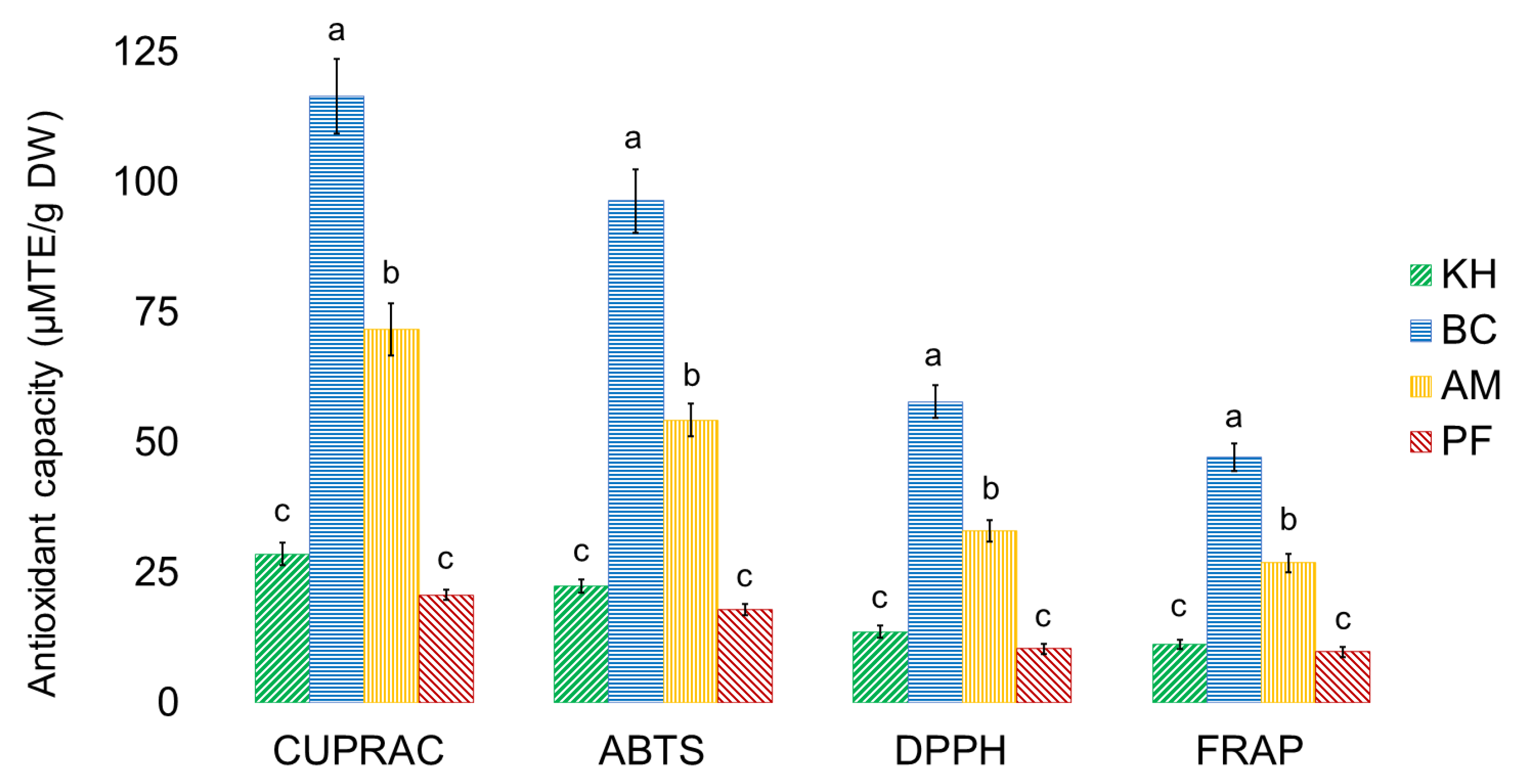
2.4. Antioxidant Capacities of Investigated Samples

The antioxidant capacities of investigated samples are shown in Figure 6.
The lowest values of CUPRAC, ABTS, DPPH, FRAP (µM TE/g DW) were 20.72 ± 1.23, 17.96 ± 1.02, 10.45 ± 0.35, 9.86 ± 0.61 for PF, and the highest values were 116.63 ± 7.21, 96.48 ± 6.14, 57.87 ± 3.12, 47.37 ± 2.05 (Figure 6). These results are similar to some reports. ‘Hayward’ water extracts had higher FRAP, ABTS, CUPRAC and DPPH values than the investigated methanol extracts [33]. A. eriantha ‘Bidan’ extracts had higher DPPH, ABTS, FRAP and CUPRAC values than A. arguta and A. deliciosa ‘Hayward’ [13,46]. TPC was the greatest antioxidant contributor in the DPPH and FRAP assays, which is in line with other findings [38], as well as with ABTS and CUPRAC (Figure 6). Diversity in the results of bioactive metabolites depends on the varieties and plant parts, extraction, analytical and processing methods, and this affects the physicochemical and biological properties of kiwifruit-derived ingredients [47,48]. The results obtained in this research can be compared with recent reports. Apart from the treatment used, changes in the content of metabolites are also affected markedly by the persimmon variety. Soluble tannins were 23.8 ± 4.3 and 14.3 ± 1.6; soluble non-tannins were 17.4 ± 2.4 and 15.9 ± 0.5; and total phenolic compounds were estimated as 89.1 ± 5.8 and 78.6 ± 4.5, expressed as g of epicatechin equivalents (EE)/kg DW for Rojo Brillante and Kaki Tipo, respectively. Gallic acid was the predominant phenolic compound found in the Rojo Brillante variety (0.953 mg/100 g), whereas the concentration of p-hydroxybenzoic acid was higher in the Triumph variety (0.119 mg/100 g). The antioxidant activity values ranged from 1.280 ± 0.069 to 8.865 ± 0.056 µmol TE/g when measured by ABTS, 0.458 ± 0.05 to 2.633 ± 0.03 µmol TE/g when measured by DPPH, and 0.206 ± 0.01 to 0.965 ± 0.005 µmol TE/g when the FRAP method was used. The greatest ABTS scavenging capacity was detected in Rojo Brillante (6.572 µmol TE/g), while the lowest was found in the non-astringent variety Triumph (41.484 µmol TE/g). The antioxidant activities of all extracts determined as DPPH radical scavenging ability ranged from 2.633 to 0.458 µmol TE/g. The ferric-reducing antioxidant power of Rojo Brillante and Triumph extracts was similar to the DPPH scavenging activity and ABTS scavenging capacity, with higher values for the astringent variety (0.965 ± 0.005 µmol TE/g). In general, the astringent variety (Rojo Brillante) showed much higher antioxidant activity than the non-astringent variety (Triumph) for both ABTS (6.572 µmol TE/g and 1.484 µmol TE/g, respectively), DPPH (2.417 µmol TE/g and 0.492 µmol TE/G, respectively), and FRAP assay (0.731 µmol TE/g and 0.242 µmol TE/g, respectively). The Rojo Brillante variety also had the highest values of total phenol content, as measured by the Folin method (380.786 µg GAE/g and 81.568 µg GAE/g, respectively). For persimmon samples, the results were similar to those of other authors, which ranged from 1027.03 to 1667.65 µmol/kg, and the concentrations very close to those reported in our study, although very few papers discussed antioxidants in the fruits considered in this study. Our results are similar and consistent with the data from other research reports, which investigated diverse persimmon genotypes [48,49,50,51].

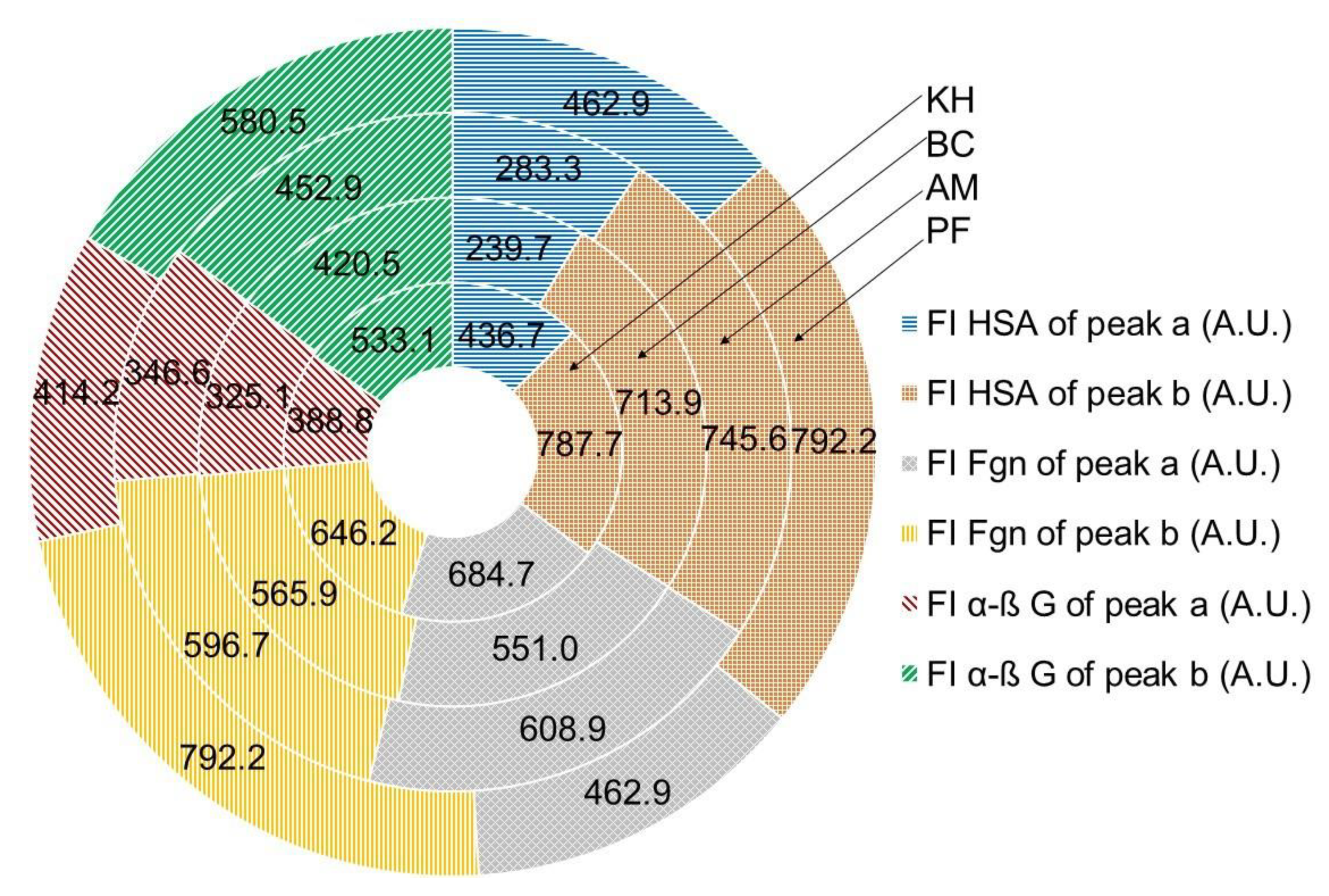
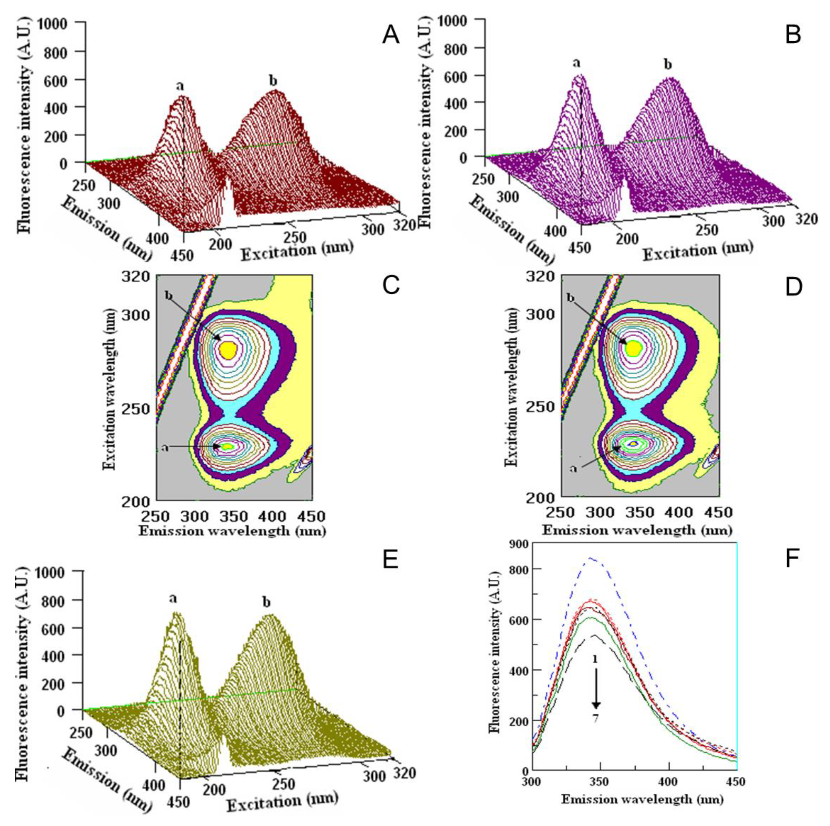
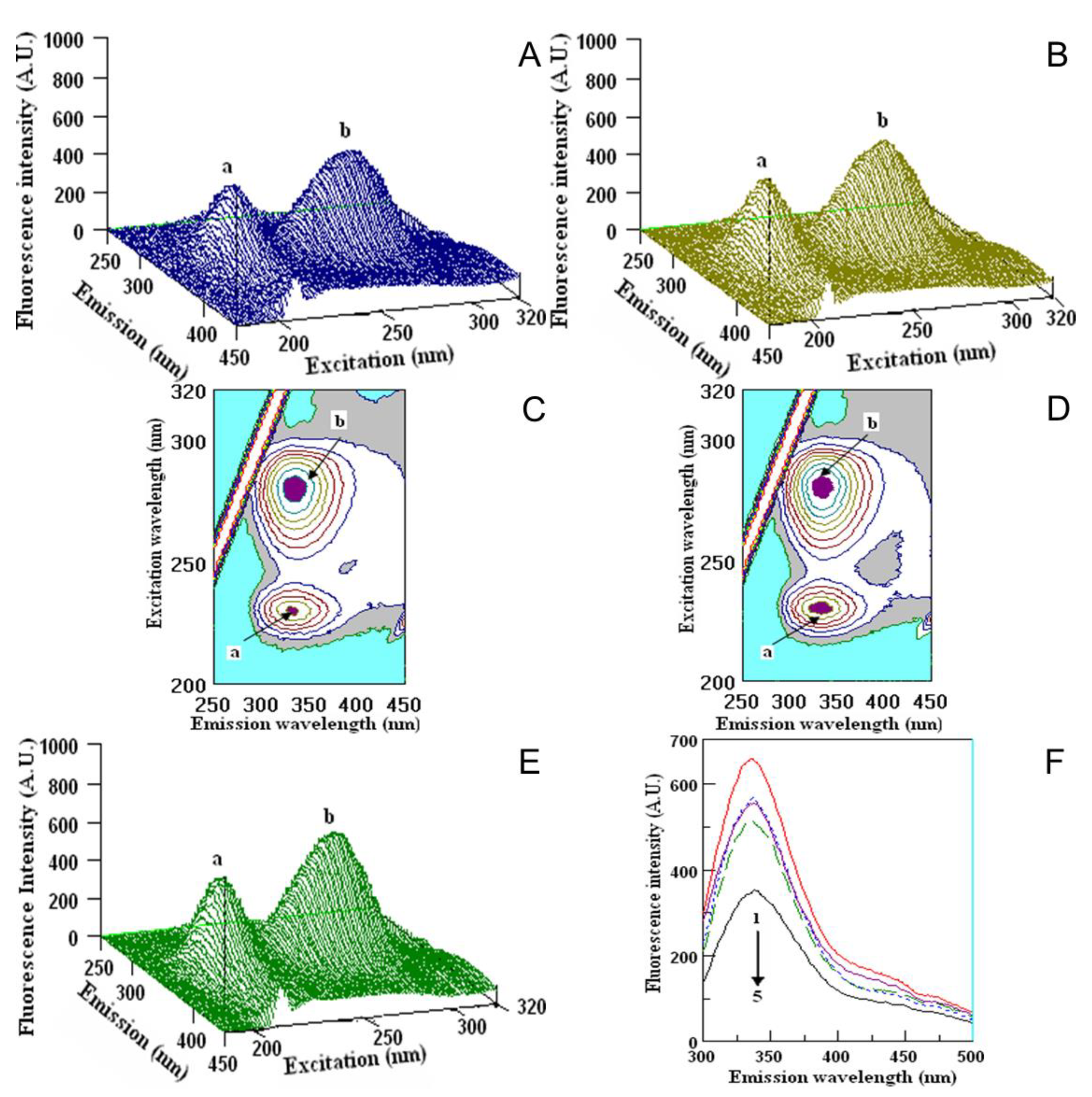
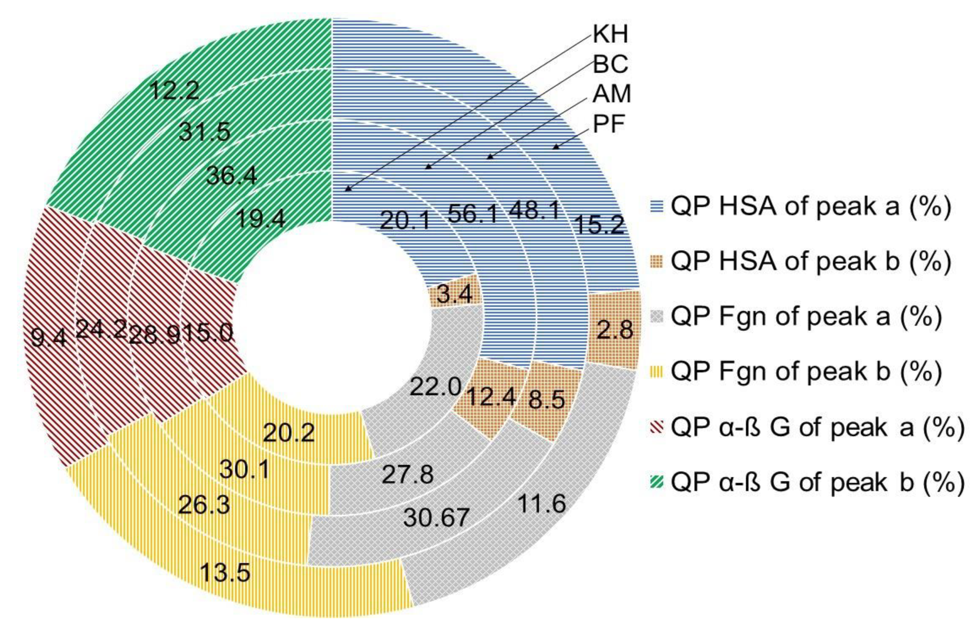
2.5. Quenching Properties of Phenolic Compounds of Investigated Fruits with Human Serum Proteins

The interactions of fruit extracts with human serum albumin (HSA), fibrinogen (Fgn) and α, β-globulin (α, β-G) are shown in Figure 7, Figure 8 and Figure 9.
The interaction with the above serum proteins and extracted fruit polyphenols is evaluated by the changes in the fluorescence intensity of the proteins. The changes appeared mostly in the position and value of peak a. The values in the fluorescence intensity (F.I.) of peak a in HSA with fruit extracts were the highest for PF (462.9 ± 6.9 A.U.) and the lowest for BC (239.7 ± 5.1). Small changes appeared in the position and value of peak b. The fluorescence intensity of HSA with fruit extracts was the highest for PF (792.2 ± 9.3 A.U.) and the lowest for BC (713.9 ± 9.9 A.U.), in comparison with the initial ones (Figure 7). The FI of peak a of Fgn after interaction with fruit extracts was the highest for PF (775.2 ± 8.2 A.U.) and the lowest for BC (551.0 ± 6.9). Lower changes in comparison with peak a appeared in the position of peak b: the highest peak was measured for PF (700.2 ± 7.5 A.U.) and the lowest one was measured for BC (565.9 ± 5.9 A.U.). The exact locations of peaks a and b during the interaction of KH and PF are presented in Figure 7 and Figure 8.
The images of the interaction of Fgn with KH and PF (Figure 8A–D) and the Fgn (Figure 8F) showed the maximum peaks a and b and their locations. The comparison of the values of fluorescence intensity of the native Fgn (Figure 8F, line 1 from the top) showed that the lowest value was obtained by its interaction with tannic acid (Figure 8F, line 7). In 2D-FL, the values of fluorescence intensities with extracts of KH and PF were nearly similar (Figure 8F, lines 4 and 5).
The fluorescence measurements with serum globulin and fruit extracts are presented in Figure 7 and Figure 9. Peak a of α-β-globulin (α-β G) after interaction with fruit extracts was the highest for PF (414.2 ± 7.4 A.U.) and the lowest for BC (325.1 ± 3.9). Lower changes appeared in the position of peak b: the highest peak was measured for PF (580.5 ± 7.5 A.U.) and the lowest was measured for BC (420.5 ± 3.3 A.U.). The exact locations of peaks a and b during the interaction with KH and PF are presented in Figure 7 and Figure 9.
The images of the interaction of α-β G with KH and PF (Figure 9A–D) and the α-β G (Figure 9E) showed the maximum of peaks a and b and their locations. The comparison of the values of fluorescence intensity of the native α-β G (Figure 9F, line 1 from the top) showed that the lowest value was obtained by its interaction with tannic acid (Figure 9F, line 5). In 2D-FL, the changes in fluorescence intensities with extracts of KH and PF were almost similar to Fgn, but showed slightly different values in reaction with globulin, and KH was more reactive than PF (Figure 9F, lines 4 and 2, respectively).
Albumin, fibrinogen, lipoproteins and α, β, and γ globulins play an important role in the pharmacokinetic properties of food nutriments. Globulins make up 35% of plasma proteins and are used in the transport of ions, drugs and lipids [21,22]. It was important to compare the standard metabolites with the polyphenol extracts. The polyphenols-protein interaction is reversible in that the polyphenols–protein complex can dissociate and release free polyphenols. Polyphenols and their metabolites rapidly exchange between free and bound forms within the circulation. Reversible binding to plasma proteins may have consequences for the delivery of the polyphenols and their metabolites to cells and tissues [21,22].
The quenching properties (%) between HSA and polyphenols for PF and BC were in the range of 15.2 ± 0.9 to 56.15.8, calculated by peak a and 2.8 ± 0.1 to 12.4 ± 1.1, according to peak b, respectively. Different values of quenching properties (%) were calculated with fibrinogen interaction: PF and BC were in the range of 11.6 ± 1.1 to 37.8 ± 2.4, calculated by peak a, and 13.5 ± 1.1 to 28.9 ± 1.2, according to peak b, respectively, and globulin showed 9.4 ± 0.9 and 37.8 ± 2.4, calculated by peak a, and 12.2 ± 1.1 and 36.4 ± 2.4, according to peak b, respectively (Figure 10).
The presented data varied between the varieties and the used serum proteins, but showed the same correlation between samples, where BC was the strongest and KH and PF were in the same range of their bioactivities (Figure 7, Figure 8, Figure 9 and Figure 10).
In the present report, we have used a simplified measure to show only the decrease in fluorescence emission after the addition of a single concentration of ligands. This can be regarded as a relative measure of quenching, providing that the inner filter is similarly negligible within the series of ligands. Thus, the % decrease of fluorescence represents the fraction of the binding sites of the protein occupied by the ligand, rather than the fraction of the total ligand bound to the protein [52].
The obtained metabolite results of persimmon samples showed relatively high amounts of tannins in comparison with the investigated three samples of kiwifruits and are in line with several reports [5,6,11,12]. The estimation of quenching of serum proteins such as fibrinogen (Figure 8F, line 8) and globulin with tannic acid (Figure 9, line 5) are in full agreement with the amount of tannic acid in persimmon samples. Based on these results, PF showed nearly the same quenching properties as KH. The presently determined high quenching properties of persimmon in vitro with relatively new metabolite indices such as fibrinogen and globulin, showing protective action and preventing CAD, are in line with some reported in vitro and in vivo studies. Evaluation of the prognostic significance of changes in serum albumin levels among patients that underwent percutaneous coronary intervention (PCI) showed that a decrease in albumin levels following PCI is an independent prognostic marker of worse long-term outcomes [24]. It was found that, similar to traditional risk factors, plasma fibrinogen and albumin levels showed a close relation with the presence and severity of CAD [25]. The fibrinogen-to-albumin ratio index is a valuable biomarker associated with ST- elevation myocardial infarction and may be useful in the prediction of the long-term prognosis of patients with such diseases [26]. So, following the recent reports on humans discussed above and the results of the supplementation of fruits, it was shown that the triglyceride (TG)/cholesterol profile depended on the treatment of persimmon water extracts, and tannin-enriched persimmon concentrate stimulated hypocholesterolemic actions [6,53,54]. Similar action was obtained by a combination of Diospyros kaki fruit and Citrus unshiu peel mixture as a potential therapeutic agent for treating nonalcoholic fatty liver disease with the remarkable growth of obesity [55]. Variation in tannin amount depends on the cultivars of persimmon, even in co-products from cvs. Rojo Brillante’ and Triumph’ persimmon juice extraction processed to obtain flours rich in the main metabolites, such as sugars, organic acids, tannins, and bioactive compounds, suggesting their use as a functional ingredient with antioxidant properties in different food products [56]. Similar results for the quenching properties of kiwifruit polyphenols were obtained in the present study. As such, the health properties of kiwifruit polyphenols results shown in Figure 7, Figure 8, Figure 9 and Figure 10 are in compliance with previous studies, where the intervention of green kiwifruit effectively lowered the total cholesterol (TC) and increased the high-density lipoprotein cholesterol (HDL) concentration in hypercholesterolaemic and healthy individuals. It was proven that the bioavailability of polyphenols depends on physicochemical stability, complex formation, food interaction, gastrointestinal absorption, and hepatic and gut metabolism [19]. Consumption of fruits influences the total cholesterol, LDL, HDL, proteins, lipid peroxidation and oxidative stress biomarkers [17,18,20]. Consumption of at least one kiwi/week is associated with lower plasma concentrations of fibrinogen and improved plasma lipid profile in the context of a normal diet and regular exercise [28,57,58]. Similar results were reported on antihypercholesterolemia Male Wistar rats when 1% cholesterol-enriched diet induced-hypercholesterolemia improved liver somatic index and lipid profiles after supplementing with 5% lyophilized Polish grown kiwifruit. A. arguta ‘Geneva’, ‘Anna’, and ‘Weiki’ showed the most significant results [46,59]. Cellular antioxidant activity (CAA) assays, combined with clinical trials, will more effectively identify antioxidant phytochemicals in fruits that can be used as dietary additives or drugs for human health. In spite of advanced methods in the determination of antioxidant activities, in vitro studies of the interaction of polyphenols with human serum proteins, in vivo experiments, or clinical trials are still required to verify the efficacious activity when fruit polyphenols are used as dietary supplements or drugs to combat oxidative stress [60].

3. Materials and Methods

3.1. Chemicals and Materials

The chemicals 2,4,6,-tripyridyl-s-triazine (TPTZ), 6-Hydroxy-2,5,7,8-tetra-methylchroman-2-carboxylic acid (Trolox), 1,1-diphenyl-2-picrylhydrazyl (DPPH), lanthanum(III) chloride heptahydrate, CuCl2·2H2O, 2,9-dimethyl-1,10-phenanthroline (neocuproine), 2,2-azino-bis(3-ethylbenzothiazloine-6-sulphonic acid) (ABTS) radical cation, ferric chloride, caffeic acid, quercetin, tannic acid, catechin, human serum albumin (HSA), fibrinogen, globulin, phosphate buffer and Folin-Ciocalteu reagent (FCR) were purchased from Sigma (St. Louis, MO, USA) and Fluka Chemie Gm bH, Buchs, Switzerland. All NMR chemicals, including 3-trimethylsilylpropanoic acid (TSP), potassium phosphate monobasic (KH2PO4), methanol-d4 (CD3OD, 99.8%), sodium deuterium oxide (NaOD), and deuterium oxide (D2O, 99.9%), were purchased from Merck (Darmstadt, Germany).

3.2. Sampling and NMR Metabolomics

Three batches of organic kiwifruits, including Actinidia (A.) deliciosa cv. Hayward (KH), A. eriantha cv. Bidan (BC), A. arguta Cheongsan (AM) and one batch of Diospyros kaki Thunb. cv. Fuyu (PF), were collected in different commercial orchards from Boseong and Muan counties, Jeonnam and Wonju-si, Gangwon-do provinces, South Korea [61]. Each batch was composed of 25 fruits, about two kg in weight. The cultivars reached the commercial maturity stage. The samples were washed with tap water and dried. The fruits were fractionated into an edible fraction (pulps), peels and seeds. Only for PF, 5–8 seeds were separated from pulps. Their edible parts were prepared manually without using steel knives. The peeled fruits (pulps) were weighed, chopped and homogenized in liquid nitrogen in a high-speed blender (Silex professional model, Hamilton Beach, Virginia, USA). A weighed portion (50–100 g) was then lyophilized for 48 h (Virtis model 10–324, Midland, ON, Canada), and the dry weight was determined. The samples were ground to pass through a 60-mesh sieve and stored at −20 °C until the bioactive substances were analyzed.
The proton (1H) and two-dimensional (2D) J-resolved NMR procedure was carried out according to the previously reported protocols with small modifications. The extraction of samples was carried out by transferring 100 mg of each sample into a 2 mL Eppendorf tube, followed by the addition of 375 µL of both CD3OD solvent and KH2PO4 buffer in D2O (pH 6.0) containing 0.1% TSP. The solution was then vortexed for 1 min before being subjected to sonication for 15 min at a controlled temperature. To get a clear supernatant, the mixture was afterward centrifuged at rpm for 10 min, and 600 µL of it was pipetted to a NMR tube prior to analysis. The 1H-NMR analysis was performed at 25 °C on an INOVA 500 MHz spectrometer (Varian Inc., Palo Alto, CA, USA). For each sample, the required time was 3.53 min, recording 64 scans with an acquisition time, a pulse width, and a relaxation delay of 220 s, 3.75 ms, and 1.0 s, respectively. These settings were for presaturation prior to 1H-NMR, which is required to suppress the water signal using low power selective irradiation. In addition, the spectral width of the recorded spectra was 20 ppm. The processing for all spectra, including phasing and baseline corrections, was performed manually with Chenomx software (Version 6.2, Edmonton, AB, Canada). Moreover, the 2D J-resolved was conducted to endorse metabolite identification [13,32,62].

3.3. Determination of Bioactive Compounds

The detailed procedures of the extraction, determination of bioactive compounds and their antioxidant capacities were described in our very recent reports [52,63,64]. For polyphenol extraction, the freeze-dried powders of investigated samples were immersed in absolute methanol (1/10 w/v). The filtrate was collected three times with constant stirring of the mixture at every 24 h interval of a 72 h total collection period at room temperature. The extract was then concentrated under reduced pressure at 45 °C using a vacuum rotary evaporator.
A Folin–Ciocalteu assay was used for the determination of total polyphenol content (TPC) in methanol fruit extracts of 0.25 mL with 1 mL of Folin–Ciocalteu reagent (Sigma, St. Louis, MO, USA). Then, 0.75 mL of 1% sodium carbonate was added. Absorbance of the mixture was measured on a Hewlett-Packard model 8452A spectrophotometer (Hewlett-Packard, Rockville, MD, USA) at 750 nm. The results were calculated in mg gallic acid equivalents (GAE) per g DW [65]. Total flavonoid content (TFC, mg catechin equivalents (CE) per g DW) was measured at 510 nm after extraction with 5% NaNO2, 10% AlCl3xH2O and 1 M of NaOH [66]. The absorbance of total flavanols (TFL, µg CE per g DW) was measured at 640 nm following the p-dimethylaminocinnamaldehyde (DMACA) method: 1 mL of DMACA solution was added to 0.2 mL of fruit extracts [67]. Condensed tannin content (CTC, mg CE per g DW) was estimated by spectrophotometric measurements in the mixture of methanol fruit extracts and the addition of 4% methanol vanillin solution. Absorbance was measured at 500 nm after the end of the reaction [68]. Total ascorbic acid content (TAAC, mg ascorbic acid (AA) per g DW) was evaluated in water fruit extracts, where 100 mg of the freeze-dried sample was extracted with 5 mL water. Then, CUPRAC method was conducted, and formed bis (Nc)-copper (I) chelate was determined spectrophotometrically at 450 nm [69].

3.4. Determination of Antioxidant Capacities

Total antioxidant capacity was determined by the following assays, which are also described in our recent reports [52,63,64].
Cupric reducing antioxidant (CUPRAC) assay is based on utilizing the copper (II)−neocuproine reagent as the chromogenic oxidizing agent. Absorbance at 450 nm was measured in a mixture of [Cu (II)-Nc] and NH4Ac buffer solution and fruit methanol extracts [70].
2,2′-azino-bis(3-ethyl-benzothiazoline-6-sulfonic acid) diammonium salt (ABTS•+) was generated by the interaction of ABTS (7 mM) and K2S2O8 (2.45 mM). This solution was diluted with methanol and measured at 734 nm [71]. Scavenging free radical potentials were tested in a methanolic solution (3.9 mL) of 1, 1-diphenyl-2-picrylhydrazyl (DPPH) with the sample extracts in methanol (0.1 mL) [72]. Ferric-reducing/antioxidant power (FRAP) assay measures the ability of the antioxidants in the investigated samples to reduce ferric-tripiridyltriazine (Fe3+-TPTZ) to a ferrous form (Fe2+) [73]. All values of antioxidant capacities were expressed in µM trolox equivalent (TE)/g DW.

3.5. Fluorometric Studies

The profiles and properties of polyphenols in methanol extracts were determined by two (2D-FL) and three-dimensional (3D-FL) fluorescence (model FP-6500, Jasco spectrofluorometer, serialN261332, Tokyo, Japan). The 2D-FL measurements were taken at emission wavelengths from 310 to 500 nm and at excitation of 295 nm. The 3D-FL was measured at emission wavelengths between 200 and 795 nm and the initial excitation wavelength at 200 nm. For comparison of the obtained results, caffeic acid, quercetin, tannic acid and catechin were used [36]. The quenching properties of phenolic compounds in kiwifruit and persimmon extracts to human serum albumin (HSA), fibrinogen and globulin were evaluated by 2D and 3D-FL. For the fluorescence measurements, 3.0 mL of 1.0 × 10⁻⁵ mol/L HSA were prepared in 0.05 mol/L Tris–HCl buffer (pH 7.4), containing 0.1 mol/L NaCl. Fibrinogen and globulin stock solutions were made by dissolving in phosphate buffer (10 mM, pH 7.4) to obtain a concentration of 20 µM. Standards phenolic solutions, such as tannic acid, quercetin, catechin, and caffeic acid stock solution, were prepared daily by dissolving at a concentration of 10 mM in methanol and then diluting with 10 mM phosphate buffer at pH 7.4. Samples were prepared by mixing fibrinogen, fruit extracts and standards of phenolic compound solutions in varying proportions. The highest resulting methanol concentration was about 1%, which had no appreciable effect on protein structure. All samples were kept at 4 °C prior to the analysis. The initial fluorescence intensities of HSA, globulin and Fgn were measured before interaction with the investigated samples and pure substances and after interaction with the samples (quenching of the fluorescence emission of proteins, in our case of HSA, globulin and fibrinogen and polyphenols of fruits). As mentioned above, changes in the fluorescence intensities were used in the estimation of quenching activities. [52,63,64].

3.6. Data Analysis

NMR data analysis followed the reported procedure [74]. The conversion of 1H-NMR spectra to an ASCII file using Chenomx software was carried out prior to multivariate data analysis (MVDA) and performed using SIMCA-P+ version 13.0 (Umetrics AB, Umeå, Sweden). This analysis consists of the exclusion of the residual water (4.70–4.90 ppm) and methanol (3.23–3.34 ppm) signals range. Next, all spectra were scaled to TSP and bucketed to bins with a width of 0.04 ppm, forming a spectral region of 0.52–9.99 ppm. The binned integral of 1H-NMR data were then subjected to principal component analysis (PCA), which was applied to clearly differentiate the 1H-NMR spectra of the kiwifruit and persimmon samples. The Pareto method was also used for scaling purposes to ensure the same importance was given to all x variables in the analyses. All obtained data were calculated on the basis of statistical analysis of Duncan’s multiple range test. Values are means ± SD per gram of dry weight (DW) of 25 measurements, representing the commercial maturity status of fruits and their replicates. Five biological replications of five extracts from each cultivar were performed. To determine the statistical significance as a 95% interval of reliability, one-way analysis of variance (ANOVA), was used.

4. Conclusions

We obtained relatively high amounts of antioxidants in the raw pulp of investigated fruits and high quenching properties of fruit extracts in comparison with pure metabolites. The addition of such fruits to generally accepted diets could be beneficial for hyperlipidemic, especially hypertriglyceridemic, patients suffering from coronary atherosclerosis. We expect that HSA, Fgn and α-β G will serve as predictors of cardiovascular events.

Author Contributions

Conceptualization, M.L.-S. and S.G.; data curation, Y.-M.K., Y.-K.P., K.-S.H. and A.E.; formal analysis, Y.-M.K., Y.-S.P. and S.-G.K.; investigation, F.A.; methodology, Y.-S.P. and K.-S.H.; software, Y.-K.P. and A.E.; validation, S.-G.K.; supervision, S.G.; writing—original draft preparation, F.A.; M.L.-S. and S.G.; writing—review and editing, F.A., M.L.-S. and S.G. All authors have read and agreed to the published version of the manuscript.

Funding

This research received no external funding.

Institutional Review Board Statement

Not applicable.

Informed Consent Statement

Not applicable.

Data Availability Statement

The data presented in this study are available on request from the Corresponding author. The data are not publicly available due to privacy reasons.

Acknowledgments

Thanks from all authors of the manuscript to Elena Katrich from Institute for Drug Research, Hebrew University of Jerusalem, Nur Ashikin Abdul Hamid and M. Maulidiani, Laboratory of Natural Products, Institute of Bioscience, Universiti Putra Malaysia, for their assistance in the measuring of some indices in fruits.

Conflicts of Interest

The authors declare no conflict of interest.

Sample Availability

Samples of the compounds are not available from the authors.

References

  1. Nardozza, S.; Cooney, J.; Boldingh, H.L.; Hewitt, K.G.; Trower, T.; Jones, D.; Thrimawithana, A.H.; Allan, A.C.; Richardson, A.C. Phytohormone and transcriptomic analysis reveals endogenous cytokinins affect kiwifruit growth under restricted carbon supply. Metabolites 2020, 10, 23. [Google Scholar] [CrossRef] [PubMed] [Green Version]
  2. Lim, V.; Gorji, S.G.; Daygon, V.D.; Fitzgerald, M. Untargeted and targeted metabolomic profiling of australian indigenous fruits. Metabolites 2020, 10, 114. [Google Scholar] [CrossRef] [PubMed] [Green Version]
  3. Dembitsky, V.M.; Poovarodom, S.; Leontowicz, H.; Leontowicz, M.; Vearasilp, S.; Trakhtenberg, S.; Gorinstein, S. The multiple nutrition properties of some exotic fruits: Biological activity and active metabolites. Food Res. Int. 2011, 44, 1671–1701. [Google Scholar] [CrossRef]
  4. Vázquez-Manjarrez, N.; Vázquez-Manjarrez, N.; Vázquez-Manjarrez, N.; Ulaszewska, M.; Garcia-Aloy, M.; Garcia-Aloy, M.; Mattivi, F.; Mattivi, F.; Praticò, G.; Dragsted, L.O.; et al. Biomarkers of intake for tropical fruits. Genes Nutr. 2020, 15. [Google Scholar] [CrossRef] [PubMed]
  5. Park, Y.S.; Leontowicz, H.; Leontowicz, M.; Namiesnik, J.; Jesion, I.; Gorinstein, S. Nutraceutical value of persimmon (Diospyros kaki Thunb.) and its influence on some indices of atherosclerosis in an experiment on rats fed cholesterol-containing diet. Adv. Hortic. Sci. 2008, 22, 250–254. [Google Scholar] [CrossRef]
  6. Park, S.Y.; Oh, E.K.; Lim, Y.; Shin, J.Y.; Jung, H.A.; Park, S.Y.; Lee, J.H.; Choe, J.S.; Kwon, O. Metabolites profiling and hypolipidemic/hypocholesterolemic effects of persimmon (Diosyros kaki Thumb.) by different processing procedures: In vitro and in vivo studies. J. Nutr. Health 2018, 51, 275–286. [Google Scholar] [CrossRef]
  7. Yoshimura, M.; Mochizuki, A.; Amakura, Y. Identification of phenolic constituents and inhibitory activity of persimmon calyx and Shiteito against tumor cell proliferation. Chem. Pharm. Bull. 2021, 32–39. [Google Scholar] [CrossRef]
  8. Hunter, D.C.; Greenwood, J.; Zhang, J.; Skinner, M.A. Antioxidant and “natural protective” properties of kiwifruit. Curr. Top. Med. Chem. 2011, 11, 1811–1820. [Google Scholar] [CrossRef]
  9. Worley, B.; Powers, R. Multivariate Analysis in Metabolomics. Curr. Metab. 2013, 1, 92–107. [Google Scholar] [CrossRef]
  10. Koda, M.; Furihata, K.; Wei, F.; Miyakawa, T.; Tanokura, M. Metabolic discrimination of mango juice from various cultivars by band-selective NMR spectroscopy. J. Agric. Food Chem. 2012, 60, 1158–1166. [Google Scholar] [CrossRef]
  11. Ryu, S.; Furihata, K.; Koda, M.; Wei, F.; Miyakawa, T.; Tanokura, M. NMR-based analysis of the chemical composition of Japanese persimmon aqueous extracts. Magn. Reson. Chem. 2016, 54, 213–221. [Google Scholar] [CrossRef]
  12. Ryu, S.; Muramatsu, T.; Furihata, K.; Wei, F.; Koda, M.; Miyakawa, T.; Tanokura, M. NMR-based metabolic profiling and comparison of Japanese persimmon cultivars. Sci. Rep. 2019, 9, 1–11. [Google Scholar] [CrossRef]
  13. Abdul Hamid, N.A.; Mediani, A.; Maulidiani, M.; Abas, F.; Park, Y.S.; Leontowicz, H.; Leontowicz, M.; Namiesnik, J.; Gorinstein, S.; Namieśnik, J.; et al. Characterization of metabolites in different kiwifruit varieties by NMR and fluorescence spectroscopy. J. Pharm. Biomed. Anal. 2017, 138, 80–91. [Google Scholar] [CrossRef]
  14. Xiong, Y.; Yan, P.; Du, K.; Li, M.; Xie, Y.; Gao, P. Nutritional component analyses of kiwifruit in different development stages by metabolomic and transcriptomic approaches. J. Sci. Food Agric. 2020, 100, 2399–2409. [Google Scholar] [CrossRef] [PubMed]
  15. Tomás-Barberán, F.A.; González-Sarrías, A.; García-Villalba, R.; Núñez-Sánchez, M.A.; Selma, M.V.; García-Conesa, M.T.; Espín, J.C. Urolithins, the rescue of “old” metabolites to understand a “new” concept: Metabotypes as a nexus among phenolic metabolism, microbiota dysbiosis, and host health status. Mol. Nutr. Food Res. 2017, 61, 1–36. [Google Scholar] [CrossRef] [PubMed]
  16. Rimac, H.; Debeljak, Ž.; Šakić, D.; Weitner, T.; Gabričević, M.; Vrček, V.; Zorc, B.; Bojić, M. Structural and electronic determinants of flavonoid binding to human serum albumin: An extensive ligand-based study. RSC Adv. 2016, 6, 75014–75022. [Google Scholar] [CrossRef] [Green Version]
  17. King, E.S.; Bolling, B.W. Composition, polyphenol bioavailability, and health benefits of aronia berry: A review. J. Food Bioact. 2020, 11, 13–30. [Google Scholar] [CrossRef]
  18. Shahidi, F.; Ramakrishnam, V.V.; Oh, W.Y. Bioavailability and Metabolism of Food Bioactives and their Health Effects: A Review. J. Food Bioact. 2019, 8, 6–41. [Google Scholar] [CrossRef] [Green Version]
  19. Luca, S.V.; Macovei, I.; Bujor, A.; Miron, A.; Skalicka-Woźniak, K.; Aprotosoaie, A.C.; Trifan, A. Bioactivity of dietary polyphenols: The role of metabolites. Crit. Rev. Food Sci. Nutr. 2020, 60, 626–659. [Google Scholar] [CrossRef] [PubMed]
  20. Xie, L.; Vance, T.; Kim, B.; Lee, S.G.; Caceres, C.; Wang, Y.; Hubert, P.A.; Lee, J.Y.; Chun, O.K.; Bolling, B.W. Aronia berry polyphenol consumption reduces plasma total and low-density lipoprotein cholesterol in former smokers without lowering biomarkers of inflammation and oxidative stress: A randomized controlled trial. Nutr. Res. 2017, 37, 67–77. [Google Scholar] [CrossRef]
  21. Xiao, J.; Kai, G.; Yang, F.; Liu, C.; Xu, X.; Yamamoto, K. Molecular structure-affinity relationship of natural polyphenols for bovine γ-globulin. Mol. Nutr. Food Res. 2011, 55, S86–S92. [Google Scholar] [CrossRef]
  22. Xiao, J.; Zhao, Y.; Wang, H.; Yuan, Y.; Yang, F.; Zhang, C.; Yamamoto, K. Noncovalent interaction of dietary polyphenols with common human plasma proteins. J. Agric. Food Chem. 2011, 59, 10747–10754. [Google Scholar] [CrossRef] [PubMed]
  23. Gorinstein, S.; Caspi, A.; Libman, I.; Lerner, H.T.; Huang, D.; Leontowicz, H.; Leontowicz, M.; Tashma, Z.; Katrich, E.; Feng, S.; et al. Red grapefruit positively influences serum triglyceride level in patients suffering from coronary atherosclerosis: Studies in vitro and in humans. J. Agric. Food Chem. 2006, 54, 1887–1892. [Google Scholar] [CrossRef]
  24. Shiyovich, A.; Bental, T.; Assali, A.; Vaknin-Assa, H.; Kornowski, R.; Perl, L. Changes over time in serum albumin levels predict outcomes following percutaneous coronary intervention. J. Cardiol. 2020, 75, 381–386. [Google Scholar] [CrossRef]
  25. Deveci, B.; Gazi, E. Relation between globulin, fibrinogen, and albumin with the presence and severity of coronary artery disease. Angiology 2021, 72, 174–180. [Google Scholar] [CrossRef] [PubMed]
  26. Liu, G.; Fan, C.-M.; Guo, H.; Fan, W.-N.; Li, M.-L.; Cui, G.-X. Fibrinogen-to-albumin ratio predicts long-term outcomes for patients with ST-elevation myocardial infarction and multivessel disease: A prospective observational cohort study. Exp. Ther. Med. 2021, 21, 465. [Google Scholar] [CrossRef]
  27. Gorinstein, S.; Haruenkit, R.; Poovarodom, S.; Park, Y.S.; Vearasilp, S.; Suhaj, M.; Ham, K.S.; Heo, B.G.; Cho, J.Y.; Jang, H.G. The comparative characteristics of snake and kiwi fruits. Food Chem. Toxicol. 2009, 47, 1884–1891. [Google Scholar] [CrossRef] [PubMed]
  28. Recio-Rodriguez, J.I.; Gomez-Marcos, M.A.; Patino-Alonso, M.C.; Puigdomenech, E.; Notario-Pacheco, B.; Mendizabal-Gallastegui, N.; De La Cal De La Fuente, A.; Otegui-Ilarduya, L.; Maderuelo-Fernandez, J.A.; De Cabo Laso, A.; et al. Effects of kiwi consumption on plasma lipids, fibrinogen and insulin resistance in the context of a normal diet. Nutr. J. 2015, 14, 1–9. [Google Scholar] [CrossRef] [Green Version]
  29. Zhu, L. Tannic Acid Inhibits Protein Disulfide Isomerase, Platelet Activation and Thrombus Formation. Blood 2018, 132, 2418. [Google Scholar] [CrossRef]
  30. Luo, X.; Du, C.; Cheng, H.; Chen, J.; Lin, C. Study on the anticoagulant or procoagulant activities of type II phenolic acid derivatives. Molecules 2017, 22, 2047. [Google Scholar] [CrossRef] [Green Version]
  31. Capitani, D.; Mannina, L.; Proietti, N.; Sobolev, A.P.; Tomassini, A.; Miccheli, A.; Di Cocco, M.E.; Capuani, G.; De Salvador, F.R.; Delfini, M. Metabolic profiling and outer pericarp water state in zespri, CI.GI, and hayward kiwifruits. J. Agric. Food Chem. 2013, 61, 1727–1740. [Google Scholar] [CrossRef]
  32. Maulidiani, M.; Mediani, A.; Abas, F.; Park, Y.K.Y.S.; Park, Y.K.Y.S.; Kim, Y.M.; Gorinstein, S. 1H-NMR and antioxidant profiles of polar and non-polar extracts of persimmon (Diospyros kaki L.)—Metabolomics study based on cultivars and origins. Talanta 2018, 184, 277–286. [Google Scholar] [CrossRef]
  33. Park, Y.K.Y.S.; Leontowicz, M.; Leontowicz, H.; Ham, K.S.; Kang, S.G.; Park, Y.K.Y.S.; Rombolà, A.D.; Katrich, E.; Gorinstein, S. Fluorescence and ultraviolet spectroscopic evaluation of phenolic compounds, antioxidant and binding activities in some kiwi fruit cultivars. Spectrosc. Lett. 2015, 48, 586–592. [Google Scholar] [CrossRef]
  34. Li, D.; Zhu, F. Physicochemical, functional and nutritional properties of kiwifruit flour. Food Hydrocoll. 2019, 92, 250–258. [Google Scholar] [CrossRef]
  35. Hettihewa, S.K.; Hemar, Y.; Vasantha Rupasinghe, H.P. Flavonoid-rich extract of actinidia macrosperma (a wild kiwifruit) inhibits angiotensin-converting enzyme in vitro. Foods 2018, 7, 146. [Google Scholar] [CrossRef] [Green Version]
  36. Park, Y.K.Y.S.; Ham, K.S.; Park, Y.K.Y.S.; Leontowicz, H.; Leontowicz, M.; Namieśnik, J.; Katrich, E.; Gorinstein, S. The effects of treatment on quality parameters of smoothie-type “Hayward” kiwi fruit beverages. Food Control 2016, 70, 221–228. [Google Scholar] [CrossRef]
  37. Pinto, D.; Sut, S.; Dall’Acqua, S.; Delerue-Matos, C.; Rodrigues, F. Actinidia arguta Pulp: Phytochemical Composition, Radical Scavenging Activity, and in Vitro Cells Effects. Chem. Biodivers. 2021, 18, e2000925. [Google Scholar] [CrossRef]
  38. Zhang, H.; Zhao, Q.; Lan, T.; Geng, T.; Gao, C.; Yuan, Q.; Zhang, Q.; Xu, P.; Sun, X.; Liu, X.; et al. Comparative analysis of physicochemical characteristics, nutritional and functional components and antioxidant capacity of fifteen kiwifruit (actinidia) cultivars—comparative analysis of fifteen kiwifruit (actinidia) cultivars. Foods 2020, 9, 1267. [Google Scholar] [CrossRef] [PubMed]
  39. Latocha, P. The Nutritional and Health Benefits of Kiwiberry (Actinidia arguta)—a Review. Plant Foods Hum. Nutr. 2017, 72, 325–334. [Google Scholar] [CrossRef] [PubMed] [Green Version]
  40. Ma, T.; Lan, T.; Ju, Y.; Cheng, G.; Que, Z.; Geng, T.; Fang, Y.; Sun, X. Comparison of the nutritional properties and biological activities of kiwifruit (Actinidia) and their different forms of products: Towards making kiwifruit more nutritious and functional. Food Funct. 2019, 10, 1317–1329. [Google Scholar] [CrossRef] [PubMed]
  41. Li, H.Y.; Yuan, Q.; Yang, Y.L.; Han, Q.H.; He, J.L.; Zhao, L.; Zhang, Q.; Liu, S.X.; Lin, D.R.; Wu, D.T.; et al. Phenolic profiles, antioxidant capacities, and inhibitory effects on digestive enzymes of different kiwifruits. Molecules 2018, 23, 2957. [Google Scholar] [CrossRef] [Green Version]
  42. Almeida, D.; Pinto, D.; Santos, J.; Vinha, A.F.; Palmeira, J.; Ferreira, H.N.; Rodrigues, F.; Oliveira, M.B.P.P. Hardy kiwifruit leaves (Actinidia arguta): An extraordinary source of value-added compounds for food industry. Food Chem. 2018, 259, 113–121. [Google Scholar] [CrossRef] [PubMed]
  43. Liao, G.; Chen, L.; He, Y.; Li, X.; Lv, Z.; Yi, S.; Zhong, M.; Huang, C.; Jia, D.; Qu, X.; et al. Three metabolic pathways are responsible for the accumulation and maintenance of high AsA content in kiwifruit (Actinidia eriantha). BMC Genom. 2021, 22, 1–11. [Google Scholar] [CrossRef]
  44. Tavarini, S.; Degl’Innocenti, E.; Remorini, D.; Massai, R.; Guidi, L. Antioxidant capacity, ascorbic acid, total phenols and carotenoids changes during harvest and after storage of Hayward kiwifruit. Food Chem. 2008, 107, 282–288. [Google Scholar] [CrossRef]
  45. Iwasawa, H.; Morita, E.; Yui, S.; Yamazaki, M. Anti-oxidant effects of kiwi fruit in vitro and in vivo. Biol. Pharm. Bull. 2011, 34, 128–134. [Google Scholar] [CrossRef] [PubMed] [Green Version]
  46. Leontowicz, H.; Leontowicz, M.; Latocha, P.; Jesion, I.; Park, Y.S.; Katrich, E.; Barasch, D.; Nemirovski, A.; Gorinstein, S. Bioactivity and nutritional properties of hardy kiwi fruit Actinidia arguta in comparison with Actinidia deliciosa “Hayward” and Actinidia eriantha “Bidan”. Food Chem. 2016, 196, 281–291. [Google Scholar] [CrossRef]
  47. Wang, S.; Qiu, Y.; Zhu, F. Kiwifruit (Actinidia spp.): A review of chemical diversity and biological activities. Food Chem. 2021, 350, 128469. [Google Scholar] [CrossRef] [PubMed]
  48. McGhie, T.K. Secondary Metabolite Components of Kiwifruit. In Advances in Food and Nutrition Research; Academic Press Inc.: Cambridge, MA, USA, 2013; Volume 68, pp. 101–124. [Google Scholar]
  49. Gorinstein, S.; Kulasek, G.W.; Bartnikowska, E.; Leontowicz, M.; Zemser, M.; Morawiec, M.; Trakhtenberg, S. The effects of diets, supplemented with either whole persimmon or phenol-free persimmon, on rats fed cholesterol. Food Chem. 2000, 70, 303–308. [Google Scholar] [CrossRef]
  50. Persic, M.; Jakopic, J.; Hudina, M. The effect of post-harvest technologies on selected metabolites in persimmon (Diospyros kaki Thunb.) fruit. J. Sci. Food Agric. 2019, 99, 854–860. [Google Scholar] [CrossRef]
  51. Esteban-Muñoz, A.; Sánchez-Hernández, S.; Samaniego-Sánchez, C.; Giménez-Martínez, R.; Olalla-Herrera, M. Differences in the phenolic profile by uplc coupled to high resolution mass spectrometry and antioxidant capacity of two diospyros kaki varieties. Antioxidants 2021, 10, 31. [Google Scholar] [CrossRef]
  52. Park, Y.S.; Jung, S.T.; Kang, S.G.; Delgado-Licon, E.; Leticia Martinez Ayala, A.; Tapia, M.S.; Martín-Belloso, O.; Trakhtenberg, S.; Gorinstein, S. Drying of persimmons (Diospyros kaki L.) and the following changes in the studied bioactive compounds and the total radical scavenging activities. LWT—Food Sci. Technol. 2006, 39, 748–755. [Google Scholar] [CrossRef]
  53. Shafreen, R.M.B.; Lakshmi, S.A.; Pandian, S.K.; Park, Y.S.; Kim, Y.M.; Paśko, P.; Deutsch, J.; Katrich, E.; Gorinstein, S. Unraveling the Antioxidant, Binding and Health-Protecting Properties of Phenolic Compounds of Beers with Main Human Serum Proteins: In Vitro and In Silico Approaches. Molecules 2020, 25, 4962. [Google Scholar] [CrossRef] [PubMed]
  54. Hwang, K.A.; Hwang, Y.J.; Hwang, I.G.; Song, J.; Cho, S.M. Cholesterol-lowering effect of astringent persimmon fruits (Diospyros kaki Thunb.) extracts. Food Sci. Biotechnol. 2017, 26, 229–235. [Google Scholar] [CrossRef] [PubMed]
  55. Shin, M.R.; Shin, S.H.; Roh, S.S. Diospyros kaki and Citrus unshiu Mixture Improves Disorders of Lipid Metabolism in Nonalcoholic Fatty Liver Disease. Can. J. Gastroenterol. Hepatol. 2020, 2020, 8812634. [Google Scholar] [CrossRef]
  56. Lucas-González, R.; Fernández-López, J.; Pérez-Álvarez, J.Á.; Viuda-Martos, M. Effect of particle size on phytochemical composition and antioxidant properties of two persimmon flours from Diospyros kaki Thunb. vars. ‘Rojo Brillante’ and ‘Triumph’ co-products. J. Sci. Food Agric. 2018, 98, 504–510. [Google Scholar] [CrossRef]
  57. Alim, A.; Li, T.; Nisar, T.; Ren, D.; Zhai, X.; Pang, Y.; Yang, X. Antioxidant, antimicrobial, and antiproliferative activity-based comparative study of peel and flesh polyphenols from Actinidia chinensis. Food Nutr. Res. 2019, 63, 1577. [Google Scholar] [CrossRef]
  58. Richardson, D.P.; Ansell, J.; Drummond, L.N. The nutritional and health attributes of kiwifruit: A review. Eur. J. Nutr. 2018, 57, 2659–2676. [Google Scholar] [CrossRef] [PubMed] [Green Version]
  59. Leontowicz, M.; Leontowicz, H.; Jesion, I.; Bielecki, W.; Najman, K.; Latocha, P.; Park, Y.S.; Gorinstein, S. Actinidia arguta supplementation protects aorta and liver in rats with induced hypercholesterolemia. Nutr. Res. 2016, 36, 1231–1242. [Google Scholar] [CrossRef]
  60. Meng, D.; Zhang, P.; Zhang, L.; Wang, H.; Ho, C.T.; Li, S.; Shahidi, F.; Zhao, H. Detection of cellular redox reactions and antioxidant activity assays. J. Funct. Foods 2017, 37, 467–479. [Google Scholar] [CrossRef]
  61. Chung, K.H.; Nam, E.Y.; Kwon, J.H.; Hur, Y.Y.; Kwon, S., Il; Kim, Y.K.; Ma, K.B.; Yun, S.H.; Lee, M.H.; Park, Y.S.; et al. The History, Current Status and Future Prospects of Fruit Breeding in Korea. Korean J. Breed. Sci. 2020, 52, 144–160. [Google Scholar] [CrossRef]
  62. Kim, H.K.; Choi, Y.H.; Verpoorte, R. NMR-based metabolomic analysis of plants. Nat. Protoc. 2010, 5, 536–549. [Google Scholar] [CrossRef]
  63. Kim, Y.M.; Park, Y.K.Y.S.; Park, Y.K.Y.S.; Ham, K.S.; Kang, S.G.; Barasch, D.; Nemirovski, A.; Gorinstein, S. Phytochemical analysis of two main varieties of persimmon and kiwifruit and their antioxidative and quenching capacities. Eur. Food Res. Technol. 2020, 246, 1259–1268. [Google Scholar] [CrossRef]
  64. Kim, Y.M.; Park, Y.K.Y.S.; Park, Y.K.Y.S.; Ham, K.S.; Kang, S.G.; Shafreen, R.M.B.; Lakshmi, S.A.; Gorinstein, S. Characterization of bioactive ligands with antioxidant properties of kiwifruit and persimmon cultivars using in vitro and in silico studies. Appl. Sci. 2020, 10, 4218. [Google Scholar] [CrossRef]
  65. Singleton, V.L.; Orthofer, R.; Lamuela-Raventós, R.M. Analysis of total phenols and other oxidation substrates and antioxidants by means of folin-ciocalteu reagent. Methods Enzymol. 1999, 299, 152–178. [Google Scholar] [CrossRef]
  66. Zhishen, J.; Mengcheng, T.; Jianming, W. The determination of flavonoid contents in mulberry and their scavenging effects on superoxide radicals. Food Chem. 1999, 64, 555–559. [Google Scholar] [CrossRef]
  67. Feucht, W.; Polster, J. Nuclei of Plants as a Sink for Flavanols. Z. Naturforsch. Sect. C J. Biosci. 2001, 56, 479–481. [Google Scholar] [CrossRef] [PubMed]
  68. Broadhurst, R.B.; Jones, W.T. Analysis of condensed tannins using acidified vanillin. J. Sci. Food Agric. 1978, 29, 788–794. [Google Scholar] [CrossRef]
  69. Özyürek, M.; Güçlü, K.; Bektaşoǧlu, B.; Apak, R. Spectrophotometric determination of ascorbic acid by the modified CUPRAC method with extractive separation of flavonoids-La(III) complexes. Anal. Chim. Acta 2007, 588, 88–95. [Google Scholar] [CrossRef]
  70. Apak, R.; Güçlü, K.; Özyürek, M.; Karademir, S.E. Novel total antioxidant capacity index for dietary polyphenols and vitamins C and E, using their cupric ion reducing capability in the presence of neocuproine: CUPRAC method. J. Agric. Food Chem. 2004, 52, 7970–7981. [Google Scholar] [CrossRef]
  71. Re, R.; Pellegrini, N.; Proteggente, A.; Pannala, A.; Yang, M.; Rice-Evans, C. Antioxidant activity applying an improved ABTS radical cation decolorization assay. Free Radic. Biol. Med. 1999, 26, 1231–1237. [Google Scholar] [CrossRef]
  72. Brand-Williams, W.; Cuvelier, M.E.; Berset, C. Use of a free radical method to evaluate antioxidant activity. LWT Food Sci. Technol. 1995, 28, 25–30. [Google Scholar] [CrossRef]
  73. Benzie, I.F.F.; Strain, J.J. The ferric reducing ability of plasma (FRAP) as a measure of “antioxidant power”: The FRAP assay. Anal. Biochem. 1996, 239, 70–76. [Google Scholar] [CrossRef] [PubMed] [Green Version]
  74. Maulidiani, H.; Khatib, A.; Shaari, K.; Abas, F.; Shitan, M.; Kneer, R.; Neto, V.; Lajis, N.H. Discrimination of three pegaga (Centella) varieties and determination of growth-lighting effects on metabolites content based on the chemometry of 1H nuclear magnetic resonance spectroscopy. J. Agric. Food Chem. 2012, 60, 410–417. [Google Scholar] [CrossRef]
Figure 1. The presence of bioactive compounds in fruit samples. Abbreviations: KH, Actinidia (A.) deliciosa cv. Hayward; BC, A. eriantha cv. Bidan; AM, A. arguta cv. Cheongsan; PF, Diospyros kaki Thunb. cv. Fuyu.
Molecules 26 04405 g001
Figure 2. (A) The representative 1H-NMR spectra in the δ 3.00 to 5.00 range of kiwifruits (a) Hayward, (b) Bidan, (c) A. arguta and (d) persimmon Fuyu; (B) the representative expanded 1D-NMR spectra in the δ 0.02 to 3.00 range from top to bottom: Hayward, Bidan, A. arguta and persimmon Fuyu.
Molecules 26 04405 g002
Figure 3. (A) Principal Component Analysis (PCA) was performed on the NMR chemical shifts and revealed a significant separation among the samples. PCA score plot of three different kiwifruits: Hayward (KH), Bidan (BC), A. Arguta (AM) and persimmon Fuyu (PF). (B) PCA score plot after excluding the sugar region of KH, BC, AM and PF.
Molecules 26 04405 g003
Figure 4. (A) PCA loading column plot of three different kiwifruits: Hayward (KH), Bidan (BC), A. arguta (AM) and persimmon Fuyu (PF). (B) Hierarchical cluster analysis (HCA) for KH, BC, AM and PF, based on group average cluster analysis of the different metabolite components. (C) PCA loading column plot for KH, BC, AM and PF after removing sugar regions. (D) HCA for KH, BC, AM and PF, based on group average cluster analysis of the different metabolite components after removing sugar regions.
Molecules 26 04405 g004
Figure 5. The content of bioactive compounds in investigated fruits. (A) Total phenolic content (TPC, mg GAE/g DW); (B) total flavonoid content (TFC, mg CE/g DW); (C) total flavanols content (TFL, µg CE/g DW); (D) condensed tannin content (CTC, mg CE/g DW); (E) total ascorbic acid content (TAAC, mg AA/g DW). In figures (AE) values are ± SD per gram dry weight (DW); n = 5 samples per cultivar, each subsampled and analyzed five times. Values in bars with different superscript letters are significantly different between the groups of investigated fruit samples in each independent analysis of their quality (p < 0.05). Abbreviations: KH, Actinidia (A.) deliciosa cv. Hayward; BC, A. eriantha cv. Bidan; AM, A. arguta cv. Cheongsan; PF, Diospyros kaki Thunb. cv. Fuyu; GAE, gallic acid equivalent; CE, catechin equivalent; AA, ascorbic acid; DW, dry weight.
Molecules 26 04405 g005
Figure 6. Antioxidant capacities in kiwifruits and persimmons. Values are ± SD per gram of dry weight (DW); n = 5 samples per cultivar, each subsampled and analyzed 5 times. Values in bars with different superscript letters are different between the groups of investigated fruit samples in each independent analysis of their quality (p < 0.05). Abbreviations: KH, Actinidia (A.) deliciosa cv. Hayward; BC, A. eriantha cv. Bidan; AM, A. arguta cv. Cheongsan; PF, Diospyros kaki Thunb. cv. Fuyu; CUPRAC, Cupric reducing antioxidant capacity; ABTS, 2, 2-Azino-bis (3-ethyl-benzothiazoline-6-sulfonic acid) diammonium salt; DPPH, 1, 1-Diphenyl-2-picrylhydrazyl method; FRAP, Ferric-reducing/antioxidant power; TE, Trolox equivalent.
Molecules 26 04405 g006
Figure 7. Fluorometric measurements in three-dimensional fluorescence analysis (3D-FL) of kiwifruit and persimmon extracts after interaction with HSA, Fng and α-β-G. Abbreviations: KH, Actinidia (A.) deliciosa cv. Hayward; BC, A. eriantha cv. Bidan; AM, A. arguta cv. Cheongsan; PF, Diospyros kaki Thunb. cv. Fuyu; HSA, human serum albumin, α-β G, α-β-globulin; Fgn, fibrinogen, FI, fluorescence intensity, A.U, arbitral units. The values of fluorescence intensity for used human serum proteins before interaction with extracted proteins were the following: FI HSA of peak a (A.U.) = 545.9; FI HSA of peak b (A.U.) = 814.9; FI Fgn of peak a (A.U.) = 877.4; FI Fgn of peak b (A.U.) = 809.6; FI α-β G of peak a (A.U.) = 457.3; FI α-β G of peak b (A.U.) = 661.1. The locations of peaks a and b are shown in Figure 8 and Figure 9 (for interpretation of the references to color in this figure legend, the reader is referred to the web version of this article).
Molecules 26 04405 g007
Figure 8. Fluorometric measurements in three-dimensional fluorescence analysis (3D-FL) of kiwifruit and persimmon extracts after interaction with fibrinogen. (A,B) 3D-FL of KH and PF after interaction with Fng; and (C,D) their cross images. (E) Fibrinogen. (F) Spectral data of two-dimensional fluorescence measurements (2D-FL) of fruit extracts and Fng from the top: 1, 2, 3, 4, 5, 6, 7, Fgn (FI = 834.71 A.U.), Fgn + quercetin (FI = 680.42 A.U.), Fgn + caffeic acid (FI = 671.79 A.U.), Fgn + KH (FI = 649.13 A.U.), Fgn + PF (FI = 648.29 A.U.), Fgn + catechin (FI = 606.17 A.U.), Fgn + tannic acid (FI = 538.23 A.U.). Abbreviations: KH, Actinidia (A.) deliciosa cv. Hayward; PF, Diospyros kaki Thunb. cv. Fuyu; Fgn, fibrinogen, FI, fluorescence intensity, A.U, arbitral units. The locations of peaks a and b are shown in Figure 7 and Figure 9 (for interpretation of the references to color in this figure legend, the reader is referred to the web version of this article).
Molecules 26 04405 g008
Figure 9. Fluorometric measurements in three-dimensional fluorescence analysis (3D-FL) of kiwifruit and persimmon extracts after interaction with globulin. (A,B) 3D-FL of KH and PF after interaction with α-β G, and (C,D) their cross images. (E) α-β G; (F) spectral data of two-dimensional fluorescence measurements (2D-FL) of fruit extracts and α-β G from the top: 1, 2, 3, 4, 5, α-β G (FI = 658.57.71 A.U.), α-β G + PF (FI = 567.49 A.U.); α-β G + quercetin (FI = 554.04 A.U.); α-β G + KH (FI = 515.68 A.U.), α-β G + tannic acid (FI = 353.19 A.U.). Abbreviations: KH, Actinidia (A.) deliciosa cv. Hayward; PF, Diospyros kaki Thunb. cv. Fuyu; α-β G, α-β-globulin; FI, fluorescence intensity; A.U, arbitral units. The locations of peaks a and b are shown in Figure 7 and Figure 9 (for interpretation of the references to color in this figure legend, the reader is referred to the web version of this article).
Molecules 26 04405 g009
Figure 10. The quenching properties (QP, %) of fruit-extracted polyphenols based on interaction with human serum proteins and fluorometric measurements. Abbreviations: KH, Actinidia (A.) deliciosa cv. Hayward; BC, A. eriantha cv. Bidan; AM, A. arguta cv. Cheongsan; PF, Diospyros kaki Thunb. cv. Fuyu; HSA, human serum albumin; α-β G, α-β-globulin; Fgn, fibrinogen. The locations and values of peaks a and b are shown in Figure 7, Figure 8 and Figure 9 (for interpretation of the references to color in this figure legend, the reader is referred to the web version of this article).
Molecules 26 04405 g010
Table 1. The identified bioactive compounds and their characteristics.
No.CompoundCASStructureδH(ppm), Multiplicity, J Value (Hz)
1Phenylalanine63-91-2 Molecules 26 04405 i0017.40, m (2H)
7.35, m
7.30, d, 7.4 (2H)
2Kaempferol520-18-3 Molecules 26 04405 i0028.01, d, 8.0
6.95, d, 8.0
6.32, br d (small d)
6.10, br d (small d)
3Rutin153-18-4 Molecules 26 04405 i0037.65, d, 2.0
7.60, dd,
6.82, d, 8.5
6.38, d,
6.19, d,
1.05, d, 7.0
4.51, br s (small d)
5.05, d, 8.0
4Tryptophan73-22-3 Molecules 26 04405 i0047.70, d, 8.0
7.54, d, 8.0
7.20, t, 7.0
5Tyrosine60-18-4 Molecules 26 04405 i0053.94, m
7.15, d, 8.0
6.82, d, 8.0
6Caffeic acid
derivatives
Molecules 26 04405 i0067.57, d, 13.0
7.28, br s (small d)
7.22, d, 8.0
6.95, d, 8.0
6.55, d, 13.0
7Protocatechuic acid99-50-3 Molecules 26 04405 i0077.39, br s (small d)
7.35, br d (dd), 8.0
6.92, d, 8.0
8Catechol120-80-9 Molecules 26 04405 i0086.77–6.84, m
4.52, d, 7.20
2.94, dd, 15.7, 6.2
2.47, dd, 15.0, 8.0
9Syringic acid530-57-4 Molecules 26 04405 i0097.26, s, 2H
3.89, s
10Afzelechin2545-00-8 Molecules 26 04405 i0102.83; 2.80; 2.79,
dd,15.6, 4.8
2.68, dd
6.85, d, 8.0 (2H)
7.17, d, 8.0 (2H)
11Kaempferol
derivatives
Molecules 26 04405 i0116.97, d, 2.7
6.46, d, 2.7
12Quercetin
derivatives
Molecules 26 04405 i0127.52, d, 3.5
6.66, d, 3.5
13Gallic acid149-91-7 Molecules 26 04405 i0137.01 (s)
Publisher’s Note: MDPI stays neutral with regard to jurisdictional claims in published maps and institutional affiliations.

