Article

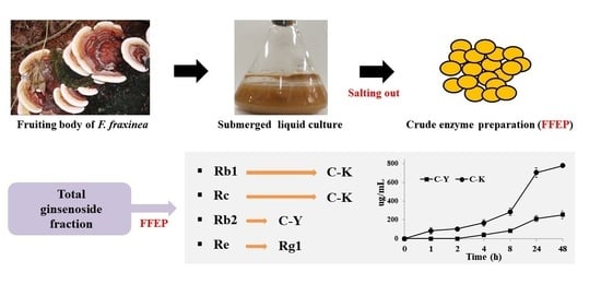
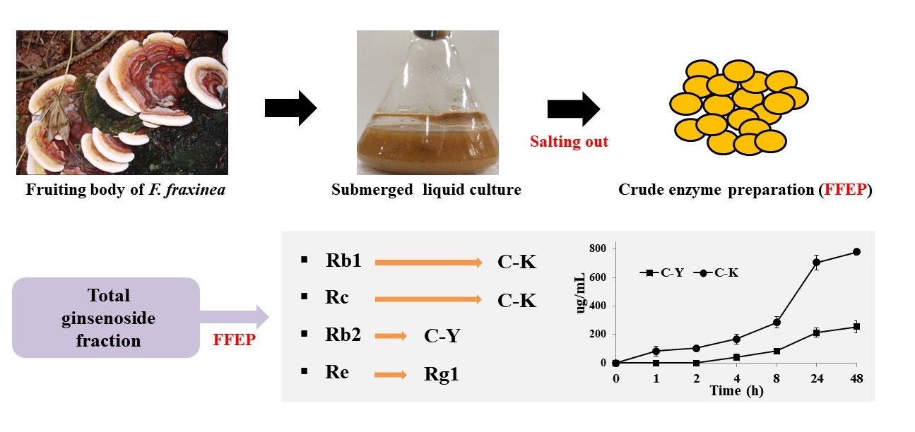
Production of Minor Ginsenosides C-K and C-Y from Naturally Occurring Major Ginsenosides Using Crude β-Glucosidase Preparation from Submerged Culture of Fomitella fraxinea

1
Department of Food Science and Technology, Jeonbuk National University, Jeonju 54896, Korea
2
Institute for Liver and Digestive Diseases, Hallym University, Daehak-ro 1, Chuncheon 24252, Korea
3
Department of Bioresource Engineering, McGill University, Montreal, QC 3A 0G4, Canada
4
Bonghwang Cheonjong Ginseng Research Institute, Daegu 34544, Korea
*
Author to whom correspondence should be addressed.
Submission received: 13 July 2021 / Revised: 2 August 2021 / Accepted: 6 August 2021 / Published: 9 August 2021
(This article belongs to the Special Issue Microbial Biotransformation of Natural Products)

Abstract

:
Minor ginsenosides, such as compounds (C)-K and C-Y, possess relatively better bioactivity than those of naturally occurring major ginsenosides. Therefore, this study focused on the biotransformation of major ginsenosides into minor ginsenosides using crude β-glucosidase preparation isolated from submerged liquid culture of Fomitella fraxinea (FFEP). FFEP was prepared by ammonium sulfate (30–80%) precipitation from submerged culture of F. fraxinea. FFEP was used to prepare minor ginsenosides from protopanaxadiol (PPD)-type ginsenoside (PPDG-F) or total ginsenoside fraction (TG-F). In addition, biotransformation of major ginsenosides into minor ginsenosides as affected by reaction time and pH were investigated by TLC and HPLC analyses, and the metabolites were also identified by UPLC/negative-ESI-Q-TOF-MS analysis. FFEP biotransformed ginsenosides Rb1 and Rc into C-K via the following pathways: Rd → F2 → C-K for Rb1 and both Rd → F2→ C-K and C-Mc1 → C-Mc → C-K for Rc, respectively, while C-Y is formed from Rb2 via C-O. FFEP can be applied to produce minor ginsenosides C-K and C-Y from PPDG-F or TG-F. To the best of our knowledge, this study is the first to report the production of C-K and C-Y from major ginsenosides by basidiomycete F. fraxinea.

Graphical Abstract

1. Introduction

Ginseng (root of Panax ginseng Meyer) has been traditionally applied as an herbal medicine and healthy food for maintenance and restoration of human homeostasis and for enhancement of the immune system, as well as for its adaptogenic and anti-aging properties in East Asian countries, mainly Korea, China and Japan [1,2,3]. Nowadays, it is attracting more attention due to its potential in pharmaceutical and healthy/functional food development. Recent research results reported that ginseng root and processed ginseng products (especially red ginseng extract) have various biological activities, including enhancement of the immune system; anti-fatigue, antioxidant and anti-aging properties; and improvement of blood circulation and climacteric symptoms with anti-tumor, anti-diabetic, neuroprotective and anti-stress effects [4,5,6,7].
Ginseng root contains various biologically active ingredients, including saponins (ginsenosides), polysaccharides and phenolic and nitrogenous compounds [6,8,9]. Among the ginsenosides (dammarane-type and oleanane-type) present in ginseng, dammarane-type ginsenosides can be classified into two types—i.e., protopanaxadiol (PPD)-type and protopanaxatriol (PPT)-type—according to bound position or the number of hydroxyl groups present in their aglycones. Ginsenosides Rb1, Rb2, Rc, Rd, Rh2, C-O, C-Y and C-K belong to the PPD-type ginsenosides group, and Re, Rg1, Rg2 and Rh1 are in the PPT-type group [10,11].
Until recently, more than 80 ginsenosides have been found in ginseng and its processed products [11,12,13]. Among these compounds, ginsenosides Rb1, Rb2, Rc, Re and Rg1 account for about 80–90% of the total ginsenosides, while minor ginsenosides including C-O, C-Y and C-K are present in trace amounts or not detected in raw ginseng [14,15]. However, the absorption of major ginsenosides such as Rb1, Rb2, Rc, Re and Rg1 in the intestinal tract is reported to be difficult due to their large molecular size, poor water solubility and poor cell membrane permeability. In contrast, minor ginsenosides can be effectively absorbed by the human intestine due to their relatively lower molecular size and the ease of cell membrane permeability [16,17,18,19]. Especially, C-K, a major intestinal metabolite of the PPD-type ginsenosides, has been attracting considerable interest due to its various biological activities, including anti-tumor, anti-inflammatory, anti-diabetic, anti-aging, antiallergic, hepatoprotective and anti-skin aging properties [20,21,22,23]. Therefore, many studies have been conducted to convert major ginsenosides into minor ginsenosides by various methods, such as physico-chemical methods [24,25], microbial fermentation and enzymatic biotransformation [26,27,28]. However, physico-chemical methods can bring about the random hydrolysis of attached sugars and unexpected side reactions, including hydration and epimerization [25]. In contrast, enzymatic biotransformation is considered to be a more desirable approach due to high substrate specificity, fewer by-products, higher biotransformation efficiency and an eco-friendlier process than physico-chemical methods [26,27].
In particular, ginsenosides Rc and Rb2 have α-L-arabinofuranosyl or α-L-arabinopyranosyl moiety, respectively, at the C-20 terminal position, with three β-glucosyl moieties in PPD-type aglycone. As a result, three types of enzymes (α-L-arabinofuranosidase, α-L-arabinopyranosidase and β-glucosidase) or glycosidases possessing multiple enzyme activity may be required to convert ginsenosides Rb1, Rc or Rb2 into their subsequent minor ginsenosides. However, most of the previously reported studies on the production of minor ginsenosides from major ginsenosides by enzyme-mediated transformation used purified individual ginsenosides or highly purified enzyme sources of high cost. It is therefore preferable to use crude ginseng extracts as a substrate and crude enzyme preparations or whole cells of relatively low cost to economically produce minor ginsenosides from major ginsenosides [26,27,28].
On the other hand, basidiomycetes, commonly known as mushrooms, have received much attention as potentially valuable enzyme sources for production of bioactive compounds [29,30]. Previous studies reported that minor ginsenosides were found to be produced with a high yield from major ginsenosides using crude enzyme preparation obtained from the mycelia of Armillaria mellea [31,32]. During our ongoing investigation on biotransformation of major ginsenosides using crude enzyme extracts obtained from various mushrooms, FFEP has a remarkable biotransformation activity of PPD-type major ginsenosides into the minor ginsenosides C-K or C-Y. Basidiomycete F. fraxinea (Polyporaceae), which is mainly distributed in the East Asia region, is one of the medicinal mushrooms, and its fruiting body (Korean name; Jangsu mushroom) is traditionally used in Korean folk medicine. In this study, we report on the production of C-K and C-Y from PPDG-F and TG-F containing Rb1, Rc and Rb2 as major ginsenosides by an FFEP-mediated biotransformation method.

2. Results and Discussion

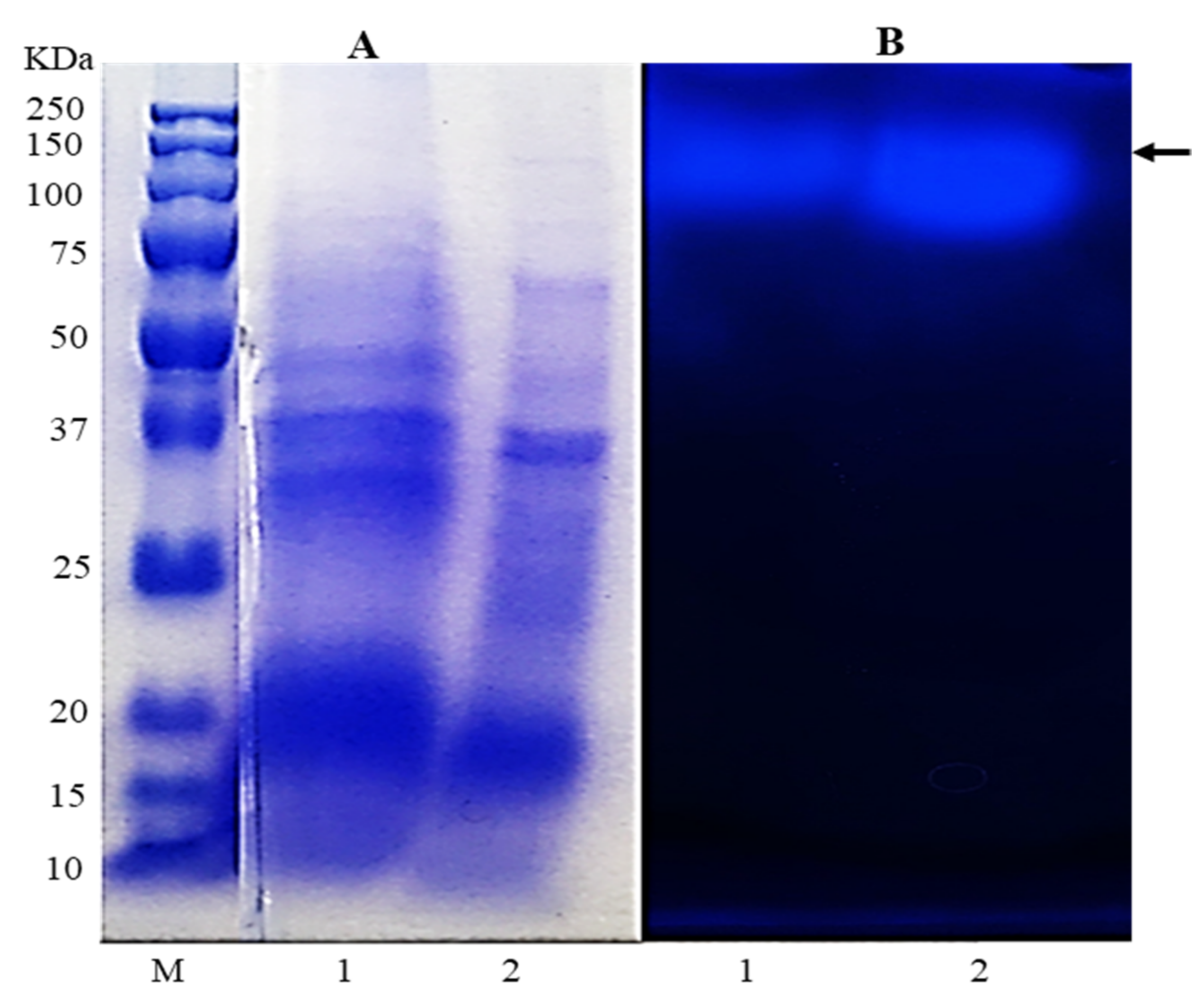
2.1. β-Glucosidase Activity in FFEP

Mushrooms are not only valuable natural resources for healthy foods and therapeutics, but they are also very interesting fungi that secrete various glycoside hydrolases that can be applicable to biotransformation of various glycosides [29,30,33]. A few studies have reported that fermentation of ginseng extract or ginsenoside-rich fraction by mushrooms could increase the content of minor ginsenosides in fermented products [34,35,36]. These results demonstrate that the enzymes from different strains of mushrooms may convert major ginsenosides to minor ginsenosides by their glycoside hydrolases. Our preliminary study revealed that FFEP has a potent activity for biotransformation of PPD-type ginsenosides (Rb1, Rc and Rb2).
As shown in Figure 1, the result of SDS-PAGE and native PAGE showed that FFEP contained β-glucosidase, and its molecular weight was estimated to be between 100 and 150 kDa based on relative migration distance of protein molecular weight markers in electrophoresis. The β-glucosidase activity in FFEP toward p-NP-β-D-glucopyranoside was 0.45 U/mg (as a dry weight base). Based on their substrate specificity, microbial β-glucosidases can also be divided into three groups—i.e., group Ⅰ, which possesses only aryl β-glucosidase activities; group Ⅱ, which possesses cellobioses; and group Ⅲ, which contains β-glucosidases showing wide substrate specificity [37]. Among them, the β-glucosidases that belong to group Ⅲ can hydrolyze α-1,3, α-1,4 and α-1,6 glycosidic linkages as well as β-1,2, β-1,4 and β-1,6 glycosidic linkages. The results of the present study show that the β-glucosidase from FFEP used in this study has enzymatic properties belonging to group Ⅲ type β-glucosidase rather than to group Ⅰ or Ⅱ.

2.2. Biotransformation of PPDG-F by FFEP

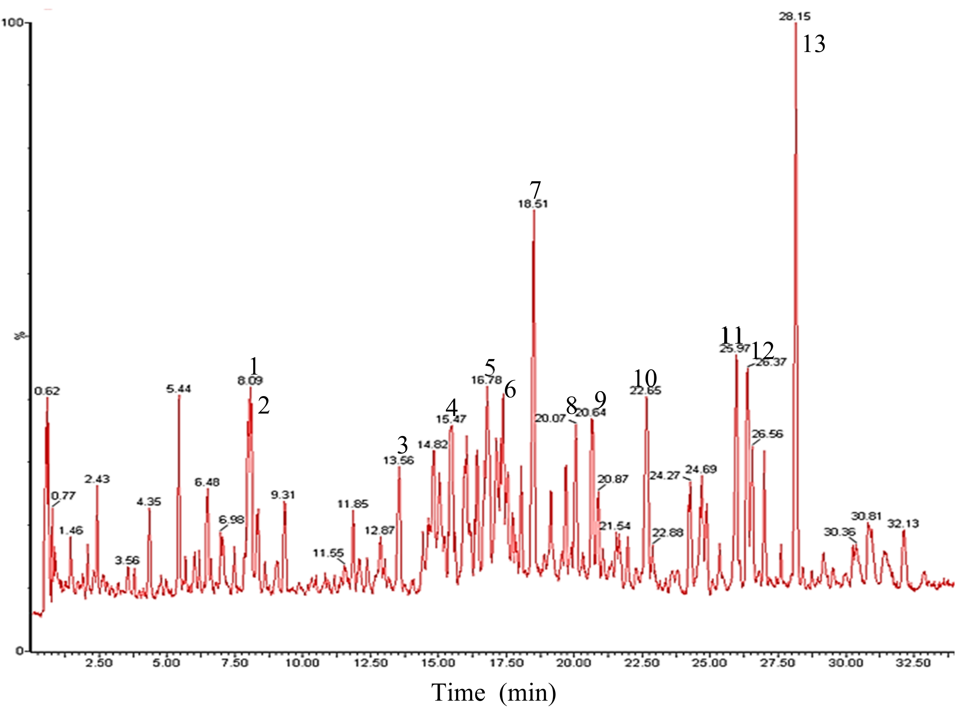
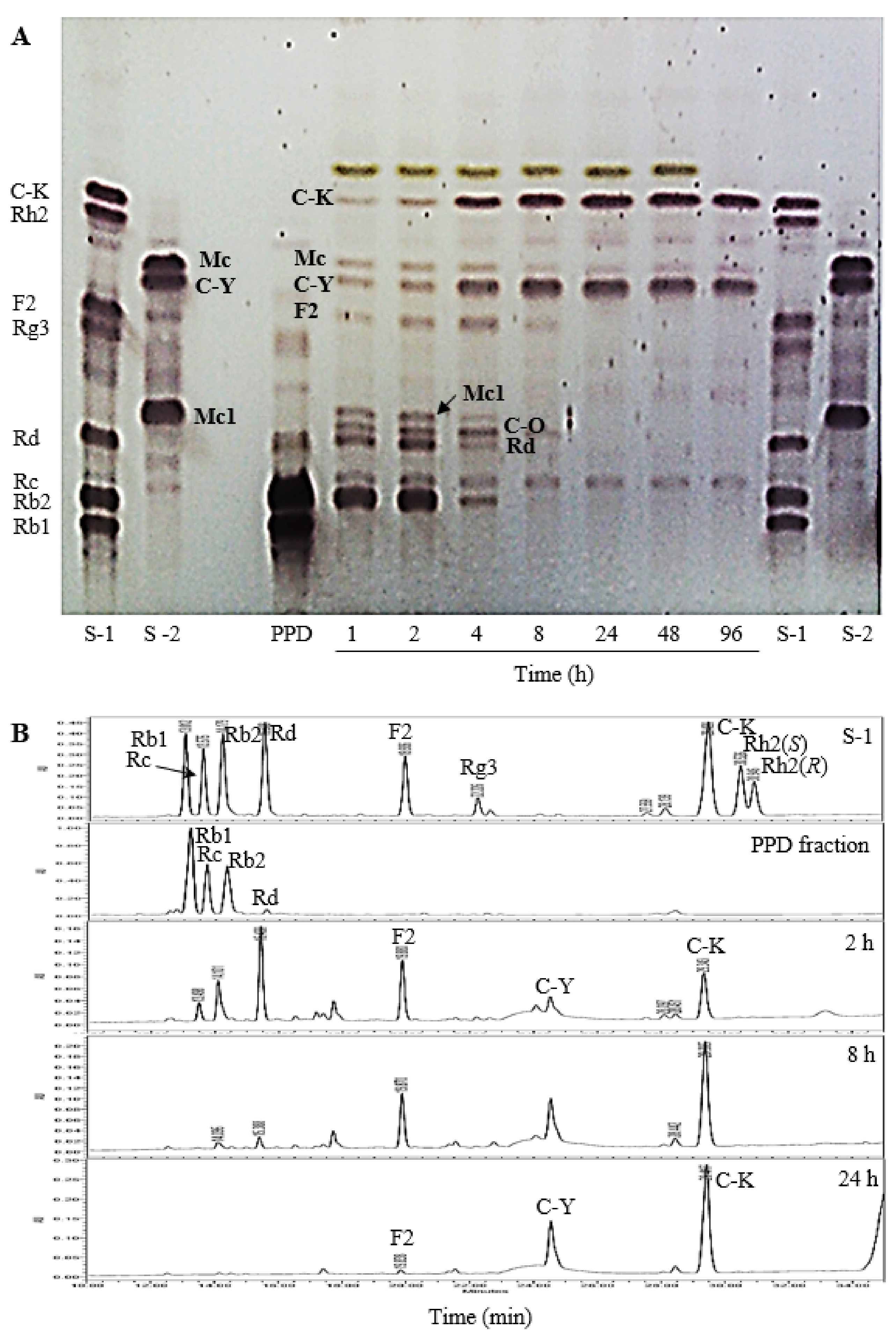
2.2.1. UPLC/ESI-Q-TOF-MS Analysis

Recently, UPLC/Q-TOF-MS has been efficiently applied for the structural identification of ginsenosides [38,39]. In the present study, the reaction mixture of PPDG-F with FFEP was withdrawn after 8 h reaction and then was analyzed by UPLC/negative-ESI-Q-TOF-MS to characterize the biotransformation products. The total ion current chromatogram (TIC) is shown in Figure 2.
The identifications of constituents detected in the reaction mixture were also compared with those of individual ginsenoside standards. The results are listed in Table 1. The largest peak 13 in the TIC chromatogram showed protonated ion peaks at m/z 621.6896 [M − H] and m/z 667.7121 [M − H + HCOOH], corresponding to the molecular formula C36H62O8 with molecular weight (MW) of m/z 622.4445. From these results, the compound of peak 13 was determined as C-K. Peak 11 and 12 showed the same molecular ion peaks at m/z 753.7594 [M − H] and m/z 753.7576 [M − H], which corresponded to C41H70O12 as the MW, respectively.
Accordingly, both compounds were estimated as C-Mc and C-Y originating from ginsenosides Rc and Rb2, respectively. Peaks 8 and 9 in Q-TOF–MS analyses were observed as ion peaks at m/z 915.8387 and m/z 915.8281 [M − H] in addition to m/z 961.8601 and m/z 961.8512 [M − H + HCOOH], which corresponds to C47H80O17 as the MW, respectively. In addition, HPLC retention times of peaks 8 and 9 were consistent with those of authentic standards of C-Mc1 and C-O, respectively, Therefore, peak 8 and 9 were estimated as C-Mc1 and C-O, respectively. Similarly, peaks 7, 10, 11, 12 and 13 were elucidated as ginsenoside Rd, F2, C-Mc, C-Y and C-K, respectively, by comparing the Q-TOF-MS data and HPLC retention time with those of reliable standards.

2.2.2. The Effect of Reaction Time

During biotransformation of PPDG-F by FFEP, it was regularly monitored by TLC (Figure 3A) and HPLC (Figure 3B). Ginsenosides Rb1, Rc and Rb2 were transformed into their intermediate products, mainly ginsenoside Rd, F2, C-Mc1 or C-O, between 1 and 4 h of reaction, and thereafter, these compounds were transformed into C-K or C-Y by increasing the reaction time. However, the concentration of C-K and C-Y increased continuously and reached maxima at 8 h of reaction, and thereafter, no meaningful changes were observed. These results suggest that FFEP can be used as an enzyme source to produce C-K and C-Y or their mixture from PPDG-F.

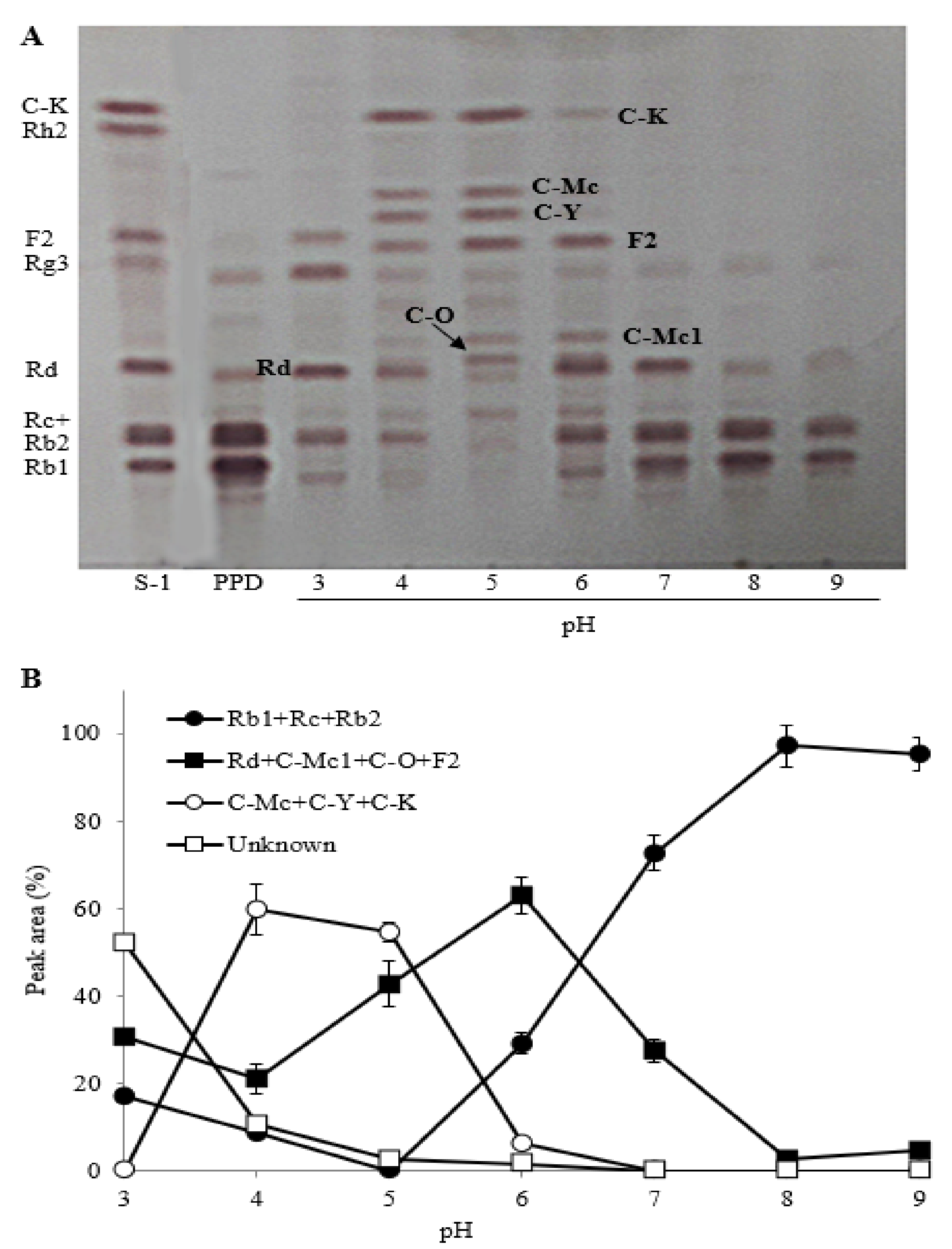
2.2.3. The Effect of Reaction pH

The biotransformation of PPDG-F by FFEP was investigated at a range from pH 3 to 9. The TLC analysis showed that the optimum biotransformation of Rb1, Rc and Rb1 occurred between pH 4 and 5 (Figure 4). Under this pH range, most of the ginsenoside Rb1, Rc and Rb2 were transformed into C-K and C-Y via Rd, F2, C-Mc1 or C-O. PPD-type ginsenosides in acidic buffer solution (pH 3) were hydrolyzed into unknown compounds, which did not match with the Rf values of authentic standards on TLC (Figure 4A). Ginsenosides can be easily deglycosylated and dehydrated at the C-20 position of aglycones when heated under mild acidic condition [24,25]. When each reaction mixture was investigated by HPLC analysis, most of the parent ginsenosides (Rb1, Rc and Rb2) were biotransformed into minor ginsenosides (C-Mc, C-Y or C-K) between pH 4 and 5, whereas efficient biotransformation could not occur in neutral and alkaline ranges (pH 6–9) (Figure 4B). These results suggest that the biotransformation of PPD-type ginsenosides by FFEP was more desirable at weak acidic condition (pH 4–5) rather than at neutral and basic conditions. Ginsenoside-hydrolyzing enzymes from different microbial sources differ from each other in terms of enzymatic properties and the mode of ginsenoside hydrolysis. Many microbial enzymes showed optimal ginsenoside-hydrolyzing activities at pH 4–5, but neutral β-glycosidases have also been reported from Pythium irregular, Fusobacterium sp. Bacteroides sp. and recombinant β-glucosidase from Microbacterium esteraromaticum, with an optimal pH range of 6.5–7.0 [21,26].

2.3. Biotransformation of TG-F by FFEP

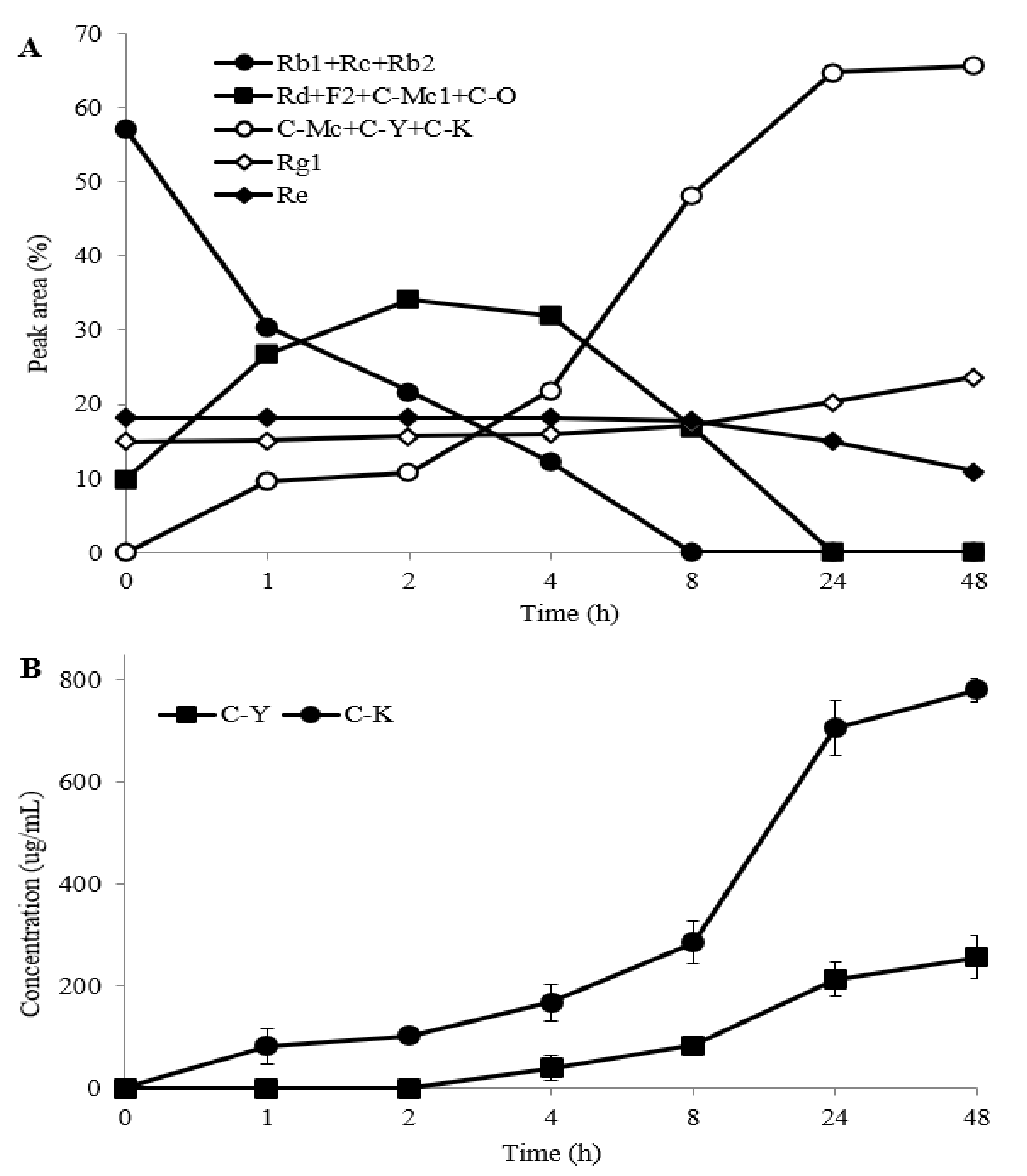
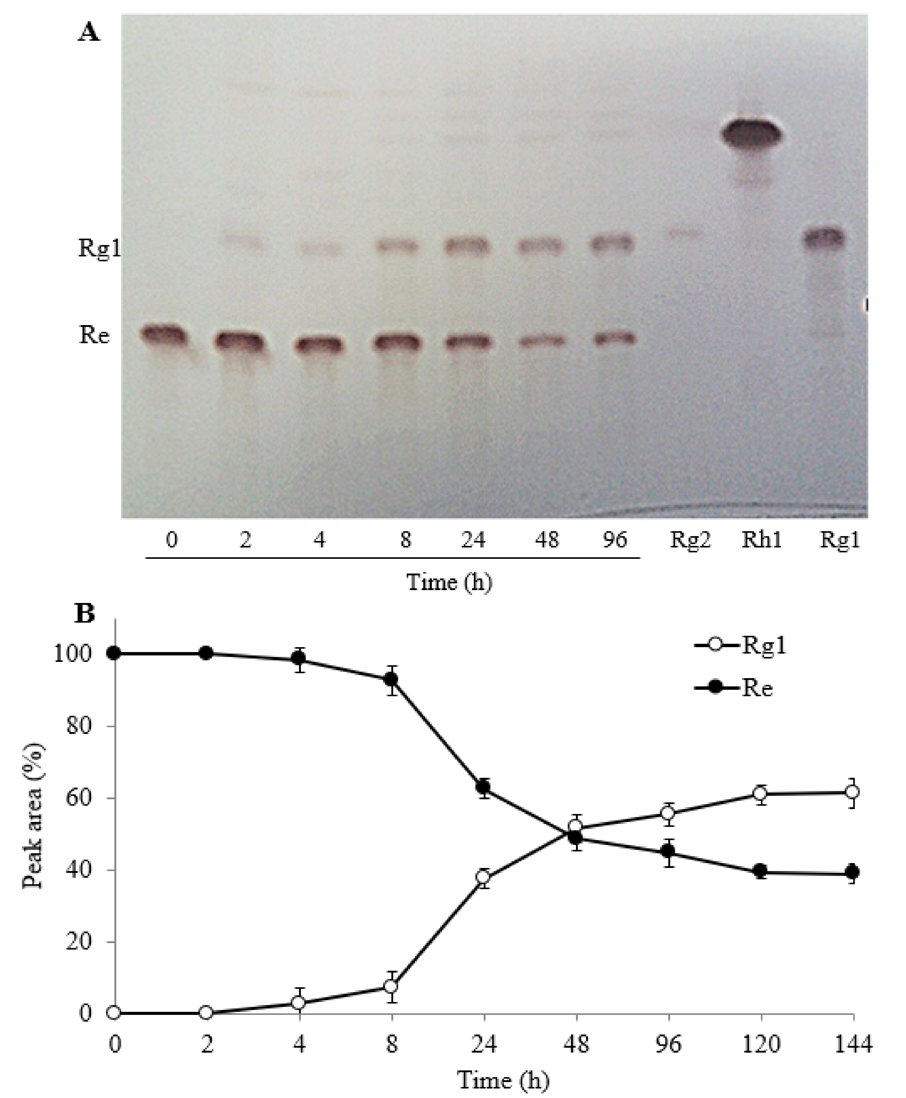
2.3.1. The Effect of Reaction Time

Reaction mixture samples were taken at different time intervals to investigate the biotransformation properties of TG-F by FFEP and were analyzed by HPLC. As shown in Figure 5A, ginsenosides Rb1, Rc, Rb2 and Rd that comprised the major portion of TG-F were rapidly transformed into F2, C-Mc1 and C-O in the early stage of the reaction (1–4 h). After 8 h reaction, most of the parent ginsenosides (Rb1, Rc, Rb2) and intermediate products (Rd, F2, C-Mc and C-O) were transformed into C-K and C-Y. When the products after 22–48 h reaction were analyzed by HPLC, C-K and C-Y were mainly observed, but C-Mc was hardly detected. These results suggest that C-Mc1 and C-Mc formed from ginsenoside Rc were further hydrolyzed to C-K. Figure 5B shows the time course for the production of C-K and C-Y from TG-F by FFEP. The contents of C-K and C-Y increased with increasing reaction time and yielded 0.780 mg C-K and 0.26 mg C-Y, respectively, from 2 mg TG-F/mL after 48 h reaction.
On the other hand, during biotransformation of TG-F by FFEP, ginsenoside Re, one of the major PPT-type ginsenosides, was gradually transformed into ginsenoside Rg1 with increasing reaction time. To verify whether PPT-type ginsenosides Re and Rg1 can be hydrolyzed by FFEP, the reaction mixture of Re or Rg1 with FFEP was taken at regular time intervals during 144 h reaction in acetate buffer solution (pH 4.5) and then was analyzed by TLC and HPLC. Ginsenoside Re was gradually transformed into Rg1 in a time-dependent manner (Figure 6A). When ginsenoside Re was used as a substrate (1 mg/mL), about 50% of ginsenoside Re after 48 h reaction was transformed into Rg1 and 60% was transformed after 144 h (Figure 6B). However, additional biotransformation of ginsenoside Rg1 into Rh1 or F1 was not observed.

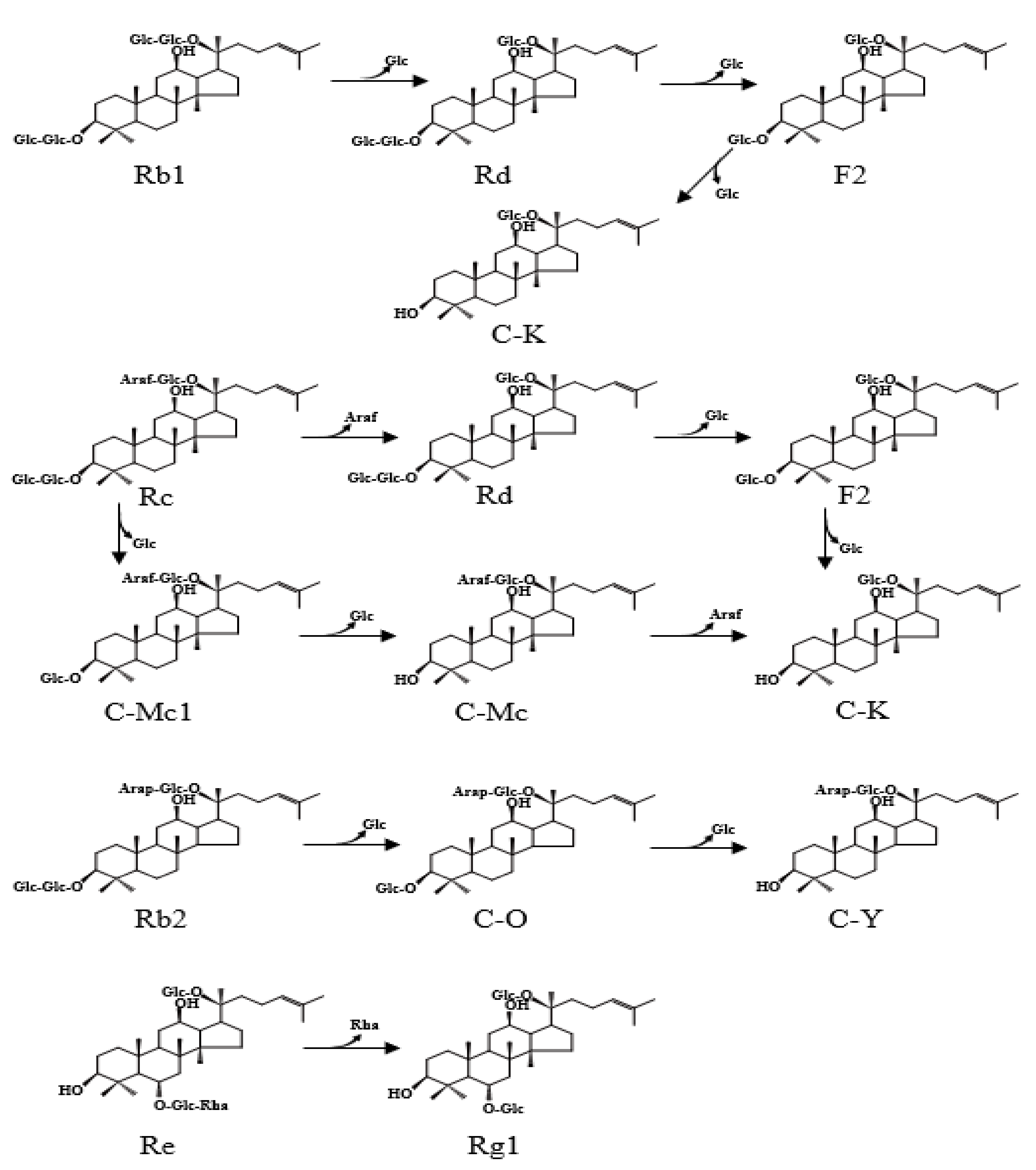
2.3.2. Biotransformation Pathway of Ginsenosides

Ginsenoside Rc contains one α-arabinofuranosyl moiety with three β-glucosyl moieties, and Rb2 contains one α-arabinopyranosyl moiety with three β-glucosyl moieties, whereas Rb1 contains four β-glucosyl moieties in PPD-type aglycone, respectively. Therefore, the biotransformation pathways may depend on the stereospecific preferences of the enzymes toward the glycosidic linkages attached to the C-3 and C-20 positions of the aglycone [26]. As shown in Figure 7, the biotransformation of ginsenosides by FFEP is thought to occur along the following pathways: Rb1 → Rd → F2 → C-K for Rb1 as a substrate, both Rc → Rd → F2 → C-K and Rc → C-Mc1 → C-Mc → C-K for Rc and Rb2 → C-O → C-Y for Rb2.
The preference in biotransformation pathways of PPD-type ginsenosides also depends on microbial sources. Biotransformation of Rb1 into C-K can occur through pathways of two types, depending on its structure. One pathway is through hydrolysis of β-(1→6)-glucosyl moiety at the C-20 position and β-(1→2)-glucosyl moiety at the C-3 of aglycone for the formation and Rd and F2 as the intermediates. Another one is by sequentially hydrolyzing the β-(1→2)-glucosyl (outer) and β-glucosyl moieties (inner) at the C-3 position of aglycone for the formation of gypenoside XVII and gypenoside LXXV as the intermediates. Microbial β-glucosidases from Fusodobacterium K-10, Paecilomyces bainier, Sulfolobus solfataricusm, Cladosporium fulvum and Aspergillus oryzae transformed ginsenoside Rb1 into C-K through the pathway of Rb1 → Rd → F2 → C-K, while Terrabacter ginsenosidimutans performed the transformation via the Rb1 → gypenoside ⅩⅦ → gypenoside LⅩⅩⅤ → C-K pathway [21,26]. Ginsenoside Rc can also be hydrolyzed via multiple pathways by microbial β-glucosidases. The results from the present study show that biotransformation of ginsenoside Rc by FFEP occurred through two pathways, as presented in Figure 7. First, FFEP hydrolyzed the α-(1→6)-arabinofuranosidic linkage (outer) linked to the C-20 position of the aglycone to produce Rd from Rc, followed by hydrolysis of β-(1→2)-glucosidic (outer) and β-glucosidic (inner) linkages linked to the C-3 position to produce C-K. At the same time, this enzyme preparation hydrolyzed β-(1→2)-glucosidic (outer) and inner β-glucosidic linkages linked to the C-3 position to produce C-Mc1 and C-Mc from Rc. Thereafter, C-Mc was further hydrolyzed into C-K as a final product.
The ginsenoside Rb2 molecule contains one α-(1→6)-arabinopyranosyl (outer) and one β-glucopyranosyl moiety (inner) at the C-20 position, with two β-glucopyranosyl moieties at the C-3 position of aglycone. Accordingly, Rb2 can also be hydrolyzed by multiple pathways. Based on the results obtained by TLC and HPLC analysis, we suggest that Rb2 by FFEP was biotransformed into C-Y via C-O (Figure 7). This enzyme attacked the outer β-(1→2)-glucosidic linkage linked to the C-3 position of aglycone to produce C-O from Rb2 and then followed by hydrolysis of the inner β-glucosidic linkage linked to the C-3 position to produce C-Y from C-O. In this study, ginsenoside Re, one of the PPT-type ginsenosides, was gradually converted into ginsenoside Rg1 (Figure 7). This result indicates that FFEP possessed hydrolytic activity toward β-glucosyl (1-1)-α-rhamnosyl linkage at the C-6 position of the PPT-type aglycone. Until now, enzymatic transformation of PPT-type ginsenosides were rarely reported [28,40,41]. However, FFEP had no hydrolytic activity toward β-glucosidic linkages at the C-6 or C-20 positions of ginsenoside Rg1.

3. Materials and Methods

3.1. Reagents

β-D-Glucopyranoside, α-L-arabinofuranoside, α-L-arabinopyranoside of p-nitrophenol (p-NP) as chromogenic substrates, bovine serum albumin (BSA), sodium dodecyl sulfate (SDS), 4-methylumbelliferyl-β-D-glucopyranoside (MUG) and dialysis membrane tubing (14,000 cutoff) were purchased from Sigma-Aldrich Korea (Gangnam-gu, Seoul, Korea). Mini protean TGX precast gel and protein molecular markers and Coomassie blue R-250 were purchased from Bio-Rad Laboratories, Inc. (Kangnam-gu, Seoul, Korea). Silica gel TLC plate was purchased from Merck KGaA (Darmstadt, Germany). Deionized water was manufactured by using a water purification system (model New Human Power I, Human Corp., Songpa-ku, Seoul, Korea). Acetonitrile and methanol (HPLC grade) were purchased from Avantor Performance Materials Korea (Suwon, Gyeonggi-do, Korea). Other reagents were obtained from a commercial source (Daejung Chemical Co., Siheung, Gyonggi-do, Korea).

3.2. Preparation of Ginsenosides and Ginseng Extracts

Authentic standards (ginsenosides Rb1, Rb2, Rc, Rd, Rg3, F2, Rh2, C-K, Re and Rg1), PPDG-F and TG-F were kindly provided by the Korea Ginseng Research Institute (Daejeon, Korea). The compositions of Rb1, Rc, Rb2 and Rd in the PPDG-F were 50.8, 21.2, 27.7 and 0.3%, respectively, as peak area in HPLC analysis. Authentic standards of C-Mc1, C-Mc, C-O and C-Y were prepared from ginsenoside Rc or Rb2 using crude enzyme preparation isolated from the mycelial mass of Armillaria mellea according to our previously reported methods [31,32]. Separately, ginsenoside Rb1, Rc and Rb2 were also isolated from PPDG-F according to a previously reported method [10]. The isolated compounds were identified by comparison of NMR and Q-TOF-MS data and HPLC retention times with those of authentic standards.

3.3. Microorganism

Strain of F. fraxinea (KACC 42289) was provided by the Korean Agricultural Culture Collection (KACC) of Rural Development Administration (Wanju, Jeonbuk, Korea). The strain was kept on an agar slant media including 2% malt extract at 4 °C.

3.4. Mushroom Cultivation

Strain of F. fraxinea was previously incubated on potato dextrose agar for 2 weeks at 24–25 °C. After the incubated strain was inoculated into saccharified malt media (11°Brix), it was cultivated for 2 weeks at 24–25 °C with orbital shaking (120 rpm). Scale-up cultivation of the strain was performed in a 3-L Erlenmeyer flask containing 1 L of malt media (11°Brix) for 2 weeks at 24–25 °C with shaking (120 rpm). All nutrient media were sterilized at 121 °C for 30 min.

3.5. Preparation of Crude Enzyme

The cultivated liquid culture with mycelial mass was homogenized at maximum speed with a homogenizer (Omni International, Kennesaw, GA, USA) for 1 min and then centrifuged at 12,000× g for 20 min. After the supernatant was treated with ammonium sulfate powder (30% saturation) for 6 h, the solution was centrifuged to remove the protein precipitate and then ammonium sulfate was added in the supernatant to 80% saturation and maintained overnight to induce the protein precipitate including β-glucosidase and followed by centrifuging (12,000× g) for 20 min. After the precipitate was dissolved in a small volume of 10 mM sodium acetate buffer (pH 4.5) and purified by overnight dialysis with 10 mM acetate buffer (pH 4.5), the supernatant obtained was lyophilized for 4 days. All experiments were carried out at 4 °C unless otherwise mentioned.

3.6. Enzyme Electrophoresis

SDS-polyacrylamide gel electrophoresis (PAGE) was done with a precast gel (12%) for 1 h at 110 mA according to Laemmli’s method [42]. The gel was stained with Coomassie blue R-250 and destaining of the stained gel was performed with distilled water containing methanol and acetic acid (each 10%, v/v). Native PAGE was performed with a precast gel (12%) under the same conditions as above. Thereafter, the gel was maintained in acetate buffer (0.1 M, pH 4.5) containing 0.1% MUG (w/v) for 30 min at 37 °C. The band due to aglycone (4-methylumbelliferol) liberated in the gel was detected under UV light (365 nm).

3.7. Enzyme Activity and Protein Assays

β-Glucosidase activity was assayed according to the method of Mfombep et al. [30], with slight modifications. The reaction mixture (1.0 mL)—containing 0.1 mL of p-NP-β-D-glucopyranoside (10 mM) as a substrate, enzyme solution (0.1 mL) and 0.1 M acetate buffer (pH 4.5) (0.8 mL)—was incubated for 30 min at 37 °C. After termination of the reaction by adding 0.5 M sodium carbonate (1.0 mL), the liberated p-nitrophenol (aglycone) was measured using a UV-Vis spectrophotometer (UV-1601, Shimadzu, Kyoto, Japan) at 400 nm. The amount of p-nitrophenol liberated was quantified based on a calibration curve prepared using an authentic standard. An amount of 1 unit of enzyme activity was defined as the amount of enzyme required to release 1 μM p-nitrophenol per min. Protein content was assayed according to the Bradford method, using BSA as a standard [43].

3.8. Enzymatic Biotransformation

The reaction mixtures (2.0 mL) each containing 2.0 mg of PPDG-F or TG-F in methanol (0.2 mL) and FFEP possessing 2.7 U as β-glucosidase activity (6.0 mg, 0.45 U/mg) in 1.8 mL of 0.1 M acetate buffer (pH 4.5) were incubated at 45 °C with shaking (90 rpm), respectively. The reaction mixtures were sampled periodically and then heated for 10 min in boiling water to inactivate the enzyme, followed by extraction twice (each 2 mL) with saturated n-butanol with water. After the butanol extract was washed with water (twice) to minimize residual sugars, the solution was subjected to dryness in vacuo. The residue was dissolved in 1.0 mL methanol, and was analyzed by TLC, HPLC or ultra-performance liquid chromatography/negative-time-of-flight mass spectrometry (UPLC/Q-TOF-MS). The effect of pH on the biotransformation of PPDG-F by FFEP was incubated for 24 h at 45 °C in the following buffers (each at 0.1 M): glycine-HCl (pH 3), sodium acetate (pH 4 and 5), sodium phosphate (pH 6 and 7), Tris-HCl (pH 8), and glycine-NaOH (pH 9). Separately, the mixture (20 mL) containing TG-F (20 mg) in methanol (2 mL) and FFEP containing β-glucosidase (27 U, 60 mg) in 18 mL of 0.1 M acetate buffer (pH 4.5) was reacted for 48 h at 45 °C. The reaction mixture (2.0 mL) was sampled periodically and then digoxin (480 μg/mL) was added as an internal standard, followed by extraction twice with saturated n-butanol with water. The residues after vacuum evaporation were dissolved in methanol (1.0 mL).

3.9. General Analytical Methods

The analyses by TLC, HPLC and UPLC/negative-ESI-Q-TOF-MS were conducted as described in our previous studies [31,32]. The ratio of each peak in HPLC analysis was calculated based on relative percentage of the detected peak area. The concentrations of C-K and C-Y produced during biotransformation of TG-F were quantified by the internal standard method using digoxin as an internal standard without considering the detector response factor. HPLC analyses for quantitation of C-K and C-Y in FFEP-mediated reaction mixtures of TG-F were done in triplicate, and the results are presented as average ± standard deviation (SD).

4. Conclusions

Production of minor ginsenosides by using highly purified major ginsenosides or purified enzymes is an economically disadvantaged, complicated and time-consuming process. Crude enzyme preparation isolated from submerged culture of F. fraxinea containing β-glucosidase exhibited potent biotransformation activity toward PPD-type ginsenosides such as Rb1, Rc and Rb2. Furthermore, ginsenoside Re was converted into Rg1, resulting in a transformation yield of about 50% after 48 h reaction. The results of the present study suggest that FFEP can be used to produce valuable minor ginsenosides C-K and C-Y from PPDG-F or TG-F, with high biotransformation efficiency. However, further studies are necessary to optimize the cultivation condition of this mushroom strain, improve the recovery of the enzyme from a liquid culture, as well as optimize the ratio of enzyme-to-substrate for improving the biotransformation yield.

Author Contributions

Conceptualization and methodology, M.-K.K.; investigation and resources, Y.-H.K.; funding acquisition, M.-K.K.; formal analysis, S.-R.K.; project administration, D.-W.K.; data curation, W.-J.L. and J.U.; writing—original draft preparation, Y.-H.K.; writing—review and editing, Y.A.G. All authors have read and agreed to the published version of the manuscript.

Funding

This paper was supported by research funds from Jeonbuk National University in 2021.

Institutional Review Board Statement

Not applicable.

Informed Consent Statement

Not applicable.

Data Availability Statement

The data that support the findings of this study are available from the corresponding author, upon reasonable request.

Acknowledgments

We are grateful to the Korea Ginseng Research Institute and the Korea Ginseng Corporation for support of UPLC/Q-TOF-MS analysis and provision of ginsenoside fractions (PPDG-F and TG-F) and ginsenoside standards.

Conflicts of Interest

All contributing authors declare no conflicts of interest.

Sample Availability

Samples of the compounds are available from the authors.

References

  1. Nocerino, E.; Amato, M.; Izzo, A.A. The aphrodisiac and adaptogenic properties of ginseng. Fitoterapia 2000, 71, S1–S5. [Google Scholar] [CrossRef]
  2. Radad, K.; Gille, G.; Liu, L.; Rausch, W.D. Use of ginseng in medicine with emphasis on neurodegenerative disorders. J. Pharmacol. Sci. 2006, 100, 175–186. [Google Scholar] [CrossRef] [PubMed] [Green Version]
  3. He, Y.C.; Yang, J.; Lv, Y.H.; Chen, J.C.; Yin, F.; Huang, J.H.; Zheng, Q.S. A review of ginseng clinical trials registered in the WHO international clinical trials registry platform. BioMed. Res. Inter. 2018, 1843142. [Google Scholar] [CrossRef] [Green Version]
  4. Park, J.D.; Rhee, D.K.; Lee, Y.H. Biological activities and chemistry of saponins from Panax ginseng C. A. Meyer. Phytochem. Rev. 2005, 4, 159–175. [Google Scholar] [CrossRef] [Green Version]
  5. Choi, K.T. Botanical characteristics, pharmacological effects and medicinal components of Korean Panax ginseng C. A. Meyer. Acta Pharmacol. Sin. 2008, 29, 1109–1118. [Google Scholar] [CrossRef] [PubMed] [Green Version]
  6. Park, S.Y.; Park, J.H.; Kim, H.S.; Lee, C.Y.; Lee, H.J.; Kang, K.S.; Kim, C.E. Systems-level mechanisms of action of Panax ginseng: A network pharmacological approach. J. Ginseng Res. 2018, 42, 98–106. [Google Scholar] [CrossRef]
  7. Shergis, J.L.; Zhang, A.L.; Zhou, W.; Xue, C.C. Panax ginseng in randomised controlled trials: A systematic review. Phytother. Res. 2013, 27, 949–965. [Google Scholar] [CrossRef] [PubMed]
  8. Kim, D.H. Chemical diversity of Panax ginseng, Panax quinquifolium, and Panax notoginseng. J. Ginseng Res. 2012, 36, 1–15. [Google Scholar] [CrossRef] [Green Version]
  9. Ru, W.W.; Wang, D.L.; Xu, Y.P.; He, X.X.; Sun, Y.E.; Qian, L.Y.; Zhou, X.S.; Qin, Y.F. Chemical constituents and bioactivities of Panax ginseng (C. A. Mey.). Drug Discov. Ther. 2015, 9, 23–32. [Google Scholar] [CrossRef] [PubMed] [Green Version]
  10. Sanada, S.; Kondo, N.; Shoji, J.; Tanaka, O.; Shibata, S. Studies on the saponins of ginseng. Ⅰ. Structures of ginsenoside-Ro, -Rb1, -Rb2, -Rc, and -Rd. Chem. Pharm. Bull. 1974, 22, 421–428. [Google Scholar] [CrossRef] [Green Version]
  11. Christensen, L.P. Ginsenosides chemistry, biosynthesis, analysis, and potential health effects. Adv Food Nutr. Res. 2009, 55, 1–99. [Google Scholar] [CrossRef] [PubMed]
  12. Shin, B.K.; Kwon, S.W.; Park, J.H. Chemical diversity of ginseng saponins from Panax ginseng. J. Ginseng Res. 2015, 39, 287–298. [Google Scholar] [CrossRef] [Green Version]
  13. Kim, J.H. Cardiovascular diseases and Panax ginseng: A review on molecular mechanisms and medical applications. J. Ginseng Res. 2012, 1, 16–26. [Google Scholar] [CrossRef] [PubMed] [Green Version]
  14. Sharma, A.; Lee, H.J. Ginsenoside compound K: Insights into recent studies on pharmacokinetics and health-promoting activities. Biomolecules 2020, 10, 1028. [Google Scholar] [CrossRef]
  15. Tawab, M.A.; Bahr, U.; Karas, M.; Wurglics, M.; Schubert-Zsilavecz, M. Degradation of ginsenosides in humans after oral administration. Drug Metab. Dispos. 2003, 31, 1065–1071. [Google Scholar] [CrossRef] [Green Version]
  16. Kobashi, K.; Alkao, T. Relation of intestinal bacteria to pharmacological effects of glycosides. Biosci. Microflora 1997, 16, 1–7. [Google Scholar] [CrossRef] [Green Version]
  17. Hasegawa, H. Proof of the mysterious efficacy of ginseng: Basic and clinical trials: Metabolic activation of ginsenoside: Deglycosylation by intestinal bacteria and esterification with fatty acid. J. Pharmacol. Sci. 2004, 95, 153–157. [Google Scholar] [CrossRef] [Green Version]
  18. Xu, Q.F.; Fang, X.L.; Chen, D.F. Pharmacokinetics and bioavailability of ginsenoside Rb1 and Rg1 from Panax notoginseng in rats. J. Ethnopharmacol. 2003, 84, 187–192. [Google Scholar] [CrossRef]
  19. Won, H.J.; Kim, H.I.; Park, T.J.; Kim, H.M.; Jo, K.H.; Jeon, H.J.; Ha, S.J.; Hyun, J.M.; Jeong, A.R.; Kim, J.S.; et al. Non-clinical pharmacokinetic behavior of ginsenosides. J. Ginseng Res. 2019, 43, 354–360. [Google Scholar] [CrossRef]
  20. Shin, K.C.; Choi, H.Y.; Seo, M.J.; Oh, D.K. Compound K production from red ginseng extract by β-glycosidase from Sulfolobus solfataricus supplemented with α-L-arabinofuranosidase from Caldicellulosiruptor saccharolyticus. PLoS ONE 2015, 10, e0145876. [Google Scholar] [CrossRef]
  21. Yang, X.D.; Yang, Y.Y.; Ouyang, D.S.; Yang, G.P. A review of biotransformation and pharmacology of ginsenoside compound K. Fitoterapia 2015, 100, 208–220. [Google Scholar] [CrossRef]
  22. Oh, J.S.; Kim, J.S. Compound K derived from ginseng: Neuroprotection and cognitive improvement. Food Funct. 2016, 7, 4506–4515. [Google Scholar] [CrossRef]
  23. Kim, E.H.; Kim, W.N. An insight into ginsenoside metabolite compound K as a potential tool for skin disorder. Evid. Based Complement. Altern. Med. 2018, 8075870. [Google Scholar] [CrossRef]
  24. Han, B.H.; Park, M.H.; Han, Y.N.; Woo, L.K.; Sankawa, U.; Yahara, S.; Tanaka, O. Degradation of ginseng saponins under mild acidic conditions. Planta Med. 1982, 44, 146–149. [Google Scholar] [CrossRef] [PubMed]
  25. Zheng, M.M.; Xu, F.X.; Li, Y.J.; Xi, X.Z.; Cui, X.W.; Han, C.C.; Zhang, X.L. Study on ttransformation of ginsenosides in different methods. BioMed. Res. Inter. 2017, 8601027. [Google Scholar] [CrossRef] [Green Version]
  26. Park, C.S.; Yoo, M.H.; Noh, K.H.; Oh, D.K. Biotransformation of ginsenosides by hydrolyzing the sugar moieties of ginsenosides using microbial glycosidases. Appl. Microbiol. Biotechnol. 2010, 87, 9–19. [Google Scholar] [CrossRef]
  27. He, W.N.; Wang, J.; Zhang, L.X.; Liu, Z.H. Biotransformation of ginsenosides and their aglycones. Inter. J. Biomed. Pharm. Sci. 2012, 6, 45–55. [Google Scholar]
  28. Jin, F.; Yu, H.; Fu, Y.; An, D.S.; Im, W.T.; Lee, S.T.; da Silva, J.A.T. Biotransformation of ginsenosides (ginseng saponins). Inter. J. Biomed. Pharm. Sci. 2012, 6, 33–44. [Google Scholar]
  29. Baldrian, P.; Valášková, V. Degradation of cellulose by basiomycetous fungi. FEBS Microbiol. Rev. 2008, 32, 501–521. [Google Scholar] [CrossRef] [PubMed] [Green Version]
  30. Mfombep, P.M.; Senwo, Z.N.; Isikhuemhen, O.S. Enzymatic activites and kinetic properties of β-glucosidase from selected white rot fungi. Adv. Biol. Chem. 2013, 3, 198–207. [Google Scholar] [CrossRef] [Green Version]
  31. Upadhyaya, J.; Yoon, M.S.; Kim, M.J.; Ryu, N.S.; Song, Y.E.; Kim, Y.H.; Kim, M.K. Purification and characterization of a novel ginsenoside Rc-hydrolyzing β-glucosidase from Armillaria mellea mycelia. AMB Expr. 2016, 6, 1–13. [Google Scholar] [CrossRef] [PubMed] [Green Version]
  32. Kim, M.J.; Upadhyaya, J.; Yoon, M.S.; Ryu, N.S.; Song, Y.E.; Park, H.W.; Kim, Y.H.; Kim, M.K. Highly regioselective biotransformation of ginsenoside Rb2 into compound Y and compound K by β-glycosidase purified from Armillaria mellea mycelia. J. Ginseng Res. 2017, 42, 504–511. [Google Scholar] [CrossRef]
  33. Manavalan, T.; Manavalan, A.; Heese, K. Characterization of lignocellulolytic enzymes from white-rot fungi. Curr. Microbiol. 2015, 70, 485–498. [Google Scholar] [CrossRef]
  34. Bae, S.H.; Lee, H.S.; Kim, M.R.; Kim, S.Y.; Kim, J.M.; Suh, H.J. Changes of ginsenoside content by mushroom mycelial fermentation in red ginseng extract. J. Ginseng Res. 2011, 35, 235–242. [Google Scholar] [CrossRef] [Green Version]
  35. Hsu, B.Y.; Lu, T.J.; Chen, C.H.; Wang, S.J.; Hwang, L.S. Biotransformation of ginsenoside Rd in the ginseng extraction residue by fermentation with lingzhi (Ganoderma lucidum). Food Chem. 2013, 141, 4186–4193. [Google Scholar] [CrossRef]
  36. Ryu, J.S.; Lee, H.J.; Bae, S.H.; Kim, S.Y.; Park, Y.; Suh, H.J.; Jeong, Y.H. The bioavailability of red ginseng extract fermented by Phellinus linteus. J. Ginseng Res. 2013, 37, 108–116. [Google Scholar] [CrossRef] [Green Version]
  37. Bhatia, Y.; Mishra, S.; Bisaria, V. Microbial β-glucosidases: Cloning, properties, and applications. Crit. Rev. Biotechnol. 2002, 22, 375–407. [Google Scholar] [CrossRef] [PubMed]
  38. Chu, C.; Xu, S.J.; Li, X.N.; Yan, J.H.; Liu, L. Profiling the ginsenosides of three ginseng products by Lc-Q-Tof/Ms. J. Food Sci. 2013, 78, C653–C659. [Google Scholar] [CrossRef] [PubMed]
  39. Zhang, H.M.; Li, S.L.; Zhang, H.; Wang, Y.; Zhao, Z.L.; Chen, S.L.; Xu, H.X. Holistic quality evaluation of commercial white and red ginseng using a UPLC-QTOF-MS/MS-based metabolomics approach. J. Pharm. Biomed. Anal. 2012, 62, 258–273. [Google Scholar] [CrossRef] [PubMed]
  40. Ko, S.R.; Suzuki, Y.; Choi, K.J.; Kim, Y.H. Enzymatic preparation of genuine prosapogenin, 20(S)-ginsenoside Rh1, from ginsenosides Re and Rg1. Biosci. Biotechnol. Biochem. 2000, 64, 2739–2743. [Google Scholar] [CrossRef] [Green Version]
  41. Chi, H.; Ji, G.E. Transformation of ginsenosides Rb1 and Re from Panax ginseng by food microorganisms. Biotechnol. Lett. 2005, 27, 765–771. [Google Scholar] [CrossRef] [PubMed]
  42. Laemmli, U.K. Cleavage of structural proteins during the assembly of the head of bacteriophage T4. Nature 1970, 227, 680–685. [Google Scholar] [CrossRef] [PubMed]
  43. Bradford, M.M. A rapid and sensitive method for the quantitation of microgram quantities of protein utilizing the principle of protein-dye binding. Anal. Biochem. 1976, 72, 248–254. [Google Scholar] [CrossRef]
Figure 1. Results of SDS-PAGE (A) and native PAGE (B) of crude enzyme preparation from F. fraxinea grown in submerged culture. M, protein molecular weight marker; 1, total extract; 2, crude enzyme preparation by ammonium sulfate (30–80%) precipitate (FFEP).
Molecules 26 04820 g001
Figure 2. Total ion chromatogram of UPLC/negative-ESI-Q-TOF-MS of biotransformation product of PPDG-F by FFEP.
Molecules 26 04820 g002
Figure 3. Time course of biotransformation of PPDG-F by FFEP. (A) TLC; (B) HPLC chromatogram.
Molecules 26 04820 g003
Figure 4. Effect of reaction pH on the biotransformation of PPDG-F by FFEP. (A) TLC; (B) relative peak area percentage (%) by HPLC.
Molecules 26 04820 g004
Figure 5. Time course of the biotransformation of total ginsenoside fraction by FFEP. (A) relative peak area percentage (%) of each compound by HPLC analysis; (B) concentrations of C-K and C-Y.
Molecules 26 04820 g005
Figure 6. Time course of biotransformation of ginsenoside Re by FFEP. (A) TLC; (B) relative peak area percentage (%) of ginsenoside Re and Rg1 by HPLC analysis.
Molecules 26 04820 g006
Figure 7. Proposed biotransformation pathways of major ginsenosides by FFEP.
Molecules 26 04820 g007
Table 1. UPLC/Q-TOF-MS analysis of biotransformation products from PPDG-F by FFEP.
Peak
no.
RT
(min)
Identification[M − H][M − H + HCOOH]Molecular
Formula
Theoretical MassDetected Mass
18.09G-Rg1799.4845799.7836845.7795C42H72O14
28.11G-Re945.5248945.8526991.8694C48H82O18
313.56G-Rf799.4815799.7791845.7988C42H72O14
415.47G-Rh1637.4316637.4324683.1782C36H62O9
516.78G-Rc1077.57811077.90371123.9282C53H90O22
617.31G-Rb21077.56831077.90221123.9231C53H90O22
718.51G-Rd945.5248945.8588991.8710C48H82O18
820.07C-Mc1915.5396915.8387961.8601C47H80O17
920.64C-O915.7124915.8281961.8512C47H80O17
1022.65G-F2783.4895783.7744829.7908C42H72O13
1125.97C-Mc753.4844753.7594799.7759C41H70O12
1226.37C-Y753.4789753.7576799.7819C41H70O12
1328.15C-K621.4366621.6896667.7121C36H62O8
Publisher’s Note: MDPI stays neutral with regard to jurisdictional claims in published maps and institutional affiliations.

