
PLA Electrospun Fibers Reinforced with Organic and Inorganic Nanoparticles: A Comparative Study

 ,
,  , , ,
, , ,  and
and
Abstract
:1. Introduction
2. Results and Discussion
3. Materials and Methods
4. Conclusions
Supplementary Materials
Author Contributions
Funding
Institutional Review Board Statement
Informed Consent Statement
Data Availability Statement
Acknowledgments
Conflicts of Interest
Sample Availability
References
- Raquez, J.M.; Habibi, Y.; Murariu, M.; Dubois, P. Polylactide (PLA)-based nanocomposites. Prog. Polym. Sci. 2013, 38, 1504–1542. [Google Scholar] [CrossRef]
- Maraveas, C. The Sustainability of Plastic Nets in Agriculture. Sustainability 2020, 12, 3625. [Google Scholar] [CrossRef]
- Arrieta, M.; Samper, M.; Aldas, M.; López, J. On the Use of PLA-PHB Blends for Sustainable Food Packaging Applications. Materials 2017, 10, 1008. [Google Scholar] [CrossRef] [PubMed]
- Tyler, B.; Gullotti, D.; Mangraviti, A.; Utsuki, T.; Brem, H. Polylactic acid (PLA) controlled delivery carriers for biomedical applications. Adv. Drug Deliv. Rev. 2016, 107, 163–175. [Google Scholar] [CrossRef] [PubMed]
- Islam, G.M.N.; Collie, S.; Qasim, M.; Ali, M.A. Highly Stretchable and Flexible Melt Spun Thermoplastic Conductive Yarns for Smart Textiles. Nanomaterials 2020, 10, 2324. [Google Scholar] [CrossRef]
- Leonés, A.; Peponi, L.; Lieblich, M.; Benavente, R.; Fiori, S. In vitro degradation of plasticized PLA electrospun fiber mats: Morphological, thermal and crystalline evolution. Polymers 2020, 12, 2975. [Google Scholar] [CrossRef]
- Farah, S.; Anderson, D.G.; Langer, R. Physical and mechanical properties of PLA, and their functions in widespread applications—A comprehensive review. Adv. Drug Deliv. Rev. 2016, 107, 367–392. [Google Scholar] [CrossRef] [Green Version]
- Leonés, A.; Sonseca, A.; López, D.; Fiori, S.; Peponi, L. Shape memory effect on electrospun PLA-based fibers tailoring their thermal response. Eur. Polym. J. 2019, 117, 217–226. [Google Scholar] [CrossRef]
- Mujica-Garcia, A.; Hooshmand, S.; Skrifvars, M.; Kenny, J.M.; Oksman, K.; Peponi, L. Poly(lactic acid) melt-spun fibers reinforced with functionalized cellulose nanocrystals. RSC Adv. 2016, 6, 9221–9231. [Google Scholar] [CrossRef]
- Sonseca, A.; Madani, S.; Muñoz-Bonilla, A.; Fernández-García, M.; Peponi, L.; Leonés, A.; Rodríguez, G.; Echeverría, C.; López, D. Biodegradable and Antimicrobial PLA–OLA Blends Containing Chitosan-Mediated Silver Nanoparticles with Shape Memory Properties for Potential Medical Applications. Nanomaterials 2020, 10, 1065. [Google Scholar] [CrossRef] [PubMed]
- Peponi, L.; Navarro-Baena, I.; Báez, J.E.; Kenny, J.M.; Marcos-Fernández, A. Effect of the molecular weight on the crystallinity of PCL-b-PLLA di-block copolymers. Polymer 2012, 53, 4561–4568. [Google Scholar] [CrossRef] [Green Version]
- Navarro-Baena, I.; Kenny, J.M.; Peponi, L. Crystallization and thermal characterization of biodegradable tri-block copolymers and poly(ester-urethane)s based on PCL and PLLA. Polym. Degrad. Stab. 2014, 108, 140–150. [Google Scholar] [CrossRef]
- Arrieta, M.P.; Gil, A.L.; Yusef, M.; Kenny, J.M.; Peponi, L. Electrospinning of PCL-based blends: Processing optimization for their scalable production. Materials 2020, 13, 3853. [Google Scholar] [CrossRef]
- Mujica-Garcia, A.; Navarro-Baena, I.; Kenny, J.M.; Peponi, L. Influence of the Processing Parameters on the Electrospinning of Biopolymeric Fibers. J. Renew. Mater. 2014, 2, 23–34. [Google Scholar] [CrossRef]
- Khorshidi, S.; Solouk, A.; Mirzadeh, H.; Mazinani, S.; Lagaron, J.M.; Sharifi, S.; Ramakrishna, S. A review of key challenges of electrospun scaffolds for tissue-engineering applications. J. Tissue Eng. Regen. Med. 2016, 10, 715–738. [Google Scholar] [CrossRef] [PubMed]
- Toncheva, A.; Spasova, M.; Paneva, D.; Manolova, N.; Rashkov, I. Polylactide (PLA)-Based Electrospun Fibrous Materials Containing Ionic Drugs as Wound Dressing Materials: A Review. Int. J. Polym. Mater. Polym. Biomater. 2014, 63, 657–671. [Google Scholar] [CrossRef]
- Müller, K.; Bugnicourt, E.; Latorre, M.; Jorda, M.; Echegoyen Sanz, Y.; Lagaron, J.; Miesbauer, O.; Bianchin, A.; Hankin, S.; Bölz, U.; et al. Review on the Processing and Properties of Polymer Nanocomposites and Nanocoatings and Their Applications in the Packaging, Automotive and Solar Energy Fields. Nanomaterials 2017, 7, 74. [Google Scholar] [CrossRef] [PubMed] [Green Version]
- Arrieta, M.P.; Perdiguero, M.; Fiori, S.; Kenny, J.M.; Peponi, L. Biodegradable electrospun PLA-PHB fibers plasticized with oligomeric lactic acid. Polym. Degrad. Stab. 2020, 109226. [Google Scholar] [CrossRef]
- Leonés, A.; Lieblich, M.; Benavente, R.; Gonzalez, J.L.; Peponi, L. Potential applications of magnesium-based polymeric nanocomposites obtained by electrospinning technique. Nanomaterials 2020, 10, 1524. [Google Scholar] [CrossRef]
- Habibi, Y.; Lucia, L.A.; Rojas, O.J. Cellulose nanocrystals: Chemistry, self-assembly, and applications. Chem. Rev. 2010, 110, 3479–3500. [Google Scholar] [CrossRef] [PubMed]
- Vatansever, E.; Arslan, D.; Nofar, M. Polylactide cellulose-based nanocomposites. Int. J. Biol. Macromol. 2019, 137, 912–938. [Google Scholar] [CrossRef]
- Li, T.; Chen, C.; Brozena, A.H.; Zhu, J.Y.; Xu, L.; Driemeier, C.; Dai, J.; Rojas, O.J.; Isogai, A.; Wågberg, L.; et al. Developing fibrillated cellulose as a sustainable technological material. Nature 2021, 590, 47–56. [Google Scholar] [CrossRef]
- Tavakolian, M.; Jafari, S.M.; van de Ven, T.G.M. A Review on Surface-Functionalized Cellulosic Nanostructures as Biocompatible Antibacterial Materials. Nano-Micro Lett. 2020, 12, 73. [Google Scholar] [CrossRef] [PubMed] [Green Version]
- Leonés, A.; Garcia, A.M.; Arrieta, M.P.; Salaris, V.; Lopez, D.; Kenny, J.M.; Peponi, L. Organic and Inorganic PCL—Based Electrospun Fibers. Polymers 2020, 12, 1325. [Google Scholar] [CrossRef] [PubMed]
- Sessini, V.; Navarro-Baena, I.; Arrieta, M.P.; Dominici, F.; López, D.; Torre, L.; Kenny, J.M.; Dubois, P.; Raquez, J.M.; Peponi, L. Effect of the addition of polyester-grafted-cellulose nanocrystals on the shape memory properties of biodegradable PLA/PCL nanocomposites. Polym. Degrad. Stab. 2018, 152, 126–138. [Google Scholar] [CrossRef]
- Zhou, J.; Li, H.; Li, Y.; Li, X. V-Shaped amphiphilic polymer brushes grafted on cellulose nanocrystals: Synthesis, characterization and properties. J. Phys. Chem. Solids 2021, 154, 110056. [Google Scholar] [CrossRef]
- Sivanesan, I.; Muthu, M.; Gopal, J.; Hasan, N.; Kashif Ali, S.; Shin, J.; Oh, J.-W. Nanochitosan: Commemorating the Metamorphosis of an ExoSkeletal Waste to a Versatile Nutraceutical. Nanomaterials 2021, 11, 821. [Google Scholar] [CrossRef]
- Pillai, C.K.S.; Paul, W.; Sharma, C.P. Chitin and chitosan polymers: Chemistry, solubility and fiber formation. Prog. Polym. Sci. 2009, 34, 641–678. [Google Scholar] [CrossRef]
- Castro Marín, A.; Colangelo, D.; Lambri, M.; Riponi, C.; Chinnici, F. Relevance and perspectives of the use of chitosan in winemaking: A review. Crit. Rev. Food Sci. Nutr. 2020, 0, 1–15. [Google Scholar] [CrossRef]
- Yang, H.; Zhang, Y.; Zhou, F.; Guo, J.; Tang, J.; Han, Y.; Li, Z.; Fu, C. Preparation, Bioactivities and Applications in Food Industry of Chitosan-Based Maillard Products: A Review. Molecules 2020, 26, 166. [Google Scholar] [CrossRef] [PubMed]
- Devlieghere, F.; Vermeulen, A.; Debevere, J. Chitosan: Antimicrobial activity, interactions with food components and applicability as a coating on fruit and vegetables. Food Microbiol. 2004, 21, 703–714. [Google Scholar] [CrossRef]
- Su, W.; Yu, S.; Wu, D.; Xia, M.; Wen, Z.; Yao, Z.; Tang, J.; Wu, W. A critical review of cast-off crab shell recycling from the perspective of functional and versatile biomaterials. Environ. Sci. Pollut. Res. 2019, 26, 31581–31591. [Google Scholar] [CrossRef] [PubMed]
- Martău, G.A.; Mihai, M.; Vodnar, D.C. The Use of Chitosan, Alginate, and Pectin in the Biomedical and Food Sector—Biocompatibility, Bioadhesiveness, and Biodegradability. Polymers 2019, 11, 1837. [Google Scholar] [CrossRef] [Green Version]
- Bayer, I.S. Thermomechanical properties of polylactic acid-graphene composites: A state-of-the-art review for biomedical applications. Materials 2017, 10, 748. [Google Scholar] [CrossRef] [PubMed] [Green Version]
- Sun, X.; Huang, C.; Wang, L.; Liang, L.; Cheng, Y.; Fei, W.; Li, Y. Recent Progress in Graphene/Polymer Nanocomposites. Adv. Mater. 2021, 33, 2001105. [Google Scholar] [CrossRef] [PubMed]
- Geim, A.K.; Novoselov, K.S. The rise of graphene. Nat. Mater. 2007, 6, 183–191. [Google Scholar] [CrossRef]
- Peponi, L.; Tercjak, A.; Verdejo, R.; Lopez-Manchado, M.A.; Mondragon, I.; Kenny, J.M. Confinement of functionalized graphene sheets by triblock copolymers. J. Phys. Chem. C 2009, 113, 17973–17978. [Google Scholar] [CrossRef]
- Verdejo, R.; Barroso-Bujans, F.; Rodriguez-Perez, M.A.; De Saja, J.A.; Lopez-Manchado, M.A. Functionalized graphene sheet filled silicone foam nanocomposites. J. Mater. Chem. 2008, 18, 2221–2226. [Google Scholar] [CrossRef]
- Su, Z.; Ding, J.; Wei, G. Electrospinning: A facile technique for fabricating polymeric nanofibers doped with carbon nanotubes and metallic nanoparticles for sensor applications. RSC Adv. 2014, 4, 52598–52610. [Google Scholar] [CrossRef] [Green Version]
- Safari, B.; Aghanejad, A.; Roshangar, L.; Davaran, S. Osteogenic effects of the bioactive small molecules and minerals in the scaffold-based bone tissue engineering. Colloids Surf. B Biointerfaces 2021, 198, 111462. [Google Scholar] [CrossRef]
- Zhou, H.; Lee, J. Nanoscale hydroxyapatite particles for bone tissue engineering. Acta Biomater. 2011, 7, 2769–2781. [Google Scholar] [CrossRef]
- Ribeiro Neto, W.A.; Pereira, I.H.L.; Ayres, E.; De Paula, A.C.C.; Averous, L.; Góes, A.M.; Oréfice, R.L.; Suman Bretas, R.E. Influence of the microstructure and mechanical strength of nanofibers of biodegradable polymers with hydroxyapatite in stem cells growth. Electrospinning, characterization and cell viability. Polym. Degrad. Stab. 2012, 97, 2037–2051. [Google Scholar] [CrossRef]
- Peponi, L.; Sessini, V.; Arrieta, M.P.; Navarro-Baena, I.; Sonseca, A.; Dominici, F.; Gimenez, E.; Torre, L.; Tercjak, A.; López, D.; et al. Thermally-activated shape memory effect on biodegradable nanocomposites based on PLA/PCL blend reinforced with hydroxyapatite. Polym. Degrad. Stab. 2018, 151, 36–51. [Google Scholar] [CrossRef]
- Sonseca, A.; Peponi, L.; Sahuquillo, O.; Kenny, J.M.; Giménez, E. Electrospinning of biodegradable polylactide/hydroxyapatite nanofibers: Study on the morphology, crystallinity structure and thermal stability. Polym. Degrad. Stab. 2012, 97, 2052–2059. [Google Scholar] [CrossRef] [Green Version]
- Venkatram, M.; Narasimha Murthy, H.N.R.; Gaikwad, A.; Mankunipoyil, S.A.; Ramakrishna, S.; Ayalasomayajula Ratna, P. Antibacterial and Flame Retardant Properties of Ag-MgO/Nylon 6 Electrospun Nanofibers for Protective Applications. Cloth. Text. Res. J. 2018, 36, 296–309. [Google Scholar] [CrossRef]
- Carbone, M.; Donia, D.T.; Sabbatella, G.; Antiochia, R. Silver nanoparticles in polymeric matrices for fresh food packaging. J. King Saud Univ. Sci. 2016, 28, 273–279. [Google Scholar] [CrossRef] [Green Version]
- Mikelonis, A.M.; Lawler, D.F.; Passalacqua, P. Multilevel modeling of retention and disinfection efficacy of silver nanoparticles on ceramic water filters. Sci. Total Environ. 2016, 566–567, 368–377. [Google Scholar] [CrossRef]
- Sotiriou, G.A.; Pratsinis, S.E. Antibacterial activity by nanosilver ions and particles. AIChE Annu. Meet. Conf. Proc. 2010, 44, 5649–5654. [Google Scholar] [CrossRef]
- Kim, J.S.; Kuk, E.; Yu, K.N.; Kim, J.-H.; Park, S.J.; Lee, H.J.; Kim, S.H.; Park, Y.K.; Park, Y.H.; Hwang, C.-Y.; et al. Antimicrobial effects of silver nanoparticles. Nanomed. Nanotechnol. Biol. Med. 2007, 3, 95–101. [Google Scholar] [CrossRef] [PubMed]
- Reneker, D.H.; Yarin, A.L. Electrospinning jets and polymer nanofibers. Polymer 2008, 49, 2387–2425. [Google Scholar] [CrossRef] [Green Version]
- Liu, C.; Shen, J.; Liao, C.Z.; Yeung, K.W.K.; Tjong, S.C. Novel electrospun polyvinylidene fluoride-graphene oxide-silver nanocomposite membranes with protein and bacterial antifouling characteristics. Express Polym. Lett. 2018, 12, 365–382. [Google Scholar] [CrossRef]
- Au, H.T.; Pham, L.N.; Vu, T.H.T.; Park, J.S. Fabrication of an antibacterial non-woven mat of a poly(lactic acid)/chitosan blend by electrospinning. Macromol. Res. 2012, 20, 51–58. [Google Scholar] [CrossRef]
- Fontes, M.R.V.; da Rosa, M.P.; Fonseca, L.M.; Beck, P.H.; da Rosa Zavareze, E.; Dias, A.R.G. Thermal stability, hydrophobicity and antioxidant potential of ultrafine poly (lactic acid)/rice husk lignin fibers. Braz. J. Chem. Eng. 2021, 38, 133–144. [Google Scholar] [CrossRef]
- Arrieta, M.P.; López, J.; López, D.; Kenny, J.M.; Peponi, L. Biodegradable electrospun bionanocomposite fibers based on plasticized PLA–PHB blends reinforced with cellulose nanocrystals. Ind. Crops Prod. 2016, 93, 290–301. [Google Scholar] [CrossRef]
- Sonseca, A.; Madani, S.; Rodríguez, G.; Hevilla, V.; Echeverría, C.; Fernández-García, M.; Muñoz-Bonilla, A.; Charef, N.; López, D. Multifunctional PLA Blends Containing Chitosan Mediated Silver Nanoparticles: Thermal, Mechanical, Antibacterial, and Degradation Properties. Nanomaterials 2019, 10, 22. [Google Scholar] [CrossRef] [Green Version]
- Cacciotti, I.; Fortunati, E.; Puglia, D.; Kenny, J.M.; Nanni, F. Effect of silver nanoparticles and cellulose nanocrystals on electrospun poly(lactic) acid mats: Morphology, thermal properties and mechanical behavior. Carbohydr. Polym. 2014, 103, 22–31. [Google Scholar] [CrossRef] [PubMed] [Green Version]
- Kotrotsos, A.; Yiallouros, P.; Kostopoulos, V. Fabrication and characterization of polylactic acid electrospun scaffolds modified with multi-walled carbon nanotubes and hydroxyapatite nanoparticles. Biomimetics 2020, 5, 43. [Google Scholar] [CrossRef] [PubMed]
- Ramos, M.; Fortunati, E.; Peltzer, M.; Jimenez, A.; Kenny, J.M.; Garrigós, M.C. Characterization and disintegrability under composting conditions of PLA-based nanocomposite films with thymol and silver nanoparticles. Polym. Degrad. Stab. 2016, 132, 2–10. [Google Scholar] [CrossRef] [Green Version]
- Iglesias-Montes, M.L.; Luzi, F.; Dominici, F.; Torre, L.; Manfredi, L.B.; Cyras, V.P.; Puglia, D. Migration and degradation in composting environment of active polylactic acid bilayer nanocomposites films: Combined role of umbelliferone, lignin and cellulose nanostructures. Polymers 2021, 13, 282. [Google Scholar] [CrossRef]
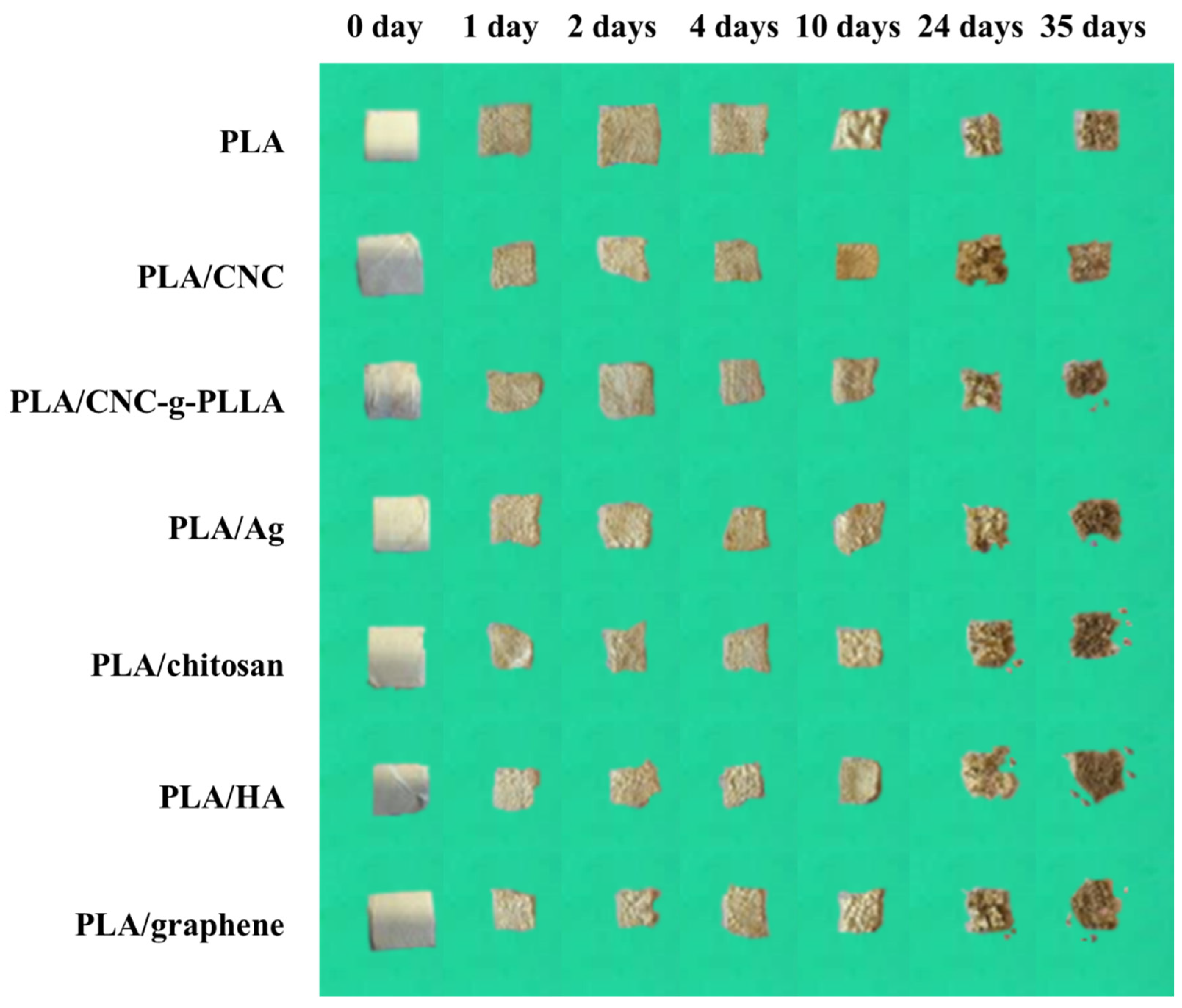
- UNE-EN-ISO. Determination of the Degree of Disintegration of Plastic Materials under Simulated Composting Conditions in a Laboratory-scale Test; International Organization for Standarization: Geneve, Switzerland, 2015. [Google Scholar]


| Samples | C (% wt) | Qs (mL/h) | Qp (mL/h) | V+ (kV) | V− (kV) | Fibers Diameter (µm) |
|---|---|---|---|---|---|---|
| PLA1 | 1 | 0.3 | 2.0 | 6.3 | −8.0 | No fiber formation |
| PLA2 | 1 | 0.3 | 0.3 | 6.4 | −0.5 | No fiber formation |
| PLA3 | 1 | 0.1 | 0.1 | 10.0 | −10.0 | No fiber formation |
| PLA4 | 1 | 0.3 | 0.3 | 10.6 | −10.5 | No fiber formation |
| PLA5 | 1 | 0.8 | 0.8 | 10.6 | −10.5 | No fiber formation |
| PLA6 | 1 | 0.8 | 0.8 | 11.4 | −1.4 | No fiber formation |
| PLA7 | 2 | 0.3 | 2.0 | 6.3 | −8.0 | No fiber formation |
| PLA8 | 2 | 0.3 | 0.3 | 6.4 | −0.5 | No fiber formation |
| PLA9 | 2 | 0.1 | 0.1 | 10.0 | −10.0 | 0.18 ± 0.01 |
| PLA10 | 2 | 0.3 | 0.3 | 10.6 | −10.5 | No fiber formation |
| PLA11 | 2 | 0.8 | 0.8 | 10.6 | −10.5 | No fiber formation |
| PLA12 | 2 | 0.8 | 0.8 | 11.4 | −1.4 | No fiber formation |
| PLA13 | 4 | 0.3 | 2.0 | 6.3 | −8.0 | 0.25 ± 0.01 |
| PLA14 | 4 | 0.3 | 0.3 | 6.4 | −0.5 | No fiber formation |
| PLA15 | 4 | 0.1 | 0.1 | 10.0 | −10.0 | 0.36 ± 0.03 |
| PLA16 | 4 | 0.3 | 0.3 | 10.6 | −10.5 | 0.33 ± 0.02 |
| PLA17 | 4 | 0.8 | 0.8 | 10.6 | −10.5 | 0.33 ± 0.02 |
| PLA18 | 4 | 0.8 | 0.8 | 11.4 | −1.4 | No fiber formation |
| PLA19 | 5 | 0.3 | 2.0 | 6.3 | −8.0 | 0.31 ± 0.02 |
| PLA20 | 5 | 0.3 | 0.3 | 6.4 | −0.5 | No fiber formation |
| PLA21 | 5 | 0.1 | 0.1 | 10.0 | −10.0 | 0.76 ± 0.05 |
| PLA22 | 5 | 0.1 | 0.0 | 10.0 | −10.0 | 1.16 ± 0.10 |
| PLA23 | 5 | 1.0 | 0.1 | 10.0 | −10.0 | 0.70 ± 0.06 |
| PLA24 | 5 | 1.0 | 1.0 | 10.0 | −10.0 | 0.76 ± 0.04 |
| PLA25 | 5 | 0.8 | 0.8 | 10.0 | −10.0 | 0.75 ± 0.06 |
| PLA26 | 5 | 0.1 | 0.3 | 10.0 | −10.0 | 0.43 ± 0.04 |
| PLA27 | 5 | 0.1 | 0.5 | 10.0 | −10.0 | 0.61 ± 0.03 |
| PLA28 | 5 | 0.1 | 0.7 | 10.0 | −10.0 | 0.34 ± 0.02 |
| PLA29 | 5 | 0.1 | 1.0 | 10.0 | −10.0 | 0.37 ± 0.02 |
| PLA30 | 5 | 0.3 | 0.3 | 10.6 | −10.5 | 0.40 ± 0.03 |
| PLA31 | 5 | 0.8 | 0.8 | 10.6 | −10.5 | 0.32 ± 0.02 |
| PLA32 | 5 | 0.8 | 0.8 | 11.4 | −1.4 | No fiber formation |
| PLA33 | 6 | 0.3 | 2.0 | 6.3 | −8.0 | 0.80 ± 0.05 |
| PLA34 | 6 | 0.3 | 0.3 | 6.4 | −0.5 | 0.51 ± 0.03 |
| PLA35 | 6 | 0.1 | 0.1 | 10.0 | −10.0 | 0.83 ± 0.07 |
| PLA36 | 6 | 0.3 | 0.3 | 10.6 | −10.5 | 0.73 ± 0.05 |
| PLA37 | 6 | 0.8 | 0.8 | 10.6 | −10.5 | 0.68 ± 0.05 |
| PLA38 | 6 | 0.8 | 0.8 | 11.4 | −1.4 | 0.68 ± 0.06 |
| PLA39 | 7 | 0.3 | 2.0 | 6.3 | −8.0 | 0.80 ± 0.05 |
| PLA40 | 7 | 0.3 | 0.3 | 6.4 | −0.5 | 0.88 ± 0.06 |
| PLA41 | 7 | 0.1 | 0.1 | 10.0 | −10.0 | 0.91 ± 0.07 |
| PLA42 | 7 | 0.3 | 0.3 | 10.6 | −10.5 | 0.83 ± 0.06 |
| PLA43 | 7 | 0.8 | 0.8 | 10.6 | −10.5 | 0.85 ± 0.05 |
| PLA44 | 7 | 0.8 | 0.8 | 11.4 | −1.4 | 0.82 ± 0.03 |
| PLA45 | 8 | 0.3 | 2.0 | 6.3 | −8.0 | 1.12 ± 0.09 |
| PLA46 | 8 | 1.0 | 1.0 | 10.0 | −10.0 | 0.92 ± 0.10 |
| PLA47 | 8 | 0.3 | 0.3 | 10.6 | −10.5 | 0.82 ± 0.09 |
| PLA48 | 8 | 0.8 | 0.8 | 10.6 | −10.5 | 1.22 ± 0.10 |
| PLA49 | 8 | 0.8 | 0.8 | 11.4 | −1.4 | 0.99 ± 0.10 |
| PLA50 | 10 | 0.3 | 2.0 | 6.3 | −8.0 | 2.03 ± 0.22 |
| Samples | Diameter (µm) |
|---|---|
| PLA | 0.92 ± 0.10 a |
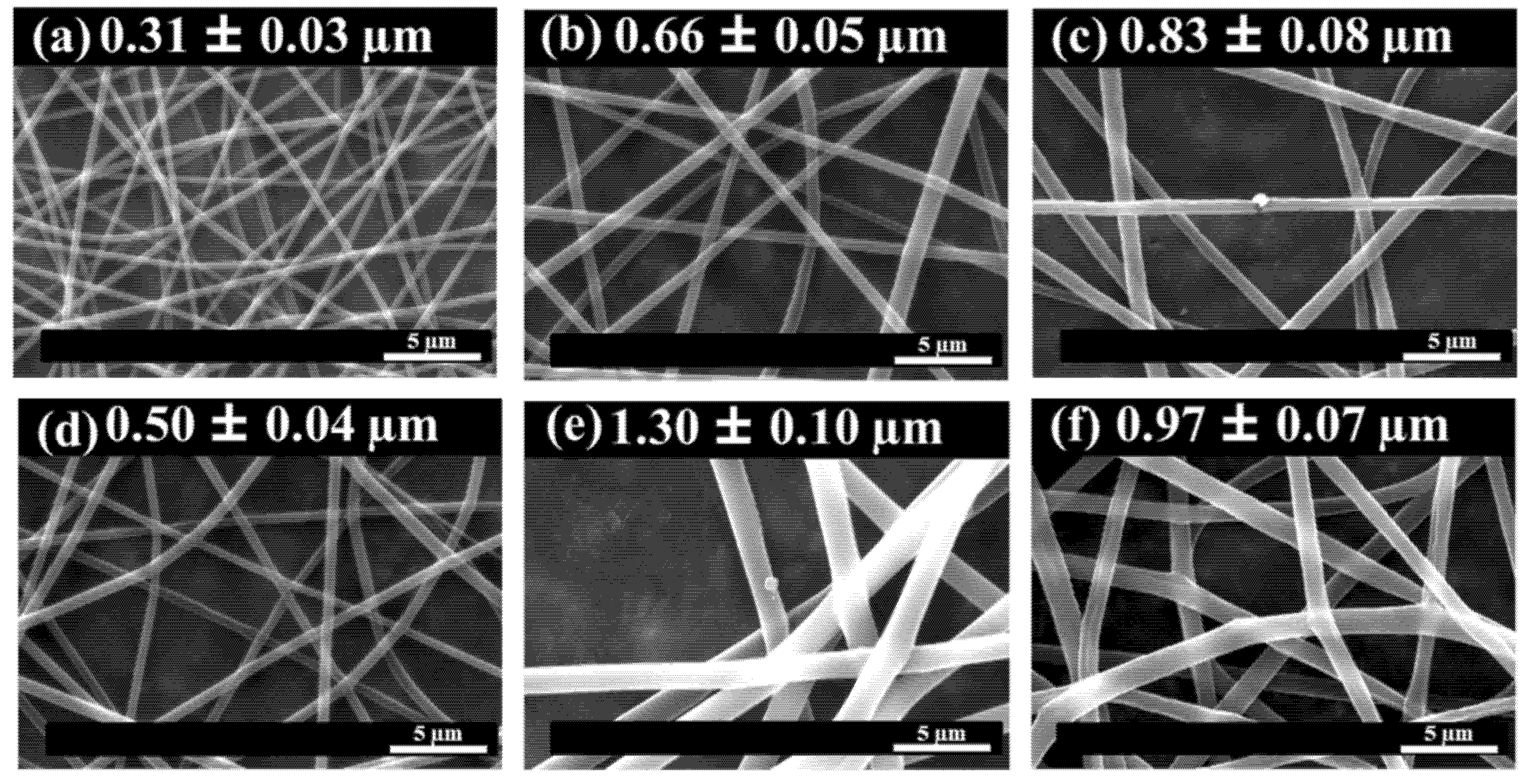
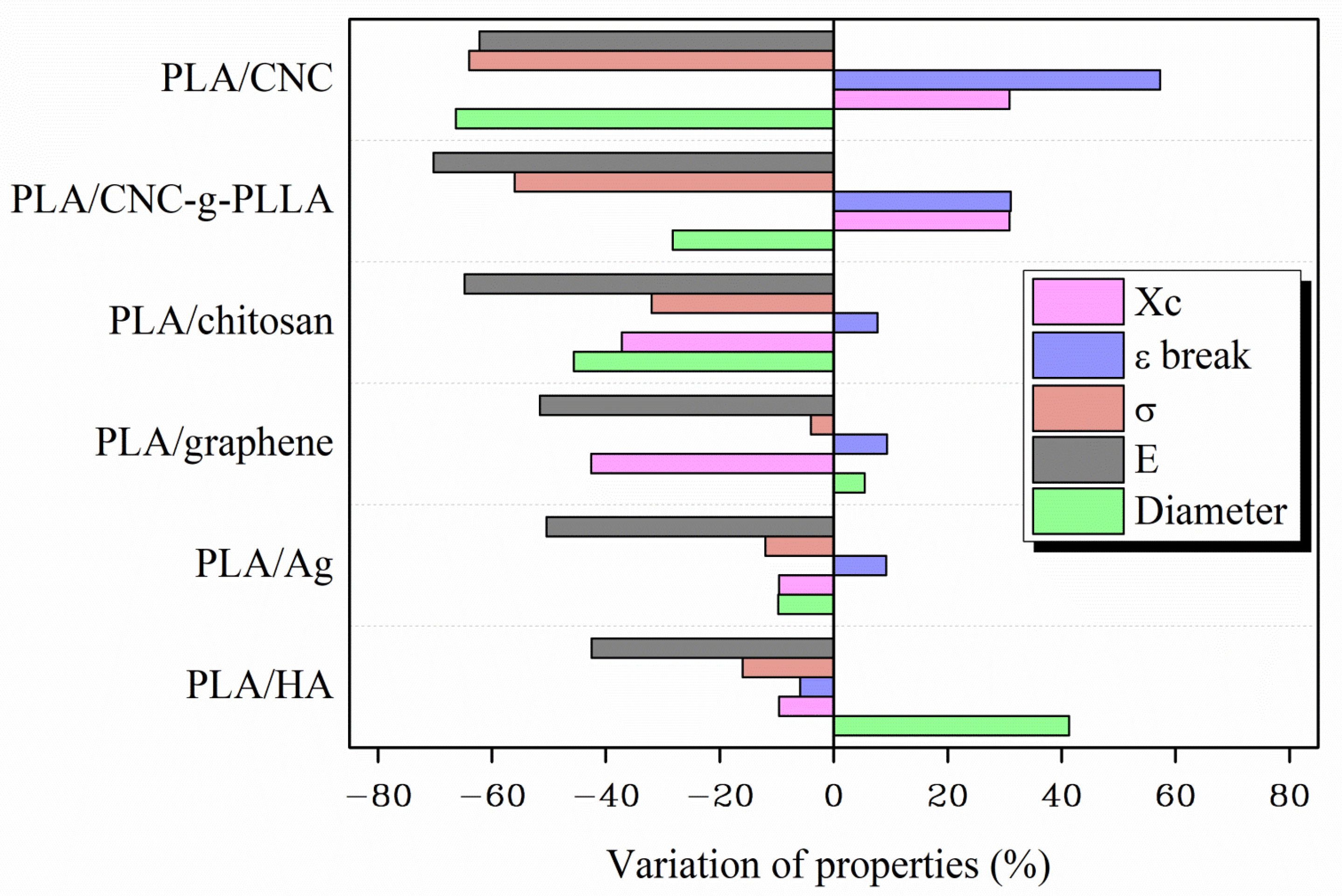
| PLA/CNC | 0.31 ± 0.03 d |
| PLA/CNC-g-PLLA | 0.66 ± 0.05 b,c |
| PLA/Ag | 0.83 ± 0.08 a,b |
| PLA/chitosan | 0.50 ± 0.04 c,d |
| PLA/HA | 1.30 ± 0.10 e |
| PLA/graphene | 0.97 ± 0.07 a |
| F ratio | 61.92 |
| p-Value | 0.0000 * |
| Samples | Tg (°C) | Tm (°C) | Xc (%) | Tmax (°C) |
|---|---|---|---|---|
| PLA | 55 | 152 | 9.4 | 340 |
| PLA/CNC | 55 | 150 | 12.3 | 364 |
| PLA/CNC-g-PLLA | 54 | 151 | 12.3 | 335 |
| PLA/Ag | 56 | 150 | 8.5 | 334 |
| PLA/chitosan | 60 | 151 | 5.4 | 326 |
| PLA/HA | 56 | 149 | 8.5 | 329 |
| PLA/graphene | 55 | 151 | 5.9 | 340 |
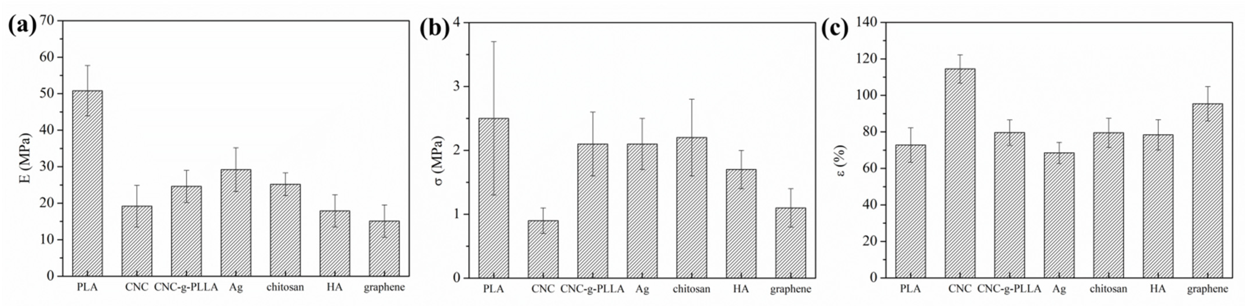
| Samples | E (MPa) | σ (MPa) | ε Break (%) |
|---|---|---|---|
| PLA | 50.8 ± 6.9 a | 2.5 ± 1.2 a | 72.8 ± 9.4 a |
| PLA/CNC | 19.2 ± 5.7 b,c | 0.9 ± 0.2 b | 114.5 ± 7.7 b |
| PLA/CNC-g-PLLA | 15.1 ± 4.4 b | 1.1 ± 0.3 b | 95.4 ± 9.4 c |
| PLA/Ag | 25.2 ± 3.1 c,d | 2.2 ± 0.6 a | 79.5 ± 8.0 a |
| PLA/chitosan | 17.9 ± 4.4 b,c | 1.7 ± 0.3 a,b | 78.4 ± 8.3 a |
| PLA/HA | 29.2 ± 6.0 d | 2.1 ± 0.4 a | 68.5 ± 5.8 a |
| PLA/graphene | 24.6 ± 4.4 c,d | 2.4 ± 0.5 a | 79.6 ± 7.0 a |
| F ratio | 22.33 | 9.80 | 27.10 |
| p-Value | 0.0000 * | 0.0000 * | 0.0000 * |
Publisher’s Note: MDPI stays neutral with regard to jurisdictional claims in published maps and institutional affiliations. |
© 2021 by the authors. Licensee MDPI, Basel, Switzerland. This article is an open access article distributed under the terms and conditions of the Creative Commons Attribution (CC BY) license (https://creativecommons.org/licenses/by/4.0/).
Share and Cite
Leonés, A.; Salaris, V.; Mujica-Garcia, A.; Arrieta, M.P.; Lopez, D.; Lieblich, M.; Kenny, J.M.; Peponi, L. PLA Electrospun Fibers Reinforced with Organic and Inorganic Nanoparticles: A Comparative Study. Molecules 2021, 26, 4925. https://0-doi-org.brum.beds.ac.uk/10.3390/molecules26164925
Leonés A, Salaris V, Mujica-Garcia A, Arrieta MP, Lopez D, Lieblich M, Kenny JM, Peponi L. PLA Electrospun Fibers Reinforced with Organic and Inorganic Nanoparticles: A Comparative Study. Molecules. 2021; 26(16):4925. https://0-doi-org.brum.beds.ac.uk/10.3390/molecules26164925
Chicago/Turabian StyleLeonés, Adrián, Valentina Salaris, Alicia Mujica-Garcia, Marina P. Arrieta, Daniel Lopez, Marcela Lieblich, José Maria Kenny, and Laura Peponi. 2021. "PLA Electrospun Fibers Reinforced with Organic and Inorganic Nanoparticles: A Comparative Study" Molecules 26, no. 16: 4925. https://0-doi-org.brum.beds.ac.uk/10.3390/molecules26164925