
Biotransformation of (−)-α-Bisabolol by Absidia coerulea


College of Pharmacy, Chonnam National University, Gwangju 61186, Korea
*
Author to whom correspondence should be addressed.
†
Current address: Advanced Radiation Technology Institute, Korea Atomic Energy Research Institute, Jeongeup-si 56212, Korea.
‡
These authors contributed equally to this work.
Molecules 2022, 27(3), 881; https://0-doi-org.brum.beds.ac.uk/10.3390/molecules27030881
Submission received: 31 December 2021
/
Revised: 25 January 2022
/
Accepted: 26 January 2022
/
Published: 27 January 2022
(This article belongs to the Special Issue Microbial Biotransformation of Natural Products)
Abstract
:(−)-α-Bisabolol, a bioactive monocyclic sesquiterpene alcohol, has been used in pharmaceutical and cosmetic products with anti-inflammatory, antibacterial and skin-caring properties. However, the poor water solubility of (−)-α-bisabolol limits its pharmaceutical applications. It has been recognized that microbial transformation is a very useful approach to generate more polar metabolites. Fifteen microorganisms were screened for their ability to metabolize (−)-α-bisabolol in order to obtain its more polar derivatives, and the filamentous fungus Absidia coerulea was selected for scale-up fermentation. Seven new and four known metabolites were obtained from biotransformation of (−)-α-bisabolol (1), and all the metabolites exhibited higher aqueous solubility than that of the parent compound 1. The structures of newly formed metabolites were established as (1R,5R,7S)- and (1R,5S,7S)-5-hydroxy-α-bisabolol (2 and 3), (1R,5R,7S,10S)-5-hydroxybisabolol oxide B (4), (1R,7S,10S)-1-hydroxybisabolol oxide B (5), 12-hydroxy-α-bisabolol (7), (1S,3R,4S,7S)- and (1S,3S,4S,7S)-3,4-dihydroxy-α-bisabolol (8 and 10) on the basis of spectroscopic analyses. These compounds could also be used as reference standards for the detection and identification of the metabolic products of 1 in the mammalian system.
1. Introduction
(−)-α-Bisabolol (1), a natural monocyclic sesquiterpene alcohol also known as levomenol, has been found in essential oils of various plants such as Matricaria recutita and Alpinia officinarum Hance [1,2]. It was reported to be nontoxic and regarded as safe by the Food and Drug Administration (FDA) and has been used in pharmaceutical and cosmetic products for its anti-inflammatory, skin soothing and moisturizing properties [3,4,5]. (−)-α-Bisabolol was shown to significantly reduce LPS-induced production of NO and PGE2 in a dose-dependent manner and to downregulate the expression of pro-inflammatory mediators iNOS and COX-2 via inhibition of AP-1 and NF-κB [5]. Control of the overproduction of inflammatory mediators may facilitate the treatment of inflammation linked diseases [6]. 12-O-Tetradecanoyl-phorbol-13-acetate (TPA)-induced ear thickness and histopathological damage in the ear tissue were significantly inhibited by (−)-α-bisabolol [6]. It exhibited significant antileishmanial activity against promastigotes with IC50 values of 8.07 μg/mL (24 h) and 4.26 μg/mL (48 h) during the in vitro studies and did not lead to the appearance of toxicity or side effects after the administration of (−)-α-bisabolol in healthy and infected hamsters [7,8]. The biological studies also revealed that 1 exhibited anti-irritant, antibacterial, antinociceptive, gastroprotective, cardioprotective and anticancer properties [4,9,10,11,12,13].
(−)-α-Bisabolol shows a tendency to be oxidized, with bisabolol oxides A and B being major oxidation products (Figure 1) [4]. α-Bisabolol oxide A has been reported to show higher antioxidant activity than α-bisabolol, with IC50 at 1.50 mg/mL and 43.88 mg/mL for DPPH radical scavenging ability, respectively [14]. The bisabolol oxide-rich matricaria oil (25.5% α-bisabolol oxide B and 21.5% α-bisabolol oxide A) was reported to exhibit antihyperalgesic and antiedematous effects [15]. Bisabolol oxide showed higher antibacterial activity than α-bisabolol during the studies against gram-positive and gram-negative bacteria [16]. Miyazawa and colleagues have reported that bisabolol oxide B was transformed from 1 by Aspergillus niger and Glomerella cingulata, and then further converted into its hydroxylated derivatives [17]. More recently, Bipolaris sorokiniana and Thamnidium elegans were applied as the biocatalysts to produce bisabolol oxide B from 1 [18,19].
Microbial transformation is a well-established green technique with high selectivity and efficiency to produce new and unique derivatives of bioactive substrates which are difficult to acquire by chemical synthesis under mild conditions [20,21]. It has been reported that microbial transformation of sesquiterpenes can result in derivatives with enhanced biological activities [22,23]. For example, 13-hydroxynootkatone exhibited stronger cytotoxic activity than its parent compound nootkatone against the lung cancer cell line A549 (IC50: 36 μg/mL and 58.4 μg/mL, respectively) [24], and 14-hydroxymethyl caryophyllene oxide showed more potent inhibitory activity against the enzyme butyryl cholinesterase than its parent compound caryophyllene oxide (IC50: 10.9 μM and 208.4 μM, respectively) (Supplementary Figure S1) [22]. Furthermore, microbial transformation is highly useful in producing more polar metabolites, leading to improved aqueous solubility [25,26]. In addition, microbial transformation has been regarded as an effective and useful experimental method to mimic and predict the mammalian metabolism of pharmaceutical agents [21,27].
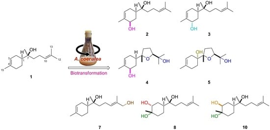
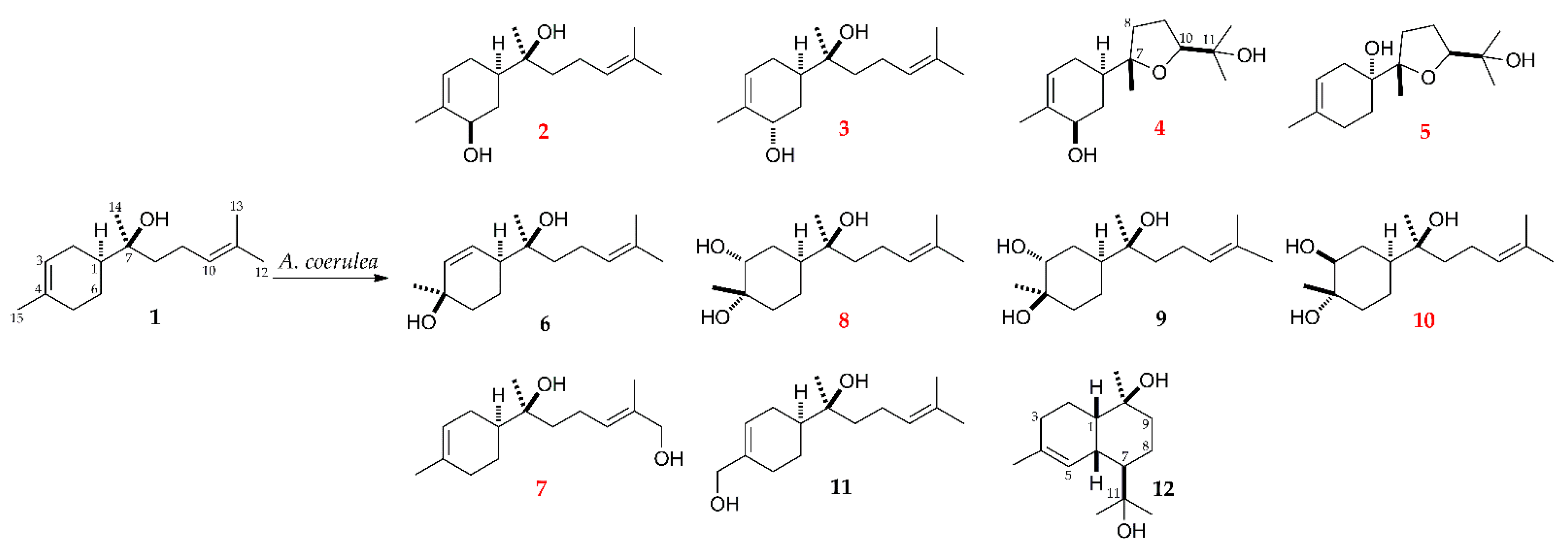
(−)-α-Bisabolol is a highly lipophilic compound that is almost insoluble in water, which limits its pharmaceutical applications [9,28]. In addition, its metabolic fate has not been extensively investigated in the previously reported metabolism studies of (−)-α-bisabolol. Thus, in order to obtain new and polar metabolites of 1, biotransformation studies using microorganisms as biocatalysts were carried out. The fungus Absidia coerulea was selected to transform 1, and the subsequent scale-up fermentation led to the isolation of seven new and four known hydroxylated metabolites (2–12, Figure 2). Moreover, hydroxylation of (−)-α-bisabolol using microorganisms could present a valuable method to generate oxygenated bisabolane-type sesquiterpenoids, which are potentially useful for pharmaceuticals and cosmetics.
2. Results and Discussion
A total of 15 microbial cultures were evaluated for their ability to metabolize (−)-α-bisabolol (1) using the usual two-stage fermentation procedure. Based on TLC analyses and control studies, it was observed that Absidia coerulea, Aspergillus fumigatus, Cunninghamella elegans var. elegans and Mucor hiemalis showed the ability to metabolize 1 (Supplementary Table S1). Among the four active strains, A. coerulea exhibited the highest transformational capability and produced a greater number of metabolites. Thus, A. coerulea was chosen for preparative-scale fermentation of 1 to produce sufficient quantities of metabolites for structural elucidation.
Seven new and four known metabolites were obtained (Figure 2). The four known metabolites were identified as (1S,7S)-4,7-dihydroxy-α-bisabolol (6) [29], (1S,3R,4R,7S)-3,4-dihydroxy-α-bisabolol (9) [17], 15-hydroxy-α-bisabolol (11) [30], and 11-hydroxy-T-muurolol (12) [31] by the combined analysis of NMR and IR spectroscopic data.
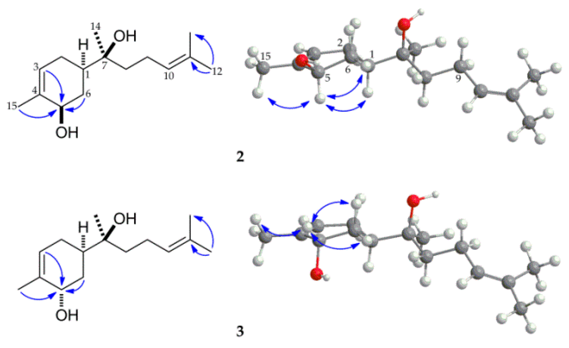
Compound 2 was obtained as a colorless oil, and its molecular formula was deduced as C15H26O2 based on its HRESIMS analysis (m/z 261.1831, calcd for C15H26O2Na, 261.1830), which indicated that it was a monohydroxylated metabolite of 1. The additional oxygen-bearing secondary carbon resonating at δC 71.0 was considered to be C-5 after comparing the 13C NMR data of related compounds with the hydroxyl group substituted at position C-5 [32]. Moreover, this was further supported by the long-range correlations of H-3 (δH 5.47), H-6 (δH 2.25, 1.35) and H-15 (δH 1.76), with C-5 (δC 71.0) in the HMBC spectrum of 2 (Figure 3). The relative configuration of 5-OH was deduced as β by the NOE (nuclear Overhauser effect) correlations among H-5 (δH 4.17) and H-1 (δH 1.72), H-6eq (δH 2.25), and H-15 (δH 1.76) (Figure 3). On the basis of the above analysis and comparison with previously reported NMR data of related compounds [32,33], compound 2 was identified as (1R,5R,7S)-5-hydroxy-α-bisabolol.
Compound 3 was obtained as a colorless oil. Its HRESIMS showed an [M + Na]+ peak at m/z 261.1830 (calcd for C15H26O2Na, 261.1830), which established a molecular formula of C15H26O2, indicating that it was a monohydroxylated derivative of 1. The presence of the hydroxyl-bearing methine proton at δH 4.05 (1H, br s) with corresponding carbon at δC 68.6 was identified based on the 1H and 13C NMR spectral analysis of 3 with the help of an HSQC experiment (Supplementary Figure S15). Compounds 2 and 3 had similar NMR spectral data, suggesting that they were epimers. The location of the hydroxyl group at the C-5 position was supported by the long-range correlations from H-3 (δH 5.56), H-6 (δH 1.44, 2.03), and H-15 (δH 1.79) to C-5 (δC 68.6) in the HMBC spectrum of 3. The correlation between H-5 and H-1 was not observed in the NOE spectrum of 3 (Figure 3) indicating that the hydroxyl group at C-5 position was α-oriented, and the broad singlet peak of H-5 in its 1H NMR spectrum also supported this elucidation. Therefore, compound 3 was established as (1R,5S,7S)-5-hydroxy-α-bisabolol.

Table 1.
1H-NMR data of metabolites 2–5, 7, 8 and 10.
| Position | Compound | ||||||
|---|---|---|---|---|---|---|---|
| 2 a | 3 a | 4 b | 5 b | 7 b | 8 b | 10 b | |
| 1 | 1.72, 1H, m | 1.79, 1H, m | 1.77, 1H, m | - | 1.57, 1H, m | 1.72, 1H, m | 1.53, 1H, tt (11.8, 2.8) |
| 2 | 1.98, 1H, m | 2.06, 1H, m | 1.97, 2H, m | 2.14, 1H, m | 1.98, 1H, m | 1.78, 1H, d (3.0) | 1.89, 1H, d (11.8) |
| 1.86, 1H, m | 1.77, 1H, m | 1.92, 1H, m | 1.79, 1H, m | 1.73 1H, dt (3.0, 13.7) | 1.18, 1H, m | ||
| 3 | 5.47, 1H, m | 5.56, 1H, dt (5.4, 1.5) | 5.47, 1H, m | 5.30, 1H, m | 5.37, 1H, m | 3.63, 1H, br s | 3.53, 1H, dd (11.6, 4.0) |
| 5 | 4.17, 1H, brs | 4.05, 1H, brs | 4.19, 1H, m | 2.21, 2H, m | 1.99, 2H, m | 1.74, 1H, m 1.55, 1H, m | 1.80, 1H, dt (3.1, 12.9) 1.42, 1H, td (12.9, 3.1) |
| 6 | 2.25, 1H, ddt (12.0, 6.0, 2.3) | 2.03, 1H, m | 2.26, 1H, dd (11.8, 5.6) | 1.65, 1H, dd (5.8, 12.8) | 1.91, 1H, m | 1.60, 1H, m | 1.72, 2H, m |
| 1.35, 1H, td (12.0, 10.0) | 1.44, 1H td (13.0, 3.8) | 1.28, 1H, m | 1.76, 1H, ddt (12.8, 5.8, 2.0) | 1.29, 1H, m | 1.42, 1H, m | ||
| 8 | 1.51, 2H, m | 1.52, 2H, m | 1.85, 1H, m 1.62, 1H, m | 2.29, 1H, dt (12.2, 7.5), 1.57, 1H, dd (12.2, 7.5) | 1.52, 2H, m | 1.50, 2H, m | 1.49, 2H, m |
| 9 | 2.04, 2H, m | 2.07, 2H, m | 1.83, 2H, m 1.78, 2H, m | 1.90, 2H, m 1.85, 2H, m | 2.12, 2H, ddd (7.8) | 2.05, 2H, m | 2.04, 2H, m |
| 10 | 5.12, 1H, tq (7.0, 1.4) | 5.13, 1H, tt (7.3, 1.3) | 3.68, 1H, dd (9.2, 5.7) | 3.76, 1H, dd (10.6, 5.4) | 5.42, 1H, tq (7.2, 1.6) | 5.13, 1H, t (7.0) | 5.12, 1H, t (6.6) |
| 12 | 1.69, 3H, s | 1.69, 3H, s | 1.11, 3H, s | 1.13, 3H, s | 4.00, 3H, s | 1.69, 3H, s | 1.69, 3H, s |
| 13 | 1.62, 3H, s | 1.62, 3H, s | 1.21, 3H, s | 1.23, 3H, s | 1.68, 3H, s | 1.63, 3H, s | 1.62, 3H, s |
| 14 | 1.13, 3H, s | 1.12, 3H, s | 1.13, 3H, s | 1.16, 3H, s | 1.12, 3H, s | 1.14, 3H, s | 1.14, 3H, s |
| 15 | 1.76, 3H, s | 1.79, 3H, s | 1.75, 3H, s | 1.70, 3H, s | 1.65, 3H, s | 1.26, 3H, s | 1.18, 3H, s |
Coupling constants (J) are given in Hz; a Spectra recorded at 500 MHz in CDCl3; b Spectra recorded at 400 MHz in CDCl3.
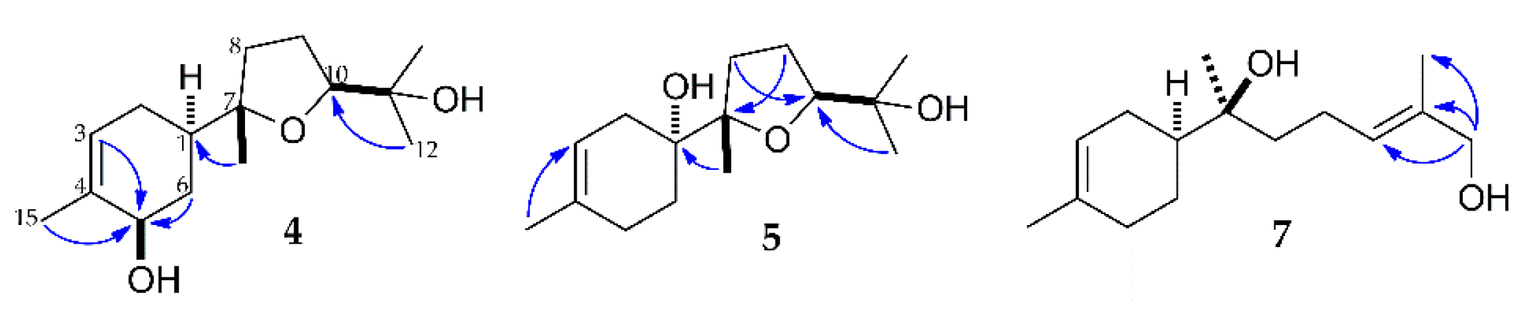
Compound 4 was obtained as colorless oil. Its molecular formula was determined to be C15H26O3 by the [M + Na]+ peak at m/z 277.1782 (calcd for C15H26O3Na, 277.1780) from its HRESIMS spectrum, indicating that it was a di-oxygenated metabolite of 1. Similar resonance signals consisting of one methyl group, two methylene groups, three methine groups (including one oxygen-bearing methine), and one quaternary carbon of the cyclohexane system were observed for 4 in comparison to the 1H and 13C NMR data of 2 (Table 1 and Table 2), suggesting the presence of one hydroxyl group at the C-5 position. In addition, the NMR spectral features of 4 were quite similar to those of bisabolol oxide B [34], except for the existence of the C-5 hydroxyl group. The presence of the tetrahydrofuran ring was evident from the oxymethine signal at δH 3.68, two non-equivalent methylene signals at δH 1.85/1.62 and 1.83/1.78, together with three oxygenated carbon signals at δH 84.4, 86.1 and 70.5. These elucidations were supported by the long-range correlations from H-3/6/15 to C-5, from H-8/12/13 to C-11, from H-14 to C-1, and from H-9 to C-7 in the HMBC spectrum of 4 (Figure 4). The relative configurations of C-5 and C-10 were established to be R and S, respectively, according to the nuclear Overhauser correlations of H-5 with H-1, H-15 and H-6eq, along with no correlation between H-10 and H-14 in the NOESY spectrum of 4 (Supplementary Figure S23). After comparison with the previously reported data of bisabolol oxide B and xylcarpin D [34,35], compound 4 was established as (1R,5R,7S,10S)-5-hydroxy-bisabolol oxide B.
Compound 5 was obtained as a colorless oil. The molecular formula for 5 was deduced as C15H26O3 on the basis of the [M + Na]+ peak at m/z 277.1780 (calcd for C15H26O3Na, 277.1780) in its HRESIMS spectrum, indicating that it was also a di-oxygenated metabolite of 1. Comparison of the 1H and 13C NMR data of 5 with those of 4 and bisabolol oxide B suggested that the double bond in the C-3(4) position remained and the double bond in the C-10(11) position was oxidized to form the tetrahydrofuran ring (Table 1 and Table 2) [17]. Similar to compound 4, the configuration of C-10 was established as S, and no correlation between H-10 and H-14 was observed in the NOESY spectrum of 5. With three methylene groups, one trisubstituted C=C group existed in the cyclohexane moiety of 5, indicating that the oxygenated quaternary carbon should be located at the C-1 position. Moreover, the location was confirmed by the long-range correlation between H-14 (δH 1.16) and C-1 (δC 73.6) in its HMBC spectrum (Figure 4). In addition, the hydroxyl group at C-1 was supposed to be α-oriented according to its specific rotation, as 1α-OH exhibited a significantly negative rotation in the studies of β-bisabolol [36,37]. Thus, compound 5 was identified as (1R,7S,10S)-1-hydroxybisabolol oxide B.
Compound 7 was obtained as a colorless oil. The molecular formula for 7 was determined to be C15H26O2 according to the [M + Na]+ peak at m/z 261.1831 (calcd for C15H26O2Na, 261.1830) in its HRESIMS spectrum, indicating that it was a monohydroxylated metabolite of 1. A notable change was observed in the 1H NMR spectrum of 7 compared to that of 1, as one allylic methyl group signal was replaced with the hydroxymethylene group at δH 4.00. The significant downfield chemical shift of H-10 at δH 5.42 indicated that the hydroxyl group was attached to the C-12. This interpretation was supported by the long-range correlations from H-10/13 to C-12, and H-12 to C-10/11/13 in the HMBC spectrum of 7 (Figure 4). In addition, the geometry of the Δ10 double bond was assigned as trans configuration based on the correlations between H-12 (δH 4.00) and H-10 (δH 5.42), and H-13 (δH 1.68) in the NOE spectrum of 7. Based on the above analysis and comparison with previously reported NMR data of 7,13-dihydroxybisabol-2,10-diene [38], compound 7 was identified as 12-hydroxy-α-bisabolol.
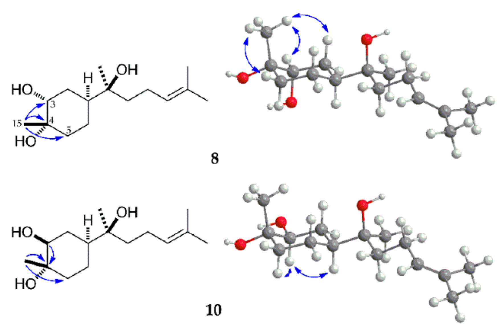
Compound 8 was obtained as a colorless oil. The molecular formula for 8, C15H28O3, was deduced from the [M + Na]+ peak at m/z 279.1937 (calcd for C15H28O3Na, 279.1936) in its HRESIMS, indicating that 8 was a dihydroxylated derivative of the parent compound 1. The 1H and 13C NMR spectra of 8 showed signals of four tert-methyl groups, five methylene groups, three methine groups, and three quaternary carbons. The olefinic proton and carbon signals at C-3 disappeared with the appearance of one oxygen-bearing proton signal at δH 3.63 and two oxidized carbon signals at δC 74.0 and 70.9, which indicated that the double bond at the C-3(4) position was oxidized to a vicinal-diol, suggesting the structure of 8 was 3,4-dihydroxy-α-bisabolol. It was confirmed by the correlations from H-15 to C-3 and C-5 in the HMBC spectrum of 8 (Figure 5). The 3-OH group was deduced to be α-oriented based on the appearance of a broad singlet proton signal for H-3 in its 1H-NMR spectrum. The stereochemistry of 8 was indicated as 3α,4α-diol based on the nuclear Overhauser correlations among H-15 with H-3eq, H-5eq, H-2ax in the NOE spectrum (Figure 5). Based on the above analysis, compound 8 was characterized as (1S,3R,4S,7S)-3,4-dihydroxy-α-bisabolol.
Compound 10 was obtained as a colorless oil. Its molecular formula was deduced as C15H28O3 on the basis of the [M + Na]+ peak at m/z 279.1935 (calcd for C15H28O3Na, 279.1936) in its HRESIMS. The 1H and 13C NMR spectral data of 10 were very similar to those of 8, indicating that 10 is a stereoisomer of 8. The orientation of H-3 was determined to be axial from the large coupling constant (J = 11.6 Hz) of the H-3 signal in its 1H NMR [17,39]. The chemical shift of C-15 in compound 10 was shown in a higher magnetic field than that of 8 (Table 2), which was dependent on the γ-gauche effect of the equatorial γ-OH [17], suggesting that the methyl group at the C-4 position existed in the axial orientation. The configuration of 10 was further confirmed by the correlations among H-3 to H-5ax, H-1ax and H-2eq, without the correlation between H-3 and H-15 in the NOE spectrum (Figure 5). Therefore, based on the above analysis and comparison with the NMR data of (1S,3S,4S,7S,10S)-3,4-dihydroxy-bisabolol oxide B [17], the structure of 10 was assigned (1S,3S,4S,7S)-3,4-dihydroxy-α-bisabolol.
All the metabolites obtained during this study were more polar than their parent compound (−)-α-bisabolol (1) and proposed to have higher aqueous solubility. In order to confirm the improvement in solubility, a modified kinetic solubility test was carried out using the stock solutions of compounds 1–12 (20 mM in dimethyl sulfoxide). White turbidity was observed for 1 after diluting 10 μL of its stock solution with 100 μL water, while no precipitates were found for the metabolites 2–12, and thus their kinetic solubility was expected to be higher than 1.82 mM [40]. This was confirmed by measuring the light transmittance of the diluted solutions. For example, compounds 2, 5, 8 and 11, showed 90% transmittance at the wavelength of 390 to 650 nm, whereas, (−)-α-bisabolol showed less than 50% transmittance in this wavelength range (Supplementary Figure S2) [41]. In addition, the solubility of 1 was determined to be 0.26 mM based on the peak area shown in its HPLC chromatogram, as the area under a peak is a measure of the concentration of the compound it represents [42].
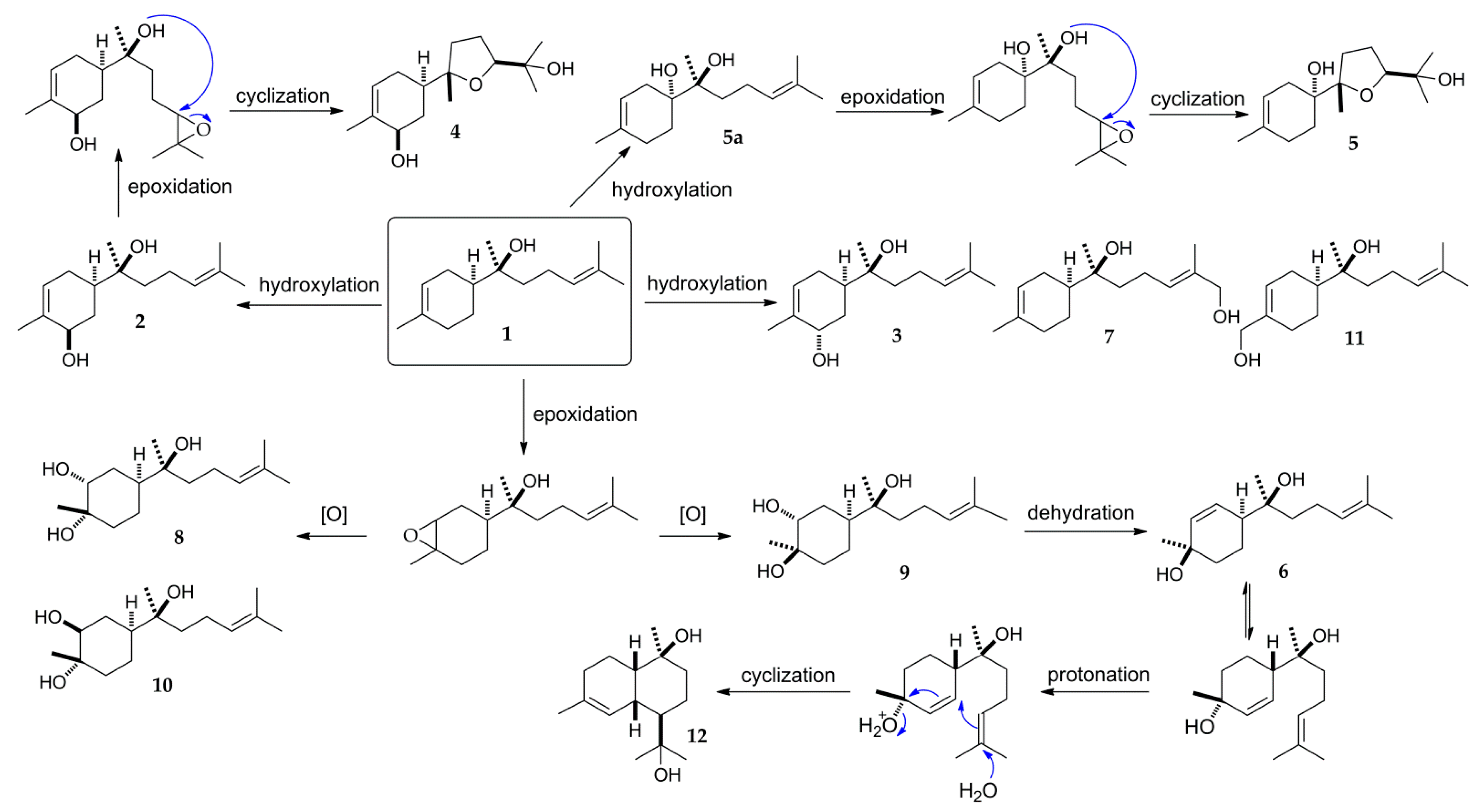
In the biotransformation studies of (−)-α-bisabolol carried out by Miyazawa and colleagues using the fungus G. cingulata, all the metabolites were identified as oxidized products in the carbon-carbon double bond positions [17]. Their major metabolites (1S,3R,4R,7S,10S)- and (1S,3S,4S,7S,10S)-3,4-dihydroxy-bisabolol oxide B were proposed to have been further transformed from the currently isolated metabolites (1S,3R,4R,7S)- and (1S,3S,4S,7S)-3,4-dihydroxy-α-bisabolol (9 and 10). In the present study, four hydroxylated metabolites were also obtained (2, 3, 7, 11) without modification of any double bonds in the skeleton of α-bisabolol. The metabolic pathway in the biotransformation of 1 by A. coerulea was proposed (Figure 6) based on the relevant reaction procedures described in the previously reported literatures [17,43,44,45]. During the transformation process, α-bisabolol was directly hydroxylated to form compounds 2, 3, 5a, 7, and 11. Next, epoxidation of the double bond in the prenyl side chain of compounds 2 and 5a followed by a cyclization process led to the formation of compounds 4 and 5. Metabolites 8–10 were formed through oxidation of the double bond existing in the cyclohexene system. In addition, it was thought that metabolite 6 was further converted from 9 through dehydration reaction. The hydroxyl group of 6 at C-4 position was postulated to undergo protonation and dehydration leading to a ring closure, followed by a hydration process to form the cadinane-type bicyclic sesquiterpene 12.
3. Materials and Methods
3.1. General Experimental Procedures
Optical rotations were measured with a 343 Plus polarimeter (Perkin Elmer, Waltham, MA, USA). UV spectra were recorded on a JASCO V-530 spectrophotometer, and IR spectra were obtained on a JASCO FT/IR-300E spectrometer (Jasco, Tokyo, Japan). NMR experiments were recorded using a Varian Unity Inova 500 spectrometer (Varian, Palo Alto, CA, USA) and a Bruker Avance III HD 400 spectrometer (Bruker, Billerica, MA, USA) with TMS as an internal standard. HRESIMS analysis was performed on a Waters Synapt G2 mass spectrometer (Waters, Milford, MA, USA). TLC was carried out on precoated silica gel 60 F254 glass plates (Merck, Darmstadt, Germany). Visualization of the silica gel TLC was performed using an anisaldehyde-H2SO4 spray reagent. The adsorbent used for column chromatography was silica gel 70–230 mesh. HPLC was performed on a Waters 600E Multisolvent Delivery System (Waters, Milford, MA, USA) connected to a Waters 996 Photodiode Array Detector using Zorbax SB-CN (10 μm, 9.4 × 250 mm) and Chiralpak AD-H (5 μm, 250 × 4.6 mm) columns.
3.2. Materials and Microorganisms
(−)-α-Bisabolol (1) was purchased from Sigma-Aldrich (St. Louis, MO, USA). All the ingredients for microbial media including D-glucose, peptone, yeast extract, malt extract, and potato dextrose medium were purchased from Becton, Dickinson and Company (Sparks, MD, USA).
All the microorganisms were obtained from the Korean Collection for Type Cultures (KCTC). The microorganisms used for preliminary screening were as follows: Absidia coerulea KCTC 6936, Alternaria alternata 6005, Aspergillus fumigatus 6145, Cunninghamella elegans var. elegans 6992, Filobasidium neoformans 7902, Fusarium merismoides 6153, Gliocladium deliquescens 6173, Glomerella cingulata 6075, Hormoconis resinae 6966, Kluyveromyces marxianus 7155, Microbacterium lacticum 9230, Mortierella ramanniana var. angulispora 6137, Mucor hiemalis 26779, Penicillium chrysogenum 6933, Trichoderma koningii 6042.
Fermentation experiments were performed in three types of media: A. coerulea, A. alternata, A. fumigatus, M. hiemalis, P. chrysogenum and T. koningii were incubated on malt medium (malt extract 20 g/L, D-glucose 20 g/L, peptone 1 g/L); F. neoformans, K. marxianus, and M. lacticum were cultured on yeast-malt medium (D-glucose 10 g/L, peptone 5 g/L, malt extract 3 g/L, and yeast extract 3 g/L); other microorganisms were cultured on potato dextrose medium (potato dextrose broth 24 g/L).
3.3. Screening Procedures
Microbial metabolism studies were carried out according to the two-stage procedure [46,47]. In the screening studies, the actively growing microbial cultures were inoculated in 250 mL flasks containing 50 mL of a suitable medium and incubated with gentle agitation (200 rpm) at 25 °C in a temperature-controlled shaking incubator. After inoculation for 24 h, 100 μL of ethanol solution (20 mg/mL) of 1 was added to each flask, and the transformation was continued under the same condition for an additional seven days. Both substrate and culture controls were carried out under the same conditions. Sampling and TLC monitoring were performed at an interval of 24 h. Culture controls consisted of fermentation cultures in which the microorganisms were grown without the addition of 1.
3.4. Extraction and Isolation of Metabolites
Scale-up fermentations were carried out under the same condition with twenty-six 1L flasks, each containing 200 mL media and 520 mg of 1 dissolved in EtOH (20 mg/mL) was evenly distributed among flasks. The cultures were extracted with equal volume of EtOAc (5.2 L × 3) after seven day incubation and the organic layers were combined and concentrated under reduced pressure [46]. The crude EtOAc extracts (895 mg) were separated by silica gel column chromatography using n-hexane-EtOAc gradient solvent system (10:1→2:3) to give 10 fractions. Fraction 3 was subjected to HPLC with 50% MeOH as elution solvent to give compound 12 (6.02 mg, 1.08%) and subfraction 3-1. Subfraction 3-1 was purified by HPLC using 45% MeOH isocratic solvent system to afford compound 5 (2.74 mg, 0.46%). Fraction 5 was chromatographed by HPLC using 45% MeOH isocratic solvent system to give compounds 4 (6.35 mg, 1.07%), 6 (3.42 mg, 0.61%), 11 (15 mg, 2.69%), 2 (17 mg, 3.05%) and subfraction 5-5. Subfraction 5-5 was further chromatographed by HPLC with a chiral column using 100% MeOH as mobile phase to afford compounds 7 (4.2 mg, 0.75%) and 3 (2.45 mg, 0.44%). Fraction 10 was chromatographed by HPLC using 35% MeOH isocratic solvent system to afford compound 8 (2.85 mg, 0.48%) and subfraction 10-4. Subfraction 10-4 was further subjected to HPLC with a chiral column using 100% MeOH as mobile phase to give compounds 10 (6.09 mg, 1.03%) and 9 (2.45 mg, 0.41%).
3.5. Spectroscopic Data of Metabolites
(1R,5R,7S)-5-hydroxy-α-bisabolol (2)
(1R,5S,7S)-5-hydroxy-α-bisabolol (3)
(1R,5R,7S,10S)-5-hydroxybisabolol oxide B (4)
(1R,7S,10S)-1-hydroxybisabolol oxide B (5)
12-hydroxy-α-bisabolol (7)
(1S,3R,4S,7S)-3,4-dihydroxy-α-bisabolol (8)
(1S,3S,4S,7S)-3,4-dihydroxy-α-bisabolol (10)
3.6. Determination of Solubility
The solubility of compounds 1–12 was evaluated by the modified kinetic solubility assay [40,48]. Compounds 1–12 were dissolved in dimethyl sulfoxide (DMSO) to yield 20 mM stock solutions, then 10 μL of the compound stock solution was diluted with 100 μL water. After all the solutions were prepared, the vials were placed under room temperature and allowed to mix for 1 h. After this, the light transmittance was measured at a scan width of 5 nm over the range of 390 to 650 nm using a SpectraMax 190 Microplate reader (Molecular Devices, Sunnyvale, CA, USA). After centrifugation, the supernatant was analyzed by HPLC under 200 nm using a Zorbax SB-CN (250 × 10 mm) column with 60% methanol as mobile phase at a flow rate of 2 mL/min.
4. Conclusions
In this study, microorganisms were used as biocatalysts for the structural modification of (−)-α-bisabolol (1). After screening of 15 different microbial strains for their ability to metabolize xenobiotics, the filamentous fungus A. coerulea KCTC 6936 was selected for transformation of (−)-α-bisabolol according to its higher catalytic capability. Eleven hydroxylated metabolites including seven previously unreported compounds (2–5, 7, 8, 10) were obtained from the culture of A. coerulea incubated with (−)-α-bisabolol. Among them, compounds 9 and 10 were proposed to be the transformation intermediates for the production of (1S,3R,4R,7S,10S)- and (1S,3S,4S,7S,10S)-3,4-dihydroxy-bisabolol oxide B. This evidence indicated that biocatalytic oxidation by A. coerulea is a feasible and effective approach to obtain more polar compounds which can be used as reference standards for the detection and identification of the metabolic products of (−)-α-bisabolol in a mammalian system. In addition, all of the isolated metabolites showed increased aqueous solubility compared with the parent compound 1, and further studies are needed to evaluate the biological activities of the identified metabolites and find microorganisms which can produce the bioactive metabolites in higher yields.
Supplementary Materials
The following supporting information can be downloaded online. Table S1: Screening for the microorganisms that transform (−)-α-bisabolol (1); Figure S1: Chemical structures of nootkatone, 13-hydroxynootkatone, caryophyllene oxide and 14-hydroxymethyl caryophyllene oxide; Figure S2: Light transmittance of the diluted solutions of (−)-α-bisabolol (1) and its metabolites in water; Figure S3: Calibration of measurement for solubility of (−)-α-bisabolol (1); Figures S4–S62: NMR and HRESIMS spectra of compounds 1–12.
Author Contributions
Conceptualization, I.-S.L.; methodology, J.P., F.H. and I.-S.L.; validation, J.P.; formal analysis, J.P. and F.H.; investigation, J.P., F.H. and I.-S.L.; resources, J.P. and I.-S.L.; data curation, J.P. and F.H.; writing—original draft preparation, J.P. and F.H.; writing—review and editing, I.-S.L.; visualization, J.P. and F.H.; supervision, I.-S.L.; project administration, I.-S.L.; funding acquisition, F.H. and I.-S.L. All authors have read and agreed to the published version of the manuscript.
Funding
This research was supported by the Basic Science Research Program through the National Research Foundation of Korea (NRF), funded by the Ministry of Education (NRF-2019R1I1A3A01043084 and NRF-2019R1I1A1A01059410).
Institutional Review Board Statement
Not applicable.
Informed Consent Statement
Not applicable.
Data Availability Statement
Data is contained within the article and Supplementary Materials.
Acknowledgments
The authors are grateful for the NMR experimental supports of the Center for Research Facilities, Chonnam National University, and for the NMR and HRESIMS analyses of Korea Basic Science Institute (KBSI).
Conflicts of Interest
The authors declare that they have no conflicts of interest.
Sample Availability
Samples of the compounds are not available from the authors.
References
- Ma, Y.; Li, W.; Mai, J.; Wang, J.; Wei, Y.; Ledesma-Amaro, R.; Ji, X.J. Engineering Yarrowia lipolytica for sustainable production of the chamomile sesquoterpene (−)-α-bisabolol. Green Chem. 2021, 23, 780–787. [Google Scholar] [CrossRef]
- Zhang, L.; Pan, C.; Ou, Z.; Liang, X.; Shi, Y.; Chi, L.; Zhang, Z.; Zheng, X.; Li, C.; Xiang, H. Chemical profiling and bioactivity of essential oils from Alpinia officinarum Hance from ten localities in China. Ind. Crops Prod. 2020, 153, 112583. [Google Scholar] [CrossRef]
- Han, G.H.; Kim, S.K.; Yoon, P.K.S.; Kang, Y.; Kim, B.S.; Fu, Y.; Sung, B.H.; Jung, H.C.; Lee, D.H.; Kim, S.W.; et al. Fermentative production and direct extraction of (−)-α-bisabolol in metabolically engineered Escherichia coli. Microb. Cell Fact. 2016, 15, 185. [Google Scholar] [CrossRef] [PubMed] [Green Version]
- Kamatou, G.P.P.; Viljoen, A.M. A review of the application and pharmacological properties of α-bisabolol and α-bisabolol-rich oils. J. Am. Oil Chem. Soc. 2010, 87, 1–7. [Google Scholar] [CrossRef]
- Kim, S.; Jung, E.; Kim, J.H.; Park, Y.H.; Lee, J.; Park, D. Inhibitory effects of (−)-α-bisabolol on LPS-induced inflammatory response in RAW264.7 macrophages. Food Chem. Toxicol. 2011, 49, 2580–2585. [Google Scholar] [CrossRef] [PubMed]
- Mautya, A.K.; Singh, M.; Dubey, V.; Srivastava, S.; Luqman, S.; Bawankule, D.U. (−)-α-Bisabolol reduces pro-inflammatory cytokine production and ameliorates skin inflammation. Curr. Pharm. Biotechnol. 2014, 15, 173–181. [Google Scholar]
- Rottini, M.M.; Amaral, A.C.F.; Ferreira, J.L.P.; Silva, J.R.A.; Taniwaki, N.N.; de Souza, C.S.F.; d’Escoffier, L.N.; Almeida-Souza, F.; Hardoim, D.J.; da Costa, S.C.G.; et al. In vitro evaluation of (−)-α-bisabolol as a promising agent against Leishmania amazonensis. Exp. Parasitol. 2015, 148, 66–72. [Google Scholar] [CrossRef]
- Corpas-López, V.; Merino-Espinosa, G.; López-Viota, M.; Gijón-Robles, P.; Morillas-Mancilla, M.J.; López-Viota, J.; Díaz-Sáez, V.; Morillas-Márquez, F.; Moll, M.C.N.; Martín-Sábchez, J. Topical treatment of Leishmania tropica infection using (−)-α-bisabolol ointment in a hamster model: Effectiveness and safety assessment. J. Nat. Prod. 2016, 79, 2403–2407. [Google Scholar] [CrossRef]
- Rodrigues, F.F.G.; Colares, A.V.; Nonato, C.F.A.; Galvāo-Rodrigues, F.F.; Mota, M.L.; Braga, M.F.B.M.; Costa, J.G.M. In vitro antimicrobial activity of the essrntial oil from Vanillosmopsis arborea Barker (Asteraceae) and its major constituent, α-bisabolol. Microb. Pathog. 2018, 125, 144–149. [Google Scholar] [CrossRef]
- Teixeira, G.F.D.; Costa, F.N.; Campos, A.R. Corneal antinociceptive effect of (−)-α-bisabolol. Pharm. Biol. 2017, 55, 1089–1092. [Google Scholar] [CrossRef]
- Terroso, T.F.; Condotta, K.B.; Fonseca, F.N.; Jornada, D.S.; Ferreira, G.O.; Ellwanger, J.H.; Schmidt, J.A.; Pohlmann, A.R.; Guterres, S.S. In vivo prophylactic gastroprotection using α-bisabolol encapsulated in lipid-core nanocapsules and in cocoa-theospheres. J. Drug Deliv. Sci. Technol. 2016, 36, 99–109. [Google Scholar] [CrossRef]
- Meeran, M.F.N.; Azimullah, S.; Laham, F.; Tariq, S.; Goyal, S.N.; Adeghate, E.; Ojha, S. α-Bisabolol protects against β-adrenergic agonist-induced myocardial infarction in rats by attenuating inflammation, lysosomal dysfunction, NLRP3 inflammasome activation and modulating autophagic flux. Food Funct. 2020, 11, 965–976. [Google Scholar] [CrossRef] [PubMed]
- Wu, S.; Peng, L.; Sang, H.; Li, Q.P.; Cheng, S. Anticancer effects of α-bisabolol in human non-small cell lung carcinoma cells are mediated via apoptosis induction, cell cycle arrest, inhibition of cell migration and invasion and upregulation of P13K/AKT signalling pathway. J. BUON 2018, 23, 1407–1412. [Google Scholar] [PubMed]
- Fırat, Z.; Demirci, F.; Demirci, B. Antioxidant activity of chamomile essential oil and main components. Nat. Volatiles Essent. Oils 2018, 5, 11–16. [Google Scholar]
- Tomić, M.; Popović, V.; Petrović, S.; Stepanović-Petrović, R.; Micov, A.; Pavlović-Drobac, M.; Couladis, M. Antihyperalgesic and antiedematous activities of bisabolol-oxides-rich matricaria oil in a rat model of inflammation. Phytother. Res. 2014, 28, 759–766. [Google Scholar] [CrossRef]
- Kazemi, M. Chemical composition and antimicrobial activity of essential oil of Matricaria recutita. Int. J. Food Prop. 2015, 18, 1784–1792. [Google Scholar] [CrossRef] [Green Version]
- Miyazawa, M.; Nankai, H.; Kameoka, H. Biotransformation of (−)-α-bisabolol by plant pathogenic fungus, Glomerella cingulata. Phytochemistry 1995, 39, 1077–1080. [Google Scholar] [CrossRef]
- Limberger, R.P.; Ferreira, L.; Castilhos, T.; Aleixo, A.M.; Petersen, R.Z.; Germani, J.C.; Zuanazzi, J.A.; Fett-Neto, A.G.; Henriques, A.T. The ability of Bipolaris sorokiniana to modify geraniol and (−)-alpha-bisabolol as exogenous substrates. Appl. Microbiol. Biotechnol. 2003, 61, 552–555. [Google Scholar] [CrossRef]
- Firat, Z.; Demirci, F.; Demirci, B.; Kırmızıbekmez, H.; Başer, K.H.C. Microbial transformation of (−)-α-bisabolol towards bioactive metabolites. Rec. Nat. Prod. 2021, 15, 593–601. [Google Scholar] [CrossRef]
- Han, F.; Xiao, Y.; Lee, I.-S. Microbial transformation of galangin derivatives and cytotoxicity evaluation of their metabolites. Catalysts 2021, 11, 1020. [Google Scholar] [CrossRef]
- Cano-Flores, A.; Gómez, J.; Escalona-Torres, I.S.; Velasco-Bejarano, B. Microorganisms as biocatalysts and enzyme sources. In Microorganisms; Blumenberg, M., Shaaban, M., Elgaml, A., Eds.; IntechOpen: London, UK, 2020. [Google Scholar]
- Sultana, N.; Saify, Z.S. Enzymatic biotransformation of terpenes as bioactive agents. J. Enzym. Inhib. Med. Chem. 2013, 28, 1113–1128. [Google Scholar] [CrossRef] [PubMed]
- Bhatti, H.N.; Zubair, M.; Rasool, N.; Hassan, Z.; Ahmad, V.U. Microbial transformation of sesquiterpenoids. Nat. Prod. Commun. 2009, 4, 1155–1168. [Google Scholar] [CrossRef] [Green Version]
- Gliszczyńska, A.; Łysek, A.; Janeczko, T.; Świtalska, M.; Wietrzyk, J.; Wawrzeńczyk, C. Microbial transformation of (+)-nootkatone and the antiproliferative activity of its metabolites. Bioorg. Med. Chem. 2011, 19, 2464–2469. [Google Scholar] [CrossRef] [PubMed]
- Han, F.; Xiao, Y.; Lee, I.-S. Microbial conjugation studies of licochalcones and xanthohumol. Int. J. Mol. Sci. 2021, 22, 6893. [Google Scholar] [CrossRef] [PubMed]
- Brzonova, I.; Asina, F.; Andrianova, A.A.; Kubátová, A.; Smoliakova, I.P.; Kozliak, E.I.; Ji, Y. Fungal biotransformation of insoluble kraft lignin into a water soluble polymer. Ind. Eng. Chem. Res. 2017, 56, 6103–6113. [Google Scholar] [CrossRef]
- Shah, S.A.A.; Tan, H.L.; Sultan, S.; Faridz, M.A.B.M.; Shah, M.A.B.M.; Nurfazilah, S.; Hussain, M. Microbial-catalyzed biotransformation of multifunctional triterpenoids derived from phytonutrients. Int. J. Mol. Sci. 2014, 15, 12027–12060. [Google Scholar] [CrossRef] [PubMed]
- D’Almeida, A.P.; de Oliveira, M.T.P.; de Souza, É.T.; Coutinho, D.S.; Ciambarella, B.T.; Gomes, C.R.; Terroso, T.; Guterres, S.S.; Pohlmann, A.R.; Silva, P.M.R.; et al. α-Bisabolol-loaded lipid-core nanocapsules reduce lipopolysaccharide-induced pulmonary inflammation in mice. Int. J. Nanomed. 2017, 12, 4479–4491. [Google Scholar] [CrossRef] [PubMed] [Green Version]
- Mori, K.; Kato, M. Synthesis of (6S,1′S)-(+)-hernandulcin, a sweetner, and its stereoisomers. Tetrahedron 1986, 42, 5895–5900. [Google Scholar] [CrossRef]
- Matos, M.E.O.; De Sousa, M.P.; Matos, F.J.A.; Craveiro, A.A. Sesquiterpenes from Vanillosmopsis arborea. J. Nat. Prod. 1988, 51, 780–782. [Google Scholar] [CrossRef]
- Ding, L.; Pfoh, R.; Ruhl, S.; Qin, S.; Laatsch, H. T-muurolol sesquiterpenes from the marine Streptomyces sp. M491 and revision of the configuration of previously reported amorphanes. J. Nat. Prod. 2009, 72, 99–101. [Google Scholar] [CrossRef]
- Campagnuolo, C.; Fattorusso, E.; Petrucci, F.; Taglialatela-Scafati, O.; Appendino, G.; Marquez, N.; Muñoz, E. A prenylbisabolane with NF-κB inhibiting properties from Cascarilla (Croton eluteria). Bioorg. Med. Chem. 2005, 13, 4238–4242. [Google Scholar] [CrossRef] [PubMed]
- Smitt, O.; Högberg, H. Syntheses of a prenylbisabolane diterpene, a natural insecticide from Corton linearis, and the biabolane sesquiterpenes (−)-delobanone and (−)-epi-delobanone. Tetrahedron 2002, 58, 7691–7700. [Google Scholar] [CrossRef]
- Flaskamp, E.; Nonnenmacher, G.; Zimmermann, G.; Isaac, O. On the stereochemistry of the bisaboloids from Matricaria chamomilla L. Z. Naturforsch. 1981, 36b, 1023–1030. [Google Scholar] [CrossRef]
- Yin, X.; Feng, T.; Li, Z.H.; Su, J.; Li, Y.; Tan, N.H.; Liu, J.K. Chemical investigation on the cultures of the fungus Xylaria carpophila. Nat. Prod. Bioprospect. 2011, 1, 75–80. [Google Scholar] [CrossRef] [Green Version]
- Ohloff, G.; Giersch, W.; Näf, R.; Delay, F. The absolute configuration of β-bisabolol. Helv. Chim. Acta 1986, 69, 698–703. [Google Scholar] [CrossRef]
- Fráter, G.; Müller, U. Synthesis of (+)-(4S,8R)-8-epi- and (−)-(4R,8S)-4-epi-β-bisabolol. Helv. Chim. Acta 1989, 72, 653–658. [Google Scholar] [CrossRef]
- Alpha, T.; Raharivelomanana, P.; Bianchini, J.; Faure, R.; Cambon, A. Bisabolane sesquiterpenoids from Santalum austrocaledonicum. Phytochemistry 1997, 44, 1519–1522. [Google Scholar] [CrossRef]
- Matsumura, T.; Ishikawa, T.; Kitajima, J. Water-soluble constituents of caraway: Carvone derivatives and their glucosides. Chem. Pharm. Bull. 2002, 50, 66–72. [Google Scholar] [CrossRef] [Green Version]
- Chellan, P.; Land, K.M.; Shoker, A.; Au, A.; An, S.H.; Taylor, D.; Simth, P.J.; Riedel, T.; Dyson, P.J.; Chibale, K.; et al. Synthesis and evaluation of new polynuclear organometallic Ru(II), Rh(III) and Ir(III) pyridyl ester complexes as in vitro antiparasitic and antitumor agents. Dalton Trans. 2014, 43, 513–526. [Google Scholar] [CrossRef] [Green Version]
- Nagai, Y.; Kawano, S.; Motoda, K.; Tomida, M.; Tatebe, C.; Sato, K.; Akiyama, H. Solubility testing of sucrose esters of fatty acids in international food additive specifications. Biol. Pharm. Bull. 2017, 40, 284–289. [Google Scholar] [CrossRef] [Green Version]
- Chawla, R.C.; Doura, K.F.; McKay, D. Effect of alcohol cosolvents on the aqueous solubility of trichloroethylene. In Proceedings of the 2001 Conference on Environmental Research, Manhattan, KA, USA, 21–24 May 2001; pp. 52–66. [Google Scholar]
- Blair, M.; Tuck, K.L. A new diastereoselective entry to the (1S,4R)- and (1S,4S)-isomers of 4-isopropyl-1-methyl-2-cyclohexen-1-ol, aggregation pheromones of the ambrosia beetle Platypus quercivorus. Tetrahedron Asymmetry 2009, 20, 2149–2153. [Google Scholar] [CrossRef]
- Liu, Q.; Beyraghdar Kashkooli, A.; Manzano, D.; Pateraki, I.; Richard, L.; Kolkman, P.; Lucas, M.F.; Guallar, V.; de Vos, R.C.H.; Franssen, M.C.R.; et al. Kauinolide synthase is P450 with unusaual hydroxylation and cyclization-elimination activity. Nat. Commun. 2018, 9, 4657. [Google Scholar] [CrossRef] [PubMed]
- Esmaeili, A.; Hashemi, E. Biotransformation of myrcene by Pseudomonas aeruginosa. Chem. Cent. J. 2011, 5, 26. [Google Scholar] [CrossRef] [PubMed] [Green Version]
- Han, F.; Lee, I.-S. Microbial transformation of the antimalarial sesquiterpene endoperoxide dihydroartemisinin. Nat. Prod. Res. 2017, 31, 883–889. [Google Scholar] [CrossRef] [PubMed]
- Han, F.; Lee, I.-S. Microbial transformation of bavachin by Absidia coerulea. Phytochem. Lett. 2016, 18, 136–139. [Google Scholar] [CrossRef]
- Hoelke, B.; Gieringer, B.; Arlt, M.; Saal, C. Comparison of nephelometric, UV-spectroscopic, and HPLC methods for high-throughput determination of aqueous drug solubility in microtiter plates. Anal. Chem. 2009, 81, 3165–3172. [Google Scholar] [CrossRef]
Figure 1.
Chemical structures of α-bisabolol, bisabolol oxides A and B.

Figure 2.
Chemical structures of (−)-α-bisabolol (1) and its metabolites (2–11).

Figure 3.
Selected HMBC (1H→13C) and NOE (1H↔1H) correlations of metabolites 2 and 3.

Figure 4.
Selected HMBC (1H→13C) correlations of metabolites 4, 5, and 7.

Figure 5.
Selected HMBC (1H→13C) and NOE (1H↔1H) correlations of metabolites 8 and 10.

Figure 6.
A proposed metabolic pathway of (−)-α-bisabolol (1) transformed by A. coerulea.

Table 2.
13C-NMR data of metabolites 2–5, 7, 8 and 10.
| Position | Compound | ||||||
|---|---|---|---|---|---|---|---|
| 2 a | 3 a | 4 b | 5 b | 7 b | 8 b | 10 b | |
| 1 | 42.1 | 36.8 | 43.5 | 73.6 | 43.0 | 39.3 | 45.6 |
| 2 | 27.1 | 27.1 | 27.3 | 32.3 | 27.0 | 29.9 | 32.1 |
| 3 | 123.7 | 125.3 | 123.8 | 117.9 | 120.5 | 74.0 | 77.4 |
| 4 | 136.6 | 134.4 | 136.6 | 134.0 | 134.2 | 70.9 | 74.0 |
| 5 | 71.0 | 68.6 | 71.1 | 26.7 | 31.0 | 33.6 | 38.4 |
| 6 | 33.8 | 32.0 | 34.9 | 28.5 | 23.3 | 21.3 | 23.6 |
| 7 | 73.9 | 73.8 | 84.4 | 88.0 | 74.3 | 74.3 | 73.9 |
| 8 | 39.9 | 40.2 | 35.5 | 32.2 | 39.8 | 39.5 | 39.8 |
| 9 | 22.1 | 22.1 | 26.4 | 26.3 | 21.7 | 22.2 | 22.2 |
| 10 | 124.3 | 124.4 | 86.1 | 87.9 | 126.2 | 124.5 | 124.2 |
| 11 | 131.9 | 131.8 | 70.5 | 70.4 | 134.9 | 131.9 | 132.1 |
| 12 | 25.7 | 25.7 | 24.0 | 24.0 | 68.9 | 25.7 | 25.8 |
| 13 | 17.7 | 17.7 | 27.7 | 27.8 | 13.7 | 17.7 | 17.7 |
| 14 | 23.4 | 23.3 | 23.5 | 22.9 | 23.2 | 24.2 | 24.1 |
| 15 | 18.8 | 20.8 | 18.8 | 23.5 | 23.4 | 27.5 | 18.9 |
a Spectra recorded at 125 MHz in CDCl3; b Spectra recorded at 100 MHz in CDCl3.
Publisher’s Note: MDPI stays neutral with regard to jurisdictional claims in published maps and institutional affiliations. |
© 2022 by the authors. Licensee MDPI, Basel, Switzerland. This article is an open access article distributed under the terms and conditions of the Creative Commons Attribution (CC BY) license (https://creativecommons.org/licenses/by/4.0/).
Share and Cite
MDPI and ACS Style
Park, J.; Han, F.; Lee, I.-S. Biotransformation of (−)-α-Bisabolol by Absidia coerulea. Molecules 2022, 27, 881. https://0-doi-org.brum.beds.ac.uk/10.3390/molecules27030881
AMA Style
Park J, Han F, Lee I-S. Biotransformation of (−)-α-Bisabolol by Absidia coerulea. Molecules. 2022; 27(3):881. https://0-doi-org.brum.beds.ac.uk/10.3390/molecules27030881
Chicago/Turabian StylePark, Jisu, Fubo Han, and Ik-Soo Lee. 2022. "Biotransformation of (−)-α-Bisabolol by Absidia coerulea" Molecules 27, no. 3: 881. https://0-doi-org.brum.beds.ac.uk/10.3390/molecules27030881