
Molecular Hybridization Strategy on the Design, Synthesis, and Structural Characterization of Ferrocene-N-acyl Hydrazones as Immunomodulatory Agents
 , and
, and
Abstract
:
1. Introduction
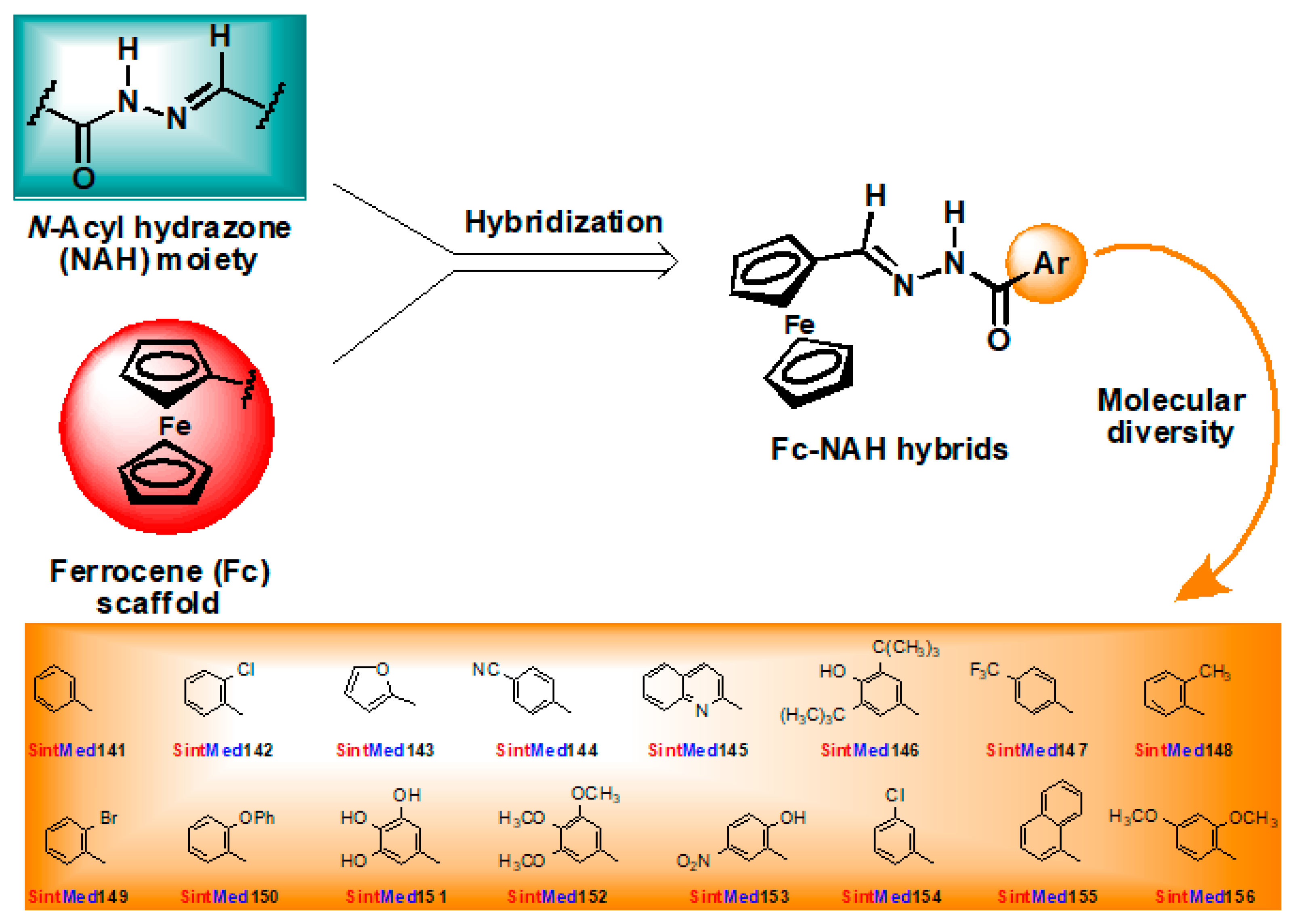
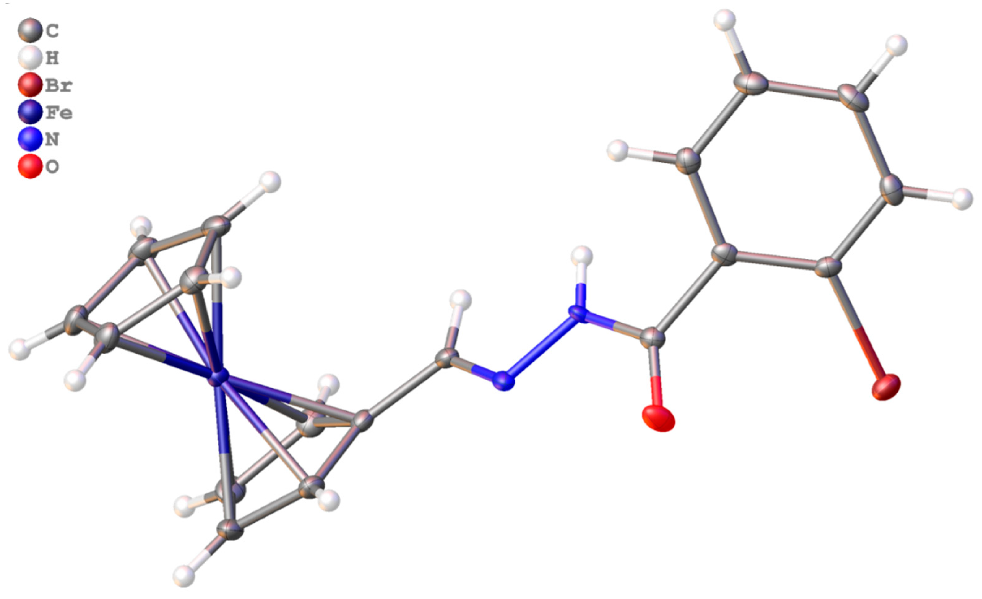
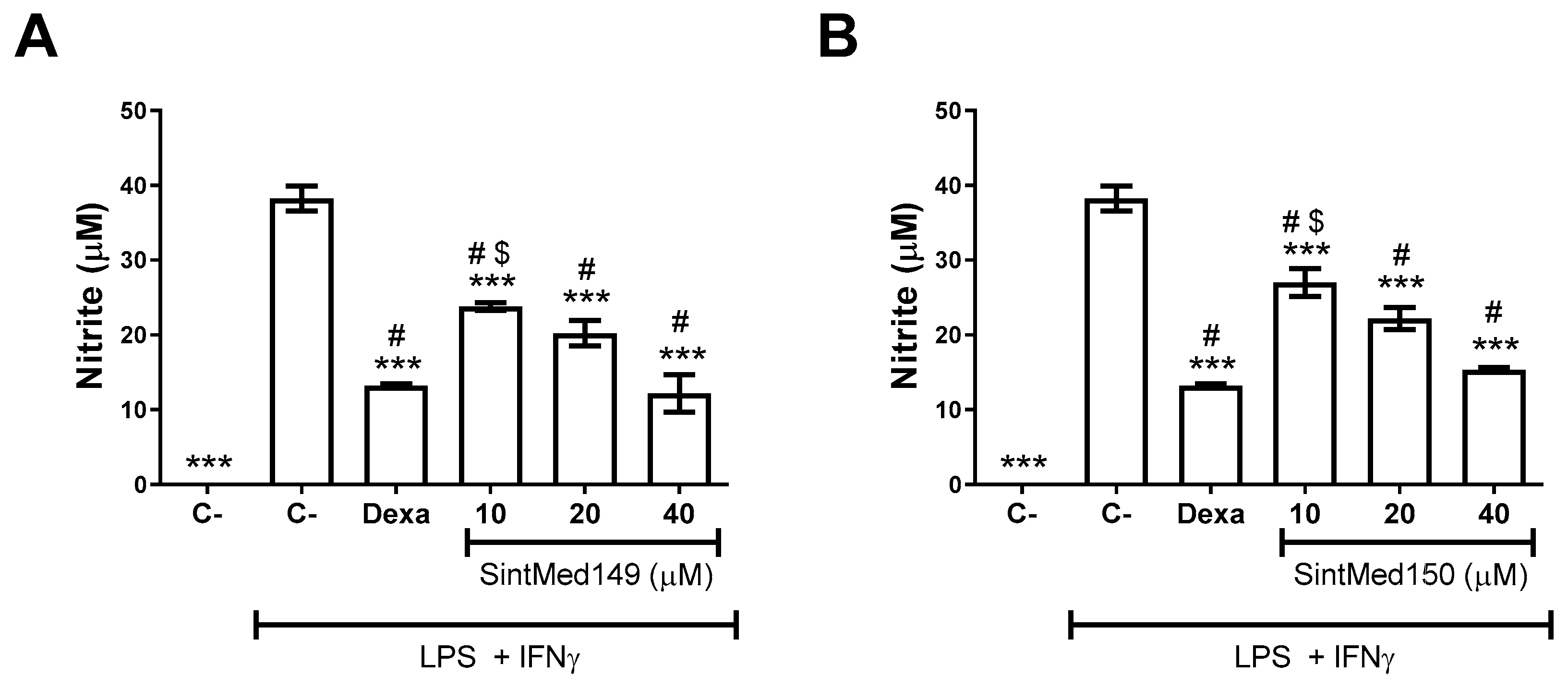
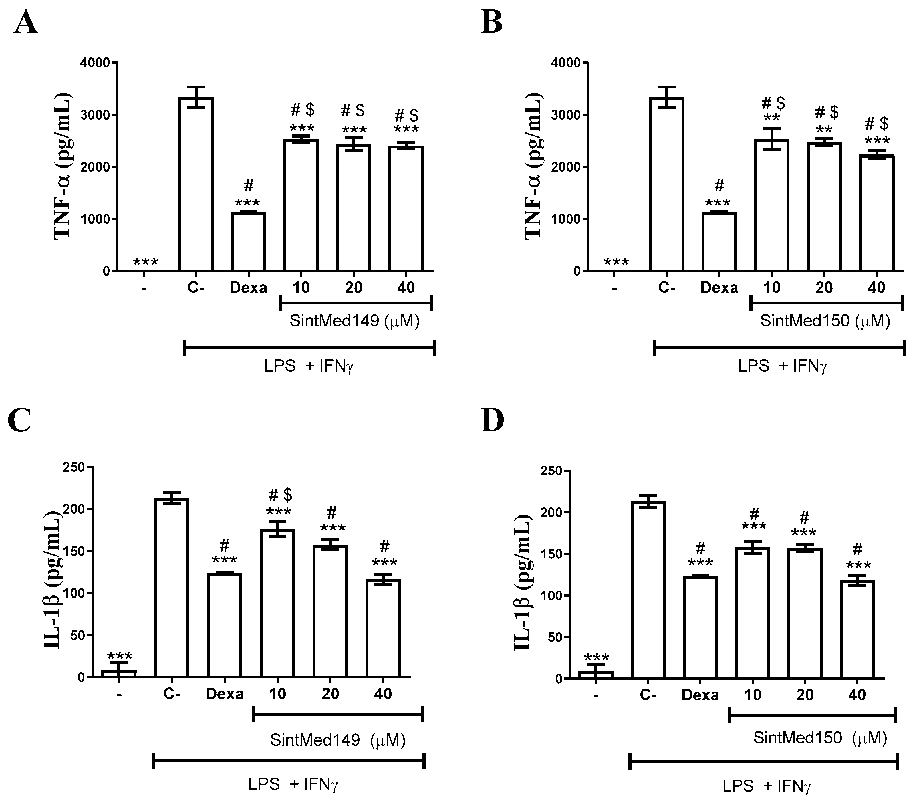
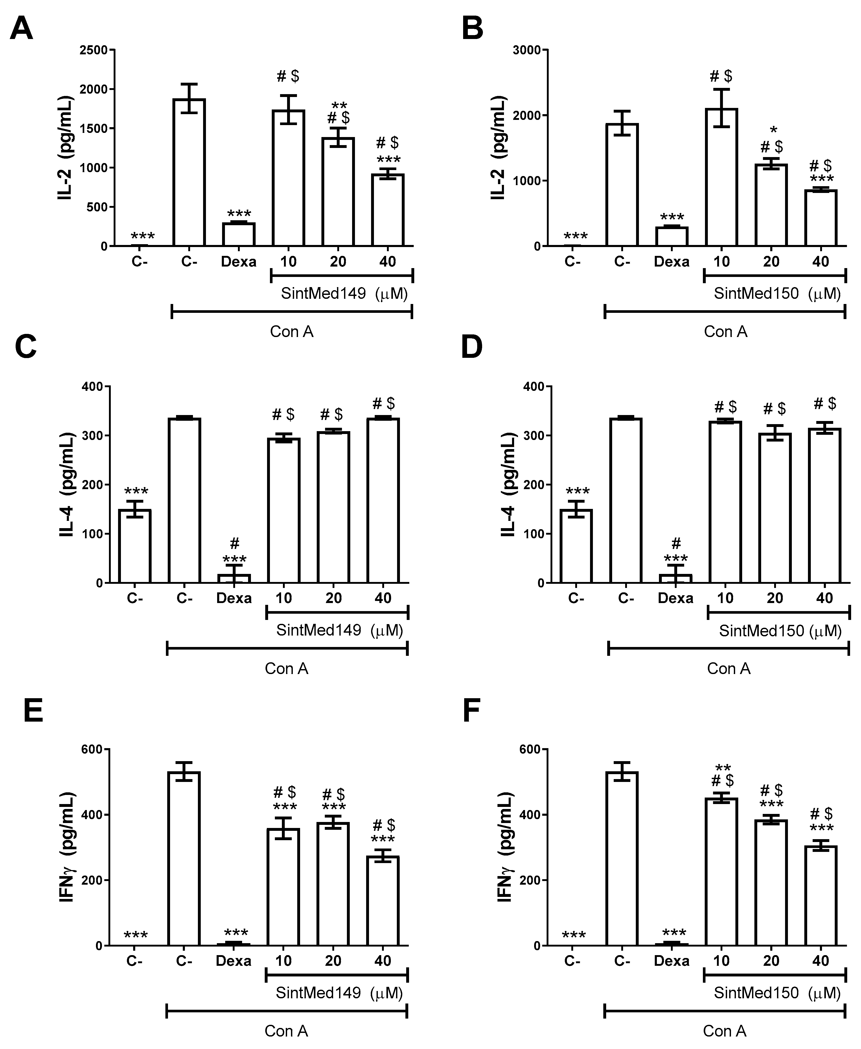
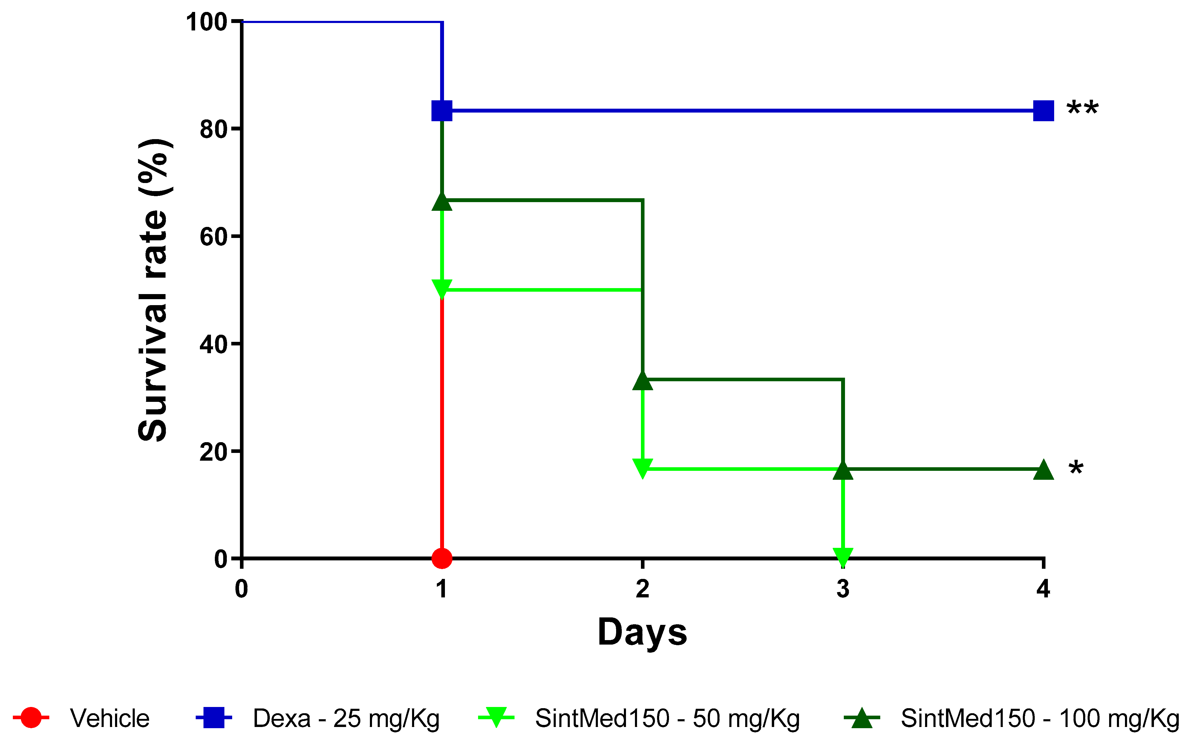
2. Results
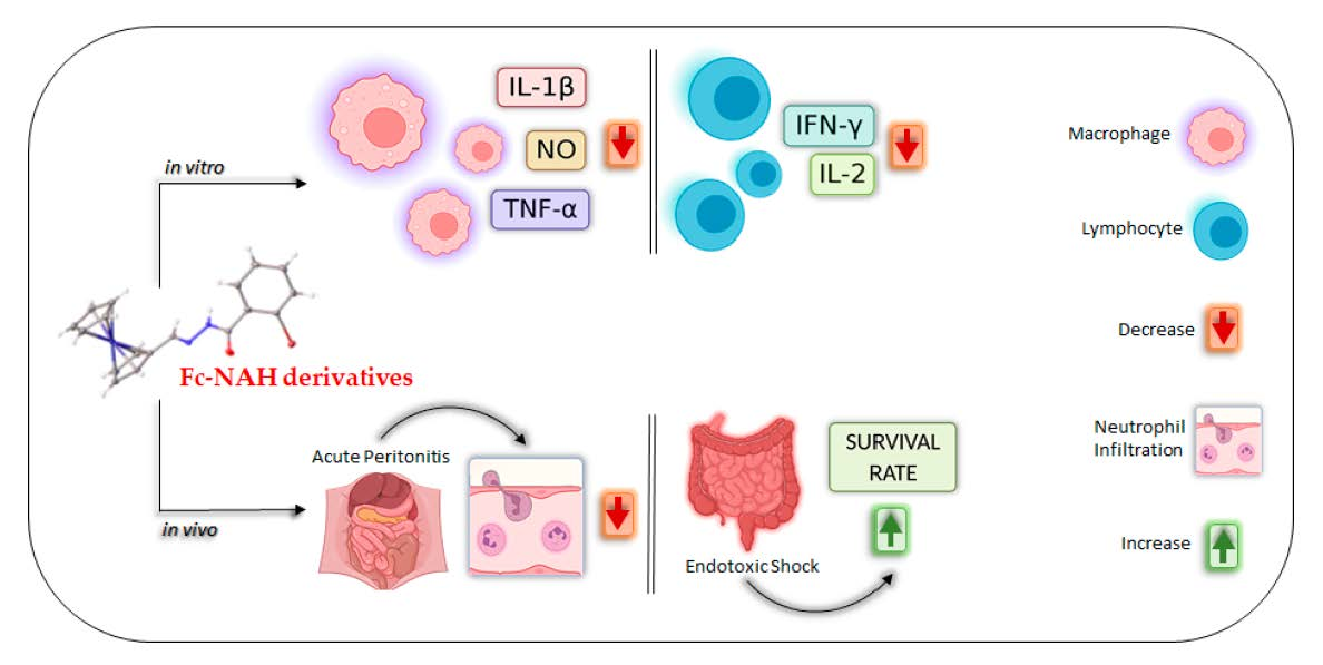
3. Discussion
4. Materials and Methods
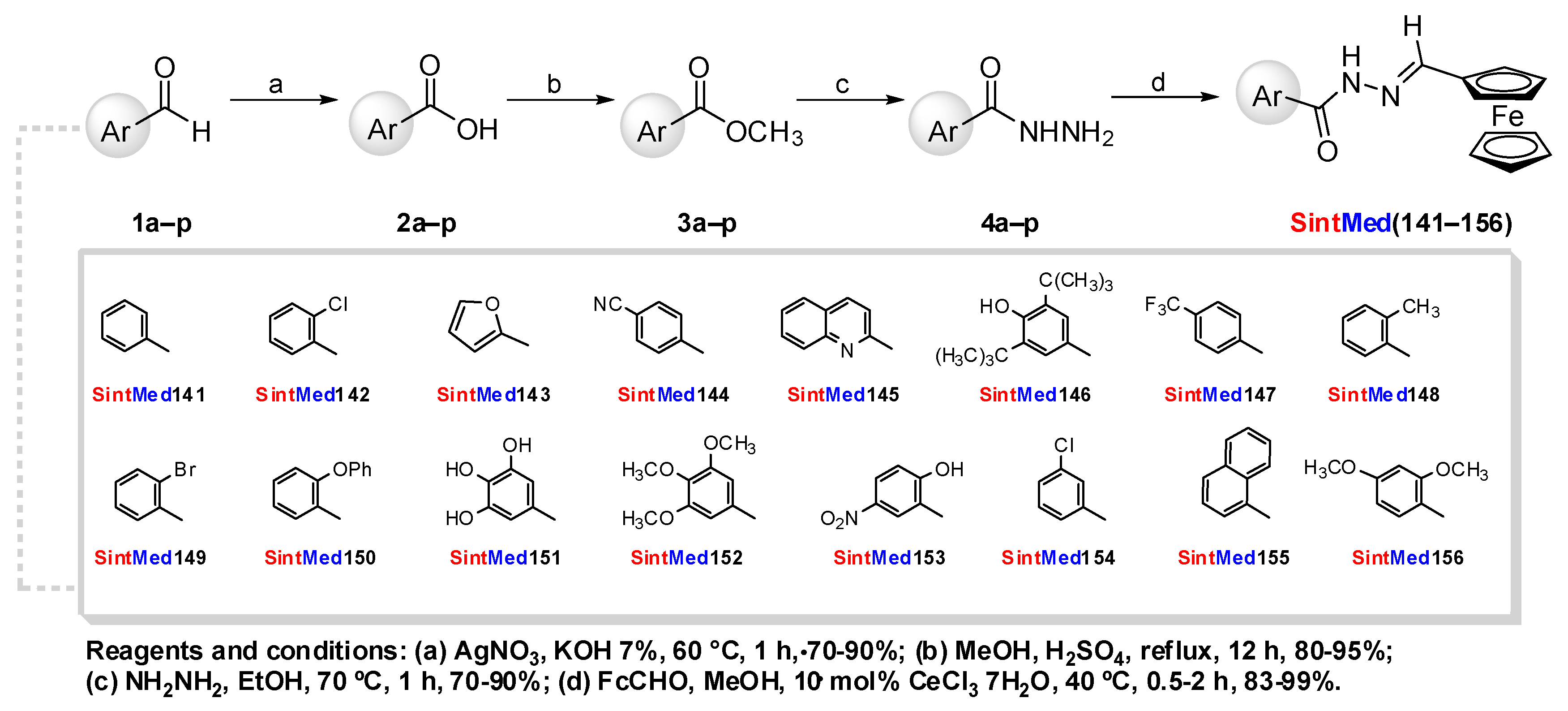
4.1. Chemistry
4.2. Preparation of Aryl Carboxylic Acids 2a–p [30]
4.3. Preparation of Aryl Carboxylate Methyl Esters 3a–p [29]
4.4. Preparation of Aryl Carbo Hydrazides 4a–p [29]
4.5. Preparation of Ferrocenyl-N-acyl Hydrazones SintMed(141–156)
4.6. X-ray Crystallographic Analysis
4.7. Drugs
4.8. Animals
4.9. Cytotoxicity to Mammalian Cells
4.10. Macrophage Cultures
4.11. Splenocyte Cultures
4.12. Assessment of Cytokine and Nitric Oxide Production
4.13. Acute Toxicity in Mice
4.14. LPS-Induced Endotoxin Shock
4.15. Induction of Acute Peritonitis in Mice
4.16. Statistical Analysis
5. Conclusions
Supplementary Materials
Author Contributions
Funding
Institutional Review Board Statement
Informed Consent Statement
Data Availability Statement
Acknowledgments
Conflicts of Interest
Abbreviations
References
- Chovatiya, R.; Medzhitov, R. Stress, inflammation, and defense of homeostasis. Mol. Cell 2014, 54, 281–288. [Google Scholar] [CrossRef] [PubMed] [Green Version]
- Chen, L.; Deng, H.; Cui, H.; Fang, J.; Zuo, Z.; Deng, J.; Li, Y.; Wang, X.; Zhao, L. Inflammatory responses and inflammation-associated diseases in organs. Oncotarget 2017, 14, 7204–7218. [Google Scholar] [CrossRef] [PubMed] [Green Version]
- Kuprash, D.V.; Nedospasov, S.A. Molecular and cellular mechanisms of inflammation. Biochmestry 2016, 81, 1237–1239. [Google Scholar] [CrossRef] [PubMed]
- Seelaender, M.; Rosa-Neto, J.C.; Pimentel, G.D.; Goldszmid, R.S.; Lira, F.S. Inflammation in the disease: Mechanism and therapies 2014. Mediat. Inflamm. 2015, 2015, 169852. [Google Scholar] [CrossRef] [PubMed]
- Alessandri, A.L.; Sousa, L.P.; Lucas, C.D.; Rossi, A.G.; Pinho, V.; Teixeira, M.M. Resolution of inflammation: Mechanisms and opportunity for drug development. Pharm. Ther. 2013, 139, 189–212. [Google Scholar] [CrossRef] [PubMed] [Green Version]
- Ronchetti, S.; Migliorati, G.; Bruscoli, S.; Riccardi, C. Defining the role of glucocorticoids in inflammation. Clin. Sci. 2018, 132, 1529–1543. [Google Scholar] [CrossRef]
- Bacchi, S.; Palumbo, P.; Sponta, A.; Coppolino, M.F. Clinical pharmacology of non-steroidal anti-inflammatory drugs: A review. Antiinflamm. Antiallergy Agents Med. Chem. 2012, 11, 52–64. [Google Scholar] [CrossRef]
- Bindu, S.; Mazumder, S.; Bandyopadhyay, U. Non-steroidal anti-inflammatory drugs (NSAIDs) and organ damage: A current perspective. Biochem. Pharmacol. 2020, 180, 114147. [Google Scholar] [CrossRef]
- Oray, M.; Samra, K.A.; Ebrahimiadib, N.; Messe, H.; Foster, C.S. Long-term side effects of glucocorticoids. Expert Opin. Drug Saf. 2016, 15, 457–465. [Google Scholar] [CrossRef]
- Verma, G.; Marella, A.; Shaquiquzzaman, M.; Akhtar, M.; Ali, M.R.; Alam, M.M. A review exploring biological activities of hydrazones. J. Pharm. Bioallied Sci. 2014, 6, 69–80. [Google Scholar]
- Guimarães, E.T.; Santos, T.B.; Silva, D.K.C.; Meira, C.S.; Moreira, D.R.M.; Silva, T.F.; Salmon, D.; Barreiro, E.J.; Soares, M.B.P. Potent immunosuppressive activity of a phosphodiesterase-4 inhibitor N-acylhydrazone in models of lipopolysaccharide-induced shock and delayed-type hypersensitivity reaction. Int. Imunopharmacol. 2018, 65, 108–118. [Google Scholar] [CrossRef] [PubMed]
- Cequeira, J.V.; Meira, C.S.; Santos, E.S.; França, L.S.A.; Vasconcelos, J.F.; Nonaka, C.K.V.; Melo, T.L.; Filho, J.M.S.; Moreira, D.R.M.; Soares, M.B.P. Anti-inflammatory activity of SintMed65, an N-acylhydrazone derivative, in a mouse model of allergic airway inflammation. Int. Imunopharmacol. 2019, 75, 105735. [Google Scholar] [CrossRef]
- Meira, C.S.; Filho, J.M.S.; Sousa, C.C.; Anjos, P.S.; Cerqueira, J.V.; Neto, H.A.D.; Silveira, R.G.; Russo, H.M.; Wolfender, J.L.; Queiroz, E.F.; et al. Structural design, synthesis and substituent effect of hydrazone-N-acylhydrazones reveal potent immunomodulatory agents. Bioorg. Med. Chem. 2018, 26, 1971–1985. [Google Scholar] [CrossRef] [PubMed]
- Gundla, R.; Shaik, B.B.; Gorantla, V. N-Acyl Hydrazones: Nonsteroidal Anti-Inflammatory Agents (NSAIDs), 1st ed.; LAP LAMBERT Academic Publishing: Hyderabad, India, 2021; pp. 1–112. [Google Scholar]
- Kajal, A.; Bala, S.; Sharma, N.; Kamboj, S.; Saini, V. Therapeutic Potential of Hydrazones as Anti-Inflammatory Agents. Int. J. Med. Chem. 2014, 2014, 761030. [Google Scholar] [CrossRef] [PubMed]
- De Melo, T.R.F.; Chelucci, R.C.; Pires, M.E.L.; Dutra, L.A.; Barbieri, K.P.; Bosquesi, P.L.; Trossini, G.H.G.; Chung, M.C.; Santos, J.L. Pharmacological Evaluation and Preparation of Nonsteroidal Anti-Inflammatory Drugs Containing an N-Acyl Hydrazone Subunit. Int. J. Mol. Sci. 2014, 15, 5821–5837. [Google Scholar] [CrossRef] [Green Version]
- Medeiros, M.A.M.B.; Gama, E.; Silva, M.; De Menezes Barbosa, J.; Martins de Lavor, É.; Ribeiro, T.F.; Macedo, C.A.F.; De Souza Duarte-Filho, L.A.M.; Feitosa, T.A.; De Jesus Silva, J.; et al. Antinociceptive and anti-inflammatory effects of hydrazone derivatives and their possible mechanism of action in mice. PLoS ONE 2021, 16, e0258094. [Google Scholar] [CrossRef]
- Viegas-Junior, C.; Danuello, A.; Bolzani, V.S.; Barreiro, E.J.; Fraga, C.A.M. Molecular hybridization: A useful tool in the design of new drug prototypes. Curr. Med. Chem. 2007, 14, 1829–1852. [Google Scholar] [CrossRef]
- Ivasiv, V.; Albertini, C.; Gonçalves, A.E.; Rossi, M.; Bolognesi, M.L. Molecular Hybridization as a Tool for Designing Multitarget Drug Candidates for Complex Diseases. Curr. Top. Med. Chem. 2019, 19, 1694–1711. [Google Scholar] [CrossRef]
- Singh, A.; Lumb, I.; Mehra, V.; Kumar, V. Ferrocene-appended pharmacophores: An exciting approach for modulating the biological potential of organic scaffolds. Dalton Trans. 2019, 48, 2840–2860. [Google Scholar] [CrossRef]
- Altowyan, M.S.; Ali, M.; Soliman, S.M.; Al-Majid, A.M.; Islam, M.S.; Yousuf, S.; Choudhary, M.I.; Ghabbour, H.A.; Barakat, A. Synthesis, computational studies and biological activity of oxamohydrazide derivatives bearing isatin and ferrocene scaffolds. J. Mol. Struct. 2019, 1202, 127372. [Google Scholar] [CrossRef]
- Guo, W.-Y.; Chen, L.-Z.; Shen, B.-N.; Liu, X.-H.; Tai, G.-P.; Li, Q.-S.; Gao, L.; Ruan, B.-F. Synthesis and in vitro and in vivo anti-inflammatory activity of novel 4-ferrocenylchroman-2-one derivatives. J. Enzym. Inhib. Med. Chem. 2019, 34, 1678–1689. [Google Scholar] [CrossRef] [PubMed]
- Barišić, L.; Roščić, M.; Kovačević, M.; Semenčić, M.Č.; Horvat, Š.; Rapić, V. The first ferrocene analogues of muramyldipeptide. Carbohydr. Res. 2011, 346, 678–684. [Google Scholar] [CrossRef] [PubMed]
- Soares, M.B.P.; Costa, J.F.O.; de Sá, M.S.; Ribeiro-dos-Santos, R.; Pigeon, P.; Jaouen, G.; Santana, A.E.G.; Goulart, M.O.F.; Hillard, E. Antiparasitic and immunomodulatory activities of 1,1-bis(4-hydroxyphenyl)-2-phenyl-but-1-ene and its protected and free 2-ferrocenyl derivatives. Drug Dev. Res. 2010, 71, 69–75. [Google Scholar] [CrossRef]
- Chaudhary, A.; Poonia, K. The redox mechanism of ferrocene and its phytochemical and biochemical compounds in anticancer therapy: A mini review. Inorg. Chem. Commun. 2021, 134, 109044. [Google Scholar] [CrossRef]
- Shoukat, H.; Altaf, A.A.; Badshah, A. Ferrocene-Based Metallodrugs. In Advances in Metallodrugs: Preparation and Applications in Medicinal Chemistry; Ul-Islam, S., Hashmi, A.A., Khan, S.A., Eds.; Wiley-Scrivener: Beverly, CA, USA, 2020; pp. 115–136. [Google Scholar]
- Song, J.; Liu, H.; Lei, M.; Tan, H.; Chen, Z.; Antoshin, A.; Payne, G.F.; Qu, X.; Liu, C. Redox-Channeling Polydopamine-Ferrocene (PDA-Fc) Coating To Confer Context-Dependent and Photothermal Antimicrobial Activities. ACS Appl. Mater. Interfaces 2020, 12, 8915–8928. [Google Scholar] [CrossRef]
- Larik, F.A.; Saeed, A.; Fattah, T.A.; Muqadar, U.; Channar, P.A. Recent advances in the synthesis, biological activities and various applications of ferrocene derivatives. Appl. Organometal. Chem. 2016, 31, e3664. [Google Scholar] [CrossRef]
- dos Santos Filho, J.M.; Queiroz e Silva, D.M.A.; Macedo, T.S.; Teixeira, H.M.P.; Moreira, D.R.M.; Challal, S.; Wolfender, J.; Queiroz, E.F.; Soares, M.B.P. Conjugation of N-acylhydrazone and 1,2,4-oxadiazole leads to the identification of active antimalarial agents. Bioorg. Med. Chem. 2016, 24, 5693–5701. [Google Scholar] [CrossRef]
- Mu, H.; Gong, R.; Ren, L.; Zhong, C.; Sun, Y.; Fu, E. An intramolecular charge transfer fluorescent probe: Synthesis and selective fluorescent sensing of Ag+. Spectrochim. Acta Part A 2008, 70, 923–928. [Google Scholar] [CrossRef]
- dos Santos Filho, J.M. Mild, Stereoselective, and Highly Efficient Synthesis of N-Acylhydrazones Mediated by CeCl3·7H2O in a Broad Range of Solvents. Eur. J. Org. Chem. 2014, 29, 6411–6417. [Google Scholar] [CrossRef]
- Lopes, A.B.; Miguez, E.; Kümmerle, A.E.; Rumjanek, V.M.; Fraga, C.A.M.; Barreiro, E.J. Characterization of Amide Bond Conformers for a Novel Heterocyclic Template of N-acylhydrazone Derivatives. Molecules 2013, 18, 11683–11704. [Google Scholar] [CrossRef]
- da Silva, T.F.; Bispo Júnior, W.; Alexandre-Moreira, M.S.; Costa, F.N.; Monteiro, C.E.S.; Ferreira, F.F.; Barroso, R.C.R.; Noël, F.; Sudo, R.T.; Zapata-Sudo, G.; et al. Novel Orally Active Analgesic and Anti-Inflammatory Cyclohexyl-N-Acylhydrazone Derivatives. Molecules 2015, 20, 3067–3088. [Google Scholar] [CrossRef] [PubMed]
- Thota, S.; Rodrigues, D.A.; Pinheiro, P.S.M.; Lima, L.M.; Fraga, C.A.M.; Barreiro, E.J. N-Acylhydrazones as drugs. Bioorg. Med. Chem. Lett. 2018, 28, 2797–2803. [Google Scholar] [CrossRef] [PubMed]
- Cordeiro, N.M.; Freitas, R.H.C.N.; Fraga, C.A.M.; Fernandes, P.D. Discovery of Novel Orally Active Tetrahydro-Naphthyl-N-Acylhydrazones with In Vivo Anti-TNF-α Effect and Remarkable Anti-Inflammatory Properties. PLoS ONE 2016, 11, e0156271. [Google Scholar] [CrossRef] [PubMed] [Green Version]
- Moraes, A.D.T.O.; Miranda, M.D.S.; Jacob, Í.T.T.; Amorim, C.A.D.C.; Moura, R.O.; Silva, S.Â.S.D.; Soares, M.B.P.; Almeida, S.M.V.; Souza, T.R.C.L.; Oliveira, J.F.; et al. Synthesis, in vitro and in vivo biological evaluation, COX-1/2 inhibition and molecular docking study of indole-N-acylhydrazone derivatives. Bioorg. Med. Chem. 2018, 26, 5388–5396. [Google Scholar] [CrossRef] [PubMed]
- Chellan, P.; Sadler, P.J. Enhancing the Activity of Drugs by Conjugation to Organometallic Fragments. Chem. Eur. J. 2020, 26, 8676–8688. [Google Scholar] [CrossRef]
- Dos Santos Filho, J.M.; Moreira, D.R.; De Simone, C.A.; Ferreira, R.S.; McKerrow, J.H.; Meira, C.S.; Guimarães, E.T.; Soares, M.B.P. Optimization of anti-Trypanosoma cruzi oxadiazoles leads to identification of compounds with efficacy in infected mice. Bioorg. Med. Chem. 2012, 20, 6423–6433. [Google Scholar] [CrossRef]
- Guzik, T.J.; Korbut, R.; Adamek-Guzik, T. Nitric oxide and superoxide in inflammation and immune regulation. J. Physiol. Pharm. 2003, 54, 469–487. [Google Scholar]
- Parameswaran, N.; Patial, S. Tumor necrosis factor-α signaling in macrophages. Crit. Rev. Eukaryot. Gene Expr. 2010, 20, 87–103. [Google Scholar] [CrossRef]
- Lopez-Castejon, G.; Brough, D. Understanding the mechanism of IL-1β secretion. Cytokine Growth Factor Rev. 2011, 22, 189–195. [Google Scholar] [CrossRef]
- Rollas, S.; Küçükgüzel, S.G. Biological activities of hydrazone derivatives. Molecules 2007, 12, 1910–1939. [Google Scholar] [CrossRef] [Green Version]
- Zeeshan, S.; Naveed, M.; Khan, A.; Atiq, A.; Arif, M.; Ahmed, M.N.; Kim, Y.S.; Khan, S. N-Pyrazoloyl and N-thiopheneacetyl hydrazone of isatin exhibited potent anti-inflammatory and anti-nociceptive properties through suppression of NF-κB, MAPK and oxidative stress signaling in animal models of inflammation. Inflamm. Res. 2019, 68, 613–632. [Google Scholar] [CrossRef] [PubMed]
- Debnath, U.; Mukherjee, S.; Joardar, N.; Sinha Babu, S.P.; Jana, K.; Misra, A.K. Aryl quinolinyl hydrazone derivatives as anti-inflammatory agents that inhibit TLR4 activation in the macrophages. Eur. J. Pharm. Sci. 2019, 134, 102–115. [Google Scholar] [CrossRef]
- Kohno, K.; Kataoka, J.; Ohtsuki, T.; Suemoto, Y.; Okamoto, I.; Usui, M.; Ikeda, M.; Kurimoto, M. IFN-gamma-inducing factor (IGIF) is a costimulatory factor on the activation of Th1 but not Th2 cells and exerts its effect independently of IL-12. J. Immunol. 1997, 15, 1541–1550. [Google Scholar]
- Boyman, O.; Sprent, J. The role of interleukin-2 during homeostasis and activation of the immune system. Nat. Rev. Immunol. 2012, 17, 180–190. [Google Scholar] [CrossRef] [PubMed]
- Sheldrick, G.M. SHELXT—Integrated space-group and crystalstructure determination. Acta Crystallogr. Sect. A Found. Adv. 2015, 71, 3–8. [Google Scholar] [CrossRef] [PubMed] [Green Version]
- Sheldrick, G.M. Crystal structure refinement with SHELXL. Acta Crystallogr. Sect. C Struct. Chem. 2015, 71, 3–8. [Google Scholar] [CrossRef] [PubMed] [Green Version]
- Puschmann, H.; Bourhis, L.J.; Dolomanov, O.V.; Gildea, R.J.; Howard, J.A.K. OLEX2—A complete package for molecular crystallography. Acta Crystallogr. Sect. A Found. Crystallogr. 2011, 67, C593. [Google Scholar] [CrossRef] [Green Version]
- Green, L.C.; Wagner, D.A.; Glogowski, J.; Skipper, P.L.; Wishnok, J.S.; Tannenbaum, S.R. Analysis of nitrate, nitrite, and [15N]nitrate in biological fluids. Anal. Biochem. 1982, 126, 131–138. [Google Scholar] [CrossRef]


 | |||
|---|---|---|---|
| Compounds | R | CC50 (µM) JJ74 a | Inhibition of NO (%) Production at 40 μM b |
| SintMed141 |  | >50 | 19.7 (±3.0) |
| SintMed142 |  | >50 | 49.3 (±0.8) |
| SintMed143 |  | >50 | 49.2 (±1.9) |
| SintMed144 |  | >50 | 55.2 (±0.6) |
| SintMed145 |  | >50 | 49.6 (±2.6) |
| SintMed146 |  | >50 | 14.4 (±2.2) |
| SintMed147 |  | >50 | 59.9 (±0.3) |
| SintMed148 |  | >50 | 56.6 (±2.1) |
| SintMed149 |  | >50 | 71.7 (±3.6) |
| SintMed150 |  | >50 | 74.2 (±0.7) |
| SintMed151 |  | >50 | 25.2 (±2.1) |
| SintMed152 |  | >50 | 55.5 (±1.7) |
| SintMed153 |  | >50 | 43.7 (±3.3) |
| SintMed154 |  | >50 | 45.2 (±2.1) |
| SintMed155 |  | >50 | 35.6 (±0.7) |
| SintMed156 |  | >50 | 46.7 (±3.0) |
| GV c | 0.8 (±0.1) | - | |
| Dexamethasone d | - | 64.9 (±6.7) | |
| Behavior and General Appearance | Observations | ||
|---|---|---|---|
| Vehicle | SintMed150 (50 mg/kg) | SintMed150 (100 mg/kg) | |
| Changes in the eyes | No changes | No changes | No changes |
| Changes in the fur | No changes | No changes | No changes |
| Changes in the skin | No changes | No changes | No changes |
| Coma | Absent | Absent | Absent |
| Convulsions | Absent | Absent | Absent |
| Diarrhea | Absent | Absent | Absent |
| Lethargy | Absent | Absent | Absent |
| Salivation | Absent | Absent | Absent |
| Sleep | Usual | Usual | Usual |
| Tremors | Absent | Absent | Absent |
| Days | Vehicle | SintMed150 (50 mg/kg) | SintMed150 (100 mg/kg) |
|---|---|---|---|
| 0 | 21.6 (±1.3) | 20.9 (±1.1) | 20.1 (±0.8) |
| 7 | 21.8 (±1.0) | 21.1 (±1.1) | 20.4 (±0.7) |
| 14 | 22.3 (±1.1) | 21.7 (±0.9) | 21.0 (±0.7) |
Publisher’s Note: MDPI stays neutral with regard to jurisdictional claims in published maps and institutional affiliations. |
© 2022 by the authors. Licensee MDPI, Basel, Switzerland. This article is an open access article distributed under the terms and conditions of the Creative Commons Attribution (CC BY) license (https://creativecommons.org/licenses/by/4.0/).
Share and Cite
Silva, L.P.; Santos, I.P.; Silva, D.K.C.; dos Reis, B.P.Z.C.; Meira, C.S.; Castro, M.V.B.d.S.; dos Santos Filho, J.M.; Araujo-Neto, J.H.d.; Ellena, J.A.; Silveira, R.G.d.; et al. Molecular Hybridization Strategy on the Design, Synthesis, and Structural Characterization of Ferrocene-N-acyl Hydrazones as Immunomodulatory Agents. Molecules 2022, 27, 8343. https://0-doi-org.brum.beds.ac.uk/10.3390/molecules27238343
Silva LP, Santos IP, Silva DKC, dos Reis BPZC, Meira CS, Castro MVBdS, dos Santos Filho JM, Araujo-Neto JHd, Ellena JA, Silveira RGd, et al. Molecular Hybridization Strategy on the Design, Synthesis, and Structural Characterization of Ferrocene-N-acyl Hydrazones as Immunomodulatory Agents. Molecules. 2022; 27(23):8343. https://0-doi-org.brum.beds.ac.uk/10.3390/molecules27238343
Chicago/Turabian StyleSilva, Laís Peres, Ivanilson Pimenta Santos, Dahara Keyse Carvalho Silva, Bruna Padilha Zurita Claro dos Reis, Cássio Santana Meira, Marcos Venícius Batista de Souza Castro, José Maurício dos Santos Filho, João Honorato de Araujo-Neto, Javier Alcides Ellena, Rafael Gomes da Silveira, and et al. 2022. "Molecular Hybridization Strategy on the Design, Synthesis, and Structural Characterization of Ferrocene-N-acyl Hydrazones as Immunomodulatory Agents" Molecules 27, no. 23: 8343. https://0-doi-org.brum.beds.ac.uk/10.3390/molecules27238343