
Surface Functionalization of Nanofibers: The Multifaceted Approach for Advanced Biomedical Applications

 , , , and
Abstract
:1. Introduction
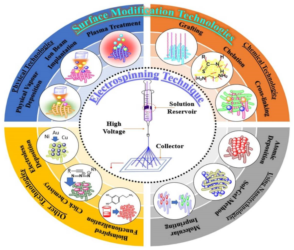
2. Electrospinning Technique
2.1. Melt Electrospinning
2.2. Co-Axial Electrospinning
2.3. Emulsion Electrospinning
2.4. Rotary Jet Spinning (RJS)
3. Methods of Surface Functionalization of Nanofibers
3.1. Surface Functionalization Using Physical Technologies
3.1.1. Plasma Treatment
3.1.2. Physical Vapor Deposition
3.1.3. Ion Beam Implantation
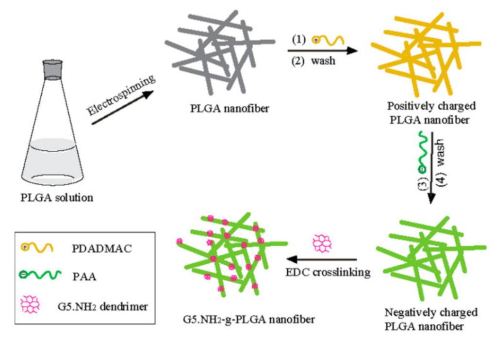
3.2. Surface Grafting, Cross-Linking and Chelation
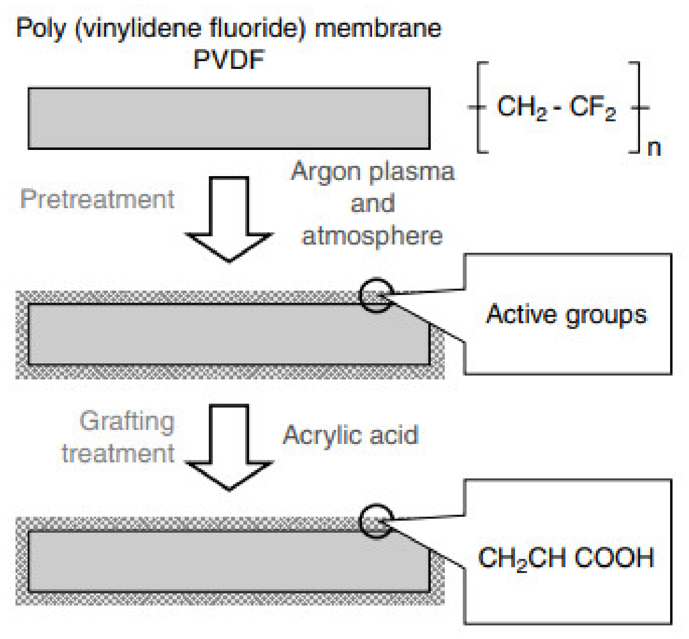
3.2.1. Grafting
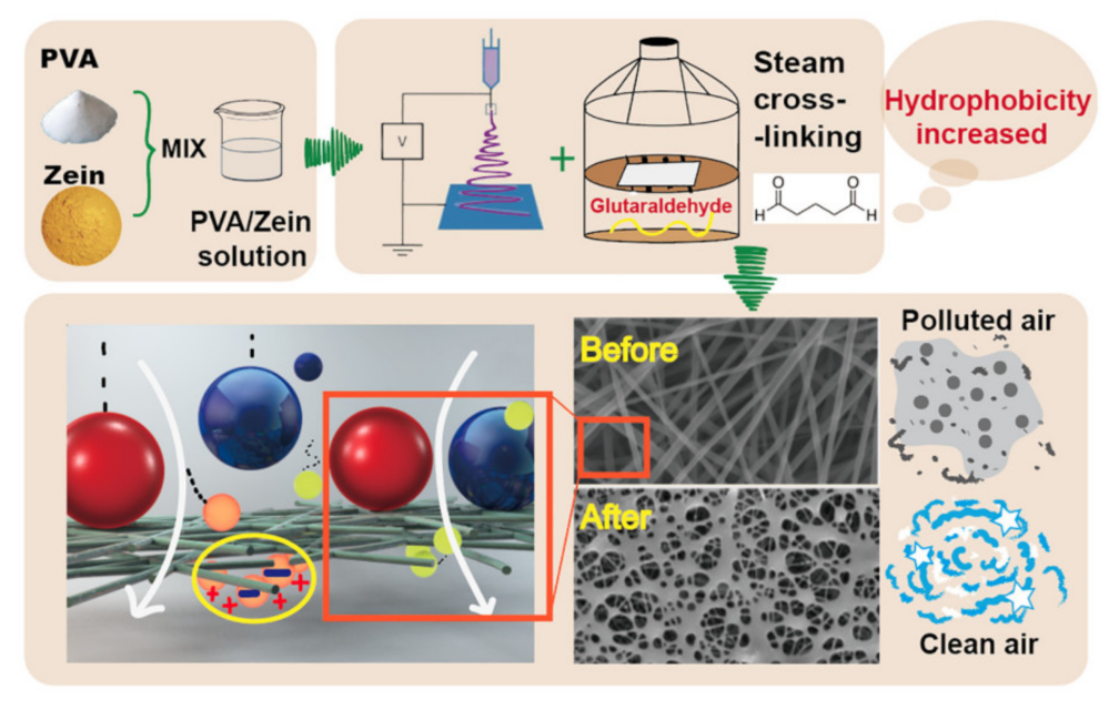
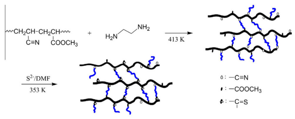
3.2.2. Cross Linking
3.2.3. Chelation
3.3. Electroless Deposition
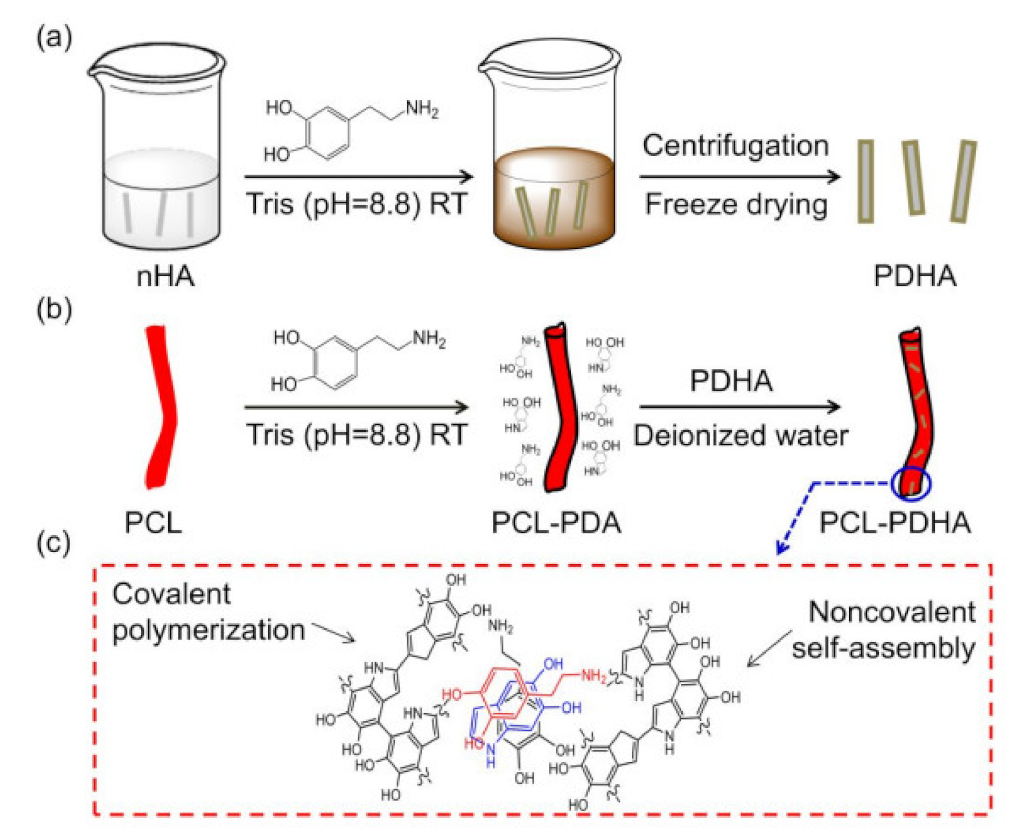
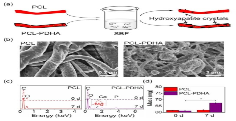
3.4. Bioinspired Surface Functionalization
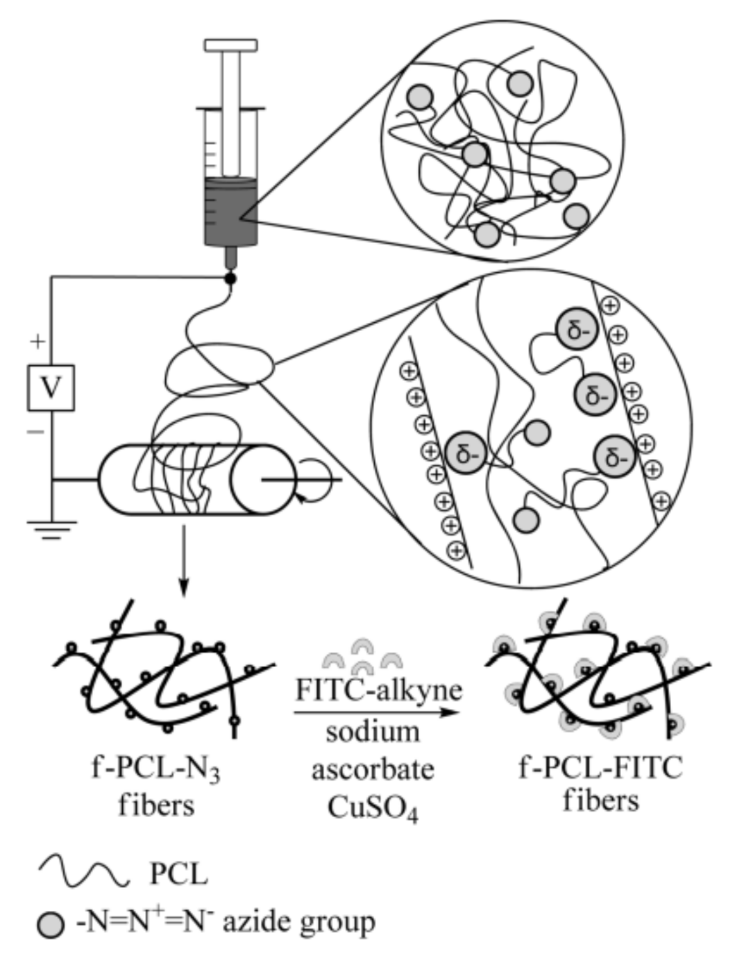
3.5. Click Chemistry
3.6. Surface Functionalization Using Nanotechnologies
3.6.1. Sol-Gel Method
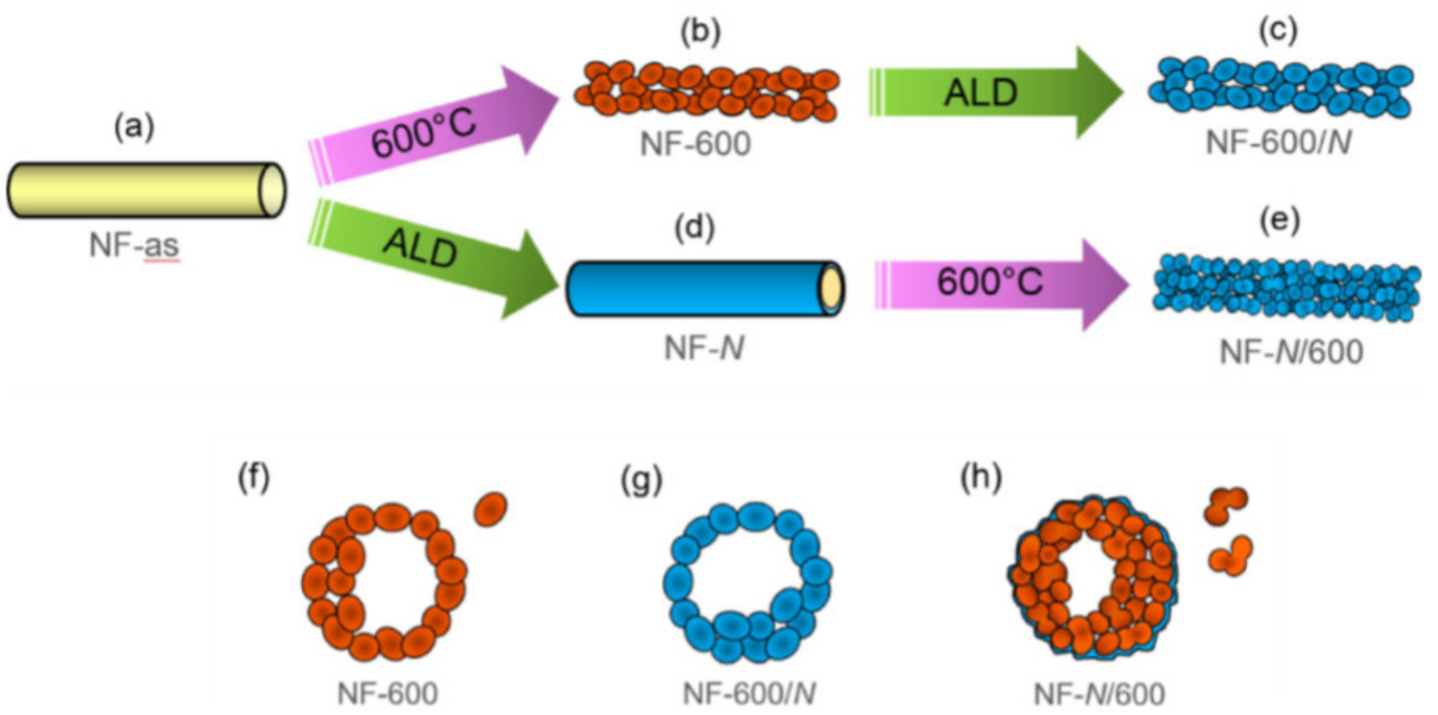
3.6.2. Atomic Deposition
3.6.3. Layer-By-Layer (Lbl) Deposition
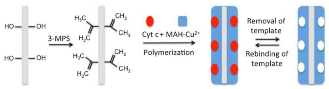
3.6.4. Molecular Imprinting
3.7. Surface Functionalization Using Biotechnology
4. Reagents Used for Surface Functionalization
4.1. Fe2O3 (Ferrous Oxide/Iron Oxide)
4.2. Gelatin
4.3. Silver
4.4. Plasma Treatment (Ar or O2 Gas)
4.5. Graphene Oxide-Silver
4.6. ZnO-Ag
4.7. Nano-Hydroxyapatite (nHA)
4.8. Collagen Coating
4.9. Polyelectrolyte: Poly (Acrylic Acid) (PAA), Chitosan (CS) and Polydiallyl Dimethyl Ammonium Chloride (pDADMAC)
4.10. Avidin
4.11. Glutaraldehyde
4.12. HNO3 (Nitric Acid)
4.13. Stainless Steel
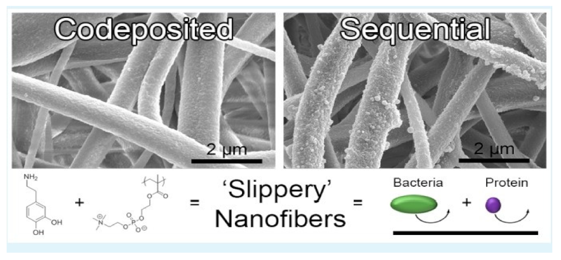
4.14. Zwitterionic Sulfobetaine
4.15. Weak Acid Cation-Exchange Ligand
4.16. Primary Amine
4.17. Beta-Cyclodextrins
4.18. COOH-Containing Polymer and TiCaPCON Film
4.19. Glycine-Phenylalanine-Hydroxyproline-Glycine-Glutamate-Arginine (GEOGER) Peptide
4.20. Polydopamine
4.21. Biotin
4.22. Poly (2-Methacryloyloxyethyl Phosphorylcholine) (Poly MPC)
5. Applications of Surface Functionalized Nanofibers
5.1. Wound Healing
5.2. Drug Delivery
5.3. Bacterial and Viral Pathogen Detection
5.4. Tissue Engineering and Regenerative Medicine
5.5. Targeting Strategies
5.6. Anti-Bacterial Application
5.7. Biomedical Application
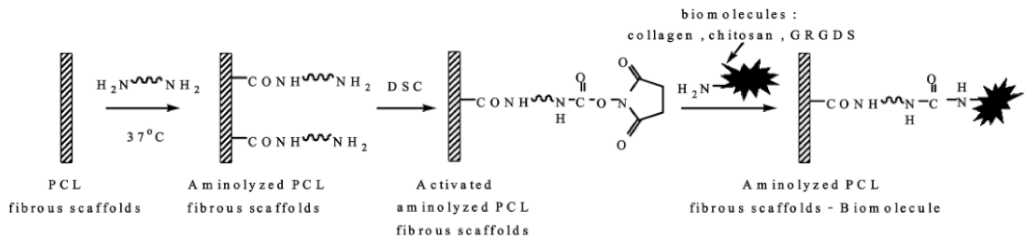
5.7.1. Immobilization of Bioactive Molecules
5.7.2. Chemotherapy-Cancer Theranostics Application
5.7.3. Implantable Smart Magnetic Nanofiber
5.7.4. Liver Cancer Therapy
5.8. Cell Culture Application
5.9. Anticoagulant Activity
6. Toxicity Study of Surface Functionalized Nanofiber
7. Conclusions
Author Contributions
Funding
Data Availability Statement
Acknowledgments
Conflicts of Interest
References
- Suárez, D.F.; Pinzón-García, A.D.; Sinisterra, R.D.; Dussan, A.; Mesa, F.; Ramírez-Clavijo, S. Uniaxial and Coaxial Nanofibers PCL/Alginate or PCL/Gelatine Transport and Release Tamoxifen and Curcumin Affecting the Viability of MCF7 Cell Line. Nanomaterials 2022, 12, 3348. [Google Scholar] [CrossRef] [PubMed]
- Garkal, A.; Kulkarni, D.; Musale, S.; Mehta, T.; Giram, P. Electrospinning Nanofiber Technology: A Multifaceted Paradigm in Biomedical Applications. New J. Chem. 2021, 45, 21508–21533. [Google Scholar] [CrossRef]
- Xue, J.; Wu, T.; Dai, Y.; Xia, Y. Electrospinning and Electrospun Nanofibers: Methods, Materials, and Applications. Chem. Rev. 2019, 119, 5298–5415. [Google Scholar] [CrossRef] [PubMed]
- Xie, X.; Chen, Y.; Wang, X.; Xu, X.; Shen, Y.; Khan, A.u.R.; Aldalbahi, A.; Fetz, A.E.; Bowlin, G.L.; El-Newehy, M.; et al. Electrospinning Nanofiber Scaffolds for Soft and Hard Tissue Regeneration. J. Mater. Sci. Technol. 2020, 59, 243–261. [Google Scholar] [CrossRef]
- Ibrahim, N.A.; Fouda, M.M.G.; Eid, B.M. Chapter 20—Functional Nanofibers: Fabrication, Functionalization, and Potential Applications. In Handbook of Functionalized Nanomaterials for Industrial Applications; Mustansar Hussain, C., Ed.; Micro and Nano Technologies; Elsevier: Amsterdam, The Netherlands, 2020; pp. 581–609. ISBN 978-0-12-816787-8. [Google Scholar]
- Kumbar, S.G.; James, R.; Nukavarapu, S.P.; Laurencin, C.T. Electrospun nanofiber scaffolds: Engineering soft tissues. Biomed. Mater. 2008, 3, 34002. [Google Scholar] [CrossRef] [Green Version]
- ScienceDirect. Electrospinning of Polymeric Nanofibers for Drug Delivery Applications. Available online: https://0-www-sciencedirect-com.brum.beds.ac.uk/science/article/abs/pii/S0168365914002363 (accessed on 2 May 2022).
- Elsadek, N.E.; Nagah, A.; Ibrahim, T.M.; Chopra, H.; Ghonaim, G.A.; Emam, S.E.; Cavalu, S.; Attia, M.S. Electrospun Nanofibers Revisited: An Update on the Emerging Applications in Nanomedicine. Materials 2022, 15, 1934. [Google Scholar] [CrossRef]
- ScienceDirect. Electrospun Polymeric Nanofibers: New Horizons in Drug Delivery. Available online: https://0-www-sciencedirect-com.brum.beds.ac.uk/science/article/abs/pii/S0928098717304001 (accessed on 2 May 2022).
- Sharma, R.; Singh, H.; Joshi, M.; Sharma, A.; Garg, T.; Goyal, A.; Rath, G. Recent Advances in Polymeric Electrospun Nanofibers for Drug Delivery. CRT 2014, 31, 187–217. [Google Scholar] [CrossRef]
- ScienceDirect. A Review on the Properties of Electrospun Cellulose Acetate and Its Application in Drug Delivery Systems: A New Perspective. Available online: https://0-www-sciencedirect-com.brum.beds.ac.uk/science/article/abs/pii/S000862152030015X (accessed on 2 May 2022).
- Kajdič, S.; Planinšek, O.; Gašperlin, M.; Kocbek, P. Electrospun Nanofibers for Customized Drug-Delivery Systems. J. Drug Deliv. Sci. Technol. 2019, 51, 672–681. [Google Scholar] [CrossRef]
- Tort, S.; Acartürk, F. Preparation and Characterization of Electrospun Nanofibers Containing Glutamine. Carbohydr. Polym. 2016, 152, 802–814. [Google Scholar] [CrossRef]
- Cavalu, S.; Roiu, G.; Pop, O.; Heredea, D.A.P.; Costea, T.O.; Costea, C.F. Nano-Scale Modifications of Amniotic Membrane Induced by UV and Antibiotic Treatment: Histological, AFM and FTIR Spectroscopy Evidence. Materials 2021, 14, 863. [Google Scholar] [CrossRef]
- ScienceDirect. A Review on Toxicity of Turmeric Derived Nano-Formulates against Bacterial and Fungal Cells with Special Emphasis on Electrospun Nanofibers. Available online: https://0-www-sciencedirect-com.brum.beds.ac.uk/science/article/pii/S2214785320344345 (accessed on 2 May 2022).
- Torres-Giner, S.; Pérez-Masiá, R.; Lagaron, J.M. A Review on Electrospun Polymer Nanostructures as Advanced Bioactive Platforms. Polym. Eng. Sci. 2016, 56, 500–527. [Google Scholar] [CrossRef]
- Balogh, A.; Cselkó, R.; Démuth, B.; Verreck, G.; Mensch, J.; Marosi, G.; Nagy, Z.K. Alternating Current Electrospinning for Preparation of Fibrous Drug Delivery Systems. Int. J. Pharm. 2015, 495, 75–80. [Google Scholar] [CrossRef] [PubMed]
- Sofi, H.S.; Ashraf, R.; Khan, A.H.; Beigh, M.A.; Majeed, S.; Sheikh, F.A. Reconstructing Nanofibers from Natural Polymers Using Surface Functionalization Approaches for Applications in Tissue Engineering, Drug Delivery and Biosensing Devices. Mater. Sci. Eng. C 2019, 94, 1102–1124. [Google Scholar] [CrossRef] [PubMed]
- Sell, S.A.; Wolfe, P.S.; Garg, K.; McCool, J.M.; Rodriguez, I.A.; Bowlin, G.L. The Use of Natural Polymers in Tissue Engineering: A Focus on Electrospun Extracellular Matrix Analogues. Polymers 2010, 2, 522–553. [Google Scholar] [CrossRef]
- Bedian, L.; Villalba-Rodríguez, A.M.; Hernández-Vargas, G.; Parra-Saldivar, R.; Iqbal, H.M.N. Bio-Based Materials with Novel Characteristics for Tissue Engineering Applications—A Review. Int. J. Biol. Macromol. 2017, 98, 837–846. [Google Scholar] [CrossRef]
- Jordan, A.M.; Viswanath, V.; Kim, S.-E.; Pokorski, J.K.; Korley, L.T.J. Processing and Surface Modification of Polymer Nanofibers for Biological Scaffolds: A Review. J. Mater. Chem. B 2016, 4, 5958–5974. [Google Scholar] [CrossRef]
- Abdul Khalil, H.; Adnan, A.; Yahya, E.B.; Olaiya, N.; Safrida, S.; Hossain, M.S.; Balakrishnan, V.; Gopakumar, D.A.; Abdullah, C.; Oyekanmi, A.; et al. A Review on Plant Cellulose Nanofibre-Based Aerogels for Biomedical Applications. Polymers 2020, 12, 1759. [Google Scholar] [CrossRef]
- Dalton, P.D.; Grafahrend, D.; Klinkhammer, K.; Klee, D.; Möller, M. Electrospinning of Polymer Melts: Phenomenological Observations. Polymer 2007, 48, 6823–6833. [Google Scholar] [CrossRef]
- Brown, T.D.; Edin, F.; Detta, N.; Skelton, A.D.; Hutmacher, D.W.; Dalton, P.D. Melt Electrospinning of Poly(ε-Caprolactone) Scaffolds: Phenomenological Observations Associated with Collection and Direct Writing. Mater. Sci. Eng. C 2014, 45, 698–708. [Google Scholar] [CrossRef]
- Hemamalini, T.; Giri Dev, V.R. Comprehensive Review on Electrospinning of Starch Polymer for Biomedical Applications. Int. J. Biol. Macromol. 2018, 106, 712–718. [Google Scholar] [CrossRef]
- Cavalu, S.; Bisboaca, S.; Mates, I.M.; Pasca, P.M.; Laslo, V.; Costea, T.; Fritea, L.; Vicas, S. Novel Formulation Based on Chitosan-Arabic Gum Nanoparticles Entrapping Propolis Extract Production, Physico-Chemical and Structural Characterization. Rev. Chim. 2018, 69, 3756–3760. [Google Scholar] [CrossRef]
- Castilho, M.; Feyen, D.; Flandes-Iparraguirre, M.; Hochleitner, G.; Groll, J.; Doevendans, P.A.F.; Vermonden, T.; Ito, K.; Sluijter, J.P.G.; Malda, J. Melt Electrospinning Writing of Poly-Hydroxymethylglycolide-Co-ε-Caprolactone-Based Scaffolds for Cardiac Tissue Engineering. Adv. Healthc. Mater. 2017, 6, 1700311. [Google Scholar] [CrossRef] [PubMed] [Green Version]
- Yarin, A.L. Coaxial Electrospinning and Emulsion Electrospinning of Core-Shell Fibers. Polym. Adv. Technol. 2011, 22, 310–317. [Google Scholar] [CrossRef]
- Briggs, T.; Arinzeh, T.L. Examining the Formulation of Emulsion Electrospinning for Improving the Release of Bioactive Proteins from Electrospun Fibers. J. Biomed. Mater. Res. Part A 2014, 102, 674–684. [Google Scholar] [CrossRef] [PubMed]
- Yoon, J.; Yang, H.-S.; Lee, B.-S.; Yu, W.-R. Recent Progress in Coaxial Electrospinning: New Parameters, Various Structures, and Wide Applications. Adv. Mater. 2018, 30, 1704765. [Google Scholar] [CrossRef]
- Sun, Z.; Zussman, E.; Yarin, A.L.; Wendorff, J.H.; Greiner, A. Compound Core-Shell Polymer Nanofibers by Co-Electrospinning. Adv. Mater. 2003, 15, 1929–1932. [Google Scholar] [CrossRef]
- He, C.; Huang, Z.; Han, X.; Liu, L.; Zhang, H.; Chen, L. Coaxial Electrospun Poly(L-Lactic Acid) Ultrafine Fibers for Sustained Drug Delivery. J. Macromol. Sci. Part B 2006, 45, 515–524. [Google Scholar] [CrossRef]
- Qin, X. 3—Coaxial Electrospinning of Nanofibers. In Electrospun Nanofibers; Woodhead Publishing Series in Textiles; Afshari, M., Ed.; Woodhead Publishing: Cambridge, UK, 2017; pp. 41–71. ISBN 978-0-08-100907-9. [Google Scholar]
- Nam, J.; Johnson, J.; Lannutti, J.J.; Agarwal, S. Modulation of Embryonic Mesenchymal Progenitor Cell Differentiation via Control over Pure Mechanical Modulus in Electrospun Nanofibers. Acta Biomater. 2011, 7, 1516–1524. [Google Scholar] [CrossRef] [Green Version]
- Zhang, Y.; Huang, Z.-M.; Xu, X.; Lim, C.T.; Ramakrishna, S. Preparation of Core−Shell Structured PCL-r-Gelatin Bi-Component Nanofibers by Coaxial Electrospinning. Chem. Mater. 2004, 16, 3406–3409. [Google Scholar] [CrossRef]
- Xu, X.; Zhuang, X.; Chen, X.; Wang, X.; Yang, L.; Jing, X. Preparation of Core-Sheath Composite Nanofibers by Emulsion Electrospinning. Macromol. Rapid Commun. 2006, 27, 1637–1642. [Google Scholar] [CrossRef]
- Buzgo, M.; Mickova, A.; Rampichova, M.; Doupnik, M. 11—Blend Electrospinning, Coaxial Electrospinning, and Emulsion Electrospinning Techniques. In Core-Shell Nanostructures for Drug Delivery and Theranostics; Woodhead Publishing Series in Biomaterials; Focarete, M.L., Tampieri, A., Eds.; Woodhead Publishing: Cambridge, UK, 2018; pp. 325–347. ISBN 978-0-08-102198-9. [Google Scholar]
- Lin, S.; Cai, Q.; Ji, J.; Sui, G.; Yu, Y.; Yang, X.; Ma, Q.; Wei, Y.; Deng, X. Electrospun Nanofiber Reinforced and Toughened Composites through in Situ Nano-Interface Formation. Compos. Sci. Technol. 2008, 68, 3322–3329. [Google Scholar] [CrossRef]
- Samanta, A.; Takkar, S.; Kulshreshtha, R.; Nandan, B.; Srivastava, R.K. Hydroxyapatite Stabilized Pickering Emulsions of Poly(ε-Caprolactone) and Their Composite Electrospun Scaffolds. Colloids Surf. A Physicochem. Eng. Asp. 2017, 533, 224–230. [Google Scholar] [CrossRef]
- Abdul Hameed, M.M.; Mohamed Khan, S.A.P.; Thamer, B.M.; Al-Enizi, A.; Aldalbahi, A.; El-Hamshary, H.; El-Newehy, M.H. Core-Shell Nanofibers from Poly(Vinyl Alcohol) Based Biopolymers Using Emulsion Electrospinning as Drug Delivery System for Cephalexin Drug. J. Macromol. Sci. Part A 2021, 58, 130–144. [Google Scholar] [CrossRef]
- Lamarra, J.; Calienni, M.N.; Rivero, S.; Pinotti, A. Electrospun Nanofibers of Poly (Vinyl Alcohol) and Chitosan-Based Emulsions Functionalized with Cabreuva Essential Oil. Int. J. Biol. Macromol. 2020, 160, 307–318. [Google Scholar] [CrossRef]
- Rogalski, J.J.; Bastiaansen, C.W.M.; Peijs, T. Rotary Jet Spinning Review—A Potential High Yield Future for Polymer Nanofibers. Nanocomposites 2017, 3, 97–121. [Google Scholar] [CrossRef] [Green Version]
- Zhang, X.; Lu, Y. Centrifugal Spinning: An Alternative Approach to Fabricate Nanofibers at High Speed and Low Cost. Polym. Rev. 2014, 54, 677–701. [Google Scholar] [CrossRef]
- Golecki, H.M.; Yuan, H.; Glavin, C.; Potter, B.; Badrossamay, M.R.; Goss, J.A.; Phillips, M.D.; Parker, K.K. Effect of Solvent Evaporation on Fiber Morphology in Rotary Jet Spinning. Langmuir 2014, 30, 13369–13374. [Google Scholar] [CrossRef] [Green Version]
- Wang, L.; Shi, J.; Liu, L.; Secret, E.; Chen, Y. Fabrication of Polymer Fiber Scaffolds by Centrifugal Spinning for Cell Culture Studies. Microelectron. Eng. 2011, 88, 1718–1721. [Google Scholar] [CrossRef]
- Rampichová, M.; Buzgo, M.; Chvojka, J.; Prosecká, E.; Kofroňová, O.; Amler, E. Cell Penetration to Nanofibrous Scaffolds. Cell Adhes. Migr. 2014, 8, 36–41. [Google Scholar] [CrossRef] [Green Version]
- Yao, T.; Baker, M.B.; Moroni, L. Strategies to Improve Nanofibrous Scaffolds for Vascular Tissue Engineering. Nanomaterials 2020, 10, 887. [Google Scholar] [CrossRef]
- Wei, Q.F.; Gao, W.D.; Hou, D.Y.; Wang, X.Q. Surface Modification of Polymer Nanofibers by Plasma Treatment. Appl. Surf. Sci. 2005, 245, 16–20. [Google Scholar] [CrossRef]
- ScienceDirect. Surface Functionalization of Polymer Nanofibers. Available online: https://0-www-sciencedirect-com.brum.beds.ac.uk/science/article/pii/B9780857090690500069 (accessed on 17 May 2022).
- Bank-Srour, B.; Becker, P.; Krasovitsky, L.; Gladkikh, A.; Rosenberg, Y.; Barkay, Z.; Rosenman, G. Physical Vapor Deposition of Peptide Nanostructures. Polym. J. 2013, 45, 494–503. [Google Scholar] [CrossRef]
- Wong, K.K.H.; Hutter, J.L.; Zinke-Allmang, M.; Wan, W. Physical Properties of Ion Beam Treated Electrospun Poly(Vinyl Alcohol) Nanofibers. Eur. Polym. J. 2009, 45, 1349–1358. [Google Scholar] [CrossRef]
- Xiao, S.; Peng, Q.; Yang, Y.; Tao, Y.; Zhou, Y.; Xu, W.; Shi, X. Preparation of [Amine-Terminated Generation 5 Poly(Amidoamine)]-Graft-Poly(Lactic-Co-Glycolic Acid) Electrospun Nanofibrous Mats for Scaffold-Mediated Gene Transfection. ACS Appl. Bio Mater. 2020, 3, 346–357. [Google Scholar] [CrossRef] [Green Version]
- Schiffman, J.D.; Schauer, C.L. Cross-Linking Chitosan Nanofibers. Biomacromolecules 2007, 8, 594–601. [Google Scholar] [CrossRef]
- ScienceDirect. Hydrophobic Cross-Linked Zein-Based Nanofibers with Efficient Air Filtration and Improved Moisture Stability. Available online: https://0-www-sciencedirect-com.brum.beds.ac.uk/science/article/abs/pii/S1385894720313656 (accessed on 17 May 2022).
- ScienceDirect. Available Efficient Adsorption of Gold Ions from Aqueous Systems with Thioamide-Group Chelating Nanofiber Membranes. Available online: https://0-www-sciencedirect-com.brum.beds.ac.uk/science/article/abs/pii/S1385894713007894 (accessed on 17 May 2022).
- Tao, D.; Wei, Q.; Cai, Y.; Xu, Q.; Sun, L. Functionalization of Polyamide 6 Nanofibers by Electroless Deposition of Copper. J. Coat. Technol. Res. 2008, 5, 399–403. [Google Scholar] [CrossRef]
- Zhang, K.; Wang, Y.; Sun, T.; Wang, B.; Zhang, H. Bioinspired Surface Functionalization for Improving Osteogenesis of Electrospun Polycaprolactone Nanofibers. Langmuir 2018, 34, 15544–15550. [Google Scholar] [CrossRef]
- ScienceDirect. Immobilization of Bovine Serum Albumin via Mussel-Inspired Polydopamine Coating on Electrospun Polyethersulfone (PES) Fiber Mat for Effective Bilirubin Adsorption. Available online: https://0-www-sciencedirect-com.brum.beds.ac.uk/science/article/abs/pii/S0169433218312200 (accessed on 17 May 2022).
- Cheng, L.; Sun, X.; Zhao, X.; Wang, L.; Yu, J.; Pan, G.; Li, B.; Yang, H.; Zhang, Y.; Cui, W. Surface Biofunctional Drug-Loaded Electrospun Fibrous Scaffolds for Comprehensive Repairing Hypertrophic Scars. Biomaterials 2016, 83, 169–181. [Google Scholar] [CrossRef]
- Lin, F.; Yu, J.; Tang, W.; Zheng, J.; Xie, S.; Becker, M.L. Postelectrospinning “Click” Modification of Degradable Amino Acid-Based Poly(Ester Urea) Nanofibers. Macromolecules 2013, 46, 9515–9525. [Google Scholar] [CrossRef]
- Lancuški, A.; Fort, S.; Bossard, F. Electrospun Azido-PCL Nanofibers for Enhanced Surface Functionalization by Click Chemistry. ACS Appl. Mater. Interfaces 2012, 4, 6499–6504. [Google Scholar] [CrossRef]
- ScienceDirect. Surface Functionalization of Carbon Nanofibers by Sol–Gel Coating of Zinc Oxide. Available online: https://0-www-sciencedirect-com.brum.beds.ac.uk/science/article/abs/pii/S0169433208007241 (accessed on 17 May 2022).
- Simon, V.; Cavalu, S.; Simon, S.; Mocuta, H.; Vanea, E.; Prinz, M.; Neumann, M. Surface Functionalisation of Sol-Gel Derived Aluminosilicates in Simulated Body Fluids. Solid State Ion. 2009, 180, 764–769. [Google Scholar] [CrossRef]
- Ponti, A.; Raza, M.H.; Pantò, F.; Ferretti, A.M.; Triolo, C.; Patanè, S.; Pinna, N.; Santangelo, S. Structure, Defects, and Magnetism of Electrospun Hematite Nanofibers Silica-Coated by Atomic Layer Deposition. Langmuir 2020, 36, 1305–1319. [Google Scholar] [CrossRef] [PubMed]
- Murphy, M.A.; Wilcox, G.D.; Dahm, R.H.; Marken, F. Electrochemical Characterisation of Ultrathin Carbon Nanofiber-Chitosan Multi-Layer Films. Indian J. Chem. 2005, 44A, 924–931. [Google Scholar]
- ScienceDirect. Surface Imprinted Bacterial Cellulose Nanofibers for Cytochrome c Purification. Available online: https://0-www-sciencedirect-com.brum.beds.ac.uk/science/article/abs/pii/S1359511315300933 (accessed on 17 May 2022).
- Mattanavee, W.; Suwantong, O.; Puthong, S.; Bunaprasert, T.; Hoven, V.P.; Supaphol, P. Immobilization of Biomolecules on the Surface of Electrospun Polycaprolactone Fibrous Scaffolds for Tissue Engineering. ACS Appl. Mater. Interfaces 2009, 1, 1076–1085. [Google Scholar] [CrossRef]
- Cai, Y.; Huang, F.; Wei, Q.; Wu, E.; Gao, W. Surface Functionalization, Morphology and Thermal Properties of Polyamide6/O-MMT Composite Nanofibers by Fe2O3 Sputter Coating. Appl. Surf. Sci. 2008, 254, 5501–5505. [Google Scholar] [CrossRef]
- Ma, Z.; He, W.; Yong, T.; Ramakrishna, S. Grafting of Gelatin on Electrospun Poly (Caprolactone) Nanofibers to Improve Endothelial Cell Spreading and Proliferation and to Control Cell Orientation. Tissue Eng. 2005, 11, 1149–1158. [Google Scholar] [CrossRef]
- Unnithan, A.R.; Nejad, A.G.; Sasikala, A.R.K.; Thomas, R.G.; Jeong, Y.Y.; Murugesan, P.; Nasseri, S.; Wu, D.; Park, C.H.; Kim, C.S. Electrospun Zwitterionic Nanofibers with in Situ Decelerated Epithelialization Property for Non-Adherent and Easy Removable Wound Dressing Application. Chem. Eng. J. 2016, 287, 640–648. [Google Scholar] [CrossRef]
- Cavalu, S.; Antoniac, I.V.; Mohan, A.; Bodog, F.; Doicin, C.; Mates, I.; Ulmeanu, M.; Murzac, R.; Semenescu, A. Nanoparticles and Nanostructured Surface Fabrication for Innovative Cranial and Maxillofacial Surgery. Materials 2020, 13, 5391. [Google Scholar] [CrossRef]
- Martins, A.; Pinho, E.D.; Faria, S.; Pashkuleva, I.; Marques, A.P.; Reis, R.L.; Neves, N.M. Surface Modification of Electrospun Polycaprolactone Nanofiber Meshes by Plasma Treatment to Enhance Biological Performance. Small 2009, 5, 1195–1206. [Google Scholar] [CrossRef] [Green Version]
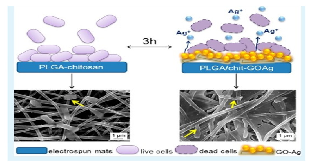
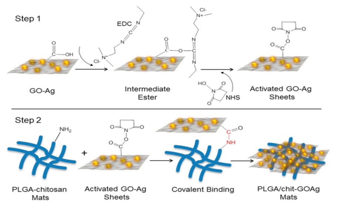
- de Faria, A.F.; Perreault, F.; Shaulsky, E.; Arias Chavez, L.H.; Elimelech, M. Antimicrobial Electrospun Biopolymer Nanofiber Mats Functionalized with Graphene Oxide-Silver Nanocomposites. ACS Appl. Mater. Interfaces 2015, 7, 12751–12759. [Google Scholar] [CrossRef]
- Patel, S.; Konar, M.; Sahoo, H.; Hota, G. Surface Functionalization of Electrospun PAN Nanofibers with ZnO–Ag Heterostructure Nanoparticles: Synthesis and Antibacterial Study. Nanotechnology 2019, 30, 205704. [Google Scholar] [CrossRef] [PubMed]
- Fadeeva, I.V.; Goldberg, M.A.; Preobrazhensky, I.I.; Mamin, G.V.; Davidova, G.A.; Agafonova, N.V.; Fosca, M.; Russo, F.; Barinov, S.M.; Cavalu, S.; et al. Improved Cytocompatibility and Antibacterial Properties of Zinc-Substituted Brushite Bone Cement Based on β-Tricalcium Phosphate. J. Mater. Sci. Mater. Med. 2021, 32, 99. [Google Scholar] [CrossRef]
- Cavalu, S.; Fritea, L.; Brocks, M.; Barbaro, K.; Murvai, G.; Costea, T.O.; Antoniac, I.; Verona, C.; Romani, M.; Latini, A.; et al. Novel Hybrid Composites Based on PVA/SeTiO2 Nanoparticles and Natural Hydroxyapatite for Orthopedic Applications: Correlations between Structural, Morphological and Biocompatibility Properties. Materials 2020, 13, 2077. [Google Scholar] [CrossRef] [PubMed]
- Cavalu, S.; Simon, V.; Goller, G.; Akin, I. Bioactivity and antimicrobial properties of PMMA/Ag2O acrylic bone cement collagen coated. Dig. J. Nanomater. Biostructures 2011, 6, 779–790. [Google Scholar]
- Zhang, Y.Z.; Venugopal, J.; Huang, Z.-M.; Lim, C.T.; Ramakrishna, S. Characterization of the Surface Biocompatibility of the Electrospun PCL-Collagen Nanofibers Using Fibroblasts. Biomacromolecules 2005, 6, 2583–2589. [Google Scholar] [CrossRef]
- Rieger, K.A.; Porter, M.; Schiffman, J.D. Polyelectrolyte-Functionalized Nanofiber Mats Control the Collection and Inactivation of Escherichia Coli. Materials 2016, 9, 297. [Google Scholar] [CrossRef] [Green Version]
- ScienceDirect. Surface Functionalization of Dual Growth Factor on Hydroxyapatite-Coated Nanofibers for Bone Tissue Engineering. Available online: https://0-www-sciencedirect-com.brum.beds.ac.uk/science/article/abs/pii/S0169433220310679 (accessed on 23 May 2022).
- Borah, R.; Kumar, A.; Das, M.K.; Ramteke, A. Surface Functionalization-Induced Enhancement in Surface Properties and Biocompatibility of Polyaniline Nanofibers. RSC Adv. 2015, 5, 48971–48982. [Google Scholar] [CrossRef]
- Cuervo, M.R.; Asedegbega-Nieto, E.; Díaz, E.; Vega, A.; Ordóñez, S.; Castillejos-López, E.; Rodríguez-Ramos, I. Effect of Carbon Nanofiber Functionalization on the Adsorption Properties of Volatile Organic Compounds. J. Chromatogr. A 2008, 1188, 264–273. [Google Scholar] [CrossRef]
- ScienceDirect. Selective Adhesion and Growth of Vascular Endothelial Cells on Bioactive Peptide Nanofiber Functionalized Stainless Steel Surface. Available online: https://0-www-sciencedirect-com.brum.beds.ac.uk/science/article/abs/pii/S0142961211009239 (accessed on 23 May 2022).
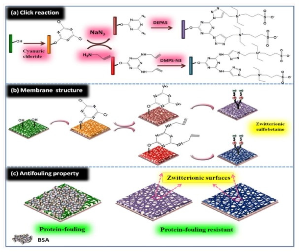
- Huang, J.; Wang, D.; Lu, Y.; Li, M.; Xu, W. Surface Zwitterionically Functionalized PVA-Co-PE Nanofiber Materials by Click Chemistry. RSC Adv. 2013, 3, 20922–20929. [Google Scholar] [CrossRef]
- Schneiderman, S.; Zhang, L.; Fong, H.; Menkhaus, T.J. Surface-Functionalized Electrospun Carbon Nanofiber Mats as an Innovative Type of Protein Adsorption/Purification Medium with High Capacity and High Throughput. J. Chromatogr. A 2011, 1218, 8989–8995. [Google Scholar] [CrossRef]
- Kim, T.G.; Park, T.G. Surface Functionalized Electrospun Biodegradable Nanofibers for Immobilization of Bioactive Molecules. Biotechnol. Prog. 2006, 22, 1108–1113. [Google Scholar] [CrossRef] [PubMed]
- Chen, P.; Liang, H.-W.; Lv, X.-H.; Zhu, H.-Z.; Yao, H.-B.; Yu, S.-H. Carbonaceous Nanofiber Membrane Functionalized by Beta-Cyclodextrins for Molecular Filtration. ACS Nano 2011, 5, 5928–5935. [Google Scholar] [CrossRef] [PubMed]
- Permyakova, E.S.; Kiryukhantsev-Korneev, P.V.; Gudz, K.Y.; Konopatsky, A.S.; Polčak, J.; Zhitnyak, I.Y.; Gloushankova, N.A.; Shtansky, D.V.; Manakhov, A.M. Comparison of Different Approaches to Surface Functionalization of Biodegradable Polycaprolactone Scaffolds. Nanomaterials 2019, 9, 1769. [Google Scholar] [CrossRef] [PubMed] [Green Version]
- Guilak, F.; Butler, D.L.; Goldstein, S.A.; Baaijens, F.P.T. Biomechanics and Mechanobiology in Functional Tissue Engineering. J. Biomech. 2014, 47, 1933–1940. [Google Scholar] [CrossRef] [Green Version]
- Jiang, J.; Xie, J.; Ma, B.; Bartlett, D.E.; Xu, A.; Wang, C.-H. Mussel-Inspired Protein-Mediated Surface Functionalization of Electrospun Nanofibers for PH-Responsive Drug Delivery. Acta Biomater. 2014, 10, 1324–1332. [Google Scholar] [CrossRef] [PubMed] [Green Version]
- González, E.; Shepherd, L.M.; Saunders, L.; Frey, M.W. Surface Functional Poly(Lactic Acid) Electrospun Nanofibers for Biosensor Applications. Materials 2016, 9, 47. [Google Scholar] [CrossRef] [PubMed]
- Kolewe, K.W.; Dobosz, K.M.; Rieger, K.A.; Chang, C.-C.; Emrick, T.; Schiffman, J.D. Antifouling Electrospun Nanofiber Mats Functionalized with Polymer Zwitterions. ACS Appl. Mater. Interfaces 2016, 8, 27585–27593. [Google Scholar] [CrossRef]
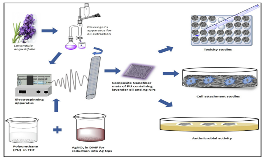
- Sofi, H.S.; Akram, T.; Tamboli, A.H.; Majeed, A.; Shabir, N.; Sheikh, F.A. Novel Lavender Oil and Silver Nanoparticles Simultaneously Loaded onto Polyurethane Nanofibers for Wound-Healing Applications. Int. J. Pharm. 2019, 569, 118590. [Google Scholar] [CrossRef]
- Ullah, S.; Hashmi, M.; Kharaghani, D.; Khan, M.Q.; Saito, Y.; Yamamoto, T.; Lee, J.; Kim, I.S. Antibacterial Properties of in Situ and Surface Functionalized Impregnation of Silver Sulfadiazine in Polyacrylonitrile Nanofiber Mats. Int. J. Nanomed. 2019, 14, 2693–2703. [Google Scholar] [CrossRef] [Green Version]
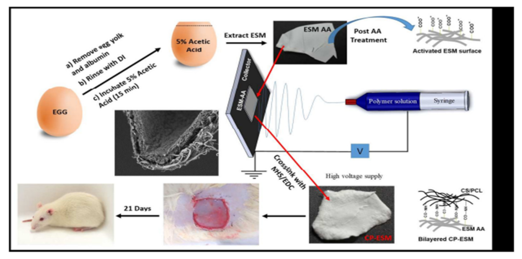
- Ray, P.G.; Pal, P.; Srivas, P.K.; Basak, P.; Roy, S.; Dhara, S. Surface Modification of Eggshell Membrane with Electrospun Chitosan/Polycaprolactone Nanofibers for Enhanced Dermal Wound Healing. ACS Appl. Bio Mater. 2018, 1, 985–998. Available online: https://0-pubs-acs-org.brum.beds.ac.uk/doi/abs/10.1021/acsabm.8b00169 (accessed on 25 May 2022).
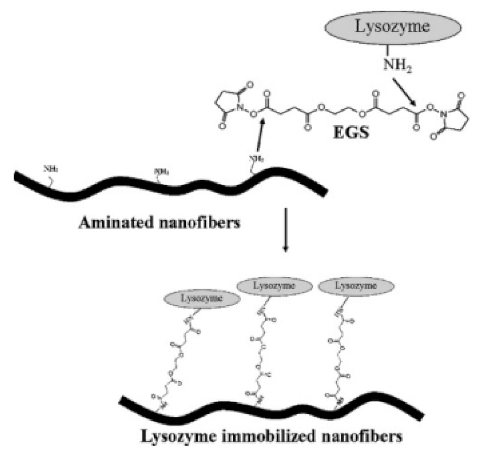
- Miao, J.; Pangule, R.C.; Paskaleva, E.E.; Hwang, E.E.; Kane, R.S.; Linhardt, R.J.; Dordick, J.S. Lysostaphin-Functionalized Cellulose Fibers with Antistaphylococcal Activity for Wound Healing Applications. Biomaterials 2011, 32, 9557–9567. [Google Scholar] [CrossRef] [PubMed]
- ACS Omega. Silver-Functionalized Bacterial Cellulose as Antibacterial Membrane for Wound-Healing Applications. Available online: https://0-pubs-acs-org.brum.beds.ac.uk/doi/full/10.1021/acsomega.7b00442 (accessed on 25 May 2022).
- Chen, X.; Li, H.; Lu, W.; Guo, Y. Antibacterial Porous Coaxial Drug-Carrying Nanofibers for Sustained Drug-Releasing Applications. Nanomaterials 2021, 11, 1316. [Google Scholar] [CrossRef]
- Topuz, F.; Uyar, T. Electrospinning of Cyclodextrin Functional Nanofibers for Drug Delivery Applications. Pharmaceutics 2019, 11, 6. [Google Scholar] [CrossRef] [PubMed]
- Luo, Y.; Nartker, S.; Miller, H.; Hochhalter, D.; Wiederoder, M.; Wiederoder, S.; Setterington, E.; Drzal, L.T.; Alocilja, E.C. Surface Functionalization of Electrospun Nanofibers for Detecting E. coli O157:H7 and BVDV Cells in a Direct-Charge Transfer Biosensor. Biosens. Bioelectron. 2010, 26, 1612–1617. [Google Scholar] [CrossRef] [PubMed]
- Massoumi, B.; Ghandomi, F.; Abbasian, M.; Eskandani, M.; Jaymand, M. Surface Functionalization of Graphene Oxide with Poly(2-Hydroxyethyl Methacrylate)-Graft-Poly(ε-Caprolactone) and Its Electrospun Nanofibers with Gelatin. Appl. Phys. A 2016, 122, 1000. [Google Scholar] [CrossRef]
- Patil, Y.B.; Toti, U.S.; Khdair, A.; Ma, L.; Panyam, J. Single-Step Surface Functionalization of Polymeric Nanoparticles for Targeted Drug Delivery. Biomaterials 2009, 30, 859–866. [Google Scholar] [CrossRef] [Green Version]
- ScienceDirect. Preparation of Polyamide-6/Chitosan Composite Nanofibers by a Single Solvent System via Electrospinning for Biomedical Applications. Available online: https://0-www-sciencedirect-com.brum.beds.ac.uk/science/article/abs/pii/S0927776510006405 (accessed on 25 May 2022).
- Fritea, L.; Pasca, P.M.; Vlase, L.; Gheldiu, A.-M.; Moldovan, L.; Banica, F.; Dobjanschi, L.; Cavalu, S. Electrochemical Methods for Evaluation of Antioxidant Properties of Propolis Extract Incorporated in Chitosan Nanoparticles. Mater. Plast. 2021, 57, 96–108. [Google Scholar] [CrossRef]
- Wang, S.; Wen, S.; Shen, M.; Guo, R.; Cao, X.; Wang, J.; Shi, X. Aminopropyltriethoxysilane-Mediated Surface Functionalization of Hydroxyapatite Nanoparticles: Synthesis, Characterization, and in Vitro Toxicity Assay. Int. J. Nanomed. 2011, 6, 3449–3459. [Google Scholar] [CrossRef] [Green Version]
- Ormanci, O.; Akin, I.; Sahin, F.; Yucel, O.; Simon, V.; Cavalu, S.; Goller, G. Spark plasma sintered Al2O3-YSZ-TiO2 composites: Processing, characterization and in vivo evaluation. Mater. Sci. Eng. C 2014, 40, 16–23. [Google Scholar] [CrossRef]
- Lv, H.; Wu, C.; Liu, X.; Bai, A.; Cao, Y.; Shang, W.; Hu, L.; Liu, Y. Folate-Functionalized Mesoporous Hollow SnO2 Nanofibers as a Targeting Drug Carrier to Improve the Antitumor Effect of Paclitaxel for Liver Cancer Therapy. BioMed Res. Int. 2018, 2018, e8526190. [Google Scholar] [CrossRef] [Green Version]
- Hou, L.; Udangawa, W.M.R.N.; Pochiraju, A.; Dong, W.; Zheng, Y.; Linhardt, R.J.; Simmons, T.J. Synthesis of Heparin-Immobilized, Magnetically Addressable Cellulose Nanofibers for Biomedical Applications. ACS Biomater. Sci. Eng. 2016, 2, 1905–1913. [Google Scholar] [CrossRef] [PubMed]
- ScienceDirect. Chitosan-Functionalized Nanofibers: A Comprehensive Review on Challenges and Prospects for Food Applications. Available online: https://0-www-sciencedirect-com.brum.beds.ac.uk/science/article/pii/S0141813018353819 (accessed on 26 May 2022).
- ScienceDirect. Synthesis and Characterization of Chitosan/Polyvinylpyrrolidone Coated Nanoporous γ-Alumina as a PH-Sensitive Carrier for Controlled Release of Quercetin. Available online: https://0-www-sciencedirect-com.brum.beds.ac.uk/science/article/abs/pii/S0141813021009338 (accessed on 3 December 2021).
- Tseng, H.; Peterson, T.E.; Berk, B.C. Fluid Shear Stress Stimulates Mitogen-Activated Protein Kinase in Endothelial Cells. Circ. Res. 1995, 77, 869–878. [Google Scholar] [CrossRef] [PubMed]
- Kingsley, G.R. The Determination of Serum Total Protein, Albumin, and Globulin by the Biuret Reaction. J. Biol. Chem. 1939, 131, 197–200. [Google Scholar] [CrossRef]
- Cadman, E.; Bostwick, J.R.; Eichberg, J. Determination of Protein by a Modified Lowry Procedure in the Presence of Some Commonly Used Detergents. Anal. Biochem. 1979, 96, 21–23. [Google Scholar] [CrossRef]
- Shitole, A.A.; Giram, P.S.; Raut, P.W.; Rade, P.P.; Khandwekar, A.P.; Sharma, N.; Garnaik, B. Clopidogrel Eluting Electrospun Polyurethane/Polyethylene Glycol Thromboresistant, Hemocompatible Nanofibrous Scaffolds. J. Biomater. Appl. 2019, 33, 1327–1347. [Google Scholar] [CrossRef]
- Borah, R.; Ingavle, G.C.; Kumar, A.; Sandeman, S.R.; Mikhalovsky, S.V. Surface-Functionalized Conducting Nanofibers for Electrically Stimulated Neural Cell Function. Biomacromolecules 2021, 22, 594–611. [Google Scholar] [CrossRef]


| Fabrication Technique | Advantages | Disadvantages |
|---|---|---|
| Drawing | Simple equipment |
|
| Template synthesis |
|
|
| Temperature-induced phase separation |
|
|
| Molecular self- assembly |
|
|
| Electrospinning |
|
|
| Methods of Modification | Advantages | Disadvantages |
|---|---|---|
| Physical blends |
|
|
| Core-shell electrospinning |
|
|
Post functionalization
|
|
|
| Sr. No. | Surface Functionalized Nanofibers | Material Used for Surface Functionalization | Applications | References |
|---|---|---|---|---|
| 1 | Polyamide 6/O-MMT composite nanofibers by Fe2O3 sputter coating | Fe2O3 (Ferrous oxide/Iron oxide) | Improved thermal stability properties of composite nanofibers | [68] |
| 2 | Gelatin grafted Electrospun Poly (caprolactone) nanofibers | Gelatin | Blood vessel tissue engineering | [69] |
| 3 | Size controlled silver Nanoparticle coated nanofibers | Silver | Wound Dressing | [70,71] |
| 4 | Surface modified polycaprolactone electrospun nanofiber | Plasma treatment (ar or O2 gas) | Cell morphology, cell adhesion and proliferation study | [72] |
| 5 | Graphene oxide-silver nanocomposites functionalized biopolymer nanofiber mats | Graphene Oxide-silver | Antimicrobial | [73] |
| 6 | PAN nanofibers with zno-Ag heterostructure nanoparticle | Zno-Ag | Anti-Bacterial | [74,75] |
| 7 | Bioinspired surface functionalized electrospun polycaprolactone nanofibers | Nano-hydroxyapatite (nha) | Bone tissue engineering | [57,76] |
| 8 | PCL-collagen nanofibers | Collagen Coating | Tissue engineering | [78] |
| 9 | Polyelectrolyte functionalized nanofiber mats | Polyelectrolyte: Poly (acrylic acid) (PAA), Chitosan (CS) and polydiallyl dimethyl ammonium chloride (pdadmac) | Anti-microbial (Collection and inactivation of E. coli) | [79] |
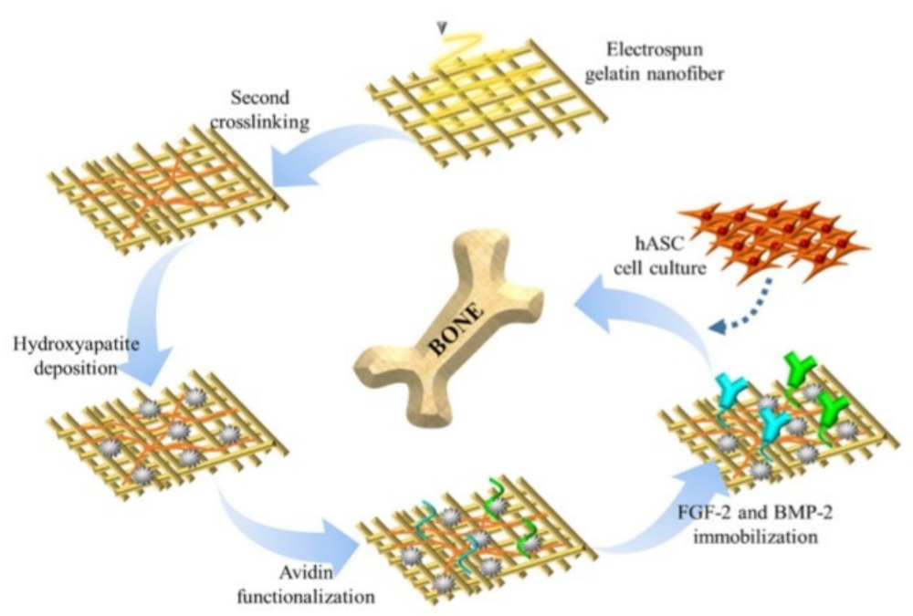
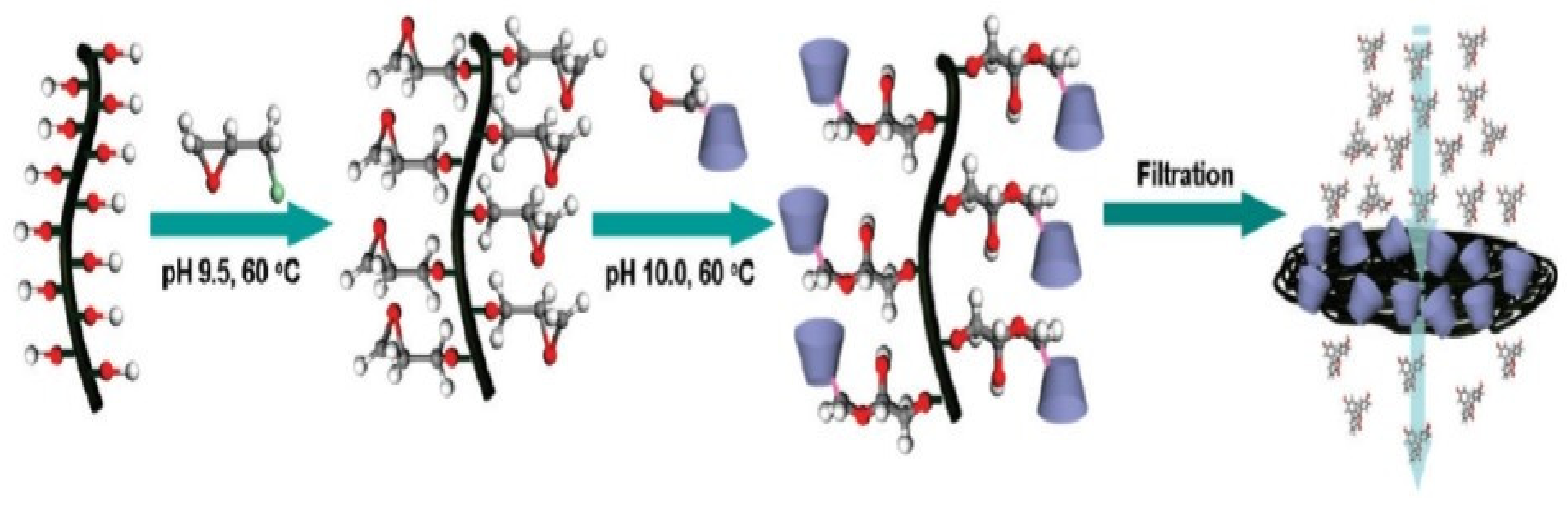
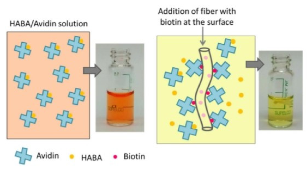
| 10 | Surface functionalization of dual growth factor on hydroxyapatite-coated nanofibers for bone tissue engineering | Avidin-Biotin | Bone tissue engineering | [80] |
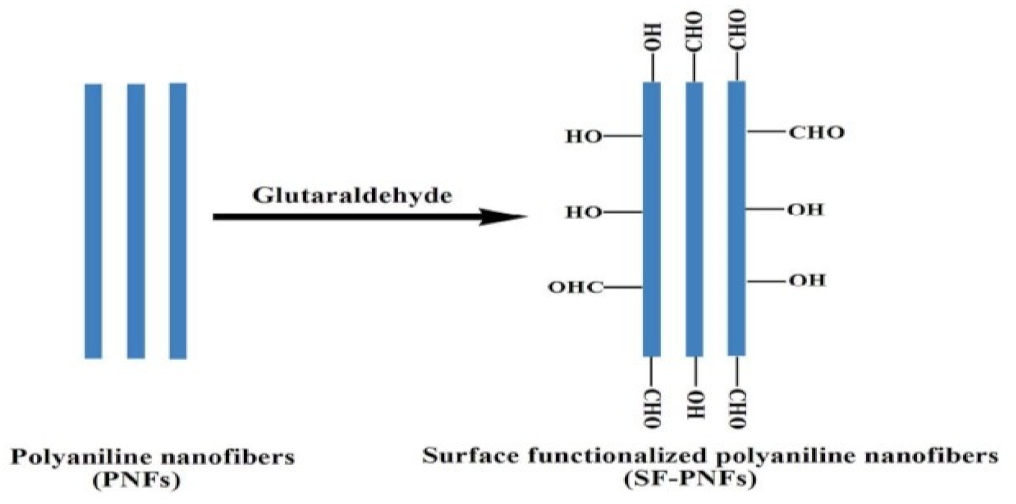
| 11 | Surface functionalized Polyaniline nanofiber | Glutaraldehyde | Bio-sensor | [81] |
| 12 | Carbon nanofiber functionalized with volatile organic compounds | HNO3 (Nitric Acid) | Improved adsorption properties | [82] |
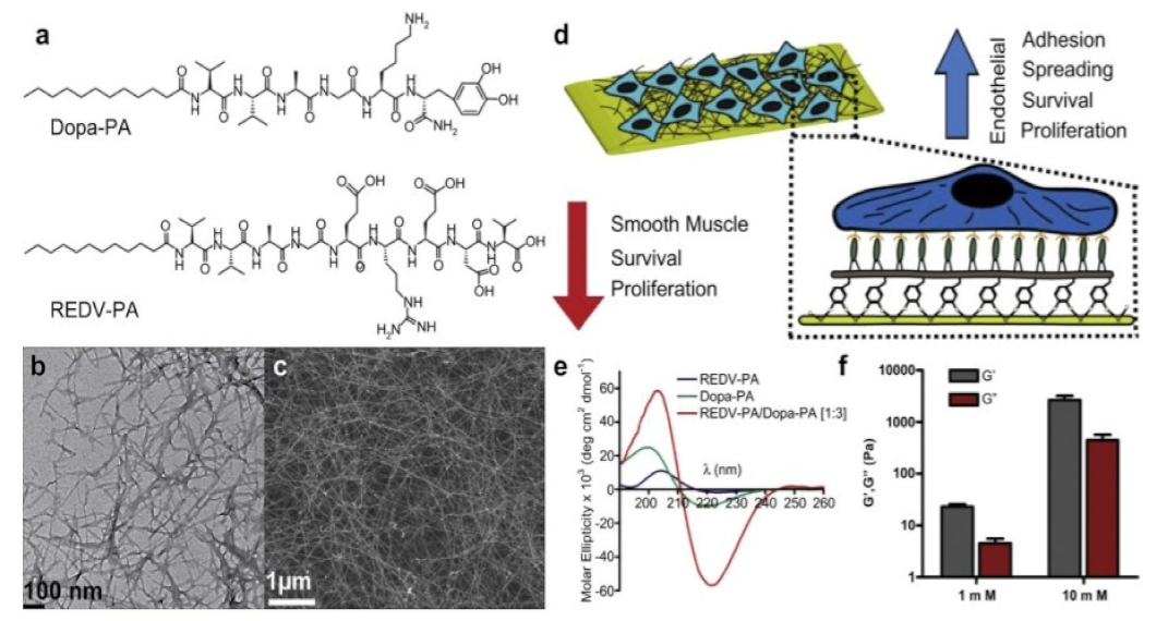
| 13 | Peptide nanofiber functionalized ith stainless steel | Stainless Steel | Treatment of cardiovascular diseases | [83] |
| 14 | Surface (zwittenrionically) functionalized PVA-co-PE nanofiber materials by click chemistry | Zwitterionic Sulfobetaine | Antifouling performance | [84] |
| 15 | Surface—functionalized Electrospun carbon nanofiber mats | Weak acid cation-exchange ligand | Protein adsorption/purification and bio-separation | [85] |
| 16 | Surface functionalized electrospun biodegradable nanofibers | Primary amine | Immobilization of bioactive molecules | [86] |
| 17 | Carbonaceous nanofiber membrane functionalized by beta-cyclodextrins for molecular filtration | Beta-cyclodextrins | Molecular Filtration, Chiral Separation and drug delivery | [87] |
| 18 | Carbonaceous Nanofiber Membrane Functionalized by beta-cyclodextrins for Molecular Filtration | COOH-containing polymer and ticapcon film | Skin reparation and wound dressing | [88] |
| 19 | Surface functionalized nanofibers | Glycine-phenylalanine-hydroxyproline-glycine-glutamate-arginine (GEOGER) peptide | Tissue Regeneration | [89] |
| 20 | Mussel-inspired protein-mediated surface functionalized electrospun nanofibers | Polydopamine | pH-responsive drug delivery | [90] |
| 21 | Surface functionalized Poly (lactic acid) electrospun nanofibers for biosensor applications | Biotin | Bio-sensor | [91] |
| 22 | Antifouling electrospun nanofiber mats Functionalized with polymer zwitterions | Poly (2-methacryloyloxyethyl phosphorylcholine) (poly MPC) | Tissue engineering and water purification | [92] |
| Description of Surface Functionalized Nanofibers | Application | Reference |
|---|---|---|
| Electrospun PCL per nanofiber with bioactive nano-hydroxy apatite (nHA) using dopamine effective bioadhesive agent | Improving Osteogenesis | [57] |
| Gelatin grafted poly-(caprolactone) (PCL) nanofibers | Tissue Engineering | [69] |
| Cellulose nanofiber mats surface functionalized using three polyelectrolytes: poly (acrylic acid) (PAA), chitosan (CS), and polydiallyldimethylammonium chloride (pDADMAC) | Control the Collection and Inactivation of Escherichia coli | [79] |
| Biodegradable poly (e-caprolactone) (PCL) and poly (D,L-lactic-co-glycolic acid)-poly(ethylene glycol)-NH2 (PLGA-b-PEG-NH2) block copolymernanofibers | Immobilization of Bioactive Molecules | [86] |
| Lavender oil and Silver loaded nanofibers | Wound Healing | [93] |
| Silver sulfadiazine (AgSD) loaded polyacrylonitrile nanofiber | Antibacterial activity | [94] |
| Modified eggshell membrane using chitosan/poly-caprolactone (CS/PCL) nanofibers | Wound healing | [95] |
| Lysostaphin functionalized cellulose fibers | Antimicrobial material in wound healing | [96] |
| Bacterial cellulose (BC) functionalized with silver nanofibers. | wound healing (Antibacterial activity against the gram-negative bacteria) | [97] |
| N, N-dimethylformamide and polydopamine loaded poly (caprolactone) nanofibers | Drug Delivery | [90] |
| Polycaprolactone (PCL)/polylactic acid (PLA) core-shell porous drug-carrying nanofibers | Sustained release drug application | [98] |
| Cyclodextrin-functional nanofibers | Drug Delivery | [99] |
| Glutaraldehyde cross linked nitrocellulose nanofiber | Bacterial and viral pathogen detection | [100] |
| Graphiene oxide (GO) with poly (2-hydroxymethyl methacrylate)-graft-poly (caprolactone) [P (HEM-g-CL)] using polymerization approach and fabricated electrospun nanofibers with gelatine | Application in Regenerative Medicine | [101] |
| Polymeric ethylene glycol (PLA-PEG) loaded nanofibers | Targeted Strategies | [102] |
| Chitosan blended poly-amide-6 nano fibers | Human osteoblastic (HOB) cell culture application | [103,104] |
| Aminopropyltriethoxysilane (APTS)-mediated surface modification of nanohydroxyapatite | Different biomedical applications | [105] |
| The mussel-inspired surface 1191 functionalization using 2-(3,4-dihydroxyphenyl) ethylamine (dopamine) to conjugate the 1192 borate-containing BTZ anticancer drug | Implantable Smart Magnetic Nanofiber | [106] |
| PTX-loaded mesoperous hollow SnO2 nanofiber conjugated with folic acid | Liver cancer therapy | [107] |
| Magnetically responsive heparin—immobilized cellulose nanofiber | Tissue Engineering | [108] |
Publisher’s Note: MDPI stays neutral with regard to jurisdictional claims in published maps and institutional affiliations. |
© 2022 by the authors. Licensee MDPI, Basel, Switzerland. This article is an open access article distributed under the terms and conditions of the Creative Commons Attribution (CC BY) license (https://creativecommons.org/licenses/by/4.0/).
Share and Cite
Kulkarni, D.; Musale, S.; Panzade, P.; Paiva-Santos, A.C.; Sonwane, P.; Madibone, M.; Choundhe, P.; Giram, P.; Cavalu, S. Surface Functionalization of Nanofibers: The Multifaceted Approach for Advanced Biomedical Applications. Nanomaterials 2022, 12, 3899. https://0-doi-org.brum.beds.ac.uk/10.3390/nano12213899
Kulkarni D, Musale S, Panzade P, Paiva-Santos AC, Sonwane P, Madibone M, Choundhe P, Giram P, Cavalu S. Surface Functionalization of Nanofibers: The Multifaceted Approach for Advanced Biomedical Applications. Nanomaterials. 2022; 12(21):3899. https://0-doi-org.brum.beds.ac.uk/10.3390/nano12213899
Chicago/Turabian StyleKulkarni, Deepak, Shubham Musale, Prabhakar Panzade, Ana Cláudia Paiva-Santos, Pratiksha Sonwane, Monika Madibone, Puja Choundhe, Prabhanjan Giram, and Simona Cavalu. 2022. "Surface Functionalization of Nanofibers: The Multifaceted Approach for Advanced Biomedical Applications" Nanomaterials 12, no. 21: 3899. https://0-doi-org.brum.beds.ac.uk/10.3390/nano12213899